General embryo
130
-
Upload
shahmohammadzulfiquar -
Category
Health & Medicine
-
view
1.849 -
download
1
Transcript of General embryo

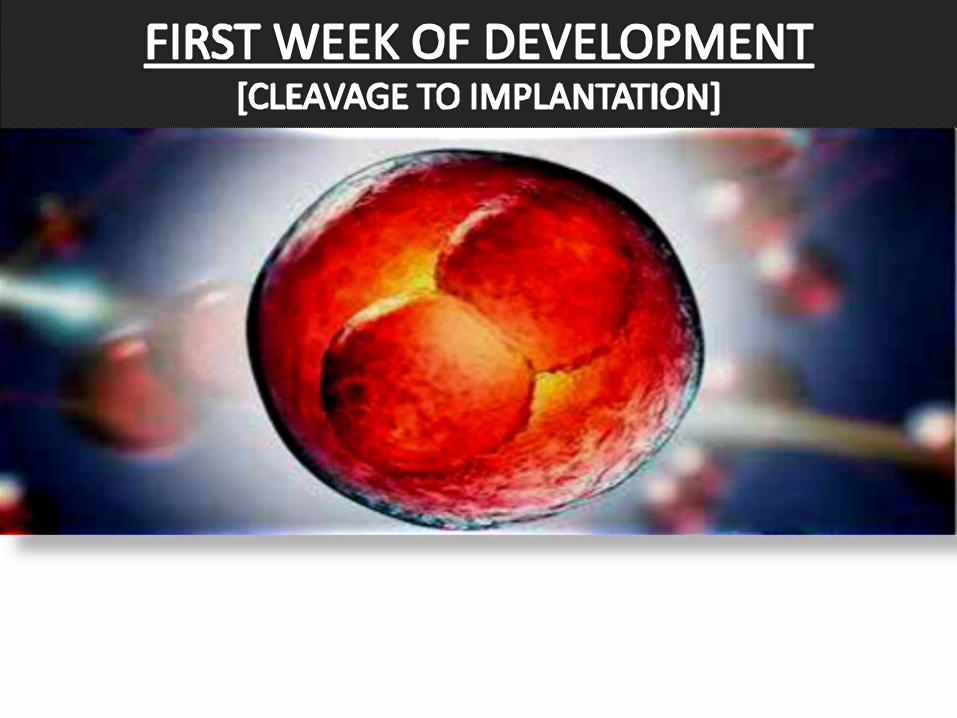
GENERAL EMBRYOLOGY (1ST WEEK -3RD
WEEK)
Fertilization
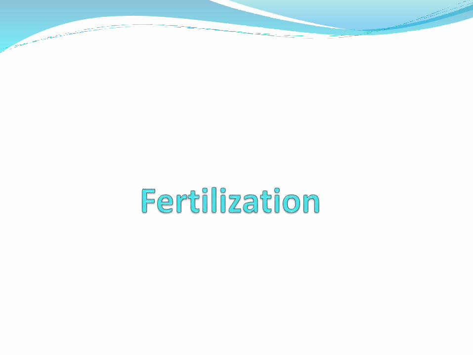
Fertilization
• The process of union of male & female gamate
• Site-ampullary region
Aim of fertilization
• Restoration of diploid number chromosome
• Determination of sex
• Initiation of clevageFig: Fertilization to implantation
Movement of sperm from cervix to uterine tube
• Muscular contraction of uterus & uterine tube
• By own propulsion
Changes in sperm before fertilization
• Capacitation
• Acrosomal reaction

Capacitation
• Period of conditioning in female reproductive tract
• In uterus & uterine tube
• Epithelial interaction between sperm & uterine tube
• Can pass through corona cells & undergo acrosomal reaction
Acrosomal reaction
• Mutual action
• Both sperm & follicular cell release enzyme
• Sperm penetrate zona pellucida
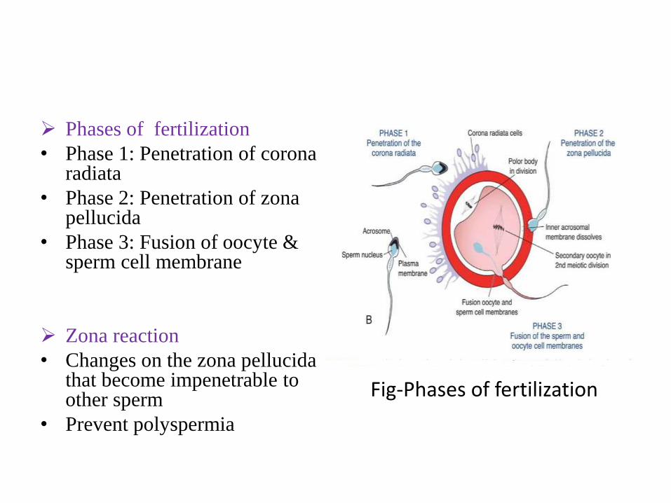
Phases of fertilization
• Phase 1: Penetration of corona radiata
• Phase 2: Penetration of zona pellucida
• Phase 3: Fusion of oocyte & sperm cell membrane
Zona reaction
• Changes on the zona pellucida that become impenetrable to other sperm
• Prevent polyspermia
Fig-Phases of fertilization
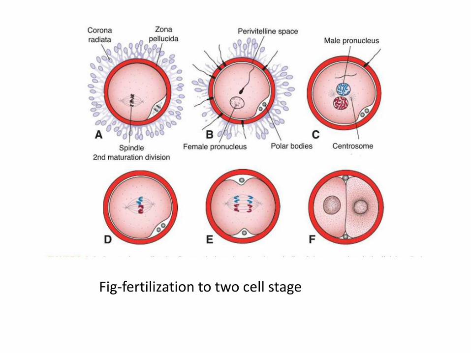
Fig-fertilization to two cell stage
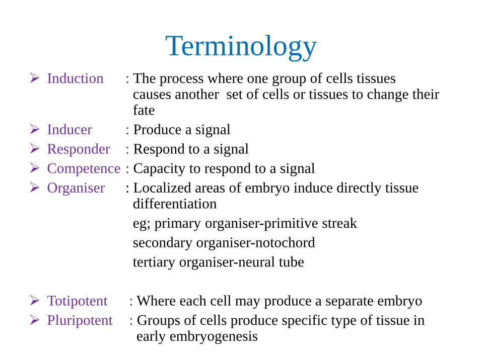
Terminology Induction : The process where one group of cells tissues
causes another set of cells or tissues to change their fate
Inducer : Produce a signal
Responder : Respond to a signal
Competence : Capacity to respond to a signal
Organiser : Localized areas of embryo induce directly tissue differentiation
eg; primary organiser-primitive streak
secondary organiser-notochord
tertiary organiser-neural tube
Totipotent : Where each cell may produce a separate embryo
Pluripotent : Groups of cells produce specific type of tissue in early embryogenesis
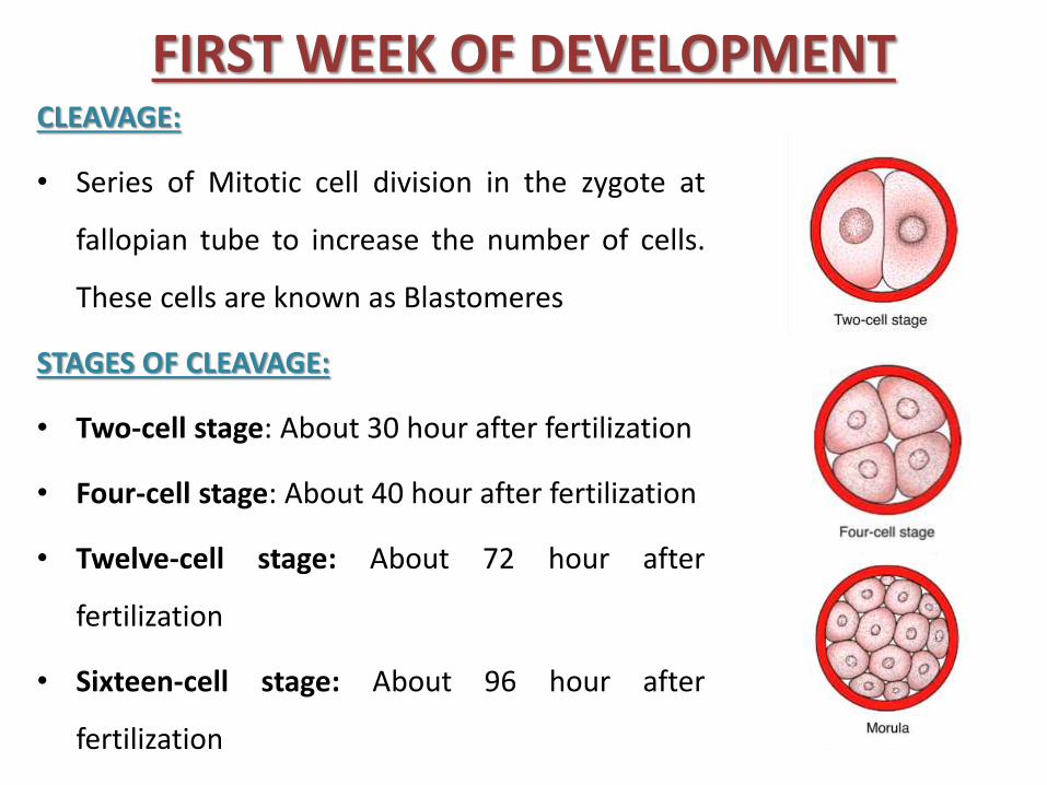
FIRST WEEK OF DEVELOPMENTCLEAVAGE:
• Series of Mitotic cell division in the zygote at
fallopian tube to increase the number of cells.
These cells are known as Blastomeres
STAGES OF CLEAVAGE:
• Two-cell stage: About 30 hour after fertilization
• Four-cell stage: About 40 hour after fertilization
• Twelve-cell stage: About 72 hour after
fertilization
• Sixteen-cell stage: About 96 hour after
fertilization
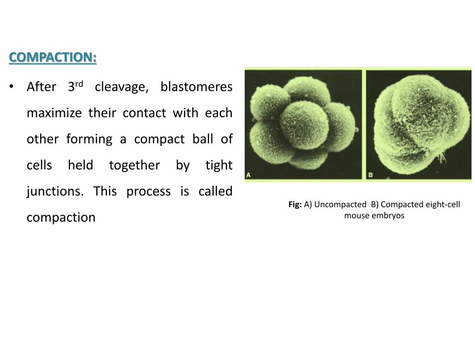
COMPACTION:
• After 3rd cleavage, blastomeres
maximize their contact with each
other forming a compact ball of
cells held together by tight
junctions. This process is called
compactionFig: A) Uncompacted B) Compacted eight-cell
mouse embryos
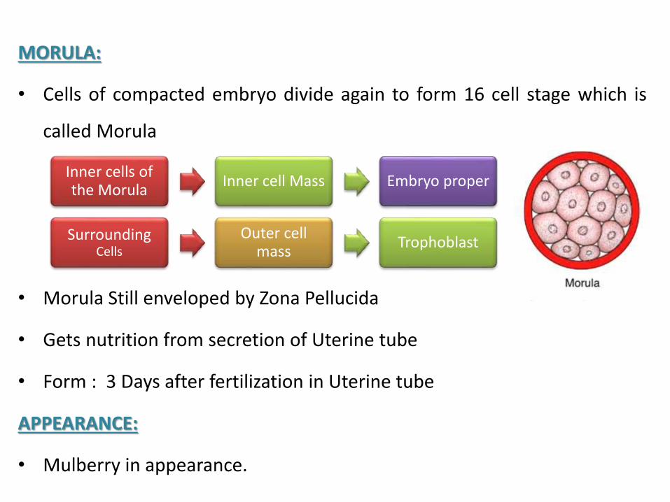
MORULA:
• Cells of compacted embryo divide again to form 16 cell stage which is
called Morula
• Morula Still enveloped by Zona Pellucida
• Gets nutrition from secretion of Uterine tube
• Form : 3 Days after fertilization in Uterine tube
APPEARANCE:
• Mulberry in appearance.
Inner cells of the Morula
Inner cell Mass Embryo proper
SurroundingCells
Outer cell mass
Trophoblast
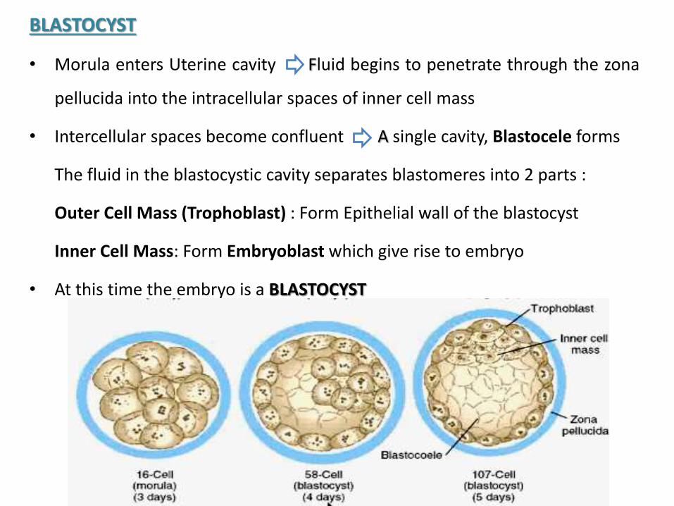
BLASTOCYST
• Morula enters Uterine cavity Fluid begins to penetrate through the zona
pellucida into the intracellular spaces of inner cell mass
• Intercellular spaces become confluent A single cavity, Blastocele forms
The fluid in the blastocystic cavity separates blastomeres into 2 parts :
Outer Cell Mass (Trophoblast) : Form Epithelial wall of the blastocyst
Inner Cell Mass: Form Embryoblast which give rise to embryo
• At this time the embryo is a BLASTOCYST
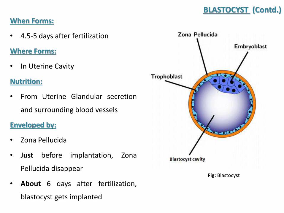
When Forms:
• 4.5-5 days after fertilization
Where Forms:
• In Uterine Cavity
Nutrition:
• From Uterine Glandular secretion
and surrounding blood vessels
Enveloped by:
• Zona Pellucida
• Just before implantation, Zona
Pellucida disappear
• About 6 days after fertilization,
blastocyst gets implanted
BLASTOCYST (Contd.)
Fig: Blastocyst
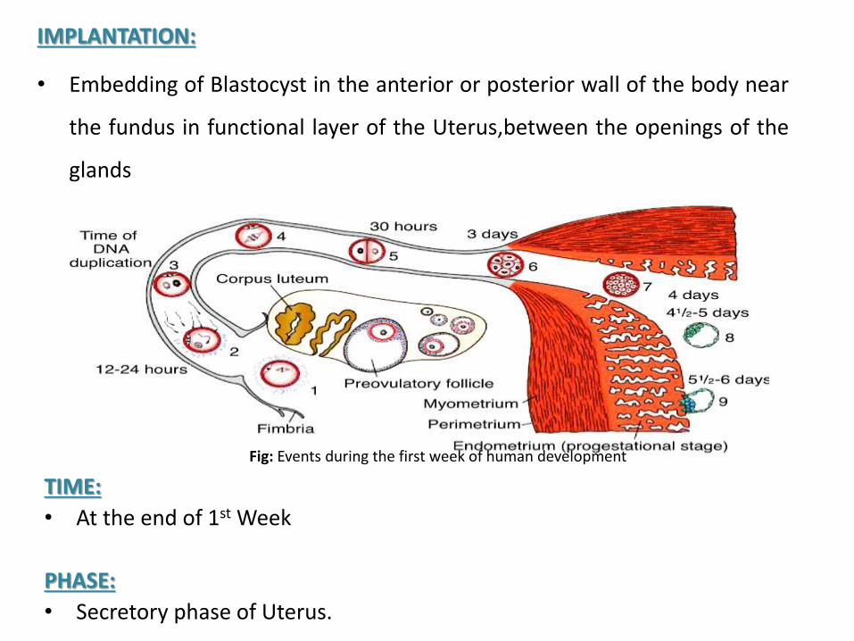
IMPLANTATION:
• Embedding of Blastocyst in the anterior or posterior wall of the body near
the fundus in functional layer of the Uterus,between the openings of the
glands
TIME:
• At the end of 1st Week
PHASE:
• Secretory phase of Uterus.
Fig: Events during the first week of human development
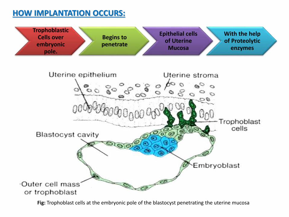
TrophoblasticCells over embryonic
pole.
Begins to penetrate
Epithelial cells of Uterine
Mucosa
With the help of Proteolytic
enzymes
HOW IMPLANTATION OCCURS:
Fig: Trophoblast cells at the embryonic pole of the blastocyst penetrating the uterine mucosa
ATTACHMENT:
• By L-Selectin on Trophoblast attached with CHO receptor on
uterine epithelium
• This is called Capture of blastocyst by uterine epithelium from
uterine cavity
• Integrin on Trophoblast for laminin promote attachment
• Integrin for Fibronectin stimulate migration
So, Implantation is the result of mutual trophoblastic and
endometrial action
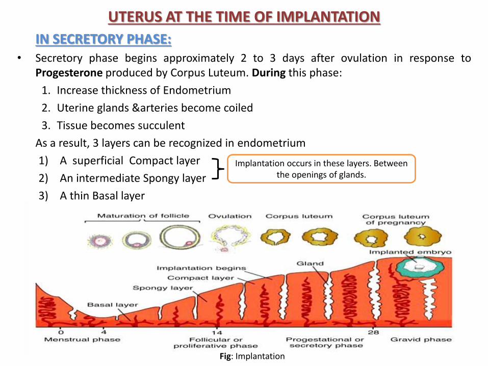
UTERUS AT THE TIME OF IMPLANTATION
IN SECRETORY PHASE: • Secretory phase begins approximately 2 to 3 days after ovulation in response to
Progesterone produced by Corpus Luteum. During this phase:
1. Increase thickness of Endometrium
2. Uterine glands &arteries become coiled
3. Tissue becomes succulent
As a result, 3 layers can be recognized in endometrium
1) A superficial Compact layer
2) An intermediate Spongy layer
3) A thin Basal layer
Implantation occurs in these layers. Between the openings of glands.
Fig: Implantation

IF FERTILIZATION DOES NOT OCCUR:
• The Corpus Luteum degenerates
• Estrogen and Progesterone levels fall
• Menstruation occurs
• Following 3 or 4 days compact& spongy layers are expelled from uterus. Basallayer is retained, functions as the regenerative layer
• Uterine lining epithelium reappears from glandular lining epithelium (simplecolumnar)
Fig: Menstrual cycle without fertilization
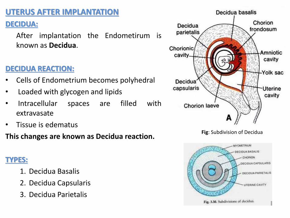
UTERUS AFTER IMPLANTATION
DECIDUA:
After implantation the Endometirum isknown as Decidua.
DECIDUA REACTION:
• Cells of Endometrium becomes polyhedral
• Loaded with glycogen and lipids
• Intracellular spaces are filled withextravasate
• Tissue is edematus
This changes are known as Decidua reaction.
TYPES:
1. Decidua Basalis
2. Decidua Capsularis
3. Decidua Parietalis
Fig: Subdivision of Decidua
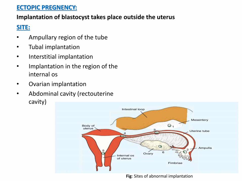
ECTOPIC PREGNENCY:
Implantation of blastocyst takes place
SITE:
• Ampullary region of the tube
• Tubal implantation
• Interstitial implantation
• Implantation in the region of the internal os
• Ovarian implantation
• Abdominal cavity (rectouterinecavity)
outside the uterus
Fig: Sites of abnormal implantation
FATE OF ECTOPIC PREGNANCY:
• Implantation in the region of the internal os, resulting placenta
previa cause severe bleeding, In second part of the pregnancy and
during delivery
• In ectopic pregnancy, embryo dies about the second month of
gestation cause severe hemorrhage & abdominal pain in the
mother – a surgical emergency
2nd WEEK OF DEVELOPMENT
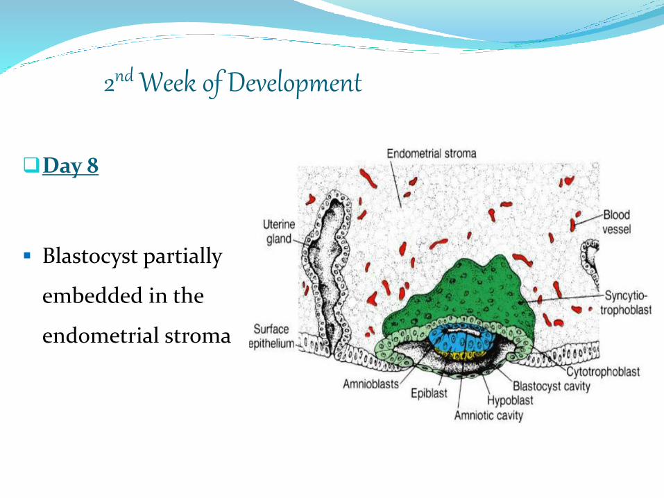
2nd Week of Development
Day 8
Blastocyst partially
embedded in the
endometrial stroma
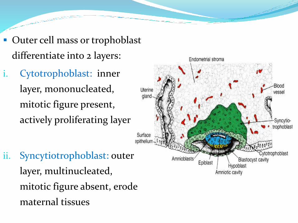
Outer cell mass or trophoblast
differentiate into 2 layers:
i. Cytotrophoblast: inner
layer, mononucleated,
mitotic figure present,
actively proliferating layer
ii. Syncytiotrophoblast: outer
layer, multinucleated,
mitotic figure absent, erode
maternal tissues
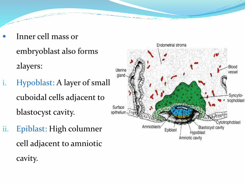
Inner cell mass or
embryoblast also forms
2layers:
i. Hypoblast: A layer of small
cuboidal cells adjacent to
blastocyst cavity.
ii. Epiblast: High columner
cell adjacent to amniotic
cavity.
A Small cavity appear with in
the epiblast.
The cavity enlarges to become
the amniotic cavity.
Epiblast cell adjacent to the
cytotrophoblast called
amnioblast
together with the rest of the
epiblast they line the amniotic
cavity.
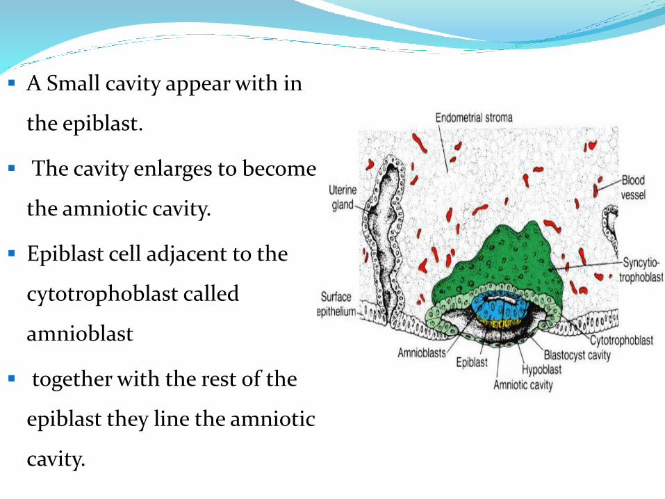
Day 9
Blastocyst more deeply
embedded in the
endometrium
At the embryonic pole
vacuoles appear in
syncytium vacuoles fuse
and form large
lacunae(lacunar stage)
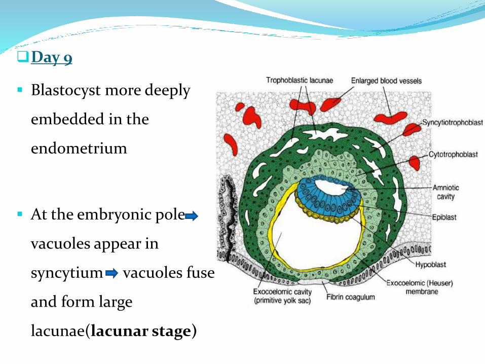
At the abembryonic pole,
flattened cells originating from
the hypoblast form a thin
exocoelomic membrane that
lines inner surface of the
cytotrophoblast
This membrane, together with
hypoblast, forms the lining of
the exocoelomic cavity or
primitive yolk sac.
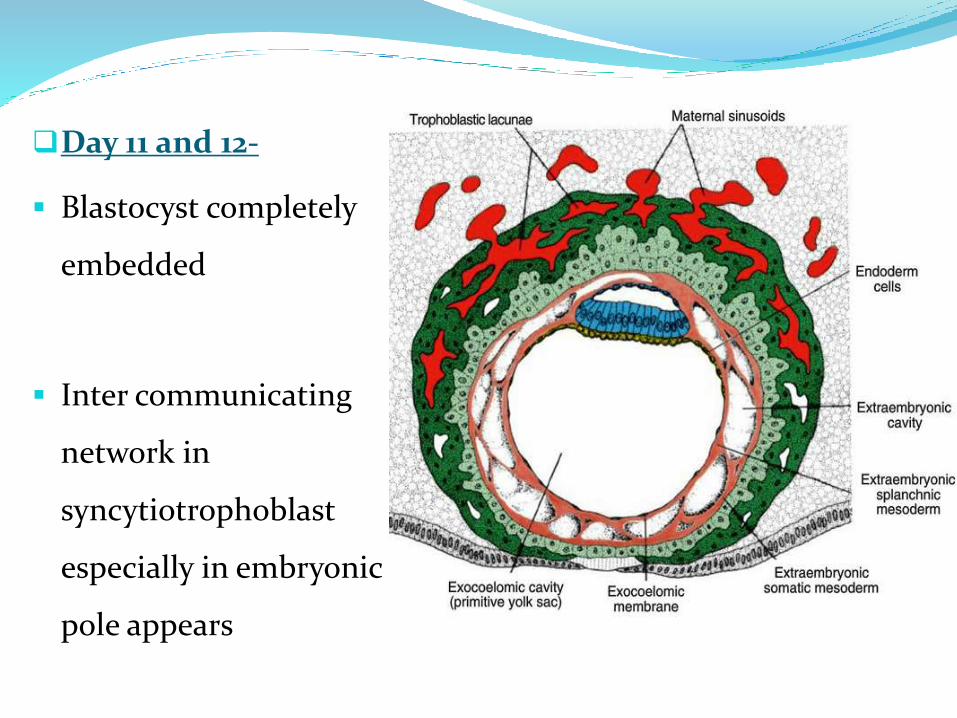
Day 11 and 12-
Blastocyst completely
embedded
Inter communicating
network in
syncytiotrophoblast
especially in embryonic
pole appears
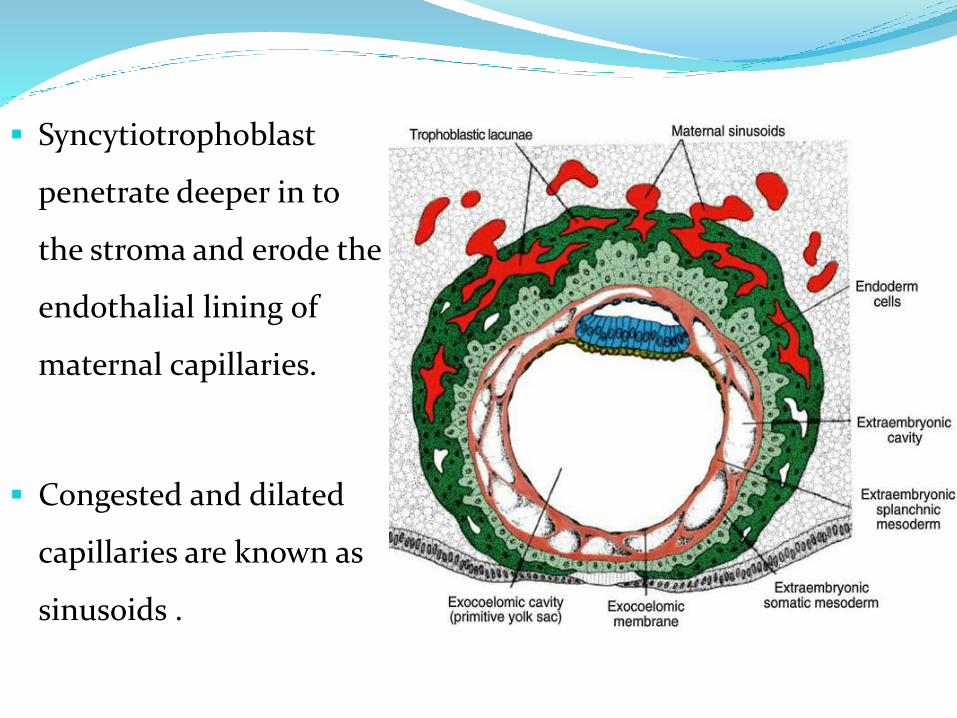
Syncytiotrophoblast
penetrate deeper in to
the stroma and erode the
endothalial lining of
maternal capillaries.
Congested and dilated
capillaries are known as
sinusoids .
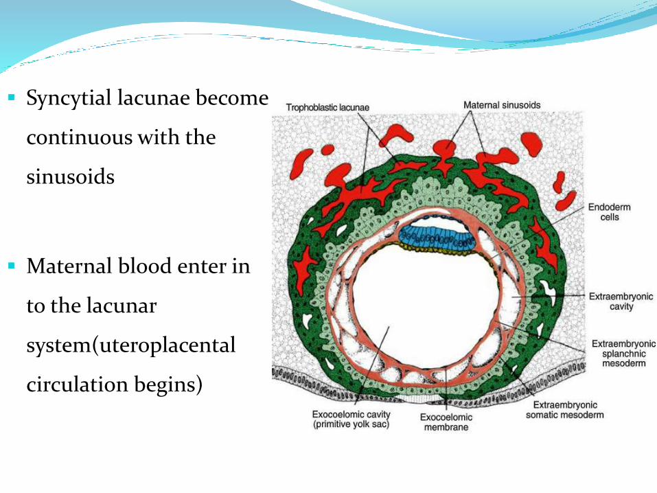
Syncytial lacunae become
continuous with the
sinusoids
Maternal blood enter in
to the lacunar
system(uteroplacental
circulation begins)
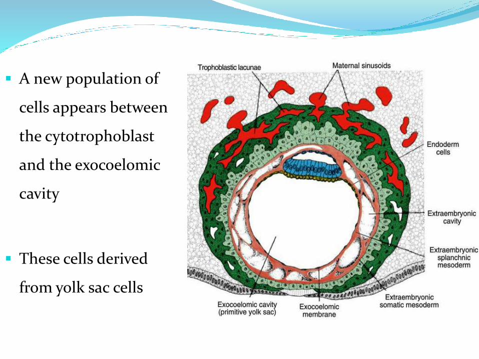
A new population of
cells appears between
the cytotrophoblast
and the exocoelomic
cavity
These cells derived
from yolk sac cells
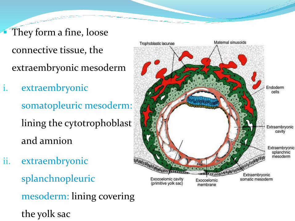
They form a fine, loose
connective tissue, the
extraembryonic mesoderm
i. extraembryonic
somatopleuric mesoderm:
lining the cytotrophoblast
and amnion
ii. extraembryonic
splanchnopleuric
mesoderm: lining covering
the yolk sac
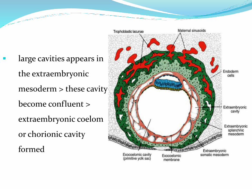
large cavities appears in
the extraembryonic
mesoderm > these cavity
become confluent >
extraembryonic coelom
or chorionic cavity
formed
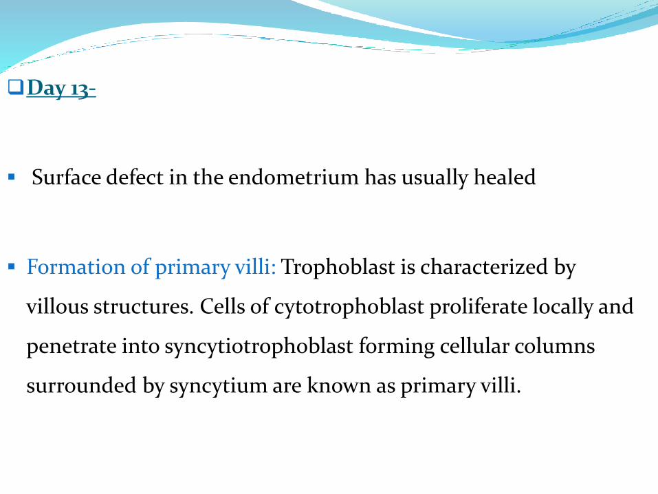
Day 13-
Surface defect in the endometrium has usually healed
Formation of primary villi: Trophoblast is characterized by
villous structures. Cells of cytotrophoblast proliferate locally and
penetrate into syncytiotrophoblast forming cellular columns
surrounded by syncytium are known as primary villi.
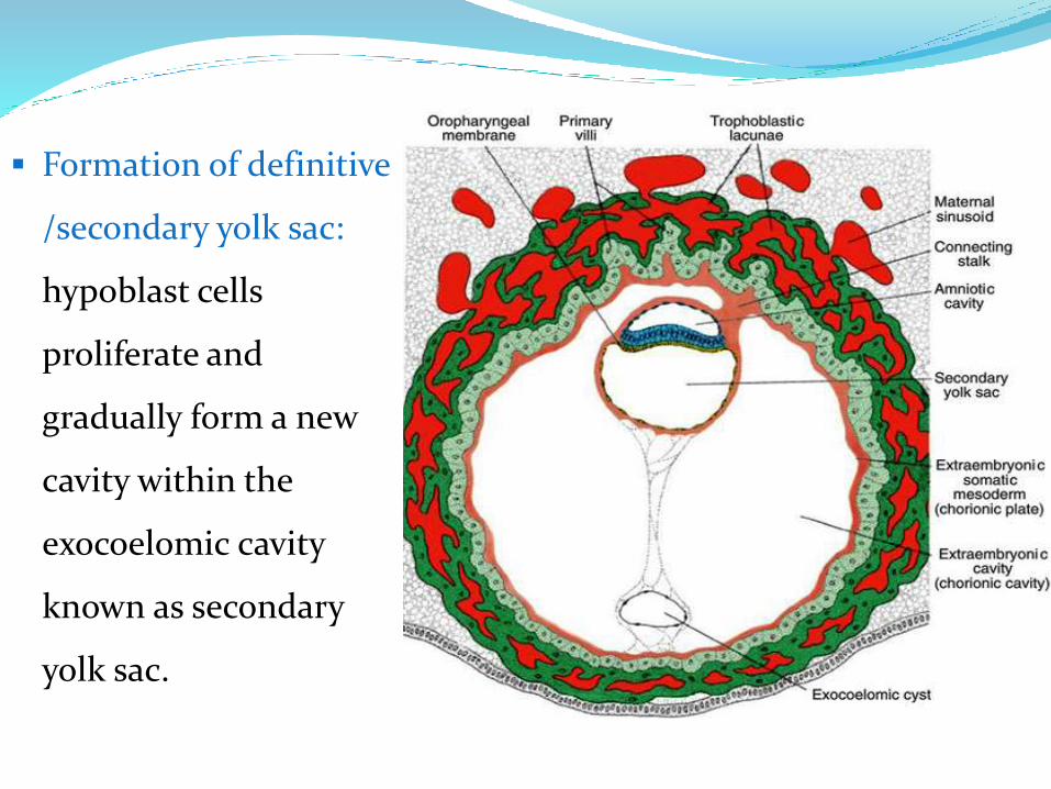
Formation of definitive
/secondary yolk sac:
hypoblast cells
proliferate and
gradually form a new
cavity within the
exocoelomic cavity
known as secondary
yolk sac.
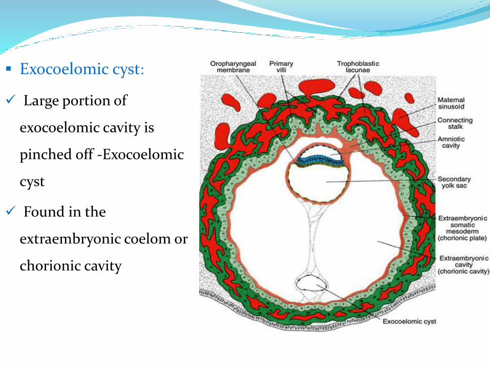
Exocoelomic cyst:
Large portion of
exocoelomic cavity is
pinched off -Exocoelomic
cyst
Found in the
extraembryonic coelom or
chorionic cavity
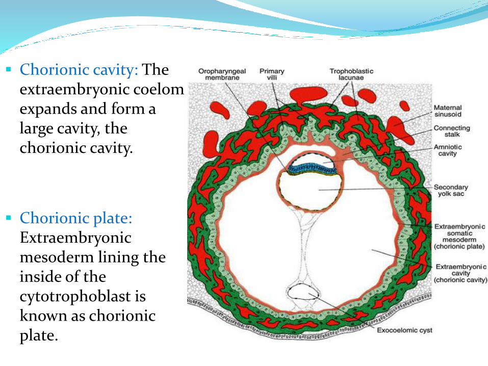
Chorionic cavity: The extraembryonic coelomexpands and form a large cavity, the chorionic cavity.
Chorionic plate: Extraembryonicmesoderm lining the inside of the cytotrophoblast is known as chorionic plate.
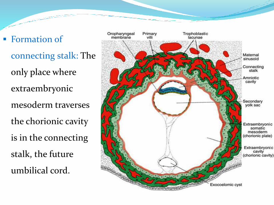
Formation of
connecting stalk: The
only place where
extraembryonic
mesoderm traverses
the chorionic cavity
is in the connecting
stalk, the future
umbilical cord.
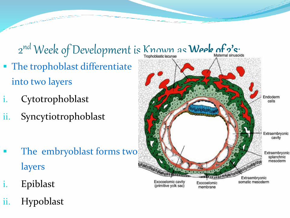
2nd Week of Development is Known as Week of 2’s: The trophoblast differentiate
into two layers
i. Cytotrophoblast
ii. Syncytiotrophoblast
The embryoblast forms two
layers
i. Epiblast
ii. Hypoblast
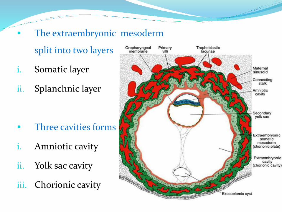
The extraembryonic mesoderm
split into two layers
i. Somatic layer
ii. Splanchnic layer
Three cavities forms
i. Amniotic cavity
ii. Yolk sac cavity
iii. Chorionic cavity
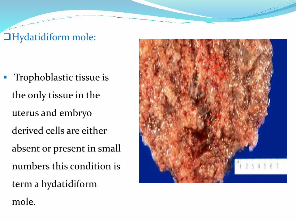
Hydatidiform mole:
Trophoblastic tissue is
the only tissue in the
uterus and embryo
derived cells are either
absent or present in small
numbers this condition is
term a hydatidiform
mole.
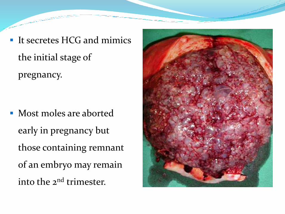
It secretes HCG and mimics
the initial stage of
pregnancy.
Most moles are aborted
early in pregnancy but
those containing remnant
of an embryo may remain
into the 2nd trimester.
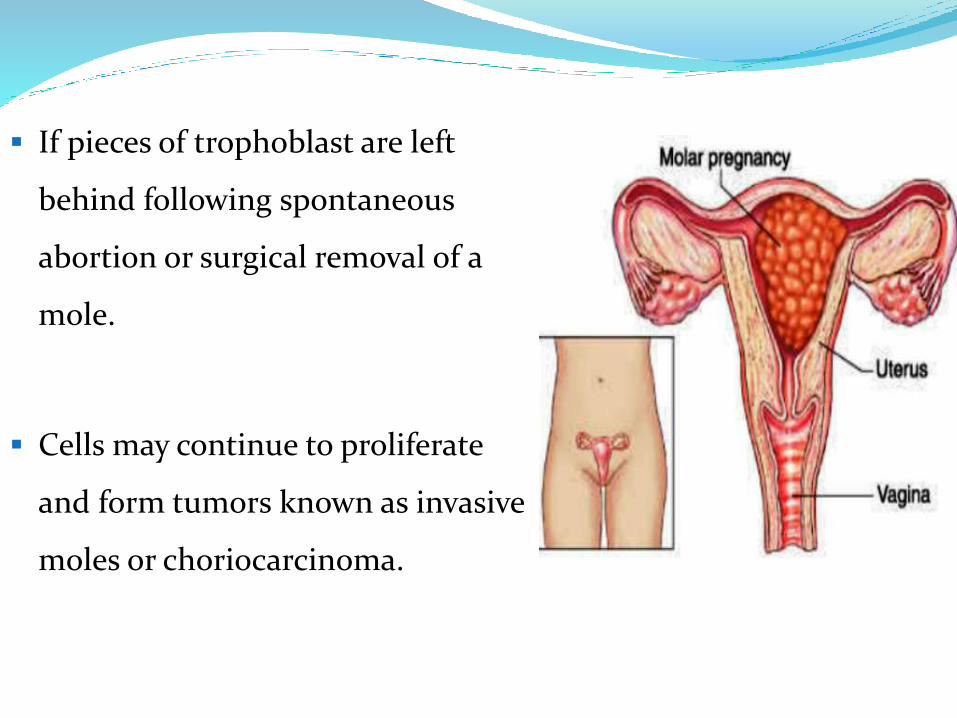
If pieces of trophoblast are left
behind following spontaneous
abortion or surgical removal of a
mole.
Cells may continue to proliferate
and form tumors known as invasive
moles or choriocarcinoma.

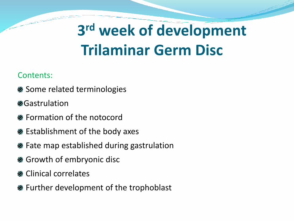
3rd week of developmentTrilaminar Germ Disc
Contents:
Some related terminologies
Gastrulation
Formation of the notocord
Establishment of the body axes
Fate map established during gastrulation
Growth of embryonic disc
Clinical correlates
Further development of the trophoblast
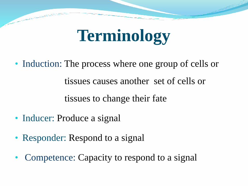
Terminology
• Induction: The process where one group of cells or
tissues causes another set of cells or
tissues to change their fate
• Inducer: Produce a signal
• Responder: Respond to a signal
• Competence: Capacity to respond to a signal
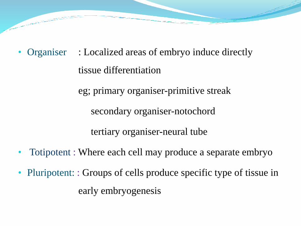
• Organiser : Localized areas of embryo induce directly
tissue differentiation
eg; primary organiser-primitive streak
secondary organiser-notochord
tertiary organiser-neural tube
• Totipotent : Where each cell may produce a separate embryo
• Pluripotent: : Groups of cells produce specific type of tissue in
early embryogenesis
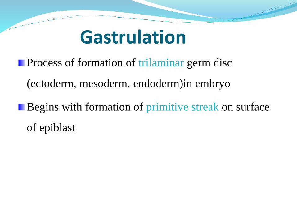
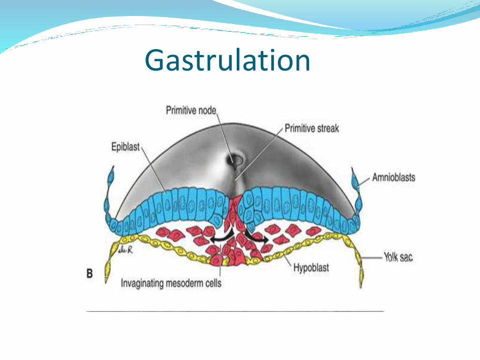
GastrulationProcess of formation of trilaminar germ disc
(ectoderm, mesoderm, endoderm)in embryo
Begins with formation of primitive streak on surface
of epiblast
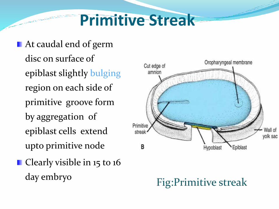
Primitive StreakAt caudal end of germ
disc on surface of
epiblast slightly bulging
region on each side of
primitive groove form
by aggregation of
epiblast cells extend
upto primitive node
Clearly visible in 15 to 16
day embryoFig:Primitive streak
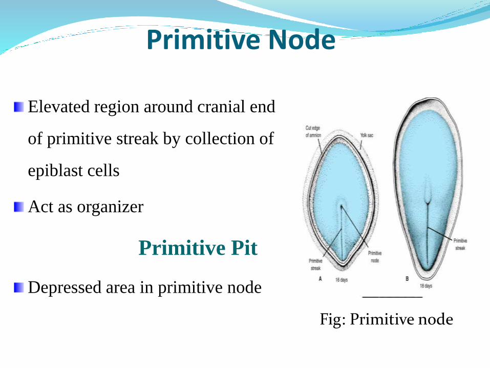
Primitive Node
Elevated region around cranial end
of primitive streak by collection of
epiblast cells
Act as organizer
Primitive Pit
Depressed area in primitive node
Fig: Primitive node
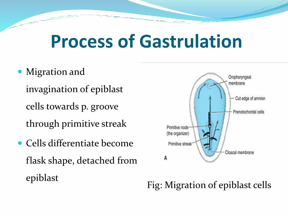
Process of Gastrulation
Migration and
invagination of epiblast
cells towards p. groove
through primitive streak
Cells differentiate become
flask shape, detached from
epiblastFig: Migration of epiblast cells
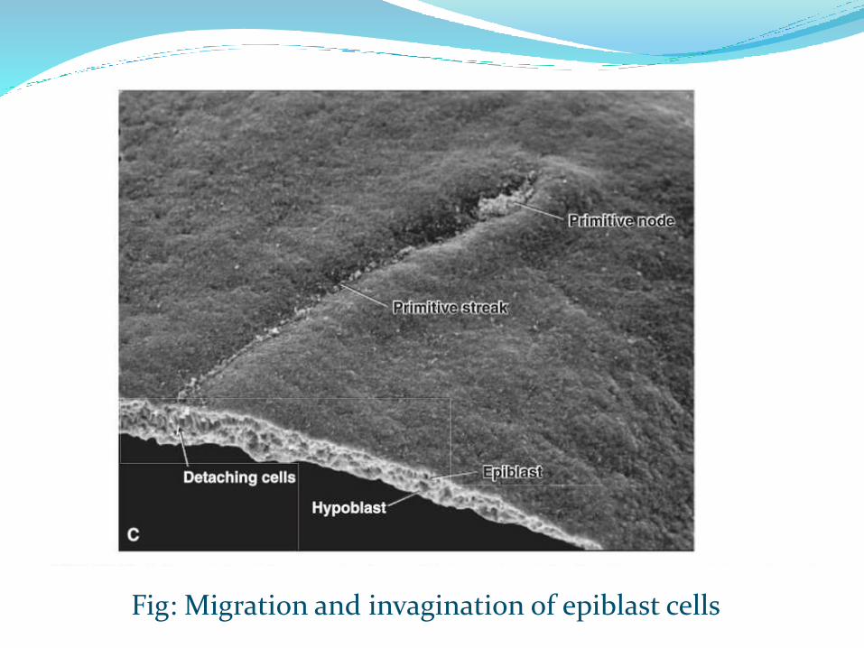
Fig: Migration and invagination of epiblast cells
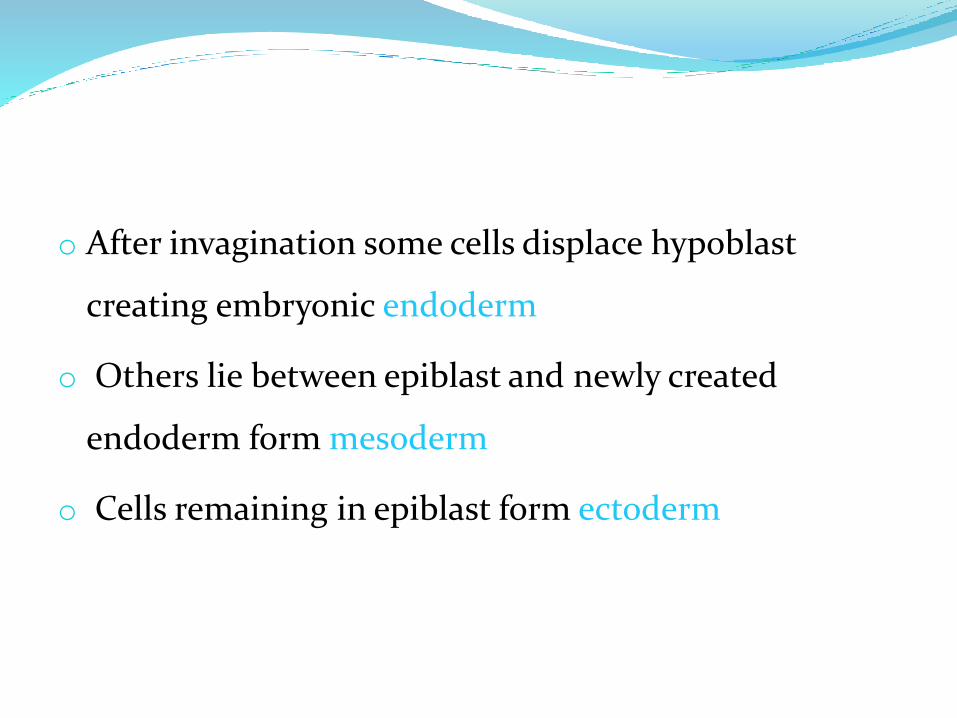
o After invagination some cells displace hypoblast
creating embryonic endoderm
o Others lie between epiblast and newly created
endoderm form mesoderm
o Cells remaining in epiblast form ectoderm
Gastrulation
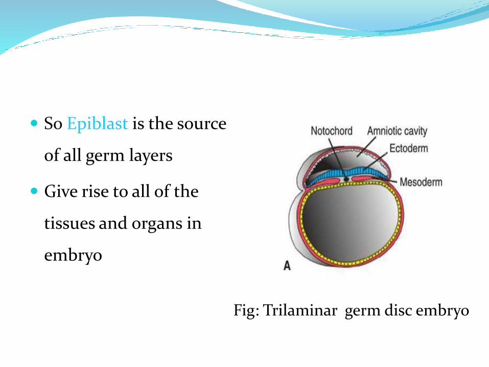
So Epiblast is the source
of all germ layers
Give rise to all of the
tissues and organs in
embryo
Fig: Trilaminar germ disc embryo
Oropharyngeal membrane
A small region of tightly adherent ectoderm and
endoderm cells at the cranial end of disc with no
intervening mesoderm
Future opening of oral cavity
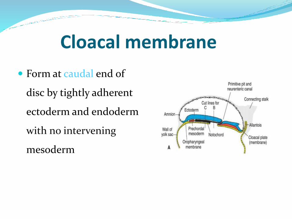
Cloacal membrane
Form at caudal end of
disc by tightly adherent
ectoderm and endoderm
with no intervening
mesoderm
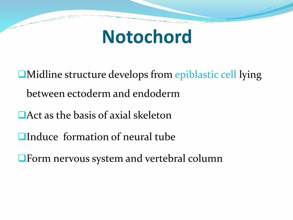
Notochord
Midline structure develops from epiblastic cell lying
between ectoderm and endoderm
Act as the basis of axial skeleton
Induce formation of neural tube
Form nervous system and vertebral column
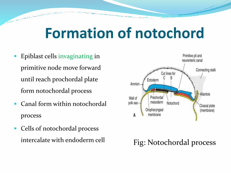
Formation of notochord
Epiblast cells invaginating in
primitive node move forward
until reach prochordal plate
form notochordal process
Canal form within notochordal
process
Cells of notochordal process
intercalate with endoderm cell Fig: Notochordal process
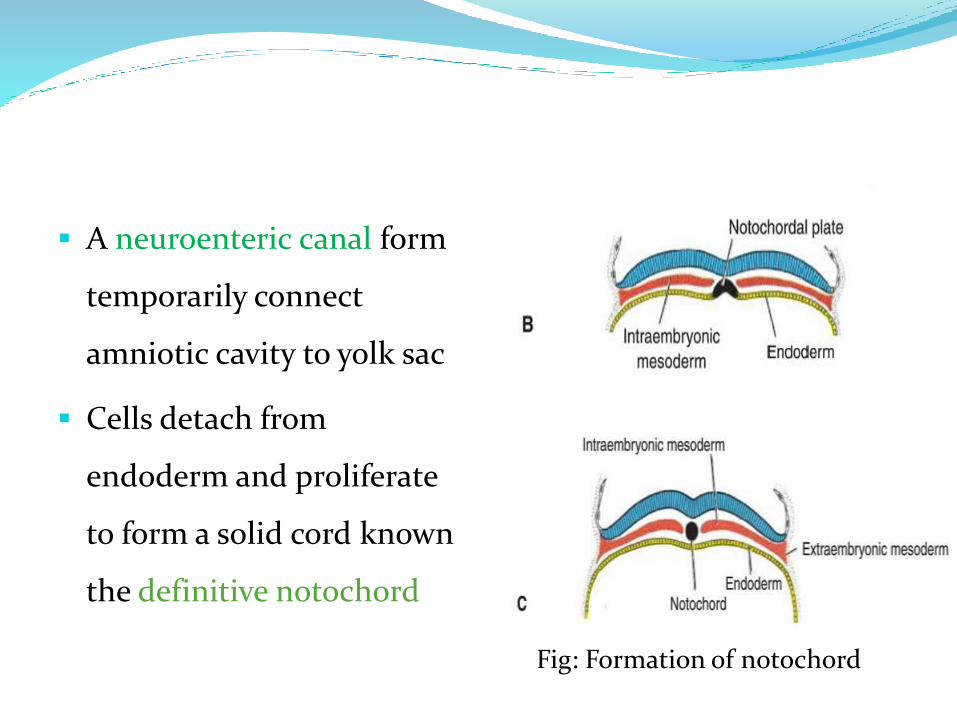
A neuroenteric canal form
temporarily connect
amniotic cavity to yolk sac
Cells detach from
endoderm and proliferate
to form a solid cord known
the definitive notochord
Fig: Formation of notochord
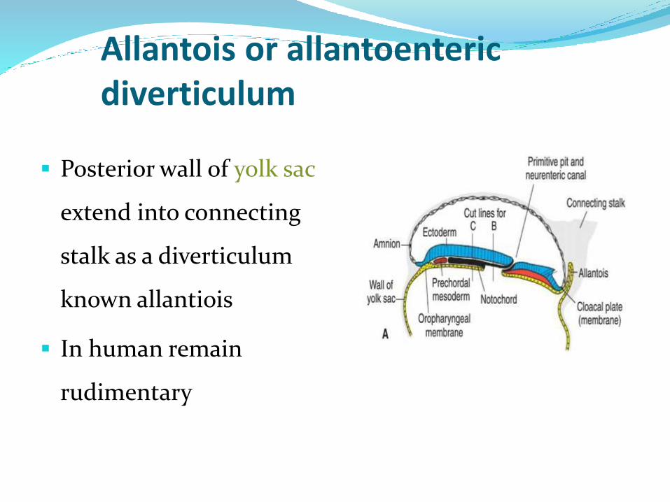
Allantois or allantoentericdiverticulum
Posterior wall of yolk sac
extend into connecting
stalk as a diverticulum
known allantiois
In human remain
rudimentary
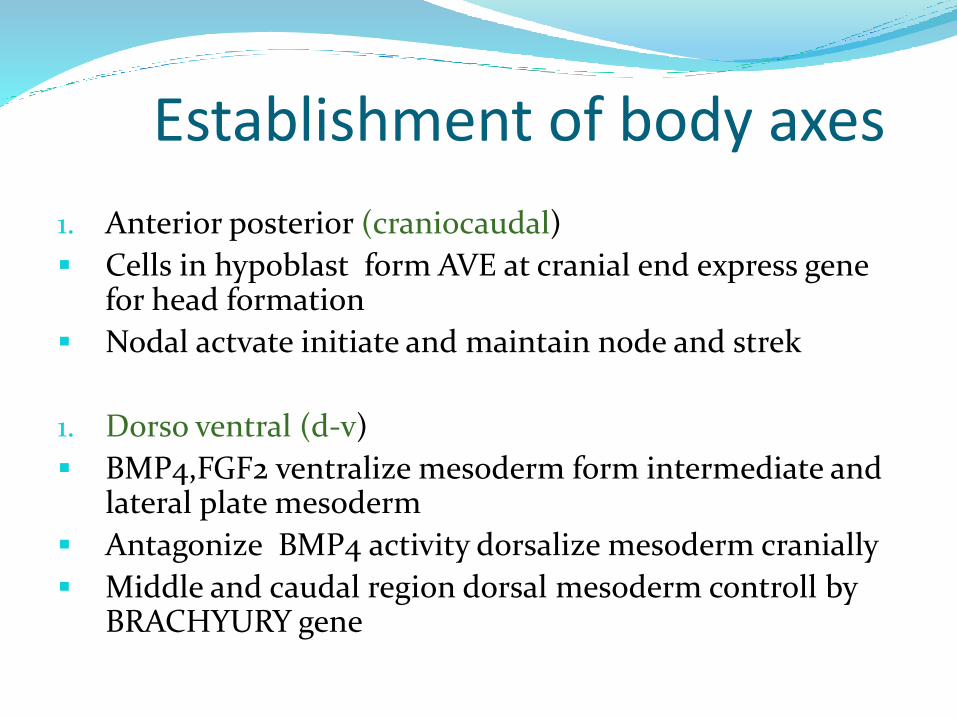
Establishment of body axes
1. Anterior posterior (craniocaudal)
Cells in hypoblast form AVE at cranial end express gene for head formation
Nodal actvate initiate and maintain node and strek
1. Dorso ventral (d-v)
BMP4,FGF2 ventralize mesoderm form intermediate and lateral plate mesoderm
Antagonize BMP4 activity dorsalize mesoderm cranially
Middle and caudal region dorsal mesoderm controll by BRACHYURY gene

Middle and caudal region dorsal mesoderm controll by
BRACHYURY gene
Left-right axis
FGF8 induces nodal expression restricted to left side by
accumulation of serotonin
Laterality defect
Situs inversus,dextrocardia
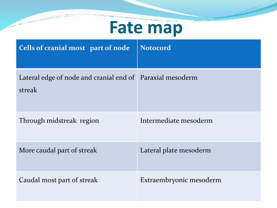
Fate map Cells of cranial most part of node Notocord
Lateral edge of node and cranial end of
streak
Paraxial mesoderm
Through midstreak region Intermediate mesoderm
More caudal part of streak Lateral plate mesoderm
Caudal most part of streak Extraembryonic mesoderm
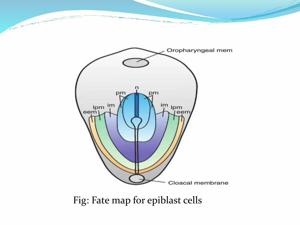
Fig: Fate map for epiblast cells
Growth of embryonic disc
Embryo develop cephalocaudally
Initially flat and almost round
Gradually elongated broad cephalic and narrow caudal
end
Invagination and migration cells continue until end of
fourth week
Primitive streak disappear at the end of fourth week
Clinical correlation
1. Insufficient mesoderm in caudal most region
resulting caudal dysgenesis (sirenomelia)
2. Sometimes remnants of primitive streak persist in
sacrococcygeal region and form sacrococcygeal
Teratoma
3. Situs inversus in which abdominal and thoracic
viscera become inverse
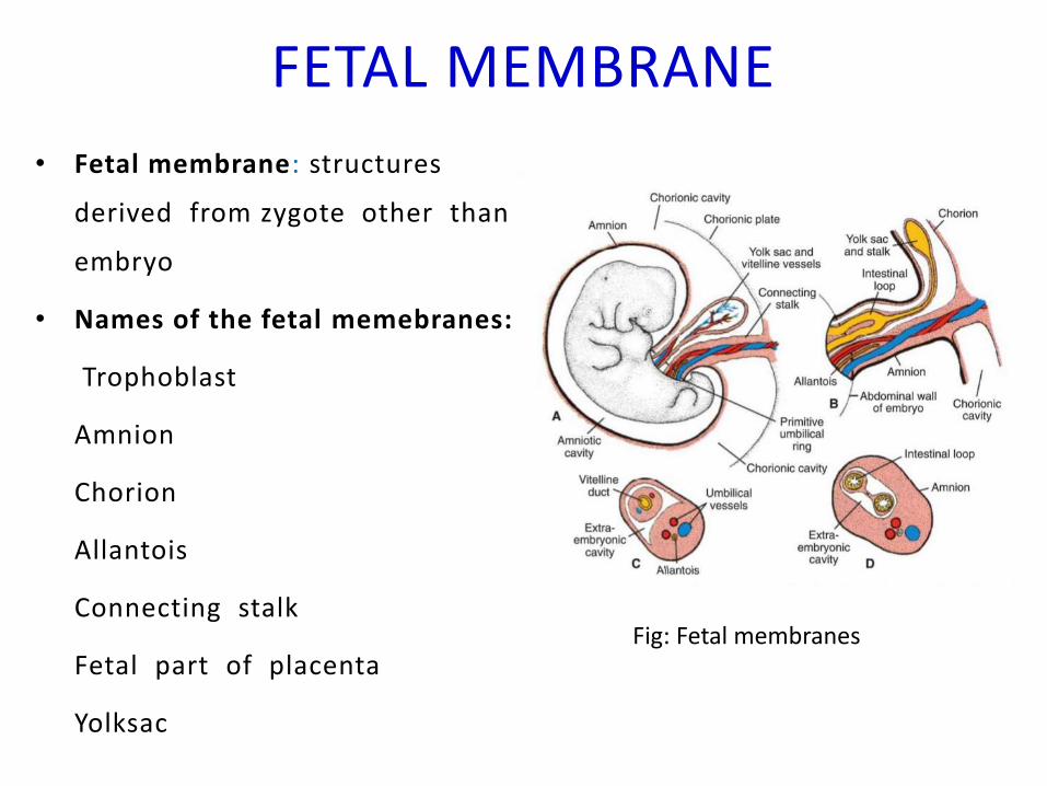
FETAL MEMBRANE
• Fetal membrane: structures
derived from zygote other than
embryo
• Names of the fetal memebranes:
Trophoblast
Amnion
Chorion
Allantois
Connecting stalk
Fetal part of placenta
Yolksac
Fig: Fetal membranes
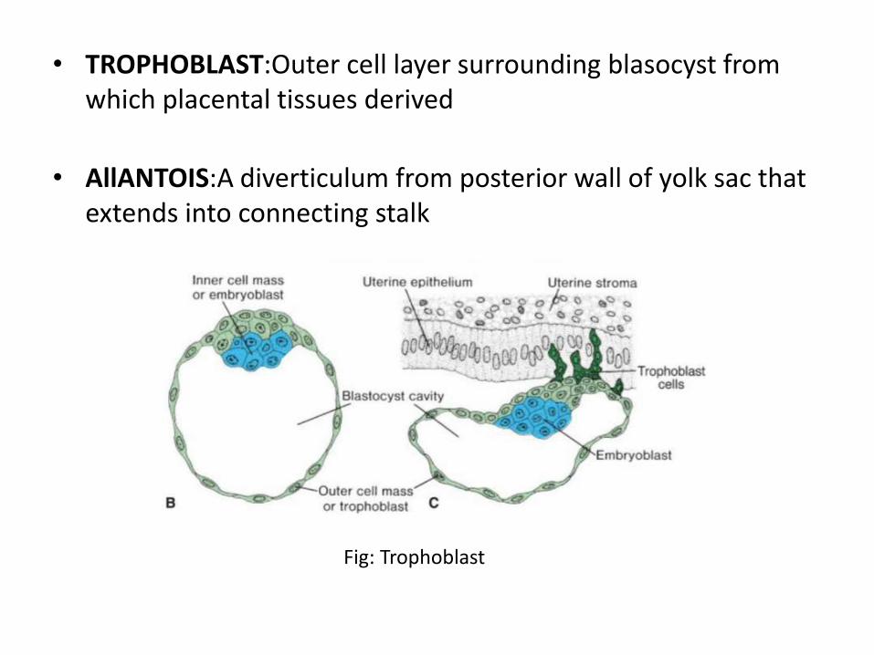
• TROPHOBLAST:Outer cell layer surrounding blasocyst from which placental tissues derived
• AllANTOIS:A diverticulum from posterior wall of yolk sac that extends into connecting stalk
Fig: Trophoblast
• AMNION: Membrane derived from
epiblast which surrounds amniotic
cavity
• CHORION:Multilayered structure
consisting of somatic layer
extraembryonic
mesoderm,cytotrophoblast &
syncytiotrophoblast
• CONNECTING STALK:Unsplitted part of
extraembryonic mesoderm ,connects
embryo with trophoblast
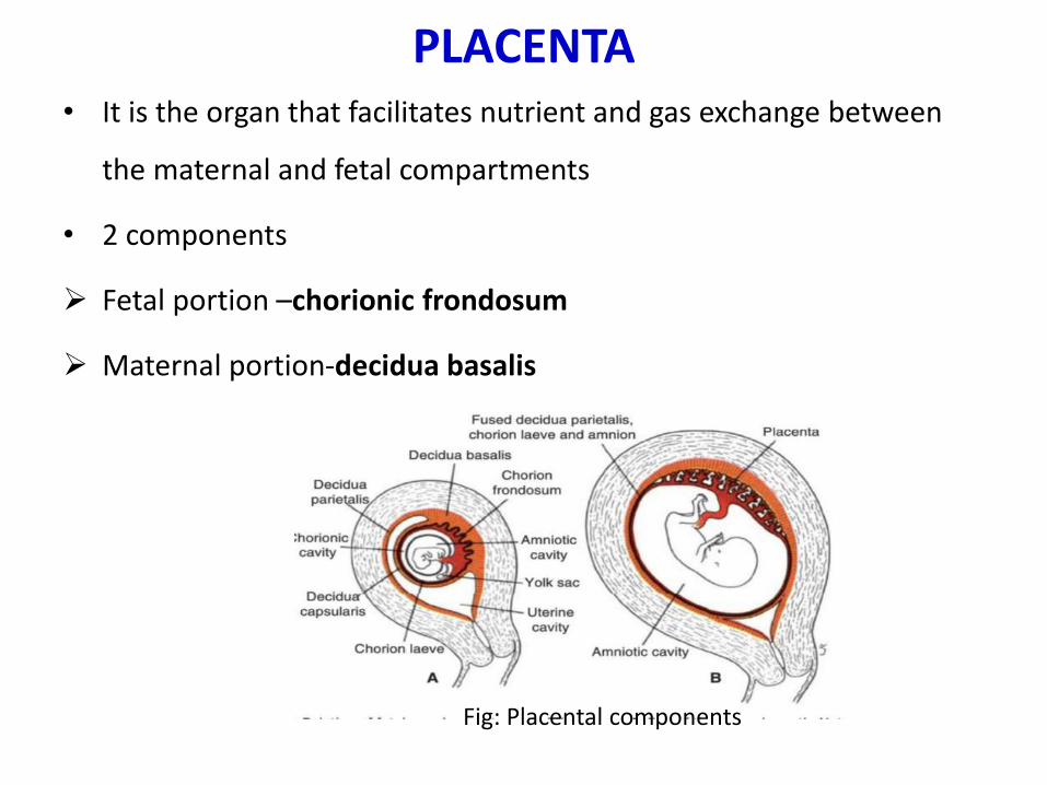
PLACENTA• It is the organ that facilitates nutrient and gas exchange between
the maternal and fetal compartments
• 2 components
Fetal portion –chorionic frondosum
Maternal portion-decidua basalis
Fig: Placental components
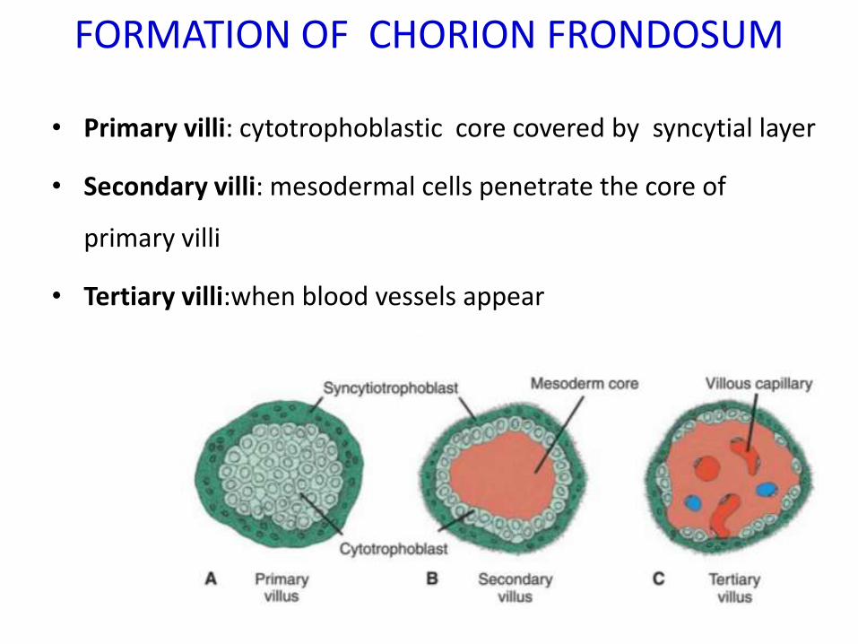
FORMATION OF CHORION FRONDOSUM
• Primary villi: cytotrophoblastic core covered by syncytial layer
• Secondary villi: mesodermal cells penetrate the core of
primary villi
• Tertiary villi:when blood vessels appear
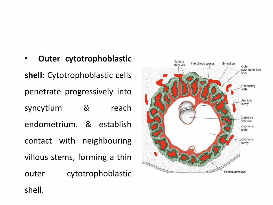
• Outer cytotrophoblastic
shell: Cytotrophoblastic cells
penetrate progressively into
syncytium & reach
endometrium. & establish
contact with neighbouring
villous stems, forming a thin
outer cytotrophoblastic
shell.
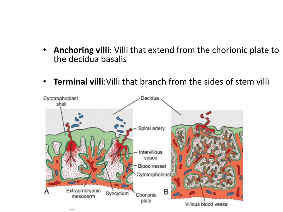
• Anchoring villi: Villi that extend from the chorionic plate to the decidua basalis
• Terminal villi:Villi that branch from the sides of stem villi
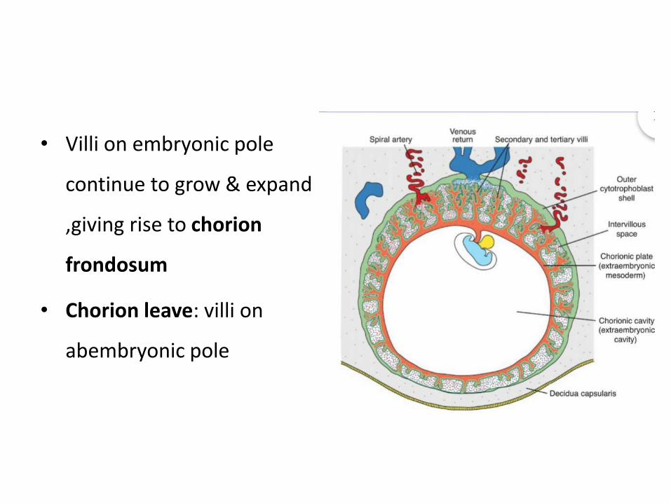
• Villi on embryonic pole
continue to grow & expand
,giving rise to chorion
frondosum
• Chorion leave: villi on
abembryonic pole
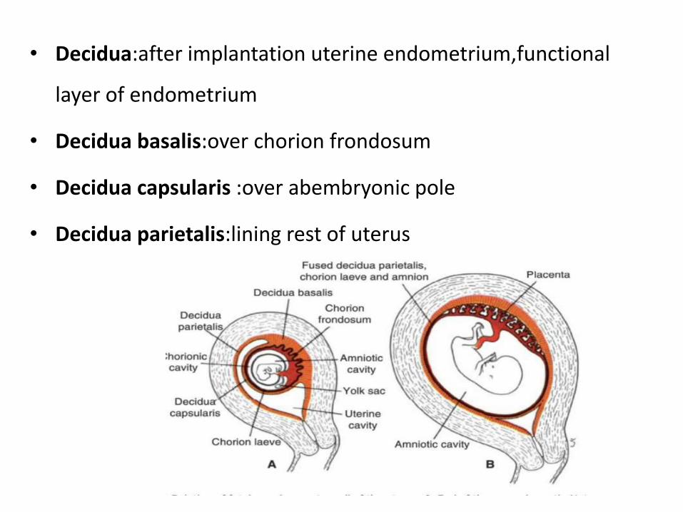
• Decidua:after implantation uterine endometrium,functional
layer of endometrium
• Decidua basalis:over chorion frondosum
• Decidua capsularis :over abembryonic pole
• Decidua parietalis:lining rest of uterus
STRUCTURE OF PLACENTA
• On fetal side placenta bordered by chorionic plate
• On maternal side by decidual plate
• In between –intervillous spaces,filled with maternal blood
• Decidual septa formed
• Placenta divides into cotyledons
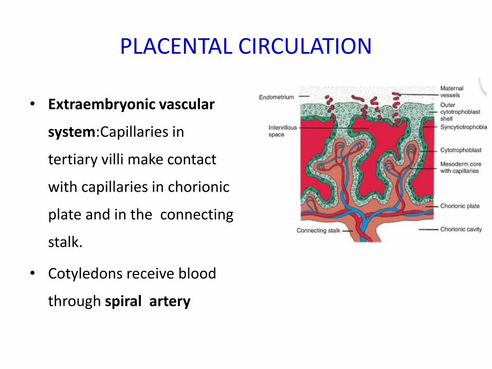
PLACENTAL CIRCULATION
• Extraembryonic vascular
system:Capillaries in
tertiary villi make contact
with capillaries in chorionic
plate and in the connecting
stalk.
• Cotyledons receive blood
through spiral artery
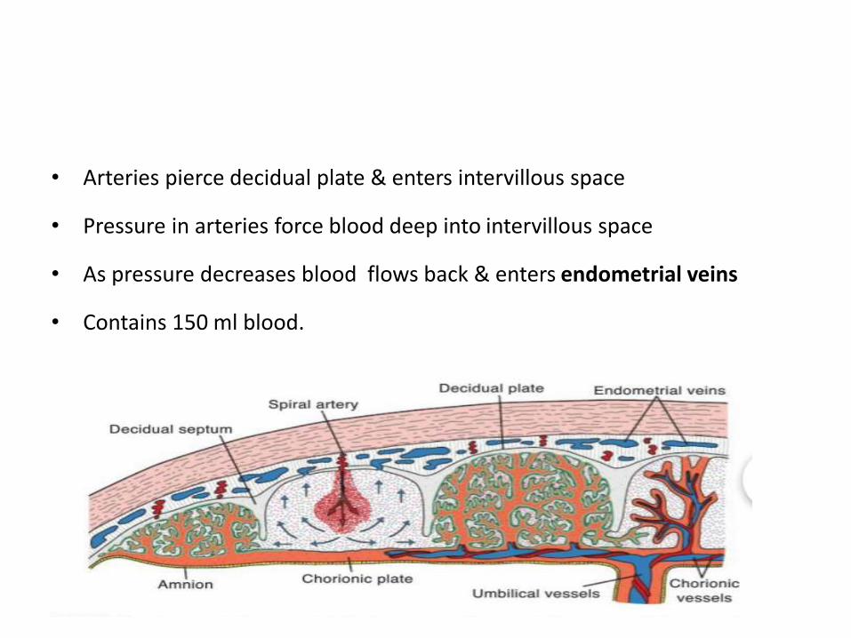
• Arteries pierce decidual plate & enters intervillous space
• Pressure in arteries force blood deep into intervillous space
• As pressure decreases blood flows back & enters endometrial veins
• Contains 150 ml blood.

PLACENTAL BARRIER
• Initially 4 layer
Endothelial linining of fetal
vessels
Connective tissue in villous core
Cytotrophoblastic layer
Syncytium
• From 4 months
Endothelium
Syncytium
FUNCTION OF PLACENTA
• Exchange of gases
• Exchange of nutrient & electrolyte
• Transmission of maternal antibodies
• Hormone production
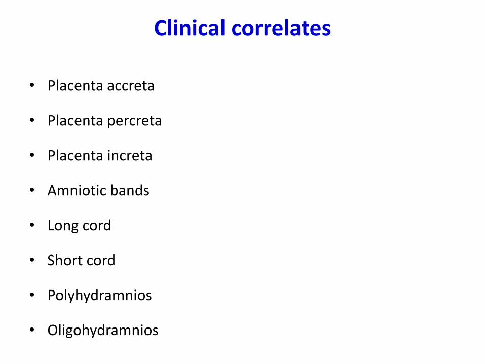
Clinical correlates
• Placenta accreta
• Placenta percreta
• Placenta increta
• Amniotic bands
• Long cord
• Short cord
• Polyhydramnios
• Oligohydramnios
Embryonic period & derivatives of the Ectodermal germ layer
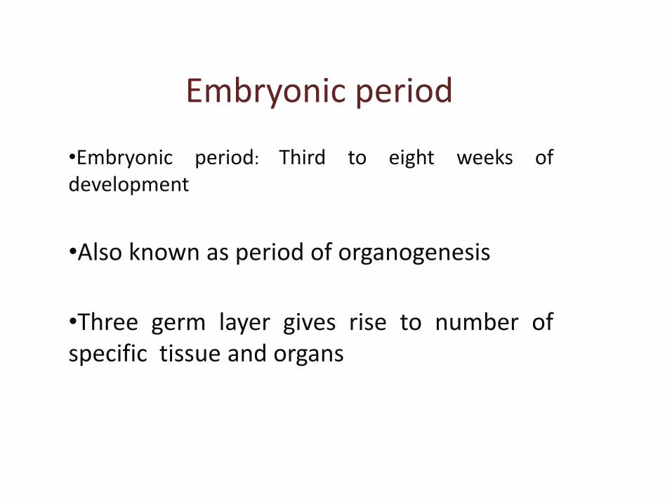
Embryonic period
•Embryonic period: Third to eight weeks ofdevelopment
•Also known as period of organogenesis
•Three germ layer gives rise to number ofspecific tissue and organs
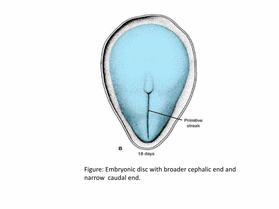
Figure: Embryonic disc with broader cephalic end and narrow caudal end.
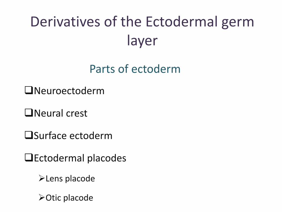
Derivatives of the Ectodermal germ layer
Parts of ectoderm
Neuroectoderm
Neural crest
Surface ectoderm
Ectodermal placodes
Lens placode
Otic placode
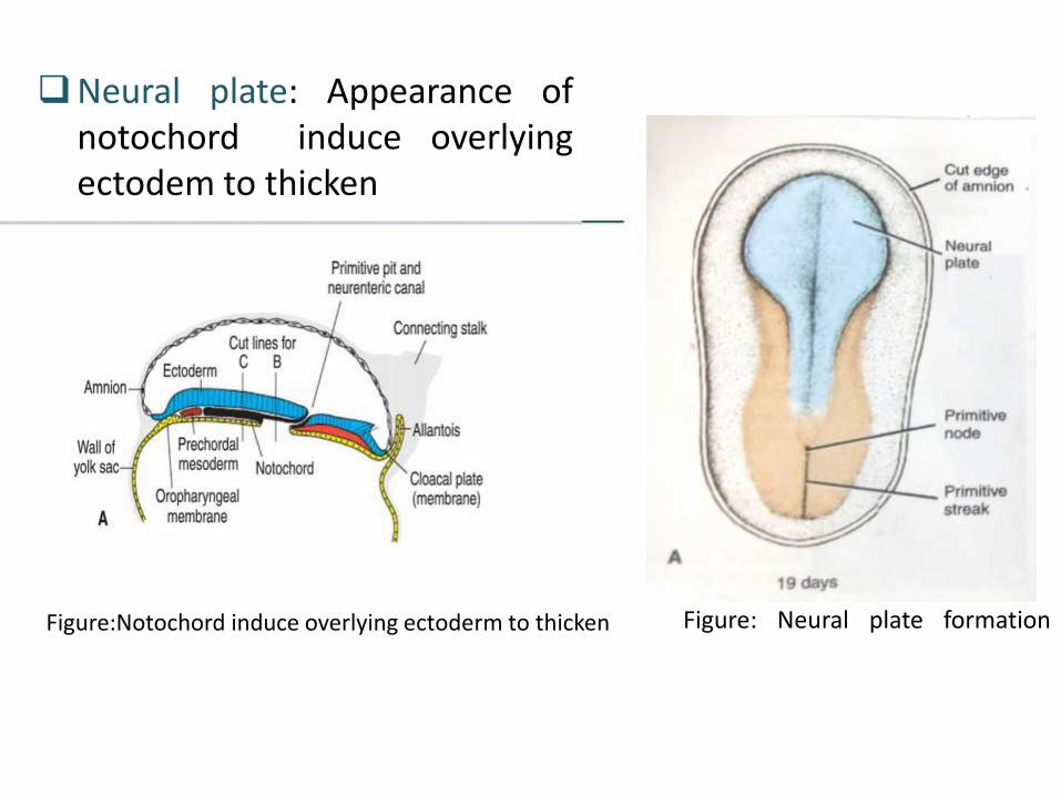
Figure:
Figure: Neural plate formation
Neural plate: Appearance ofnotochord induce overlyingectodem to thicken
Figure:Notochord induce overlying ectoderm to thicken
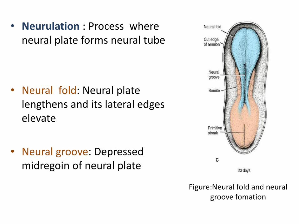
Figure:Neural fold and neural groove fomation
• Neurulation : Process where neural plate forms neural tube
• Neural fold: Neural plate lengthens and its lateral edges elevate
• Neural groove: Depressed midregoin of neural plate
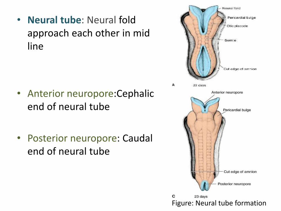
Figure: Neural tube formation
• Neural tube: Neural fold approach each other in mid line
• Anterior neuropore:Cephalicend of neural tube
• Posterior neuropore: Caudal end of neural tube
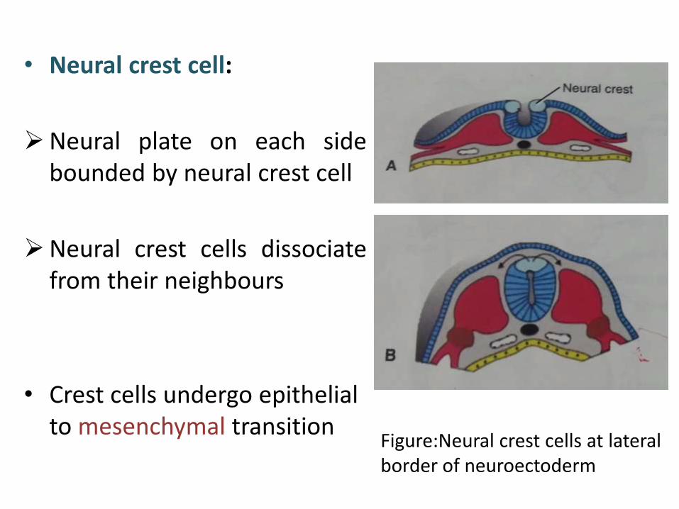
Figure:Neural crest cells at lateralborder of neuroectoderm
• Neural crest cell:
Neural plate on each sidebounded by neural crest cell
Neural crest cells dissociatefrom their neighbours
• Crest cells undergo epithelial to mesenchymal transition
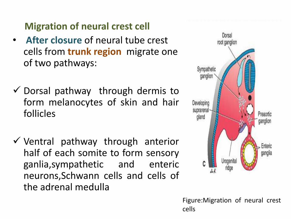
Figure:Migration of neural crestcells
Migration of neural crest cell
• After closure of neural tube crest cells from trunk region migrate one of two pathways:
Dorsal pathway through dermis toform melanocytes of skin and hairfollicles
Ventral pathway through anteriorhalf of each somite to form sensoryganlia,sympathetic and entericneurons,Schwann cells and cells ofthe adrenal medulla
• Before closure of neural tube crest cells from cranialregion migrate to form craniofacial skeleton as neuronfor cranial ganglia, glial cells,melanocytes
• Neural crest cell contributes to so many organs and tissues that sometimes they are referred as the fourth germ layer
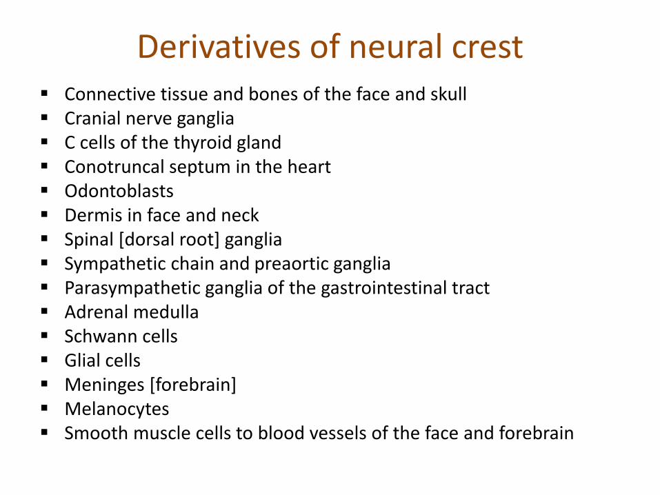
Derivatives of neural crest Connective tissue and bones of the face and skull Cranial nerve ganglia C cells of the thyroid gland Conotruncal septum in the heart Odontoblasts Dermis in face and neck Spinal [dorsal root] ganglia Sympathetic chain and preaortic ganglia Parasympathetic ganglia of the gastrointestinal tract Adrenal medulla Schwann cells Glial cells Meninges [forebrain] Melanocytes Smooth muscle cells to blood vessels of the face and forebrain
Surface ectoderm: After closure of neural tube remaining ectoderm
Derivatives of surface ectoderm
EpidermisHair and hair follicleNailMammary glandSebaceous glandSweat gland
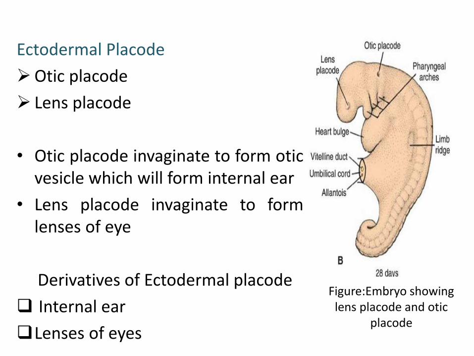
Figure:Embryo showing lens placode and otic
placode
Ectodermal Placode
Otic placode
Lens placode
• Otic placode invaginate to form oticvesicle which will form internal ear
• Lens placode invaginate to formlenses of eye
Derivatives of Ectodermal placode
Internal ear
Lenses of eyes
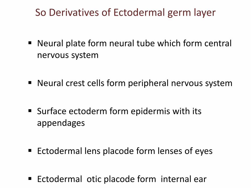
So Derivatives of Ectodermal germ layer
Neural plate form neural tube which form central nervous system
Neural crest cells form peripheral nervous system
Surface ectoderm form epidermis with its appendages
Ectodermal lens placode form lenses of eyes
Ectodermal otic placode form internal ear
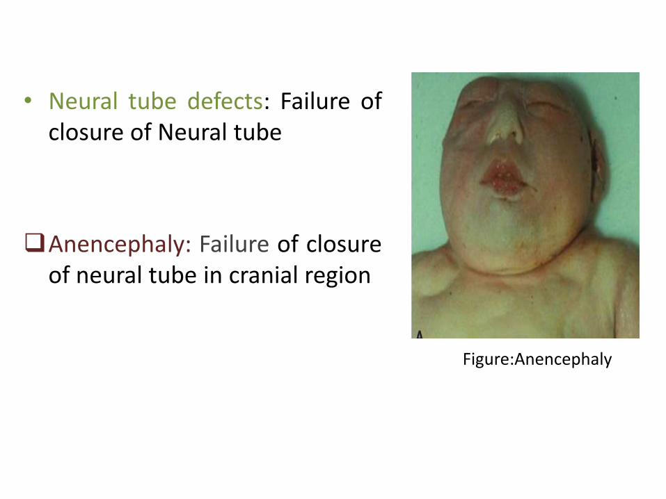
Figure:Anencephaly
• Neural tube defects: Failure ofclosure of Neural tube
Anencephaly: Failure of closureof neural tube in cranial region
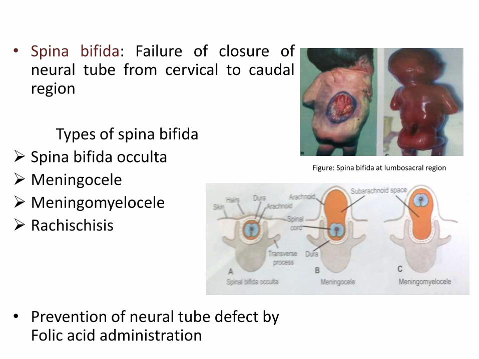
Figure: Spina bifida at lumbosacral region
• Spina bifida: Failure of closure ofneural tube from cervical to caudalregion
Types of spina bifida
Spina bifida occulta
Meningocele
Meningomyelocele
Rachischisis
• Prevention of neural tube defect by Folic acid administration
DERIVATIVES OFMESODERMAL GERM LAYER
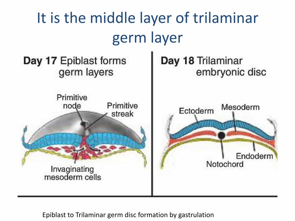
It is the middle layer of trilaminargerm layer
Epiblast to Trilaminar germ disc formation by gastrulation
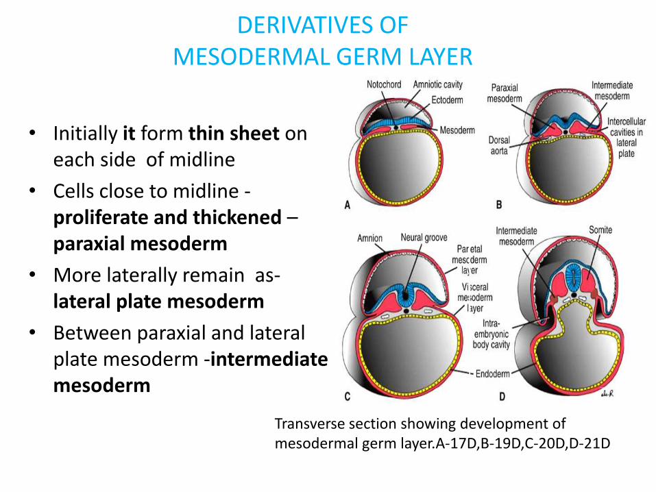
DERIVATIVES OFMESODERMAL GERM LAYER
• Initially it form thin sheet on each side of midline
• Cells close to midline -proliferate and thickened –paraxial mesoderm
• More laterally remain as-lateral plate mesoderm
• Between paraxial and lateral plate mesoderm -intermediate mesoderm
Transverse section showing development of mesodermal germ layer.A-17D,B-19D,C-20D,D-21D
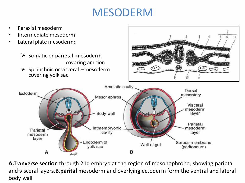
MESODERM• Paraxial mesoderm• Intermediate mesoderm• Lateral plate mesoderm:
Somatic or parietal -mesoderm covering amnion
Splanchnic or visceral –mesoderm covering yolk sac
A.Tranverse section through 21d embryo at the region of mesonephrone, showing parietal and visceral layers.B.parital mesoderm and overlying ectoderm form the ventral and lateral body wall
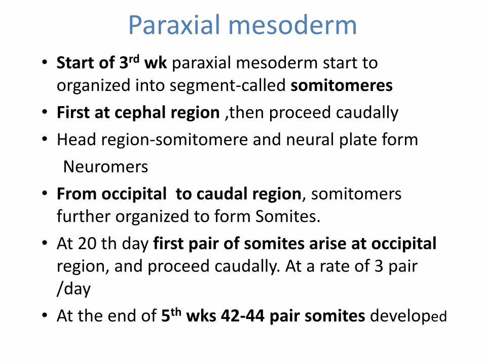
Paraxial mesoderm• Start of 3rd wk paraxial mesoderm start to
organized into segment-called somitomeres
• First at cephal region ,then proceed caudally
• Head region-somitomere and neural plate form
Neuromers
• From occipital to caudal region, somitomersfurther organized to form Somites.
• At 20 th day first pair of somites arise at occipital region, and proceed caudally. At a rate of 3 pair /day
• At the end of 5th wks 42-44 pair somites developed
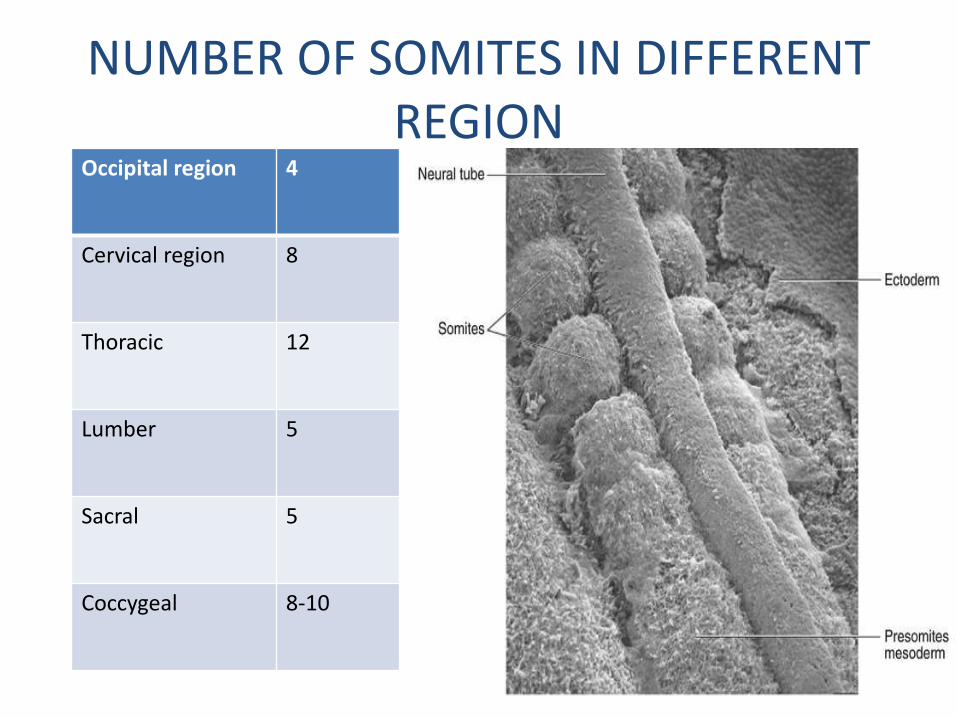
NUMBER OF SOMITES IN DIFFERENT REGION
Occipital region 4
Cervical region 8
Thoracic 12
Lumber 5
Sacral 5
Coccygeal 8-10
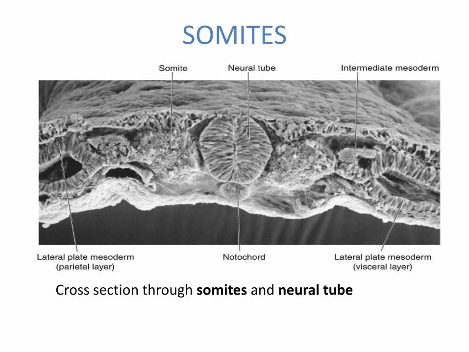
SOMITES
Cross section through somites and neural tube
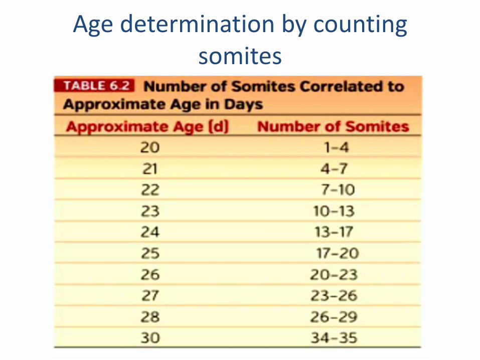
Age determination by counting somites
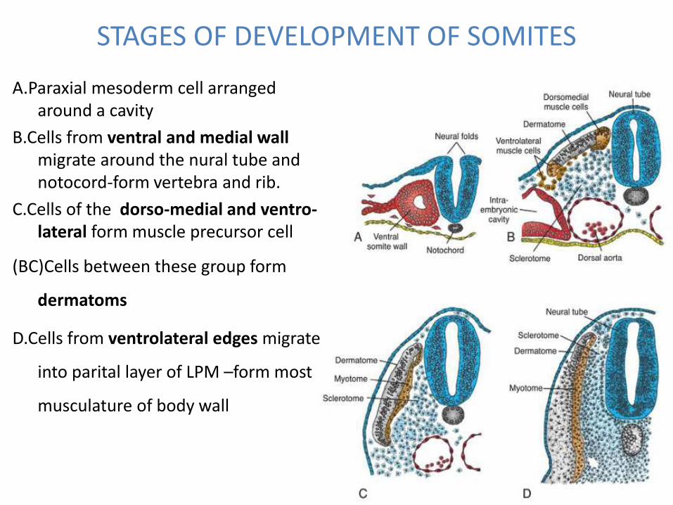
STAGES OF DEVELOPMENT OF SOMITES
A.Paraxial mesoderm cell arranged around a cavity
B.Cells from ventral and medial wall migrate around the nural tube and notocord-form vertebra and rib.
C.Cells of the dorso-medial and ventro-lateral form muscle precursor cell
(BC)Cells between these group form
dermatoms
D.Cells from ventrolateral edges migrate
into parital layer of LPM –form most
musculature of body wall
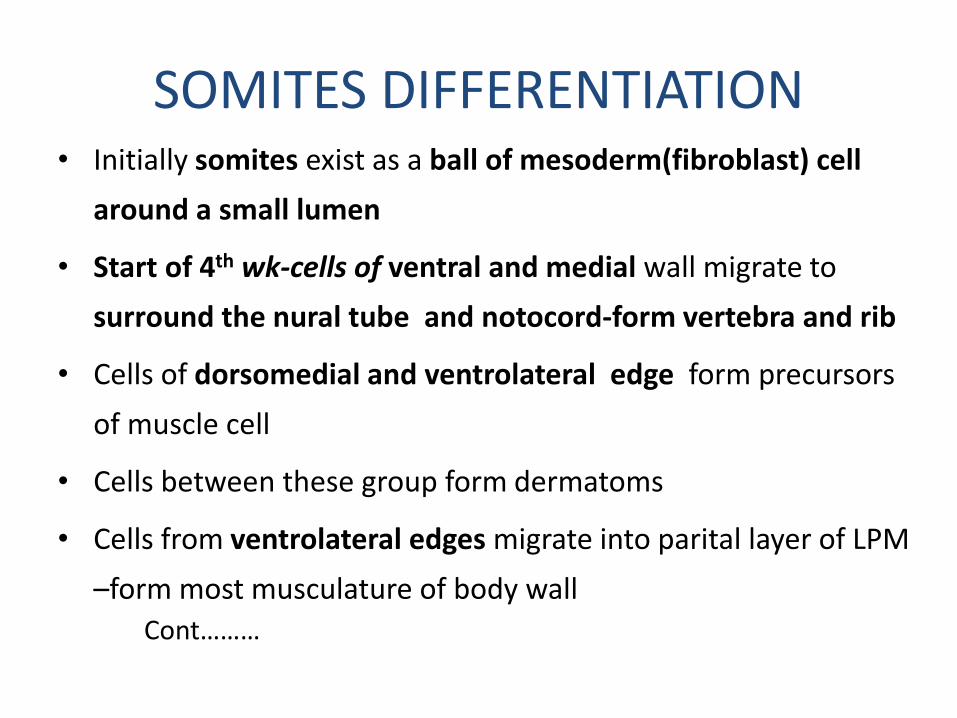
SOMITES DIFFERENTIATION• Initially somites exist as a ball of mesoderm(fibroblast) cell
around a small lumen
• Start of 4th wk-cells of ventral and medial wall migrate to
surround the nural tube and notocord-form vertebra and rib
• Cells of dorsomedial and ventrolateral edge form precursors
of muscle cell
• Cells between these group form dermatoms
• Cells from ventrolateral edges migrate into parital layer of LPM
–form most musculature of body wall
Cont………
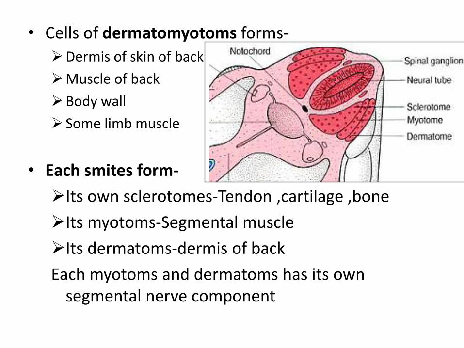
• Cells of dermatomyotoms forms-
Dermis of skin of back
Muscle of back
Body wall
Some limb muscle
• Each smites form-
Its own sclerotomes-Tendon ,cartilage ,bone
Its myotoms-Segmental muscle
Its dermatoms-dermis of back
Each myotoms and dermatoms has its own segmental nerve component
LATERAL PLATE MESODERM
Parietal(somatic)-
• Surround the intra embryonic cavity,
• Form thin membrane(mesothelium) line the –peritoneal, pleural,
pericardial cavity
• Mesoderm of parietal layer together with overlying ectoderm form
the lateral body wall fold
• These fold with head and tail fold close the body wall.
• Parietal layer of LPM-
i. Dermis of the skin of body wall and limb
ii. The bones and Connective tissue of limb and sternum
• Sclerotome and muscular precursor cell migrate into parietal
layer- costal cartilage , limb muscle, and most of the body wal
muscle
Visceral(splanchnic)-
• surround the organ, form thin membrane around each organ
• Visceral layer of LPM with embryonic endoderm form the wall
of gut tube.
Intermediate mesoderm:Differentiated into urogenital system
• Cervical and upper thoracic region-segmented cell cluster(future-nephrotoms)
• Caudally-unsegmented mass-neohrogeniccord
• Excretory unit of Urinary system and gonad –developed from partially segmented and partially unsegmented intermediate mesoderm
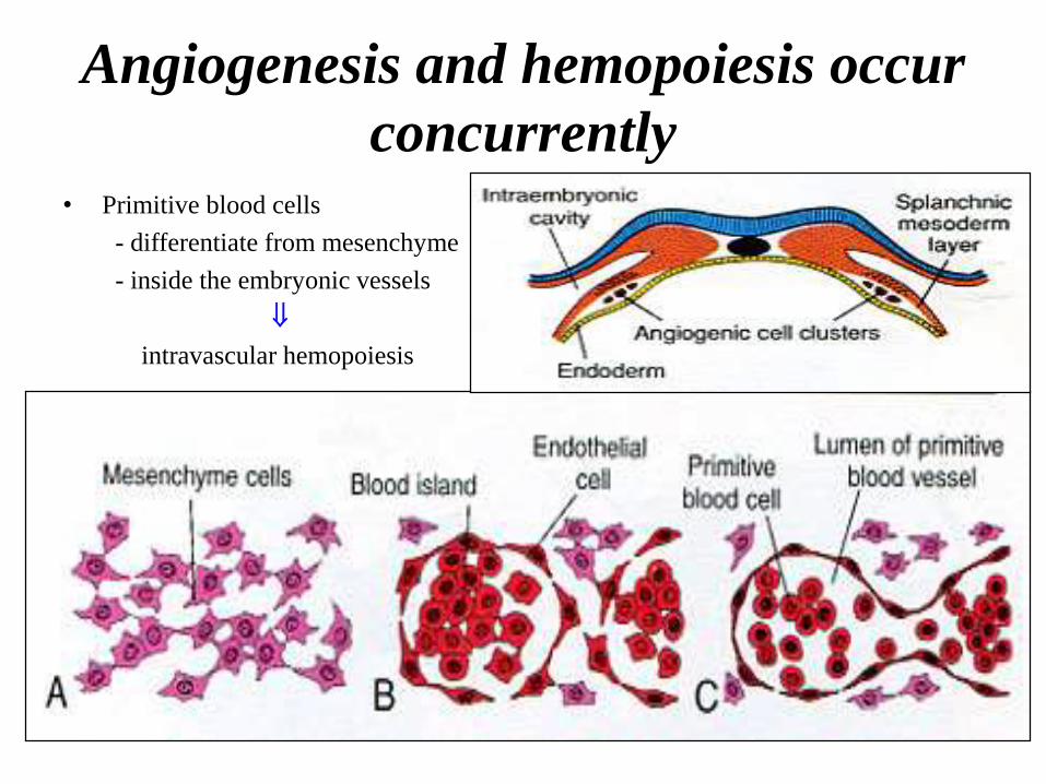
Angiogenesis and hemopoiesis occur
concurrently• Primitive blood cells
- differentiate from mesenchyme
- inside the embryonic vessels
intravascular hemopoiesis
Blood and blood vessels
• Blood vessels forms in 2 way:
– Vasculogenesis-vesells arise from blood island
– Angiogenesis:sprouting from existing blood vessels
• First blood island apear in mesoderm surrounding the wall of the yolk sac at 3rd wk ,
• later lateral plate mesoderm and other region
• Island arise from mesodermal cells –induced to form hem-angioblast-commom precoursure of vessels and blood cell.
• Definitive haemopoitic stem call are drive from mesoderm surrounding the aorta
• Then cell colonize to liver – major haemopoitic organ for 2nd
to 7th months.
• Stem cells of liver colonized –bone marrow at the 7th
months of gastation and liver loss its haemopoetic function.
IN SUMMARY: following tissue and organ developed from mesoderm
• Supporting tissue-connective tissue, cartilage and
bones
• Striated and smooth muscle
• Vascular system-The heart, arteries, vain, lymph
vessels, and all blood and lymph cells
• Urogenital system-Kidney gonad and their
corrosponding duct (except UB)
• The cortical portion of suprarenal gland and spleen
DERIVATIVES OF ENDODERM
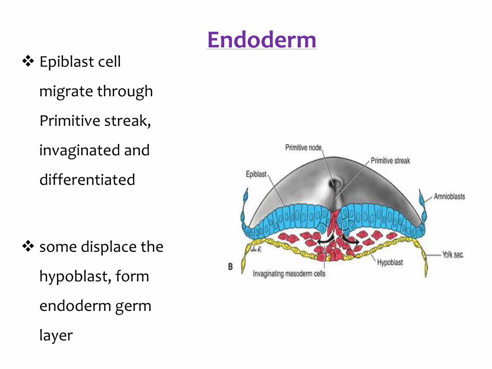
Endoderm Epiblast cell
migrate through
Primitive streak,
invaginated and
differentiated
some displace the
hypoblast, form
endoderm germ
layer
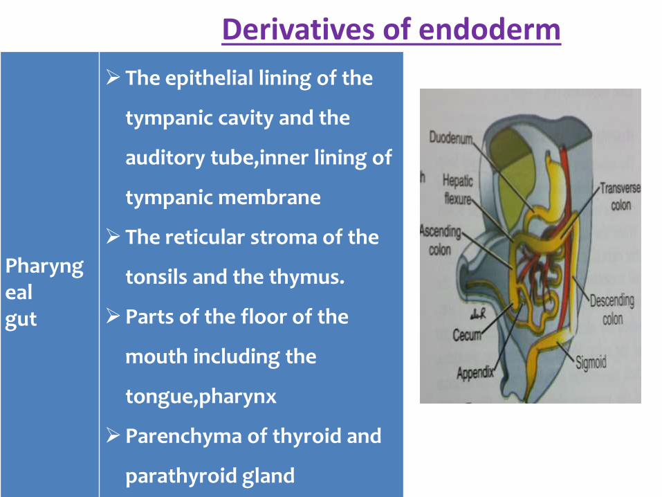
Pharyngealgut
The epithelial lining of the
tympanic cavity and the
auditory tube,inner lining of
tympanic membrane
The reticular stroma of the
tonsils and the thymus.
Parts of the floor of the
mouth including the
tongue,pharynx
Parenchyma of thyroid and
parathyroid gland
Derivatives of endoderm
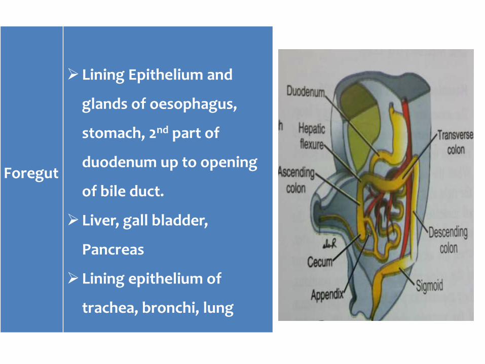
Foregut
Lining Epithelium and
glands of oesophagus,
stomach, 2nd part of
duodenum up to opening
of bile duct.
Liver, gall bladder,
Pancreas
Lining epithelium of
trachea, bronchi, lung
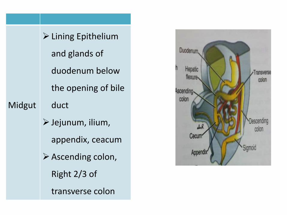
Midgut
Lining Epithelium
and glands of
duodenum below
the opening of bile
duct
Jejunum, ilium,
appendix, ceacum
Ascending colon,
Right 2/3 of
transverse colon
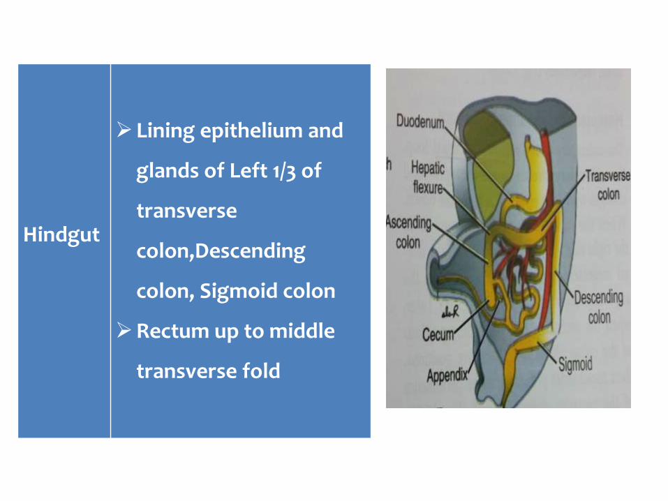
Hindgut
Lining epithelium and
glands of Left 1/3 of
transverse
colon,Descending
colon, Sigmoid colon
Rectum up to middle
transverse fold
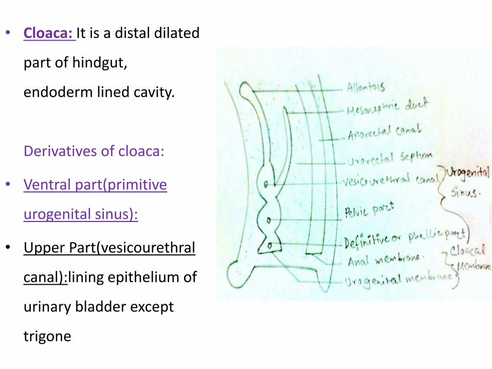
• Cloaca: It is a distal dilated
part of hindgut,
endoderm lined cavity.
Derivatives of cloaca:
• Ventral part(primitive
urogenital sinus):
• Upper Part(vesicourethral
canal):lining epithelium of
urinary bladder except
trigone
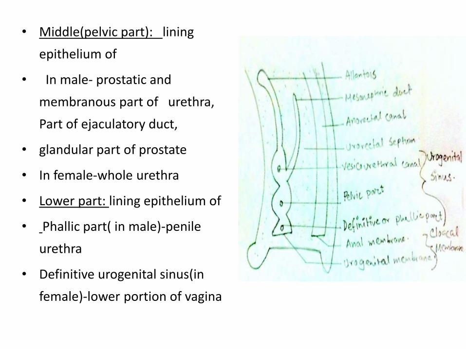
• Middle(pelvic part): lining
epithelium of
• In male- prostatic and
membranous part of urethra,
Part of ejaculatory duct,
• glandular part of prostate
• In female-whole urethra
• Lower part: lining epithelium of
• Phallic part( in male)-penile
urethra
• Definitive urogenital sinus(in
female)-lower portion of vagina
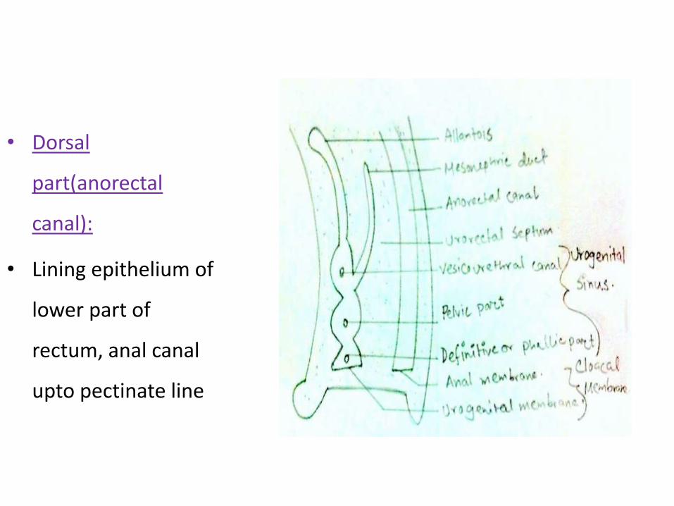
• Dorsal
part(anorectal
canal):
• Lining epithelium of
lower part of
rectum, anal canal
upto pectinate line
References :
Sadler,T.W.,2015.Langman’s Medical Embryology, 13th ed. New Delhi: Lippincott Willliam and Wilkins.
Singh, I.,2014.Human Embryology, 10th ed.Kundi: JaypeeBrothers Medical Publishers (P) Ltd.
Datta, A.K.,2012.Human Embryology, 6th ed.Kolkata: Current Books International.

THANK YOU