Gd-complexes of DOTA conjugates of tranexamates: A new ...
1
Gd-complexes of DOTA conjugates of tranexamates: A new class of non-aromatic, non-ionic MRI blood-pool contrast agents H-K. Kim 1 , K-H. Jung 2 , M-K. Kang 1 , J-A. Park 3 , S-T. Woo 4 , J-H. Kim 4 , T-J. Kim 2 , and Y. Chang 1,5 1 Department of Medical & Biological Engineering, Kyungpook National University, Daegu, Korea, Republic of, 2 Department of Applied Chemistry, Kyungpook National University, Daegu, Korea, Republic of, 3 Laboratory of Nuclear Medicine Research, Molecular Imaging Center, Korea Institute of Radiological & Medical Science, Seoul, Korea, Republic of, 4 Bayer Schering Pharma Korea, Seoul, Korea, Republic of, 5 Department of Diagnostic Radiology and Molecular Medicine, Kyungpook National University, Daegu, Korea, Republic of Introduction Magnetic resonance imaging (MRI) has been one of the most powerful techniques for the non-invasive diagnostic technique of the human anatomy, physiology and pathophysiology on the basis of superior spatial resolution and contrast. In general, contrast agents (CAs) are employed in MRI to improve the contrast effect, and Gd(III), with its high magnetic moment and long electron spin relaxation time, is an ideal candidate for such a proton relaxation agent and is the most famous metal center for contrast enhancement. There are now known four types of Gd-Chelate. They are: (1) anionic Gd-(acyclic chelates) such as [Gd(DTPA)(H 2 O)] 2- ; (2) neutral Gd-(acyclic chelates) such as [Gd(DTPA-BMA)(H 2 O)]; (3) anionic Gd-(cyclic chelates) such as [Gd(DOTA)(H 2 O)] 1- ; and (4) neutral Gd-(cyclic chelates) such as [Gd(HP-DO3A)(H 2 O)]. Each type possesses its own merits as well as drawbacks. For instance, a neutral type is preferred over an anionic one in that the former exhibits relatively low osmotic pressure in the body fluids after intravenous administration. As for the thermodynamic stability and kinetic inertness of CAs, a macrocyclic chelate such as DOTA or DO3A might be preferred over an acyclic, open-chain counterpart such as DTPA of DTPA-bis(amide). All these MRI CAs listed above have a common feature in that they are extracellular fluid (ECF) agents. Therefore, despite of their successful applications, they have revealed some inherent drawbacks such as poor contrast enhancement due to low R 1 -relaxivity, non-specificity, and rapid renal excretion. There thus arises the need for new MRI CAs with organ or tumor targeting and blood-pool effect. The purpose of the present work is to design and synthesize some new MRI blood-pool contrast agents (BPCAs) with improved R 1 - relaxivity and thermodynamic stability as well as kinetic inertness. Herein, we report the design and the synthesis of a series of DOTA conjugates (4a-c) and their Gd- complexes of the type [Gd(L)(H 2 O)] ⋅xH 2 O (5a-c; L= 4a-c) for use as a new class of bi-functional MRI CAs with strong liver enhancement and blood-pool properties. Material and Methods All reagents were purchased from commercial sources and used as received. T 1 measurements were carried out using an inversion recovery method with a variable inversion time (TI) at 1.5 T (64 MHz). For T 2 measurements the CPMG (Carr-Purcell-Meiboon-Gill) pulse sequence was adapted for multiple spin-echo measurements. Relaxivities (R 1 and R 2 ) were then calculated as an inverse of relaxation time per mM. For in vivo MRI, the mice were anesthetized by 1.5% isoflurane in oxygen. MR images of anaesthetized ICR mice (n=4) were obtained pre- and post- 5c (0.1 mmol Gd/kg) injection by tail vein with a 1.5 Tesla (T) MR unit (GE Healthcare, Milwaukee, WI, USA) with home-made small animal RF coil. The coil was of the receiver type, and the inner diameter of the coil was 50 mm. The imaging parameters for 3D fast SPGR (spoiled GRASS images) were as follows: repetition time (TR) = 8.8 ms; echo time (TE) = 3.9 ms; 10 mm field of view (FOV); 256×192 matrix size; 1.0 mm slice thickness; number of acquisition (NEX) = 4. The imaging parameters for Spin echo are as follows : TR = 500 ms; TE = 13 ms; 6 mm FOV; 192×128 matrix size; 1.5mm slice thickness; NEX = 4. Each image was taken at the interval of 3 minutes and 16 seconds. MR Images were obtained for 26 h after injection. Potentiometric titrations were carried out with an automatic titrator (798 MPT Titroprocessor, a 728 stirrer and a PT-100 combination pH electrode, Metrohm) to determine the protonation constants of the amides and the stability constants of corresponding metal complexes. Transmetallation is based on measurement of the evolution of the water proton longitudinal relaxation rate (R 1 P ) of a buffered solution (phosphate buffer, pH 7.4) containing 2.5 mmol/L gadolinium complex and 2.5 mmol/L ZnCl 2 . The trnasmetallation measurements were performed on a 3 T whole body system (Magnetom Tim Trio, simens), at room temperature. Results and Discussion The formation of 4 and 5 (Scheme 1) was confirmed by microanalysis and spectroscopic techniques. They exhibit a strong blood-pool effect with concomitant contrast enhancement in liver as well as R 1 -relaxivities higher than those of any of current MRI CAs. For instance, the R 1 relaxivity of 5c is 9.5 mM −1 sec −1 , which is about 2.6 times as high as that of structurally related Dotarem ® (R 1 =3.7 mM −1 sec −1 ). Furthermore they all possess pretty high kinetic stability (inertness) to retain more than 98% of the paramagnetic relaxation rate during the initial measurement for 72 h (Figure 1). The evolution pattern of 5c is nearly the same as that of Gd-DOTA. These observations may be as expected judging from their structural similarity. The structural uniqueness of 5 lies in the fact that, unlike their predecessors such as Gd- BOPTA and Gd-EOB-DTPA, they adopt macrocyclic DOTA instead of acyclic DTPA as ligand and that they carry no aromatic substituent(s) in the ligand backbone. A strong and long enhancement for hepatocellular uptake shown in Figure 2 may be due to slow excretion through bile duct. The slow excretion may be explained in terms of longer circulation time as demonstrated from the blood-pool effect. Increased lipophilicity of 5c due to the presence of tranexamic unit might have caused such a unique blood-pool effect. Conclusion We have synthesized Gd-DOTA conjugates of tranexamic esters (5) for use as a new and practical class of MRI BPCAs. Their thermodynamic stabilities as well as kinetic inertness are comparable with or better than most of current ECF and liver-specific MRI CAs. What is demonstrated by the presence of tranexamate is that it plays a key role in many respects such as an increase in R 1 -relaxivity, an increase in thermodynamic stability as well as kinetic inertness, and blood-pool enhancement through non-covalent aliphatic interactions with organs or blood cells. In every respect, the present series may put an entry into a new family of practical MRI BPCAs. Scheme. Figure 2. The T 1 -weighted images of six-week male ICR mice: (a) before injection; after injection with (b) 5c; (c) Gd-BOPTA ; (d) Gd-EOB-DTPA at the dosage of 0.1 mmol/kg. Table 1. R 1 and R 2 values for 5a-c and Gd-DOTA Figure 1. Evolution of R 1 p (t)/R 1 p (0) as a function of time for various MRI CAs. Proc. Intl. Soc. Mag. Reson. Med. 19 (2011) 1681
Transcript of Gd-complexes of DOTA conjugates of tranexamates: A new ...
Gd-complexes of DOTA conjugates of tranexamates: A new class of non-aromatic, non-ionic MRI blood-pool contrast agents
H-K. Kim1, K-H. Jung2, M-K. Kang1, J-A. Park3, S-T. Woo4, J-H. Kim4, T-J. Kim2, and Y. Chang1,5 1Department of Medical & Biological Engineering, Kyungpook National University, Daegu, Korea, Republic of, 2Department of Applied Chemistry, Kyungpook
National University, Daegu, Korea, Republic of, 3Laboratory of Nuclear Medicine Research, Molecular Imaging Center, Korea Institute of Radiological & Medical Science, Seoul, Korea, Republic of, 4Bayer Schering Pharma Korea, Seoul, Korea, Republic of, 5Department of Diagnostic Radiology and Molecular Medicine,
Kyungpook National University, Daegu, Korea, Republic of
Introduction Magnetic resonance imaging (MRI) has been one of the most powerful techniques for the non-invasive diagnostic technique of the human anatomy, physiology and
pathophysiology on the basis of superior spatial resolution and contrast. In general, contrast agents (CAs) are employed in MRI to improve the contrast effect, and Gd(III), with its high magnetic moment and long electron spin relaxation time, is an ideal candidate for such a proton relaxation agent and is the most famous metal center for contrast enhancement. There are now known four types of Gd-Chelate. They are: (1) anionic Gd-(acyclic chelates) such as [Gd(DTPA)(H2O)]2- ; (2) neutral Gd-(acyclic chelates) such as [Gd(DTPA-BMA)(H2O)]; (3) anionic Gd-(cyclic chelates) such as [Gd(DOTA)(H2O)]1- ; and (4) neutral Gd-(cyclic chelates) such as [Gd(HP-DO3A)(H2O)]. Each type possesses its own merits as well as drawbacks. For instance, a neutral type is preferred over an anionic one in that the former exhibits relatively low osmotic pressure in the body fluids after intravenous administration. As for the thermodynamic stability and kinetic inertness of CAs, a macrocyclic chelate such as DOTA or DO3A might be preferred over an acyclic, open-chain counterpart such as DTPA of DTPA-bis(amide). All these MRI CAs listed above have a common feature in that they are extracellular fluid (ECF) agents. Therefore, despite of their successful applications, they have revealed some inherent drawbacks such as poor contrast enhancement due to low R1-relaxivity, non-specificity, and rapid renal excretion. There thus arises the need for new MRI CAs with organ or tumor targeting and blood-pool effect. The purpose of the present work is to design and synthesize some new MRI blood-pool contrast agents (BPCAs) with improved R1-relaxivity and thermodynamic stability as well as kinetic inertness. Herein, we report the design and the synthesis of a series of DOTA conjugates (4a-c) and their Gd-complexes of the type [Gd(L)(H2O)] ⋅xH2O (5a-c; L= 4a-c) for use as a new class of bi-functional MRI CAs with strong liver enhancement and blood-pool properties. Material and Methods
All reagents were purchased from commercial sources and used as received. T1 measurements were carried out using an inversion recovery method with a variable inversion time (TI) at 1.5 T (64 MHz). For T2 measurements the CPMG (Carr-Purcell-Meiboon-Gill) pulse sequence was adapted for multiple spin-echo measurements. Relaxivities (R1 and R2) were then calculated as an inverse of relaxation time per mM. For in vivo MRI, the mice were anesthetized by 1.5% isoflurane in oxygen. MR images of anaesthetized ICR mice (n=4) were obtained pre- and post- 5c (0.1 mmol Gd/kg) injection by tail vein with a 1.5 Tesla (T) MR unit (GE Healthcare, Milwaukee, WI, USA) with home-made small animal RF coil. The coil was of the receiver type, and the inner diameter of the coil was 50 mm. The imaging parameters for 3D fast SPGR (spoiled GRASS images) were as follows: repetition time (TR) = 8.8 ms; echo time (TE) = 3.9 ms; 10 mm field of view (FOV); 256×192 matrix size; 1.0 mm slice thickness; number of acquisition (NEX) = 4. The imaging parameters for Spin echo are as follows : TR = 500 ms; TE = 13 ms; 6 mm FOV; 192×128 matrix size; 1.5mm slice thickness; NEX = 4. Each image was taken at the interval of 3 minutes and 16 seconds. MR Images were obtained for 26 h after injection. Potentiometric titrations were carried out with an automatic titrator (798 MPT Titroprocessor, a 728 stirrer and a PT-100 combination pH electrode, Metrohm) to determine the protonation constants of the amides and the stability constants of corresponding metal complexes. Transmetallation is based on measurement of the evolution of the water proton longitudinal relaxation rate (R1
P) of a buffered solution (phosphate buffer, pH 7.4) containing 2.5 mmol/L gadolinium complex and 2.5 mmol/L ZnCl2. The trnasmetallation measurements were performed on a 3 T whole body system (Magnetom Tim Trio, simens), at room temperature. Results and Discussion
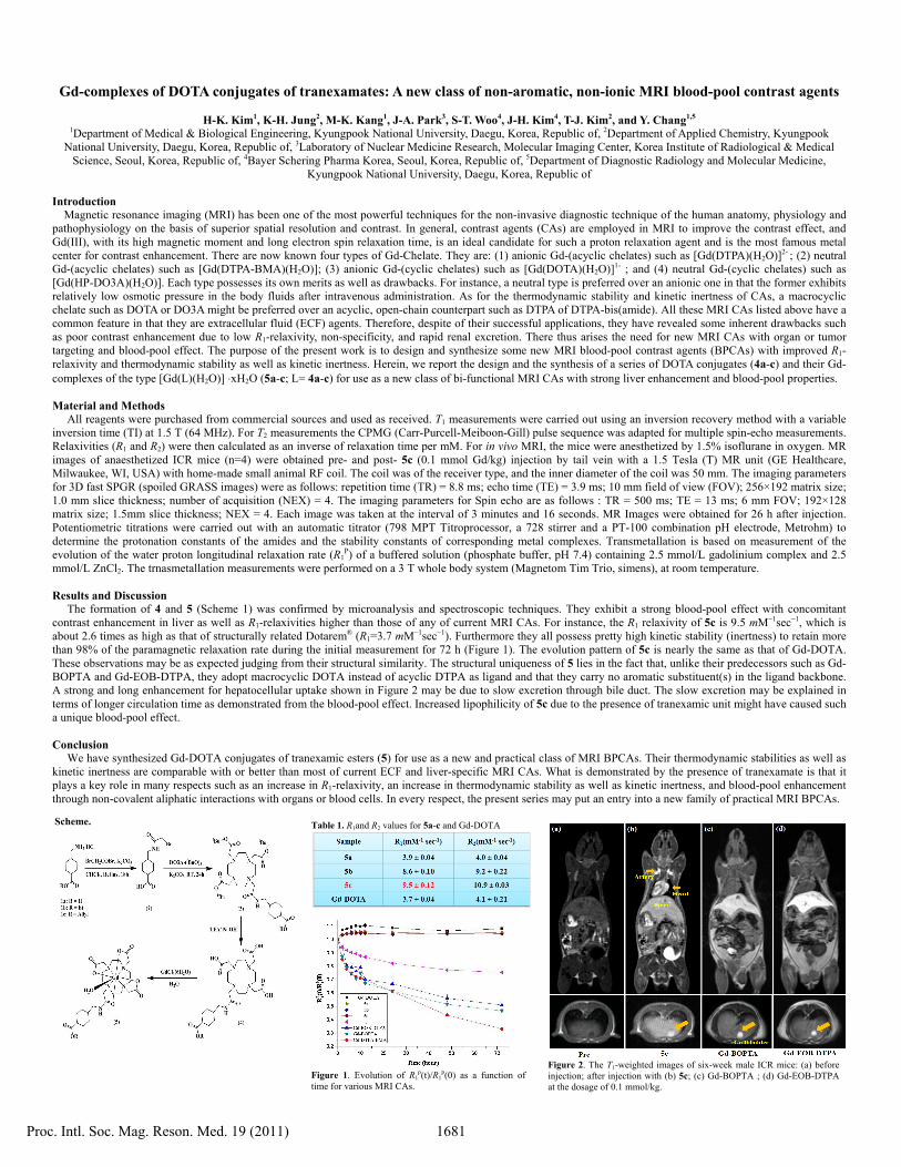
The formation of 4 and 5 (Scheme 1) was confirmed by microanalysis and spectroscopic techniques. They exhibit a strong blood-pool effect with concomitant contrast enhancement in liver as well as R1-relaxivities higher than those of any of current MRI CAs. For instance, the R1 relaxivity of 5c is 9.5 mM−1sec−1, which is about 2.6 times as high as that of structurally related Dotarem® (R1=3.7 mM−1sec−1). Furthermore they all possess pretty high kinetic stability (inertness) to retain more than 98% of the paramagnetic relaxation rate during the initial measurement for 72 h (Figure 1). The evolution pattern of 5c is nearly the same as that of Gd-DOTA. These observations may be as expected judging from their structural similarity. The structural uniqueness of 5 lies in the fact that, unlike their predecessors such as Gd-BOPTA and Gd-EOB-DTPA, they adopt macrocyclic DOTA instead of acyclic DTPA as ligand and that they carry no aromatic substituent(s) in the ligand backbone. A strong and long enhancement for hepatocellular uptake shown in Figure 2 may be due to slow excretion through bile duct. The slow excretion may be explained in terms of longer circulation time as demonstrated from the blood-pool effect. Increased lipophilicity of 5c due to the presence of tranexamic unit might have caused such a unique blood-pool effect. Conclusion
We have synthesized Gd-DOTA conjugates of tranexamic esters (5) for use as a new and practical class of MRI BPCAs. Their thermodynamic stabilities as well as kinetic inertness are comparable with or better than most of current ECF and liver-specific MRI CAs. What is demonstrated by the presence of tranexamate is that it plays a key role in many respects such as an increase in R1-relaxivity, an increase in thermodynamic stability as well as kinetic inertness, and blood-pool enhancement through non-covalent aliphatic interactions with organs or blood cells. In every respect, the present series may put an entry into a new family of practical MRI BPCAs.
Scheme.
Figure 2. The T1-weighted images of six-week male ICR mice: (a) before injection; after injection with (b) 5c; (c) Gd-BOPTA ; (d) Gd-EOB-DTPA at the dosage of 0.1 mmol/kg.
Table 1. R1and R2 values for 5a-c and Gd-DOTA
Figure 1. Evolution of R1
p(t)/R1p(0) as a function of
time for various MRI CAs.
Proc. Intl. Soc. Mag. Reson. Med. 19 (2011) 1681