Gastrointestinal Dysfunctions in Parkinson’s Disease ... · drooling(sialorrhea) ... Motor...
23
Review Article Gastrointestinal Dysfunctions in Parkinson’s Disease: Symptoms and Treatments Andrée-Anne Poirier, 1,2 Benoit Aubé, 1 Mélissa Côté, 1,3 Nicolas Morin, 4 Thérèse Di Paolo, 1,2 and Denis Soulet 1,2,3 1 Axe Neurosciences, Centre de Recherche du CHU de Qu´ ebec (Pavillon CHUL), Quebec City, QC, Canada 2 Faculty of Pharmacy, Laval University, Quebec City, QC, Canada 3 Department of Psychiatry and Neuroscience, Faculty of Medicine, Laval University, Quebec City, QC, Canada 4 Faculty of Medicine, Laval University, Quebec City, QC, Canada Correspondence should be addressed to Denis Soulet; [email protected] Received 15 August 2016; Accepted 16 October 2016 Academic Editor: Shey-Lin Wu Copyright © 2016 Andr´ ee-Anne Poirier et al. is is an open access article distributed under the Creative Commons Attribution License, which permits unrestricted use, distribution, and reproduction in any medium, provided the original work is properly cited. A diagnosis of Parkinson’s disease is classically established aſter the manifestation of motor symptoms such as rigidity, bradykinesia, and tremor. However, a growing body of evidence supports the hypothesis that nonmotor symptoms, especially gastrointestinal dysfunctions, could be considered as early biomarkers since they are ubiquitously found among confirmed patients and occur much earlier than their motor manifestations. According to Braak’s hypothesis, the disease is postulated to originate in the intestine and then spread to the brain via the vagus nerve, a phenomenon that would involve other neuronal types than the well-established dopaminergic population. It has therefore been proposed that peripheral nondopaminergic impairments might precede the alteration of dopaminergic neurons in the central nervous system and, ultimately, the emergence of motor symptoms. Considering the growing interest in the gut-brain axis in Parkinson’s disease, this review aims at providing a comprehensive picture of the multiple gastrointestinal features of the disease, along with the therapeutic approaches used to reduce their burden. Moreover, we highlight the importance of gastrointestinal symptoms with respect to the patients’ responses towards medical treatments and discuss the various possible adverse interactions that can potentially occur, which are still poorly understood. 1. The Importance of Nonmotor Symptoms in Parkinson’s Disease In the early 19th century (1817), with the publication of An Essay on the Shaking Palsy [1], Dr. James Parkinson was the first to provide a clear clinical description of the disease that now bears his name [2, 3]. ere are currently four motor features characterizing this neurological disorder, namely, muscle rigidity, tremor at rest, bradykinesia, and postural instability [3, 4]. However, a definitive diagnosis of Parkinson’s disease (PD) is difficult to establish and can be obtained only postmortem by the demonstration of the presence of Lewy bodies [3]. erefore, clinicians currently rely not only on motor symptoms manifestations but also on a positive response to levodopa (L-DOPA) treatment [4]. Progressive alterations of dopaminergic (DAergic) neu- rons in the nigrostriatal pathway are at the core of the abovementioned motor symptoms, resulting in a dysfunction of the somatomotor system. e extent of dopamine (DA) loss in the substantia nigra is already about 50–70% when the first motor symptoms emerge, and although PD is a progressive neurological disorder, DAergic deterioration is usually very slow and varies from one person to another [4]. An early diagnosis of the disease based on the Unified Parkinson’s Disease Rating Scale (UPDRS) has a favorable long-term impact on the quality of life of patients [3]. Over the course of PD progression, motor impairments are generally preceded by nonmotor symptoms (NMS) such as depression, olfactory deficit, sleep behavior disorder, and constipation, sometimes by up to ten years [5–8]. In his essay, Hindawi Publishing Corporation Parkinson’s Disease Volume 2016, Article ID 6762528, 23 pages http://dx.doi.org/10.1155/2016/6762528
Transcript of Gastrointestinal Dysfunctions in Parkinson’s Disease ... · drooling(sialorrhea) ... Motor...

Review ArticleGastrointestinal Dysfunctions in Parkinson’s Disease:Symptoms and Treatments
Andrée-Anne Poirier,1,2 Benoit Aubé,1 Mélissa Côté,1,3 Nicolas Morin,4
Thérèse Di Paolo,1,2 and Denis Soulet1,2,3
1Axe Neurosciences, Centre de Recherche du CHU de Quebec (Pavillon CHUL), Quebec City, QC, Canada2Faculty of Pharmacy, Laval University, Quebec City, QC, Canada3Department of Psychiatry and Neuroscience, Faculty of Medicine, Laval University, Quebec City, QC, Canada4Faculty of Medicine, Laval University, Quebec City, QC, Canada
Correspondence should be addressed to Denis Soulet; [email protected]
Received 15 August 2016; Accepted 16 October 2016
Academic Editor: Shey-Lin Wu
Copyright © 2016 Andree-Anne Poirier et al. This is an open access article distributed under the Creative Commons AttributionLicense, which permits unrestricted use, distribution, and reproduction in any medium, provided the original work is properlycited.
A diagnosis of Parkinson’s disease is classically established after themanifestation ofmotor symptoms such as rigidity, bradykinesia,and tremor. However, a growing body of evidence supports the hypothesis that nonmotor symptoms, especially gastrointestinaldysfunctions, could be considered as early biomarkers since they are ubiquitously found among confirmed patients and occurmuch earlier than their motor manifestations. According to Braak’s hypothesis, the disease is postulated to originate in theintestine and then spread to the brain via the vagus nerve, a phenomenon that would involve other neuronal types than thewell-established dopaminergic population. It has therefore been proposed that peripheral nondopaminergic impairments mightprecede the alteration of dopaminergic neurons in the central nervous system and, ultimately, the emergence of motor symptoms.Considering the growing interest in the gut-brain axis in Parkinson’s disease, this review aims at providing a comprehensive pictureof themultiple gastrointestinal features of the disease, alongwith the therapeutic approaches used to reduce their burden.Moreover,we highlight the importance of gastrointestinal symptoms with respect to the patients’ responses towards medical treatments anddiscuss the various possible adverse interactions that can potentially occur, which are still poorly understood.
1. The Importance of Nonmotor Symptomsin Parkinson’s Disease
In the early 19th century (1817), with the publication ofAn Essay on the Shaking Palsy [1], Dr. James Parkinsonwas the first to provide a clear clinical description of thedisease that now bears his name [2, 3]. There are currentlyfour motor features characterizing this neurological disorder,namely, muscle rigidity, tremor at rest, bradykinesia, andpostural instability [3, 4]. However, a definitive diagnosisof Parkinson’s disease (PD) is difficult to establish and canbe obtained only postmortem by the demonstration of thepresence of Lewy bodies [3]. Therefore, clinicians currentlyrely not only on motor symptoms manifestations but also ona positive response to levodopa (L-DOPA) treatment [4].
Progressive alterations of dopaminergic (DAergic) neu-rons in the nigrostriatal pathway are at the core of theabovementionedmotor symptoms, resulting in a dysfunctionof the somatomotor system. The extent of dopamine (DA)loss in the substantia nigra is already about 50–70% whenthe first motor symptoms emerge, and although PD is aprogressive neurological disorder, DAergic deterioration isusually very slow and varies from one person to another[4]. An early diagnosis of the disease based on the UnifiedParkinson’s Disease Rating Scale (UPDRS) has a favorablelong-term impact on the quality of life of patients [3].
Over the course of PD progression, motor impairmentsare generally preceded by nonmotor symptoms (NMS) suchas depression, olfactory deficit, sleep behavior disorder, andconstipation, sometimes by up to ten years [5–8]. In his essay,
Hindawi Publishing CorporationParkinson’s DiseaseVolume 2016, Article ID 6762528, 23 pageshttp://dx.doi.org/10.1155/2016/6762528
2 Parkinson’s Disease
James Parkinson had mentioned some of these nonmotorfeatures, namely, constipation, sleep disorders, dysphagia,drooling (sialorrhea), bladder dysfunction, and a slight stateof confusion [1]. Nowadays, NMS are increasingly associatedwith PD, although they have not yet received extensiveattention [6]. Indeed, patients report less than 40% of theirnonmotor problems to healthcare professionals, either outof embarrassment or because these symptoms are seen ascommonplace and inconsequential events [8]. To compoundthis problem, only a few NMS are recorded in medicalfiles and are associated as such with PD, although thoseproblems have been shown to result from the disease itselfrather than being unremarkable manifestations of normalaging [9–12]. Therefore, these NMS, which are very oftenoverlooked and are poorly investigated and treated, can havea major negative impact on the clinical care and quality oflife of PD patients [6, 13–15]. Patients also often indicate thattheir NMS are more difficult to manage than their motorproblems and may sometimes result in their hospitalizationand institutionalization [6, 15, 16]. In addition, it has beendemonstrated that attenuating NMS greatly improves thequality of life of patients, particularly those who positivelyrespond to a DAergic therapy [15, 17]. Thus, the recentlydeveloped awareness on the detection of the different NMSearly in the course of PD has led to amore critical appraisal ofits etiology, the identification of risk factors, and the currentadvances in neuroprotective and therapeutic biomarkers ofPD [5, 6, 18–20]. In light of these lines of evidence, PD canno longer be viewed solely as a complex disorder of motorfunctions, but rather as a progressive condition involvingbothmotor and nonmotor features [5, 15, 21]. Some investiga-tors have even proposed that PD could be divided into threephases, namely, preclinical, premotor (corresponding to theNMS), andmotor phases [6, 20]. In some patients, nonmotorproblems can be reminiscent of complications resulting frompharmacological and surgical interventions for the treatmentofmotor symptoms [16]. NMS can also bemore predominantin the “off” medication state and some might be alleviatedby DAergic therapy or, on the contrary, be exacerbated bythe latter [8]. Furthermore, the high costs associated withmedical care and the aging population strongly stress theneed to expand our knowledge base on all aspects of PD[13]. The various effects of which NMS are comprised andtheir highly divergent patterns of progression between PDpatients further raise the challenge imposed by NMS in themanagement of PD [15].
About a decade ago, Dr. Braak et al. proposed theintriguing hypothesis that PD might result from an infectionspreading first by intestinal and olfactory mucosae [22, 23].This proposal followed the first description of Lewy bodiesin the dorsal vagal nucleus by Friederick Lewy in the early20th century [6, 15]. Based on Lewy bodies distributionin PD postmortem patients, Braak et al. also suggested sixneuropathological stages, corresponding to disease evolution[23]. As such, the first signs of Lewy pathology appearin projection neurons of the dorsal motor nucleus of thevagus nerve at the early stage of PD [23]. Despite itspotential interest, this hypothesis is not widely accepted,mainly because of the paucity of patients studied and the lack
of associated clinical data [24]. However, the manifestationof NMS, preceding motor diagnosis, closely correspondsto the progression of Lewy pathology, supporting Braak’shypothesis [8]. Some studies have further suggested that thepathological process leading to PD could be initiated in theenteric nervous system (ENS) before spreading to the centralnervous system (CNS) via autonomous connections such asthrough the vagus nerve [25, 26]. In connectionwith the latterobservation, a recent study has demonstrated that differentforms of human alpha-synuclein (𝛼-syn), the major proteincomponent in Lewy bodies, injected in the intestine of micecan propagate to the brain via the vagus nerve and reach thedorsal motor nucleus in the brainstem, supporting Braak’shypothesis [27].
There are several different approaches to categorize thenonmotor features encountered in PD, but they have usuallybeen separated into five major classes, namely, cognitiveimpairment, neuropsychiatric disorders, autonomic dysfunc-tion, sleep disturbances, and other NMS [4–7]. Confusionand dementia are the most commonly reported cogni-tive impairments, whereas neuropsychiatric disorders ratheroccur as hallucinations, anxiety, depression, and impulse con-trol disorders. Importantly, PD medication can potentiallyexacerbate some of the latter problems [13]. For example, theeffects of DA agonists on the mesolimbic pathway could beresponsible for impulse control disorders such as compulsivegambling, compulsive shopping, and hypersexuality [7, 28].In addition, an injury to the autonomic nervous system canbe observed in various peripheral NMS such as orthostatichypotension, functional bladder disorder, excessive sweating,erectile dysfunction, and gastrointestinal (GI) symptomssuch as constipation, drooling, dysphagia, and nausea [4,6, 13, 16, 26, 28]. Other nonmotor features that are stillpoorly categorized include pain, fatigue, unexplained weightchanges, and visual as well as olfactory disturbances. To betteridentify these elements, Chaudhuri et al. developed the Non-Motor Symptoms Scale, which allows for a more accuratemeasurement of the frequency and severity of NMS andallows determining the impact of treatment on these symp-toms [15, 29]. In addition, the Non-Motor Questionnaire, theScales for Outcomes in Parkinson’s Disease, and a revisedversion of the UPDRS (sponsored by theMovement DisorderSociety) also contribute to the establishment of standardizedand reliable means to assess NMS in PD [8, 30].
2. GI Manifestations in Autonomic Disorders
Early PD, when left untreated, is often accompanied by auto-nomic nervous system impairments among which GI symp-toms represent the most common NMS [31]. Indeed, severalstudies relying on nonmotor rating scales have underscoredthe particular significance of GI symptoms in assessing thequality of life and have shown that thesemanifestations occurin 60% to 80% of patients [13, 16, 32, 33]. GI disorders areamong the most common causes of emergency admissionand often result in severe complications such as malnutri-tion (15% of PD patients), pulmonary aspiration (2.4% ofPD patients), megacolon (mostly asymptomatic; incidence

Parkinson’s Disease 3
unknown), intestinal obstruction (rarely reported; incidenceunknown), and even intestinal perforation (a few casesreported; incidence unknown) [34–38]. Moreover, older age,DAergic medication, and higher disease severity are usuallyassociated with these nonmotor features [28]. Hence, GIsymptoms reflect disturbances of GI tractmotility at all levels.
There are twomajor neural influences that regulate theGItract, namely, the extrinsic pathway, which is associated withthe vagus nerve, and the ENS, a component of the autonomicnervous system [39]. Due to its capacity to operate inde-pendently of the CNS and its 100 million neurons, the ENSis often considered as the second brain of the human body[39–41]. The ENS contains the myenteric and submucosalplexi, which are responsible for controlling smooth muscleactivity in the GI tract [40, 41]. The latter intestinal function,which is regulated by the ENS, requires the involvement ofseveral types of neurotransmitters such as DA, serotonin,acetylcholine, vasoactive intestinal peptide (VIP), substanceP, and nitric oxide synthase (NOS) [42]. Although the ENShas the ability to function independently of external stimuli,it also closely interacts with the vagal system [39, 41].
2.1. Constipation. Constipation is one of the initial NMSrelated to PD pathophysiology, affecting about 50–80% ofpatients. It often occurs early in the course of the disease andmay precede the appearance of motor symptoms by severalyears [6, 13, 28, 31, 43, 44]. Constipation is usually defined asfewer than three bowel movements per week and straining topass stools [45]. Although constipation is mainly consideredas a delay of the GI transit, some evidence suggests that itcan also be ascribed to a paradoxical contraction of voluntarysphincters during defecation, resulting in difficulties withrectal expulsion. In the early stages of PD, decreased GImotility has been associated with neuronal loss in the myen-teric and submucosal plexi and inclusions of Lewy bodiesin the dorsal motor nucleus of the vagus, underscoring theirpotential role in slowing down intestinal peristalsis [7, 28, 32].In addition to its association with autonomic alterations and,in some cases, urologic impairment, constipation is linked toa 2.7- to 4.5-fold increase in the risk of suffering from PD[15, 43, 46]. Constipation may also be accompanied by otherGI features that can affect intestinal transit. For instance, pain,nausea, bloating, vomiting, and distension are all symptomsof paralytic ileus, inducing complete obstruction of thegut and affecting about 7% of parkinsonians. Anismus, theabnormal contraction of the external anal sphincter andpuborectalis muscle during attempted defecation, is anotherproblem that can occur in synergy with constipation inapproximately 65% of PD patients, which is more frequentlyobserved during “off” periods [16, 28, 47]. Other intestinalcomplications such as megacolon (mostly asymptomatic),pseudoobstruction, sigmoid volvulus, and bowel perforationmay also arise in severe conditions, although their exactincidence is still currently unknown [32, 37, 38, 48].
2.2. Drooling. Also known as sialorrhea, drooling is themost common NMS of PD and is generally predominantlyobserved in the late stages of the disease and during the “off”
state medication [5, 49, 50]. Affecting 70 to 80% of parkin-sonians, sialorrhea corresponds to an exaggerated increase ofsaliva production and/or retention in the mouth cavity, withoccasional overflow into the pharynx [13, 32, 49–51].The sub-mandibular, sublingual, and parotid glands are the three pairsof salivary glands responsible for most of the approximately1.5 liters of saliva secreted daily and are controlled by theautonomic nervous system, mainly under parasympatheticcholinergic innervations [52, 53]. Sialorrhea may result fromthree phenomena, namely, abnormal production of saliva,impairment of salivary clearance, and/or inability tomaintainsaliva in the mouth [51]. Furthermore, excessive salivaryproduction may sometimes lead to serious complications,including saliva-induced asphyxiation and aspiration pneu-monia [31, 45]. Different scales, such as Drooling Severityand Frequency Scales, Drooling Rating Scale, and SialorrheaClinical Scale for PD, have been proposed to assess sialorrheaaccording to standard criteria [52, 54, 55]. However, droolingis rarely due to overproduction of saliva but is rather morecommon due to dysphagia, which itself is essentially amanifestation of bradykinesia [50, 56]. Indeed, in most PDpatients, decreased salivary production is in fact observed[51, 56, 57]. Studies have shown that patients do not produceexcessive amounts of saliva but rather have a more limitedability to swallow properly which, when associated with a for-ward head posture, might contribute to the onset of drooling[32, 49, 58]. In general, the inability to control oral secretionscan affect eating and speech and cause social embarrassment[59]. Somepatients even consider sialorrhea as theirworst PDsymptom [32].Different factors can influence sialorrhea, suchas male gender [60], aging [61], severity and duration of PD[62], hallucinations [59], orthostatic hypotension, dysphagia,dysarthria, UPDRS scores, and the use of antidepressants [51,63]. Furthermore, the peripheral autonomic nervous systemand the dorsal motor nucleus of the vagus nerve have beenimplicated in drooling, and Lewy bodies have been found inthe submandibular salivary glands in some studies [5, 64].
2.3. Dysphagia. Dysphagia, a feature of PD pathophysiology,is defined as a difficulty in swallowing food, liquids, orpills due to an impaired function of the medullary center[65, 66]. Dysphagia can result from muscular coordina-tion dysfunctions in at least one of the three phases ofdeglutition: oral, pharyngeal, and oesophageal [67]. Themain cause of swallowing difficulties, that is, a dysfunctionof the oropharyngeal phase (found in about one-third ofPD patients [68]), often results from motor symptoms ofbradykinesia and a reduced motor control of the tongue.Thus, these motor features contribute to the pathophysio-logical development of dysphagia and, by extension, mightalso play a role in the onset of sialorrhea in PD [51].Various abnormalities in the oropharyngeal phase, such as adelayed swallowing reflex, laryngeal movement deficits, andvallecular and piriform sinus residues, have been reported[66, 69]. In the oesophageal phase, complete aperistalsis,simultaneous oesophageal spasms, slower oesophageal tran-sit, and deficit in sphincter relaxation and pressure havebeen the predominantly observed abnormalities [67, 70].

4 Parkinson’s Disease
Interestingly, this involuntary component of deglutition isunder autonomic control, and Lewy bodies have been iden-tified in the oesophageal myenteric plexus [66, 67]. Thesefindings suggest that swallowing impairment could partlyresult from direct damage to the ENS. Moreover, in viewof the various aforementioned abnormalities, dysphagia isclearly linked to an increased risk of mortality by causingand/or exacerbating other PD-related complications such asaspiration pneumonia (estimated to account for 70% of themortality rates among PD patients [36]), choking, malnutri-tion, unexplainedweight loss, and dehydration [13, 66, 69, 71].Unfortunately, the degree of dysphagia cannot be predictedby PD progression because it has no direct connection withthe clinical severity of the disease as evaluated by motorcriteria [31, 70]. Moreover, data from various studies suggestthat up to about 50% of parkinsonians might suffer fromdeglutition problems, which, as with drooling, occur mainlyduring the late stages of the disease [66, 71, 72].
2.4. Nausea, Vomiting, and Gastroparesis. Nausea and vomit-ing (which are experienced by approximately 20% of patients[45]) are related, most of the time, to antiparkinsonianmedications for motor symptoms, rather than occurringas intrinsic features of PD [6, 7, 28]. Indeed, these sideeffects generally appear following the initiation of DAer-gic treatments [28]. However, nausea may likely occur inuntreated parkinsonian patients as well, and such casesmightbe explained by underlying gastroparesis [73]. Also knownas delayed gastric emptying, gastroparesis corresponds todecreased stomach motility, which may eventually affectgut transit. In addition to nausea, chronic gastroparesisis characterized by early satiety, a sensation of fullness,weight loss, and abdominal pain and bloating [74]. Thisphenomenon could well be related to the degeneration ofautonomic neurons in the myenteric plexus and brainstem[45]. Moreover, intestinal absorption of L-DOPA and othermedications might be slowed by such protracted gastricretention, thus reducing the effectiveness of treatment andpreventing the improvement of motor symptoms [75]. PD-associated gastroparesis deserves proper medical attention asits observed prevalence approaches 90% of patients [76].
2.5. Pathophysiology. Recently, several clinical and post-mortem studies exploring Lewy bodies expression and/or thepresence of neurodegeneration in the enteric nervous systemof parkinsonian patients have been conducted in order tobetter understand the etiopathogenesis of PD (see Table 1).
2.5.1. Lewy Bodies. The pathophysiological mechanismsunderlying GI dysfunctions are likely to be multifaceted,reflecting not only the involvement of the intrinsic inner-vation of the gut, but also extrinsic inputs because of thepresence of Lewy pathology in the dorsal motor nucleus ofthe vagus, sacral parasympathetic nuclei, and sympatheticganglia [77–79]. The occurrence of Lewy pathology in thegut of PD patients was first reported in an autopsy survey inwhich Qualman et al. found myenteric Lewy bodies in thecolon of one patient and in the esophagus of another [80].
A subsequent clinical study demonstrated the presence ofLewy bodies in the colon of one PD subject [81]. Theseprimary observations led Wakabayashi et al. to perform asystematic assessment of Lewy pathology in the ENS ofseveral PD patients [82]. Lewy bodies were found in the GItract of seven patients and were distributed widely from theupper esophagus to the rectum. In a follow-up study, the sameteam reported that most Lewy bodies observed within theGI tract of the three patients were located in VIP+ neuronsand to a lesser extent in neurons immunoreactive for tyrosinehydroxylase (TH) [83]. Therefore, this suggests potentialinterplay between these neurons and cholinergic neurons ofthe vagus nerve contributing to the spread of 𝛼-syn to theCNS. It was also mentioned that few Lewy bodies were foundin neurons that were negative for either VIP or TH. To date,these have been the only studies suggesting that a specificsubset of enteric neurons could bear Lewy pathology [83].No further reports regarding GI Lewy pathology in patientswith PDwere published, until 2006 when Braak et al. broughtthis topic to the forefront [84]. In this postmortem study,they investigated the gastric myenteric and submucosal plexifrom five individuals with Lewy body disease. Clinical datademonstrated that three out of the five patients with Lewybody pathology displayed motor symptoms reminiscent ofPD while the other two patients were reported to be freeof such symptoms. However, Lewy pathology was presentin both the myenteric and the submucosal plexi of all fivepatients. This led Braak and colleagues to postulate that thepathology initiates in the ENS before progressing to theCNS [84]. Despite being a potentially important finding, thishypothesis has not been widely accepted, mainly because ofthe paucity of patients studied and the lack of associatedclinical data [24]. More recently, a comprehensive survey onthe occurrence of Lewy pathology in the peripheral nervoussystem, and especially in the ENS, has been published bythe Arizona Parkinson’s Disease Consortium [79]. One ofthe most striking results of this study was the identificationof Lewy inclusions in the esophagus of 14 out of 15 PDpatients, suggesting that enteric pathology is present in thevast majority of PD cases [79]. Other recent studies havealso observed 𝛼-syn positive staining in GI tissues collectedbefore patient’s diagnosis [85] and in the vast majority ofparkinsonian patients’ colon tissues [86, 87].
The abovementioned data on the ENS in PDpatients werecollected either at autopsy or using colectomy specimens.To extend this work by analyzing enteric neuropathologyin living patients, Lebouvier et al. took advantage of anovel colonic biopsy technique [88, 89]. Twenty-nine patientswith an established PD diagnosis were enrolled togetherwith 10 healthy subjects who had undergone colonoscopyfor colorectal cancer screening. Biopsies from 21 out ofthe 29 patients with PD (72%) showed Lewy neurites intheir submucosal plexus, whereas no Lewy pathology wasobserved in any of the controls [89]. Chronic constipationwas more frequent in patients with than without Lewyneurites, suggesting a pathogenic role for these inclusions.However, Lebouvier et al. did not consider the myentericplexus, which is directly involved in the control of bowelmotility [89]. These findings are in line with other reports

Parkinson’s Disease 5
Table1:GIp
hysio
pathologic
manifestations
inPD
.Sum
maryof
clinicalstudies
exploringLewybo
dies
expressio
nand/or
presence
ofneurod
egenerationin
enteric
nervou
ssyste
mof
parkinsonian
patie
nts.
Stud
ies
GIp
art
Plexi
Dise
ases
tage
ordu
ratio
nSymptom
s(num
bero
fPDpatie
nts)
GIp
atho
logicalobservatio
ns(num
bero
fPDpatie
nts)
Qualm
anetal.,
1984
[80]
Esop
hagusa
ndcolon
Myenteric
Unk
nown
Lewybo
dies
(2/3)
Kupsky
etal.,1987
[81]
Colon
Myenteric
Unk
nown
Megacolon
(1/1)
Lewybo
dies
(1/1)
Wakabayashi
etal.,
1988
[82]
Upp
eresop
hagustothe
rectum
Myentericand
subm
ucosal
From
lessthan
1yearto27
years
IntraneuriticLewybo
dies
inmyentericneuron
sof
thee
soph
agus
(7/7),sto
mach(2/7),du
odenum
(2/7),jejunu
m(1/7),colon(1/7),andrectum
(1/7)
IntraneuriticLewybo
dies
insubm
ucosalneuron
sof
thejeju
num
(1/7),colon(2/7),andrectum
(1/7)
Intracytop
lasm
icLewybo
dies
inmyenteric
neuron
softhe
esop
hagus(1/7
)
Wakabayashi
etal.,
1990
[83]
Upp
eresop
hagustothe
rectum
Myentericand
subm
ucosal
8years,27
years,andun
know
n
Alm
ostallneuron
scon
tainingLewybo
dies
were
TH+or
VIP+(3/3)
Noapparent
lossof
TH+andVIP+neuron
scell
bodies
andprocess
Sing
aram
etal.,
1995
[99]
Ascend
ingcolon
Myentericand
subm
ucosal
Long
standing
severe
disease
(>20
yearsfor
8patie
nts)
Megacolon
(9/11
)Colon
cancer
(1/11
)Neededmanualevacuation
(7/11
)
DecreaseinDAe
rgicneuron
snum
ber(9/11)
Lewybo
dies
inmyentericneuron
s(11/11
)∗𝑀𝑜𝑠𝑡𝑙𝑦observed
inVI
PandTH
+neurons
DecreaseinDAconcentration
NodifferenceinTH+,V
IP+,and
totaln
eurons
number
Braaketal.,2006
[84]
Distalesop
hagusa
ndsto
mach
Myentericand
subm
ucosal
Stage2
tosta
ge5
IntraneuronalL
ewybo
dies
(5/5)
Lebo
uviere
tal.,
2008
[102]
Ascend
ingcolon
Subm
ucosal
>5years
Con
stipatio
nLewyneurites(4/5)
NodifferenceinTH+andtotaln
eurons
number
Beachetal.,2010
[79]
Upp
eresop
hagustothe
rectum
,sub
mandibu
lar
gland,liver,pancreas,and
gallb
ladd
er
Myentericand
subm
ucosal
Morethan80%in
stage
3or
4Lewybo
dies
inclu
sions
(11/1
7)∗14/15
patientsfor
onlyesophagusa
ndsubm
andibu
larg
land
Lebo
uviere
tal.,
2010
[89]
Ascend
inganddescending
colon
Subm
ucosal
Group
1:≤6years(9patie
nts)
Group
2:7–12
years(10
patie
nts)
Group
3:≥13
years(10
patie
nts)
Chronicc
onstipatio
n∗𝑀𝑜𝑟𝑒fre
quenta
mong
patientsw
ithLewy
neurites
Lewyneurites(21/29)
∗𝐺𝑟𝑜𝑢𝑝
1=7;Gr
oup2=5;Gr
oup3=9
∗𝑃𝑟𝑜𝑝𝑜𝑟𝑡𝑖𝑜𝑛
ofpatientsw
ithLewy
pathologyd
idnot
correla
tewith
diseasep
rogressio
nbutp
ositively
correla
tedwith
age
∗60%we
refoun
din
TH+neurons
Decreaseintotaln
eurons
number

6 Parkinson’s Disease
Table1:Con
tinued.
Stud
ies
GIp
art
Plexi
Dise
ases
tage
ordu
ratio
nSymptom
s(num
bero
fPDpatie
nts)
GIp
atho
logicalobservatio
ns(num
bero
fPDpatie
nts)
Ann
erinoetal.,
2012
[92]
Stom
ach,du
odenum
,ileum
,transversec
olon
,andrectum
Myenteric
From
4to
22years
Lewybo
dies
(12/13)
Lewyneurites(13/13
)∗<3%
werefoun
din
TH+neurons
∗𝑁𝑜correla
tionwith
ageo
rdise
asep
rogressio
nNodifferenceinTH+,V
IP+,N
OS+,and
total
neuron
snum
ber
Poucletetal.,2012
[90]
Ascend
inganddescending
colonandrectum
Subm
ucosal
From
1to24
years
Lewyneuritesinascend
ingcolon(17/26),in
descending
colon(11/2
6),and
inrectum
(6/26)
Poucletetal.,2012
[91]
Descend
ingcolon
Subm
ucosal
From
3to
15years
Lewyneurites(4/9)
Shanno
netal.,
2012
[86]
Sigm
oidcolon
Subm
ucosal
From
6mon
thsto8years
Mild
disabilities
𝛼-syn
positives
taining(9/9)
Goldetal.,2013
[87]
Colon
Myentericand
subm
ucosal
Unk
nown
𝛼-syn
positives
taining(10/10)
∗𝐻𝑖𝑔ℎ𝑒𝑟
prevalence
andgradeo
f𝛼-sy
ndetecta
bility
than
controls
Hilton
etal.,2014
[85]
Esop
hagus,sto
mach,sm
all
intestine,colon,
andgall
bladder
Subm
ucosal
From
8yearsp
riortotheo
nsetof
motor
symptom
sto15
yearsa
fter
diagno
sis
Postu
ralhypotensio
n,constip
ation,
dysphagia,
urinaryincontinence,
impo
tence,no
cturia,and
droo
ling
𝛼-syn
positives
taining(7/62)
∗11%
in“postdiagnosis”
tissues,7%in
“upto5years
priortodiagnosis”tissues,17%in
“5–10yearsp
rior
todiagnosis”tissues,and0%
in“m
orethan10
years
beforediagnosis”tissues
∗𝑃𝑟𝑜𝑝𝑜𝑟𝑡𝑖𝑜𝑛𝑠ofpositiveb
iopsies
inboth
theu
pper
andthelow
erGI
tractwe
resim
ilar
Gelp
ietal.,2014
[93]
Distalesop
hagus,sto
mach,
ileum
,colon
,and
rectum
Myenteric
Averageo
f11.5
years
∗𝐴V𝑒𝑟𝑎𝑔𝑒of18
yearsfor
PDpatientsw
ithdementia
Dem
entia
(6/10
)Lewyneuritesa
ndLewybo
dies
inclu
sions
indistal
esop
hagus,sto
mach,andcolon(8/10
)
Corbille
etal.,2014
[103]
Ascend
inganddescending
colon
Subm
ucosal
From
1to24
years
NodifferenceinTH+andtotaln
eurons
number
Beachetal.,2016
[96]
Sigm
oidcolon
Myentericand
subm
ucosal
Averageo
f15.2years
𝛼-syn
positives
tainingin
thes
ubmucosal(5/5)a
ndmyenteric(4/5)p
lexi
∗:note.

Parkinson’s Disease 7
on PD enteric pathology, which showed that, besides Lewybodies, Lewy neurites were also observed in the ENS ofpatients [79, 84, 90–93]. Using 𝛼-syn immunostaining, theauthors also demonstrated that approximately half of theLewy neurites observed in the submucosal plexus belongedto postganglionic neurons, thus supporting their extrinsicorigin [84]. The origin of the remaining Lewy neuritesremains to be determined, but it is possible that they couldoriginate both from submucosal and from myenteric neu-rons, which have been shown to project to the submucosalblood vessels [94]. This observation is in agreement withrecent studies showing 𝛼-syn immunolabeling in the submu-cosal perivascular regions [95, 96]. Depending on the typeof 𝛼-syn immunostained and the intestinal region studied,some discrepancies in the observation of Lewy bodies inGI biopsies or postmortem tissues are possible, especiallybecause 𝛼-syn is physiologically expressed by red blood cellsand vascular endothelial cells [96].
Interestingly, an animal model of PD recently developedprovides some clues on the role of ENS alterations in GIdysfunction. Transgenic 𝛼-syn SNCA, A53T, and A30P micedisplay aggregates within their enteric ganglia, which isassociated with a prolonged whole-gut total transit time andreduced colonic motility [97]. However, there is no evidenceof pathologic changes in the dorsalmotor nucleus of the vagusor autonomic cardiovascular dysfunction. These findingssuggest that ENS alterations in these mice are intrinsic inorigin, being caused by 𝛼-syn aggregation in enteric neuronsonly. It is possible in PD patients that at least some of the GIsymptoms could be caused by enteric neuropathy. It shouldbe pointed out, however, that studies on GI symptoms inPD have focused mainly on motility disorders and thereforethe role of the myenteric plexus and associated consequencesof Lewy pathology in the submucosal plexus have, to ourknowledge, not been addressed either in patients or inexperimental models of PD.
2.5.2. Neurodegeneration. Enteric neurons produce a sub-stantial amount of DA which regulates normal gut motility[67]. Interestingly, slowed GI transit and decreased gut con-traction in PD patients occur via altered DA-ENS circuitry,which normally promotes the peristaltic reflex [98]. PDpatients with severe constipation have been reported topresent lower levels of GI DA, suggesting that damage tothe enteric DAergic system might be an important factorunderlying GI dysfunction [99]. More recently, age-relatedloss of myenteric neurons has been associated with chronicconstipation, although studies are widely controversial [100,101]. Unfortunately, it is still not clear whether PD leads tothe loss of enteric neurons. Singaram et al. reported thatmost patients present DAergic neuronal loss in the colonicmyenteric and submucosal plexi, whereas other types ofneuronswere not affected based onTH immunostaining [99].Other teams also used thismarker on postmortem tissues andcolon biopsies, and none reported DAergic enteric neuronalloss [88, 92, 102, 103].
Systemic administration of the selective DAergic neu-ronal toxin 1-methyl 4-phenyl 1,2,3,6-tetrahydropyridine
(MPTP) leads to the loss of DAergic neurons in the intestinaltracts of mice [104, 105], but MPTP-treated monkeys werereported to display an increased number of neurons in theirmyenteric ganglia [106]. MPTP causes a transient increase ofstool frequency and colon relaxation lesions in mice [104],although this effect is inconsistent with the slow GI motilityof PD patients. Therefore, despite the fact that inhibitoryintestinal DAergic neurons could be impaired in PD, theseneurons are not the only neuropathological targets of thedisease [106–108]. Indeed, intestinal non-DAergic neuronscould also be impaired, but the discrepancy between datamakes it difficult to draw robust conclusions. Andersonet al. demonstrated that MPTP-treated mice presented nodifference in nitric oxidergic neurons [104]. Another studyshowed in a PD model induced by directional stereotaxicbrain injection of the neurotoxin 6-hydroxydopamine (6-OHDA) that rats exhibited slow colon motility accompaniedwith nitric oxidergic neuron loss in the myenteric plexus[109]. Other studies showed that a primate MPTP model ledto an increase in nitric oxidergic neurons [106]. Overall, mostof these studies have shown that GI cholinergic transmitterswere not significantly altered in PD [104, 106, 110].
According to these data, constipation in PD patientscannot be explained solely by a decrease in DA levels linkedto damage to neurons. Digestive tract motility would requiresophisticated synchronization from all neurotransmitters,not only DA. Moreover, the important variability betweenthe results pertaining to enteric neuronal loss refers to theneurodegenerative paradox. Even if DAergic neuronal deathis the histopathological hallmark of PD, it is one of themost difficult parameters to highlight in the ENS becauseof both the rarity of apoptosis in the neurodegenerativeprocess and the difficulty in counting neurons [111]. This hastogether led to numerous unanswered questions concerningneurodegenerative processes occurring in the ENS and theirimpact on GI impairments.
2.6. Other Outcomes of PD Therapies on GI Dysfunctions.Antiparkinsonianmedication considerably hampers the eval-uation of the potential correlation between GI dysfunctionsand the severity of PD symptoms. An individual stabilizedby drug therapy may indeed display a better overall con-dition than another patient with early PD, thus receivinga suboptimal treatment [37]. Moreover, in some situations,addressing motor symptoms only may affect GI featuresboth positively and negatively. Indeed, DAergic therapy mayimprove dysphagia and drooling but, on the other hand,might also worsen gastroparesis and reduce GI motility[69, 75, 112]. However, since nausea and vomiting are oftenside effects of various medications, they can limit the useof the latter and, as a result, preempt the benefit of suchmedications on motors symptoms [31]. Moreover, deep brainstimulation (DBS), which is widely used to treat motorsymptoms, has been shown to have a potential impact onthe manifestation of GI symptoms [113, 114]. According tosome studies, constipation and deglutition are significantlyimproved after surgery in the subthalamic nucleus [115–117].However, there is no consensus on the putative effect of DBS

8 Parkinson’s Disease
Under investigation
¤
¤
MosaprideCisaprideErythromycinGastric pacemaker
Under investigation
∗
∗
∗
¤
¤
¤
Apomorphine injectionsDuodopaPrucaloprideCisaprideMosaprideTegaserodMisoprostolNeostigmineDomperidoneTrimebutineErythromycinNeurotrophin 3Botulinum toxin injectionsSacral nerve stimulationProbiotics/prebioticsBiofeedback therapyDeep brain stimulation
∗ Not available in the United States¤ Withdrawn from the market
Effective
∗
Small and frequent mealsTaking fluids during mealsWalking after mealsDomperidoneTrimethobenzamideAvoid fatsAvoid metoclopramide
Constipation
Under investigationIpratropium bromide sprayScopolamineBenztropineClonidineModafinilTropicamideRadiotherapyNeurectomySalivary gland excisionSalivary duct ligation or relocation
EffectiveChewing gum or sucking on hard candySpeech and position therapyBotulinum toxin A/B injectionsAtropine ophthalmic dropsGlycopyrrolateAvoid cholinesterase inhibitorsAvoid clozapine, yohimbine, and quetiapine
Nausea
Vomiting
Gastroparesis
EffectiveExerciseDietary fibersIncreased fluid uptakeMacrogolLactuloseMagnesium sulfateBisacodylSodium picosulfateDocusate sodiumPsylliumSenna acutifoliaLubiprostoneMethylnaltrexoneLinaclotideAvoid opioids, tricyclicantidepressants, antimuscarinics,and some antiparkinsonian drugs
DroolingDysphagia
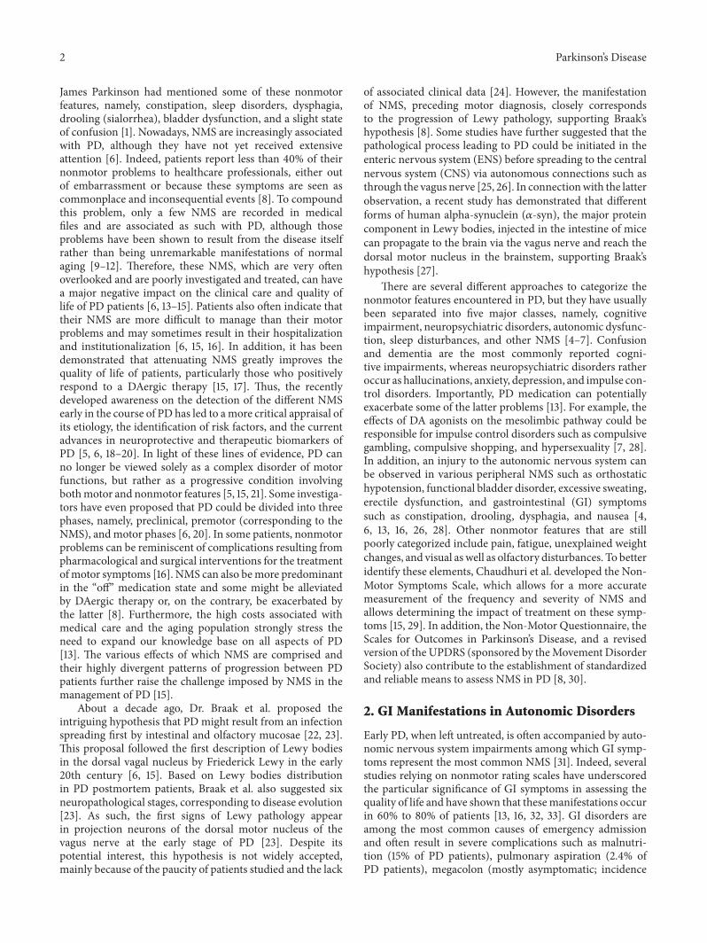
Figure 1: Treatment options for gastrointestinal dysfunctions in Parkinson’s disease. Overview of the different pharmacological treatmentsor therapeutic approaches that are currently effective or under investigation to manage constipation (left panels), drooling/dysphagia (rightpanels), and nausea/vomiting/gastroparesis (bottom panels). Please note that some drug options are not available in the USA (∗) or had tobe withdrawn from the market due to unacceptable side effects (¤).
on GI manifestations, as shown by reports that the latterneurosurgery does not improve dysphagia and drooling [51,118, 119].
3. Therapeutic Approaches to GI Symptoms
Importantly, GI impairments can impact other symptoms,which further complicates the clinical management of PD.For instance, GI problems such as gastroparesis and delayedintestinal absorptionmight lead tomore erratic absorption ofL-DOPA, which is reflected by motor fluctuations [120]. Thelatter problem emphasizes the necessity for clinicians to exertdue vigilance during office visits of PD patients and regularly
ask specific questions regarding GI manifestations. Recentstudies have also provided evidence for symptomatic treat-ments of constipation and drooling, but, unfortunately, thecurrent armamentarium for dysphagia and nausea remainsquite limited [31]. In this regard, Figure 1 and Table 2 providea summary of GI symptoms as well as the current treatmentalternatives.
3.1. Constipation
3.1.1. Effective Treatments. To prevent constipation problemsin PD, therapies aimed at accelerating colonic transit may be

Parkinson’s Disease 9
Table2:Eff
ectiv
etherapeutic
approaches.C
lassificatio
nandmechanism
sofa
ctionof
thevario
useffectiv
eop
tions
fortreatingGIs
ymptom
sexp
erienced
byPD
patie
nts,depend
ingon
efficacy
andsid
eeffects.
GIsym
ptom
sClassifi
catio
nTh
erapeutic
approaches
Mechanism
sofaction
Dosage
(adu
lt)Effi
cacy
(on
patie
nts)
Side
effects
(%of
patie
nts)
Com
ments
Stud
ies
Constip
ation
(1)Us
ewith
caution
Tricyclic
antid
epressants
Anticho
linergics
idee
ffects
[15,52]
Antim
uscarin
ics
Anticho
linergics
idee
ffects
[15,52]
Opioids
Anticho
linergics
idee
ffects
[15,52]
(2)N
onpharmac
ologica
loptions
Exercise
Intestinalstim
ulationby
movem
ents,
increased
fluids,andmuscularm
ass
[6,16,32]
Dietary
fibers
[6,16,32]
Increasedflu
idup
take
[6,16,32]
(3)L
axatives
Macrogol
(polyethyleneg
lycol)
Passes
throug
htheg
utwith
outb
eing
absorbed
anddigeste
dby
enzymes,
causingretentionof
water
intheintestin
altube
Oral:17g(∼1
tablespo
on)d
issolved
in240m
Lof
water
orjuiceo
nced
aily
Abdo
minalbloatin
g,cram
ping
,diarrhea,
flatulence,andnausea
Dono
tuse
for>
1-2weeks
[121,189]
Lactulose
Passes
throug
htheg
utwith
outb
eing
absorbed
anddigeste
dby
enzymes,
causingretentionof
water
intheintestin
altube
Oralorrectal:10
to20
g,daily
Abdo
minaldiscom
fortand
diste
ntion,
belch
ing,
cram
ping
,diarrhea
(excessiv
edose),flatulence,
nausea,and
vomiting
[190]
Magnesiu
msulfate
Blocks
perip
heralm
uscular
contractions
and
neurotransmission
Oral:2–4level
teaspo
onso
fgranu
les
dissolvedin
240m
Lof
water;m
ayrepeat
in6hours
Hypermagnesemia,
flushing,hypo
tension,
and
vasodilatation
Dono
texceed2do
ses
perd
ay[19
1]
Bisacodyl
Stim
ulates
enteric
nerves
tocausec
olon
iccontractions
Oralorrectal:
5–15mgas
single
dose
<1%
:abd
ominalmild
cram
ps,m
etabolicacidosis
oralkalosis
,hypocalcemia,
nausea,rectalirritatio
n,vertigo,andvomiting
[124]
Sodium
picosulfate
Stim
ulates
peris
talsisa
ndprom
otes
water
and
electro
lytesa
ccum
ulation
inthec
olon
Oral:150m
Lin
the
eveningbefore
the
colono
scop
y,follo
wed
byas
econ
ddo
se∼5ho
ursb
efore
thep
rocedu
re
Hypermagnesemia(12%
),hypo
kalemia(7%),
increasedserum
creatin
ine
(5%),hypo
chloremia(4%),
hypo
natre
mia(4%),
headache
(3%),nausea
(3%),andvomiting
(1%)
Mainlyused
for
colono
scop
yprocedure
[124]

10 Parkinson’s Disease
Table2:Con
tinued.
GIsym
ptom
sClassifi
catio
nTh
erapeutic
approaches
Mechanism
sofaction
Dosage
(adu
lt)Effi
cacy
(on
patie
nts)
Side
effects
(%of
patie
nts)
Com
ments
Stud
ies
Docusates
odium
(alone
orin
combinatio
nwith
psylliu
m)
Unclear;m
ayinhibitfl
uids
absorptio
nor
stimulate
secretionin
jejunu
m
Oral:50
to360m
g,once
daily
orin
divideddo
ses
Throatirr
itatio
n(1to10%)
[192]
Senn
aacutifolia
Redu
cesfl
uidabsorptio
nfro
mthefaecesa
ndinflu
encesfl
uidsecretions
bythec
olon
Long
-term
useisn
otrecommended
[126]
(4)O
ther
pharmacologica
loptio
ns
Lubiprostone
IntestinalClC-
2chlorid
echannelactivator
Oral:24𝜇gtwice
daily
64%
Interm
ittentloo
sesto
ols
(48%
),nausea
(29%
),diarrhea
(12%
),abdo
minal
pain
(8%),flatulence(6%
),dizziness(3%
),and
vomiting
(3%)
[126,
127]
Methylnaltre
xone
𝜇-O
pioidantagonist
Subcutaneous:12m
g,once
daily
60%
Abdo
minalpain
(45%
),flatulence(33%),diarrhea
(30%
),andnausea
(24%
)
Disc
ontin
ueall
laxativ
espriortouse;if
respon
seisno
toptim
alaft
er3days,laxative
therapymay
bereinitiated
[128]
Linaclo
tide
Guanylatecycla
seCagon
istOral:145𝜇
g,once
daily
Abdo
minalcram
ping
(4%),
discom
fort(4%),and
diarrhea
(4%)
Con
traind
icated
inpediatric
patie
nts(<6
yearso
fage)
[129,
130,193]
Droolinga
nddysphagia
(1)Us
ewith
caution
Cholinesterase
inhibitors
[51]
Clozapine
Serotoninantagonist
Dem
onstr
ated
effectiv
enessa
gainst
dyskinesias
[51,161,
194]
Yohimbine
Presyn
aptic𝛼2-adrenergic
blocking
agent
[51,162]
Quetia
pine
D2receptors(mesolim
bic
pathway)a
nd5H
T2A
(fron
talcortex)
antagonist
Dem
onstr
ated
effectiv
enessa
gainst
dyskinesias
[51,195]
(2)N
onpharma-
cologicaloptions
Chew
inggum
orsuckingon
hard
cand
y5tim
esim
proved
[158]
Speech
andpo
sition
therapy
Self-motivationisan
impo
rtantfactorto
obtain
apositive
outcom
e
[159,
160]

Parkinson’s Disease 11
Table2:Con
tinued.
GIsym
ptom
sClassifi
catio
nTh
erapeutic
approaches
Mechanism
sofaction
Dosage
(adu
lt)Effi
cacy
(on
patie
nts)
Side
effects
(%of
patie
nts)
Com
ments
Stud
ies
(3)P
harm
acologi-
caloptions
Botulin
umtoxinA/B
injections
(parotid
andsubm
andibu
lar
glands)
Inhibitsthec
holin
ergic
parasympatheticand
postg
anglionics
ympathetic
activ
ity
Atoxin:
500un
itsdivided
amon
gaffected
glands
Atoxin:
dryn
esso
fmou
thandmild
transitorysw
allowing
difficulties
(6%)
Prod
uced
byClostridium
botulin
umbacterium
[163,165,
166,168]
Btoxin:
1,000
units
into
each
parotid
glandand250
units
into
each
subm
andibu
larg
land
Btoxin:
dryn
esso
fmou
th(40%
),worsenedgait(25%
),diarrhea
(15%),neck
pain
(15%),andmild
transitory
swallowingdifficulties
(16%)
[50,163,
167]
Atropine
ophthalm
icdrop
s(sublingual
administratio
n)Anticho
linergicthatb
locks
muscarin
icreceptor
M3
1dropof
1%atropine
solutio
n,twice
daily
for1
week
Hallucinatio
ns(29%
)and
delirium
(14%)
Lack
ofclinical
evidence
fortreatments
lasting
longer
than
afewweeks
Use
with
cautionin
the
elderly;increased
risk
fora
nticho
linergic
effects,
confusion,
and
hallu
cinatio
ns
[170]
Glycopyrrolate
Anticho
linergicthatb
locks
muscarin
icreceptor
M3
Oral:1m
g3tim
es,
daily
95to100%
Dry
mou
th(52%
),urinary
retention(13%
),visio
nprob
lems(13%),
constip
ation(13%
),and
nausea
(4%)
[171,172,
174,175]
Nausea,
vomiting
and
gastroparesis
(1)Us
ewith
caution
High-fatfoo
ds[31]
Metoclopram
ide
Dop
aminea
ntagon
ist
Con
traind
icated
forP
Dpatie
ntsb
ecause
itworsens
motor
symptom
sbyblocking
dopaminer
eceptorsin
theC
NS
[31]
(2)N
onpharma-
cologicaloptions
Smalland
frequ
ent
meals
[31]
Drin
king
durin
gmeals
[31]
Walking
after
meals
[31]

12 Parkinson’s Disease
Table2:Con
tinued.
GIsym
ptom
sClassifi
catio
nTh
erapeutic
approaches
Mechanism
sofaction
Dosage
(adu
lt)Effi
cacy
(on
patie
nts)
Side
effects
(%of
patie
nts)
Com
ments
Stud
ies
(3)P
harm
acologi-
caloptions
Dom
perid
one
Dop
aminea
ntagon
ist
Oral:initiatingat
10mg
3tim
es,daily
(maxim
um:
30mg/day)
100%
Xerosto
mia(2%)a
ndheadache
(1%)
Doesn
otreadily
cross
theB
BB∗𝑈𝑠𝑒
thelow
esteffective
doseforthe
shortest
duratio
nnecessa
ry∗𝑁𝑜𝑡
availableinthe
UnitedStates
[149,196,
197]
Trim
etho
benzam
ide
Unclear;m
ostlikely
involves
thec
hemoreceptor
triggerz
one(throug
hwhich
emeticim
pulse
sare
transportedto
thev
omiting
center)
Oral:300m
g;intram
uscular:
200m
g,3or
4tim
esdaily
20%
Dizziness,headache,
blurredvisio
n,anddiarrhea
May
masktoxicityof
otherd
rugs
orcond
ition
s[19
8]
∗:note.

Parkinson’s Disease 13
effective. Increasing the levels of daily activity and introduc-ing dietary changes are the first options to consider. Patientsshould be encouraged to maximize dietary fibers (cereals,bran, citrus fruits, etc.), as well as ensure adequate fluidintake to avoid dehydration [6, 15, 16, 32]. Nevertheless, anexhaustive pharmaceutical evaluation of the drug treatmentsalready prescribed to patients is important before introducingadditional measures. Indeed, the dosage of medicationsknown to increase constipation symptoms should be opti-mized as much as possible. Some antiparkinsonian drugs aswell as opioids, tricyclic antidepressants, and antimuscarinicsare recurrent sources of severe constipation, likely due totheir anticholinergic effects [15, 52]. Other available optionsto increase the frequency of bowel movements and improvestool consistency are (i) osmotic laxatives such as macrogol(polyethylene glycol), lactulose, and magnesium sulfate, (ii)stimulant laxatives such as bisacodyl and sodium picosulfate,and (iii) stool softeners [28, 30, 121–123]. The safety profileassociated with the long-term use of osmotic agents makesthem the preferred group of laxatives. Macrogol, which isavailable in the USA and is recommended by the Ameri-can Academy of Neurology and the Movement DisordersSociety, is considered to be an effective and safe osmoticlaxative for PD patients [15, 32, 121]. Bisacodyl and sodiumpicosulfate, which both act by stimulating colonic smoothmuscle contractions as well as electrolyte andwater secretion,may represent additional alternatives to treat constipation[124]. Moreover, stool softeners such as docusate sodiummay be used alone or in combination with psyllium husksto increase stool volume and, therefore, peristalsis reflex [6,7, 125]. By increasing intestinal fluid secretion, lubiprostone,an intestinal ClC-2 chloride channel activator, also improvesconstipation issues (64% of PD patients) [7, 28, 52, 126].Themost common adverse events observedwere intermittentloose stools (48% of PD patients), nausea (29%), diarrhea(12%), abdominal pain (8%), flatulence (6%), dizziness (3%),and vomiting (3%) [52, 126, 127]. Methylnaltrexone (𝜇-opioid antagonist) is another medicinal agent approved inthe USA and indicated for the treatment of opioid-inducedconstipation, with approximately 60% of patients havingreported beneficial intestinal effects [28, 128]. In 2008, aclinical trial led by Portenoy et al. showed that adverse effectsexperienced by patients taking methylnaltrexone are mostlyabdominal pain (45%), flatulence (33%), diarrhea (30%),and nausea (24%) [128]. Linaclotide, a guanylate cyclase Cagonist, has also recently been approved by the Food andDrugAdministration (FDA) as a treatment for irritable bowelsyndrome and chronic constipation. Abdominal cramping,discomfort, and diarrhea are the adverse events commonlyreported by patients for linaclotide (about 4%) [52, 129, 130].Finally, several other studies have also demonstrated theeffectiveness of the Senna acutifolia plant, but the long-termuse of this well-known laxative is not recommended [122].
3.1.2. Treatments under Investigation. Treating constipationremains an active research area and various studies haveassessed the impact and clinical relevance of options that
could help relieve the discomfort and adverse effects associ-ated with this GI problem encountered in PD. For example,subcutaneous injections of apomorphine have translated topositive effects on intestinal motility (improvement of thedefecatory mechanisms and anorectal dysfunction [6, 32,131, 132]) and UPDRS motor scores (in about 70% of PDpatients [133]), although adverse effects such as orthostatichypotension (in 50% of patients), nausea, and drowsiness(in 75% of patients) may occur following administrationof this DA agonist [8, 134]. It is also recommended thatpatients use an antiemetic as a pretreatment before receivinginjections in order to avoid the unpleasant effect of nausea[31]. Therefore, due to these various secondary effects, thelong-term use of apomorphine appears to be inadvisable [32].Intrajejunal infusion of L-DOPA/carbidopa (or duodopa) hasalso proved beneficial relatively to constipation problems(in approximately 70% of PD patients) [135, 136]. Moreover,a body of research has been heavily focused on differentligands (agonists or antagonists) of the 5-HT
4serotonin
receptors. These receptors, which are located partly in thesmooth musculature and cholinergic nerves of the GI tract,are, among others, capable of increasing gastric and colonicmotility by facilitating acetylcholine release [137–139], thusmaking them an attractive target for treating constipation.The main 5-HT
4agonists studied to date are prucalopride,
cisapride, mosapride, and tegaserod [137, 140–142]. Unfortu-nately, although these agonists were found to be effective inthe treatment of constipation in PD patients, those prokineticagents have been removed from the US market or havenot been approved by the FDA due to possible adversecardiovascular effects (less than 1% of patients) [141, 143–145].Other medicinal agents are also under investigation, suchas misoprostol (a prostaglandin E
1analogue; 55% efficacy)
[32, 146], neostigmine (an acetylcholinesterase inhibitor;50% efficacy) [7, 147], and domperidone (a DA antagonist;about 35% efficacy) [148]. However, even if the promotilityagent domperidone could be potentially effective, due toits absence of permeation through the blood-brain barrier(BBB) [149], there is insufficient evidence to recommend itsutilization for constipation, as in the case of trimebutine (anenkephalinergic agonist) and erythromycin (the well-knownmacrolide antibiotic) [143]. In recent years, the NGF receptoragonist neurotrophin 3 has also been studied to improveGI motility dysfunction in PD. Although its mechanism ofaction with respect to GI motility remains unknown, thisneurotrophic factor was found to be effective in treatingconstipation (in about 20% of patients) [52, 150]. In a clinicaltrial conducted by Pfeiffer et al., a reduced colonic transittime, an increase in stool frequency, and shortening ofthe intervals without stool were observed [151]. However,abdominal cramps and diarrhea were noted in three patients,who were forced to reduce neurotrophin 3 dosage (300𝜇g/kgthree times weekly) [151]. Injections of botulinum toxin(BTX), a neurotoxin produced by the Clostridium botulinumbacterium that inhibits acetylcholine release, have also beenproposed to help reduce constipation burden in PD [51, 152].However, not only are such injections technically challenging,including ultrasound guidance, but also there is insufficientevidence that this method offers an effective treatment [30,

14 Parkinson’s Disease
153, 154]. For example, Albanese et al. reported a beneficialclinical effect of BTX injections on constipation, but only ina single patient [153]. In another clinical study, Cadeddu etal. observed an improvement of constipation symptoms in 10out of 18 patients after two months of BTX treatment [154].However, the authors mentioned that repeated injectionscould be necessary to maintain this clinical improvementsince the effects of the toxin wear off within three monthsof administration. Nonpharmacological strategies have alsobeen put forward to treat constipation, such as sacral nervestimulation (with 57% efficacy) [28, 155], synbiotic yogurt(i.e., probiotic- and prebiotic-enriched yogurt) [7, 16, 52],biofeedback therapy (79% efficacy) [52], and DBS (about25% efficacy after two years of treatment, a percentage thatmight however be influenced by the postoperative reductionin DAergic therapy and an improvement in motor fluctu-ations) [116, 117]. Milk fermented with the probiotic strainLactobacillus casei Shirota has also been suggested to dampenconstipation problems by modulating the host immuneresponse, enhancing mucosal function, suppressing growthof pathogenic bacteria, and blocking epithelial attachmentby pathogens, resulting in an improvement in 70 constipatedadults [156, 157]. A decrease in abdominal pain, bloating, andsensation of incomplete emptying is also observed in patientsusing probiotics [52].
3.2. Drooling and Dysphagia
3.2.1. Effective Treatments. For patients with mild symptomsof drooling and/or dysphagia, chewing gum or sucking onhard candy may be effective in ameliorating swallowing (anapproximately 5-fold improvement) and thus reduce drooling[13, 67, 152, 158]. Speech and position therapies can alsoprove efficient for easing these GI symptoms (with 60 to 90%efficacy) [159]. These therapies consist basically in trainingto learn voluntary airway protection techniques throughadequate swallowing methods and improved head postures.Marks et al. investigated such techniques and observed thatself-motivation was an important factor in obtaining a posi-tive outcome [160]. It is strongly recommended to consider allthese nonpharmacological options first to improve droolingand dysphagia symptoms before changing over to drug-based solutions. However, such drug-free approaches mayonly provide temporary improvement and might not beeffective or suitable for all patients. Indeed, pharmacologicaltreatments are generally considered when more aggressiveintervention is required [31]. Itmust be emphasized that somecategories of medications used to treat other PD symptomsmay in fact aggravate drooling and dysphagia and should thusbe avoided as much as possible. Such medications includeacetylcholinesterase inhibitors, the antipsychotic quetiapine,and adrenergic receptor agonists such as clozapine andyohimbine [51, 161, 162]. The pharmacological treatmentmost often mentioned for drooling/dysphasia is undoubt-edly BTX injections. Local injections of this toxin in theparotid and submandibular glands inhibit the cholinergicparasympathetic and postganglionic sympathetic activity,thereby reducing saliva production [163]. This treatment,
which denervates the salivary glands, was shown to be effec-tive in reducing drooling severity and frequency (in about80 to 90% of patients) without compromising swallowing[50, 51, 164–167]. Unfortunately, published studies cannotbe easily compared due to the important disparity betweenthe methodologies employed. Indeed, there is no standardtechnique for the injection (gland, ultrasound guidance,etc.) and no compliance regarding the optimal dose to beadministered [51]. The sole guideline for achieving the besteffect using this therapeutic approach is to inject the toxinbilaterally and periodically [31, 163]. Dryness of the mouth(or xerostomia) is the common adverse effect observed withBTX [51]. Importantly, submandibular glands injections arerecommended only under the supervision of a specialist dueto potential side effects caused by spreading of the toxin tonearby structures and should be performed exclusively whentreatment of the parotid gland alone is insufficient [163].Among the several serotypes of BTX, only A and B have beenstudied and are commercially available [51]. In the majorityof these studies, no side effects were observed with BTX-A,although BTX-B injections inducedmild adverse events suchas dry mouth (in about 40% of patients), diarrhea (∼15%),neck pain (∼15%), and worsened gait (∼25%) [50, 168, 169].This suggests a preferential action of BTX-B on autonomicneurons and therefore might point to its higher effectivenesscompared to BTX-A [152]. However, in two different clinicaltrials, Lagalla et al. observed that some patients experiencedmild transitory swallowing difficulties 7 days after a BTX-Ainjection (in about 6% of patients) [166] and 10 days aftera BTX-B injection (∼16%) [167], but they recovered within10 to 14 days. In spite of these potential drawbacks, thesestudies, which are the only ones that have compared theA andB serotypes, failed to demonstrate a significant difference inthe effectiveness between both neurotoxins [166, 167]. Otherpharmacological alternatives to BTX in the treatment ofdrooling/dysphagia include anticholinergic drugs that blockmuscarinic receptors and particularly theM3 subtype. Never-theless, the currently available agents are not selective for M3receptors and might thus give rise to several undesirable sideeffects (e.g., confusion, hallucinations, drowsiness, urinaryretention, and constipation) [51]. Thus, some of these drugshave yet to be considered truly effective, which warrantfurther investigations. A few studies have claimed that thetwo anticholinergics, atropine and glycopyrrolate, are theonly potentially useful therapies available for improvingdrooling/dysphagia [51, 52, 123]. Despite being effective,atropine still causes a wide range of undesirable adverseeffects such as hallucinations (2/7 patients) and delirium (1/7patients; but this was confounded by a concomitant urinarytract infection) [170]. Since glycopyrrolate does not crossthe BBB, unlike atropine, it is therefore the preferred agentbecause it is less likely to cause adverse effects in the CNS[152, 171]. Between 95 and 100% of patients who completedclinical studies reported improvement in drooling/dysphagiawith glycopyrrolate [172–174]. As expected, the side effectsobserved occurred in the periphery and mostly includedxerostomia (in approximately 52% of patients), urinary reten-tion (13%), constipation (13%), vision problems (13%), and

Parkinson’s Disease 15
nausea (∼4%) [171, 175]. While anticholinergics might be effi-cient for treating drooling/dysphagia, they do not representa suitable option for PD patients since other NMS can besubsequently worsened. Moreover, there is a lack of clinicalevidence for treatments lasting longer than a few weeks andthe long-term adverse effects of atropine and glycopyrrolatehave not been documented, thus leaving important safetyissues unresolved [51, 171]. All the pharmacological optionslisted above may thus be regarded as effective treatmentsfor drooling/dysphagia, but, considering their potential sideeffects, they should remain a secondary choice compared tononpharmacological therapies.
3.2.2. Treatments under Investigation. Other anticholinergictreatments such as ipratropium bromide spray, transdermalscopolamine, and benztropine have also been investigated fortreating drooling/dysphagia [123, 176–178]. However, previ-ous studies on the effectiveness of anticholinergic treatmentshad failed to conclude on the superiority of one drug overanother [179]. The ipratropium bromide spray (which hasinduced a significant effect on the UPDRS part 6 subscore[178]) is used sublingually as a bronchodilator and does notcross the BBB, thereby reducing systemic side effects [152].Unfortunately, there is insufficient data about its safety andefficacy to draw definite conclusions on its potential interestin drooling/dysphagia management [51, 123]. Adrenergicreceptors agonists have also been explored in this context.Clonidine, a selective 𝛼2-adrenergic receptor agonist, sig-nificantly improved the frequency at which patients had toclear their mouths [51, 52, 152]. The most common adverseevents observed with clonidine were diurnal somnolence(2/17 patients), dizziness (1/17), and dry mouth (1/17) [180].The 𝛼1-adrenergic agonist modafinil has also been reportedto exert rather beneficial effects on drooling/dysphagia inPD patients (6/6 patients), although this improvement wasmostly related to dysphagia rather than hypersalivation [51,181]. Moreover, Lloret et al. have investigated tropicamide, ashort-actingmuscarinic receptor antagonist, in the treatmentof drooling/dysphagia. So far, this treatment has shownpotential efficacy (33% average reduction in salivary volumefor 16 patients who completed the study) along with alack of noticeable side effects and no side effects, althoughthe data must still be considered as preliminary [182].Radiotherapy has also been suggested as a treatment fordrooling/dysphagia and studies in this context have shown asignificant improvement in symptoms (79% of patients), aneffect that could be maintained for at least one year [183].Common side effects were xerostomia (40% of patients) anda loss of taste (45%), which were mostly transient (25% and35%, resp.). Regrettably, the success of radiotherapy is largelycompromised by its potential to induce neoplasia [183, 184].Therefore, this treatment should only be considered whenall other options discussed above have proved ineffective.Finally, surgical options such as neurectomy, salivary glandexcision, salivary duct ligation or relocation, and DBS havealso been explored to ameliorate drooling/dysphagia [50,118, 184–187]. Neurectomy, that is, the surgical sectioningof the chorda tympani nerves, reduces salivary production
(improvement in 74% of patients) but might induce seri-ous complications such as hearing loss and a loss of taste[152, 188]. These invasive options (neurectomy and salivarygland/duct surgeries) can be realized individually or incombination (with>75% success) and possible adverse effectsinclude dental caries (10% of patients), cracked lips (10%),aspiration pneumonia, and xerostomia [152, 184–186]. Dueto their high risk of irreversible adverse effects, all theseinterventions are considered only when all other availableoptions have failed to bring about a positive outcome [32].DBS intervention has not been studied much to date in thecontext of drooling/dysphagia improvement, but, with thelimited information obtained so far, it seems unlikely thatDBS represents a useful option [51, 118].
3.3. Nausea, Vomiting, and Gastroparesis
3.3.1. Effective Treatments. Despite substantial progress inrecent research on constipation and drooling treatment, thearmamentarium of useful agents for other PD-associatedGI symptoms, such as nausea, vomiting, and gastroparesis,remains severely limited [31]. The effective antiemetic med-ications that have been investigated so far include domperi-done (100% efficacy) and trimethobenzamide (∼20% efficacy)[123, 198, 199]. Domperidone is a peripheral DA antagonistthat does not cross the BBB and has been reported to safelyimprove gastroparesis and associated GI symptoms in PDpatients [199]. This antiemetic agent is not available in theUSA but is prescribed in many other countries across theworld [13, 16]. Interestingly, metoclopramide, another DAreceptor antagonist often employed in gastroparesis treat-ment, is contraindicated for PD patients because it worsensmotor symptoms by blocking DA receptors in the CNS[31]. Finally, changes in the lifestyle of patients with nausea,vomiting, and gastroparesis symptoms are also strongly rec-ommended.Thus, having small and frequent meals, avoidinghigh-fat foods, drinking during meals, and walking aftermeals are the suggested options [31].
3.3.2. Treatments under Investigation. Other treatments havebeen considered to improve nausea, vomiting, and gastro-paresis in PD patients. Mosapride and cisapride, two mild5-HT4 serotonin receptor agonists that act as prokineticagents, have been shown to reduce gastroparesis symptomsin PD (3/5 and 8/12 patients, resp.) [200, 201]. However,due to their cardiotoxicity, these drugs have been removedfrom the US market [31]. Other potential options such aserythromycin and the implantation of a gastric pacemakermight be beneficial to correct gastroparesis, but they have notyet been specifically tested in PD patients [31]. Furthermore,electric stimulation, surgery, or application of BTX in thepyloric sphincter can be employed, albeit exclusively inextreme cases [16].
3.4. Possible Interactions of PD Treatment with GI Dysfunc-tions. As mentioned above, treatments for motor symptomsmay influence GI symptoms, but the opposite may also holdtrue [31]. These considerations hamper interpretations as

16 Parkinson’s Disease
to whether symptoms observed in a given patient reflectthe disease per se or, on the contrary, are iatrogenic. Forinstance, L-DOPA is usually administered in combinationwith carbidopa, which is well known to exacerbate nausea[13]. In the periphery, carbidopa prevents the conversionof L-DOPA to DA, and as its half-life exceeds that of L-DOPA, one might theoretically expect residual effects ofcarbidopa outside the CNS [202]. This treatment might wellprevent the conversion of endogenous peripheral L-DOPAin addition to the exogenous L-DOPA that is concomitantlyadministrated. Such potentially protracted effects of thecombination therapy due to putative residual carbidopa couldresult in decreased DA production in the periphery, whichwould then affectNMS, includingGI features. It has also beenshown that carbidopa might influence DA concentrationsin the kidney [203]. Therefore, the potential impact ofcarbidopa on peripheral organs involved in NMS deservescareful evaluation. This concept may be of importance whenconsidering the administration of L-DOPA by intestinal gelinfusion, which may act directly on GI tract [202].
4. Discussion
Despite increased interest in the recent years in PD-associated NMS, there is still a paucity of knowledge on theGI features of PD. This is an unfortunate state of affairs sincethese features are more difficult to manage thanmotor symp-toms and are therefore of great concern for parkinsonianpatients. In addition to their adverse effects on quality of life,GI problems are even more relevant to the understanding ofthe etiology of PD, insofar as Braak's hypothesis holds true.Accordingly, by collecting more clinical data on peripheralsymptoms in putative cases of PD, an early diagnosis andbetter preventive action, aswell asmore efficientmanagementof this disorder at its critical initiation and developmentstages, might be possible. For the time being, such atherapeutic approach is still purely speculative since PD isdiagnosed solely following the recurrent manifestation ofmotor symptoms. Therefore, inasmuch as the importance ofthe ENS is further confirmed by future PD research, it mightbecome essential to target the earliest manifestations of thedisease in order to delay or even prevent neurodegenerationand thus the apparition of motor symptoms in PD patients.
This review summarizes the range of effective as wellas potential therapeutic approaches to the management ofGI symptoms in PD patients. Unfortunately, all existingtreatments for both motor and nonmotor symptoms arepurely symptomatic and result in merely temporary relief ofthese manifestations. Furthermore, it is very difficult to ade-quately treat GI symptoms because the exact target remainsoften unknown due to the lack of basic knowledge on thepathophysiology of the ENS component in the etiology of PD.Indeed, the main objective of current therapeutic research onPD is still oriented towards its management within the limitsof present knowledge, that is, mainly reducing the side effectsof medication, rather towards the further investigation of PDpathogenesis.
To date, several hypotheses have been proposed to under-stand the GI aspects in the physiopathology of PD. The mostpromising among these hypotheses include neurodegenera-tion, 𝛼-syn overexpression, inflammation, intestinal hyper-permeability, and microbiota disturbance as likely mecha-nisms involved in GI dysfunction [83, 84, 99, 204–208].Furthermore, some factors have been suggested to participatein the initiation of the PD process, namely, disruption ofthe lysosomal and proteasomal systems, abnormal autophagy,endoplasmic reticulum stress, mitochondrial dysfunction,and oxidative stress [209–215]. Unfortunately, none of thelatter putative factors could be confirmed as a PD biomarkerdue to the lack of an animal or cellular model that faithfullyreproduces all features of PD. In the current state of our basicknowledge onPDpathophysiology,more optimal therapeuticavenues might be obtained by targeting a subset of theseelements, given the fact that PD is clearly a multifactorialdisease. However, a better insight into the etiology andmechanisms of the disease is crucial in order to find moretargeted and effective treatments.
As summarized in the present review, there are nowseveral lines of evidence that clearly demonstrate that GIdysfunctions not only are painful symptomswhose treatmentconstantly challenges clinicians, but also are relevant to thevery process that causes PD, likely as reflections of processesthat are under control by the ENS.Thus, GI symptoms in PDdefinitely should deserve much closer attention and warrantmore detailed investigation in order to grasp the causativemechanisms at the core of this complex disease, which is anecessary prelude to the proper management of the disease’ssymptoms and, ultimately, to an actual curative strategy.Undoubtedly, further critical aspects of the mechanism lead-ing to PD remain to be discovered and should call for areassessment of the whole medical approach to this devastat-ing disorder. Thus, in view of the recent developments in PDresearch emphasized in the present coverage of the literature,the peripheral aspects of PD should remain a priority in orderto improve the therapeutic approaches to the disease, whichare clearly in need of major improvements.
Abbreviations
𝛼-syn: Alpha-synucleinBBB: Blood-brain barrierBTX: Botulinum toxinCNS: Central nervous systemDA: DopamineDAergic: DopaminergicDBS: Deep brain stimulationENS: Enteric nervous systemGI: GastrointestinalL-DOPA: Levodopa, L-3,4-dihydroxyphenylalanineNMS: Nonmotor symptomsNOS: Nitric oxide synthasePD: Parkinson’s diseaseUPDRS: Unified Parkinson’s Disease Rating ScaleVIP: Vasoactive intestinal peptide.

Parkinson’s Disease 17
Competing Interests
The authors declare that there are no competing interestsregarding the publication of this paper.
Acknowledgments
The authors acknowledge the grant support of the CanadianInstitute of Health Research (Therese Di Paolo and DenisSoulet) and the Canadian Foundation for Innovation (DenisSoulet). Andree-Anne Poirier holds studentship awards fromthe Fonds de la Recherche du Quebec-Sante (FRQS), Parkin-son Quebec, Societe Parkinson Canada and La Fondationdu CHU de Quebec (Scholarship Didier-Mouginot). DenisSoulet holds a career award Chercheur-Boursier Junior 2from Fonds de la Recherche du Quebec-Sante (FRQS). Theauthors also wish to thank Dr. Jaclyn I. Wamsteeker Cusulin(Hoffmann-La Roche, Basel, Switzerland) and Dr. RichardPoulin (Centre de Recherche du CHU de Quebec, Quebec,Canada) for their critical reading of the manuscript.
References
[1] J. Parkinson, “An essay on the shaking palsy. 1817,” The Journalof Neuropsychiatry & Clinical Neurosciences, vol. 14, no. 2, pp.222–236, 2002.
[2] A. J. Lees, “Unresolved issues relating to the Shaking Palsy onthe celebration of James Parkinson’s 250th birthday,”MovementDisorders, vol. 22, supplement 17, pp. S327–S334, 2007.
[3] J. Jankovic, “Parkinson’s disease: clinical features and diagnosis,”Journal of Neurology, Neurosurgery and Psychiatry, vol. 79, no. 4,pp. 368–376, 2008.
[4] M. W. Hayes, V. S. Fung, T. E. Kimber, and J. D. O’Sullivan,“Current concepts in the management of Parkinson disease,”Medical Journal of Australia, vol. 192, no. 3, pp. 144–149, 2010.
[5] J. G. Goldman and R. Postuma, “Premotor and nonmotorfeatures of Parkinson’s disease,” Current Opinion in Neurology,vol. 27, no. 4, pp. 434–441, 2014.
[6] A. Sauerbier and K. R. Chaudhuri, “Non-motor symptoms: thecore of multi-morbid Parkinson’s disease,” British Journal ofHospital Medicine, vol. 75, no. 1, pp. 18–24, 2014.
[7] A. Park and M. Stacy, “Non-motor symptoms in Parkinson’sdisease,” Journal of Neurology, vol. 256, supplement 3, pp. 293–298, 2009.
[8] K. R. Chaudhuri and A. H. Schapira, “Non-motor symptoms ofParkinson’s disease: dopaminergic pathophysiology and treat-ment,”The Lancet Neurology, vol. 8, no. 5, pp. 464–474, 2009.
[9] R. Pahwa and K. E. Lyons, “Early diagnosis of Parkinson’sdisease: recommendations from diagnostic clinical guidelines,”The American Journal of Managed Care, vol. 16, pp. S94–S99,2010.
[10] M. Horstink, E. Tolosa, U. Bonuccelli et al., “Review of the ther-apeutic management of Parkinson’s disease. Report of a jointtask force of the European Federation of Neurological Societiesand theMovement Disorder Society-European Section—part I:early (uncomplicated) Parkinson’s disease,” European Journal ofNeurology, vol. 13, no. 11, pp. 1170–1185, 2006.
[11] S. Krishnan, G. Sarma, S. Sarma, andA. Kishore, “Do nonmotorsymptoms in Parkinson’s disease differ from normal aging?”Movement Disorders, vol. 26, no. 11, pp. 2110–2113, 2011.
[12] D. A. Gallagher, A. J. Lees, and A. Schrag, “What are the mostimportant nonmotor symptoms in patients with Parkinson’sdisease and are wemissing them?”Movement Disorders, vol. 25,no. 15, pp. 2493–2500, 2010.
[13] K. E. Lyons and R. Pahwa, “The impact and management ofnonmotor symptoms of Parkinson’s disease,” American Journalof Managed Care, pp. S308–S314, 2011.
[14] S.-E. Soh, M. E. Morris, and J. L. McGinley, “Determinants ofhealth-related quality of life in Parkinson’s disease: a systematicreview,” Parkinsonism and Related Disorders, vol. 17, no. 1, pp.1–9, 2011.
[15] A. Todorova, P. Jenner, and K. Ray Chaudhuri, “Non-motorparkinson’s: integral to motor parkinson’s, yet often neglected,”Practical Neurology, vol. 14, no. 5, pp. 310–322, 2014.
[16] O. Bernal-Pacheco, N. Limotai, C. L. Go, and H. H. Fernandez,“Nonmotor manifestations in parkinson disease,” Neurologist,vol. 18, no. 1, pp. 1–16, 2012.
[17] A. Todorova and K. Ray Chaudhuri, “Subcutaneous apo-morphine and non-motor symptoms in Parkinson’s disease,”Parkinsonism and Related Disorders, vol. 19, no. 12, pp. 1073–1078, 2013.
[18] D. Berg, R. B. Postuma, B. Bloem et al., “Time to redefine PD?Introductory statement of theMDSTask Force on the definitionof Parkinson’s disease,” Movement Disorders, vol. 29, no. 4, pp.454–462, 2014.
[19] A. Siderowf and A. E. Lang, “Premotor Parkinson’s disease:concepts and definitions,” Movement Disorders, vol. 27, no. 5,pp. 608–616, 2012.
[20] M. B. Stern, A. Lang, and W. Poewe, “Toward a redefinition ofParkinson’s disease,”Movement Disorders, vol. 27, no. 1, pp. 54–60, 2012.
[21] K. A. Jellinger, “Neuropathology of sporadic Parkinson’s dis-ease: evaluation and changes of concepts,”Movement Disorders,vol. 27, no. 1, pp. 8–30, 2012.
[22] H. Braak, U. Rub, W. P. Gai, and K. Del Tredici, “IdiopathicParkinson’s disease: possible routes by which vulnerable neu-ronal types may be subject to neuroinvasion by an unknownpathogen,” Journal of Neural Transmission, vol. 110, no. 5, pp.517–536, 2003.
[23] H. Braak, E. Ghebremedhin, U. Rub, H. Bratzke, and K. DelTredici, “Stages in the development of Parkinson’s disease-related pathology,” Cell and Tissue Research, vol. 318, no. 1, pp.121–134, 2004.
[24] K. A. Jellinger, “In dementia with lewy bodies, braak stagedetermines phenotype, not lewy body distribution,” Neurology,vol. 70, no. 5, pp. 407–408, 2008.
[25] R. J. Phillips, G. C. Walter, S. L. Wilder, E. A. Baronowsky,and T. L. Powley, “Alpha-synuclein-immunopositive myentericneurons and vagal preganglionic terminals: autonomic pathwayimplicated in Parkinson’s disease?” Neuroscience, vol. 153, no. 3,pp. 733–750, 2008.
[26] M. G. Cersosimo and E. E. Benarroch, “Pathological correlatesof gastrointestinal dysfunction in Parkinson’s disease,”Neurobi-ology of Disease, vol. 46, no. 3, pp. 559–564, 2012.
[27] S. Holmqvist, O. Chutna, L. Bousset et al., “Direct evidence ofParkinson pathology spread from the gastrointestinal tract tothe brain in rats,”ActaNeuropathologica, vol. 128, no. 6, pp. 805–820, 2014.
[28] M. Stacy, “Nonmotor symptoms in Parkinson’s disease,” Inter-national Journal of Neuroscience, vol. 121, supplement 2, pp. 9–17,2011.

18 Parkinson’s Disease
[29] K. R. Chaudhuri, P. Martinez-Martin, R. G. Brown et al., “Themetric properties of a novel non-motor symptoms scale forParkinson’s disease: results from an international pilot study,”Movement Disorders, vol. 22, no. 13, pp. 1901–1911, 2007.
[30] T. A. Zesiewicz, K. L. Sullivan, I. Arnulf et al., “Practice param-eter: treatment of nonmotor symptoms of Parkinson disease:report of the quality standards subcommittee of the Americanacademy of neurology,” Neurology, vol. 74, no. 11, pp. 924–931,2010.
[31] L. J. Cloud and J. G. Greene, “Gastrointestinal features of Par-kinson’s disease,” Current Neurology and Neuroscience Reports,vol. 11, no. 4, pp. 379–384, 2011.
[32] J. S. Dubow, “Autonomic dysfunction in Parkinson’s disease,”Disease-a-Month, vol. 53, no. 5, pp. 265–274, 2007.
[33] P. Barone, A. Antonini, C. Colosimo et al., “The PRIAMOstudy: a multicenter assessment of nonmotor symptoms andtheir impact on quality of life in Parkinson’s disease,”MovementDisorders, vol. 24, no. 11, pp. 1641–1649, 2009.
[34] O. Guneysel, O. Onultan, andO. Onur, “Parkinson’s disease andthe frequent reasons for emergency admission,” Neuropsychi-atric Disease and Treatment, vol. 4, no. 4, pp. 711–714, 2008.
[35] J. M. Sheard, S. Ash, G. D. Mellick, P. A. Silburn, and G. K. Kerr,“Malnutrition in a sample of community-dwelling people withParkinson’s disease,” PLoS ONE, vol. 8, no. 1, Article ID e53290,2013.
[36] D. Martinez-Ramirez, L. Almeida, J. C. Giugni et al., “Rateof aspiration pneumonia in hospitalized Parkinson’s diseasepatients: a cross-sectional study,” BMCNeurology, vol. 15, article104, 2015.
[37] W. H. Jost, “Gastrointestinal motility problems in patients withParkinson’s disease. Effects of antiparkinsonian treatment andguidelines for management,”Drugs and Aging, vol. 10, no. 4, pp.249–258, 1997.
[38] C. Bozek and S. M. Calne, “The management of medical andsurgical problems in Parkinson’s disease,” BC Medical Journal,vol. 43, no. 4, pp. 219–223, 2001.
[39] S. Marrinan, A. V. Emmanuel, and D. J. Burn, “Delayed gastricemptying in Parkinson’s disease,” Movement Disorders, vol. 29,no. 1, pp. 23–32, 2014.
[40] P. Derkinderen, T. Rouaud, T. Lebouvier, S. Bruley DesVarannes, M. Neunlist, and R. De Giorgio, “Parkinson disease:the enteric nervous system spills its guts,”Neurology, vol. 77, no.19, pp. 1761–1767, 2011.
[41] T. Lebouvier, T. Chaumette, S. Paillusson et al., “The secondbrain and Parkinson’s disease,” European Journal of Neuro-science, vol. 30, no. 5, pp. 735–741, 2009.
[42] M. Schemann and M. Neunlist, “The human enteric nervoussystem,”Neurogastroenterology andMotility, vol. 16, supplement1, pp. 55–59, 2004.
[43] R. D. Abbott, H. Petrovitch, L. R. White et al., “Frequency ofbowel movements and the future risk of Parkinson’s disease,”Neurology, vol. 57, no. 3, pp. 456–462, 2001.
[44] U. Wullner, T. Schmitz-Hubsch, G. Antony et al., “Autonomicdysfunction in 3414 Parkinson’s disease patients enrolled in theGermanNetwork onParkinson’s disease (KNP e.V.): the effect ofageing,” European Journal of Neurology, vol. 14, no. 12, pp. 1405–1408, 2007.
[45] R. Postuma, S. R. Romenets, and R. Rakheja, Physician Guide:Non-Motor Symptoms of Parkinson’s Disease, McGill UniversityHealth Centre, Montreal, Canada, 2012.
[46] E. Y. Uc, M. P. McDermott, K. S. Marder et al., “Incidence ofand risk factors for cognitive impairment in an early Parkinsondisease clinical trial cohort,”Neurology, vol. 73, no. 18, pp. 1469–1477, 2009.
[47] R. Sakakibara, M. Kishi, E. Ogawa et al., “Bladder, bowel, andsexual dysfunction in Parkinson’s disease,” Parkinson’s Disease,vol. 2011, Article ID 924605, 21 pages, 2011.
[48] S. Toebosch, V. Tudyka, A. Masclee, and G. Koek, “Treatmentof recurrent sigmoid volvulus in Parkinson’s disease by percu-taneous endoscopic colostomy,”World Journal of Gastroenterol-ogy, vol. 18, no. 40, pp. 5812–5815, 2012.
[49] B. T. Johnston, Q. Li, J. A. Castell, andD. O. Castell, “Swallowingand esophageal function in Parkinson’s disease,” The AmericanJournal of Gastroenterology, vol. 90, no. 10, pp. 1741–1746, 1995.
[50] W. G. Ondo, C. Hunter, and W. Moore, “A double-blindplacebo-controlled trial of botulinum toxin B for sialorrhea inParkinson’s disease,” Neurology, vol. 62, no. 1, pp. 37–40, 2004.
[51] P. Srivanitchapoom, S. Pandey, and M. Hallett, “Drooling inParkinson’s disease: a review,” Parkinsonism & Related Disor-ders, vol. 20, no. 11, pp. 1109–1118, 2014.
[52] S. Perez-Lloret, M. V. Rey, A. Pavy-Le Traon, and O. Rascol,“Emerging drugs for autonomic dysfunction in Parkinson’sdisease,” Expert Opinion on Emerging Drugs, vol. 18, no. 1, pp.39–53, 2013.
[53] O. Dogu, D. Apaydin, S. Sevim, D. U. Talas, and M. Aral,“Ultrasound-guided versus ‘blind’ intraparotid injections ofbotulinum toxin-A for the treatment of sialorrhoea in patientswith Parkinson’s disease,” Clinical Neurology and Neurosurgery,vol. 106, no. 2, pp. 93–96, 2004.
[54] M. L. Evatt, K. R. Chaudhuri, K. L. Chou et al., “Dysautonomiarating scales in Parkinson’s disease: sialorrhea, dysphagia, andconstipation—critique and recommendations by movementdisorders task force on rating scales for Parkinson’s disease,”Movement Disorders, vol. 24, no. 5, pp. 635–646, 2009.
[55] S. P. Lloret, G. P. Arce, M. Rossi, M. L. C. Nemet, P. Salsamendi,and M. Merello, “Validation of a new scale for the evaluationof sialorrhea in patients with Parkinson’s disease,” MovementDisorders, vol. 22, no. 1, pp. 107–111, 2007.
[56] M. Proulx, F. P. de Courval, M. A. Wiseman, and M. Panisset,“Salivary production in Parkinson’s disease,” Movement Disor-ders, vol. 20, no. 2, pp. 204–207, 2005.
[57] O. R. Tumilasci, M. G. Cersosimo, J. E. Belforte, F. E. Micheli,E. E. Benarroch, and J. H. Pazo, “Quantitative study of salivarysecretion in Parkinson’s disease,” Movement Disorders, vol. 21,no. 5, pp. 660–667, 2006.
[58] A. C. Nobrega, B. Rodrigues, A. C. Torres, R. D. Scarpel, C.A. Neves, and A. Melo, “Is drooling secondary to a swallowingdisorder in patients with Parkinson’s disease?” Parkinsonismand Related Disorders, vol. 14, no. 3, pp. 243–245, 2008.
[59] J. Leibner, A. Ramjit, L. Sedig et al., “The impact of andthe factors associated with drooling in Parkinson’s disease,”Parkinsonism and Related Disorders, vol. 16, no. 7, pp. 475–477,2010.
[60] S.-M. Cheon, M.-S. Ha, M. J. Park, and J. W. Kim, “Nonmotorsymptoms of Parkinson’s disease: prevalence and awareness ofpatients and families,” Parkinsonism and Related Disorders, vol.14, no. 4, pp. 286–290, 2008.
[61] J. G. Kalf, A. M. Smit, B. R. Bloem, M. J. Zwarts, and M.Munneke, “Impact of drooling in Parkinson’s disease,” Journalof Neurology, vol. 254, no. 9, pp. 1227–1232, 2007.

Parkinson’s Disease 19
[62] A. Q. Rana, M. S. Yousuf, N. Awan, and A. Fattah, “Impact ofprogression of Parkinson’s disease on drooling in various ethnicgroups,” European Neurology, vol. 67, no. 5, pp. 312–314, 2012.
[63] B. Muller, J. P. Larsen, T. Wentzel-Larsen, G. O. Skeie, andO.-B. Tysnes, “Autonomic and sensory symptoms and signsin incident, untreated Parkinson’s disease: frequent but mild,”Movement Disorders, vol. 26, no. 1, pp. 65–72, 2011.
[64] K. Del Tredici, C. H. Hawkes, E. Ghebremedhin, and H. Braak,“Lewy pathology in the submandibular gland of individualswith incidental Lewy body disease and sporadic Parkinson’sdisease,” Acta Neuropathologica, vol. 119, no. 6, pp. 703–713,2010.
[65] A. Kikuchi, T. Baba, T. Hasegawa et al., “Hypometabolism inthe supplementary and anterior cingulate cortices is related todysphagia in Parkinson’s disease: a cross-sectional and 3-yearlongitudinal cohort study,” BMJ Open, vol. 3, no. 3, Article ID002249, 2013.
[66] A. Potulska, A. Friedman, L. Krolicki, and A. Spychala, “Swal-lowing disorders in Parkinson’s disease,” Parkinsonism andRelated Disorders, vol. 9, no. 6, pp. 349–353, 2003.
[67] R. F. Pfeiffer, “Gastrointestinal dysfunction in Parkinson’s dis-ease,”The Lancet Neurology, vol. 2, no. 2, pp. 107–116, 2003.
[68] J. G. Kalf, B. J. M. de Swart, B. R. Bloem, and M. Munneke,“Prevalence of oropharyngeal dysphagia in Parkinson’s disease:a meta-analysis,” Parkinsonism and Related Disorders, vol. 18,no. 4, pp. 311–315, 2012.
[69] J. L. Fuh, R.-C. Lee, S.-J. Wang et al., “Swallowing difficulty inParkinson’s disease,” Clinical Neurology and Neurosurgery, vol.99, no. 2, pp. 106–112, 1997.
[70] G.N.Ali, K. L.Wallace, R. Schwartz, D. J. DeCarle, A. S. Zagami,and I. J. Cook, “Mechanisms of oral-pharyngeal dysphagia inpatients with Parkinson’s disease,”Gastroenterology, vol. 110, no.2, pp. 383–392, 1996.
[71] S. Varanese, Z. Birnbaum, R. Rossi, and A. Di Rocco, “Treat-ment of advanced Parkinson’s disease,” Parkinson’s Disease, vol.2010, Article ID 480260, 9 pages, 2010.
[72] L. Edwards, E. M. M. Quigley, R. Hofman, and R. F. Pfeiffer,“Gastrointestinal symptoms in Parkinson disease: 18-monthfollow-up study,” Movement Disorders, vol. 8, no. 1, pp. 83–86,1993.
[73] L. L. Edwards, R. F. Pfeiffer, E. M. M. Quigley, R. Hofman, andM. Balluff, “Gastrointestinal symptoms in Parkinson’s disease,”Movement Disorders, vol. 6, no. 2, pp. 151–156, 1991.
[74] Z. S. Heetun and E. M. Quigley, “Gastroparesis and Parkinson’sdisease: a systematic review,” Parkinsonism and Related Disor-ders, vol. 18, no. 5, pp. 433–440, 2012.
[75] R. Hardoff, M. Sula, A. Tamir et al., “Gastric emptying time andgastric motility in patients with Parkinson’s disease,”MovementDisorders, vol. 16, no. 6, pp. 1041–1047, 2001.
[76] O. Goetze, A. B. Nikodem, J. Wiezcorek et al., “Predictors ofgastric emptying in Parkinson’s disease,” Neurogastroenterologyand Motility, vol. 18, no. 5, pp. 369–375, 2006.
[77] K. Wakabayashi and H. Takahashi, “Neuropathology of auto-nomic nervous system in Parkinson’s disease,” European Neu-rology, vol. 38, no. 2, pp. 2–7, 1997.
[78] E. E. Benarroch, A. M. Schmeichel, P. Sandroni, P. A. Low, andJ. E. Parisi, “Involvement of vagal autonomic nuclei in multiplesystem atrophy and Lewy body disease,” Neurology, vol. 66, no.3, pp. 378–383, 2006.
[79] T. G. Beach, C. H. Adler, L. I. Sue et al., “Multi-organ distribu-tion of phosphorylated 𝛼-synuclein histopathology in subjects
with Lewy body disorders,” Acta Neuropathologica, vol. 119, no.6, pp. 689–702, 2010.
[80] S. J. Qualman, H. M. Haupt, P. Yang, and S. R. Hamilton,“Esophageal Lewy bodies associated with ganglion cell loss inachalasia. Similarity to Parkinson’s disease,” Gastroenterology,vol. 87, no. 4, pp. 848–856, 1984.
[81] W. J. Kupsky, M. M. Grimes, J. Sweeting, R. Bertsch, and L. J.Cote, “Parkinson’s disease and megacolon: concentric hyalineinclusions (lewy bodies) in enteric ganglion cells,” Neurology,vol. 37, no. 7, pp. 1253–1255, 1987.
[82] K. Wakabayashi, H. Takahashi, S. Takeda, E. Ohama, and F.Ikuta, “Parkinson’s disease: the presence of Lewy bodies inAuerbach’s and Meissner’s plexuses,” Acta Neuropathologica,vol. 76, no. 3, pp. 217–221, 1988.
[83] K. Wakabayashi, H. Takahashi, E. Ohama, and F. Ikuta,“Parkinson’s disease: an immunohistochemical study of Lewybody-containing neurons in the enteric nervous system,” ActaNeuropathologica, vol. 79, no. 6, pp. 581–583, 1990.
[84] H. Braak, R. A. I. De Vos, J. Bohl, and K. Del Tredici,“Gastric 𝛼-synuclein immunoreactive inclusions in Meissner’sand Auerbach’s plexuses in cases staged for Parkinson’s disease-related brain pathology,”Neuroscience Letters, vol. 396, no. 1, pp.67–72, 2006.
[85] D. Hilton, M. Stephens, L. Kirk et al., “Accumulation of 𝛼-synuclein in the bowel of patients in the pre-clinical phase ofParkinson’s disease,” Acta Neuropathologica, vol. 127, no. 2, pp.235–241, 2014.
[86] K. M. Shannon, A. Keshavarzian, E. Mutlu et al., “𝛼-synucleinin colonic submucosa in early untreated Parkinson’s disease,”Movement Disorders, vol. 27, no. 6, pp. 709–715, 2012.
[87] A. Gold, Z. T. Turkalp, and D. G. Munoz, “Enteric alpha-synuclein expression is increased in Parkinson’s disease but notAlzheimer’s disease,” Movement Disorders, vol. 28, no. 2, pp.237–241, 2013.
[88] T. Lebouvier, E. Coron, T. Chaumette et al., “Routine colonicbiopsies as a new tool to study the enteric nervous system inliving patients,” Neurogastroenterology and Motility, vol. 22, no.1, pp. e11–e14, 2010.
[89] T. Lebouvier, M. Neunlist, S. B. des Varannes et al., “Colonicbiopsies to assess the neuropathology of Parkinson’s disease andits relationship with symptoms,” PLoS ONE, vol. 5, no. 9, ArticleID e12728, 2010.
[90] H. Pouclet, T. Lebouvier, E. Coron et al., “A comparisonbetween rectal and colonic biopsies to detect Lewy pathologyin Parkinson’s disease,” Neurobiology of Disease, vol. 45, no. 1,pp. 305–309, 2012.
[91] H. Pouclet, T. Lebouvier, E. Coron, S. B. Des Varannes, M.Neunlist, and P. Derkinderen, “A comparison between colonicsubmucosa andmucosa to detect Lewy pathology in Parkinson’sdisease,” Neurogastroenterology and Motility, vol. 24, no. 4, pp.e202–e205, 2012.
[92] D. M. Annerino, S. Arshad, G. M. Taylor, C. H. Adler, T. G.Beach, and J. G. Greene, “Parkinson’s disease is not associatedwith gastrointestinal myenteric ganglion neuron loss,” ActaNeuropathologica, vol. 124, no. 5, pp. 665–680, 2012.
[93] E. Gelpi, J. Navarro-Otano, E. Tolosa et al., “Multiple organinvolvement by alpha-synuclein pathology in lewy body disor-ders,”Movement Disorders, vol. 29, no. 8, pp. 1010–1018, 2014.
[94] D. E. Reed and S. J. Vanner, “Long vasodilator reflexes projectingthrough the myenteric plexus in guinea-pig ileum,” Journal ofPhysiology, vol. 553, no. 3, pp. 911–924, 2003.

20 Parkinson’s Disease
[95] A. Corbille, F. Letournel, J. H. Kordower et al., “Evaluation ofalpha-synuclein immunohistochemical methods for the detec-tion of Lewy-type synucleinopathy in gastrointestinal biopsies,”Acta Neuropathologica Communications, vol. 4, p. 35, 2016.
[96] T. G. Beach, A. Corbille, F. Letournel et al., “Multicenterassessment of immunohistochemical methods for pathological𝛼-synuclein in sigmoid colon of autopsied Parkinson’s diseaseand control subjects,” Journal of Parkinson’s Disease, vol. 6, no.4, pp. 761–770, 2016.
[97] Y.-M. Kuo, Z. Li, Y. Jiao et al., “Extensive enteric nervoussystem abnormalities in mice transgenic for artificial chro-mosomes containing Parkinson disease-associated 𝛼-synucleingene mutations precede central nervous system changes,”HumanMolecular Genetics, vol. 19, no. 9, Article ID ddq038, pp.1633–1650, 2010.
[98] R. Sakakibara, T. Uchiyama, T. Yamanishi, K. Shirai, and T.Hattori, “Bladder and bowel dysfunction in Parkinson’s disease,”Journal of Neural Transmission, vol. 115, no. 3, pp. 443–460,2008.
[99] C. Singaram, E. A. Gaumnitz, C. Torbey et al., “Dopaminergicdefect of enteric nervous system in Parkinson’s disease patientswith chronic constipation,” The Lancet, vol. 346, no. 8979, pp.861–864, 1995.
[100] T. Wedel, J. Spiegler, S. Soellner et al., “Enteric nerves andinterstitial cells of Cajal are altered in patients with slow-transitconstipation and megacolon,” Gastroenterology, vol. 123, no. 5,pp. 1459–1467, 2002.
[101] M. Camilleri, T. Cowen, and T. R. Koch, “Enteric neurodegen-eration in ageing,” Neurogastroenterology and Motility, vol. 20,no. 4, pp. 418–429, 2008.
[102] T. Lebouvier, T. Chaumette, P. Damier et al., “Pathologicallesions in colonic biopsies during Parkinson’s disease,” Gut, vol.57, no. 12, pp. 1741–1743, 2008.
[103] A.-G. Corbille, E. Coron, M. Neunlist, P. Derkinderen, and T.Lebouvier, “Appraisal of the dopaminergic and noradrenergicinnervation of the submucosal plexus in PD,” Journal of Parkin-son’s Disease, vol. 4, no. 4, pp. 571–576, 2014.
[104] G. Anderson, A. R. Noorian, G. Taylor et al., “Loss of entericdopaminergic neurons and associated changes in colonmotilityin anMPTPmouse model of Parkinson’s disease,” ExperimentalNeurology, vol. 207, no. 1, pp. 4–12, 2007.
[105] M. Cote, J. Drouin-Ouellet, F. Cicchetti, and D. Soulet, “Thecritical role of the MyD88-dependent pathway in non-CNSMPTP-mediated toxicity,” Brain, Behavior, and Immunity, vol.25, no. 6, pp. 1143–1152, 2011.
[106] T. Chaumette, T. Lebouvier, P. Aubert et al., “Neurochemicalplasticity in the enteric nervous system of a primate animalmodel of experimental Parkinsonism,” Neurogastroenterologyand Motility, vol. 21, no. 2, pp. 215–222, 2009.
[107] F. Blandini, B. Balestra, G. Levandis et al., “Functional andneurochemical changes of the gastrointestinal tract in a rodentmodel of Parkinson’s disease,”Neuroscience Letters, vol. 467, no.3, pp. 203–207, 2009.
[108] H. C. Zhu, J. Zhao, C. Y. Luo, and Q. Q. Li, “Gastrointestinaldysfunction in a Parkinson’s disease rat model and the changesof dopaminergic, nitric oxidergic, and cholinergic neurotrans-mitters in myenteric plexus,” Journal of Molecular Neuroscience,vol. 47, no. 1, pp. 15–25, 2012.
[109] F. Blandini, M.-T. Armentero, and E. Martignoni, “The 6-hydroxydopamine model: news from the past,” Parkinsonismand Related Disorders, vol. 14, no. 2, pp. S124–S129, 2008.
[110] J. G. Greene, A. R. Noorian, and S. Srinivasan, “Delayedgastric emptying and enteric nervous system dysfunction in therotenone model of Parkinson’s disease,” Experimental Neurol-ogy, vol. 218, no. 1, pp. 154–161, 2009.
[111] R. B. Banati, S. E. Daniel, and S. B. Blunt, “Glial pathologybut absence of apoptotic nigral neurons in long-standingParkinson’s disease,”Movement Disorders, vol. 13, no. 2, pp. 221–227, 1998.
[112] F. Tison, L. Wiart, M. Guatterie et al., “Effects of centraldopaminergic stimulation by apomorphine on swallowing dis-orders in Parkinson’s disease,” Movement Disorders, vol. 11, no.6, pp. 729–732, 1996.
[113] I. Strauss, S. K. Kalia, and A. M. Lozano, “Where are we withsurgical therapies for Parkinson’s disease?” Parkinsonism andRelated Disorders, vol. 20, no. 1, pp. S187–S191, 2014.
[114] A. Wagle Shukla and M. S. Okun, “Surgical treatment ofParkinson’s disease: patients, targets, devices, and approaches,”Neurotherapeutics, vol. 11, no. 1, pp. 47–59, 2014.
[115] M. R. Ciucci, J.M. Barkmeier-Kraemer, and S. J. Sherman, “Sub-thalamic nucleus deep brain stimulation improves deglutitionin Parkinson’s disease,” Movement Disorders, vol. 23, no. 5, pp.676–683, 2008.
[116] K. Ashkan, M. Samuel, P. Reddy, and K. Ray Chaudhuri, “Theimpact of deep brain stimulation on the nonmotor symptomsof Parkinson’s disease,” Journal of Neural Transmission, vol. 120,no. 4, pp. 639–642, 2013.
[117] M. Zibetti, E. Torre, A. Cinquepalmi et al., “Motor andnonmotor symptom follow-up in Parkinsonian patients afterdeep brain stimulation of the subthalamic nucleus,” EuropeanNeurology, vol. 58, no. 4, pp. 218–223, 2007.
[118] M. S. Troche, A. E. Brandimore, K. D. Foote et al., “Swallowingoutcomes following unilateral STN vs. GPi surgery: a retrospec-tive analysis,” Dysphagia, vol. 29, no. 4, pp. 425–431, 2014.
[119] M. S. Troche, A. E. Brandimore, K. D. Foote, and M. S. Okun,“Swallowing and deep brain stimulation in Parkinson’s disease:a systematic review,” Parkinsonism and Related Disorders, vol.19, no. 9, pp. 783–788, 2013.
[120] R. Djaldetti, J. Baron, I. Ziv, and E.Melamed, “Gastric emptyingin Parkinson’s disease: patients with and without responsefluctuations,” Neurology, vol. 46, no. 4, pp. 1051–1054, 1996.
[121] T. E. Eichhorn and W. H. Oertel, “Macrogol 3350/electrolyteimproves constipation in parkinson’s disease and multiplesystem atrophy,” Movement Disorders, vol. 16, no. 6, pp. 1176–1177, 2001.
[122] M. Coggrave, C. Norton, and J. D. Cody, “Management offaecal incontinence and constipation in adults with centralneurological diseases,” The Cochrane Database of SystematicReviews, vol. 1, Article ID CD002115, 2014.
[123] K. Seppi, D.Weintraub,M. Coelho et al., “Themovement disor-der society evidence-based medicine review update: treatmentsfor the non-motor symptoms of Parkinson’s disease,”MovementDisorders, vol. 26, supplement 3, pp. S42–S80, 2011.
[124] C. Friedrich, E. Richter, D. Trommeshauser et al., “Absenceof excretion of the active moiety of bisacodyl and sodiumpicosulfate into human breast milk: an open-label, parallel-group, multiple-dose study in healthy lactating women,” DrugMetabolism and Pharmacokinetics, vol. 26, no. 5, pp. 458–464,2011.
[125] W. Ashraf, R. F. Pfeiffer, F. Park, J. Lof, and E. M. M. Quigley,“Constipation in Parkinson’s disease: objective assessment andresponse to psyllium,” Movement Disorders, vol. 12, no. 6, pp.946–951, 1997.

Parkinson’s Disease 21
[126] W. G. Ondo, C. Kenney, K. Sullivan et al., “Placebo-controlledtrial of lubiprostone for constipation associated with Parkinsondisease,” Neurology, vol. 78, no. 21, pp. 1650–1654, 2012.
[127] J. F. Johanson and R. Ueno, “Lubiprostone, a locally actingchloride channel activator, in adult patients with chronic consti-pation: a double-blind, placebo-controlled, dose-ranging studyto evaluate efficacy and safety,” Alimentary Pharmacology andTherapeutics, vol. 25, no. 11, pp. 1351–1361, 2007.
[128] R. K. Portenoy, J. Thomas, M. L. Moehl Boatwright et al.,“Subcutaneous methylnaltrexone for the treatment of opioid-induced constipation in patients with advanced illness: ADouble-Blind, Randomized, Parallel Group, Dose-RangingStudy,” Journal of Pain and Symptom Management, vol. 35, no.5, pp. 458–468, 2008.
[129] R. Sood and A. C. Ford, “Linaclotide: new mechanisms andnew promise for treatment in constipation and irritable bowelsyndrome,”Therapeutic Advances in Chronic Disease, vol. 4, no.6, pp. 268–276, 2013.
[130] A. J. Lembo, H. A. Schneier, S. J. Shiff et al., “Two randomizedtrials of linaclotide for chronic constipation,”The New EnglandJournal of Medicine, vol. 365, no. 6, pp. 527–536, 2011.
[131] L. L. Edwards, E. M. M. Quigley, R. K. Harned, R. Hofman,and R. F. Pfeiffer, “Defecatory function in Parkinson’s disease:response to apomorphine,” Annals of Neurology, vol. 33, no. 5,pp. 490–493, 1993.
[132] S. E. Mathers, P. A. Kempster, P. J. Law et al., “Anal sphincterdysfunction in Parkinson’s disease,” Archives of Neurology, vol.46, no. 10, pp. 1061–1064, 1989.
[133] D. Deleu, Y. Hanssens, and M. G. Northway, “Subcutaneousapomorphine: an evidence-based reviewof its use in Parkinson’sdisease,” Drugs & Aging, vol. 21, no. 11, pp. 687–709, 2004.
[134] G. U. Corsini, M. D. Zompo, G. L. Gessa, and A. Man-goni, “Therapeutic efficacy of apomorphine combined with anextracerebral inhibitor of dopamine receptors in Parkinson’sdisease,”The Lancet, vol. 313, no. 8123, pp. 954–956, 1979.
[135] P. Reddy, P. Martinez-Martin, A. Rizos et al., “Intrajejunallevodopa versus conventional therapy in parkinson disease:motor and nonmotor effects,” Clinical Neuropharmacology, vol.35, no. 5, pp. 205–207, 2012.
[136] H. Honig, A. Antonini, P. Martinez-Martin et al., “Intrajejunallevodopa infusion in Parkinson’s disease: a pilot multicenterstudy of effects on nonmotor symptoms and quality of life,”Movement Disorders, vol. 24, no. 10, pp. 1468–1474, 2009.
[137] S. H. Fox, J. M. Brotchie, and A. E. Lang, “Non-dopaminergictreatments in development for Parkinson’s disease,”The LancetNeurology, vol. 7, no. 10, pp. 927–938, 2008.
[138] P. G. Leclere, N. H. Prins, J. A. J. Schuurkes, and R. A. Lefebvre,“5-HT4 receptors located on cholinergic nerves in human coloncircular muscle,”Neurogastroenterology and Motility, vol. 17, no.3, pp. 366–375, 2005.
[139] R. A. Lefebvre, S. Ferrero, I. Van Colen et al., “Influence of5-HT4 receptor activation on acetylcholine release in humanlarge intestine with endometriosis,” Neurogastroenterology andMotility, vol. 22, no. 5, pp. 557–563, 2010.
[140] Z. Liu, R. Sakakibara, T. Odaka et al., “Mosapride citrate, anovel 5-HT4 agonist and partial 5-HT3 antagonist, amerlioratesconstipation in Parkinsonian patients,” Movement Disorders,vol. 20, no. 6, pp. 680–686, 2005.
[141] J. C.Morgan and K. D. Sethi, “Tegaserod in constipation associ-ated with Parkinson disease,” Clinical Neuropharmacology, vol.30, no. 1, pp. 52–54, 2007.
[142] L. Degen, C. Petrig, D. Studer, S. Schroller, and C. Beglinger,“Effect of tegaserod on gut transit in male and female subjects,”Neurogastroenterology and Motility, vol. 17, no. 6, pp. 821–826,2005.
[143] P. Pare, R. Bridges, M. C. Champion et al., “Recommendationson chronic constipation (including constipation associatedwithirritable bowel syndrome) treatment,” Canadian Journal ofGastroenterology, vol. 21, pp. 3B–22B, 2007.
[144] Y. Kii, K. Nakatsuji, I. Nose, M. Yabuuchi, Y. Mizuki, and T. Ito,“Effects of 5-HT(4) receptor agonists, cisapride and mosapridecitrate on electrocardiogram in anaesthetized rats and guinea-pigs and conscious cats,” Pharmacology & Toxicology, vol. 89,no. 2, pp. 96–103, 2001.
[145] P. J. H. Tooley, P. Vervaet, and E. Wager, “Cardiac arrhythmiasreported during treatment with cisapride,” Pharmacoepidemiol-ogy and Drug Safety, vol. 8, no. 1, pp. 57–58, 1999.
[146] T. P. Roarty, F. Weber, I. Soykan, and R. W. McCallum, “Miso-prostol in the treatment of chronic refractory constipation:results of a long-term open label trial,” Alimentary Pharmacol-ogy andTherapeutics, vol. 11, no. 6, pp. 1059–1066, 1997.
[147] A. S. Rubiales, S. Hernansanz, C. Gutierrez, M. L. del Valle,and L. A. Flores, “Neostigmine for refractory constipationin advanced cancer patients,” Journal of Pain and SymptomManagement, vol. 32, no. 3, pp. 204–205, 2006.
[148] P. A. Cann, N. W. Read, and C. D. Holdsworth, “Oral domperi-done: double blind comparison with placebo in irritable bowelsyndrome,” Gut, vol. 24, no. 12, pp. 1135–1140, 1983.
[149] S. C. Reddymasu, I. Soykan, and R. W. McCallum, “Dom-peridone: review of pharmacology and clinical applications ingastroenterology,” American Journal of Gastroenterology, vol.102, no. 9, pp. 2036–2045, 2007.
[150] H. P. Parkman, S. S. C. Rao, J. C. Reynolds et al., “Neurotrophin-3 improves functional constipation,” The American Journal ofGastroenterology, vol. 98, no. 6, pp. 1338–1347, 2003.
[151] R. F. Pfeiffer, K. Markopoulou, E. M. M. Quigley, N. Stambler,and J. M. Cedarbaum, “Effect of NT-3 on bowel function inParkinson’s disease,”Movement Disorders, vol. 17, supplement 5,pp. S218–S263 (P714), 2002.
[152] K. L. Chou, M. Evatt, V. Hinson, and K. Kompoliti, “Sialorrheain Parkinson’s disease: a review,” Movement Disorders, vol. 22,no. 16, pp. 2306–2313, 2007.
[153] A. Albanese, G. Maria, A. Bentivoglio, G. Brisinda, E. Cassetta,and P. Tonali, “Severe constipation in Parkinson’s diseaserelieved by botulinum toxin,” Movement Disorders, vol. 12, no.5, pp. 764–766, 1997.
[154] F. Cadeddu, A. R. Bentivoglio, F. Brandara, G. Marniga, G.Brisinda, andG.Maria, “Outlet type constipation in Parkinson’sdisease: results of botulinum toxin treatment,” AlimentaryPharmacology and Therapeutics, vol. 22, no. 10, pp. 997–1003,2005.
[155] K. J. Lee, J. H. Kim, and S. W. Cho, “Short-term effects ofmagnetic sacral dermatome stimulation for idiopathic slowtransit constipation: sham-controlled, cross-over pilot study,”Journal of Gastroenterology andHepatology, vol. 21, no. 1, pp. 47–53, 2006.
[156] E. Cassani, G. Privitera, G. Pezzoli et al., “Use of probiotics forthe treatment of constipation in Parkinson’s disease patients,”Minerva Gastroenterologica e Dietologica, vol. 57, no. 2, pp. 117–121, 2011.
[157] D. Pohl, R. Tutuian, andM. Fried, “Pharmacologic treatment ofconstipation: what is new?” Current Opinion in Pharmacology,vol. 8, no. 6, pp. 724–728, 2008.

22 Parkinson’s Disease
[158] A. R. South, S. M. Somers, and M. S. Jog, “Gum chewingimproves swallow frequency and latency in Parkinson patients:a preliminary study,” Neurology, vol. 74, no. 15, pp. 1198–1202,2010.
[159] R. Speyer, L. Baijens, M. Heijnen, and I. Zwijnenberg, “Effectsof therapy in oropharyngeal dysphagia by speech and languagetherapists: A systematic review,” Dysphagia, vol. 25, no. 1, pp.40–65, 2010.
[160] L. Marks, K. Turner, J. O’Sullivan, B. Deighton, and A. Lees,“Drooling in Parkinson’s disease: a novel speech and languagetherapy intervention,” International Journal of Language andCommunication Disorders, vol. 36, pp. 282–287, 2001.
[161] S. Sockalingam, C. Shammi, and G. Remington, “Clozapine-induced hypersalivation: a review of treatment strategies,”Canadian Journal of Psychiatry, vol. 52, no. 6, pp. 377–384, 2007.
[162] E. Chatelut, Y. Rispail, M. Berlan, and J. L. Montastruc,“Yohimbine increases human salivary secretion,” British Journalof Clinical Pharmacology, vol. 28, no. 3, pp. 366–368, 1989.
[163] G. Egevad, V. Y. A. Petkova, and O. J. A. Vilholm, “Sialorrheain patients with Parkinson’s disease: safety and administrationof botulinum neurotoxin,” Journal of Parkinson’s disease, vol. 4,no. 3, pp. 321–326, 2014.
[164] W. H. Jost, “Treatment of drooling in Parkinson’s disease withbotulinum toxin,” Movement Disorders, vol. 14, no. 6, p. 1057,1999.
[165] P. K. Pal, D. B. Calne, S. Calne, and J. K. C. Tsui, “Botulinumtoxin A as treatment for drooling saliva in PD,” Neurology, vol.54, no. 1, pp. 244–247, 2000.
[166] G. Lagalla, M. Millevolte, M. Capecci, L. Provinciali, and M. G.Ceravolo, “Botulinum toxin type A for drooling in Parkinson’sdisease: a double-blind, randomized, placebo-controlled study,”Movement Disorders, vol. 21, no. 5, pp. 704–707, 2006.
[167] G. Lagalla, M. Millevolte, M. Capecci, L. Provinciali, and M. G.Ceravolo, “Long-lasting benefits of botulinum toxin type B inParkinson’s disease-related drooling,” Journal of Neurology, vol.256, no. 4, pp. 563–567, 2009.
[168] F. Mancini, R. R. Zangaglia, S. Cristina et al., “Double-blind,placebo-controlled study to evaluate the efficacy and safety ofbotulinum toxin type A in the treatment of drooling Parkin-sonism,”Movement Disorders, vol. 18, no. 6, pp. 685–688, 2003.
[169] A. Lipp, T. Trottenberg, T. Schink, A. Kupsch, and G. Arnold,“A randomized trial of botulinum toxin A for treatment ofdrooling,” Neurology, vol. 61, no. 9, pp. 1279–1281, 2003.
[170] H. C. Hyson, A. M. Johnson, and M. S. Jog, “Sublingualatropine for sialorrhea secondary to parkinsonism: a pilotstudy,”Movement Disorders, vol. 17, no. 6, pp. 1318–1320, 2002.
[171] M. E. L. Arbouw, K. L. L. Movig, M. Koopmann et al., “Gly-copyrrolate for sialorrhea in Parkinson disease: a randomized,double-blind, crossover trial,” Neurology, vol. 74, no. 15, pp.1203–1207, 2010.
[172] P. A. Blasco and J. C. K. Stansbury, “Glycopyrrolate treatmentof chronic drooling,” Archives of Pediatrics and AdolescentMedicine, vol. 150, no. 9, pp. 932–935, 1996.
[173] S. J. Bachrach, R. S. Walter, and K. Trzcinski, “Use of glycopy-rrolate and other anticholinergic medications for sialorrhea inchildren with cerebral palsy,” Clinical Pediatrics, vol. 37, no. 8,pp. 485–490, 1998.
[174] R. J.Mier, S. J. Bachrach, R. C. Lakin, T. Barker, J. Childs, andM.Moran, “Treatment of sialorrhea with glycopyrrolate: a double-blind, dose-ranging study,”Archives of Pediatrics and AdolescentMedicine, vol. 154, no. 12, pp. 1214–1218, 2000.
[175] L. S. Eiland, “Glycopyrrolate for chronic drooling in children,”Clinical Therapeutics, vol. 34, no. 4, pp. 735–742, 2012.
[176] D. W. Lewis, C. Fontana, L. K. Mehallick, and Y. Everett,“Transdermal scopolamine for reduction of drooling in devel-opmentally delayed children,” Developmental Medicine andChild Neurology, vol. 36, no. 6, pp. 484–486, 1994.
[177] J. A. Camp-Bruno, B. G. Winsberg, A. R. Green-Parsons, andJ. P. Abrams, “Efficacy of benztropine therapy for drooling,”Developmental Medicine and Child Neurology, vol. 31, no. 3, pp.309–319, 1989.
[178] T. R. Thomsen, W. R. Galpern, A. Asante, T. Arenovich, and S.H. Fox, “Ipratropium bromide spray as treatment for sialorrheain Parkinson’s disease,”Movement Disorders, vol. 22, no. 15, pp.2268–2273, 2007.
[179] P. H. Jongerius, P. Van Tiel, J. Van Limbeek, F. J. M. Gabreels,and J. J. Rotteveel, “A systematic review for evidence of efficacyof anticholinergic drugs to treat drooling,” Archives of Diseasein Childhood, vol. 88, no. 10, pp. 911–914, 2003.
[180] M. Serrano-Duenas, “Treatment of sialorrhea in Parkinson’sdisease patients with clonidine. Double-blind, comparativestudy with placebo,” Neurologia, vol. 18, no. 1, pp. 2–6, 2003.
[181] M. Kushnir, A. Eliam, and E. Heldman, “Modafinil reducesdrooling in Parkinson’s disease,” Movement Disorders, vol. 21,supplement 15, pp. S598–S599, 2006.
[182] S. P. Lloret, G. Nano, A. Carrosella, E. Gamzu, and M. Merello,“A double-blind, placebo-controlled, randomized, crossoverpilot study of the safety and efficacy of multiple doses of intra-oral tropicamide films for the short-term relief of sialorrheasymptoms in Parkinson’s disease patients,” Journal of the Neu-rological Sciences, vol. 310, no. 1-2, pp. 248–250, 2011.
[183] A.-G. Postma, M. A. A. M. Heesters, and T. van Laar, “Radio-therapy to the salivary glands as treatment of sialorrhea inpatients with parkinsonism,” Movement Disorders, vol. 22, no.16, pp. 2430–2435, 2007.
[184] P. M. Andersen, H. Gronberg, L. Franzen, and U. Funegard,“External radiation of the parotid glands significantly reducesdrooling in patients with motor neurone disease with bulbarparesis,” Journal of the Neurological Sciences, vol. 191, no. 1-2, pp.111–114, 2001.
[185] Y. Stern, R. Feinmesser, M. Collins, S. R. Shott, and R. T.Cotton, “Bilateral submandibular gland excision with parotidduct ligation for treatment of sialorrhea in children: long-termresults,”Archives of Otolaryngology-Head and Neck Surgery, vol.128, no. 7, pp. 801–803, 2002.
[186] M. De, R. Adair, K. Golchin, and M. J. Cinnamond, “Outcomesof submandibular duct relocation: a 15-year experience,” TheJournal of Laryngology & Otology, vol. 117, no. 10, pp. 821–823,2003.
[187] A. Panarese, S. Ghosh, D. Hodgson, J. McEwan, and P. D.Bull, “Outcomes of submandibular duct re-implantation forsialorrhoea,”Clinical Otolaryngology andAllied Sciences, vol. 26,no. 2, pp. 143–146, 2001.
[188] W. M. Mullins, C. W. Gross, and J. M. Moore, “Long-termfollow-up of tympanic neurectomy for sialorrhea,” Laryngo-scope, vol. 89, no. 8, pp. 1219–1223, 1979.
[189] R. W. Pelham, L. C. Nix, R. E. Chavira, M. V. Cleveland, and P.Stetson, “Clinical trial: single- and multiple-dose pharmacoki-netics of polyethylene glycol (PEG-3350) in healthy young andelderly subjects,” Alimentary Pharmacology and Therapeutics,vol. 28, no. 2, pp. 256–265, 2008.
[190] F. A. Lederle, D. L. Busch, K. M. Mattox, M. J. West, andD. M. Aske, “Cost-effective treatment of constipation in the

Parkinson’s Disease 23
elderly: a randomized double-blind comparison of sorbitol andlactulose,” The American Journal of Medicine, vol. 89, no. 5, pp.597–601, 1990.
[191] M. D. Kraft, I. F. Btaiche, G. S. Sacks, and K. A. Kudsk, “Treat-ment of electrolyte disorders in adult patients in the intensivecare unit,”American Journal ofHealth-SystemPharmacy, vol. 62,no. 16, pp. 1663–1682, 2005.
[192] A. J. Singer, E. Sauris, andA.W.Viccellio, “Ceruminolytic effectsof docusate sodium: a randomized, controlled trial,” Annals ofEmergency Medicine, vol. 36, no. 3, pp. 228–232, 2000.
[193] M. Vazquez Roque and M. Camilleri, “Linaclotide, a syntheticguanylate cyclase C agonist, for the treatment of functionalgastrointestinal disorders associated with constipation,” ExpertReview of Gastroenterology andHepatology, vol. 5, no. 3, pp. 301–310, 2011.
[194] F. Durif, B. Debilly, M. Galitzky et al., “Clozapine improvesdyskinesias in Parkinson disease: a double-blind, placebo-con-trolled study,” Neurology, vol. 62, no. 3, pp. 381–388, 2004.
[195] M. W. Nelson, R. R. Reynolds, D. L. Kelly, and R. R. Conley,“Adjunctive quetiapine decreases symptoms of tardive dyskine-sia in a patient taking risperidone,”Clinical Neuropharmacology,vol. 26, no. 6, pp. 297–298, 2003.
[196] B. Drolet, G. Rousseau, P. Daleau, R. Cardinal, and J. Turgeon,“Domperidone should not be considered a no-risk alternative tocisapride in the treatment of gastrointestinalmotility disorders,”Circulation, vol. 102, no. 16, pp. 1883–1885, 2000.
[197] M. Camilleri, H. P. Parkman, M. A. Shafi, T. L. Abell, and L.Gerson, “Clinical guideline: management of gastroparesis,”TheAmerican Journal of Gastroenterology, vol. 108, no. 1, pp. 18–38,2013.
[198] R. A. Hauser, S. Isaacson, T. Clinch et al., “Randomized,placebo-controlled trial of trimethobenzamide to control nau-sea and vomiting during initiation and continued treatmentwith subcutaneous apomorphine injection,” Parkinsonism andRelated Disorders, vol. 20, no. 11, pp. 1171–1176, 2014.
[199] I. Soykan, I. Sarosiek, J. Shifflett, G. F. Wooten, and R. W.McCallum, “Effect of chronic oral domperidone therapy ongastrointestinal symptoms and gastric emptying in patientswithParkinson’s disease,”Movement Disorders, vol. 12, no. 6, pp. 952–957, 1997.
[200] R. Djaldetti, M. Koren, I. Ziv, A. Achiron, and E. Melamed,“Effect of Cisapride on response fluctuations in Parkinson’sdisease,”Movement Disorders, vol. 10, no. 1, pp. 81–84, 1995.
[201] H. Asai, F. Udaka, M. Hirano et al., “Increased gastric motilityduring 5-HT4 agonist therapy reduces response fluctuations inParkinson’s disease,” Parkinsonism and RelatedDisorders, vol. 11,no. 8, pp. 499–502, 2005.
[202] L. Dezsi and L. Vecsei, “Clinical implications of irregularADMET properties with levodopa and other antiparkinson’sdrugs,” Expert Opinion on DrugMetabolism and Toxicology, vol.10, no. 3, pp. 409–424, 2014.
[203] G. C. Inglis and C. J. Kenyon, “Effects of long-term infusions ofdopa and carbidopa on renin and steroid secretion in the rat,”Endocrinology, vol. 131, no. 6, pp. 2941–2945, 1992.
[204] D.Devos, T. Lebouvier, B. Lardeux et al., “Colonic inflammationin Parkinson’s disease,” Neurobiology of Disease, vol. 50, pp. 42–48, 2013.
[205] C. B. Forsyth, K. M. Shannon, J. H. Kordower et al., “Increasedintestinal permeability correlates with sigmoid mucosa alpha-synuclein staining and endotoxin exposure markers in earlyParkinson’s disease,” PLoSONE, vol. 6, no. 12, Article ID e28032,2011.
[206] V. Grozdanov, C. Bliederhaeuser, W. P. Ruf et al., “Inflamma-tory dysregulation of blood monocytes in Parkinson’s diseasepatients,” Acta Neuropathologica, vol. 128, no. 5, pp. 651–663,2014.
[207] A. Keshavarzian, S. J. Green, P. A. Engen et al., “Colonic bac-terial composition in Parkinson’s disease,”Movement Disorders,vol. 30, no. 10, pp. 1351–1360, 2015.
[208] F. Scheperjans, V. Aho, P. A. B. Pereira et al., “Gut microbiotaare related to Parkinson’s disease and clinical phenotype,”Movement Disorders, vol. 30, no. 3, pp. 350–358, 2015.
[209] I. Celardo, L. M. Martins, and S. Gandhi, “Unravelling mito-chondrial pathways to Parkinson’s disease,” British Journal ofPharmacology, vol. 171, no. 8, pp. 1943–1957, 2014.
[210] D. T. Dextera and P. Jenner, “Parkinson disease: from pathologyto molecular disease mechanisms,” Free Radical Biology andMedicine, vol. 62, pp. 132–144, 2013.
[211] C. Guardia-Laguarta, E. Area-Gomez, E. A. Schon, and S.Przedborski, “A new role for 𝛼-synuclein in Parkinson’s disease:alteration of ER-mitochondrial communication,” MovementDisorders, vol. 30, no. 8, pp. 1026–1033, 2015.
[212] D. Ivankovic, K.-Y. Chau, A. H. V. Schapira, and M. E.Gegg, “Mitochondrial and lysosomal biogenesis are activatedfollowing PINK1/parkin-mediated mitophagy,” Journal of Neu-rochemistry, vol. 136, no. 2, pp. 388–402, 2016.
[213] E. D. Plowey, S. J. Cherra III, Y.-J. Liu, and C. T. Chu, “Roleof autophagy in G2019S-LRRK2-associated neurite shorteningin differentiated SH-SY5Y cells,” Journal of Neurochemistry, vol.105, no. 3, pp. 1048–1056, 2008.
[214] R. C. S. Seet, C.-Y. J. Lee, E. C. H. Lim et al., “Oxidative damagein Parkinson disease: measurement using accurate biomarkers,”Free Radical Biology and Medicine, vol. 48, no. 4, pp. 560–566,2010.
[215] W. W. Smith, H. Jiang, Z. Pei et al., “Endoplasmic reticulumstress and mitochondrial cell death pathways mediate A53Tmutant alpha-synuclein-induced toxicity,” Human MolecularGenetics, vol. 14, no. 24, pp. 3801–3811, 2005.