Gastrointestinal Bleeding by dr Mohammed Hussien
61
Gastrointestinal bleeding Dr/ Mohammed Hussien Assistant Lecturer of Gastroenterology & Hepatology Kafrelsheik University Membership at American Collage of Gastroenterology (ACG) Membership at Egyptian association for Research and training in Hepatogastroentrology Dr/ Mohammed Hussien Assistant Lecturer of Gastroenterology & Hepatology Kafrelsheik University Membership at American Collage of Gastroenterology (ACG) Membership at Egyptian association for Research and training in Hepatogastroentrology
-
Upload
kafrelsheiekh-university -
Category
Health & Medicine
-
view
59 -
download
2
Transcript of Gastrointestinal Bleeding by dr Mohammed Hussien

Gastrointestinal bleeding
Dr/ Mohammed Hussien Assistant Lecturer of Gastroenterology &
Hepatology Kafrelsheik University
Membership at American Collage of Gastroenterology (ACG)
Membership at Egyptian association for Research and training in Hepatogastroentrology
Dr/ Mohammed Hussien Assistant Lecturer of Gastroenterology &
Hepatology Kafrelsheik University
Membership at American Collage of Gastroenterology (ACG)
Membership at Egyptian association for Research and training in Hepatogastroentrology

• Bleeding from the gastrointestinal tract is one of the most common reasons for admission to the hospital.
CAUSES OF GASTROINTESTINAL BLEEDING IN THE ADULT:
The commonest causes of upper GIT bleeding are varices, erosive gastritis and peptic ulcer
The commonest causes of lower GIT bleeding are hemorrhoids, dysentery, polyps and inflammatory bowel diseases

Etiology and severity of upper gastrointestinal tract hemorrhage
[I] Local Causes: They usually cause GIT bleeding only
1- DISEASES OF THE ESOPHAGUS: •Varices secondary to portal hypertension.•Esophagitis and esophageal peptic ulcer.•Benign and malignant tumors.•Mallory-Weiss syndrome.
2- DISEASES OF THE STOMACH • Gastric ulcer- Prepyloric ulcer -Pyloric channel ulcer• Gastric erosion----- Gastritis• Varices• Portal-hypertensive gastropathy• Gastric cancer• Polyp• Dieulafoy lesion
DOUDENUM:• Ulcer• Duodenitis• Aortoenteric fistula• Pancreatic pseudocyst• Post-sphincterotomy


Esophagitis with bleeding

Esophageal ulcer

Mallory-Weiss tear

Esophageal varices (I(

Gastric ulcer

Prepyloric ulcer

Gastric varices

Bleeding gastric varix

Portal-hypertensive gastropathy

Gastric cancer

Gastric polyp
Hyperplastic polyp Inflammatory polyp Leiomyoma with bleeding

Dieulafoy lesion

Duodenal ulcer

NSAID-induced

Etiology of lower GIT bleeding
Source of hemorrhage Percentage
Colonic cancer 7
Colonic polyp 11
Diverticula 23
Colitis 11
Vascular ectasia 1
Large hemorrhoids only 12
Ulcer tear (rectum) 10
Upper gastrointestinal or small bowel source
10
No site identified 14
100

3- DISEASES OF THE SMALL INTESTINE:
•Ulcers.
•Erosions
•Vascular malformations
•Intussusception
4- DISEASES OF THE COLON AND RECTUM:
•Hemorrhoids and anal fissure
•Infections as Bilharziasis, amoebiasis, and bacillary dysentery.
•Inflammatory bowel disease
•Benign and malignant tumors
•Colonic polyposis
•Ischemic bowel disease
•Vascular telangiectasias. ( Vascular malformation )
•Diverticulosis and diverticulitis
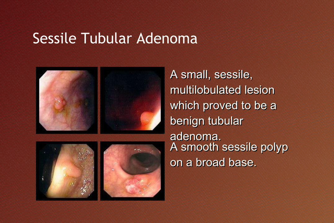
Sessile Tubular Adenoma
A small, sessile, A small, sessile, multilobulated lesion multilobulated lesion which proved to be a which proved to be a benign tubular benign tubular adenoma. adenoma. A smooth sessile polyp A smooth sessile polyp on a broad base. on a broad base.

Pedunculated Tubular Adenoma
Benign tubular adenoma Benign tubular adenoma on a long stalk .on a long stalk .The stalk is several The stalk is several times larger than the times larger than the polyp itself.polyp itself.

Sessile Villous Adenoma3-4 cm carpet-like 3-4 cm carpet-like tubulovillous adenoma of the tubulovillous adenoma of the cecum.cecum.
The orifice of the appendix is The orifice of the appendix is visible in the image on the left.visible in the image on the left.
1 cm sessile tubulovillous 1 cm sessile tubulovillous adenoma of the sigmoid colon.adenoma of the sigmoid colon.

Polyposis syndromes
• Polyposis syndromes are hereditary conditions that include: • Familial adenomatous polyposis (FAP).• Gardner syndrome.• Turcot syndrome.• Peutz-Jeghers syndrome.• Cowden disease.• Familial juvenile polyposis.
• Some of the syndromes have extraintestinal features that help differentiate one syndrome from the other.

Familial adenomatous polyposis (FAP(
Hundreds of benign Hundreds of benign tubular adenomas tubular adenomas throughout the colon.throughout the colon.
Small and large Small and large benign polyps benign polyps throughout the colon. throughout the colon.

Colon cancer

diverticula

Colitis
Pseudomembranous colitis

Colitis
Crohn’s disease Ulcerative colitis

Hemorrhoid

Angiodysplasia

Presentations & Definition 1- HEMATEMESIS:
It is bloody vomitus, either fresh and bright red
It results from upper GIT bleeding up to the 2nd part of the duodenum
proximal to ligament of Treitz at duodeno-jejunal junction.
Most frequently follows bleeding from the esophagus, stomach, or
duodenum
Melenemesis“coffee grounds” vomitingoccurs when blood is in contact with gastric acid for at least 1 hourUsually bleeding at a slower rate than those who have grossly bloody emesis

2- MELENA:
It is tarry, shiny, black, sticky stool
It usually occurs when bleeding is slow enough to allow time for degradation of
blood.
It results from slow upper GIT bleeding but occasionally hemorrhage into the jejunum,
ileum, or even right colon can cause melena if gastrointestinal transit is slow.
It should be distinguished from the black stools caused by ingestion of iron or bismuth.
3-Hematochezia
• The passage of bright red stools
• Usually a sign of distal small bowel or brisk colonic hemorrhage
• 10% : actively bleeding from an upper GIT lesion, and have accelerated GI transit
times

4- OCCULT BLOOD IN STOOL:
•Stool appears normal, but blood is detected when tested with guaiac test
and patients present with anemia. Microscopic blood when RBC are present
microscopically
5- ANEMIA:
•Patient may present without any objective signs of bleeding but rather with
symptoms of blood loss, such as dizziness, dyspnea on mild exertion, anginal
pain or fainting.

[II] Generalized causes: GIT bleeding is usually part of generalized
bleeding tendency.
1- Defects of platelet and coagulation factors: ITP, leukemia,
hemophilia and hypoprothrombinemia.
2- Disorders of the blood vessels: Hereditary hemorrhagic
telangiectasia and vascular malformations.

Severity of hemorrhage
• The most accurate non-invasive indicator of the severity of acute blood loss
Shock : acute blood volume loss of at least 15 to 20% Postural vital sign changes
Upright tachycardia, Widening of the pulse pressure &/or upright systolic hypotension
Acute intravascular volume loss of at least 10 to 15% Nasogastric lavage is helpful but highly inaccurate in
estimating the severity of upper GIT bleeding (esp. duodenal bleeding)
Presence of shock or postural changes in vital sign

Hypovolemic shock
Mild Moderate Severe
(<20% blood volume) (20-40% blood volume) (>40% blood volume)
Cool extremiyies Same, plus : Same, plus :
Increased capillary refill time
Tachycardia Hemodynamic instability
Diaphoresis Tachypnea Marked tachycardia
Collapsed veins Oliguria Hypotension
Anxiety Postural changes Mental status deterioration (coma)

Severity of hemorrhage
With acute hemorrhage – the hematocrit and hemoglobin levels are
not reliable indicators of the severity of bleeding
For the hematocrit to fall, the blood plasma must have equilibrated
with ECF or with administered intravenous fluids, and this
equilibration may require 24 to 48 hours to occur

Initial evaluation and treatment
• Vital signs• Supine and upright blood pressure• Pulse
• If blood loss is significant
iv fluids must be started immediately
• Brisk bleeding Packed RBC

The History
• Bleeding episode
• Any previous GIT hemorrhage
• Peptic ulcer
• Cancer
• Vascular ectasia
• Alcohol abuse
• Chronic liver disease – painless hematemesis of from esophageal varices
• Reflux esophagitis• Substernal burning pain• Regurgitation• Reflux symptom
• Mallory-Weiss tear• Forceful, dry retching or multiple episode of vomiting of food before the onset of
hematemesis

The history
• PUD• Epigastric burnng pain promptly relieved by food or antacid• Nocturnal pain
• Use of NSAID
• Diverticular disease
• Colorectal cancer• Gradual weight loss• Intermittent blood in the stools• Altered bowel habits
• IBD• Long-standing mucous and bloody diarrhea
• Hemorrhoid• Presence of bright red blood surrounding well-formed, normal-appearing stools

Physical examination
• Chronic liver disease
• Spider angioma
• Ascites
• Gynecomastia
• PUD or gastritis
• Localized epigastric tenderness on palpation
• LGIT malignancy
• Palpable lower abdominal mass
• Hepatomegaly
• Weight loss
• Adenopathy
• Anorectal mass lesion (polyps, cancers, or large hemorrhoids)
• Digital examination

Management
• Nasogastric tube lavage
• Using room temperature water
• Indication the rate of ongoing bleeding
• Decrease the bleeding rate by constricting smaller gastric vessels
• Hct, Hgb, PT, PTT, ABO & Rh
• Shock or postural hypotension
• 4 to 6 units of packed RBC should be cross-matched

UGIT bleeding• ulcer disease
• TMC cause of UGI bleeding
• 50% of moderately severe bleeding
• 35% of severe bleeding
• Esophageal or gastric varices
• 1/3 of massive UGI hemorrhage
• Usually associated with chronic liver disease
• Alcoholic > viral
• Large, firm liver
• Enlarged spleen
• Gross ascites
• Scleral icterus
• Palmar erythema
• Peripheral muscle wasting

UGIT bleeding
• Mallory-Weiss tears
• Tears of GEJ
• 5% of minor UGI hemorrhage
• 20% of severe UGI hemorrhage
• Usually associated antecedent, forceful rething
• Nearly 50% - alcohol abuse
• Gastritis due to alcohol or NSAID
• Esophagitis
• GIT malignancies

Endoscopy
• Diagnostic procedure of choice
• High accuracy and immediate therapeutic potential
• Must be performed only after adequate resuscitation and clinical assessment of the patient

Endoscopy
• Indication• Postural vital sign changes or shock• Multiple transfusion• Hematocrit below 30%• High index of suspicion of variceal hemorrhage• Recurrent hemorrhage from unknown sources• High risk for surgery
• Relative contraindication• Acute myocardial infarction• Severe chronic lung disease (SO2<90%)
• Hemodynamic instability• Patient agitation

Endoscopy
• Therapeutic endoscopy• Acute variceal bleeding
Endoscopic sclerotherayVarix band ligation
• UGIT hemorrhage
Endoscopic bipolar electrocoagulationHeater probe coagulationInjection of dilute epinephrine soln.

Esophageal varices (II(

Bleeding control with heater probe

Bleeding control with hemoclip

Barium radiography
• “UGI series”, when performed by double-contrast technique, identifies at least 70-80% of lesions confirmed to be associated with UGIT bleeding• Noninvasive• Costs less than endoscopy• Readily available but has significant disadvantages,
particularly in patients who are bleeding briskly• Multiple lesions may be detected by barium radiography
and the actual site of bleeding may be difficult to assess

Angiography
• When the site of of UGIT bleeding is missed on endoscopy
• Selective infusion with vasopressin or coil embolization of actively bleeding arteries may control bleeding
• Bleeding must be active (> 30 mL/h)
• Expensive, time-consuming, invasive
• Requires transporting the patient to a specialized unit

Nuclear scintigraphy
• When less active blood loss (3 mL/h)
• Technetium red cell nuclear scintigraphy (RBC scan)
• Non-invasive
• Portable gamma camera
• Often performed before any angiographic evaluation to prove the presence of active bleeding and to assist in the localization of the bleeding focus

LGIT bleeding• Colonic diverticula
• 1/4 of all episodes of hemodynamically significant bleeding from the LGIT
• Nearly always painless• Acute large-volume hematochezia
• Colonic cancer and polyps• 20% of LGI bleeding• Often present as hematochezia , particularly with lesions in the
distal sigmoid colon and rectum• Cf) proximal – IDA and frequently dark black or bloody stool
• UC and CD• Bloody diarrhea, tenesmus, long-standing history of IBD
• Vascular ectasia

Proctoscopy
• Careful evaluation of the anorectal junction are the initial diagnostic step for all patients with hematochezia
• Hemorrhoids or lacerations, diverticula, colitis, polyps, and cancer

Double-contrast barium radiography
• If blood loss is modest (as evidence by a normal hematocrit and vital sign), sigmoidoscopy may be followed by double-contrast radiography
• Highly accurate for detecting even smaller polyps and superficial mucosal abnormalities such as colitis

Colonoscopy
• Indicate LGIT hemorrhage with anemia
• Not only allows the site of hemorrhage to be determined accurately but also allows biopsy of suspicious mass lesions
• Polypectomy
• Coagulation technique

Technetium RBC scintigraphy
• Detect active bleeding at a rate of at least 3 to 10 mL/h
• Usually localizes the site but not the cause of active hemorrhage
Angiography• Bleeding at a rate exceeding 30 to 50 mL/h
• Vasopressin or embolization technique

Finally, Occult GIT hemorrhage
• 5%• 3 general categories• 1) Hematemesis or melanemesis but a “negative” upper
endoscopy• 2) Hematochezia but a “negative” colonoscopy• 3) Positive fecal OB testing and negative routine upper and
lower endoscopies• Modalities• Technetium RBC scintigraphy• Small bowel enteroclysis• Small bowel endoscopy• Angiography• Capsule endoscopy