Gastric Emptying and Secretion in Zollinger-Ellison Syndrome · Gastric Emptyingand Secretion in...
9
Gastric Emptying and Secretion in Zollinger-Ellison Syndrome ANDRE DUBOIS, PAUL VAN EERDEWEGH, AND JERRY D. GARDNER From the Section on Gastroenterology, Digestive Diseases Branch, National Institute of Arthritis, Metabolism, and Digestive Diseases, and Laboratory of Theoretical Biology, National Cancer Institute, National Institutes of Health, Bethesda, Maryland 20014 A B S T R A C T Gastric emptying and secretion, as well as intragastric volume and composition, were determined simultaneously in three patients with Zollinger-Ellison syndrome and in seven normal subjects. Gastric hyper- secretion was observed in patients with Zollinger- Ellison syndrome and in normal subjects receiving pentagastrin. In contrast, the fraction of gastric con- tents emptied per minute (fractional rate of emptying) was increased in Zollinger-Ellison patients and un- changed or decreased in normal subjects receiving pentagastrin. The increased fractional rate of gastric emptying in patients with Zollinger-Ellison syndrome persisted despite abolition of gastric hypersecretion by metiamide. Thus, the increased fractional gastric emptying seen in patients with Zollinger-Ellison syn- drome is not attributable to hypergastrinemia, or to gastric hypersecretion per se. Instead, it appears to be caused by an undefined nervous or humoral factor. INTRODUCTION The characteristic abnormalities found in patients with the Zollinger-Ellison syndrome are generally con- sidered to result directly or indirectly from increased circulating concentrations of gastrin (1). Administra- tion of exogenous gastrin (or a functional equivalent such as pentagastrin) to normal human subjects (2) or laboratory animals (3) produces changes in gastro- intestinal function similar and in most cases identical to those seen in patients with Zollinger-Ellison syn- drome (4). Insofar as gastric emptying is concerned there is an apparent discrepancy in the previously published Dr. Dubois' present address is: Department of Internal Medicine, Uniformed Services University of the Health Sciences, National Naval Medical Center, Bethesda, Md. 20014. Receivedfor publication 10June 1976 and in revisedform 15 September 1976. literature. In one patient with Zollinger-Ellison syn- drome, gastric emptying was reported to be increased (5) while administration of gastrin or pentagastrin to man or laboratory animals (6-8) has decreased gastric emptying. To explore this apparent discrepancy in greater detail we have simultaneously measured gastric emptying and secretion in normal subjects, normal subjects receiving pentagastrin, patients with Zollinger-Ellison syndrome, and patients with Zol- linger-Ellison syndrome receiving metiamide. METHODS All patients and normal volunteers were studied as inpatients at the Clinical Center, National Institutes of Health, Bethesda, Md. The experimental procedure was explained to, and in- formed consent obtained from, each subject for each study performed. The seven normal subjects (aged 22-26 yr; three females, four males) were free of and had no previous history of gastrointestinal disease and had normal serum gastrin concentrations.' The patients with Zollinger-Ellison syndrome (aged 27, 47, and 48 yr; two females, one male) had fasting serum gastrin concentrations greater than 300 pg/ml, basal gastric acid secretion greater than 20 mq/h with a ratio of basal acid secretion to maximal acid secretion (determined by Histalog administration; Eli Lilly and Co., Indianapolis, Ind.) greater than 0.6, and at some time radiological evi- dence of gastrointestinal ulceration. Two of the subjects had metastatic islet cell pancreatic carcinoma (verified histo- logically). The third patient had not undergone abdominal surgery. At the time these studies were performed none of the patients with Zollinger-Ellison syndrome had an "active ulcer" by endoscopic or radiological examination, although each had abnormalities known to be associated with Zol- linger-Ellison syndrome (e.g. scarred duodenal bulb and gastric rugal hypertrophy). The patient with endogenous hyperhistaminemia (10) had basophilic leukemia with in- creased whole blood and urinary histamine, normal serum gastrin, gastric acid hypersecretion, and duodenal ulcer. Intragastric volume and rates of gastric emptying and secretion were determined with a dilution technique. During each study 150 mM sodium chloride was adminis- 1 Normal serum immunoreactive gastrin activity in our laboratory is less than 150 pg/ml (9). The Journal of Clinical Investigation Volume 59 February 1977 255-263 255
Transcript of Gastric Emptying and Secretion in Zollinger-Ellison Syndrome · Gastric Emptyingand Secretion in...
Gastric Emptying and Secretion in
Zollinger-Ellison Syndrome
ANDREDUBOIS, PAUL VANEERDEWEGH,ANDJERRY D. GARDNER
From the Section on Gastroenterology, Digestive Diseases Branch, National Institute of Arthritis,Metabolism, and Digestive Diseases, and Laboratory of Theoretical Biology, National Cancer Institute,National Institutes of Health, Bethesda, Maryland 20014
A B S T RA C T Gastric emptying and secretion, as wellas intragastric volume and composition, were determinedsimultaneously in three patients with Zollinger-Ellisonsyndrome and in seven normal subjects. Gastric hyper-secretion was observed in patients with Zollinger-Ellison syndrome and in normal subjects receivingpentagastrin. In contrast, the fraction of gastric con-tents emptied per minute (fractional rate of emptying)was increased in Zollinger-Ellison patients and un-changed or decreased in normal subjects receivingpentagastrin. The increased fractional rate of gastricemptying in patients with Zollinger-Ellison syndromepersisted despite abolition of gastric hypersecretionby metiamide. Thus, the increased fractional gastricemptying seen in patients with Zollinger-Ellison syn-drome is not attributable to hypergastrinemia, or togastric hypersecretion per se. Instead, it appears to becaused by an undefined nervous or humoral factor.
INTRODUCTION
The characteristic abnormalities found in patients withthe Zollinger-Ellison syndrome are generally con-sidered to result directly or indirectly from increasedcirculating concentrations of gastrin (1). Administra-tion of exogenous gastrin (or a functional equivalentsuch as pentagastrin) to normal human subjects (2) orlaboratory animals (3) produces changes in gastro-intestinal function similar and in most cases identicalto those seen in patients with Zollinger-Ellison syn-drome (4).
Insofar as gastric emptying is concerned there is anapparent discrepancy in the previously published
Dr. Dubois' present address is: Department of InternalMedicine, Uniformed Services University of the HealthSciences, National Naval Medical Center, Bethesda, Md.20014.
Receivedfor publication 10June 1976 and in revisedform15 September 1976.
literature. In one patient with Zollinger-Ellison syn-drome, gastric emptying was reported to be increased(5) while administration of gastrin or pentagastrinto man or laboratory animals (6-8) has decreasedgastric emptying. To explore this apparent discrepancyin greater detail we have simultaneously measuredgastric emptying and secretion in normal subjects,normal subjects receiving pentagastrin, patients withZollinger-Ellison syndrome, and patients with Zol-linger-Ellison syndrome receiving metiamide.
METHODS
All patients and normal volunteers were studied as inpatientsat the Clinical Center, National Institutes of Health, Bethesda,Md. The experimental procedure was explained to, and in-formed consent obtained from, each subject for each studyperformed. The seven normal subjects (aged 22-26 yr;three females, four males) were free of and had no previoushistory of gastrointestinal disease and had normal serumgastrin concentrations.' The patients with Zollinger-Ellisonsyndrome (aged 27, 47, and 48 yr; two females, one male) hadfasting serum gastrin concentrations greater than 300 pg/ml,basal gastric acid secretion greater than 20 mq/h with a ratio ofbasal acid secretion to maximal acid secretion (determinedby Histalog administration; Eli Lilly and Co., Indianapolis,Ind.) greater than 0.6, and at some time radiological evi-dence of gastrointestinal ulceration. Two of the subjects hadmetastatic islet cell pancreatic carcinoma (verified histo-logically). The third patient had not undergone abdominalsurgery. At the time these studies were performed none ofthe patients with Zollinger-Ellison syndrome had an "activeulcer" by endoscopic or radiological examination, althougheach had abnormalities known to be associated with Zol-linger-Ellison syndrome (e.g. scarred duodenal bulb andgastric rugal hypertrophy). The patient with endogenoushyperhistaminemia (10) had basophilic leukemia with in-creased whole blood and urinary histamine, normal serumgastrin, gastric acid hypersecretion, and duodenal ulcer.
Intragastric volume and rates of gastric emptying andsecretion were determined with a dilution technique.During each study 150 mMsodium chloride was adminis-
1 Normal serum immunoreactive gastrin activity in ourlaboratory is less than 150 pg/ml (9).
The Journal of Clinical Investigation Volume 59 February 1977 255-263 255
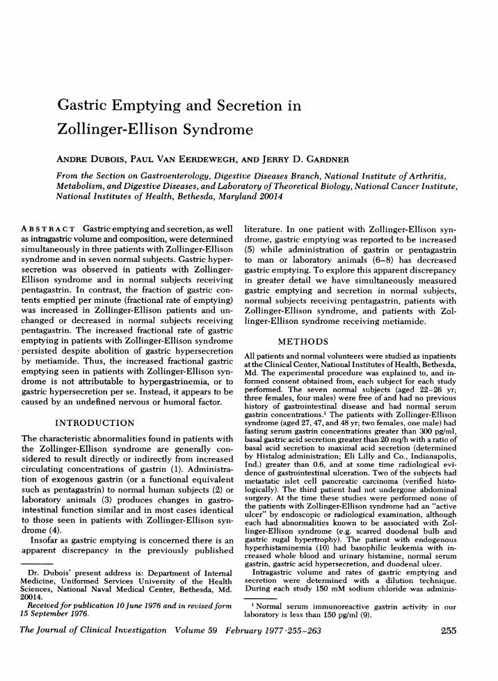
Time (min)
FIGURE 1 Dilution technique. Schematic illustration of thetechnique used to determine gastric emptying and secretion.
tered intravenously at a rate of 60 ml/h. When appropriate,pentagastrin and metiamide were infused at 5 ,4g/kg per h and4 mg/kg per h, respectively. Studies were started 30 min afterintroducing a no. 12 French, double lumen polyethyleneSalem suimp tube (bore 4 mm, wall thickness 1 mm, SherwoodMedical Industries Inc., St. Louis, Mo.) in the stomach.Proper positioning of the tube in the most dependent partof the stomach was verified by demonstrating that im-mediately after injecting 20 ml of water at least 15 mlcould be recovered.
Gastric contents were mixed for 1 min by rapid, repeatedaspiration and immediate reinsertion of 5 ml of gastriccontents with a 50-ml syringe; 2.5 ml (VAl) were sampledat time T, (Fig. 1). Immediately thereafter 5 ml (V2) of aphenol red solution in distilled water (280 mg/liter, pH 7.4)was instilled into the stomach and mixed thoroughly withgastric contents for 50 s. A second 2.5-ml sample (VA3) wasthen taken.
This 2-min mixing, sampling, and dilution procedure wasperformed at 10-min intervals for 30 min. 250 ml of a phenolred solution in distilled water ("meal"; 40 mg/liter, pH 7.4)2was then instilled into the stomach during 2.5 min with aconstant infusion pump. The 2-min mixing, sampling, anddilution procedure was then repeated at 5 and 10 min afterthe start of the meal and subsequently at 10-min intervalsfor 40 min.
Each sample of gastric juice and the phenol red solutionswere adjusted to pH 10 with 0.25% Na3PO4 and analyzedfor the concentration of phenol red with a spectrophotometerat 560 nm (Gilford Micro-sample Spectrophotometer, Gil-ford Instrument Laboratories, Inc., Oberlin, Ohio). Hydrogenion concentration was determined by endpoint titration topH 7.4 with 0.01 N NaOH(Titration assembly, RadiometerCo., Copenhagen, Denmark).
The reason for using 5- or 10-min intervals was that thetime should be long enough to allow for changes in phenolred and ion concentrations but short enough to permitthe assumption that the fractional rate of emptying andsecretory rates remained constant over the given inter-val. When the volume present in the stomach was greaterthan 10 ml, the size of the samples VA, and VA3 was 10 ml,and the volume of test solution injected (V,) was 20 ml, while,when it was less than 10 ml, volumes identical to the onesused during the fasting period were sampled and injected.Because the volume of test solution injected (V2) was equal tothe total of the volume aspirated (VAI + VA3), there was no
2 A solution containing phenol red was used instead of dis-tilled water to avoid the large relative errors which may resultfrom spectrophotometric measurements of solutions contain-ing low concentrations of phenol red.
net gain or loss of fluid resulting from the sampling anddilution procedture.
Several assumptions were needed to calculate intragastricvolume at the times of the dilution procedure as Xvell as therates of emptying and secretion during the intervals betweenthese dilution procedures. (a) Phenol red is not absorbed,secreted, or degraded by the human stomach (11). (b) A5-20-ml phenol red solution injected into the stomach canbe mixed completely with the gastric contents by aspiratingand reinjecting 5-20-ml aliquots four times during a 1-mimperiod (unpublished observations and 12). (c) Over a shortinterval of time, the rate of decline of intragastric phenolred (dP/dt) is proportional to the intragastric amouint ofphenol red (P). This assumption is expressed mathematicallyby
dP/dt =-gP,
where g is the fractional rate of emptying, i.e., the fraction ofthe intragastric contents leaving the stomach per unit of time.(d) Phenol red remains homogeneously mixed with the gastriccontents during the intervals between samplings. A logicalcorollary of this assumption is that any element present inthe stomach (water, ions) is emptied at a fractional rate identi-cal to that for phenol red. That is, if the fractional rate ofemptying for phenol red is 5%per min, 5%of the intragastricamount of water and ioIns will also leave the stomach perminute. (e) The fractional rate of emptying and the rates ofsecretion are constant during one 5- or 10-min interval butcan vary between intervals.
From assumptions a and b an initial estimate of the intra-gastric volume was calculated from the equation (Appendixand Fig. 1):
Vl = V2 (C2 - C3)/(C3 - C1) + VA1,
where VI is the volume of fluid in the stomach before thedilution procedure, C, is the concentration of phenol redin the first aspirated sample, the volume of which is VAI,V2 is the volume of test solution instilled into the stomach,C2 is the concentration of phenol red in this test solution(280 mg/liter), and C3 is the concentration of phenol red inVA3 ml aspirated after mixing the test solution with the gastriccontents. The amount of phenol red present in the stomach(Pi) was then calculated at the time of each sampling duringthe experiment. This initial estimate is obtained by neglect-ing the secretion and emptying occurring during the 1-mininterval between the two samplings VA, and VA3.
From assumptions a, c, and d, fractional rate of emptying,g, was then calculated from the change in the amount ofintragastric phenol red during any 5- or 10-min interval(Appendix). The amount of a particular substance (X) presentat the end of any given interval reflects the amotunt of Xpresent at the beginning of the interval plus the net dif-ference between the amount secreted and the amountemptied. One can calculate the rate at which X is emptied.By determining the amounts of X present at the beginningand at the end of the interval one can then calculate therate at which X was secreted during the interval (Appendix).
After rates of emptying and secretion were determined,intragastric volume was recalculated with an equation whichincludes a term for the emptying and secretion which occurduring the 1-min mixing period (Appendix). The fractionalrate of emptying and the rate of water secretion were thencomputed again and these new values were used to deter-mine the intragastric volume. This iterative process (13) wasrepeated until the calculated value for intragastric volumechanged by less than 1%. Calculations were performed with alocally developed program and a GE 265 computer (Call-
256 A. Dubois, P. Van Eerdewegh, and J. D. Gardner
a-Computer, Inc., Wellesley Hills, Mass.). For the 2.5-mmninterval corresponding to the instillation of the meal, we
have calculated secretory and emptying rates with an equa-
tion which includes a term describing this infusion (Ap-
pendix).For each time interval between two sampling and dilution
procedures, fractional rate of emptying and rates of secre-
tion were determined from the changes in intragastric
concentrations of phenol red and ions. The fraction of the test
meal remaining in the stomach was calculated at the end of
each interval. For each subject, one fasting value was cal-
culated from the mean of the results obtained before the
meal. In each group, values obtained in each subject be-
fore and for each interval after the meal were used to deter-
mine the mean (±+SEM) given in Figs. 2-7. The change in
each function (e.g., fractional emptying, water secretion, etc.)
was evaluated using a two-factor (time and group of subjects)
analysis of variance with repeated measures (14) and
multiple t tests. These calculations were pefformed with
the program LDUO40 (K. L. Dom) and an IBM 370 com-
puter (Division of Computer Research and Technology,National Institutes of Health, Bethesda, md.).
Two different techniques were used to test the validity of
the dilution technique. (a) In dogs, fractional rate of empty-
ing was measured simultaneously with the present dilution
technique and by counting externally intragastric "mTc-DTPA with a gamma camera inteffaced with a computer.
Results obtained with the two techniques were not signifi-
cantly different (15). Furthermore, repeating the isotope
study on a separate day without mixing yielded similar re-
sults, indicating that the mixing procedure does not affect
gastric emptying. (b) In humans, the gastric contents were
aspirated completely immediately before each dilution pro-
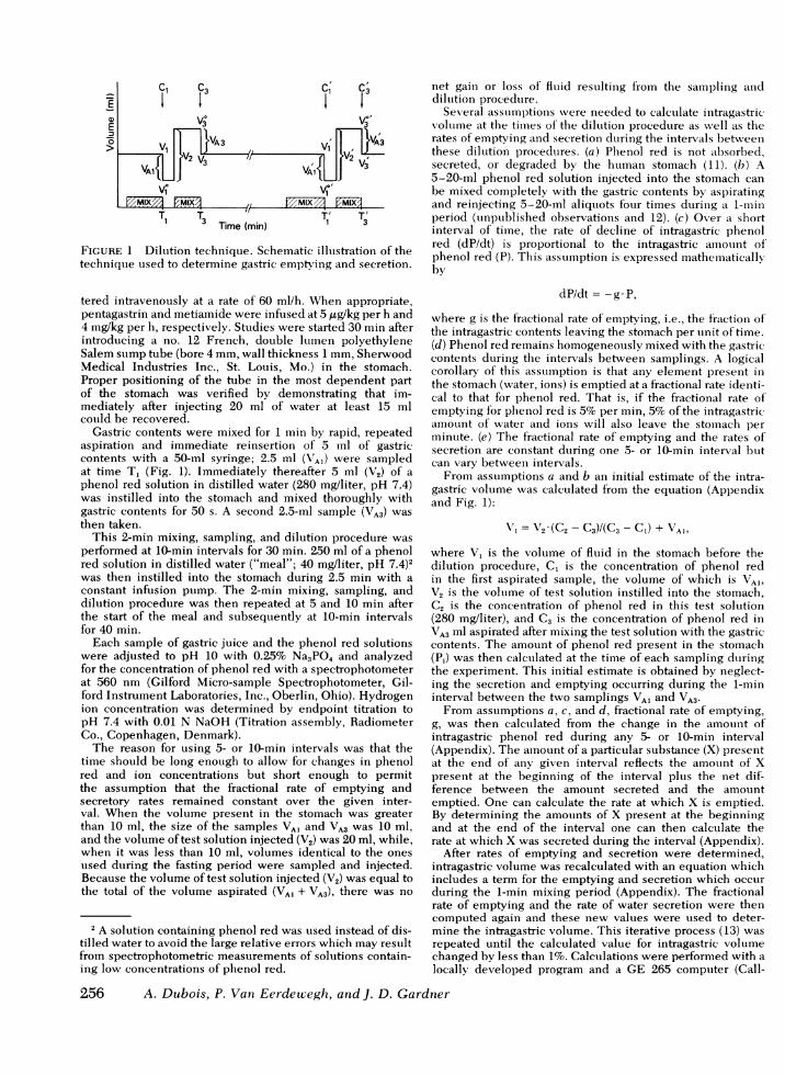
TABLE I
Intragastric Volume and Rates of Gastric Secretion
and Fractional Emptying in Fasting Subjects
Fractional
Water Hydrogen Intragastric rate of
Subjects secretion ion secretion volume emptying
ml/min meqImin ?nl %/min
Normal (7) 0.9±+0.2 0.031±+0.006 24±#.5 4±+2
Normal plus
penta-
gastrin (7) 5.0±+0.6 0.55±0.04 95± 16 6±0.7
ZES* (3) 8.2± 1.0 0.83±0.20 42±6 19±6
ZES plus
meti-
amide (3) 1.3±0.4 0 10±0.4 16±2
Hyperhista-
min-emia (1) 3.5 0.59 94 3
Intragastric volume and fractional rates of gastric emptying
and water secretion were determined at 10-mmn intervals
during a 40-mmn fasting period. Pentagastrin (5 .tig/kg per h or
metiamide (4 mg/kg per h) were given intravenously in
150 mMNaCl at a rate of 60 ml/h. When these agents were
used experimental determinations were begun 30 min after
starting infusion of the drug. Values are meanstl SEM.
The number of subjects is given in parentheses.* Zollinger-Ellison syndrome.
TABLE IIGastric Secretion of Water and Hydrogen Ion in Patients
with Zollinger-Ellison Syndrome
Rate of water Rate of hydrogen ionsecretion secretion
Dilution Aspiration Dilution AspirationPatient technique technique technique technique
ml/min meqimin
j. G. 8.97 6.70 1.14 0.90D. M. 6.27 4.12 0.45 0.46P. R. 9.27 7.25 0.90 1.07
P<O.O5* NS*
Gastric secretion of water and hydrogen ion was determinedin three patients with Zollinger-Ellison syndrome with(a) the dilution technique described in Methods and (b) con-tinuous aspiration of the gastric contents via a nasogastrictube. Results are means of four measurements.* As calculated with an analysis of variance with repeatedmeasures (14).
cedure during fasting and at the end of each experiment. Inall instances, the aspirated volume was greater than 90%/of that calculated with the dilution technique.
RESULTS
In seven normal fasting subjects administration ofpentagastrin caused a 5-fold increase in water secre-tion, an 18-fold increase in acid secretion, no changein the fractional rate of gastric emptying, and a 4-foldincrease in intragastric volume (Table I). In three fast-ing patients with Zollinger-Ellison syndrome secretoryrates of water and acid were significantly greater thanthose in normal subjects (9- and 28-fold, respectively)and fractional rate of gastric emptying was three to fivetimes greater than normal. In Zollinger-Ellison syn-drome, intragastric volume was approximately twicethat in normal subjects but approximately one-half ofthat of normal subjects receiving pentagastrin. Ad-ministration of metiamide to the patients with Zol-linger-Ellison syndrome reduced the rates of watersecretion to values that were not significantly differentfrom those in normal subjects and abolished acid secre-tion but did not alter the fractional rate of gastric empty-ing. Thus, in patients with Zollinger-Ellison syndrome,metiamide reduced the volume of the intragastric con-tents to a value which was approximately one-half thatobtained in normal fasting subjects. The patient withhyperhistaminemia had values for acid and water secre-tion, fractional rate of emptying, and intragastricvolume which were the same as those in normal sub-jects given pentagastrin.
In the three patients with Zollinger-Ellison syn-drome we compared values for gastric secretion ob-
Gastric Emptying and Secretion 257
10
EZ-
Ez0
a:wLU
n
LA-
0wc-
5
i0 t 10 20 30 40Meal TIME (min)
, ZES
, Normal+ PG
Normal
50
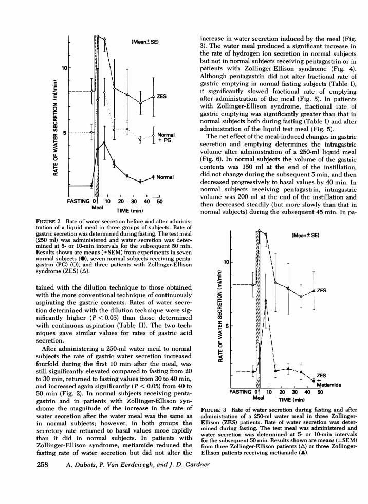
FIGURE 2 Rate of water secretion before and after adminis-tration of a liquid meal in three groups of subjects. Rate ofgastric secretion was determined during fasting. The test meal(250 ml) was administered and water secretion was deter-mined at 5- or 10-min intervals for the subsequent 50 min.Results shown are means (±SEM) from experiments in sevennormal subjects (0), seven normal subjects receiving penta-gastrin (PG) (0), and three patients with Zollinger-Ellisonsyndrome (ZES) (A).
tained with the dilution technique to those obtainedwith the more conventional technique of continuouslyaspirating the gastric contents. Rates of water secre-tion determined with the dilution technique were sig-nificantly higher (P < 0.05) than those determinedwith continuous aspiration (Table II). The two tech-niques gave similar values for rates of gastric acidsecretion.
After administering a 250-ml water meal to normalsubjects the rate of gastric water secretion increasedfourfold during the first 10 min after the meal, wasstill significantly elevated compared to fasting from 20to 30 min, returned to fasting values from 30 to 40 min,and increased again significantly (P < 0.05) from 40 to50 min (Fig. 2). In normal subjects receiving penta-gastrin and in patients with Zollinger-Ellison syn-drome the magnitude of the increase in the rate ofwater secretion after the water meal was the same asin normal subjects; however, in both groups thesecretory rate returned to basal values more rapidlythan it did in normal subjects. In patients withZollinger-Ellison syndrome, metiamide reduced thefasting rate of water secretion but did not alter the
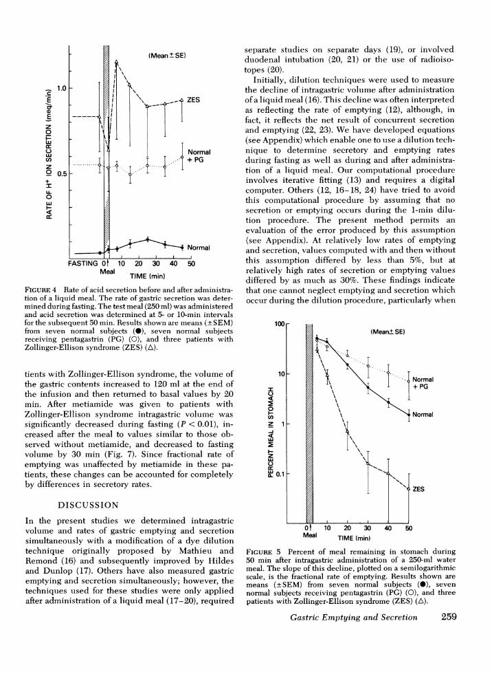
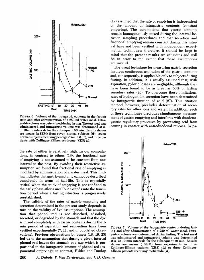
increase in water secretion induced by the meal (Fig.3). The water meal produced a significant increase inthe rate of hydrogen ion secretion in normal subjectsbut not in normal subjects receiving pentagastrin or inpatients with Zollinger-Ellison syndrome (Fig. 4).Although pentagastrin did not alter fractional rate ofgastric emptying in normal fasting subjects (Table I),it significantly slowed fractional rate of emptyingafter administration of the meal (Fig. 5). In patientswith Zollinger-Ellison syndrome, fractional rate ofgastric emptying was significantly greater than that innormal subjects both during fasting (Table I) and afteradministration of the liquid test meal (Fig. 5).
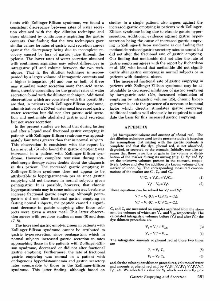
The net effect of the meal-induced changes in gastricsecretion and emptying determines the intragastricvolume after administration of a 250-ml liquid meal(Fig. 6). In normal subjects the volume of the gastriccontents was 150 ml at the end of the instillation,did not change during the subsequent 5 min, and thendecreased progressively to basal values by 40 min. Innormal subjects receiving pentagastrin, intragastricvolume was 200 ml at the end of the instillation andthen decreased steadily (but more slowly than that innormal subjects) during the subsequent 45 min. In pa-
-
EI
z0
LIc-u
uL0
(Mean± SE)
I
Meal
'\. I '1I'~~~~
i \li
1'~~~~~~-
P i i \ ZESMetiamide
10 20 30 40 50TIME (min)
FIGuRE 3 Rate of water secretion during fasting and afteradministration of a 250-ml water meal in three Zollinger-Ellison (ZES) patients. Rate of water secretion was deter-mined during fasting. The test meal was administered andwater secretion was determined at 5- or 10-min intervalsfor the subsequent 50 min. Results shown are means (+SEM)from three Zollinger-Ellison patients (A) or three Zollinger-Ellison patients receiving metiamide (A).
258 A. Dubois, P. Van Eerdewegh, and J. D. Gardner
Ea,Ez0
ccwwcnz0+IU-0wi
c-
Ot 10 20 30 40Meal
TIME (min)
ZES
Normal+ PG
Normal
50
FIGURE 4 Rate of acid secretion before and after administra-tion of a liquid meal. The rate of gastric secretion was deter-mined during fasting. The test meal (250 ml) was administeredand acid secretion was determined at 5- or 10-min intervalsfor the subsequent 50 min. Results shown are means (+SEM)from seven normal subjects (0), seven normal subjectsreceiving pentagastrin (PG) (0), and three patients withZollinger-Ellison syndrome (ZES) (A).
tients with Zollinger-Ellison syndrome, the volume ofthe gastric contents increased to 120 ml at the end ofthe infusion and then returned to basal values by 20min. After metiamide was given to patients withZollinger-Ellison syndrome intragastric volume wassignificantly decreased during fasting (P < 0.01), in-creased after the meal to values similar to those ob-served without metiamide, and decreased to fastingvolume by 30 min (Fig. 7). Since fractional rate ofemptying was unaffected by metiamide in these pa-tients, these changes can be accounted for completelyby differences in secretory rates.
DISCUSSION
In the present studies we determined intragastricvolume and rates of gastric emptying and secretionsimultaneously with a modification of a dye dilutiontechnique originally proposed by Mathieu andRemond (16) and subsequently improved by Hildesand Dunlop (17). Others have also measured gastricemptying and secretion simultaneously; however, thetechniques used for these studies were only appliedafter administration of a liquid meal (17-20), required
separate studies on separate days (19), or involvedduodenal intubation (20, 21) or the use of radioiso-topes (20).
Initially, dilution techniques were used to measurethe decline of intragastric volume after administrationof a liquid meal (16). This decline was often interpretedas reflecting the rate of emptying (12), although, infact, it reflects the net result of concurrent secretionand emptying (22, 23). Wehave developed equations(see Appendix) which enable one to use a dilution tech-nique to determine secretory and emptying ratesduring fasting as well as during and after administra-tion of a liquid meal. Our computational procedureinvolves iterative fitting (13) and requires a digitalcomputer. Others (12, 16-18, 24) have tried to avoidthis computational procedure by assuming that nosecretion or emptying occurs during the 1-min dilu-tion procedure. The present method permits anevaluation of the error produced by this assumption(see Appendix). At relatively low rates of emptyingand secretion, values computed with and then withoutthis assumption differed by less than 5%, but atrelatively high rates of secretion or emptying valuesdiffered by as much as 30%. These findings indicatethat one cannot neglect emptying and secretion whichoccur during the dilution procedure, particularly when
I
00Inz 1
I-i
zu
X, 0.1
Normal+ PG
, Normal
, ZES
Meal TIME (min)
FIGURE 5 Percent of meal remaining in stomach during50 min after intragastric administration of a 250-ml watermeal. The slope of this decline, plotted on a semilogarithmicscale, is the fractional rate of emptying. Results shown aremeans (+±SEM) from seven normal subjects (0), sevennormal subjects receiving pentagastrin (PG) (0), and threepatients with Zollinger-Ellison syndrome (ZES) (A\).
Gastric Emptying and Secretion 259
LU
D-i0
a:
re0n
Normal+ PG
0 ZES
Normal
Meal TIME (min)
FIGURE 6 Volume of the intragastric contents in the fastingstate and after administration of a 250-ml water meal. Intra-gastric volume was determined during fasting. The test meal wasadministered and intragastric volume was determined at 5-or 10-min intervals for the subsequent 50 min. Results shownare means (+SEM) from seven normal subjects (0), sevennormal subjects receiving pentagastrin (PG) (0), and three pa-tients with Zollinger-Ellison syndrome (ZES) (A).
the rate of either is relatively high. In our computa-tions, in contrast to others (18), the fractional rateof emptying is not assumed to be constant from oneinterval to the next. By avoiding their restrictive as-sumption we found that fractional rate of emptying ismodified by administration of a water meal. This find-ing indicates that gastric emptying cannot be describedcompletely in terms of half-life. This is especiallycritical when the study of emptying is not confined tothe early phase after a meal but extends into the transi-tion period when a fasting situation is progressivelyreestablished.
The validity of the rates of gastric emptying andsecretion determined in the present study depends inturn on the validity of five assumptions. The assump-tion that phenol red is not absorbed, adsorbed,secreted, or degraded by the stomach and that the dyeis mixed completely with gastric contents during the 1-min period of aspiration and reinjection have beenverified experimentally (7, 11, and unpublished obser-vations). Previous observations by others (18, 19, 25)led us to the assumption that during a given intervalphenol red leaves the stomach at a rate which is pro-portional to the intragastric amount of phenol red (ex-ponential emptying); in contrast, Hildes and Dunlop
(17) assumed that the rate of emptying is independentof the amount of intragastric contents (constantemptying). The assumptions that gastric contentsremain homogeneously mixed during the interval be-tween sampling procedures and that secretion andfractional emptying remain constant during this inter-val have not been verified with independent experi-mental techniques; therefore, it should be kept inmind that the present results are estimates and willbe in error to the extent that these assumptionsare invalid.
The usual technique for measuring gastric secretioninvolves continuous aspiration of the gastric contentsand, consequently, is applicable only to subjects duringfasting. In addition, it is usually assumed that, withaspiration, pyloric losses are negligible, although theyhave been found to be as great as 50% of fastingsecretory rates (26). To overcome these limitations,rates of hydrogen ion secretion have been determinedby intragastric titration of acid (27). This titrationmethod, however, precludes determination of secre-tory rates for other ions and water. In addition, eachof these techniques precludes simultaneous measure-ment of gastric emptying and interferes with duodeno-gastric regulatory processes by preventing acid fromcoming in contact with antroduodenal mucosa. In pa-
200 F-
E
40
z
-J0
100oo
FASTING IWeal
10
(Mean±SE)
I
4'\Z ZES
'- 44ZES +I -I Metiamide
20 30 40 50TIME (min)
FIGURE 7 Volume of the intragastric contents during fast-ing and after administration of a 250-ml water meal. Intra-gastric volume was determined during fasting. The test mealwas administered and intragastric volume was determinedat 5- or 10-min intervals for the subsequent 50 min. Resultsshown are means (±+SEM) from experiments in threeZollinger-Ellison patients (ZES) (A) or three Zollinger-Ellison patients receiving metiamide (A).
260 A. Dubois, P. Van Eerdewegh, and J. D. Gardner
I- - - -T
tients with Zollinger-Ellison syndrome, we found aconsistent discrepancy between rates of water secre-tion obtained with the dye dilution technique andthose obtained by continuously aspirating the gastriccontents. Our finding that the two techniques gavesimilar values for rates of gastric acid secretion arguesagainst the discrepancy being due to incomplete re-covery caused by loss of gastric juice through thepylorus. The lower rates of water secretion obtainedwith continuous aspiration may reflect differences inintragastric pH and volume between the two tech-niques. That is, the dilution technique is accom-panied by a larger volume of intragastric contents anda higher intragastric pH and one or both of thesemay stimulate water secretion more than acid secre-tions, thereby accounting for the greater rates of watersecretion found with the dilution technique. Additionalobservations which are consistent with this possibilityare that, in patients with Zollinger-Ellison syndrome,administration of a 250-ml water meal increased gastricwater secretion but did not alter gastric acid secre-tion and metiamide abolished gastric acid secretionbut not water secretion.
In the present studies we found that during fastingand after a liquid meal fractional gastric emptying inpatients with Zollinger-Ellison syndrome was approxi-mately four times greater than that in normal subjects.This observation is consistent with the report byLawrie et al. (5) who found that gastric emptying wasincreased in a patient with Zollinger-Ellison syn-drome. However, complete remission during anti-cholinergic therapy raises doubts about the diagnosisin this patient. The increased gastric emptying inZollinger-Ellison syndrome does not appear to beattributable to hypergastrinemia per se since gastricemptying did not increase in normal subjects givenpentagastrin. It is possible, however, that chronichypergastrinemia may in some unknown way be able toincrease fractional gastric emptying. Although penta-gastrin did not alter fractional gastric emptying infasting normal subjects, the peptide caused a signifi-cant decrease in gastric emptying after these sub-jects were given a water meal. This latter observa-tion agrees with previous studies in man (6) and dogs(7, 8).
The increased gastric emptying seen in patients withZollinger-Ellison syndrome cannot be attributed togastric hypersecretion, since pentagastrin, which innormal subjects increased gastric secretion to ratesapproaching those in the patients with Zollinger-Elli-son syndrome, decreased or did not alter fractionalgastric emptying. Furthermore, the rate of fractionalgastric emptying was normal in a patient withendogenous hyperhistaminemia and gastric secretoryrates comparable to those in the Zollinger-Ellisonsyndrome. This latter finding, although based on
studies in a single patient, also argues against theincreased gastric emptying in patients with Zollinger-Ellison syndrome being due to chronic gastric hyper-secretion. Additional evidence against gastric hyper-secretion being the cause of increased gastric empty-ing in Zollinger-Ellison syndrome is our finding thatmetiamide reduced gastric secretory rates to normal butdid not alter the fractional rate of gastric emptying.Our finding that metiamide did not alter the rate ofgastric emptying agrees with the report by Richardsonet al. (28) who found that metiamide did not signifi-cantly alter gastric emptying in normal subjects or inpatients with duodenal ulcers.
The increased fractional rate of gastric emptying inpatients with Zollinger-Ellison syndrome may be at-tributable to decreased inhibition of gastric emptyingby intragastric acid (29), increased stimulation ofemptying by intragastric volume (25), chronic hyper-gastrinemia, or to the presence of a nervous or humoralfactor which directly stimulates gastric emptying.Additional studies will obviously be required to eluci-date the basis for this increased gastric emptying.
APPENDIX
(a) Intragastric volume and amount of phenol red. Thedye dilution technique used for the present studies is based onthe assumptions that mixing with the gastric contents iscomplete and that the dye, phenol red, is not absorbed,degraded, or secreted by the stomach. Initially, one also as-sumes that there is no loss or gain in the volume of distri-bution of the marker during its mixing (Fig. 1). V,° and V30are the unknown volumes present in the stomach, respec-tively, before and after the addition of a known volume of themarker solution, V2. The corresponding measured concen-trations of the marker are Cl, C2, and C3.
V1°C1 + V2C2 = V30C3; (1)
V10 + V2 = V30. (2)These equations can be solved for V10 and V30:
V10 = V2-(C2 - C3)/(C3 - C1);V30 = V2 (C2 - C1)/(C3 - C1)-
C1 and C3 are measured on samples aspirated from the stom-ach, the volumes of which are VA, and VA3, respectively. Thecalculated intragastric volumes before (VI) and after (V3) thedye dilution procedure are
V1 = V10 + VAI;
V3 = V30 - VA3.(3)(4)
The intragastric amounts of phenol red at those two timesare
P1 =V1 C1,
P3 = V3-C3,
(5)(6)
and, for the subsequent dilution procedures, volumes of waterand amounts of phenol red will be Vl',Pl',V3',P3'; V'",P1',V3",P3"; etc. Weselected a value for V2 which was directly pro-
Gastric Emptying and Secretion 261
portional to V1 and V3 so that
C1 - 0.7 C3s 0.6 C2.
Thus, the relative error on V1 and V3 was of the same orderof magnitude as the relative error on C1, C2, and C3.
(b) Fractional rate of emptying between the dilution proce-dures. Assuming that the rate of emptying of phenol redis proportional to its intragastric amount, P:
dP/dt = - g P. (7)If g, the fractional rate of emptying, is assumed to be constantover a time interval t (= T1' - T3), one can integrate thisequation to obtain
PI' = P3 exp(- g t), (8)
where P1' and P3 are the amounts of marker at times T1' andT3, respectively. This equation can then be solved for g.
If phenol red is instilled into the stomach at a constantrate, Up (expressed in milligrams per minute), during thetime t', starting at T3 and ending at T4 (T4 < T1'), the relation is
4P1 = P3 exp(- g t) + Up exp(- g [T1' - T])dT. (9)
[T1' - r] is the time interval between any time r (T3 < T < T4)and T1' or by evaluating the integral,
P1 = P3 exp(- g t) +
Up [exp(- g [t - t']) - exp(- g t)]/g. (10)
This equation cannot be solved analytically for g, but g canbe evaluated by successive iterations (Ref. 13).
(c) Rate ofwatersecretion The rate of change dV/dt oftheintragastric amount of water (V) can be described by
dV/dt = - g V + Rw, (11)
where Rw is the rate of water secretion.If a time interval t(= T1' - T3) is considered, the relation
becomes
VI' = V3 exp(- g.t) + Rw exp(- g [T,' - T]) *dT. (12)
V3 and V1' are the volumes of water present in the stomachat the times T3 and T1' respectively. The integral can thenbe evaluated if Rw is assumed to be constant over theinterval t.
V1' = V3-exp(- g t) + Rw [1 - exp(- g t)]/g. (13)
Rwcan then be calculated.If water is instilled into the stomach at a constant rate, Uw
(expressed in milliliters per minute) during the time t',starting at the time T3 and ending at T4, the relation is
V1' = V3*exp(- gt) + Rw [1 - exp(- g t)]/g
+ Uw [exp(- g[t - t']) - exp(- gt)]/g. (14)
Similar equations can be derived for the ions whose concen-trations have been measured in the gastric contents, thusallowing the calculation of the secretory rates of ions.
(d) Correction for secretion and emptying occurring dur-ing the dilution procedure (Fig. 1). Initially, the emptyingand secretory rates are assumed to be negligible over the1-min sampling and dilution period (T1 to T3). If the fractionalrate of emptying (g) and secretory rate (Rw) are known forthe time interval T3 to T1' it can be assumed that these rates
are also valid for the sampling intervals T, to T3 and T1' toT3'. Therefore, equations (1) and (2) can be transformed to:
V10C1 exp(- g [T3 - T,])+ V2[C2K + (C3 - C2) K'] = V30C3; (15)
V,° exp(- g [T3 - T,]) + V2 K3
+{RwWexP(- g [T3 - T]) dT = V3°. (16)
[T3 - T,] is the time interval between the samplings thatpermits the measurements of C3 and C,. [T3 - r] is the timeinterval between any time T and T3 (T, < T < T3). K is a factoraccounting for the fact that, due to four aspirations and fourreinjections of volume V2 during mixing, the injected phenolred test solution is in the stomach, available for emptying,only during a fraction of the 1-min dilution interval [K = (1+ exp(- g 314))I(1 + exp(- g/12))]. K' accounts for the pro-gressive change in concentration of the injected phenol redfrom C2 to C3 [K' = (1 - exp(-g/12))-(4 + 3-exp(-g/6) + 2exp(-g/3) + exp(-g/2))/4].
These equations can be solved for V10 and V30 to give:
V0° = [V2 (C2 - C3) (K - K')
- C3Rw [l - exp(- g [T3 - Tj])]/g]I[(C3 - C1)-exp(- g [T3 - TI])]; (17)
V30 = [V2 [K (C2 - C1) - K' (C2 - C3)]- C1lRw[1 - exp(- g [T3 - T,])]/g]/(C3 - C1). (18)
By analogy with equations (3), (4), and (5), the intragastricvolumes and amounts of phenol red before and after the dyedilution procedure can be calculated as follows:
V1 = V10 + VA, and P1 = V1 Cl;
V3 = V30 - VA3 and P3 = V3 C3.
A new estimate of the rates of gastric emptying and secre-tion can then be calculated. This two-step calculation is re-peated, which gives alternatively over- and underestimatedvalues for the intragastric volumes and the secretory andemptying rates. The solutions converge and the iterationsare stopped when the fractional change of the calculatedvolume becomes less than 0.01. The initial, uncorrectedvalue for intragastric volume was always significantly dif-ferent from the final value. The absolute difference be-tween these two values was directly related to the volumeof the gastric contents and varied between 1.5 and 30 ml.The fractional difference was a function of the rates ofemptying and secretion, and varied from 10% at low ratesto up to 30% at higher rates. Similar differences were ob-served during computation of the rates of emptying andsecretion.
ACKNOWLEDGMENTS
Wethank Dr. M. Berman for his help in conceptualizing thepresent model for gastric emptying and secretion and for hisconstant advice throughout this study. Wealso thank Dr. S.Chu for assisting with the development of mathematicalequations, and K. D. Pettigrew, H. R. Baird, and F. D.Vansant for their advice on statistical methods and computerprogram use. We also thank Mrs. B. Fors for preparing themanuscript.
262 A. Duibois, P. Vant Eerdetwegh, and J. D. Gardner
REFERENCES
1. Zollinger, R. M., and E. H. Ellison. 1955. Primary pepticulcerations of the jejunum associated with islet celltumors of the pancreas. Ann. Surg. 142: 709-728.
2. Makhlouf, G. M., J. P. A. McManus, and W. I. Card. 1964.The action of gastrin II on gastric acid secretion in man.Comparison of the "maximal" secretory response togastrin II and histamine. Lancet. 2: 485-490.
3. Gregory, R. A., and H. J. Tracy. 1964. The constitutionand properties of two gastrins extracted from hog antralmucosa. Gut. 5: 103-117.
4. Isenberg, J. I., J. H. Walsh, and M. I. Grossman. 1973.Zollinger-Ellison syndrome. Gastroenterology. 65:140-165.
5. Lawrie, R. S., A. W. R. Williamson, and J. N. Hunt.1962. Zollinger-Ellison syndrome treated with poldinemethyl methosulphate. Lancet. 1: 1002-1004.
6. Hunt, J. N., and N. Ramsbottom. 1967. Effect of gastrin IIon gastric emptying and secretion during a test meal.Br. Med. J. 4: 386-387.
7. Dozois, R. R., and K. A. Kelly. 1971. Effect of a gastrinpentapeptide on canine gastric emptying of liquids.Am. J. Physiol. 221: 113-117.
8. Cooke, A. R., T. E. Chvasta, and N. W. Weisbrodt. 1972.Effect of pentagastrin on emptying and electrical andmotor activity of the dog stomach. Am. J. Physiol. 223:934-938.
9. Yalow, R. S., and S. A. Berson. 1970. Radioimmuno-assay of gastrin. Gastroenterology. 58: 1-14.
10. Olinger, E. J., D. M. McCarthy, R. C. Young, and J. D.Gardner. 1976. Hyperhistaminemia and hyperchlorhydriain basophilic granulocytic leukemia. Gastroenterology.71: 667-669.
11. Ivey, K. J., and H. P. Schedl. 1970. Gastric nonabsorb-able indicators for studies in man. Gastroenterology.59: 234-239.
12. George, J. D. 1968. New clinical method for measuringthe rate of gastric emptying: The double sampling testmeal. Gut. 9: 237-242.
13. Hamming, R. W. 1962. Numerical Methods for Scientistsand Engineers. McGraw-Hill, Inc., New York. 411 pp.
14. Kirk, R. E. 1968. Experimental Design: Procedures for theBehavioral Sciences. Brooks/Cole Publishing Co.,Monterey, Calif. 110-112.
15. Dubois, A., P. Van Eerdewegh, B. Line, P. Van Maele,and J. D. Gardner. 1975. A new method for simultaneousdetermination of gastric emptying and secretion. In
Proceedings of the 5th International Symposium onGastrointestinal Motility. 244-247.
16. Mathieu, A., et A. Remond. 1890. Note sur un moyen dedeterminer la quantit6 de liquide contenu dans l'estomacet la quantit6 de travail chlorhydropeptique effectu6 parcet organe. C. R. Seances Soc. Biol. Fil. 96 (ii): 591-593.
17. Hildes, J. A., and D. L. Dunlop. 1951. A method forestimating the rates of gastric secretion and emptying.Can. J. Med. Sci. 29: 83-89.
18. de Salamanca, F. E., and J. Tamarit Torres. 1956. Estudiosde Fisiologia Gastrica. Consejo Superior de Investiga-tiones Cientificas, Instituto de Medicina Experimental,Madrid, Spain. 128 pp.
19. Hunt, J. N. 1959. Gastric emptying and secretion inman. Physiol. Rev. 39: 491-533.
20. Johansson, C. 1974. Studies of gastrointestinal interac-tions. Scand. J. Gastroenterol. 9 (Suppl. 28): 1-60.
21. Go, V. L. W., A. F. Hofmann, and W. H. J. Summerskill.1970. Simultaneous measurements of total pancreatic,biliary, and gastric outputs in man using a perfusiontechnique. Gastroenterology. 58: 321-328.
22. Dubois, A., and M. Berman. 1974. Gastric emptying andsecretion. Kinetic analysis and physiological model. InProceedings of the 4th International Symposium onGastrointestinal Motility. Mitchell Press Ltd., Vancouver,British Columbia. 523-536.
23. Hunt, J. N. 1974. A modification of the method ofGeorge for studying gastric emptying. Gut. 15: 812-813.
24. Moberg, S. 1974. Gastric emptying and duodenal diges-tion before and after partial gastrectomy and selectiveproximal vagotomy. Scand. J. Gastroenterol. 9 (Suppl.24): 1-39.
25. Marbaix, 0. 1898. Le passage pylorique. Cellule. 14:249-331.
26. Hobsley, M., and W. Silen. 1969. Use of an inert marker(phenol red) to improve accuracy in gastric secretionstudies. Gut. 10: 787-795.
27. Fordtran, J. S., and J. H. Walsh. 1973. Gastric acidsecretion rate and buffer content of the stomach aftereating. Results in normal subjects and in patients withduodenal ulcer. J. Clin. Invest. 52: 645-657.
28. Richardson, C. T., B. A. Bailey, J. H. Walsh, and J. S.Fordtran. 1975. The effect of an H2-receptor antagoniston food-stimulated acid secretion, serum gastrin, andgastric emptying in patients with duodenal ulcers.Comparison with an anticholinergic drug.J. Clin. Invest.55: 536-542.
29. Cooke, A. R. 1974. Duodenal acidification: Role of firstpart of duodenum in gastric emptying and secretionin dogs. Gastroenterology. 67: 85-92.
Gastric Emptying and Secretion 263


![ACUT GI BLEEDING [Írásvédett] - … · Zollinger -Ellison syndrome (gastrinoma ) Crohn disease Infections ... Massive blood transfusion –hypocalcemia , hypothermia, thrombopenia](https://static.fdocuments.net/doc/165x107/5b8415857f8b9aef498b8dfa/acut-gi-bleeding-irasvedett-zollinger-ellison-syndrome-gastrinoma-.jpg)