Gallbladder Tumors Overview - UCSF Medical …10 mm PLG2 CM, consider Radical Resxn Treatment...
8
3/28/2012 1 Gallbladder Tumors Carlos U. Corvera M.D. Associate Professor Department of Surgery Chief, Liver, Biliary and Pancreatic Surgery University of California, San Francisco School of Medicine Adenmyomatosis Gallbladder Cancer Overview • Definition/Classification of Gallbladder Tumors – Non neoplastic lesions – Neoplastic lesions. • Clinical Presentation • Diagnosis/ evaluation • Management • Examples: Case Presentations • Treatment Recommendations Questions/Facts • Why is evaluation and management of Gallbladder tumors important ? 1. Advanced gallbladder cancer has a dismal prognosis despite radical surgery. 2. Cure is more likely achieved in the early stage of disease. 3. Early carcinoma of the gallbladder that is confined to the muscle wall can be safely treated with cholecystectomy alone. 4. However, gallbladder cancer is found in only ~ 3-8 % in cholecystectomies done for removal of gallbladder tumors. • How can we accurately determine which gallbladder tumors are malignant vs. those that are benign. Definition: The term “polypoid lesion of the gallbladder” (PLG)refers to any elevated lesion/mass of the mucosal surface of the gallbladder wall TRUE TUMORS • Adenoma, Papillary • Adenoma, Non-papillary • Lipomas • Hemangiomas • Leiomyomas. • Granular cell tumor PSEUDOTUMORS • Adenomyomas • Cholesterol polyps • Inflammatory polyps • Hypertrophic mucosal folds • Heterotopic mucosa – Stomach,pancreasor liver • Miscellaneous: – Fibroxanthogranulomatous inflammation – Parasitic infection – Non-echogenic calculus disease mimicking malignancy i.e. Mirrizzi’ Syndrome.
Transcript of Gallbladder Tumors Overview - UCSF Medical …10 mm PLG2 CM, consider Radical Resxn Treatment...

3/28/2012
1
Gallbladder Tumors
Carlos U. Corvera M.D. Associate Professor
Department of SurgeryChief, Liver, Biliary and Pancreatic Surgery
University of California, San Francisco School of Medicine
Adenmyomatosis
Gallbladder Cancer
Overview• Definition/Classification of Gallbladder Tumors
– Non neoplastic lesions– Neoplastic lesions.
• Clinical Presentation• Diagnosis/ evaluation• Management• Examples: Case Presentations• Treatment Recommendations
Questions/Facts• Why is evaluation and management of Gallbladder tumors
important ?1. Advanced gallbladder cancer has a dismal prognosis
despite radical surgery. 2. Cure is more likely achieved in the early stage of disease. 3. Early carcinoma of the gallbladder that is confined to the
muscle wall can be safely treated with cholecystectomyalone.
4. However, gallbladder cancer is found in only ~ 3-8 % in cholecystectomies done for removal of gallbladder tumors.
• How can we accurately determine which gallbladder tumors are malignant vs. those that are benign.
Definition: The term “polypoid lesion of the gallbladder” (PLG)refers to any elevated lesion/mass of the mucosal
surface of the gallbladder wall
TRUE TUMORS• Adenoma, Papillary• Adenoma, Non-papillary• Lipomas• Hemangiomas• Leiomyomas. • Granular cell tumor
PSEUDOTUMORS• Adenomyomas• Cholesterol polyps• Inflammatory polyps• Hypertrophic mucosal folds• Heterotopic mucosa
– Stomach,pancreas or liver• Miscellaneous:
– Fibroxanthogranulomatousinflammation
– Parasitic infection– Non-echogenic calculus disease
mimicking malignancy i.e. Mirrizzi’ Syndrome.

3/28/2012
2
Classification• Non-neoplastic (95%)
– Cholesterol polyp: lamina propria is infiltrated with lipid-laden foamy macrophages: (60-70% of all GB polyps.
• <10 mm in size• Usually multiple.
– Adenomyomatosis (~25%): hyperplastic lesion caused by excessive proliferation of the surface epithelium= invaginates into the muscularis, “Rokitansky-Ashoff”
• Appears as a solitary polyp (1-2 cm).– Inflammatory polyps(10%):Granulation and fibrous
tissue due to chronic inflammation. Solitary and <10 mm.
• mucosal folds, biliary sludge, and impacted stones lacking an acoustic shadow can mimic a gallbladder cancer.
Neoplastic (~5%)• Adenomas(4%) 5-20 mm. generally solitary and
associated with gallstones.• Miscellaneous neoplastic polyps (1%)
– leiomyomas, lipomas, neurofibromas and carcinoids• Adenoma-� Carcinoma ?? Not clear
Diagnosis• Most found on conventional Ultrasounography
– Appear fixed, hyperechoic material protruding into the lumen -/+ acoustic shadow.
– Utility of US to make Dx. 20% <1 cm, 80%>1 cm.• High- resolution ultrasonography, have contributed to improved
detection of PLG. Accuracy of ultrasonography for diagnosis of PLG~70 to 90%.
• Contrast-enhanced ultrasonography using per- flubutane(Sonazoid) is under investigation in the differential diagnosis of PLG.
• Contrast-enhanced computed tomography – shows when a mass has replaced the gallbladder or thickened the gallbladder wall.
• Magnetic resonance imaging -disadvantages of poor spatial and contrast resolution.
Endoscopic Ultrasound• The accuracy of EUS in correctly
distinguishing the polyp was 97%, superior to the 76% accuracy of abdominal ultrasound.
• Athough there is not enough evidence to suggest that EUS is the one definitive diagnostic modality for making this determination.
Sugiyama M, et al. Endoscopic ultrasonography for differential diagnosis of polypoid gallbladder lesions: analysis in surgical and follow-up series. Gut 2000;46:250–4.

3/28/2012
3
Risk Factors associated with Malignancy
Authors Year Risk factors Associated with CAYang et al 1992 Size>10 mm, single, stones, age>50 yrs*Kubota et al 1995 Sessile shape, rapid growth, isoechogenicityCollett et al 1998 Size>10 mmTerzi 2000 Age>50 yrs, size >10 mm, stoneMainprize 2000 Size>10 mmYeh et al 2001 Age>50 yrs, size>10 mmHe et al 2002 Age>50 yrs, size>10 mmSun et al 2004 Size>10 mm, age>50 yrs, sessile, stone Chattopadhyay et al 2005 Size >10 mmLeung et al 2007 Presence of Sclerosing CholangitisPark et al 2008 Size > 10 mm, age ≥57 yearsCha et al 2011 Size > 15 mm, Diabetes Mellitus Ferrone et al 2012 Size > 9 mm, age >52 years, presence of
gallstones, size >9 mm, presence of invasion at the liver interface, and wall thickening >5 mm.
*Kubota et al. 13% of cancers were smaller than 10 mm
Risk Factors for Gallbladder CA
• Size > 10 mm---- >than 4 cm, hepatic invasion and lymph node metastasis.• Age >50.• Gallstones: lead to long-term chronic inflammation.• Primary Sclerosing cholangitis• Diabetes Mellitus.• Abnormal Pancreatico-biliary anatomy:• Genetic Predispositions:. a high incidence of gallbladder cancer include
Chile, Poland, India, and Japan. There is also a very high incidence of this cancer among women in Northern India (21.5/100,000) and female Native American Indians (14.5/100,000).
Clinical Presentation• Usually asymtomatic, found incidentally.• Symptoms: RUQ pain, N, dyspepsia and mild
jaundice.– 60% have Gallstones.
• Cholesterol polyps can detach and behave like stones.
Case #132 year-old woman presenting with RUQ pain, nausea and vomiting.
•mass appears adherent to the nondependent wall of the gallbladder without posterior shadowing. •not mobile during real-time dynamic imaging with the patient in different positions.
1.5 x 0.9 cm.with arterial flow.
Grayscale
3/28/2012
4
Cholesterol Polyp58 year-old Man with HCV, HBV infection with well compensated chronic
liver disease presented with RUQ pain. Laboratory values significant for an elevated AFP of 234.
Case #2
Segmental thickening of the GB wall, the identification of intramural diverticulae
MR Imaging
MRI axial T2 MRCP (MIP)
Adenomyomatosis
• Incidence (3-6%)• Epithelial Proliferation and hypertrophy of the muscular
layer�Rokitansky-Aschoff Sinuses.• Usually asymptomatic.• Important to make diagnosis to avoid incorrect treatment.

3/28/2012
5
Case #365 year-old Man who presented with RUQ pain, 20 lb weight loss. Ultrasound
exam showed multiple gallstones, and ~ 2 cm mass in the neck of the gallbladder. LFTs = Tbil = 2.5, Alk P = 247. AST = 35
US ERCP
MRI Imaging
MRI cor T2 MRI axial T2
Operation MRI Imaging6 months Post op
ERCP MRCP (MIP)
3/28/2012
6
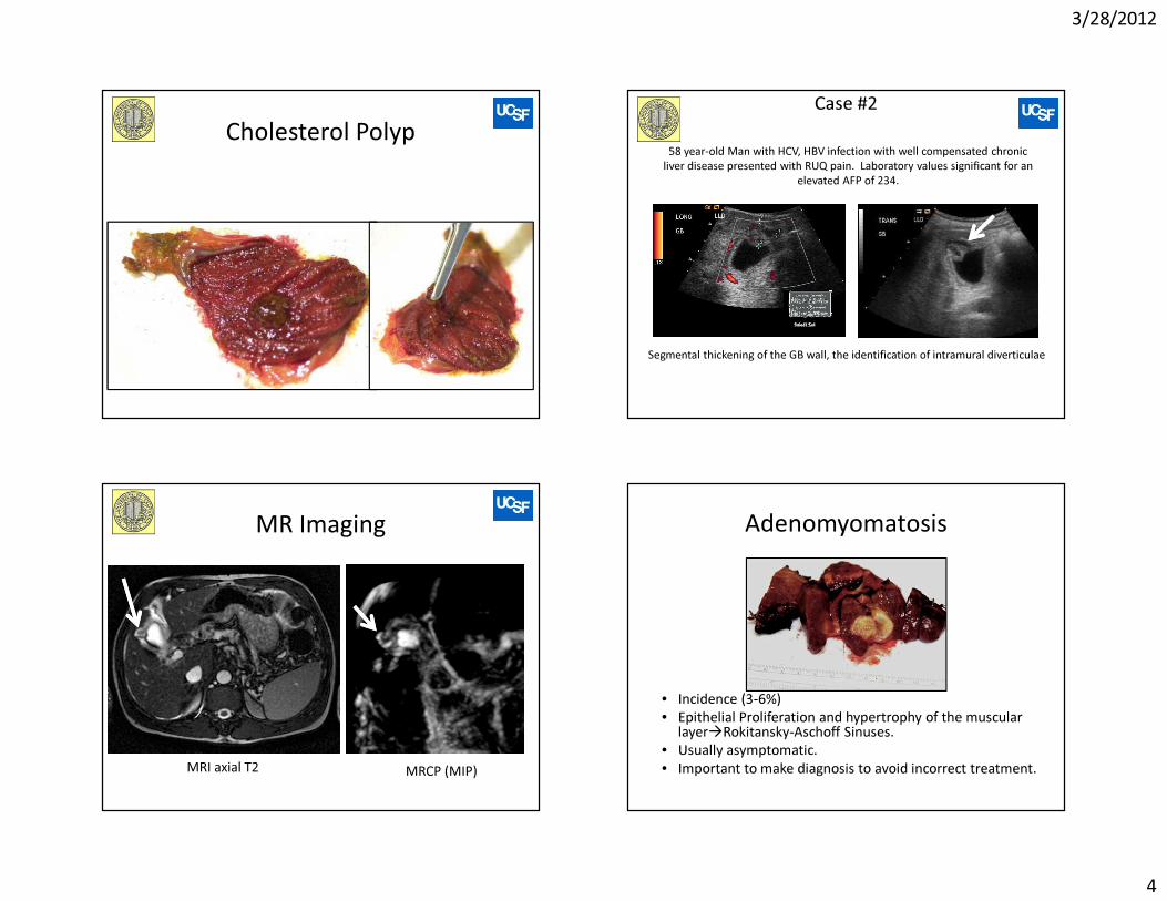
60 year-old morbidly obese man with history HCV,HIV and PSC well compensated chronic liver disease. He was found on
surveillance CT-imaging to have a 2.0 cm gallbladder mass.
Case #4 Case #572 year-old previously healthy presents with RUQ pain.
US Longitudial Axial CT
Case #4
Axial CTMRI axial T1
Misdiagnosed as symptomatic cholelithaisis. 13 months later develops recurrent RUQ pain, fever and chills. Dx with subacute
cholecystitis.
Case #4Laparoscopic Exploration

3/28/2012
7
Case #4Conversion to Open Laparotomy: Segment 4/5 Resection
Locally Advanced Gallbladder CA
• Removal of gallbladder• Removal of areas of liver
invasion• Portal lymph node
dissection• Remove and reconstruct
areas of portal vein invasion
• Biliary reconstruction
Case #4Postoperative Scan--2 years
Symptomatic Asymptomatic
Lap Cholecystectomy
**Risk factorsPresent
**Risk factors: Age>50, Presence of Gallstones, DM, PSC, High Risk Population, or APBDJ
PLG>10 mm PLG<10 mm
CLOSE OBSERVATION3-6 Months US exam
OPENSize >2 CM,
consider Radical Resxn
Treatment algorithm of polypoid lesions of the gallbladder (PLG)
The required duration of follow-up remains unknown.
3/28/2012
8
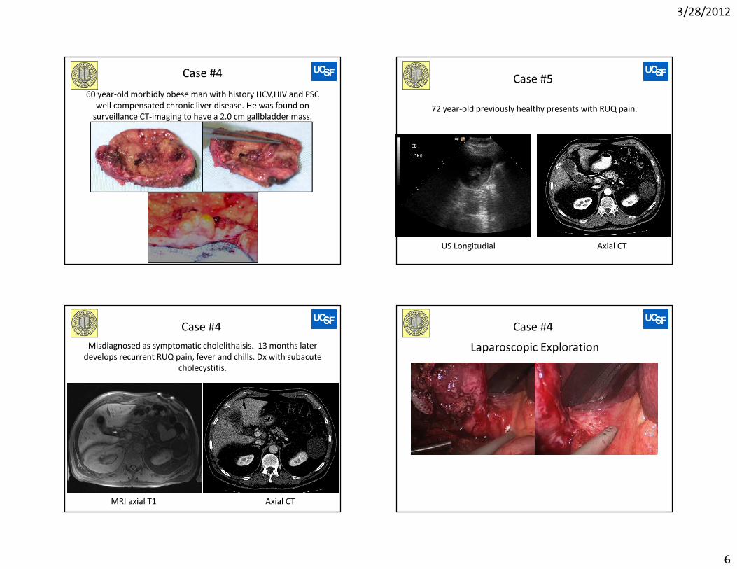
Conclusions• Polypoid lesions of the gallbladder remain a problem of concern to
both patients and surgeons.• Polyps larger > 1 cm and patient age >50 years are the two most
important predictors for malignancy.– Associated gallstones, Solitary PLG, High risk population, DM, PSC,
• Laparoscopic cholecystectomy remains the operation of choice for PLG.
• Frozen section is advised during the operation to guide extended surgery if malignancy is confirmed.
• However, if the chance of malignancy is assessed to be high before operation, for example, PLG larger than 2 cm, open surgery is recommended to reduce the risk of tumor seeding associated with laparoscopic surgery.
• For asymptomatic PLG smaller than 1 cm, it is generally agreed that follow-up USG every 3 to 6 months is necessary to exclude rapidly growing malignant tumor.