Small Cell Carcinoma in the Mammary Gland: Primary or Metastatic
CASE REPORT Open Access
Gallbladder small cell carcinoma: a casereport and literature reviewToshiyuki Adachi1, Masashi Haraguchi1*, Junji Irie2, Tomoko Yoshimoto1, Ryohei Uehara3, Shinichiro Ito1,Hirotaka Tokai1, Kazumasa Noda1, Nobuhiro Tada1, Masataka Hirabaru1, Keiji Inoue1, Shigeki Minami1
and Susumu Eguchi4
Abstract
Gallbladder small cell carcinoma (SCC) comprises only 0.5 % of all gallbladder cancer and consists of aggressivetumors with poor survival outcomes against current treatments. These tumors are most common in elderly females,particularly those with cholecystolithiasis. We report the case of a 79-year-old woman with gallbladder small cellcarcinoma. The patient had intermittent right upper quadrant abdominal pain and was admitted to our hospitaldue to suspected acute cholecystitis. She regularly received medical treatment for diabetes, hypertension, anddyslipidemia. On initial laboratory evaluation, the levels of aspartate aminotransferase (AST), total bilirubin, andC-reactive protein (CRP) were markedly elevated. She underwent computed tomography (CT) for screening. CTimages showed a thick-walled gallbladder containing multiple stones and multiple 3-cm-sized round nodularlesions, which were suggestive of metastatic lymph nodes. After percutaneous transhepatic gallbladder drainagewas performed, endoscopic ultrasound-guided fine needle aspiration of enlarged lymph nodes resulted in adiagnosis of small cell carcinoma or adenocarcinoma. However, we could not identify the primary lesion beforethe surgery because of no decisive factors. We performed cholecystectomy because there was a possibility ofcholecystitis recurrence risk and also partial liver resection because we suspected tumor invasion. The finalpathological diagnosis was neuroendocrine carcinoma of the gallbladder, small cell type. The tumor stage was IVb,T3aN1M1. The patient died 13 weeks after the surgery. In the present paper, we review the current availableEnglish-language literature of gallbladder SCC.
Keywords: Small cell carcinoma, Gallbladder, Neuroendocrine tumor
BackgroundPrimary gallbladder neuroendocrine tumors are rare, repre-senting 0.2 % of all tumors [1]. Neuroendocrine neoplasmsof the gallbladder are classified as grades 1 and 2 neuroen-docrine tumor (NET), neuroendocrine carcinoma (NEC)(large cell or small cell type), and mixed adenoneuroendo-crine carcinoma (MANEC) [2]. In particular, gallbladdersmall cell carcinoma (SCC) is extremely rare. GallbladderSCC carries a grave prognosis with the survival rates worsethan gallbladder adenocarcinoma due to its high malignantpotential and late stage at presentation [2]. The overallmedian survival is 4–6 months despite aggressive mana-gement. According to the Surveillance, Epidemiology and
End Results (SEER) data, the 1-year survival of gallbladderSCC was 21 % and 5-year was 0 % [3]. In recent years,gallbladder SCC has attracted increasing attention forimproved understanding and diagnosis. Herein, we describethe latest review of the English-language literature, includ-ing our present gallbladder SCC case [2–17].
Case presentationA 79-year-old woman visited a local internal medicineclinic because of intermittent right upper quadrantabdominal pain. The patient was admitted to our hospitaldue to suspected acute cholecystitis and had Murphy’ssign on admission. The patient was being regularly treatedfor diabetes, hypertension, and dyslipidemia, but she didnot smoke or drink alcohol. Additionally, the patienthad a history of breast cancer diagnosed 22 years previ-ously that had been treated with mastectomy. Routine
* Correspondence: [email protected] of Surgery, Nagasaki Harbor Medical Center City Hospital,Nagasaki 850-8555, JapanFull list of author information is available at the end of the article
© 2016 The Author(s). Open Access This article is distributed under the terms of the Creative Commons Attribution 4.0International License (http://creativecommons.org/licenses/by/4.0/), which permits unrestricted use, distribution, andreproduction in any medium, provided you give appropriate credit to the original author(s) and the source, provide a link tothe Creative Commons license, and indicate if changes were made.
Adachi et al. Surgical Case Reports (2016) 2:71 DOI 10.1186/s40792-016-0200-3
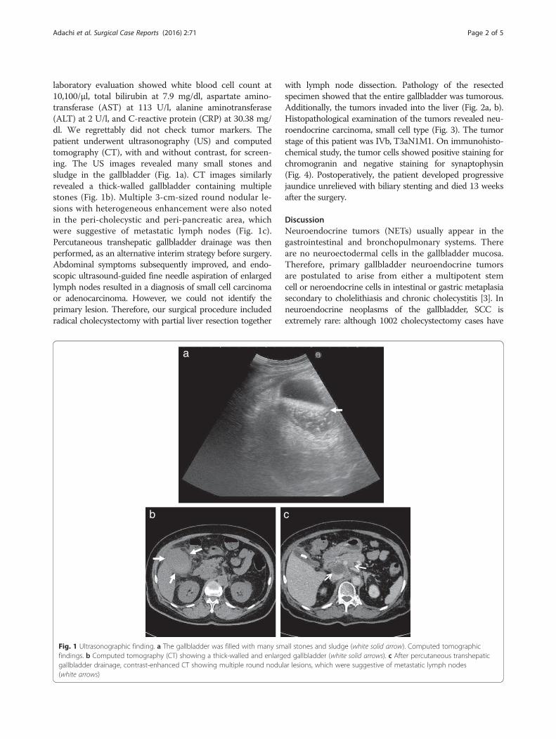
laboratory evaluation showed white blood cell count at10,100/μl, total bilirubin at 7.9 mg/dl, aspartate amino-transferase (AST) at 113 U/l, alanine aminotransferase(ALT) at 2 U/l, and C-reactive protein (CRP) at 30.38 mg/dl. We regrettably did not check tumor markers. Thepatient underwent ultrasonography (US) and computedtomography (CT), with and without contrast, for screen-ing. The US images revealed many small stones andsludge in the gallbladder (Fig. 1a). CT images similarlyrevealed a thick-walled gallbladder containing multiplestones (Fig. 1b). Multiple 3-cm-sized round nodular le-sions with heterogeneous enhancement were also notedin the peri-cholecystic and peri-pancreatic area, whichwere suggestive of metastatic lymph nodes (Fig. 1c).Percutaneous transhepatic gallbladder drainage was thenperformed, as an alternative interim strategy before surgery.Abdominal symptoms subsequently improved, and endo-scopic ultrasound-guided fine needle aspiration of enlargedlymph nodes resulted in a diagnosis of small cell carcinomaor adenocarcinoma. However, we could not identify theprimary lesion. Therefore, our surgical procedure includedradical cholecystectomy with partial liver resection together
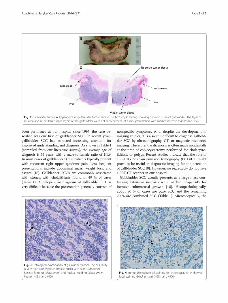
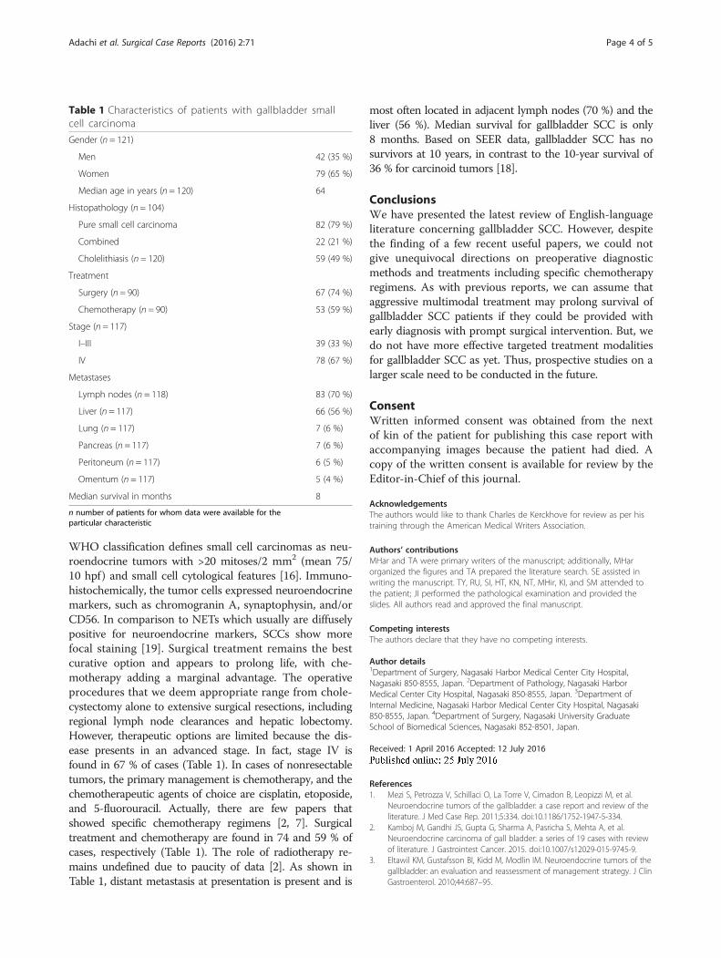
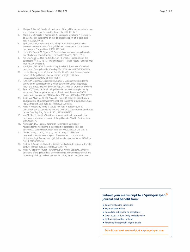
with lymph node dissection. Pathology of the resectedspecimen showed that the entire gallbladder was tumorous.Additionally, the tumors invaded into the liver (Fig. 2a, b).Histopathological examination of the tumors revealed neu-roendocrine carcinoma, small cell type (Fig. 3). The tumorstage of this patient was IVb, T3aN1M1. On immunohisto-chemical study, the tumor cells showed positive staining forchromogranin and negative staining for synaptophysin(Fig. 4). Postoperatively, the patient developed progressivejaundice unrelieved with biliary stenting and died 13 weeksafter the surgery.
DiscussionNeuroendocrine tumors (NETs) usually appear in thegastrointestinal and bronchopulmonary systems. Thereare no neuroectodermal cells in the gallbladder mucosa.Therefore, primary gallbladder neuroendocrine tumorsare postulated to arise from either a multipotent stemcell or neroendocrine cells in intestinal or gastric metaplasiasecondary to cholelithiasis and chronic cholecystitis [3]. Inneuroendocrine neoplasms of the gallbladder, SCC isextremely rare: although 1002 cholecystectomy cases have
Fig. 1 Ultrasonographic finding. a The gallbladder was filled with many small stones and sludge (white solid arrow). Computed tomographicfindings. b Computed tomography (CT) showing a thick-walled and enlarged gallbladder (white solid arrows). c After percutaneous transhepaticgallbladder drainage, contrast-enhanced CT showing multiple round nodular lesions, which were suggestive of metastatic lymph nodes(white arrows)
Adachi et al. Surgical Case Reports (2016) 2:71 Page 2 of 5
been performed at our hospital since 1997, the case de-scribed was our first of gallbladder SCC. In recent years,gallbladder SCC has attracted increasing attention forimproved understanding and diagnosis. As shown in Table 1(compiled from our literature survey), the average age ofdiagnosis is 64 years, with a male-to-female ratio of 1:1.9.In most cases of gallbladder SCCs, patients typically presentwith recurrent right upper quadrant pain. Less frequentpresentations include abdominal mass, weight loss, andascites [16]. Gallbladder SCCs are commonly associatedwith stones, with cholelithiasis found in 49 % of cases(Table 1). A preoperative diagnosis of gallbladder SCC isvery difficult because the presentation generally consists of
nonspecific symptoms. And, despite the development ofimaging studies, it is also still difficult to diagnose gallblad-der SCC by ultrasonography, CT, or magnetic resonanceimaging. Therefore, the diagnosis is often made incidentallyat the time of cholecystectomy performed for cholecysto-lithiasis or polyps. Recent studies indicate that the role of18F-FDG positron emission tomography (PET)/CT mightprove to be useful in diagnostic imaging for the detectionof gallbladder SCC [8]. However, we regrettably do not havea PET-CT scanner in our hospital.Gallbladder SCC usually presents as a large mass con-
taining extensive necrosis with marked propensity forinvasive submucosal growth [18]. Histopathologically,about 80 % of cases are pure SCC and the remaining20 % are combined SCC (Table 1). Microscopically, the
Fig. 2 Gallbladder tumor. a Appearance of gallbladder tumor section. b Microscopic finding showing necrotic tissue of gallbladder. The layer ofmucosa and muscularis propria layers of the gallbladder were not seen because of tumor proliferation with marked necrosis (panoramic view)
Fig. 3 Histological examination of gallbladder tumor. The cellularityis very high with hyperchromatic nuclei with scant cytoplasm.Rosette forming (black arrow) and nuclear molding (black arrowheads) (H&E stain, ×400)
Fig. 4 Immunohistochemical staining for chromogaranin A showedfocal staining (black arrows) (H&E stain, ×400)
Adachi et al. Surgical Case Reports (2016) 2:71 Page 3 of 5
WHO classification defines small cell carcinomas as neu-roendocrine tumors with >20 mitoses/2 mm2 (mean 75/10 hpf) and small cell cytological features [16]. Immuno-histochemically, the tumor cells expressed neuroendocrinemarkers, such as chromogranin A, synaptophysin, and/orCD56. In comparison to NETs which usually are diffuselypositive for neuroendocrine markers, SCCs show morefocal staining [19]. Surgical treatment remains the bestcurative option and appears to prolong life, with che-motherapy adding a marginal advantage. The operativeprocedures that we deem appropriate range from chole-cystectomy alone to extensive surgical resections, includingregional lymph node clearances and hepatic lobectomy.However, therapeutic options are limited because the dis-ease presents in an advanced stage. In fact, stage IV isfound in 67 % of cases (Table 1). In cases of nonresectabletumors, the primary management is chemotherapy, and thechemotherapeutic agents of choice are cisplatin, etoposide,and 5-fluorouracil. Actually, there are few papers thatshowed specific chemotherapy regimens [2, 7]. Surgicaltreatment and chemotherapy are found in 74 and 59 % ofcases, respectively (Table 1). The role of radiotherapy re-mains undefined due to paucity of data [2]. As shown inTable 1, distant metastasis at presentation is present and is
most often located in adjacent lymph nodes (70 %) and theliver (56 %). Median survival for gallbladder SCC is only8 months. Based on SEER data, gallbladder SCC has nosurvivors at 10 years, in contrast to the 10-year survival of36 % for carcinoid tumors [18].
ConclusionsWe have presented the latest review of English-languageliterature concerning gallbladder SCC. However, despitethe finding of a few recent useful papers, we could notgive unequivocal directions on preoperative diagnosticmethods and treatments including specific chemotherapyregimens. As with previous reports, we can assume thataggressive multimodal treatment may prolong survival ofgallbladder SCC patients if they could be provided withearly diagnosis with prompt surgical intervention. But, wedo not have more effective targeted treatment modalitiesfor gallbladder SCC as yet. Thus, prospective studies on alarger scale need to be conducted in the future.
ConsentWritten informed consent was obtained from the nextof kin of the patient for publishing this case report withaccompanying images because the patient had died. Acopy of the written consent is available for review by theEditor-in-Chief of this journal.
AcknowledgementsThe authors would like to thank Charles de Kerckhove for review as per histraining through the American Medical Writers Association.
Authors’ contributionsMHar and TA were primary writers of the manuscript; additionally, MHarorganized the figures and TA prepared the literature search. SE assisted inwriting the manuscript. TY, RU, SI, HT, KN, NT, MHir, KI, and SM attended tothe patient; JI performed the pathological examination and provided theslides. All authors read and approved the final manuscript.
Competing interestsThe authors declare that they have no competing interests.
Author details1Department of Surgery, Nagasaki Harbor Medical Center City Hospital,Nagasaki 850-8555, Japan. 2Department of Pathology, Nagasaki HarborMedical Center City Hospital, Nagasaki 850-8555, Japan. 3Department ofInternal Medicine, Nagasaki Harbor Medical Center City Hospital, Nagasaki850-8555, Japan. 4Department of Surgery, Nagasaki University GraduateSchool of Biomedical Sciences, Nagasaki 852-8501, Japan.
Received: 1 April 2016 Accepted: 12 July 2016
References1. Mezi S, Petrozza V, Schillaci O, La Torre V, Cimadon B, Leopizzi M, et al.
Neuroendocrine tumors of the gallbladder: a case report and review of theliterature. J Med Case Rep. 2011;5:334. doi:10.1186/1752-1947-5-334.
2. Kamboj M, Gandhi JS, Gupta G, Sharma A, Pasricha S, Mehta A, et al.Neuroendocrine carcinoma of gall bladder: a series of 19 cases with reviewof literature. J Gastrointest Cancer. 2015. doi:10.1007/s12029-015-9745-9.
3. Eltawil KM, Gustafsson BI, Kidd M, Modlin IM. Neuroendocrine tumors of thegallbladder: an evaluation and reassessment of management strategy. J ClinGastroenterol. 2010;44:687–95.
Table 1 Characteristics of patients with gallbladder smallcell carcinoma
Gender (n = 121)
Men 42 (35 %)
Women 79 (65 %)
Median age in years (n = 120) 64
Histopathology (n = 104)
Pure small cell carcinoma 82 (79 %)
Combined 22 (21 %)
Cholelithiasis (n = 120) 59 (49 %)
Treatment
Surgery (n = 90) 67 (74 %)
Chemotherapy (n = 90) 53 (59 %)
Stage (n = 117)
I–III 39 (33 %)
IV 78 (67 %)
Metastases
Lymph nodes (n = 118) 83 (70 %)
Liver (n = 117) 66 (56 %)
Lung (n = 117) 7 (6 %)
Pancreas (n = 117) 7 (6 %)
Peritoneum (n = 117) 6 (5 %)
Omentum (n = 117) 5 (4 %)
Median survival in months 8
n number of patients for whom data were available for theparticular characteristic
Adachi et al. Surgical Case Reports (2016) 2:71 Page 4 of 5
4. Mahipal A, Gupta S. Small-cell carcinoma of the gallbladder: report of a caseand literature review. Gastrointest Cancer Res. 2010;4:135–6.
5. Matsuo S, Shinozaki T, Yamaguchi S, Matsuzaki S, Takami Y, Hayashi T,et al. Small-cell carcinoma of the gallbladder: report of a case. SurgToday. 2000;30:89–93.
6. Iype S, Mirza TA, Propper DJ, Bhattacharya S, Feakins RM, Kocher HM.Neuroendocrine tumours of the gallbladder: three cases and a review ofthe literature. Postgrad Med J. 2009;85:213–8.
7. Usmani S, Pazooki M, Bilgrami SF. Small cell carcinoma of the gall bladder:role of adjuvant chemotherapy. J Gastrointest Cancer. 2010;41:84–7.
8. Kim DM, Yang SO, Han HY, Kim KS, Son HJ. Small cell carcinoma of thegallbladder: 18F-FDG PET/CT imaging features—a case report. Nucl Med MolImaging. 2010;44:213–6.
9. Nau P, Liu J, Dillhoff M, Forster M, Hazey J, Melvin S. Two cases of small cellcarcinoma of the gallbladder. Case Rep Med. 2010. doi:10.1155/2010/453624.
10. Lee JM, Hwang S, Lee SG, Lee YJ, Park KM, Kim KH, et al. Neuroendocrinetumors of the gallbladder: twelve cases in a single institution.Hepatogastroenterology. 2010;57:1064–8.
11. Furrukh M, Qureshi A, Saparamadu A, Kumar S. Malignant neuroendocrinetumour of the gallbladder with elevated carcinoembryonic antigen: casereport and literature review. BMJ Case Rep. 2013. doi:10.1136/bcr-2013-008778.
12. Tamura T, Takeuchi K. Small cell gall bladder carcinoma complicated bysyndrome of inappropriate secretion of antidiuretic hormone (SIADH)treated with mozavaptan. BMJ Case Rep. 2013. doi:10.1136/bcr-2013-010039.
13. Tunio MA, Alasiri M, Ali AM, Alsaeed EF, Shuja M, Fatani H. Distal humerusas delayed site of metastasis from small cell carcinoma of gallbladder. CaseRep Gastrointest Med. 2013. doi:10.1155/2013/946835.
14. Aiello P, Aragona F, Territo V, Caruso AM, Patti R, Buscemi S, et al.Concomitant small cell neuroendocrine carcinoma of gallbladder and breastcancer. Case Rep Surg. 2014. doi:10.1155/2014/945921.
15. Yun SP, Shin N, Seo HI. Clinical outcomes of small cell neuroendocrinecarcinoma and adenocarcinoma of the gallbladder. World J Gastroenterol.2015;21:269–75.
16. Nemenqani DM, Fuloria J, Karam RA, Hammadi H. Gallbladderneuroendocrine neoplasms: a case report of gallbladder small cellcarcinoma. J Gastrointest Cancer. 2015. doi:10.1007/s12029-015-9757-5.
17. Chen C, Wang L, Liu X, Zhang G, Zhao Y, Geng Z. Gallbladderneuroendocrine carcinoma: report of 10 cases and comparison ofclinicopathologic features with gallbladder adenocarcinoma. Int J Clin ExpPathol. 2015;8:8218–26.
18. Kanthan R, Senger JL, Ahmed S, Kanthan SC. Gallbladder cancer in the 21stcentury. J Oncol. 2015. doi:10.1155/2015/967472.
19. Maitra A, Tascilar M, Hruban RH, Offerhaus GJ, Albores-Saavedra J. Small cellcarcinoma of the gallbladder: a clinicopathologic, immunohistochemical, andmolecular pathology study of 12 cases. Am J Surg Pathol. 2001;25:595–601.
Submit your manuscript to a journal and benefi t from:
7 Convenient online submission
7 Rigorous peer review
7 Immediate publication on acceptance
7 Open access: articles freely available online
7 High visibility within the fi eld
7 Retaining the copyright to your article
Submit your next manuscript at 7 springeropen.com
Adachi et al. Surgical Case Reports (2016) 2:71 Page 5 of 5