
Gallbladder Disease · Gallbladder Disease Joel Heidelbaugh, MD, FAAFP, FACG ... for uncomplicated...
28
1 Gallbladder Disease Joel Heidelbaugh, MD, FAAFP, FACG ACTIVITY DISCLAIMER The material presented here is being made available by the American Academy of Family Physicians for educational purposes only. Please note that medical information is constantly changing; the information contained in this activity was accurate at the time of publication. This material is not intended to represent the only, nor necessarily best, methods or procedures appropriate for the medical situations discussed. Rather, it is intended to present an approach, view, statement, or opinion of the faculty, which may be helpful to others who face similar situations. The AAFP disclaims any and all liability for injury or other damages resulting to any individual using this material and for all claims that might arise out of the use of the techniques demonstrated therein by such individuals, whether these claims shall be asserted by a physician or any other person. Physicians may care to check specific details such as drug doses and contraindications, etc., in standard sources prior to clinical application. This material might contain recommendations/guidelines developed by other organizations. Please note that although these guidelines might be included, this does not necessarily imply the endorsement by the AAFP.
Transcript of Gallbladder Disease · Gallbladder Disease Joel Heidelbaugh, MD, FAAFP, FACG ... for uncomplicated...
1
Gallbladder Disease
Joel Heidelbaugh, MD, FAAFP, FACG
ACTIVITY DISCLAIMERThe material presented here is being made available by the American Academy of Family Physicians for educational purposes only. Please note that medical information is constantly changing; the information contained in this activity was accurate at the time of publication. This material is not intended to represent the only, nor necessarily best, methods or procedures appropriate for the medical situations discussed. Rather, it is intended to present an approach, view, statement, or opinion of the faculty, which may be helpful to others who face similar situations.
The AAFP disclaims any and all liability for injury or other damages resulting to any individual using this material and for all claims that might arise out of the use of the techniques demonstrated therein by such individuals, whether these claims shall be asserted by a physician or any other person. Physicians may care to check specific details such as drug doses and contraindications, etc., in standard sources prior to clinical application. This material might contain recommendations/guidelines developed by other organizations. Please note that although these guidelines might be included, this does not necessarily imply the endorsement by the AAFP.
2
DISCLOSUREIt is the policy of the AAFP that all individuals in a position to control content disclose any relationships with commercial interests upon nomination/invitation of participation. Disclosure documents are reviewed for potential conflict of interest (COI), and if identified, conflicts are resolved prior to confirmation of participation. Only those participants who had no conflict of interest or who agreed to an identified resolution process prior to their participation were involved in this CME activity.
All individuals in a position to control content for this session have indicated they have no relevant financial relationships to disclose.
The content of my material/presentation in this CME activity will not include discussion of unapproved or investigational uses of products or devices.
Joel Heidelbaugh, MD, FAAFP, FACGClinical Professor, Departments of Family Medicine and Urology/Director of Medical Student Education and Clerkship Director, Department of Family Medicine/Director, Patients and Populations Branch, University of Michigan Medical School, Ann Arbor
Dr. Heidelbaugh is a family physician who has 20 years of academic teaching experience. His specialty topics include gastrointestinal disorders, men's health, and primary care urology. He is a member of the American Gastroenterological Association guideline panels for irritable bowel syndrome, inflammatory bowel disease, and Lynch syndrome. He is the co editor and co author of the textbook ROME IV: Functional Gastrointestinal Disorders for Primary Care and Non GI Clinicians, published through the Rome Foundation. In addition, he is the consulting editor of Primary Care: Clinics in Office Practice and the president elect of the American Society for Men's Health. Dr. Heidelbaugh believes that increasing awareness and education about gastrointestinal and men's health issues is an important trend in medical education, clinical practice, and research.
3
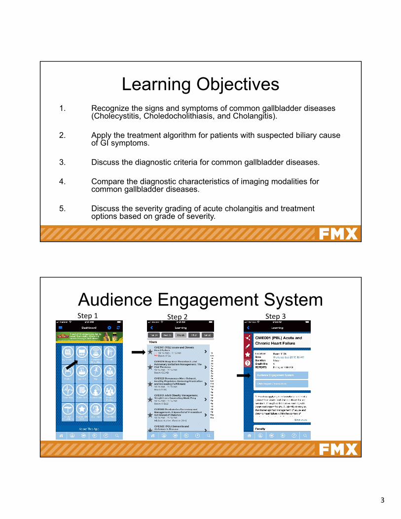
Learning Objectives1. Recognize the signs and symptoms of common gallbladder diseases
(Cholecystitis, Choledocholithiasis, and Cholangitis).
2. Apply the treatment algorithm for patients with suspected biliary cause of GI symptoms.
3. Discuss the diagnostic criteria for common gallbladder diseases.
4. Compare the diagnostic characteristics of imaging modalities for common gallbladder diseases.
5. Discuss the severity grading of acute cholangitis and treatment options based on grade of severity.
Audience Engagement SystemStep 1 Step 2 Step 3
4
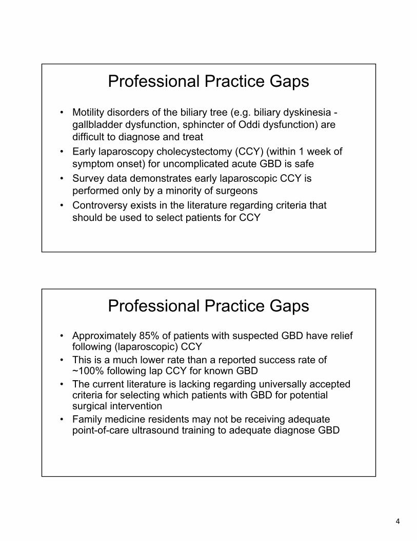
Professional Practice Gaps
• Motility disorders of the biliary tree (e.g. biliary dyskinesia -gallbladder dysfunction, sphincter of Oddi dysfunction) are difficult to diagnose and treat
• Early laparoscopy cholecystectomy (CCY) (within 1 week of symptom onset) for uncomplicated acute GBD is safe
• Survey data demonstrates early laparoscopic CCY is performed only by a minority of surgeons
• Controversy exists in the literature regarding criteria that should be used to select patients for CCY
Professional Practice Gaps
• Approximately 85% of patients with suspected GBD have relief following (laparoscopic) CCY
• This is a much lower rate than a reported success rate of ~100% following lap CCY for known GBD
• The current literature is lacking regarding universally accepted criteria for selecting which patients with GBD for potential surgical intervention
• Family medicine residents may not be receiving adequate point-of-care ultrasound training to adequate diagnose GBD
5
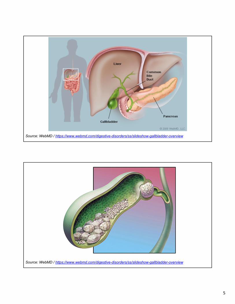
Source: WebMD / https://www.webmd.com/digestive-disorders/ss/slideshow-gallbladder-overview
Source: WebMD / https://www.webmd.com/digestive-disorders/ss/slideshow-gallbladder-overview
6
Epidemiology• 10-15% of adult population• 20-25 million Americans annually• $6.2 billion annually• Healthcare burden increased > 20% in last 3 decades• 1.8 million ambulatory care visits yearly• Leading cause of hospital admissions due to GI disease• Data likely underestimated as lap CCY is often a same-day
procedure and not captured in hospital admission data• Mortality rate in patients with GBD is < 0.6%• Data is over a decade old…
Stinton LM, et al. Gut Liver 2012;6(2):172-187.
Epidemiology• 80% of patients with gallstones will never experience biliary
pain or complications
• Acute cholecystitis, cholangitis, pancreatitis
• Often discovered incidentally on US or CT
• Only 10-20% of asymptomatic patients will become symptomatic with 5-20 of diagnosis
• 2% per year will develop symptomatic gallstones
• Fatality rates have decreased at greatest rate of any GI disease, from > 5,000 deaths in 1950 to 1,092 in 2004
Stinton LM, et al. Gut Liver 2012;6(2):172-187.
7
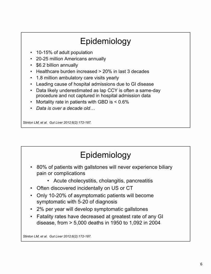
Poll Question 1
Which of the following represents the most common type of gallstone?
1. Pigment stones
2. Protein stones
3. Calcium carbonate stones
4. Cholesterol stones
5. Mixed stones
Systematic Classification of Gallstones
• 8 types, over 10 subtypes - (807)• Cholesterol stones (297 / 37%)• Pigment stones (217 / 27%)• Calcium carbonate stones (139 / 17%)• Mixed stones - 2 or more subtypes (129 / 16%)• Phosphate stones (12)• Calcium stearate stones (9)• Protein stones (3)• Cystine stones (1)
Qiao T, et al. PLoS One 2013;8(10):e74887.
8
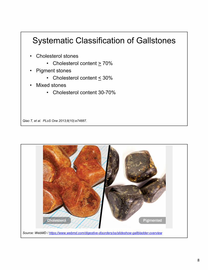
Systematic Classification of Gallstones
• Cholesterol stones
• Cholesterol content > 70%
• Pigment stones
• Cholesterol content < 30%
• Mixed stones
• Cholesterol content 30-70%
Qiao T, et al. PLoS One 2013;8(10):e74887.
Source: WebMD / https://www.webmd.com/digestive-disorders/ss/slideshow-gallbladder-overview
9
10
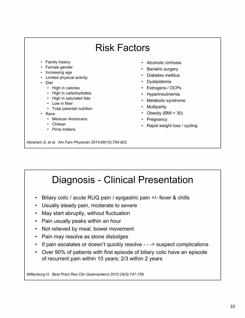
Risk Factors• Family history• Female gender• Increasing age• Limited physical activity• Diet
• High in calories• High in carbohydrates• High in saturated fats• Low in fiber• Total parental nutrition
• Race• Mexican Americans• Chilean• Pima Indians
Abraham S, et al. Am Fam Physician 2014;89(10);795-802.
• Alcoholic cirrhosis
• Bariatric surgery
• Diabetes mellitus
• Dyslipidemia
• Estrogens / OCPs
• Hyperinsulinemia
• Metabolic syndrome
• Multiparity
• Obesity (BMI > 30)
• Pregnancy
• Rapid weight loss / cycling
Diagnosis - Clinical Presentation
• Biliary colic / acute RUQ pain / epigastric pain +/- fever & chills
• Usually steady pain, moderate to severe
• May start abruptly, without fluctuation
• Pain usually peaks within an hour
• Not relieved by meal, bowel movement
• Pain may resolve as stone dislodges
• If pain escalates or doesn’t quickly resolve - - -> suspect complications
• Over 90% of patients with first episode of biliary colic have an episode of recurrent pain within 10 years; 2/3 within 2 years
Wittenburg H. Best Pract Res Clin Gastroenterol 2010;24(5):747-756.
11
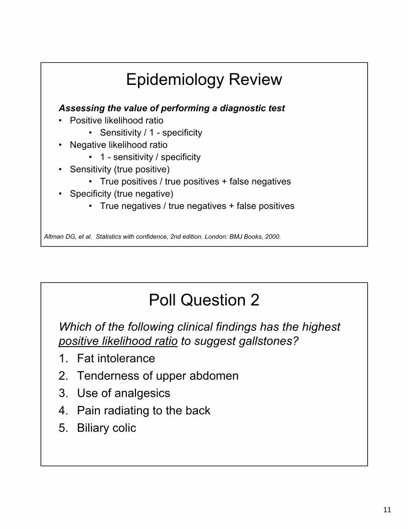
Epidemiology Review
Assessing the value of performing a diagnostic test• Positive likelihood ratio
• Sensitivity / 1 - specificity• Negative likelihood ratio
• 1 - sensitivity / specificity• Sensitivity (true positive)
• True positives / true positives + false negatives• Specificity (true negative)
• True negatives / true negatives + false positives
Altman DG, et al. Statistics with confidence, 2nd edition. London: BMJ Books, 2000.
Poll Question 2
Which of the following clinical findings has the highest positive likelihood ratio to suggest gallstones?
1. Fat intolerance
2. Tenderness of upper abdomen
3. Use of analgesics
4. Pain radiating to the back
5. Biliary colic
12
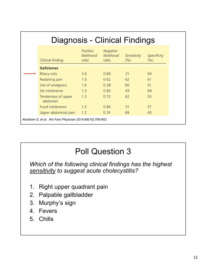
Diagnosis - Clinical Findings
Abraham S, et al. Am Fam Physician 2014;89(10);795-802.
Poll Question 3
Which of the following clinical findings has the highest sensitivity to suggest acute cholecystitis?
1. Right upper quadrant pain2. Palpable gallbladder3. Murphy’s sign4. Fevers5. Chills

13
Diagnosis - Clinical Findings
Abraham S, et al. Am Fam Physician 2014;89(10);795-802.
Complications of GallstonesAcute calculous cholecystitis
• 1/3 of all surgical emergency hospital admissions
• Accounts for 14-30% of CCY in many countries
• Second most common source of intra-abdominal infection
• Increase LFTs, WBC, +/- fevers, chills
Obstructive cholangitis due to choledocholithiasis
• Gallstones become stuck in common bile duct
• Bile backs up into the liver
• RUQ pain, jaundice
• Increase LFTs, WBC
• Can lead to gallstone pancreatitis
Gallstone pancreatitis
• Increase LFTs, amylase, lipase
• Best identified on CT; small stones best seen on US
Gomes CA, et al. World J Gastrointest Surg 2017;9(5):118-126.Baiu I, at al. JAMA 2018;320(14):1506.
14
Poll Question 4
Which of the following is the recommend initial modality to detect gallstones and acute calculous cholecystitis?
1. Plain film abdominal radiograph
2. Ultrasound
3. Abdominal CT
4. Abdominal MRI
5. Hepatobiliary iminodiacetic acid scan
Diagnostic Evaluation
Abdominal (plain film) radiography
• Will not demonstrate gallbladder or offending pathology
• Useful to exclude other causes of abdominal pain
• Perforation (free air under diaphragm)
• Constipation (significant fecal loading)
• Obstruction (dilated bowel)
• Stones (e.g. renal)
• Pancreatitis (e.g. chronic calcifications)
Abraham S, et al. Am Fam Physician 2014;89(10);795-802.
15
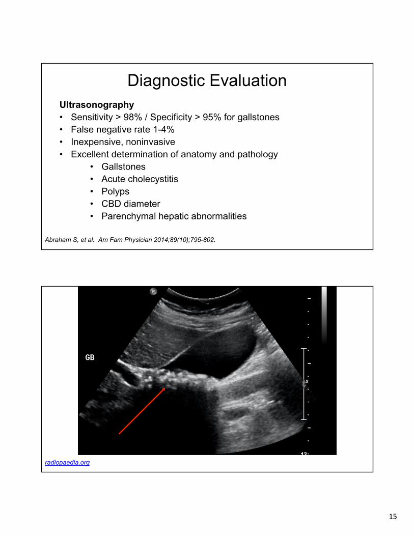
Diagnostic EvaluationUltrasonography• Sensitivity > 98% / Specificity > 95% for gallstones• False negative rate 1-4%• Inexpensive, noninvasive• Excellent determination of anatomy and pathology
• Gallstones• Acute cholecystitis• Polyps• CBD diameter• Parenchymal hepatic abnormalities
Abraham S, et al. Am Fam Physician 2014;89(10);795-802.
radiopaedia.org
16
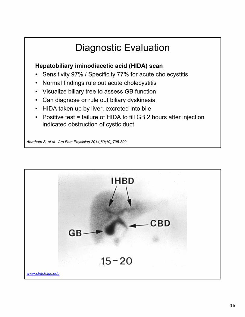
Diagnostic Evaluation
Hepatobiliary iminodiacetic acid (HIDA) scan
• Sensitivity 97% / Specificity 77% for acute cholecystitis
• Normal findings rule out acute cholecystitis
• Visualize biliary tree to assess GB function
• Can diagnose or rule out biliary dyskinesia
• HIDA taken up by liver, excreted into bile
• Positive test = failure of HIDA to fill GB 2 hours after injection indicated obstruction of cystic duct
Abraham S, et al. Am Fam Physician 2014;89(10);795-802.
www.stritch.luc.edu
17
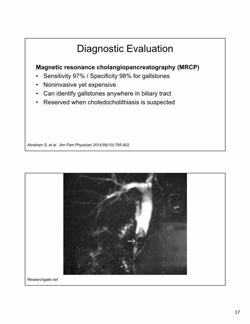
Diagnostic Evaluation
Magnetic resonance cholangiopancreatography (MRCP)
• Sensitivity 97% / Specificity 98% for gallstones
• Noninvasive yet expensive
• Can identify gallstones anywhere in biliary tract
• Reserved when choledocholithiasis is suspected
Abraham S, et al. Am Fam Physician 2014;89(10);795-802.
Researchgate.net
18
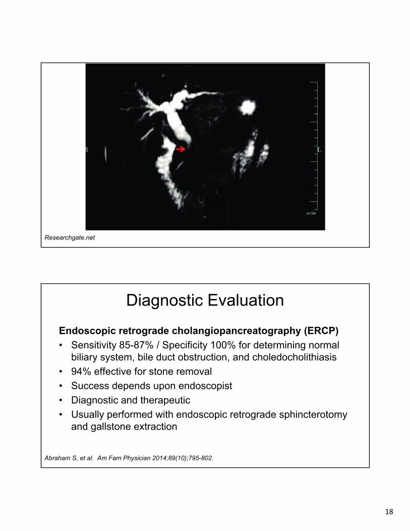
Researchgate.net
Diagnostic Evaluation
Endoscopic retrograde cholangiopancreatography (ERCP)
• Sensitivity 85-87% / Specificity 100% for determining normal biliary system, bile duct obstruction, and choledocholithiasis
• 94% effective for stone removal
• Success depends upon endoscopist
• Diagnostic and therapeutic
• Usually performed with endoscopic retrograde sphincterotomy and gallstone extraction
Abraham S, et al. Am Fam Physician 2014;89(10);795-802.
19
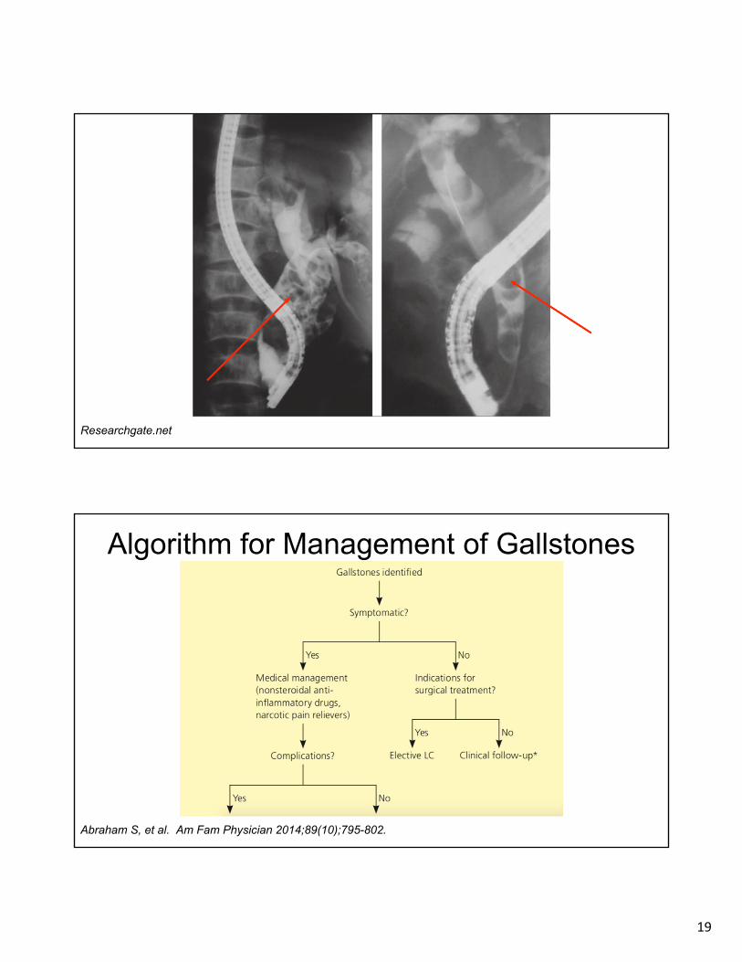
Researchgate.net
Algorithm for Management of Gallstones
Abraham S, et al. Am Fam Physician 2014;89(10);795-802.
20
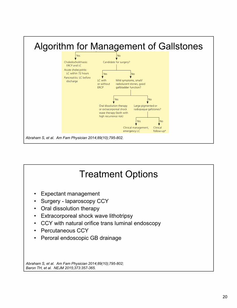
Algorithm for Management of Gallstones
Abraham S, et al. Am Fam Physician 2014;89(10);795-802.
Treatment Options
• Expectant management• Surgery - laparoscopy CCY• Oral dissolution therapy• Extracorporeal shock wave lithotripsy• CCY with natural orifice trans luminal endoscopy• Percutaneous CCY• Peroral endoscopic GB drainage
Abraham S, et al. Am Fam Physician 2014;89(10);795-802; Baron TH, et al. NEJM 2015;373:357-365.
21
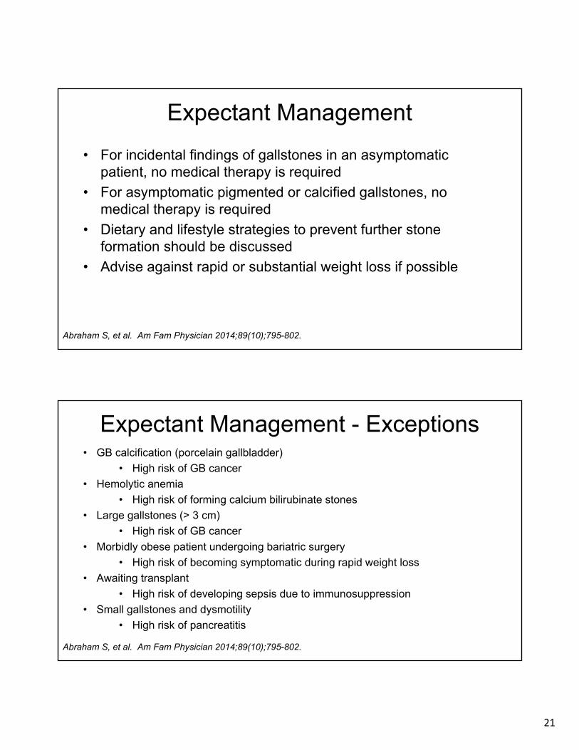
Expectant Management
• For incidental findings of gallstones in an asymptomatic patient, no medical therapy is required
• For asymptomatic pigmented or calcified gallstones, no medical therapy is required
• Dietary and lifestyle strategies to prevent further stone formation should be discussed
• Advise against rapid or substantial weight loss if possible
Abraham S, et al. Am Fam Physician 2014;89(10);795-802.
Expectant Management - Exceptions• GB calcification (porcelain gallbladder)
• High risk of GB cancer
• Hemolytic anemia
• High risk of forming calcium bilirubinate stones
• Large gallstones (> 3 cm)
• High risk of GB cancer
• Morbidly obese patient undergoing bariatric surgery
• High risk of becoming symptomatic during rapid weight loss
• Awaiting transplant
• High risk of developing sepsis due to immunosuppression
• Small gallstones and dysmotility
• High risk of pancreatitis
Abraham S, et al. Am Fam Physician 2014;89(10);795-802.
22
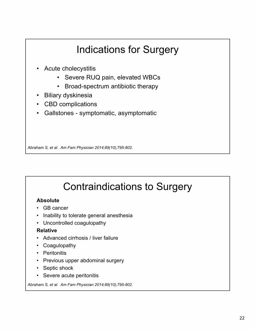
Indications for Surgery
• Acute cholecystitis
• Severe RUQ pain, elevated WBCs
• Broad-spectrum antibiotic therapy
• Biliary dyskinesia
• CBD complications
• Gallstones - symptomatic, asymptomatic
Abraham S, et al. Am Fam Physician 2014;89(10);795-802.
Contraindications to SurgeryAbsolute
• GB cancer
• Inability to tolerate general anesthesia
• Uncontrolled coagulopathy
Relative
• Advanced cirrhosis / liver failure
• Coagulopathy
• Peritonitis
• Previous upper abdominal surgery
• Septic shock
• Severe acute peritonitis
Abraham S, et al. Am Fam Physician 2014;89(10);795-802.
23
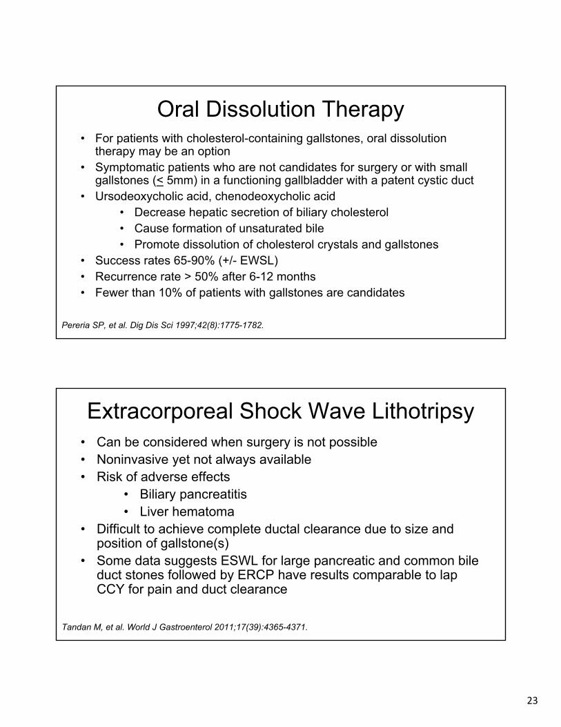
Oral Dissolution Therapy• For patients with cholesterol-containing gallstones, oral dissolution
therapy may be an option• Symptomatic patients who are not candidates for surgery or with small
gallstones (< 5mm) in a functioning gallbladder with a patent cystic duct • Ursodeoxycholic acid, chenodeoxycholic acid
• Decrease hepatic secretion of biliary cholesterol• Cause formation of unsaturated bile• Promote dissolution of cholesterol crystals and gallstones
• Success rates 65-90% (+/- EWSL)• Recurrence rate > 50% after 6-12 months• Fewer than 10% of patients with gallstones are candidates
Pereria SP, et al. Dig Dis Sci 1997;42(8):1775-1782.
Extracorporeal Shock Wave Lithotripsy• Can be considered when surgery is not possible• Noninvasive yet not always available• Risk of adverse effects
• Biliary pancreatitis• Liver hematoma
• Difficult to achieve complete ductal clearance due to size and position of gallstone(s)
• Some data suggests ESWL for large pancreatic and common bile duct stones followed by ERCP have results comparable to lap CCY for pain and duct clearance
Tandan M, et al. World J Gastroenterol 2011;17(39):4365-4371.
24
Severity Grading of Acute Cholangitis - Tokyo Guidelines 2018
https://www.mdcalc.com/tokyo-guidelines-acute-cholangitis-2018
Poll Question 5
Which of the following is a mechanism that contributes to gallstone formation during rapid weight loss?
1. Diet of 1600 kcal/day
2. Decreased bile salt production
3. Decreased bile stasis
4. Decreased cholesterol saturation
5. Enhanced gallbladder motility
25
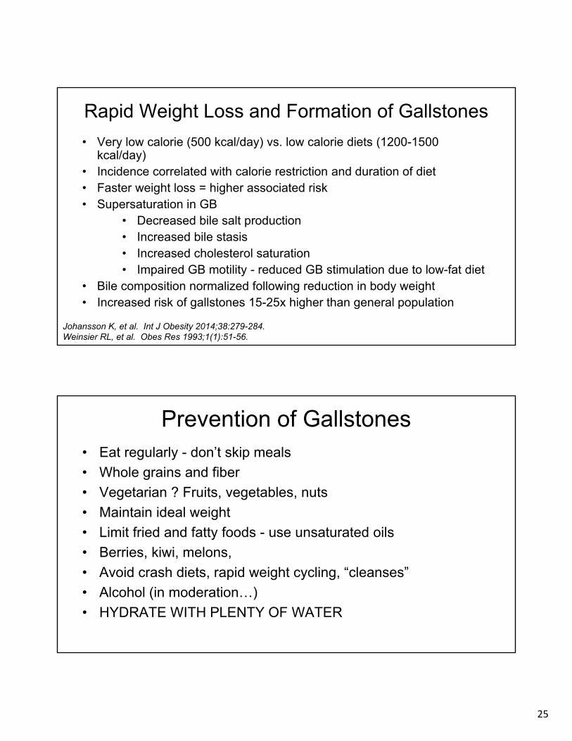
Rapid Weight Loss and Formation of Gallstones
• Very low calorie (500 kcal/day) vs. low calorie diets (1200-1500 kcal/day)
• Incidence correlated with calorie restriction and duration of diet• Faster weight loss = higher associated risk• Supersaturation in GB
• Decreased bile salt production• Increased bile stasis• Increased cholesterol saturation• Impaired GB motility - reduced GB stimulation due to low-fat diet
• Bile composition normalized following reduction in body weight• Increased risk of gallstones 15-25x higher than general population
Johansson K, et al. Int J Obesity 2014;38:279-284.Weinsier RL, et al. Obes Res 1993;1(1):51-56.
Prevention of Gallstones• Eat regularly - don’t skip meals
• Whole grains and fiber
• Vegetarian ? Fruits, vegetables, nuts
• Maintain ideal weight
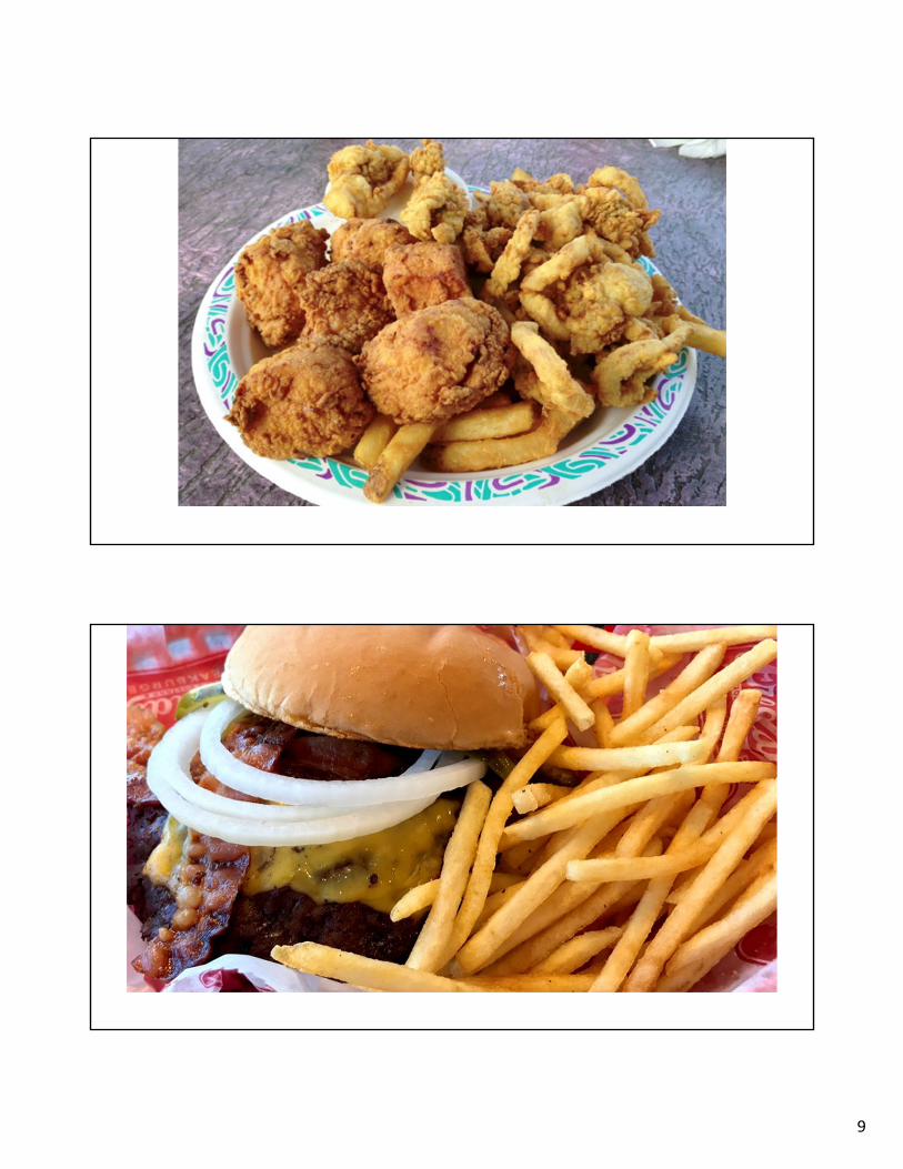
• Limit fried and fatty foods - use unsaturated oils
• Berries, kiwi, melons,
• Avoid crash diets, rapid weight cycling, “cleanses”
• Alcohol (in moderation…)
• HYDRATE WITH PLENTY OF WATER
26
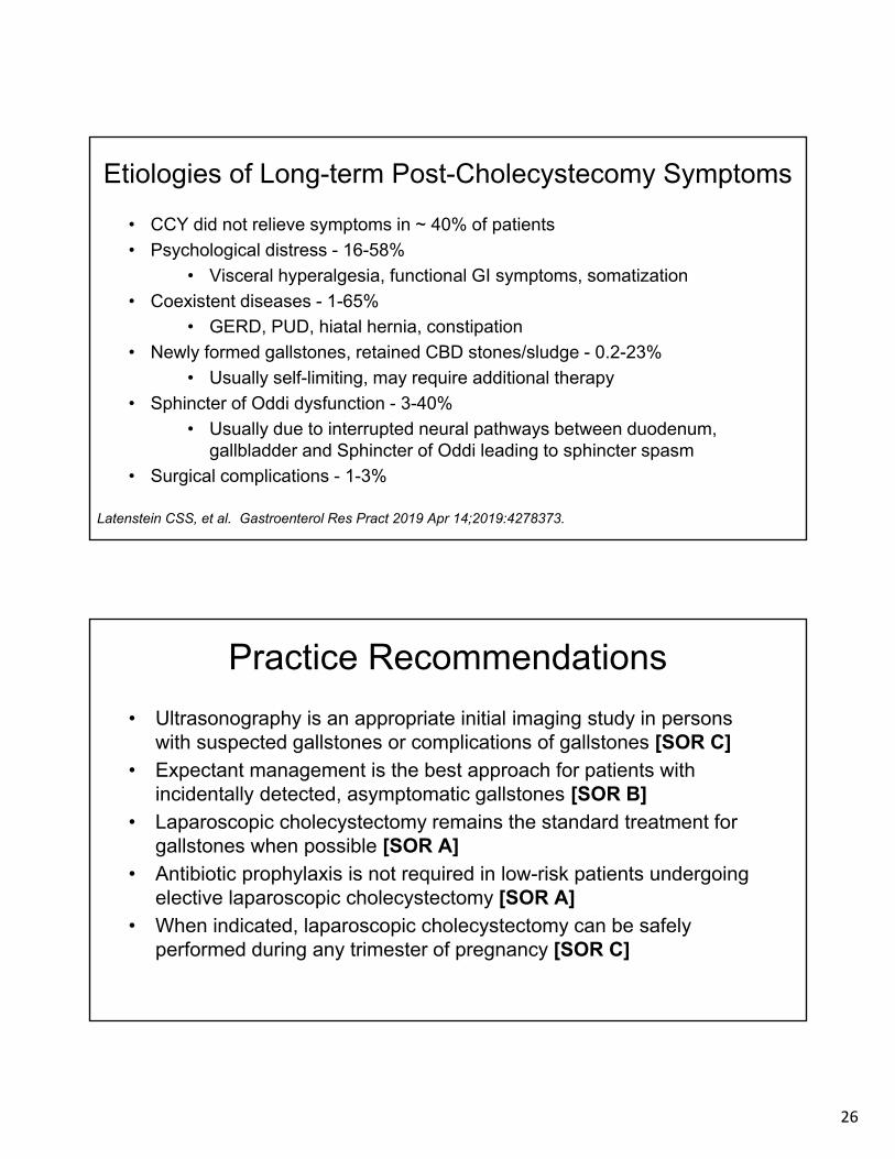
Etiologies of Long-term Post-Cholecystecomy Symptoms
• CCY did not relieve symptoms in ~ 40% of patients
• Psychological distress - 16-58%
• Visceral hyperalgesia, functional GI symptoms, somatization
• Coexistent diseases - 1-65%
• GERD, PUD, hiatal hernia, constipation
• Newly formed gallstones, retained CBD stones/sludge - 0.2-23%
• Usually self-limiting, may require additional therapy
• Sphincter of Oddi dysfunction - 3-40%
• Usually due to interrupted neural pathways between duodenum, gallbladder and Sphincter of Oddi leading to sphincter spasm
• Surgical complications - 1-3%
Latenstein CSS, et al. Gastroenterol Res Pract 2019 Apr 14;2019:4278373.
Practice Recommendations
• Ultrasonography is an appropriate initial imaging study in persons with suspected gallstones or complications of gallstones [SOR C]
• Expectant management is the best approach for patients with incidentally detected, asymptomatic gallstones [SOR B]
• Laparoscopic cholecystectomy remains the standard treatment for gallstones when possible [SOR A]
• Antibiotic prophylaxis is not required in low-risk patients undergoing elective laparoscopic cholecystectomy [SOR A]
• When indicated, laparoscopic cholecystectomy can be safely performed during any trimester of pregnancy [SOR C]
28
Key References• Abraham S, et al. Surgical and nonsurgical management of gallstones.
Am Fam Physician 2-04;89(10):795-802.• Baiu I, et al. Choledocholithiasis. JAMA 2018;320(14):1506.• Gomes CA, et al. Acute calculous cholecystitis: review of current best
practices. World J Gastrointest Surg 2017;9(5):118-126.• Wittenburg H. Best Pract Res Clin Gastroenterol 2010;24(5):747-756. • Sankarankutty A, et al. Uncomplicated acute cholecystitis: early or
delayed laparoscopic cholecystectomy? Revista do Colegio Brasilerio de Cirurgioes 2012;39(5):436-440.
• Latenstein CSS, et al. Etiologies of long-term postcholecystectomy symptoms: a systematic review. Gastroenterol Res Pract 2019 Apr 14;2019:4278373.


![Choledocholithiasis, Ascending Cholangitis, and Gallstone ... · bilirubinate to form biliary sludge, which can aggregate eventually into a gallbladder stone [10]. Black pigment stones](https://static.fdocuments.net/doc/165x107/5e04b56d64882534e3400732/choledocholithiasis-ascending-cholangitis-and-gallstone-bilirubinate-to-form.jpg)