GABA promotes human beta-cell proliferation and · PDF fileGABA promotes human beta-cell...
60
GABA promotes human beta-cell proliferation and modulates glucose homeostasis. Journal: Diabetes Manuscript ID: DB14-0153.R1 Manuscript Type: Original Article Date Submitted by the Author: n/a Complete List of Authors: Purwana, Indri; St Michael's Hospital, Division of Endocrinology and Metabolism Zheng, Juan; St Michael's Hospital, Division of Endocrinology and Metabolism Li, Xiaoming; St Michael's Hospital, Division of Endocrinology and Metabolism Deurloo, Marielle; University of Toronto, Department of Phsyiology Son, Donna; St Michael's Hospital, Division of Endocrinology and Metabolism Zhang, Zhaoyun; Huashan Hospital, Division of Endocrinology and Metabolism Liang, Christie; St Michael's Hospital, Division of Endocrinology and Metabolism; University of Toronto, Department of Phsyiology Shen, Eddie; St Michael's Hospital, Division of Endocrinology and Metabolism; University of Toronto, Department of Phsyiology Tadkase, Akshaya; St Michael's Hospital, Division of Endocrinology and Metabolism Feng, Zhong-Ping; University of Toronto, Department of Phsyiology Li, Yiming; Huashan Hospital, Division of Endocrinology and Metabolism Hasilo, Craig; McGill University, Department of Surgery Paraskevas, Steven; McGill University, Department of Surgery Bortell, Rita; University of Massachusetts Medical School, Department of Molecular Medicine Greiner, Dale; University of Massachusetts Medical School, Department of Molecular Medicine Atkinson, Mark; University of Florida, Department of Pathology, Immunology and Laboratory Medicine Prud’homme, Gerald; University of Toronto, Department of Laboratory Medicine and Pathobiology Wang, Qinghua; University of Toronto, Physiology; Key Words: Beta Cell(s), Regeneration, Xenoislet Transplantation For Peer Review Only Diabetes
Transcript of GABA promotes human beta-cell proliferation and · PDF fileGABA promotes human beta-cell...
GABA promotes human beta-cell proliferation and
modulates glucose homeostasis.
Journal: Diabetes
Manuscript ID: DB14-0153.R1
Manuscript Type: Original Article
Date Submitted by the Author: n/a
Complete List of Authors: Purwana, Indri; St Michael's Hospital, Division of Endocrinology and Metabolism Zheng, Juan; St Michael's Hospital, Division of Endocrinology and Metabolism Li, Xiaoming; St Michael's Hospital, Division of Endocrinology and Metabolism
Deurloo, Marielle; University of Toronto, Department of Phsyiology Son, Donna; St Michael's Hospital, Division of Endocrinology and Metabolism Zhang, Zhaoyun; Huashan Hospital, Division of Endocrinology and Metabolism Liang, Christie; St Michael's Hospital, Division of Endocrinology and Metabolism; University of Toronto, Department of Phsyiology Shen, Eddie; St Michael's Hospital, Division of Endocrinology and Metabolism; University of Toronto, Department of Phsyiology Tadkase, Akshaya; St Michael's Hospital, Division of Endocrinology and Metabolism Feng, Zhong-Ping; University of Toronto, Department of Phsyiology
Li, Yiming; Huashan Hospital, Division of Endocrinology and Metabolism Hasilo, Craig; McGill University, Department of Surgery Paraskevas, Steven; McGill University, Department of Surgery Bortell, Rita; University of Massachusetts Medical School, Department of Molecular Medicine Greiner, Dale; University of Massachusetts Medical School, Department of Molecular Medicine Atkinson, Mark; University of Florida, Department of Pathology, Immunology and Laboratory Medicine Prud’homme, Gerald; University of Toronto, Department of Laboratory Medicine and Pathobiology
Wang, Qinghua; University of Toronto, Physiology;
Key Words: Beta Cell(s), Regeneration, Xenoislet Transplantation
For Peer Review Only
Diabetes

Page 1 of 58
For Peer Review Only
Diabetes
1
GABA promotes human ββββ-cell proliferation and modulates glucose homeostasis
Indri Purwana1, 2,
*, Juan Zheng1, 2,
*, Xiaoming Li1, 2,
*, Marielle Deurloo2, Dong Ok Son
1, 2,
Zhaoyun Zhang3, Christie Liang
1,2, Eddie Shen
1,2, Akshaya Tadkase
1, Zhong-Ping Feng
2,
Yiming Li3, Craig Hasilo
4, Steven Paraskevas
4, Rita Bortell
5, Dale L. Greiner
5, Mark Atkinson
6,
Gerald J. Prud’homme7, and Qinghua Wang
1, 2,3
1Division of Endocrinology and Metabolism, Keenan Research Centre for Biomedical Science of
St. Michael’s Hospital, Toronto, Ontario, Canada 2 Departments of Physiology and Medicine, Faculty of Medicine, University of Toronto,
Toronto, Ontario, Canada 3Department of Endocrinology, Huashan Hospital, Fudan University, Shanghai 200040, China
4Department of Surgery, McGill University, and Human Islet Transplantation Laboratory,
McGill University Health Centre, Montreal, Quebec, Canada 5Department of Molecular Medicine, University of Massachusetts Medical School, Worcester,
MA 6Department of Pathology, Immunology and Laboratory Medicine, University of Florida, Health
Science Center, Gainesville, FL 7Department of Laboratory Medicine and Pathobiology, University of Toronto, Keenan Research
Centre for Biomedical Science of St. Michael’s Hospital, Toronto, Canada.
* These authors contributes equally in this study. (JZ’s present address: Dept of Endocrinology,
Union Hospital, Tongji Medical College, Huazhong University of Science and Technology,
Wuhan 430022, China)
Running title: GABA promotes human β-cell proliferation and survival.
Key Word: GABA, Akt, CREB, β-cell, Proliferation, Islet transplantation
Corresponding author: Qinghua Wang, Division of Endocrinology and Metabolism, St. Michael's
Hospital, Toronto, Ontario, M5B 1W8, Canada; Tel: (416)-864-6060 (Ext. 77610); Fax: (416)-
864-5140; E-mail: [email protected]
Page 2 of 58
For Peer Review Only
Diabetes
2
Abstract
γ-aminobutyric acid (GABA) exerts protective and regenerative effects on mouse islet β-cells.
However, in humans it is unknown whether it can increase β-cell mass and improve glucose
homeostasis. To address this question, we transplanted a suboptimal mass of human islets into
immunodeficient NOD-scid-gamma mice with streptozotocin-induced diabetes. GABA treatment
increased grafted β-cell proliferation, while decreasing apoptosis, leading to enhanced β-cell
mass. This was associated with increased circulating human insulin and reduced glucagon levels.
Importantly, GABA administration lowered blood glucose levels and improved glucose
excursion rates. We investigated GABA receptor expression and signaling mechanisms. In
human islets, GABA activated a calcium-dependent signaling pathway through both GABAAR
and GABABR. This activated the PI3K-Akt and CREB-IRS-2 signaling pathways that convey
GABA signals responsible for β-cell proliferation and survival. Our findings suggest that GABA
regulates human β-cell mass and may be beneficial for the treatment of diabetes or improvement
of islet transplantation.
Page 3 of 58
For Peer Review Only
Diabetes

3
Introduction
Expanding β-cell mass by promoting β-cell regeneration is a major goal of diabetes
therapy. β-cell proliferation was shown to be the major source of β-cell renewal in adult
rodents(1) and perhaps in humans as well(2). In vitro, human β-cells have generally responded
poorly to mediators that stimulate mouse β-cells. In vivo, the proliferation of adult human β-cells
is very low, but an increase has been detected in a patient with recent-onset T1D(3), suggesting a
regenerative capacity. This is supported by recent studies showing that mild hyperglycemia can
increase human β-cell proliferation in vivo(4;5). These observations suggest that it may be
possible to use stimuli to induce the β-cell regeneration and promote β-cell mass in diabetic
condition.
γ-aminobutyric acid (GABA) is produced by pancreatic β-cells in large quantities(6). It is
an inhibitory neurotransmitter in the adult brain(7), but in the developing brain it exerts trophic
effects including cell proliferation and dendritic maturation via a depolarization effect(8). In the
β-cells, GABA induces membrane depolarization and increases insulin secretion(9), while in the
α-cells it induces membrane hyperpolarization and suppresses glucagon secretion(10). In mice,
we previously observed that it enhanced β-cell proliferation and reduced β-cell death, which
reversed T1D(9). Indeed, in various disease models, GABA exerts trophic effects on β-cells and
protects against diabetes(9;11). More recent studies reveal that GABA also protects human β-
cells against apoptosis and increases their replication rate(12;13). However, it is unknown
whether it can increase functional human β-cell mass and improve glucose homeostasis under in
vivo conditions.
In this study, we investigated stimulatory effects of GABA on human β cells in vivo and in
vitro. In order to evaluate the effect of GABA on expanding functional human β-cell mass in
Page 4 of 58
For Peer Review Only
Diabetes

4
vivo, we transplanted a marginal (sub-optimal) dose of human islets under the kidney capsule of
diabetic NOD-scid-gamma (NSG) mice. We report here that GABA stimulates functional
human β-cell mass expansion and improves glucose homeostasis in diabetic condition.
Methods
Human islets isolation
Human islets were isolated as described(14). Pancreata from deceased non-diabetic adult human
donors were retrieved after consent was obtained by Transplant Quebec (Montreal, Canada). The
pancreas was intraductally loaded with cold CIzyme (Collagenase HA; VitaCyte LLP) and
neutral protease (NB Neutral Protease; SERVA Electrophoresis Gmbh) enzymes, cut into pieces
and transferred to a sterile chamber for warm digestion at 37°C in a closed loop circuit.
Dissociation was stopped using ice-cold dilution buffer containing 10% normal human AB
serum, once 50% of the islets were seen to be free of surrounding acinar tissue under dithizone
staining. Islets were purified on a continuous iodixanol-based density gradient (Optiprep, Axis-
Shield), using a COBE 2991 Cell Processor (Terumo BCT). Yield, purity, viability and glucose
stimulated insulin secretion assays were determined. The clinical data of donors used for islet
transplantation experiments are shown (Table 1).
Human islets transplantation and in vivo assays
All animal handlings were in accordance with approved Institutional Animal Care and Use
Committee protocols at St. Michael’s Hospital. Male NOD.Cg-Prkdcscid
Il2rgtm1Wjl
/SzJ (NOD-
scid IL2rγnull, NSG) (denoted NOD-scid-gamma or NSG) mice of (5 animals per group) were
rendered diabetes with streptozotocin (STZ) injections (Sigma, 125 mg/kg for two consecutive
Page 5 of 58
For Peer Review Only
Diabetes
5
days). Mice with BG≥18 mmol/l for more than 2 days were selected for experiments and
transplanted with a suboptimal dose of human islets (1,500 islet equivalents, IEQ) under the
kidney capsule as described previously(4;5). This leaves a margin to observe whether GABA
treatment improves hyperglycemia in these diabetic mice. Mice were then treated with or without
GABA (Sigma, 6 mg/ml) in drinking water. One week after transplantation, sub-therapeutic dose
of subcutaneous insulin (Novolin GE Toronto, 0.2 U/mice, Novo Nordisk) was administered
daily as treatment for dehydration due to high BG to all animals. The injection of insulin was
omitted 24h prior to the bioassays or 48h prior to the blood sampling to avoid the drug effect.
BG was measured using a glucose meter. Five weeks post-transplantation, mice were sacrificed
after receiving BrdU injection (100 mg/kg, 6 h prior); the blood was collected for the
measurement of human insulin (Human Insulin ELISA Kit, Mercodia, Sweden) and glucagon
(Glucagon ELISA kit, Millipore); the graft-containing kidneys and pancreases were paraffin-
embedded and prepared for histological analysis as described(9).
Human islet culture and in vitro assays
Isolated human islets were maintained in CMRL 1066 medium (Gibco) supplemented with 10%
FBS, penicillin, streptomycin, and L-glutamine. For mRNA and apoptosis assays, CMRL 1066
medium containing 2% heat-inactivated FBS was used. Cytokines (IL-1β, TNF-α, and IFN-γ)
were from Sigma. For phosphorylation assay, the islets were pretreated with picrotoxin (Tocris
bioscience), saclofen (Sigma), or nifedipine (Sigma).
Immunohistochemistry, β-cell count and islet mass analysis
Page 6 of 58
For Peer Review Only
Diabetes
6
Tissue harvesting and processing were performed as we described previously(9;15). For rodent
pancreas,we cut the rodent pancreatic issues from a single pancreas into 8–10 segments; they
were randomly arranged and embedded into one block, thus permitting analysis of the entire
pancreas in a single section, to avoid any orientation issues(9;15). For human islet mass analysis,
kidney grafts were cut in longitudinal orientation and random sections were analyzed. More than
4,000-5,500 β-cells were examined from at least 2-4 different random sections from individual
grafts in each group. Proliferative β-cells were identified by insulin-BrdU or insulin-Ki67 dual
staining. Islets were dual-stained for insulin and glucagon for β-cell and α-cell mass analysis.
The primary antibodies used were: guinea pig anti insulin IgG (1:800, Dako), (rabbit anti
glucagon IgG (1:500, Dako), anti-BrdU (1:100, Sigma) or anti-Ki67 (1:200, Abcam). The
staining were detected with fluorescent (Cy3- and FITC- conjugated IgG, 1:1,000; Jackson Labs)
or biotinylated secondary antibodies, and, in the latter case, samples were incubated with avidin–
biotin–peroxidase complex (Vector laboratories) before chromogen staining with DAB (Vector
laboratories) and subsequent hematoxylin counterstaining. Insulin+ BrdU+ (or Ki67+) β-cells
were counted within the human grafts. β-cell and α-cell mass were evaluated by quantification of
positive pixel in the selected graft area using Aperio count algorithm (Aperio ImageScope),
which is preconfigured for quantification of insulin+ and/or glucagon+ versus total graft area,
similar to that described previously(16).
Immunoblotting
Immunoblotting was performed as described previously(16). Primary antibodies [p-Akt (Ser437)
1:500, total Akt 1:3000, p-CREB (Ser133) 1:1000, total CREB 1:1000 cleaved caspase-3 1:1000;
total caspase-3 1:1000] were from Cell Signaling, and GAPDH antibody (1:10,000) from Boster
Page 7 of 58
For Peer Review Only
Diabetes
7
Immunoleader. HRP-conjugated secondary antibodies (1:5000-20,000) were from Jackson
Immunoresearch. Protein band densities were quantified using ImageJ program.
mRNA extraction, reverse transcription, and qPCR
RNA isolation and reverse transcription from 1 µg RNA was performed using Qiazol (Qiagen)
and reverse transcriptase (Fermentas) according to the manufacturers’ instructions. Real-time
qPCRreaction was conducted on ABI ViiA7 (Applied biosystems) in 8 µl volume of SYBR
green PCR reagents (Thermo scientific) under conditions recommended by the supplier. For
IRS-2 mRNA detection, the following primers were used: 5’TCTCTCAGGAAAAGCAGCGA3’
(forward) and 5’TGGCGATGTAGTTGAGACCA3’ (reverse). Results were normalized to β-
actin and relative quantification analysis was performed using 2-∆∆Ct
method.
ROS assay
The levels of ROS were measured using 2’, 7’-dichlorofluoresceindiacetate (DFDCA)
fluorogenic dye-based cellular reactive oxygen species detection assay kit (Abcam) according to
manufacturer’s protocol.
Intracellular Ca2+ measurement
Intracellular Ca2+
was measured using Fura-2 AM (Molecular Probes). Isolated human islet β-
cells were preloaded with Fura-2 AM (2 µM), washed, and transferred to a thermal-controlled
chamber and perfused with GABA or muscimol, with or without inhibitors focally in the re-
cording solution(3) while the recordings were made with an intensified CCD camera. The
fluorescent signal was recorded with a time-lapse protocol, and the fluorescence intensity (i.e.,
Page 8 of 58
For Peer Review Only
Diabetes
8
Poenie-Tsien) ratios of images were calculated by using ImagePro-5.
Statistical analysis
Statistical analysis was performed using Microsoft Excel and GraphPad Prism 6 (GraphPad
Software Inc.). Student’s t-test or one-way ANOVA with Dunnet’s post-hoc test were used as
appropriate. Data are mean±SE. P-value less than 0.05 is considered significant.
Results
GABA enhanced human β-cell mass and elevated human insulin levels
Direct in vivo studies of human β-cell proliferation have been limited by the lack of biopsy
material, or quantitative noninvasive methods to assess β-cell mass(17). Here, we used an in vivo
model by transplanting a marginal (suboptimal) mass of human islets. The islets were inserted
under the kidney capsule of NOD-scid-gamma (NSG) mice with streptozotocin (STZ)-induced
diabetes. We then treated the recipient mice with or without oral GABA, administered through
the drinking water (6 mg/ml) for 5 weeks. Immunohistochemical analysis showed that GABA
induced β-cell replication in the islet grafts, as demonstrated by an increased number of BrdU+ β
cells (Fig. 1a, 1b) and the ratio of β-cells to grafted islet cells (Fig. 1a, 1c). However, the ratio of
α-cells per graft was not significantly changed. In untreated mice, the rate of human β-cell
proliferation was 0.4±0.07%, consistent with previous findings under similar conditions(4;5).
Administration of GABA significantly increased the rate of β-cell proliferation by ~5-fold,
revealing a potent stimulatory effect.
Page 9 of 58
For Peer Review Only
Diabetes
9
We examined circulating insulin levels in the recipient mice using human insulin specific
ELISA kit, and found that the administration of GABA significantly increased plasma insulin
levels compared with untreated mice (Fig. 1d). The specific detection of human insulin was
further confirmed by pancreatic histochemistry which showed that STZ injection destroyed
nearly all the mouse pancreatic β-cells, with the residual islet containing mostly α-cells
(Supplementary(S)-Fig. 1). Notably, the treatment of GABA increased circulating human
insulin levels, while reduced glucagon levels as determined by the ELISA (Fig. 1e). Serial blood
glucose (BG) testing showed that marginal islet transplantation the diabetic mice reduced BG
levels in both groups. However, BG levels were significantly lower in the GABA-treated group
(Fig. 1f) and associated with improved glucose excursion rates, as demonstrated by the
intraperitoneal glucose tolerance testing (IPGTT) (Fig. 1g). The transplantation experiments
were performed three times with three different donors and similar results were obtained. Of
note, higher doses of GABA (up to 30 mg/ml in the drinking water) yielded similar results (S-
Fig. 2).
In vitro, in similarity to the in vivo results, we observed that GABA increases human β-cell
replication as determined by the increased numbers of Ki-67+ and BrdU+ β-cells (Fig. 2a, 2b).
Furthermore, GABA-increased proliferative human islet β-cells were blocked by type A receptor
(GABAAR) antagonist picrotoxin, or partially by the type B receptor (GABABR) antagonist
saclofen (S-Fig. 3a). This is consistent with observations in clonal -cells that GABA increased
β-cell thymidine incorporation, which was blocked by GABAAR and GABABR antagonists (S-
Fig. 3b) suggesting the involvement of both types of GABA receptors, and in accord with a
recent study in human islets under in vivo and in vivo settings(13).
Page 10 of 58
For Peer Review Only
Diabetes
10
GABA induced GABA receptor activation and evoked calcium influx
Pancreatic β cells express two GABA receptors; i.e., GABAAR and GABABR(18). We
recently reported that GABA stimulation activates a Ca2+
-dependent intracellular signaling
pathway involving PI3K-Akt, which appears important in conveying trophic effects to rodent -
cells(9). To investigate whether this pathway is active in human β-cells, we conducted
intracellular Ca2+
-imaging assays. We observed that both GABA, as well as the GABAAR
agonist muscimol, evoked Ca2+
influx that was diminished by GABAAR antagonism or Ca2+
channel blockage (Fig 3a), suggesting GABAAR-mediated Ca2+
channel activation. Furthermore,
GABA-stimulated elevation of intracellular Ca2+
was also attenuated by GABABR antagonist
treatment (Fig 3a). This is consistent with previous findings that activation of GABABR results
in a rise in Ca2+
concentration that, however, is released from intracellular Ca2+
stores(19). We
conclude that GABA increases intracellular Ca2+
in human -cells through the activation of both
GABAAR and GABABR.
GABA activated Akt and CREB pathways independently
The Akt signaling pathway is pivotal in regulating β-cell mass in rodents(20). We
hypothesized that, in human -cells the GABA-induced Ca2+
initiates signaling events that lead
to the activation the Akt pathway. Here, we show by Western blotting that GABA promoted Akt
activation in human β cells, which was sensitive to either GABA receptor or Ca2+
channel
blockade (Fig. 3b), consistent with our findings in rodent islet cells(9) and insulinoma cells (S-
Fig. 4).
In an effort to identify other key target molecules that mediate GABA trophic signals in
human β cells, we found that CREB, a transcription factor that is crucial in β-cell gene
Page 11 of 58
For Peer Review Only
Diabetes
11
expression and function, was remarkably phosphorylated upon GABAR activation. In particular,
our data show, in an in vitro setting of human islets, that GABA stimulation increases CREB
phosphorylation which can be inhibited by GABAAR or GABABR receptor antagonists, as well
as calcium channel blockade with nifedipine (Fig. 3b). Interestingly, this was simultaneously
associated with increased mRNA expression level of IRS-2 (Fig. 3c). Furthermore, we found that
pharmacological inhibition of PI3K/Akt did not reduce GABA-induced CREB phosphorylation,
and blockade of PKA-dependent CREB activation did not attenuate GABA-induced Akt
phosphorylation in human islets (Fig. 4a) and INS-1 cells (S-Fig. 5). This suggests that GABA-
stimulated β-cell signaling that involving the activation of Akt and CREB pathways
independently. These findings lead us to postulate that GABA acts on human β-cells through a
mechanism involving Ca2+
influx, PI3K-Akt activation, and CREB-IRS2 signaling (Fig. 4b).
GABA protected β cells against apoptosis under in vitro and in vivo conditions
Under in vitro conditions, we found that GABA significantly attenuated cytokine-induced
human islet apoptosis (S-Fig. 6a, 6b), which was associated with suppressed cytokine-induced
ROS production in human islets (S-Fig. 6c). This is consistent with anti-inflammatory and
immunosuppressive GABA-mediated effects reported by us(9;12) and others(13). Indeed, under
in vivo conditions treatment of GABA significantly reduced β-cell apoptosis in the human islet
grafts in the recipient mice, as determined by insulin-TUNEL dual staining (S-Fig. 6d),
suggesting protective effects of GABA in human islet β-cells under in vivo conditions.
Discussion
Page 12 of 58
For Peer Review Only
Diabetes
12
In this study, we demonstrated proliferative and protective effects of GABA on human
islets. Our findings indicate that GABA enhances grafted human islet β-cell mass and increases
circulating human insulin levels, while decreasing glucagon levels. Importantly, GABA
treatment decreased blood glucose levels and improved glucose tolerance in these diabetic NSG
mice. The elimination of their endogenous β cells by injection of high-dose streptozotocin
rendered the grafted human islets the sole resource of insulin production. Therefore, the
improved glucose homeostasis observed in these diabetic recipient mice is attributed to the
enhanced functional human islet β-cell mass. These findings suggest that the trophic effects of
GABA on human islet β cells are physiologically relevant, and it may contribute to the improved
glucose homeostasis in diabetic conditions.
It is very likely the improved hyperglycemia in the diabetic GABA-treated NSG mice is
due to enhanced β-cell mass, elevated insulin secretion and suppressed glucagon release. Of note,
despite the fact that the α-cell mass per graft was not significantly changed in the GABA-treated
mice, they displayed significantly reduced serum glucagon levels. This is presumably resulted, at
least in part, from increased intra-islet insulin action. Indeed, insulin is a physiological
suppressor of glucagon secretion(21). Furthermore, our previous work showed that the paracrine
insulin, in cooperation with GABA, exerts suppressive effects on glucagon secretion in the α-
cells(10).
It is interesting to note that the glucose-induced suppression of glucagon release is
significantly blunted in the islets of T2D patients. This has been attributed in part to declined
intra-islet insulin(21), decreased intra-islet GABA levels(22), and/or reduced GABA receptor
signaling(23). These reports are consistent with clinical observations that T2D patients have
exaggerated glucagon responses under glucagon stimulatory conditions(24).
Page 13 of 58
For Peer Review Only
Diabetes
13
The molecular mechanisms by which GABA exerts trophic effects on β-cells are not well
understood. GABAAR is a pentameric ligand-activated chloride channel that consists of various
combinations of several subunits (i.e., 2 α, 2 β and a third subunit) such that multiple GABAAR
can be assembled. Activation of GABAAR allows movement of Cl- in or out of the cells, thus
modulating membrane potentials(25). In developing neurons, GABA induces depolarizing
effects that result in Ca2+
influx and activation of Ca2+
-dependent signaling events involving
PI3K(26) in modulating a variety of cellular processes such as proliferation and
differentiation(8). Here, we report that GABA induces membrane depolarization and promotes
calcium influx into β-cells. In rodents, the PI3K-Akt signaling pathway is known to be pivotal
for the regulation of β-cell mass and function in response to glucagon-like peptide-1 (GLP-1)
and glucose(27). We hypothesized that, the GABA-induced membrane depolarization and
elevation of intracellular Ca2+
initiate Ca2+
-dependent signaling events that result in the
activation the PI3K-Akt pathway in human -cells. In accord with this hypothesis, we found that
GABA triggered the Ca2+
-dependent signaling pathway involving activation PI3K-Akt signaling,
and this is likely an important mechanism underlying its action in the human β-cells.
Our observations also show that GABA stimulated CREB activation. This cAMP-
responsive element-binding protein is a key transcription factor for the maintenance of
appropriate glucose sensing, insulin exocytosis, insulin gene transcription and β-cell growth and
survival(28-30). Activation of CREB, in response to a variety of pharmacological stimuli
including GLP-1 initiates the transcription of target genes in the β cells(30). The role of CREB in
regulating β-cell mass homeostasis is supported by the finding that mice lacking CREB in their β
cells have diminished expression of IRS-2(28) and display excessive β-cell apoptosis(31).
Previous findings suggested that the activation of CREB is one of the key targets of the Akt
Page 14 of 58
For Peer Review Only
Diabetes
14
signaling pathway(32). Interestingly, our findings showed that in human islets GABA-induced
CREB activation was not suppressed upon inhibiting PI3K-Akt signaling pathway, whereas,
blockade of PKA-dependent CREB activation did not affect GABA-stimulated Akt activation.
This suggests the two signaling pathways downstream of GABAAR and GABABR are both
actively involved in conveying GABA actions in the human β-cells. GABABR is a G-protein
coupled receptor consisting of two non-variable subunits, and upon activation it initiates cAMP
signaling and Ca2+
-dependent signaling. Of note, previous studies demonstrated that under
membrane depolarizing conditions, activation of GABAAR triggered voltage-gated calcium
channel-dependent Ca2+
influx and Ca2+
release from intracellular stores, whereas GABABR
activation evoked intracellular Ca2+
only(33). Our observations that the GABABR mediated
activation of the cAMP-PKA signaling is independent of PI3K-Akt pathway appear of
physiological relevance. This provides a plausible mechanism by which GABA may be
bypassing Akt activation in subjects with insulin resistance.
Pancreatic β-cells are susceptible to injury under glucolipotoxicity or inflammatory
cytokine-producing conditions. This appears to be due at least in part to the production of
reactive oxygen species (ROS)(34) causing β-cell apoptosis and a loss of functional β-cell
mass(35). The NSG mice used in this study have impaired innate and adaptive immunity, and
likely produce much reduced inflammatory cytokines(36). However, the transplanted organs in
these mice are also exposed to other nonspecific inflammatory stimuli that generate nitric oxide
and ROS. Notably, the newly transplanted islets are essentially avascular. This ischemic
microenvironment followed by reperfusion as a result of revascularization produces conditions
known to induce detrimental ROS in transplanted organs(37). In this study, we show that GABA
protects human β-cells from apoptosis induced by inflammatory cytokines in vitro, and from the
Page 15 of 58
For Peer Review Only
Diabetes
15
spontaneous apoptosis observed in transplanted islets in vivo. This protective effect undoubtedly
contributes to the increase in β-cell mass observed.
The trophic effects of GABA in human islets appear to be physiologically relevant and
might contribute to the recovery or preservation of β-cell mass in diabetic patients. In T1D, a
major limitation of β-cell replacement therapy by islet transplantation or other methods is the
persistent autoimmune destruction of these cells, primarily by apoptosis. Thus, there is only a
limited chance of therapeutic success, unless this immune component is suppressed. GABA has
the rare combination of properties of stimulating β-cell growth, suppressing inflammation
(insulitis) and inhibiting apoptosis. These features point to a possible application in the treatment
of T1D. Moreover, our findings suggest that GABA might improve the outcome of clinical islet
transplantation.
Reference List
1. Dor,Y, Brown,J, Martinez,OI, Melton,DA: Adult pancreatic beta-cells are formed by
self-duplication rather than stem-cell differentiation. Nature 429:41-46, 2004
2. Bonner-Weir,S, Li,WC, Ouziel-Yahalom,L, Guo,L, Weir,GC, Sharma,A: Beta-cell growth
and regeneration: replication is only part of the story. Diabetes 59:2340-2348, 2010
3. Meier,JJ, Lin,JC, Butler,AE, Galasso,R, Martinez,DS, Butler,PC: Direct evidence of
attempted beta cell regeneration in an 89-year-old patient with recent-onset type 1
diabetes. Diabetologia 49:1838-1844, 2006
4. Levitt,HE, Cyphert,TJ, Pascoe,JL, Hollern,DA, Abraham,N, Lundell,RJ, Rosa,T,
Romano,LC, Zou,B, O'Donnell,CP, Stewart,AF, Garcia-Ocana,A, Alonso,LC: Glucose
stimulates human beta cell replication in vivo in islets transplanted into NOD-severe
combined immunodeficiency (SCID) mice. Diabetologia 54:572-582, 2011
5. Diiorio,P, Jurczyk,A, Yang,C, Racki,WJ, Brehm,MA, Atkinson,MA, Powers,AC,
Shultz,LD, Greiner,DL, Bortell,R: Hyperglycemia-induced proliferation of adult
human beta cells engrafted into spontaneously diabetic immunodeficient NOD-
Rag1null IL2rgammanull Ins2Akita mice. Pancreas 40:1147-1149, 2011
Page 16 of 58
For Peer Review Only
Diabetes
16
6. Adeghate,E, Ponery,AS: GABA in the endocrine pancreas: cellular localization and
function in normal and diabetic rats. Tissue Cell 34:1-6, 2002
7. Owens,DF, Kriegstein,AR: Is there more to GABA than synaptic inhibition? Nat Rev
Neurosci 3:715-727, 2002
8. Represa,A, Ben-Ari,Y: Trophic actions of GABA on neuronal development. Trends
Neurosci 28:278-283, 2005
9. Soltani,N, Qiu,H, Aleksic,M, Glinka,Y, Zhao,F, Liu,R, Li,Y, Zhang,N, Chakrabarti,R, Ng,T,
Jin,T, Zhang,H, Lu,WY, Feng,ZP, Prud'homme,GJ, Wang,Q: GABA exerts protective and
regenerative effects on islet beta cells and reverses diabetes. Proc Natl Acad Sci U S A
108:11692-11697, 2011
10. Xu,E, Kumar,M, Zhang,Y, Ju,W, Obata,T, Zhang,N, Liu,S, Wendt,A, Deng,S, Ebina,Y,
Wheeler,MB, Braun,M, Wang,Q: Intra-islet insulin suppresses glucagon release via
GABA-GABAA receptor system. Cell Metab 3:47-58, 2006
11. Tian,J, Dang,HN, Yong,J, Chui,WS, Dizon,MP, Yaw,CK, Kaufman,DL: Oral treatment
with gamma-aminobutyric acid improves glucose tolerance and insulin sensitivity
by inhibiting inflammation in high fat diet-fed mice. PLoS One 6:e25338, 2011
12. Prud'homme,GJ, Glinka,Y, Hasilo,C, Paraskevas,S, Li,X, Wang,Q: GABA protects
human islet cells against the deleterious effects of immunosuppressive drugs and
exerts immunoinhibitory effects alone. Transplantation 96:616-623, 2013
13. Tian,J, Dang,H, Chen,Z, Guan,A, Jin,Y, Atkinson,MA, Kaufman,DL: gamma-
Aminobutyric acid regulates both the survival and replication of human beta-cells.
Diabetes 62:3760-3765, 2013
14. Negi,S, Jetha,A, Aikin,R, Hasilo,C, Sladek,R, Paraskevas,S: Analysis of beta-cell gene
expression reveals inflammatory signaling and evidence of dedifferentiation
following human islet isolation and culture. PLoS One 7:e30415, 2012
15. Wang,Q, Brubaker,PL: Glucagon-like peptide-1 treatment delays the onset of
diabetes in 8 week-old db/db mice. Diabetologia 45:1263-1273, 2002
16. Soltani,N, Kumar,M, Glinka,Y, Prud'homme,GJ, Wang,Q: In vivo expression of GLP-
1/IgG-Fc fusion protein enhances beta-cell mass and protects against
streptozotocin-induced diabetes. Gene Ther 14:981-988, 2007
17. Lysy,PA, Weir,GC, Bonner-Weir,S: Concise review: pancreas regeneration: recent
advances and perspectives. Stem Cells Transl Med 1:150-159, 2012
18. Braun,M, Ramracheya,R, Bengtsson,M, Clark,A, Walker,JN, Johnson,PR, Rorsman,P:
Gamma-aminobutyric acid (GABA) is an autocrine excitatory transmitter in human
pancreatic beta-cells. Diabetes 59:1694-1701, 2010
Page 17 of 58
For Peer Review Only
Diabetes
17
19. Schwirtlich,M, Emri,Z, Antal,K, Mate,Z, Katarova,Z, Szabo,G: GABA(A) and GABA(B)
receptors of distinct properties affect oppositely the proliferation of mouse
embryonic stem cells through synergistic elevation of intracellular Ca(2+). FASEB J
24:1218-1228, 2010
20. Tuttle,RL, Gill,NS, Pugh,W, Lee,JP, Koeberlein,B, Furth,EE, Polonsky,KS, Naji,A,
Birnbaum,MJ: Regulation of pancreatic beta-cell growth and survival by the
serine/threonine protein kinase Akt1/PKBalpha. Nat Med 7:1133-1137, 2001
21. Bansal,P, Wang,Q: Insulin as a physiological modulator of glucagon secretion. Am J
Physiol Endocrinol Metab 295:E751-E761, 2008
22. Li,C, Liu,C, Nissim,I, Chen,J, Chen,P, Doliba,N, Zhang,T, Nissim,I, Daikhin,Y, Stokes,D,
Yudkoff,M, Bennett,MJ, Stanley,CA, Matschinsky,FM, Naji,A: Regulation of glucagon
secretion in normal and diabetic human islets by gamma-hydroxybutyrate and
glycine. J Biol Chem 288:3938-3951, 2013
23. Taneera,J, Jin,Z, Jin,Y, Muhammed,SJ, Zhang,E, Lang,S, Salehi,A, Korsgren,O,
Renstrom,E, Groop,L, Birnir,B: gamma-Aminobutyric acid (GABA) signalling in
human pancreatic islets is altered in type 2 diabetes. Diabetologia 55:1985-1994,
2012
24. Tsuchiyama,N, Takamura,T, Ando,H, Sakurai,M, Shimizu,A, Kato,K, Kurita,S,
Kaneko,S: Possible role of alpha-cell insulin resistance in exaggerated glucagon
responses to arginine in type 2 diabetes. Diabetes Care 30:2583-2587, 2007
25. Farrant,M, Kaila,K: The cellular, molecular and ionic basis of GABA(A) receptor
signalling. Prog Brain Res 160:59-87, 2007
26. Porcher,C, Hatchett,C, Longbottom,RE, McAinch,K, Sihra,TS, Moss,SJ, Thomson,AM,
Jovanovic,JN: Positive feedback regulation between gamma-aminobutyric acid type
A (GABA(A)) receptor signaling and brain-derived neurotrophic factor (BDNF)
release in developing neurons. J Biol Chem 286:21667-21677, 2011
27. Elghazi,L, Rachdi,L, Weiss,AJ, Cras-Meneur,C, Bernal-Mizrachi,E: Regulation of beta-
cell mass and function by the Akt/protein kinase B signalling pathway. Diabetes
Obes Metab 9 Suppl 2:147-157, 2007
28. Jhala,US, Canettieri,G, Screaton,RA, Kulkarni,RN, Krajewski,S, Reed,J, Walker,J, Lin,X,
White,M, Montminy,M: cAMP promotes pancreatic beta-cell survival via CREB-
mediated induction of IRS2. Genes Dev 17:1575-1580, 2003
29. Hussain,MA, Porras,DL, Rowe,MH, West,JR, Song,WJ, Schreiber,WE, Wondisford,FE:
Increased pancreatic beta-cell proliferation mediated by CREB binding protein gene
activation. Mol Cell Biol 26:7747-7759, 2006
Page 18 of 58
For Peer Review Only
Diabetes
18
30. Dalle,S, Quoyer,J, Varin,E, Costes,S: Roles and regulation of the transcription factor
CREB in pancreatic beta -cells. Curr Mol Pharmacol 4:187-195, 2011
31. Withers,DJ, Gutierrez,JS, Towery,H, Burks,DJ, Ren,JM, Previs,S, Zhang,Y, Bernal,D,
Pons,S, Shulman,GI, Bonner-Weir,S, White,MF: Disruption of IRS-2 causes type 2
diabetes in mice. Nature 391:900-904, 1998
32. Du,K, Montminy,M: CREB is a regulatory target for the protein kinase Akt/PKB. J Biol
Chem 273:32377-32379, 1998
33. Schwirtlich,M, Emri,Z, Antal,K, Mate,Z, Katarova,Z, Szabo,G: GABA(A) and GABA(B)
receptors of distinct properties affect oppositely the proliferation of mouse
embryonic stem cells through synergistic elevation of intracellular Ca(2+). FASEB J
24:1218-1228, 2010
34. Hansen,JB, Tonnesen,MF, Madsen,AN, Hagedorn,PH, Friberg,J, Grunnet,LG, Heller,RS,
Nielsen,AO, Storling,J, Baeyens,L, Anker-Kitai,L, Qvortrup,K, Bouwens,L, Efrat,S,
Aalund,M, Andrews,NC, Billestrup,N, Karlsen,AE, Holst,B, Pociot,F, Mandrup-
Poulsen,T: Divalent metal transporter 1 regulates iron-mediated ROS and pancreatic
beta cell fate in response to cytokines. Cell Metab 16:449-461, 2012
35. Robertson,RP: Chronic oxidative stress as a central mechanism for glucose toxicity
in pancreatic islet beta cells in diabetes. J Biol Chem 279:42351-42354, 2004
36. Ito,M, Hiramatsu,H, Kobayashi,K, Suzue,K, Kawahata,M, Hioki,K, Ueyama,Y,
Koyanagi,Y, Sugamura,K, Tsuji,K, Heike,T, Nakahata,T: NOD/SCID/gamma(c)(null)
mouse: an excellent recipient mouse model for engraftment of human cells. Blood
100:3175-3182, 2002
37. Li,X, Chen,H, Epstein,PN: Metallothionein protects islets from hypoxia and extends
islet graft survival by scavenging most kinds of reactive oxygen species. J Biol Chem
279:765-771, 2004
38. Egea,J, Espinet,C, Soler,RM, Dolcet,X, Yuste,VJ, Encinas,M, Iglesias,M, Rocamora,N,
Comella,JX: Neuronal survival induced by neurotrophins requires calmodulin. J Cell
Biol 154:585-597, 2001
39. Bito,H, Deisseroth,K, Tsien,RW: CREB phosphorylation and dephosphorylation: a
Ca(2+)- and stimulus duration-dependent switch for hippocampal gene expression.
Cell 87:1203-1214, 1996
40. Park,S, Dong,X, Fisher,TL, Dunn,S, Omer,AK, Weir,G, White,MF: Exendin-4 uses Irs2
signaling to mediate pancreatic beta cell growth and function. J Biol Chem 281:1159-
1168, 2006
Author contributions
Page 19 of 58
For Peer Review Only
Diabetes

19
IP and JZ performed the experiments, analyzed data, and wrote the manuscript; XL performed
islets transplantation; MD and ZPF performed the calcium measurement; DOS contributed to in
vitro apoptosis studies and data analysis; CL, AT contributed to cell line western blot assays; ES
contributed to islet image capture and data analysis. CH and SP isolated the human islets; RB
provide technique assistance in islet transplantation, ZZ, YL, MA, DG gave intellectual input;
GJP contributed to experimental design and preparation of the manuscript; QW conceived and
designed the study, analyzed data, writing the manuscript and was responsible for the overall
study.
Acknowledgement
This work was supported by grants from Juvenile Diabetes Research Foundation (JDRF Grant
Key: 17-2012-38); and the Canadian Institute for Health Research (CIHR) to QW. IP was a
recipient of Postdoctoral Fellowship from Banting and Best Diabetes Centre (BBDC), University
of Toronto. The funders had no role in study design, data collection and analysis, decision to
publish, or preparation of the manuscript. Dr. Qinghua Wang is the guarantor of this work and,
as such, had full access to all the data in the study and takes responsibility for the integrity of the
data and the accuracy of the data analysis.
Financial Interests statement
Q.W. is an inventor of GABA-related patents. Q.W. serves on the Scientific Advisory Board of
Diamyd Medical. The authors state no other potential conflicts of interest relevant to this article.
Figure legends
Page 20 of 58
For Peer Review Only
Diabetes
20
Figure 1, GABA increased ββββ-cell proliferation of transplanted human islet and improved
glucose homeostasis.
Streptozotocin (STZ)-induced NOD-SCID mice (n=5 for each group) were transplanted with
human islets of suboptimal number (1,500 islet equivalents, IEQ) under the kidney capsule and
treated with or without GABA (6 mg/ml in drinking water). (a) Representative images of islet
grafts double-stained with insulin (red) and BrdU (brown). (b) GABA treatment increased β-cell
proliferation determined by counting the number of BrdU+ β- cells over the total of β-cell
number. 4,000-5,500 β-cells were counted from at least 2-4 different sections from individual
grafts in each group. Scale bars are 200µm. (c) GABA increased β-cell mass but not -cell mass,
which were evaluated by quantifying the positive pixel in the selected entire graft, and quantified
as percentage of islet cells (c’), and islet cell mass per graft (c’’) . Circulating human insulin (d)
and glucagon (e) levels were measured by specific ELISA Kits. (f) Blood glucose levels were
measured in the course of GABA administration. (g) IPGTT was performed at the end of the
feeding course, and area under curve (AUC) is shown (h). Data are mean ± SEM * P<0.05, **
P<0.01 vs. control, n=5.
Figure 2, GABA induces ββββ-cell proliferation in vitro.
Human islets were incubated with BrdU (50 µM) and treated with or without GABA (100 µM)
for 24 h. Islets were double-stained for insulin (red) and Ki67 (green) and visualized with a
fluorescent microscope. A portion of treated islets were paraffin-embedded and double-stained
for insulin (red) and BrdU (brown). GABA induced Ki67+ (a) and BrdU+ (b) β-cells. The bar
graph shows quantitative results of 3 assays of islets from 3 donors. *P<0.05, ** P<0.01 vs.
control, # P<0.05, ## P<0.01.
Page 21 of 58
For Peer Review Only
Diabetes
21
Figure 3, GABA induced Akt and CREB phosphorylation, and increased IRS2 mRNA
expression in human islets.
(a) GABA evokes Ca2+
influx in isolated adult human islets. Islets were perfused with GABA
(200 µM) or the GABAAR agonist muscimol, in the presence or absence of antagonists as
indicated. Intracellular Ca2+
was measured using Fura-2 AM. The shown represents n=11-26
from two different donors. (b) GABA induced Akt and CREB phosphorylation in human islets,
which was inhibited by picrotoxin, nifedipine, and saclofen. Representative blots are shown. The
bar graph represents quantitative results of 3-9 assays from 5 islet donors. (c) GABA increased
IRS2 mRNA expression in isolated human islets. Islets were treated with GABA (100 µM) in the
presence of indicated inhibitors for 16 h, followed by mRNA extraction and qPCR using specific
primers for human IRS2. The data shown are representative result of 3 assays using islets from 2
donors. GABAAR agonist: muscimol (Mus, 20 µM); GABAAR antagonists: picrotoxin (Pic, 100
µM); bicuculline (Bic 100 µM), or SR-95531(SR, 1000 µM); GABABR antagonist: saclofen
(Sac, 100 µM); Ca2+
channel blocker: nifedipine (Nif, 1 µM). *P<0.05 vs. control, **P<0.01 vs.
controls, #P<0.05 vs. GABA-treated group.
Figure 4, GABA-induced Akt and CREB phosphorylation independently.
(a) GABA-induced Akt and CREB phosphorylation are independent of each other. Human islets
were serum-starved for 2 h and treated with GABA for 1 h in the presence or absence of PI3K
inhibitor LY294002 (LY, 1 µM) or PKA inhibitor H89 (1 µM) as indicated. Inhibitors were
added 30 min prior to GABA treatment. (b) Model of GABA signaling in the β-cells: 1)
GABAAR activation causes membrane depolarization, which leads to Ca2+
influx. 2) Activation
Page 22 of 58
For Peer Review Only
Diabetes
22
of GABABR causes release of Ca2+
from intracellular storage and PKA activation(19). 3) Ca2+
-
dependent activation of PI3K/Akt(38) and CREB(39), which regulates IRS2 expression(40). 4)
Increased intracellular Ca2+
promotes insulin secretion and autocrine insulin action activates
PI3K/Akt signaling pathway. Data are mean ± SEM * P<0.05, ** P<0.01 vs. control, n=3.
S-Fig. 1, Streptozotocin injections eliminated endogenous mouse islet β-cell in mice.
NOD-SCID mice were injected with streptozotocin (STZ, 125 mg/kg for two consecutive days)
prior to transplantation. At the end of experiment, pancreatic sections were immunostained for
insulin (red) and glucagon (brown). The STZ injections eliminated nearly all β-cells with the
residual islet containing mostly α-cells in the pancreas of water (a) or GABA (b) treated mice.
The glucagon-insulin dual stained normal pancreatic is shown (c). Scale bars are 50µm.
S-Fig. 2, GABA lowered blood glucose and improved glucose tolerance in diabetic NOD-
SCID mice transplanted with suboptimal dose human islets.
Induction of hyperglycemia was made by injections of streptozotocin (STZ, 125 mg/kg for two
consecutive days), and a suboptimal number of human islets (1500 IEQ) was inserted under the
kidney capsule of the mice. The low numbers of human islets cannot maintain blood glucose
levels in the normal physiological range, thus providing a margin to observe whether GABA
treatment improves hyperglycemia in the recipient mice. A sub-therapeutic dose of insulin was
used, but only during the phase when the individual blood glucose levels exceeded 22 mM, in
order to prevent severe hyperglycemia and potential complications or death. The injection of
insulin was omitted 48h prior to the blood sampling. The arrow(s) indicates the time point of
STZ injection, islet transplantation, administration of GABA (through the drinking water, 30
Page 23 of 58
For Peer Review Only
Diabetes
23
mg/ml, and the intraperitoneal glucose tolerance test (IPGTT). (a) Blood glucose levels were
monitored during the feeding course. (b) IPGTT conducted at 1 week and 3 weeks (c) after the
GABA treatment. The quantification is expressed as the area under curve (AUC). Data are mean
± SEM * P<0.05, vs. control.
S-Fig. 3, GABA-induced β-cell proliferation is GABAAR, GABABR, and Ca2+
channel-
dependent
(a) Isolated adult human islets incubated for 24 h with BrdU (50 µM) with or without GABA
(100 µM), in the presence or absence of GABAAR antagonist picrotoxin (Pic, 100µM) or
GABABR antagonist saclofen (Sac, 100µM). The islets were paraffin-embedded and subjected to
insulin-BrdU dual staining and determination of BrdU+ β-cell numbers. (b) Similar experiments
were conducted in clonal INS-1 cells, and also assessed by the3H-thymidine incorporation assay.
The data is a representative of three independent experiments in triplicate, expressed as mean ±
SEM. **P≤0.01 vs. control, ##P≤0.01 vs. GABA.
S-Fig. 4, GABA induces Akt and CREB phosphorylation in time- and dose-dependent
manner
INS-1 cells were serum-starved for 2 h and treated with GABA for the indicated time (a) or dose
(b) for 30 min. Cell lysates were subjected to Western blot analysis for Akt and CREB
phosphorylation. GAPDH was used as the loading control. Representative blots are shown.
Results were quantified and expressed in the histogram as mean ± SEM of 4-5 independent
experiments. *P≤0.05, **P≤0.01 vs. controls.
Page 24 of 58
For Peer Review Only
Diabetes
24
S-Fig. 5, GABA-induced Akt and CREB phosphorylation that is independent of each other
in INS-1 cells.
(a) INS-1 cells were serum-starved for 2 h and treated with GABA for 1 h in the presence or
absence of PI3K inhibitor LY294002 (LY, 1 µM) or PKA inhibitor H89 (1 µM) as indicated.
Inhibitors were added 30 min prior to GABA treatment. Representative blots are shown. Two
sets came from non-adjacent lanes on the same gel. Results were quantified and expressed in
histogram as mean ± SEM of 3 independent experiments. *P≤0.05, **P≤0.01 vs. respective
controls.
S-Fig. 6, GABA protected human β-cells from apoptosis under in vitro and in vivo
conditions. (a) Western blot analysis shows GABA attenuated cytokine-induced expression of
cleaved caspase-3 expression in isolated adult human islets; the bar graphs (b) show the
quantitative analysis. (c) DFDCA-fluorogenic dye-based cellular reactive oxygen species (ROS)
detection shows that GABA significantly attenuated cytokine-induced ROS production in
isolated human islets. (d) Insulin-Tunel double staining was conducted on xenotransplanted
human islets grafts in mice that were treated or not with GABA. The islets had been inserted
under the kidney capsule of NSG mice. Representative staining shows the Insulin+ cells and
TUNEL+ cells that have undergone apoptosis. Scale bars are 50µm. (e) Quantification of
TUNEL+ β-cells in the grafts. Data are mean ± SEM of 3-5 independent experiments. *P≤0.05,
**P≤0.01 vs. respective controls.
Page 25 of 58
For Peer Review Only
Diabetes
1
GABA promotes human ββββ-cell proliferation and modulates glucose homeostasis
Indri Purwana1, 2,
*, Juan Zheng1, 2,
*, Xiaoming Li1, 2,
*, Marielle Deurloo2, Dong Ok Son
1, 2,
Zhaoyun Zhang3, Christie Liang
1,2, Eddie Shen
1,2, Akshaya Tadkase
1, Zhong-Ping Feng
2,
Yiming Li3, Craig Hasilo
4, Steven Paraskevas
4, Rita Bortell
5, Dale L. Greiner
5, Mark Atkinson
6,
Gerald J. Prud’homme7, and Qinghua Wang
1, 2,3
1Division of Endocrinology and Metabolism, Keenan Research Centre for Biomedical Science of
St. Michael’s Hospital, Toronto, Ontario, Canada 2 Departments of Physiology and Medicine, Faculty of Medicine, University of Toronto,
Toronto, Ontario, Canada 3Department of Endocrinology, Huashan Hospital, Fudan University, Shanghai 200040, China
4Department of Surgery, McGill University, and Human Islet Transplantation Laboratory,
McGill University Health Centre, Montreal, Quebec, Canada 5Department of Molecular Medicine, University of Massachusetts Medical School, Worcester,
MA 6Department of Pathology, Immunology and Laboratory Medicine, University of Florida, Health
Science Center, Gainesville, FL 7Department of Laboratory Medicine and Pathobiology, University of Toronto, Keenan Research
Centre for Biomedical Science of St. Michael’s Hospital, Toronto, Canada.
* These authors contributes equally in this study. (JZ’s present address: Dept of Endocrinology,
Union Hospital, Tongji Medical College, Huazhong University of Science and Technology,
Wuhan 430022, China)
Running title: GABA promotes human β-cell proliferation and survival.
Key Word: GABA, Akt, CREB, β-cell, Proliferation, Islet transplantation
Corresponding author: Qinghua Wang, Division of Endocrinology and Metabolism, St. Michael's
Hospital, Toronto, Ontario, M5B 1W8, Canada; Tel: (416)-864-6060 (Ext. 77610); Fax: (416)-
864-5140; E-mail: [email protected]
Page 26 of 58
For Peer Review Only
Diabetes

2
Abstract
γ-aminobutyric acid (GABA) exerts protective and regenerative effects on mouse islet β-cells.
However, in humans it is unknown whether it can increase β-cell mass and improve glucose
homeostasis. To address this question, we transplanted a suboptimal mass of human islets into
immunodeficient NOD-scid-gamma mice with streptozotocin-induced diabetes. GABA treatment
increased grafted β-cell proliferation, while decreasing apoptosis, leading to enhanced β-cell
mass. This was associated with increased circulating human insulin and reduced glucagon levels.
Importantly, GABA administration lowered blood glucose levels and improved glucose
excursion rates. We investigated GABA receptor expression and signaling mechanisms. In
human islets, GABA activated a calcium-dependent signaling pathway through both GABAAR
and GABABR. This activated the PI3K-Akt and CREB-IRS-2 signaling pathways that convey
GABA signals responsible for β-cell proliferation and survival. Our findings suggest that GABA
regulates human β-cell mass and may be beneficial for the treatment of diabetes or improvement
of islet transplantation.
Page 27 of 58
For Peer Review Only
Diabetes
3
Introduction
Expanding β-cell mass by promoting β-cell regeneration is a major goal of diabetes
therapy. β-cell proliferation was shown to be the major source of β-cell renewal in adult
rodents(1) and perhaps in humans as well(2). In vitro, human β-cells have generally responded
poorly to mediators that stimulate mouse β-cells. In vivo, the proliferation of adult human β-cells
is very low, but an increase has been detected in a patient with recent-onset T1D(3), suggesting a
regenerative capacity. This is supported by recent studies showing that mild hyperglycemia can
increase human β-cell proliferation in vivo(4;5). These observations suggest that it may be
possible to use stimuli to induce the β-cell regeneration and promote β-cell mass in diabetic
condition.
γ-aminobutyric acid (GABA) is produced by pancreatic β-cells in large quantities(6). It is
an inhibitory neurotransmitter in the adult brain(7), but in the developing brain it exerts trophic
effects including cell proliferation and dendritic maturation via a depolarization effect(8). In the
β-cells, GABA induces membrane depolarization and increases insulin secretion(9), while in the
α-cells it induces membrane hyperpolarization and suppresses glucagon secretion(10). In mice,
we previously observed that it enhanced β-cell proliferation and reduced β-cell death, which
reversed T1D(9). Indeed, in various disease models, GABA exerts trophic effects on β-cells and
protects against diabetes(9;11). More recent studies reveal that GABA also protects human β-
cells against apoptosis and increases their replication rate(12;13). However, it is unknown
whether it can increase functional human β-cell mass and improve glucose homeostasis under in
vivo conditions.
In this study, we investigated stimulatory effects of GABA on human β cells in vivo and in
vitro. In order to evaluate the effect of GABA on expanding functional human β-cell mass in
Page 28 of 58
For Peer Review Only
Diabetes
4
vivo, we transplanted a marginal (sub-optimal) dose of human islets under the kidney capsule of
diabetic NOD-scid-gamma (NSG) mice. We report here that GABA stimulates functional
human β-cell mass expansion and improves glucose homeostasis in diabetic condition.
Methods
Human islets isolation
Human islets were isolated as described(14). Pancreata from deceased non-diabetic adult human
donors were retrieved after consent was obtained by Transplant Quebec (Montreal, Canada). The
pancreas was intraductally loaded with cold CIzyme (Collagenase HA; VitaCyte LLP) and
neutral protease (NB Neutral Protease; SERVA Electrophoresis Gmbh) enzymes, cut into pieces
and transferred to a sterile chamber for warm digestion at 37°C in a closed loop circuit.
Dissociation was stopped using ice-cold dilution buffer containing 10% normal human AB
serum, once 50% of the islets were seen to be free of surrounding acinar tissue under dithizone
staining. Islets were purified on a continuous iodixanol-based density gradient (Optiprep, Axis-
Shield), using a COBE 2991 Cell Processor (Terumo BCT). Yield, purity, viability and glucose
stimulated insulin secretion assays were determined. The clinical data of donors used for islet
transplantation experiments are shown (Table 1).
Human islets transplantation and in vivo assays
All animal handlings were in accordance with approved Institutional Animal Care and Use
Committee protocols at St. Michael’s Hospital. Male NOD.Cg-Prkdcscid
Il2rgtm1Wjl
/SzJ (NOD-
scid IL2rγnull, NSG) (denoted NOD-scid-gamma or NSG) mice of (5 animals per group) were
rendered diabetes with streptozotocin (STZ) injections (Sigma, 125 mg/kg for two consecutive
Page 29 of 58
For Peer Review Only
Diabetes

5
days). Mice with BG≥18 mmol/l for more than 2 days were selected for experiments and
transplanted with a suboptimal dose of human islets (1,500 islet equivalents, IEQ) under the
kidney capsule as described previously(4;5). This leaves a margin to observe whether GABA
treatment improves hyperglycemia in these diabetic mice. Mice were then treated with or without
GABA (Sigma, 6 mg/ml) in drinking water. One week after transplantation, sub-therapeutic dose
of subcutaneous insulin (Novolin GE Toronto, 0.2 U/mice, Novo Nordisk) was administered
daily as treatment for dehydration due to high BG to all animals. The injection of insulin was
omitted 24h prior to the bioassays or 48h prior to the blood sampling to avoid the drug effect.
BG was measured using a glucose meter. Five weeks post-transplantation, mice were sacrificed
after receiving BrdU injection (100 mg/kg, 6 h prior); the blood was collected for the
measurement of human insulin (Human Insulin ELISA Kit, Mercodia, Sweden) and glucagon
(Glucagon ELISA kit, Millipore); the graft-containing kidneys and pancreases were paraffin-
embedded and prepared for histological analysis as described(9).
Human islet culture and in vitro assays
Isolated human islets were maintained in CMRL 1066 medium (Gibco) supplemented with 10%
FBS, penicillin, streptomycin, and L-glutamine. For mRNA and apoptosis assays, CMRL 1066
medium containing 2% heat-inactivated FBS was used. Cytokines (IL-1β, TNF-α, and IFN-γ)
were from Sigma. For phosphorylation assay, the islets were pretreated with picrotoxin (Tocris
bioscience), saclofen (Sigma), or nifedipine (Sigma).
Immunohistochemistry, β-cell count and islet mass analysis
Page 30 of 58
For Peer Review Only
Diabetes
6
Tissue harvesting and processing were performed as we described previously(9;15). For rodent
pancreas,we cut the rodent pancreatic issues from a single pancreas into 8–10 segments; they
were randomly arranged and embedded into one block, thus permitting analysis of the entire
pancreas in a single section, to avoid any orientation issues(9;15). For human islet mass analysis,
kidney grafts were cut in longitudinal orientation and random sections were analyzed. More than
4,000-5,500 β-cells were examined from at least 2-4 different random sections from individual
grafts in each group. Proliferative β-cells were identified by insulin-BrdU or insulin-Ki67 dual
staining. Islets were dual-stained for insulin and glucagon for β-cell and α-cell mass analysis.
The primary antibodies used were: guinea pig anti insulin IgG (1:800, Dako), (rabbit anti
glucagon IgG (1:500, Dako), anti-BrdU (1:100, Sigma) or anti-Ki67 (1:200, Abcam). The
staining were detected with fluorescent (Cy3- and FITC- conjugated IgG, 1:1,000; Jackson Labs)
or biotinylated secondary antibodies, and, in the latter case, samples were incubated with avidin–
biotin–peroxidase complex (Vector laboratories) before chromogen staining with DAB (Vector
laboratories) and subsequent hematoxylin counterstaining. Insulin+ BrdU+ (or Ki67+) β-cells
were counted within the human grafts. β-cell and α-cell mass were evaluated by quantification of
positive pixel in the selected graft area using Aperio count algorithm (Aperio ImageScope),
which is preconfigured for quantification of insulin+ and/or glucagon+ versus total graft area,
similar to that described previously(16).
Immunoblotting
Immunoblotting was performed as described previously(16). Primary antibodies [p-Akt (Ser437)
1:500, total Akt 1:3000, p-CREB (Ser133) 1:1000, total CREB 1:1000 cleaved caspase-3 1:1000;
total caspase-3 1:1000] were from Cell Signaling, and GAPDH antibody (1:10,000) from Boster
Page 31 of 58
For Peer Review Only
Diabetes

7
Immunoleader. HRP-conjugated secondary antibodies (1:5000-20,000) were from Jackson
Immunoresearch. Protein band densities were quantified using ImageJ program.
mRNA extraction, reverse transcription, and qPCR
RNA isolation and reverse transcription from 1 µg RNA was performed using Qiazol (Qiagen)
and reverse transcriptase (Fermentas) according to the manufacturers’ instructions. Real-time
qPCRreaction was conducted on ABI ViiA7 (Applied biosystems) in 8 µl volume of SYBR
green PCR reagents (Thermo scientific) under conditions recommended by the supplier. For
IRS-2 mRNA detection, the following primers were used: 5’TCTCTCAGGAAAAGCAGCGA3’
(forward) and 5’TGGCGATGTAGTTGAGACCA3’ (reverse). Results were normalized to β-
actin and relative quantification analysis was performed using 2-∆∆Ct
method.
ROS assay
The levels of ROS were measured using 2’, 7’-dichlorofluoresceindiacetate (DFDCA)
fluorogenic dye-based cellular reactive oxygen species detection assay kit (Abcam) according to
manufacturer’s protocol.
Intracellular Ca2+ measurement
Intracellular Ca2+
was measured using Fura-2 AM (Molecular Probes). Isolated human islet β-
cells were preloaded with Fura-2 AM (2 µM), washed, and transferred to a thermal-controlled
chamber and perfused with GABA or muscimol, with or without inhibitors focally in the re-
cording solution(3) while the recordings were made with an intensified CCD camera. The
fluorescent signal was recorded with a time-lapse protocol, and the fluorescence intensity (i.e.,
Page 32 of 58
For Peer Review Only
Diabetes

8
Poenie-Tsien) ratios of images were calculated by using ImagePro-5.
Statistical analysis
Statistical analysis was performed using Microsoft Excel and GraphPad Prism 6 (GraphPad
Software Inc.). Student’s t-test or one-way ANOVA with Dunnet’s post-hoc test were used as
appropriate. Data are mean±SE. P-value less than 0.05 is considered significant.
Results
GABA enhanced human β-cell mass and elevated human insulin levels
Direct in vivo studies of human β-cell proliferation have been limited by the lack of biopsy
material, or quantitative noninvasive methods to assess β-cell mass(17). Here, we used an in vivo
model by transplanting a marginal (suboptimal) mass of human islets. The islets were inserted
under the kidney capsule of NOD-scid-gamma (NSG) mice with streptozotocin (STZ)-induced
diabetes. We then treated the recipient mice with or without oral GABA, administered through
the drinking water (6 mg/ml) for 5 weeks. Immunohistochemical analysis showed that GABA
induced β-cell replication in the islet grafts, as demonstrated by an increased number of BrdU+ β
cells (Fig. 1a, 1b) and the ratio of β-cells to grafted islet cells (Fig. 1a, 1c). However, the ratio of
α-cells per graft was not significantly changed. In untreated mice, the rate of human β-cell
proliferation was 0.4±0.07%, consistent with previous findings under similar conditions(4;5).
Administration of GABA significantly increased the rate of β-cell proliferation by ~5-fold,
revealing a potent stimulatory effect.
Page 33 of 58
For Peer Review Only
Diabetes
9
We examined circulating insulin levels in the recipient mice using human insulin specific
ELISA kit, and found that the administration of GABA significantly increased plasma insulin
levels compared with untreated mice (Fig. 1d). The specific detection of human insulin was
further confirmed by pancreatic histochemistry which showed that STZ injection destroyed
nearly all the mouse pancreatic β-cells, with the residual islet containing mostly α-cells
(Supplementary(S)-Fig. 1). Notably, the treatment of GABA increased circulating human
insulin levels, while reduced glucagon levels as determined by the ELISA (Fig. 1e). Serial blood
glucose (BG) testing showed that marginal islet transplantation the diabetic mice reduced BG
levels in both groups. However, BG levels were significantly lower in the GABA-treated group
(Fig. 1f) and associated with improved glucose excursion rates, as demonstrated by the
intraperitoneal glucose tolerance testing (IPGTT) (Fig. 1g). The transplantation experiments
were performed three times with three different donors and similar results were obtained. Of
note, higher doses of GABA (up to 30 mg/ml in the drinking water) yielded similar results (S-
Fig. 2).
In vitro, in similarity to the in vivo results, we observed that GABA increases human β-cell
replication as determined by the increased numbers of Ki-67+ and BrdU+ β-cells (Fig. 2a, 2b).
Furthermore, GABA-increased proliferative human islet β-cells were blocked by type A receptor
(GABAAR) antagonist picrotoxin, or partially by the type B receptor (GABABR) antagonist
saclofen (S-Fig. 3a). This is consistent with observations in clonal -cells that GABA increased
β-cell thymidine incorporation, which was blocked by GABAAR and GABABR antagonists (S-
Fig. 3b) suggesting the involvement of both types of GABA receptors, and in accord with a
recent study in human islets under in vivo and in vivo settings(13).
Page 34 of 58
For Peer Review Only
Diabetes
10
GABA induced GABA receptor activation and evoked calcium influx
Pancreatic β cells express two GABA receptors; i.e., GABAAR and GABABR(18). We
recently reported that GABA stimulation activates a Ca2+
-dependent intracellular signaling
pathway involving PI3K-Akt, which appears important in conveying trophic effects to rodent -
cells(9). To investigate whether this pathway is active in human β-cells, we conducted
intracellular Ca2+
-imaging assays. We observed that both GABA, as well as the GABAAR
agonist muscimol, evoked Ca2+
influx that was diminished by GABAAR antagonism or Ca2+
channel blockage (Fig 3a), suggesting GABAAR-mediated Ca2+
channel activation. Furthermore,
GABA-stimulated elevation of intracellular Ca2+
was also attenuated by GABABR antagonist
treatment (Fig 3a). This is consistent with previous findings that activation of GABABR results
in a rise in Ca2+
concentration that, however, is released from intracellular Ca2+
stores(19). We
conclude that GABA increases intracellular Ca2+
in human -cells through the activation of both
GABAAR and GABABR.
GABA activated Akt and CREB pathways independently
The Akt signaling pathway is pivotal in regulating β-cell mass in rodents(20). We
hypothesized that, in human -cells the GABA-induced Ca2+
initiates signaling events that result
in lead to the activation the Akt pathway. Here, we show by Western blotting that GABA
promoted Akt activation in human β cells, which was sensitive to either GABA receptor or Ca2+
channel blockade (Fig. 3b), consistent with our findings in rodent islet cells(9) and insulinoma
cells (S-Fig. 4).
In an effort to identify other key target molecules that mediate GABA trophic signals in
human β cells, we found that CREB, a transcription factor that is crucial in β-cell gene
Page 35 of 58
For Peer Review Only
Diabetes
11
expression and function, was remarkably phosphorylated upon GABAR activation. In particular,
our data show, in an in vitro setting of human islets, that GABA stimulation increases CREB
phosphorylation which can be inhibited by GABAAR or GABABR receptor antagonists, as well
as calcium channel blockade with nifedipine (Fig. 3b). Interestingly, this was simultaneously
associated with increased mRNA expression level of IRS-2 (Fig. 3c). Furthermore, we found that
pharmacological inhibition of PI3K/Akt did not reduce GABA-induced CREB phosphorylation,
and blockade of PKA-dependent CREB activation did not attenuate GABA-induced Akt
phosphorylation in human islets (Fig. 4a) and INS-1 cells (S-Fig. 5). This suggests that GABA-
stimulated β-cell signaling that involving the activation of Akt and CREB pathways
independently of each other. These findings lead us to postulate that GABA acts on human β-
cells through a mechanism involving Ca2+
influx, PI3K-Akt activation, and CREB-IRS2
signaling (Fig. 4b).
GABA protected β cells against apoptosis under in vitro and in vivo conditions
Under in vitro conditions, we found that GABA significantly attenuated cytokine-induced
human islet apoptosis (S-Fig. 6a, 6b), which was associated with suppressed cytokine-induced
ROS production in human islets (S-Fig. 6c). This is consistent with anti-inflammatory and
immunosuppressive GABA-mediated effects reported by us(9;12) and others(13). Indeed, under
in vivo conditions treatment of GABA significantly reduced β-cell apoptosis in the human islet
grafts in the recipient mice, as determined by insulin-TUNEL dual staining (S-Fig. 6d),
suggesting protective effects of GABA in human islet β-cells under in vivo conditions.
Discussion
Page 36 of 58
For Peer Review Only
Diabetes

12
In this study, we demonstrated proliferative and protective effects of GABA on human
islets. Our findings indicate that GABA enhances grafted human islet β-cell mass and increases
circulating human insulin levels, while decreasing glucagon levels. Importantly, GABA
treatment decreased blood glucose levels and improved glucose tolerance in these diabetic NSG
mice. The elimination of their endogenous β cells by injection of high-dose streptozotocin
rendered the grafted human islets the sole resource of insulin production. Therefore, the
improved glucose homeostasis observed in these diabetic recipient mice is attributed to the
enhanced functional human islet β-cell mass. These findings suggest that the trophic effects of
GABA on human islet β cells are physiologically relevant, and it may contribute to the improved
glucose homeostasis in diabetic conditions.
It is very likely the improved hyperglycemia in the diabetic GABA-treated NSG mice is
due to enhanced β-cell mass, elevated insulin secretion and suppressed glucagon release. Of note,
despite the fact that the α-cell mass per graft was not significantly changed in the GABA-treated
mice, they displayed significantly reduced serum glucagon levels. This is presumably resulted, at
least in part, from increased intra-islet insulin action. Indeed, insulin is a physiological
suppressor of glucagon secretion(21). Furthermore, our previous work showed that the paracrine
insulin, in cooperation with GABA, exerts suppressive effects on glucagon secretion in the α-
cells(10).
It is interesting to note that the glucose-induced suppression of glucagon release is
significantly blunted in the islets of T2D patients. This has been attributed in part to declined
intra-islet insulin(21), decreased intra-islet GABA levels(22), and/or reduced GABA receptor
signaling(23). These reports are consistent with clinical observations that T2D patients have
exaggerated glucagon responses under glucagon stimulatory conditions(24).
Page 37 of 58
For Peer Review Only
Diabetes

13
The molecular mechanisms by which GABA exerts trophic effects on β-cells are not well
understood. GABAAR is a pentameric ligand-activated chloride channel that consists of various
combinations of several subunits (i.e., 2 α, 2 β and a third subunit) such that multiple GABAAR
can be assembled. Activation of GABAAR allows movement of Cl- in or out of the cells, thus
modulating membrane potentials(25). In developing neurons, GABA induces depolarizing
effects that result in Ca2+
influx and activation of Ca2+
-dependent signaling events involving
PI3K(26) in modulating a variety of cellular processes such as proliferation and
differentiation(8). Here, we report that GABA induces membrane depolarization and promotes
calcium influx into β-cells. In rodents, the PI3K-Akt signaling pathway is known to be pivotal
for the regulation of β-cell mass and function in response to glucagon-like peptide-1 (GLP-1)
and glucose(27). We hypothesized that, the GABA-induced membrane depolarization and
elevation of intracellular Ca2+
initiate Ca2+
-dependent signaling events that result in the
activation the PI3K-Akt pathway in human -cells. In accord with this hypothesis, we found that
GABA triggered the Ca2+
-dependent signaling pathway involving activation PI3K-Akt signaling,
and this is likely an important mechanism underlying its action in the human β-cells.
Our observations also show that GABA stimulated CREB activation. This cAMP-
responsive element-binding protein is a key transcription factor for the maintenance of
appropriate glucose sensing, insulin exocytosis, insulin gene transcription and β-cell growth and
survival(28-30). Activation of CREB, in response to a variety of pharmacological stimuli
including GLP-1 initiates the transcription of target genes in the β cells(30). The role of CREB in
regulating β-cell mass homeostasis is supported by the finding that mice lacking CREB in their β
cells have diminished expression of IRS-2(28) and display excessive β-cell apoptosis(31).
Previous findings suggested that the activation of CREB is one of the key targets of the Akt
Page 38 of 58
For Peer Review Only
Diabetes

14
signaling pathway(32). Interestingly, our findings showed that in human islets GABA-induced
CREB activation was not suppressed upon inhibiting PI3K-Akt signaling pathway, whereas,
blockade of PKA-dependent CREB activation did not affect GABA-stimulated Akt activation.
This suggests the two signaling pathways downstream of GABAAR and GABABR are both
actively involved in conveying GABA actions in the human β-cells. GABABR is a G-protein
coupled receptor consisting of two non-variable subunits, and upon activation it initiates cAMP
signaling and Ca2+
-dependent signaling. Of note, previous studies demonstrated that under
membrane depolarizing conditions, activation of GABAAR triggered voltage-gated calcium
channel-dependent Ca2+
influx and Ca2+
release from intracellular stores, whereas GABABR
activation evoked intracellular Ca2+
only(33). Our observations that the GABABR mediated
activation of the cAMP-PKA signaling is independent of PI3K-Akt pathway appear of
physiological relevance. This provides a plausible mechanism by which GABA may be
bypassing Akt activation in subjects with insulin resistance.
Pancreatic β-cells are susceptible to injury under glucolipotoxicity or inflammatory
cytokine-producing conditions. This appears to be due at least in part to the production of
reactive oxygen species (ROS)(34) causing β-cell apoptosis and a loss of functional β-cell
mass(35). The NSG mice used in this study have impaired innate and adaptive immunity, and
likely produce much reduced inflammatory cytokines(36). However, the transplanted organs in
these mice are also exposed to other nonspecific inflammatory stimuli that generate nitric oxide
and ROS. Notably, the newly transplanted islets are essentially avascular. This ischemic
microenvironment followed by reperfusion as a result of revascularization produces conditions
known to induce detrimental ROS in transplanted organs(37). In this study, we show that GABA
protects human β-cells from apoptosis induced by inflammatory cytokines in vitro, and from the
Page 39 of 58
For Peer Review Only
Diabetes
15
spontaneous apoptosis observed in transplanted islets in vivo. This protective effect undoubtedly
contributes to the increase in β-cell mass observed.
The trophic effects of GABA in human islets appear to be physiologically relevant and
might contribute to the recovery or preservation of β-cell mass in diabetic patients. In T1D, a
major limitation of β-cell replacement therapy by islet transplantation or other methods is the
persistent autoimmune destruction of these cells, primarily by apoptosis. Thus, there is only a
limited chance of therapeutic success, unless this immune component is suppressed. GABA has
the rare combination of properties of stimulating β-cell growth, suppressing inflammation
(insulitis) and inhibiting apoptosis. These features point to a possible application in the treatment
of T1D. Moreover, our findings suggest that GABA might improve the outcome of clinical islet
transplantation.
Reference List
1. Dor,Y, Brown,J, Martinez,OI, Melton,DA: Adult pancreatic beta-cells are formed by
self-duplication rather than stem-cell differentiation. Nature 429:41-46, 2004
2. Bonner-Weir,S, Li,WC, Ouziel-Yahalom,L, Guo,L, Weir,GC, Sharma,A: Beta-cell
growth and regeneration: replication is only part of the story. Diabetes 59:2340-2348,
2010
3. Meier,JJ, Lin,JC, Butler,AE, Galasso,R, Martinez,DS, Butler,PC: Direct evidence of
attempted beta cell regeneration in an 89-year-old patient with recent-onset type 1
diabetes. Diabetologia 49:1838-1844, 2006
4. Levitt,HE, Cyphert,TJ, Pascoe,JL, Hollern,DA, Abraham,N, Lundell,RJ, Rosa,T,
Romano,LC, Zou,B, O'Donnell,CP, Stewart,AF, Garcia-Ocana,A, Alonso,LC: Glucose
stimulates human beta cell replication in vivo in islets transplanted into NOD-severe
combined immunodeficiency (SCID) mice. Diabetologia 54:572-582, 2011
5. Diiorio,P, Jurczyk,A, Yang,C, Racki,WJ, Brehm,MA, Atkinson,MA, Powers,AC,
Shultz,LD, Greiner,DL, Bortell,R: Hyperglycemia-induced proliferation of adult human
beta cells engrafted into spontaneously diabetic immunodeficient NOD-Rag1null
IL2rgammanull Ins2Akita mice. Pancreas 40:1147-1149, 2011
Page 40 of 58
For Peer Review Only
Diabetes
16
6. Adeghate,E, Ponery,AS: GABA in the endocrine pancreas: cellular localization and
function in normal and diabetic rats. Tissue Cell 34:1-6, 2002
7. Owens,DF, Kriegstein,AR: Is there more to GABA than synaptic inhibition? Nat Rev
Neurosci 3:715-727, 2002
8. Represa,A, Ben-Ari,Y: Trophic actions of GABA on neuronal development. Trends
Neurosci 28:278-283, 2005
9. Soltani,N, Qiu,H, Aleksic,M, Glinka,Y, Zhao,F, Liu,R, Li,Y, Zhang,N, Chakrabarti,R,
Ng,T, Jin,T, Zhang,H, Lu,WY, Feng,ZP, Prud'homme,GJ, Wang,Q: GABA exerts
protective and regenerative effects on islet beta cells and reverses diabetes. Proc Natl
Acad Sci U S A 108:11692-11697, 2011
10. Xu,E, Kumar,M, Zhang,Y, Ju,W, Obata,T, Zhang,N, Liu,S, Wendt,A, Deng,S, Ebina,Y,
Wheeler,MB, Braun,M, Wang,Q: Intra-islet insulin suppresses glucagon release via
GABA-GABAA receptor system. Cell Metab 3:47-58, 2006
11. Tian,J, Dang,HN, Yong,J, Chui,WS, Dizon,MP, Yaw,CK, Kaufman,DL: Oral treatment
with gamma-aminobutyric acid improves glucose tolerance and insulin sensitivity by
inhibiting inflammation in high fat diet-fed mice. PLoS One 6:e25338, 2011
12. Prud'homme,GJ, Glinka,Y, Hasilo,C, Paraskevas,S, Li,X, Wang,Q: GABA protects
human islet cells against the deleterious effects of immunosuppressive drugs and exerts
immunoinhibitory effects alone. Transplantation 96:616-623, 2013
13. Tian,J, Dang,H, Chen,Z, Guan,A, Jin,Y, Atkinson,MA, Kaufman,DL: gamma-
Aminobutyric acid regulates both the survival and replication of human beta-cells.
Diabetes 62:3760-3765, 2013
14. Negi,S, Jetha,A, Aikin,R, Hasilo,C, Sladek,R, Paraskevas,S: Analysis of beta-cell gene
expression reveals inflammatory signaling and evidence of dedifferentiation following
human islet isolation and culture. PLoS One 7:e30415, 2012
15. Wang,Q, Brubaker,PL: Glucagon-like peptide-1 treatment delays the onset of diabetes in
8 week-old db/db mice. Diabetologia 45:1263-1273, 2002
16. Soltani,N, Kumar,M, Glinka,Y, Prud'homme,GJ, Wang,Q: In vivo expression of GLP-
1/IgG-Fc fusion protein enhances beta-cell mass and protects against streptozotocin-
induced diabetes. Gene Ther 14:981-988, 2007
17. Lysy,PA, Weir,GC, Bonner-Weir,S: Concise review: pancreas regeneration: recent
advances and perspectives. Stem Cells Transl Med 1:150-159, 2012
18. Braun,M, Ramracheya,R, Bengtsson,M, Clark,A, Walker,JN, Johnson,PR, Rorsman,P:
Gamma-aminobutyric acid (GABA) is an autocrine excitatory transmitter in human
pancreatic beta-cells. Diabetes 59:1694-1701, 2010
Page 41 of 58
For Peer Review Only
Diabetes
17
19. Schwirtlich,M, Emri,Z, Antal,K, Mate,Z, Katarova,Z, Szabo,G: GABA(A) and
GABA(B) receptors of distinct properties affect oppositely the proliferation of mouse
embryonic stem cells through synergistic elevation of intracellular Ca(2+). FASEB J
24:1218-1228, 2010
20. Tuttle,RL, Gill,NS, Pugh,W, Lee,JP, Koeberlein,B, Furth,EE, Polonsky,KS, Naji,A,
Birnbaum,MJ: Regulation of pancreatic beta-cell growth and survival by the
serine/threonine protein kinase Akt1/PKBalpha. Nat Med 7:1133-1137, 2001
21. Bansal,P, Wang,Q: Insulin as a physiological modulator of glucagon secretion. Am J
Physiol Endocrinol Metab 295:E751-E761, 2008
22. Li,C, Liu,C, Nissim,I, Chen,J, Chen,P, Doliba,N, Zhang,T, Nissim,I, Daikhin,Y,
Stokes,D, Yudkoff,M, Bennett,MJ, Stanley,CA, Matschinsky,FM, Naji,A: Regulation of
glucagon secretion in normal and diabetic human islets by gamma-hydroxybutyrate and
glycine. J Biol Chem 288:3938-3951, 2013
23. Taneera,J, Jin,Z, Jin,Y, Muhammed,SJ, Zhang,E, Lang,S, Salehi,A, Korsgren,O,
Renstrom,E, Groop,L, Birnir,B: gamma-Aminobutyric acid (GABA) signalling in human
pancreatic islets is altered in type 2 diabetes. Diabetologia 55:1985-1994, 2012
24. Tsuchiyama,N, Takamura,T, Ando,H, Sakurai,M, Shimizu,A, Kato,K, Kurita,S,
Kaneko,S: Possible role of alpha-cell insulin resistance in exaggerated glucagon
responses to arginine in type 2 diabetes. Diabetes Care 30:2583-2587, 2007
25. Farrant,M, Kaila,K: The cellular, molecular and ionic basis of GABA(A) receptor
signalling. Prog Brain Res 160:59-87, 2007
26. Porcher,C, Hatchett,C, Longbottom,RE, McAinch,K, Sihra,TS, Moss,SJ, Thomson,AM,
Jovanovic,JN: Positive feedback regulation between gamma-aminobutyric acid type A
(GABA(A)) receptor signaling and brain-derived neurotrophic factor (BDNF) release in
developing neurons. J Biol Chem 286:21667-21677, 2011
27. Elghazi,L, Rachdi,L, Weiss,AJ, Cras-Meneur,C, Bernal-Mizrachi,E: Regulation of beta-
cell mass and function by the Akt/protein kinase B signalling pathway. Diabetes Obes
Metab 9 Suppl 2:147-157, 2007
28. Jhala,US, Canettieri,G, Screaton,RA, Kulkarni,RN, Krajewski,S, Reed,J, Walker,J,
Lin,X, White,M, Montminy,M: cAMP promotes pancreatic beta-cell survival via CREB-
mediated induction of IRS2. Genes Dev 17:1575-1580, 2003
29. Hussain,MA, Porras,DL, Rowe,MH, West,JR, Song,WJ, Schreiber,WE, Wondisford,FE:
Increased pancreatic beta-cell proliferation mediated by CREB binding protein gene
activation. Mol Cell Biol 26:7747-7759, 2006
30. Dalle,S, Quoyer,J, Varin,E, Costes,S: Roles and regulation of the transcription factor
CREB in pancreatic beta -cells. Curr Mol Pharmacol 4:187-195, 2011
Page 42 of 58
For Peer Review Only
Diabetes
18
31. Withers,DJ, Gutierrez,JS, Towery,H, Burks,DJ, Ren,JM, Previs,S, Zhang,Y, Bernal,D,
Pons,S, Shulman,GI, Bonner-Weir,S, White,MF: Disruption of IRS-2 causes type 2
diabetes in mice. Nature 391:900-904, 1998
32. Du,K, Montminy,M: CREB is a regulatory target for the protein kinase Akt/PKB. J Biol
Chem 273:32377-32379, 1998
33. Schwirtlich,M, Emri,Z, Antal,K, Mate,Z, Katarova,Z, Szabo,G: GABA(A) and
GABA(B) receptors of distinct properties affect oppositely the proliferation of mouse
embryonic stem cells through synergistic elevation of intracellular Ca(2+). FASEB J
24:1218-1228, 2010
34. Hansen,JB, Tonnesen,MF, Madsen,AN, Hagedorn,PH, Friberg,J, Grunnet,LG, Heller,RS,
Nielsen,AO, Storling,J, Baeyens,L, Anker-Kitai,L, Qvortrup,K, Bouwens,L, Efrat,S,
Aalund,M, Andrews,NC, Billestrup,N, Karlsen,AE, Holst,B, Pociot,F, Mandrup-
Poulsen,T: Divalent metal transporter 1 regulates iron-mediated ROS and pancreatic beta
cell fate in response to cytokines. Cell Metab 16:449-461, 2012
35. Robertson,RP: Chronic oxidative stress as a central mechanism for glucose toxicity in
pancreatic islet beta cells in diabetes. J Biol Chem 279:42351-42354, 2004
36. Ito,M, Hiramatsu,H, Kobayashi,K, Suzue,K, Kawahata,M, Hioki,K, Ueyama,Y,
Koyanagi,Y, Sugamura,K, Tsuji,K, Heike,T, Nakahata,T: NOD/SCID/gamma(c)(null)
mouse: an excellent recipient mouse model for engraftment of human cells. Blood
100:3175-3182, 2002
37. Li,X, Chen,H, Epstein,PN: Metallothionein protects islets from hypoxia and extends islet
graft survival by scavenging most kinds of reactive oxygen species. J Biol Chem
279:765-771, 2004
38. Egea,J, Espinet,C, Soler,RM, Dolcet,X, Yuste,VJ, Encinas,M, Iglesias,M, Rocamora,N,
Comella,JX: Neuronal survival induced by neurotrophins requires calmodulin. J Cell Biol
154:585-597, 2001
39. Bito,H, Deisseroth,K, Tsien,RW: CREB phosphorylation and dephosphorylation: a
Ca(2+)- and stimulus duration-dependent switch for hippocampal gene expression. Cell
87:1203-1214, 1996
40. Park,S, Dong,X, Fisher,TL, Dunn,S, Omer,AK, Weir,G, White,MF: Exendin-4 uses Irs2
signaling to mediate pancreatic beta cell growth and function. J Biol Chem 281:1159-
1168, 2006
Author contributions
IP and JZ performed the experiments, analyzed data, and wrote the manuscript; XL performed
islets transplantation; MD and ZPF performed the calcium measurement; DOS contributed to in
Page 43 of 58
For Peer Review Only
Diabetes
19
vitro apoptosis studies and data analysis; CL, AT contributed to cell line western blot assays; ES
contributed to islet image capture and data analysis. CH and SP isolated the human islets; RB
provide technique assistance in islet transplantation, ZZ, YL, MA, DG gave intellectual input;
GJP contributed to experimental design and preparation of the manuscript; QW conceived and
designed the study, analyzed data, writing the manuscript and was responsible for the overall
study.
Acknowledgement
This work was supported by grants from Juvenile Diabetes Research Foundation (JDRF Grant
Key: 17-2012-38); and the Canadian Institute for Health Research (CIHR) to QW. IP was a
recipient of Postdoctoral Fellowship from Banting and Best Diabetes Centre (BBDC), University
of Toronto. The funders had no role in study design, data collection and analysis, decision to
publish, or preparation of the manuscript. Dr. Qinghua Wang is the guarantor of this work and,
as such, had full access to all the data in the study and takes responsibility for the integrity of the
data and the accuracy of the data analysis.
Financial Interests statement
Q.W. is an inventor of GABA-related patents. Q.W. serves on the Scientific Advisory Board of
Diamyd Medical. The authors state no other potential conflicts of interest relevant to this article.
Figure legends
Figure 1, GABA increased ββββ-cell proliferation of transplanted human islet and improved
glucose homeostasis.
Page 44 of 58
For Peer Review Only
Diabetes
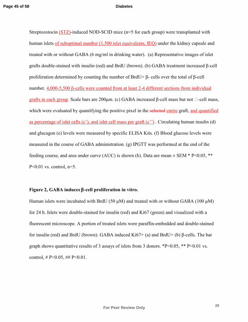
20
Streptozotocin (STZ)-induced NOD-SCID mice (n=5 for each group) were transplanted with
human islets of suboptimal number (1,500 islet equivalents, IEQ) under the kidney capsule and
treated with or without GABA (6 mg/ml in drinking water). (a) Representative images of islet
grafts double-stained with insulin (red) and BrdU (brown). (b) GABA treatment increased β-cell
proliferation determined by counting the number of BrdU+ β- cells over the total of β-cell
number. 4,000-5,500 β-cells were counted from at least 2-4 different sections from individual
grafts in each group. Scale bars are 200µm. (c) GABA increased β-cell mass but not -cell mass,
which were evaluated by quantifying the positive pixel in the selected entire graft, and quantified
as percentage of islet cells (c’), and islet cell mass per graft (c’’) . Circulating human insulin (d)
and glucagon (e) levels were measured by specific ELISA Kits. (f) Blood glucose levels were
measured in the course of GABA administration. (g) IPGTT was performed at the end of the
feeding course, and area under curve (AUC) is shown (h). Data are mean ± SEM * P<0.05, **
P<0.01 vs. control, n=5.
Figure 2, GABA induces ββββ-cell proliferation in vitro.
Human islets were incubated with BrdU (50 µM) and treated with or without GABA (100 µM)
for 24 h. Islets were double-stained for insulin (red) and Ki67 (green) and visualized with a
fluorescent microscope. A portion of treated islets were paraffin-embedded and double-stained
for insulin (red) and BrdU (brown). GABA induced Ki67+ (a) and BrdU+ (b) β-cells. The bar
graph shows quantitative results of 3 assays of islets from 3 donors. *P<0.05, ** P<0.01 vs.
control, # P<0.05, ## P<0.01.
Page 45 of 58
For Peer Review Only
Diabetes
21
Figure 3, GABA induced Akt and CREB phosphorylation, and increased IRS2 mRNA
expression in human islets.
(a) GABA evokes Ca2+
influx in isolated adult human islets. Islets were perfused with GABA
(200 µM) or the GABAAR agonist muscimol, in the presence or absence of antagonists as
indicated. Intracellular Ca2+
was measured using Fura-2 AM. The shown represents n=11-26
from two different donors. (b) GABA induced Akt and CREB phosphorylation in human islets,
which was inhibited by picrotoxin, nifedipine, and saclofen. Representative blots are shown. The
bar graph represents quantitative results of 3-9 assays from 5 islet donors. (c) GABA increased
IRS2 mRNA expression in isolated human islets. Islets were treated with GABA (100 µM) in the
presence of indicated inhibitors for 16 h, followed by mRNA extraction and qPCR using specific
primers for human IRS2. The data shown are representative result of 3 assays using islets from 2
donors. GABAAR agonist: muscimol (Mus, 20 µM); GABAAR antagonists: picrotoxin (Pic, 100
µM); bicuculline (Bic 100 µM), or SR-95531(SR, 1000 µM); GABABR antagonist: saclofen
(Sac, 100 µM); Ca2+
channel blocker: nifedipine (Nif, 1 µM). *P<0.05 vs. control, **P<0.01 vs.
controls, #P<0.05 vs. GABA-treated group.
Figure 4, GABA-induced Akt and CREB phosphorylation independently.
(a) GABA-induced Akt and CREB phosphorylation are independent of each other. Human islets
were serum-starved for 2 h and treated with GABA for 1 h in the presence or absence of PI3K
inhibitor LY294002 (LY, 1 µM) or PKA inhibitor H89 (1 µM) as indicated. Inhibitors were
added 30 min prior to GABA treatment. (b) Model of GABA signaling in the β-cells: 1)
GABAAR activation causes membrane depolarization, which leads to Ca2+
influx. 2) Activation
of GABABR causes release of Ca2+
from intracellular storage and PKA activation(19). 3) Ca2+
-
Page 46 of 58
For Peer Review Only
Diabetes

22
dependent activation of PI3K/Akt(38) and CREB(39), which regulates IRS2 expression(40). 4)
Increased intracellular Ca2+
promotes insulin secretion and autocrine insulin action activates
PI3K/Akt signaling pathway. Data are mean ± SEM * P<0.05, ** P<0.01 vs. control, n=3.
S-Fig. 1, Streptozotocin injections eliminated endogenous mouse islet β-cell in mice.
NOD-SCID mice were injected with streptozotocin (STZ, 125 mg/kg for two consecutive days)
prior to transplantation. At the end of experiment, pancreatic sections were immunostained for
insulin (red) and glucagon (brown). The STZ injections eliminated nearly all β-cells with the
residual islet containing mostly α-cells in the pancreas of water (a) or GABA (b) treated mice.
The glucagon-insulin dual stained normal pancreatic is shown (c). Scale bars are 50µm.
S-Fig. 2, GABA lowered blood glucose and improved glucose tolerance in diabetic NOD-
SCID mice transplanted with suboptimal dose human islets.
Induction of hyperglycemia was made by injections of streptozotocin (STZ, 125 mg/kg for two
consecutive days), and a suboptimal number of human islets (1500 IEQ) was inserted under the
kidney capsule of the mice. The low numbers of human islets cannot maintain blood glucose
levels in the normal physiological range In these experiments the transplanted islets briefly
restored glucose levels, but were not sufficient (suboptimal) to prevent hyperglycemia over time,
thus providing a margin to observe whether GABA treatment improves hyperglycemia in the
recipient mice. A sub-therapeutic dose of insulin was used, but only during the phase when the
individual blood glucose levels exceeded 22 mM, in order to prevent severe hyperglycemia and
potential complications or death. The injection of insulin was omitted 48h prior to the blood
sampling. The arrow(s) indicates the time point of STZ injection, islet transplantation,
Page 47 of 58
For Peer Review Only
Diabetes
23
administration of GABA (through the drinking water, 30 mg/ml, and the intraperitoneal glucose
tolerance test (IPGTT). (a) Blood glucose levels were monitored during the feeding course. (b)
IPGTT conducted at 1 week and 3 weeks (c) after the GABA treatment. the time point as
indicated. (c) The quantification is expressed as the area under curve (AUC). Data are mean ±
SEM * P<0.05, vs. control.
S-Fig. 3, GABA-induced β-cell proliferation is GABAAR, GABABR, and Ca2+
channel-
dependent
(a) Isolated adult human islets incubated for 24 h with BrdU (50 µM) with or without GABA
(100 µM), in the presence or absence of GABAAR antagonist picrotoxin (Pic, 100µM) or
GABABR antagonist saclofen (Sac, 100µM). The islets were paraffin-embedded and subjected to
insulin-BrdU dual staining and determination of BrdU+ β-cell numbers. (b) Similar experiments
were conducted in clonal INS-1 cells, and also assessed by the3H-thymidine incorporation assay.
The data is a representative of three independent experiments in triplicate, expressed as mean ±
SEM. **P≤0.01 vs. control, ##P≤0.01 vs. GABA.
S-Fig. 4, GABA induces Akt and CREB phosphorylation in time- and dose-dependent
manner
INS-1 cells were serum-starved for 2 h and treated with GABA for the indicated time (a) or dose
(b) for 30 min. Cell lysates were subjected to Western blot analysis for Akt and CREB
phosphorylation. GAPDH was used as the loading control. Representative blots are shown.
Results were quantified and expressed in the histogram as mean ± SEM of 4-5 independent
experiments. *P≤0.05, **P≤0.01 vs. controls.
Page 48 of 58
For Peer Review Only
Diabetes
24
S-Fig. 5, GABA-induced Akt and CREB phosphorylation that is independent of each other
in INS-1 cells.
(a) INS-1 cells were serum-starved for 2 h and treated with GABA for 1 h in the presence or
absence of PI3K inhibitor LY294002 (LY, 1 µM) or PKA inhibitor H89 (1 µM) as indicated.
Inhibitors were added 30 min prior to GABA treatment. Representative blots are shown. Two
sets came from non-adjacent lanes on the same gel. Results were quantified and expressed in
histogram as mean ± SEM of 3 independent experiments. *P≤0.05, **P≤0.01 vs. respective
controls.
S-Fig. 6, GABA protected human β-cells from apoptosis under in vitro and in vivo
conditions. (a) Western blot analysis shows GABA attenuated cytokine-induced expression of
cleaved caspase-3 expression in isolated adult human islets; the bar graphs (b) show the
quantitative analysis. (c) DFDCA-fluorogenic dye-based cellular reactive oxygen species (ROS)
detection shows that GABA significantly attenuated cytokine-induced ROS production in
isolated human islets. (d) Insulin-Tunel double staining was conducted on xenotransplanted
human islets grafts in mice that were treated or not with GABA. The islets had been inserted
under the kidney capsule of NSG mice. Representative staining shows the Insulin+ cells and
TUNEL+ cells that have undergone apoptosis. Scale bars are 50µm. (e) Quantification of
TUNEL+ β-cells in the grafts. Data are mean ± SEM of 3-5 independent experiments. *P≤0.05,
**P≤0.01 vs. respective controls.
Page 49 of 58
For Peer Review Only
Diabetes
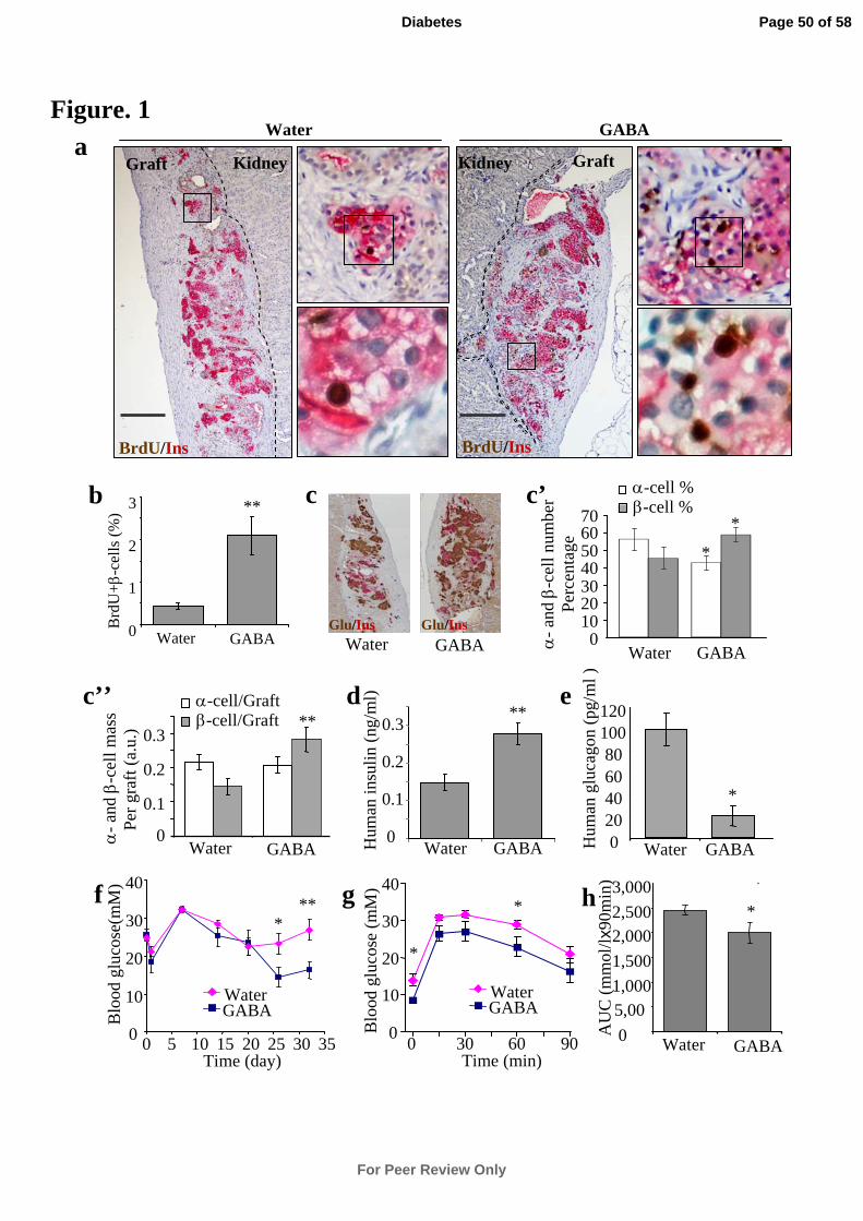
Figure. 1GABA
Kidney Graft
Water
KidneyGraft
BrdU/Ins
a
d
0
0.1
0.2
0.3
Water GABAHum
an in
suli
n (n
g/m
l)
**
b
Brd
U+-
cell
s (%
)
0
1
2
3
Water GABA
**
BrdU/Ins
-
and -
cell
mas
sP
er g
raft
(a.
u.)
0.2
0.3
0
0.1
Water GABA
-cell/Graft-cell/Graft **
e
Hum
an g
luca
gon
(pg/
ml )
020406080
100120
Water GABA
*
c
Water GABAGlu/InsGlu/Ins
f*
0
10
20
30
40
0 5 10 15 20 25 30 35Time (day)
Blo
od g
luco
se(m
M)
GABAWater
** g
0
10
20
30
40
0 30 60 90
Blo
od g
luco
se (
mM
)
*
*
GABAWater
Time (min)
0
5,00
1,000
1,500
2,0002,500
3,000
Water GABA
*
AU
C (
mm
ol/l
x90m
in)
h
-
and -
cell
num
ber
Per
cent
age
010203040506070
Water GABA
*
*
-cell %-cell %c’
c’’
Page 50 of 58
For Peer Review Only
Diabetes
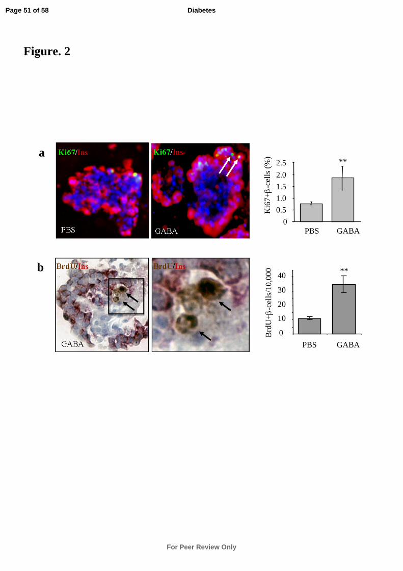
PBS GABA0
0.5
1.0
1.5
2.0
2.5
Ki6
7+-
cell
s (%
)
****
PBS GABA
0
10
20
30
40B
rdU
+-
cell
s/10
,000 ****b
a
PBS GABA
GABA
BrdU/InsBrdU/Ins
Ki67/Ins Ki67/Ins
Figure. 2
Page 51 of 58
For Peer Review Only
Diabetes
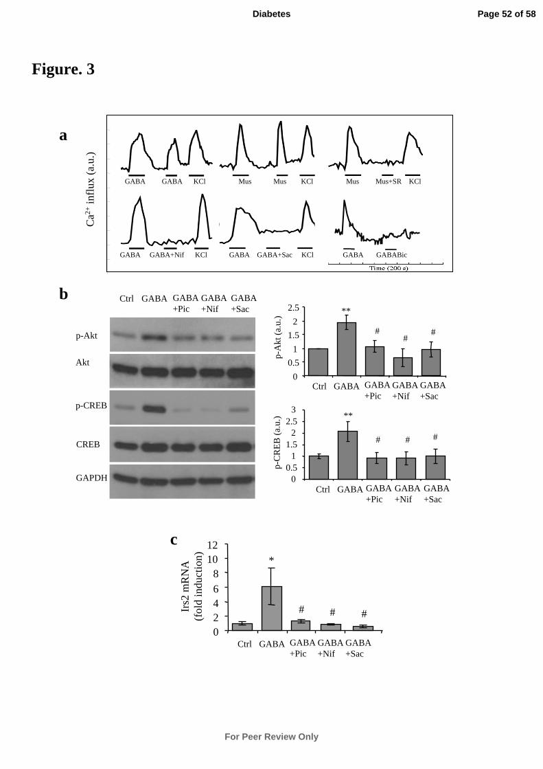
Ca2+
infl
ux (
a.u.
)
GABA GABA KCl Mus Mus KCl Mus Mus+SR KCl
GABA GABA+Nif KCl GABA GABA+Sac KCl GABA GABABic
0
0.5
1
1.5
2
2.5
Ctrl GABA GABA+Pic
GABA+Nif
GABA+Sac
p-A
kt (
a.u.
) **
##
#
00.5
11.5
22.5
3
Ctrl GABA GABA+Pic
GABA+Nif
GABA+Sac
p-C
RE
B (
a.u.
) **
# # #
p-CREB
CREB
Ctrl GABA GABA+Pic
GABA+Nif
p-Akt
Akt
GABA+Sac
GAPDH
0246
81012
Irs2
mR
NA
(fol
d in
duct
ion) *
# # #
c
a
b
Ctrl GABA GABA+Pic
GABA+Nif
GABA+Sac
Figure. 3
Page 52 of 58
For Peer Review Only
Diabetes
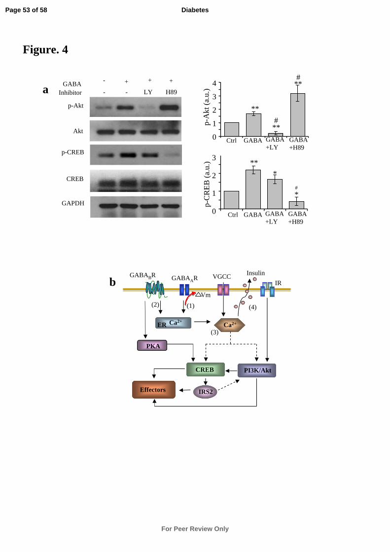
p-Akt
p-CREB
GAPDH
Akt
CREB
Inhibitor
- +
- - LY H89
+ +GABA a
0
1
2
3
p-C
RE
B (
a.u.
) **
*
*
Ctrl GABA GABA+LY
GABA+H89
0
1
2
3
4
p-A
kt (
a.u.
)
**
**
**
Ctrl GABA GABA+LY
GABA+H89
#
#
#
Figure. 4
IR
PI3K/Akt
GABAAR
Vm
(1)
VGCC
Ca2+
Insulin
(3)
CREB
IRS2
GABABR
Ca2+ER
(4)
Effectors
(2)
PKA
b
Page 53 of 58
For Peer Review Only
Diabetes
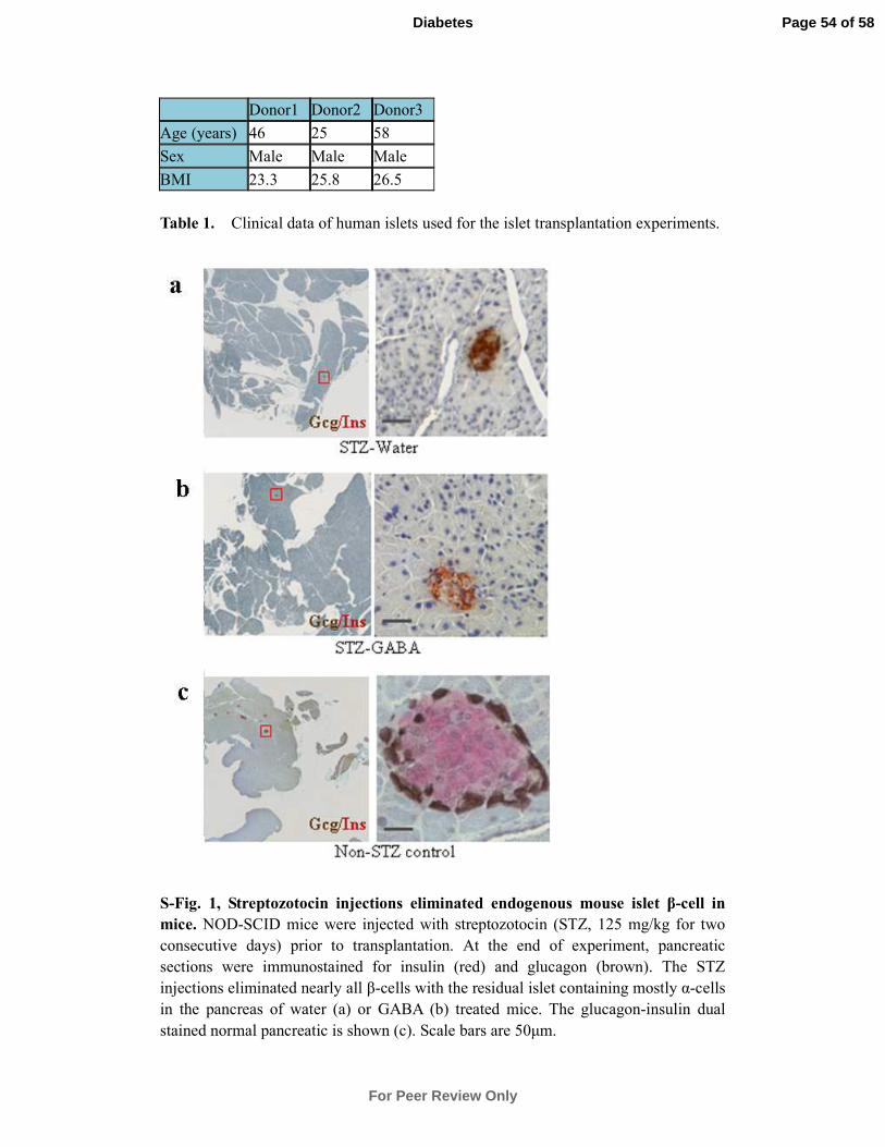
Donor1 Donor2 Donor3
Age (years) 46 25 58
Sex Male Male Male
BMI 23.3 25.8 26.5
Table 1. Clinical data of human islets used for the islet transplantation experiments.
S-Fig. 1, Streptozotocin injections eliminated endogenous mouse islet β-cell in
mice. NOD-SCID mice were injected with streptozotocin (STZ, 125 mg/kg for two
consecutive days) prior to transplantation. At the end of experiment, pancreatic
sections were immunostained for insulin (red) and glucagon (brown). The STZ
injections eliminated nearly all β-cells with the residual islet containing mostly α-cells
in the pancreas of water (a) or GABA (b) treated mice. The glucagon-insulin dual
stained normal pancreatic is shown (c). Scale bars are 50µm.
Page 54 of 58
For Peer Review Only
Diabetes
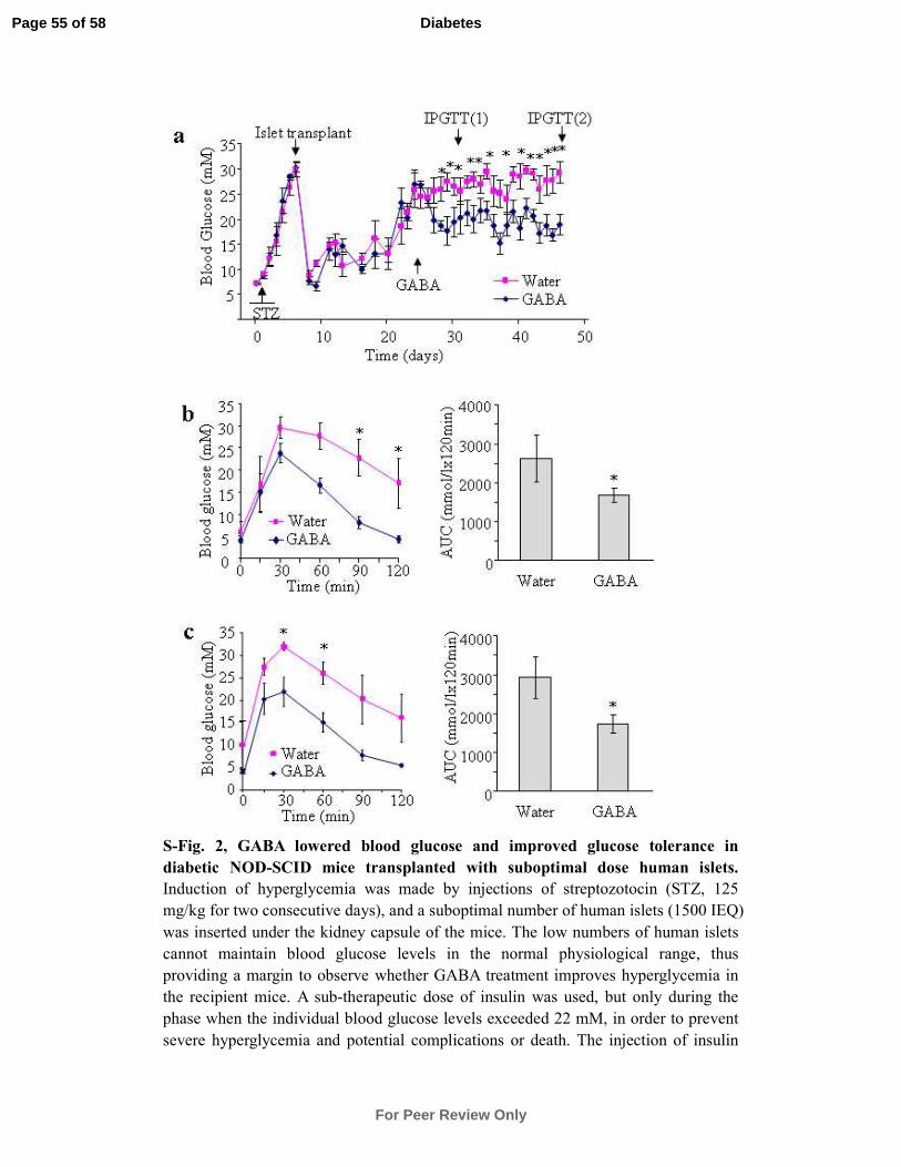
S-Fig. 2, GABA lowered blood glucose and improved glucose tolerance in
diabetic NOD-SCID mice transplanted with suboptimal dose human islets.
Induction of hyperglycemia was made by injections of streptozotocin (STZ, 125
mg/kg for two consecutive days), and a suboptimal number of human islets (1500 IEQ)
was inserted under the kidney capsule of the mice. The low numbers of human islets
cannot maintain blood glucose levels in the normal physiological range, thus
providing a margin to observe whether GABA treatment improves hyperglycemia in
the recipient mice. A sub-therapeutic dose of insulin was used, but only during the
phase when the individual blood glucose levels exceeded 22 mM, in order to prevent
severe hyperglycemia and potential complications or death. The injection of insulin
Page 55 of 58
For Peer Review Only
Diabetes
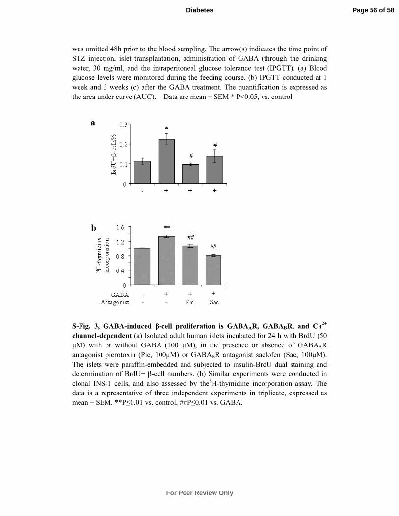
was omitted 48h prior to the blood sampling. The arrow(s) indicates the time point of
STZ injection, islet transplantation, administration of GABA (through the drinking
water, 30 mg/ml, and the intraperitoneal glucose tolerance test (IPGTT). (a) Blood
glucose levels were monitored during the feeding course. (b) IPGTT conducted at 1
week and 3 weeks (c) after the GABA treatment. The quantification is expressed as
the area under curve (AUC). Data are mean ± SEM * P<0.05, vs. control.
S-Fig. 3, GABA-induced β-cell proliferation is GABAAR, GABABR, and Ca2+
channel-dependent (a) Isolated adult human islets incubated for 24 h with BrdU (50
µM) with or without GABA (100 µM), in the presence or absence of GABAAR
antagonist picrotoxin (Pic, 100µM) or GABABR antagonist saclofen (Sac, 100µM).
The islets were paraffin-embedded and subjected to insulin-BrdU dual staining and
determination of BrdU+ β-cell numbers. (b) Similar experiments were conducted in
clonal INS-1 cells, and also assessed by the3H-thymidine incorporation assay. The
data is a representative of three independent experiments in triplicate, expressed as
mean ± SEM. **P≤0.01 vs. control, ##P≤0.01 vs. GABA.
Page 56 of 58
For Peer Review Only
Diabetes
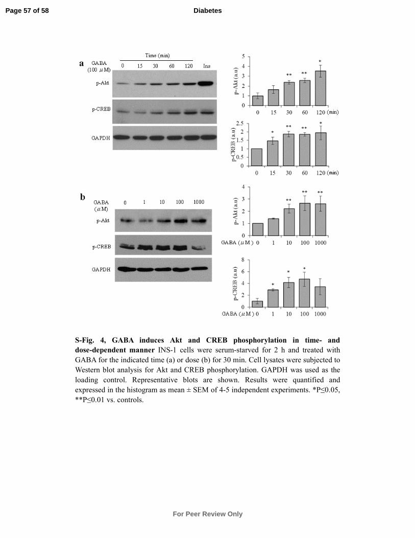
S-Fig. 4, GABA induces Akt and CREB phosphorylation in time- and
dose-dependent manner INS-1 cells were serum-starved for 2 h and treated with
GABA for the indicated time (a) or dose (b) for 30 min. Cell lysates were subjected to
Western blot analysis for Akt and CREB phosphorylation. GAPDH was used as the
loading control. Representative blots are shown. Results were quantified and
expressed in the histogram as mean ± SEM of 4-5 independent experiments. *P≤0.05,
**P≤0.01 vs. controls.
Page 57 of 58
For Peer Review Only
Diabetes
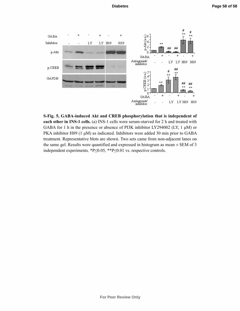
S-Fig. 5, GABA-induced Akt and CREB phosphorylation that is independent of
each other in INS-1 cells. (a) INS-1 cells were serum-starved for 2 h and treated with
GABA for 1 h in the presence or absence of PI3K inhibitor LY294002 (LY, 1 µM) or
PKA inhibitor H89 (1 µM) as indicated. Inhibitors were added 30 min prior to GABA
treatment. Representative blots are shown. Two sets came from non-adjacent lanes on
the same gel. Results were quantified and expressed in histogram as mean ± SEM of 3
independent experiments. *P≤0.05, **P≤0.01 vs. respective controls.
Page 58 of 58
For Peer Review Only
Diabetes
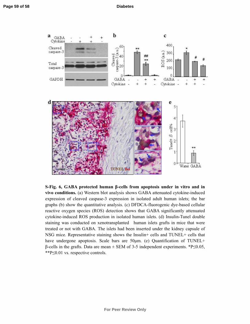
S-Fig. 6, GABA protected human β-cells from apoptosis under in vitro and in
vivo conditions. (a) Western blot analysis shows GABA attenuated cytokine-induced
expression of cleaved caspase-3 expression in isolated adult human islets; the bar
graphs (b) show the quantitative analysis. (c) DFDCA-fluorogenic dye-based cellular
reactive oxygen species (ROS) detection shows that GABA significantly attenuated
cytokine-induced ROS production in isolated human islets. (d) Insulin-Tunel double
staining was conducted on xenotransplanted human islets grafts in mice that were
treated or not with GABA. The islets had been inserted under the kidney capsule of
NSG mice. Representative staining shows the Insulin+ cells and TUNEL+ cells that
have undergone apoptosis. Scale bars are 50µm. (e) Quantification of TUNEL+
β-cells in the grafts. Data are mean ± SEM of 3-5 independent experiments. *P≤0.05,
**P≤0.01 vs. respective controls.
Page 59 of 58
For Peer Review Only
Diabetes