Quantum Computing and Quantum Programming Language Choon Oh Lee ISILab, KAIST.

This is an Open Access document downloaded from ORCA, Cardiff University's institutional
repository: http://orca.cf.ac.uk/130555/
This is the author’s version of a work that was submitted to / accepted for publication.
Citation for final published version:
Grasby, Katrina L., Jahanshad, Neda, Painter, Jodie N., Colodro-Conde, Lucía, Bralten, Janita,
Hibar, Derrek P., Lind, Penelope A., Pizzagalli, Fabrizio, Ching, Christopher R. K., McMahon,
Mary Agnes B., Shatokhina, Natalia, Zsembik, Leo C. P., Thomopoulos, Sophia I., Zhu, Alyssa H.,
Strike, Lachlan T., Agartz, Ingrid, Alhusaini, Saud, Almeida, Marcio A. A., Alnæs, Dag, Amlien,
Inge K., Andersson, Micael, Ard, Tyler, Armstrong, Nicola J., Ashley-Koch, Allison, Atkins,
Joshua R., Bernard, Manon, Brouwer, Rachel M., Buimer, Elizabeth E. L., Bülow, Robin, Bürger,
Christian, Cannon, Dara M., Chakravarty, Mallar, Chen, Qiang, Cheung, Joshua W., Couvy-
Duchesne, Baptiste, Dale, Anders M., Dalvie, Shareefa, de Araujo, Tânia K., de Zubicaray, Greig I.,
de Zwarte, Sonja M. C., den Braber, Anouk, Doan, Nhat Trung, Dohm, Katharina, Ehrlich, Stefan,
Engelbrecht, Hannah-Ruth, Erk, Susanne, Fan, Chun Chieh, Fedko, Iryna O., Foley, Sonya F., Ford,
Judith M., Fukunaga, Masaki, Garrett, Melanie E., Ge, Tian, Giddaluru, Sudheer, Goldman, Aaron
L., Green, Melissa J., Groenewold, Nynke A., Grotegerd, Dominik, Gurholt, Tiril P., Gutman, Boris
A., Hansell, Narelle K., Harris, Mathew A., Harrison, Marc B., Haswell, Courtney C., Hauser,
Michael, Herms, Stefan, Heslenfeld, Dirk J., Ho, New Fei, Hoehn, David, Hoffmann, Per, Holleran,
Laurena, Hoogman, Martine, Hottenga, Jouke-Jan, Ikeda, Masashi, Janowitz, Deborah, Jansen, Iris
E., Jia, Tianye, Jockwitz, Christiane, Kanai, Ryota, Karama, Sherif, Kasperaviciute, Dalia,
Kaufmann, Tobias, Kelly, Sinead, Kikuchi, Masataka, Klein, Marieke, Knapp, Michael, Knodt,
Annchen R., Krämer, Bernd, Lam, Max, Lancaster, Thomas M., Lee, Phil H., Lett, Tristram A.,
Lewis, Lindsay B., Lopes-Cendes, Iscia, Luciano, Michelle, Macciardi, Fabio, Marquand, Andre F.,
Mathias, Samuel R., Melzer, Tracy R., Milaneschi, Yuri, Mirza-Schreiber, Nazanin, Moreira, Jose
C. V., Mühleisen, Thomas W., Müller-Myhsok, Bertram, Najt, Pablo, Nakahara, Soichiro, Nho,
Kwangsik, Olde Loohuis, Loes M., Orfanos, Dimitri Papadopoulos, Pearson, John F., Pitcher, Toni
L., Pütz, Benno, Quidé, Yann, Ragothaman, Anjanibhargavi, Rashid, Faisal M., Reay, William R.,
Redlich, Ronny, Reinbold, Céline S., Repple, Jonathan, Richard, Geneviève, Riedel, Brandalyn C.,
Risacher, Shannon L., Rocha, Cristiane S., Mota, Nina Roth, Salminen, Lauren, Saremi, Arvin,
Saykin, Andrew J., Schlag, Fenja, Schmaal, Lianne, Schofield, Peter R., Secolin, Rodrigo,
Shapland, Chin Yang, Shen, Li, Shin, Jean, Shumskaya, Elena, Sønderby, Ida E., Sprooten, Emma,
Tansey, Katherine E., Teumer, Alexander, Thalamuthu, Anbupalam, Tordesillas-Gutiérrez, Diana,
Turner, Jessica A., Uhlmann, Anne, Vallerga, Costanza Ludovica, van der Meer, Dennis, van
Donkelaar, Marjolein M. J., van Eijk, Liza, van Erp, Theo G. M., van Haren, Neeltje E. M., van
Rooij, Daan, van Tol, Marie-José, Veldink, Jan H., Verhoef, Ellen, Walton, Esther, Wang,
Mingyuan, Wang, Yunpeng, Wardlaw, Joanna M., Wen, Wei, Westlye, Lars T., Whelan,
Christopher D., Witt, Stephanie H., Wittfeld, Katharina, Wolf, Christiane, Wolfers, Thomas, Wu,
Jing Qin, Yasuda, Clarissa L., Zaremba, Dario, Zhang, Zuo, Zwiers, Marcel P., Artiges, Eric,
Assareh, Amelia A., Ayesa-Arriola, Rosa, Belger, Aysenil, Brandt, Christine L., Brown, Gregory

G., Cichon, Sven, Curran, Joanne E., Davies, Gareth E., Degenhardt, Franziska, Dennis, Michelle
F., Dietsche, Bruno, Djurovic, Srdjan, Doherty, Colin P., Espiritu, Ryan, Garijo, Daniel, Gil,
Yolanda, Gowland, Penny A., Green, Robert C., Häusler, Alexander N., Heindel, Walter, Ho, Beng-
Choon, Hoffmann, Wolfgang U., Holsboer, Florian, Homuth, Georg, Hosten, Norbert, Jack,
Clifford R., Jang, MiHyun, Jansen, Andreas, Kimbrel, Nathan A., Kolskår, Knut, Koops, Sanne,
Krug, Axel, Lim, Kelvin O., Luykx, Jurjen J., Mathalon, Daniel H., Mather, Karen A., Mattay,
Venkata S., Matthews, Sarah, Mayoral Van Son, Jaqueline, McEwen, Sarah C., Melle, Ingrid,
Morris, Derek W., Mueller, Bryon A., Nauck, Matthias, Nordvik, Jan E., Nöthen, Markus M.,
O'Leary, Daniel S., Opel, Nils, Martinot, Marie-Laure Paillère, Pike, G. Bruce, Preda, Adrian,
Quinlan, Erin B., Rasser, Paul E., Ratnakar, Varun, Reppermund, Simone, Steen, Vidar M.,
Tooney, Paul A., Torres, Fábio R., Veltman, Dick J., Voyvodic, James T., Whelan, Robert, White,
Tonya, Yamamori, Hidenaga, Adams, Hieab H. H., Bis, Joshua C., Debette, Stephanie, Decarli,
Charles, Fornage, Myriam, Gudnason, Vilmundur, Hofer, Edith, Ikram, M. Arfan, Launer, Lenore,
Longstreth, W. T., Lopez, Oscar L., Mazoyer, Bernard, Mosley, Thomas H., Roshchupkin,
Gennady V., Satizabal, Claudia L., Schmidt, Reinhold, Seshadri, Sudha, Yang, Qiong, Alvim,
Marina K. M., Ames, David, Anderson, Tim J., Andreassen, Ole A., Arias-Vasquez, Alejandro,
Bastin, Mark E., Baune, Bernhard T., Beckham, Jean C., Blangero, John, Boomsma, Dorret I.,
Brodaty, Henry, Brunner, Han G., Buckner, Randy L., Buitelaar, Jan K., Bustillo, Juan R., Cahn,
Wiepke, Cairns, Murray J., Calhoun, Vince, Carr, Vaughan J., Caseras, Xavier, Caspers, Svenja,
Cavalleri, Gianpiero L., Cendes, Fernando, Corvin, Aiden, Crespo-Facorro, Benedicto, Dalrymple-
Alford, John C., Dannlowski, Udo, de Geus, Eco J. C., Deary, Ian J., Delanty, Norman, Depondt,
Chantal, Desrivières, Sylvane, Donohoe, Gary, Espeseth, Thomas, Fernández, Guillén, Fisher,
Simon E., Flor, Herta, Forstner, Andreas J., Francks, Clyde, Franke, Barbara, Glahn, David C.,
Gollub, Randy L., Grabe, Hans J., Gruber, Oliver, Håberg, Asta K., Hariri, Ahmad R., Hartman,
Catharina A., Hashimoto, Ryota, Heinz, Andreas, Henskens, Frans A., Hillegers, Manon H. J.,
Hoekstra, Pieter J., Holmes, Avram J., Hong, L. Elliot, Hopkins, William D., Hulshoff Pol, Hilleke
E., Jernigan, Terry L., Jönsson, Erik G., Kahn, René S., Kennedy, Martin A., Kircher, Tilo T. J.,
Kochunov, Peter, Kwok, John B. J., Le Hellard, Stephanie, Loughland, Carmel M., Martin,
Nicholas G., Martinot, Jean-Luc, McDonald, Colm, McMahon, Katie L., Meyer-Lindenberg,
Andreas, Michie, Patricia T., Morey, Rajendra A., Mowry, Bryan, Nyberg, Lars, Oosterlaan, Jaap,
Ophoff, Roel A., Pantelis, Christos, Paus, Tomas, Pausova, Zdenka, Penninx, Brenda W. J. H.,
Polderman, Tinca J. C., Posthuma, Danielle, Rietschel, Marcella, Roffman, Joshua L., Rowland,
Laura M., Sachdev, Perminder S., Sämann, Philipp G., Schall, Ulrich, Schumann, Gunter, Scott,
Rodney J., Sim, Kang, Sisodiya, Sanjay M., Smoller, Jordan W., Sommer, Iris E., St Pourcain,
Beate, Stein, Dan J., Toga, Arthur W., Trollor, Julian N., Van der Wee, Nic J. A., van t Ent, Dennis,
Völzke, Henry, Walter, Henrik, Weber, Bernd, Weinberger, Daniel R., Wright, Margaret J., Zhou,
Juan, Stein, Jason L., Thompson, Paul M. and Medland, Sarah E. 2020. The genetic architecture of
the human cerebral cortex. Science 367 (6484) , eaay6690. 10.1126/science.aay6690 file
Publishers page: http://dx.doi.org/10.1126/science.aay6690
<http://dx.doi.org/10.1126/science.aay6690>
Please note:
Changes made as a result of publishing processes such as copy-editing, formatting and page
numbers may not be reflected in this version. For the definitive version of this publication, please
refer to the published source. You are advised to consult the publisher’s version if you wish to cite
this paper.
This version is being made available in accordance with publisher policies. See
http://orca.cf.ac.uk/policies.html for usage policies. Copyright and moral rights for publications

made available in ORCA are retained by the copyright holders.

Final Submitted Manuscript
1
Title: The genetic architecture of the human cerebral cortex. Authors: Katrina L. Grasby1†*, Neda Jahanshad2†*, Jodie N. Painter1‡, Lucía Colodro-Conde1,3-
5‡, Janita Bralten6,7‡, Derrek P. Hibar2,8‡, Penelope A. Lind1,4,9‡, Fabrizio Pizzagalli2‡, Christopher R.K. Ching2,10, Mary Agnes B. McMahon2, Natalia Shatokhina2, Leo C.P. Zsembik11, Sophia I.
Thomopoulos2, Alyssa H. Zhu2, Lachlan T. Strike12, Ingrid Agartz13-16, Saud Alhusaini17,18, Marcio A.A. Almeida19, Dag Alnæs13,14, Inge K. Amlien20, Micael Andersson21,22, Tyler Ard23, Nicola J. Armstrong24, Allison Ashley-Koch25, Joshua R. Atkins26,27, Manon Bernard28, Rachel M. Brouwer29, Elizabeth E.L. Buimer29, Robin Bülow30, Christian Bürger31, Dara M. Cannon32, Mallar Chakravarty33,34, Qiang Chen35, Joshua W. Cheung2, Baptiste Couvy-Duchesne12,36,37,
Anders M. Dale38,39, Shareefa Dalvie 40, Tânia K. de Araujo41,42, Greig I. de Zubicaray43, Sonja M.C. de Zwarte29, Anouk den Braber44,45, Nhat Trung Doan13,14, Katharina Dohm31, Stefan Ehrlich46, Hannah-Ruth Engelbrecht47, Susanne Erk48, Chun Chieh Fan49, Iryna O. Fedko44, Sonya F. Foley50, Judith M. Ford51, Masaki Fukunaga52, Melanie E. Garrett25, Tian Ge53,54, Sudheer Giddaluru55, Aaron L. Goldman35, Melissa J. Green56,57, Nynke A. Groenewold40,
Dominik Grotegerd31, Tiril P. Gurholt13-15, Boris A. Gutman2,58, Narelle K. Hansell12, Mathew A. Harris59,60, Marc B. Harrison2, Courtney C. Haswell61,62, Michael Hauser25, Stefan Herms63-65, Dirk J. Heslenfeld66, New Fei Ho67, David Hoehn68, Per Hoffmann63,64,69, Laurena Holleran70,
Martine Hoogman6,7, Jouke-Jan Hottenga44, Masashi Ikeda71, Deborah Janowitz72, Iris E. Jansen73,74, Tianye Jia75-77, Christiane Jockwitz78-80, Ryota Kanai81-83, Sherif Karama33,84,85, Dalia
Kasperaviciute86,87, Tobias Kaufmann13,14, Sinead Kelly88,89, Masataka Kikuchi 90, Marieke Klein6,7,29, Michael Knapp91, Annchen R. Knodt92, Bernd Krämer93,94, Max Lam67,95, Thomas M. Lancaster50,96, Phil H. Lee53,97, Tristram A. Lett48, Lindsay B. Lewis85,98, Iscia Lopes-Cendes41,42,
Michelle Luciano99,100, Fabio Macciardi101, Andre F. Marquand7,102, Samuel R. Mathias103,104, Tracy R. Melzer105-107, Yuri Milaneschi108, Nazanin Mirza-Schreiber68,109, Jose C.V.
Moreira42,110, Thomas W. Mühleisen63,78,111, Bertram Müller-Myhsok68,112,113, Pablo Najt32, Soichiro Nakahara101,114, Kwangsik Nho115, Loes M. Olde Loohuis116, Dimitri Papadopoulos
Orfanos117, John F. Pearson118,119, Toni L. Pitcher105-107, Benno Pütz68, Yann Quidé56,57, Anjanibhargavi Ragothaman2, Faisal M. Rashid2, William R. Reay26,27, Ronny Redlich31, Céline
S. Reinbold20,63,64, Jonathan Repple31, Geneviève Richard13,14,120,121, Brandalyn C. Riedel2,115, Shannon L. Risacher115, Cristiane S. Rocha41,42, Nina Roth Mota6,7,122, Lauren Salminen2, Arvin
Saremi2, Andrew J. Saykin115,123, Fenja Schlag124, Lianne Schmaal125-127, Peter R. Schofield128,129, Rodrigo Secolin41,42, Chin Yang Shapland124, Li Shen130, Jean Shin28,131, Elena
Shumskaya6,7,132, Ida E. Sønderby13,14, Emma Sprooten7, Katherine E. Tansey96, Alexander Teumer133, Anbupalam Thalamuthu134, Diana Tordesillas-Gutiérrez135,136, Jessica A.
Turner137,138, Anne Uhlmann40,139, Costanza Ludovica Vallerga36, Dennis van der Meer140,141, Marjolein M.J. van Donkelaar142, Liza van Eijk3,12, Theo G.M. van Erp101, Neeltje E.M. van Haren29,143, Daan van Rooij7,102, Marie-José van Tol144, Jan H. Veldink145, Ellen Verhoef124, Esther Walton137,146,147, Mingyuan Wang67, Yunpeng Wang13,14, Joanna M. Wardlaw59,100,148,
Wei Wen134, Lars T. Westlye13,14,120, Christopher D. Whelan2,17, Stephanie H. Witt149, Katharina Wittfeld72,150, Christiane Wolf151, Thomas Wolfers6, Jing Qin Wu26, Clarissa L. Yasuda42,152,
Dario Zaremba31, Zuo Zhang153, Marcel P. Zwiers7,102,132, Eric Artiges154, Amelia A. Assareh134, Rosa Ayesa-Arriola136,155, Aysenil Belger61,156, Christine L. Brandt13,14, Gregory G. Brown157,158, Sven Cichon63,64,78, Joanne E. Curran19, Gareth E. Davies159, Franziska Degenhardt69, Michelle F.
Dennis62, Bruno Dietsche160, Srdjan Djurovic161,162, Colin P. Doherty163-165, Ryan Espiritu166, Daniel Garijo166, Yolanda Gil166, Penny A. Gowland167, Robert C. Green168-170, Alexander N.

Final Submitted Manuscript
2
Häusler171,172, Walter Heindel173, Beng-Choon Ho174, Wolfgang U. Hoffmann133,150, Florian Holsboer68,175, Georg Homuth176, Norbert Hosten177, Clifford R. Jack Jr.178, MiHyun Jang166, Andreas Jansen160,179, Nathan A. Kimbrel62,180, Knut Kolskår13,14,120,121, Sanne Koops29, Axel
Krug160, Kelvin O. Lim181, Jurjen J. Luykx29,182,183, Daniel H. Mathalon184,185, Karen A. Mather128,134, Venkata S. Mattay35,186,187, Sarah Matthews146, Jaqueline Mayoral Van Son136,155, Sarah C. McEwen188,189, Ingrid Melle13,14, Derek W. Morris32, Bryon A. Mueller181, Matthias
Nauck190,191, Jan E. Nordvik121, Markus M. Nöthen69, Daniel S. O'Leary174, Nils Opel31, Marie-Laure Paillère Martinot154,192, G. Bruce Pike193, Adrian Preda194, Erin B. Quinlan153, Paul E. Rasser27,195-197, Varun Ratnakar166, Simone Reppermund134,198, Vidar M. Steen162,199, Paul A.
Tooney26,197, Fábio R. Torres41,42, Dick J. Veltman108, James T. Voyvodic61, Robert Whelan200, Tonya White143,201, Hidenaga Yamamori202, Hieab H.H. Adams203-205, Joshua C. Bis206,
Stephanie Debette207,208, Charles Decarli209, Myriam Fornage210, Vilmundur Gudnason211,212, Edith Hofer213,214, M. Arfan Ikram203, Lenore Launer215, W. T. Longstreth216, Oscar L.
Lopez203,217, Bernard Mazoyer218, Thomas H. Mosley 219, Gennady V. Roshchupkin203,204,217, Claudia L. Satizabal220-222, Reinhold Schmidt213, Sudha Seshadri220,222,223, Qiong Yang224, The Alzheimer's Disease Neuroimaging Initiative#, CHARGE consortium#, EPIGEN consortium#, IMAGEN consortium#, SYS consortium#, The Parkinson’s Progression Markers Initiative#,
Marina K.M. Alvim42,152, David Ames225,226, Tim J. Anderson105-107,227, Ole A. Andreassen13,14, Alejandro Arias-Vasquez6,7,122, Mark E. Bastin59,100, Bernhard T. Baune31,228,229, Jean C.
Beckham180,230, John Blangero19, Dorret I. Boomsma44, Henry Brodaty134,231, Han G. Brunner6,7,232, Randy L. Buckner233-235, Jan K. Buitelaar7,102,236, Juan R. Bustillo237, Wiepke
Cahn238, Murray J. Cairns26,27,239, Vince Calhoun240, Vaughan J. Carr56,57,241, Xavier Caseras96, Svenja Caspers78,80,242, Gianpiero L. Cavalleri243,244, Fernando Cendes42,152, Aiden Corvin245,
Benedicto Crespo-Facorro136,155,246, John C. Dalrymple-Alford106,107,247, Udo Dannlowski31, Eco J.C. de Geus44, Ian J. Deary99,100, Norman Delanty17,165, Chantal Depondt248, Sylvane
Desrivières77,153, Gary Donohoe70, Thomas Espeseth13,120, Guillén Fernández7,102, Simon E. Fisher7,124, Herta Flor249, Andreas J. Forstner63,64,69,250,251, Clyde Francks7,124, Barbara
Franke6,7,122, David C. Glahn104,252, Randy L. Gollub97,234,235, Hans J. Grabe72,150, Oliver Gruber93, Asta K. Håberg253,254, Ahmad R. Hariri92, Catharina A. Hartman255, Ryota
Hashimoto202,256,257, Andreas Heinz258, Frans A. Henskens195,259, Manon H.J. Hillegers143,260, Pieter J. Hoekstra261, Avram J. Holmes234,262, L. Elliot Hong263, William D. Hopkins264, Hilleke
E. Hulshoff Pol29, Terry L. Jernigan39,49,157,265, Erik G. Jönsson14,16, René S. Kahn29,266, Martin A. Kennedy119, Tilo T.J. Kircher160, Peter Kochunov263, John B.J. Kwok128,129,267, Stephanie Le Hellard162,199, Carmel M. Loughland195,268, Nicholas G. Martin37, Jean-Luc Martinot154, Colm
McDonald32, Katie L. McMahon269, Andreas Meyer-Lindenberg270, Patricia T. Michie271, Rajendra A. Morey61,62, Bryan Mowry12,272, Lars Nyberg21,22,273, Jaap Oosterlaan274-276, Roel A. Ophoff116, Christos Pantelis228,229,277, Tomas Paus278-280, Zdenka Pausova28,281, Brenda W.J.H. Penninx108, Tinca J.C. Polderman73, Danielle Posthuma73,282, Marcella Rietschel149, Joshua L. Roffman234, Laura M. Rowland263, Perminder S. Sachdev134,283, Philipp G. Sämann68, Ulrich
Schall27,197, Gunter Schumann75,77,153,284,285, Rodney J. Scott26,286, Kang Sim287, Sanjay M. Sisodiya86,288, Jordan W. Smoller53,234,289, Iris E. Sommer144,260,261,290, Beate St Pourcain7,124,146, Dan J. Stein291,292, Arthur W. Toga23, Julian N. Trollor134,198, Nic J.A. Van der Wee293, Dennis van 't Ent44, Henry Völzke133, Henrik Walter48, Bernd Weber171,172, Daniel R. Weinberger35,294,
Margaret J. Wright12,295, Juan Zhou296, Jason L. Stein11§*, Paul M. Thompson2§*, Sarah E. Medland1,3,9§* on behalf of the Enhancing NeuroImaging Genetics through Meta-Analysis
Consortium - Genetics working group

Final Submitted Manuscript
3
Affiliations: 1Psychiatric Genetics, QIMR Berghofer Medical Research Institute, Brisbane, Australia. 2Imaging Genetics Center, Mark and Mary Stevens Neuroimaging and Informatics Institute, Keck School of Medicine of USC, University of Southern California, Los Angeles, USA. 3School of Psychology, University of Queensland, Brisbane, Australia. 4School of Biomedical Sciences, Queensland University of Technology, Brisbane, Australia. 5Faculty of Psychology, University of Murcia, Murcia, Spain. 6Department of Human Genetics, Radboud university medical center, Nijmegen, The Netherlands. 7Donders Institute for Brain, Cognition and Behaviour, Radboud University, Nijmegen, The Netherlands. 8Personalized Healthcare, Genentech, Inc., South San Francisco, USA. 9Faculty of Medicine, University of Queensland, Brisbane, Australia. 10Graduate Interdepartmental Program in Neuroscience, University of California Los Angeles, Los Angeles, USA. 11Department of Genetics & UNC Neuroscience Center, University of North Carolina at Chapel Hill, Chapel Hill, USA. 12Queensland Brain Institute, University of Queensland, St Lucia, Australia. 13NORMENT - K.G. Jebsen Centre for Psychosis Research, Division of Mental Health and Addiction, Oslo University Hospital, Oslo, Norway. 14NORMENT - K.G. Jebsen Centre for Psychosis Research, Institute of Clinical Medicine, University of Oslo, Oslo, Norway. 15Department of Psychiatric Research, Diakonhjemmet Hospital, Oslo, Norway. 16Centre for Psychiatric Research, Department of Clinical Neuroscience, Karolinska Institutet, Stockholm, Sweden. 17Department of Molecular and Cellular Therapeutics, Royal College of Surgeons in Ireland, Dublin, Ireland. 18Neurology Department, Yale School of Medicine, New Haven, USA. 19Department of Human Genetics and South Texas Diabetes and Obesity Institute, University of Texas Rio Grande Valley School of Medicine, Brownsville, USA. 20Centre for Lifespan Changes in Brain and Cognition, Department of Psychology, University of Oslo, Oslo, Norway. 21Department of Integrative Medical Biology, Umeå University, Umeå, Sweden. 22Umeå Center for Functional Brain Imaging, Umeå University, Umeå, Sweden. 23Laboratory of Neuro Imaging, Mark and Mary Stevens Neuroimaging and Informatics Institute, Keck School of Medicine of the University of Southern California, Los Angeles, USA. 24Mathematics and Statistics, Murdoch University, Murdoch, Australia. 25Duke Molecular Physiology Institute, Duke University Medical Center, Durham, USA. 26School of Biomedical Sciences and Pharmacy, University of Newcastle, Callaghan, Australia. 27Priority Centre for Brain and Mental Health Research, University of Newcastle, Callaghan, Australia. 28The Hospital for Sick Children, University of Toronto, Toronto, Canada. 29Department of Psychiatry, Brain Center Rudolf Magnus, University Medical Center Utrecht, Utrecht University, Utrecht, The Netherlands. 30Institute for Radiology and Neuroradiology, University Medicine, Ernst-Moritz-Arndt University, Greifswald, Germany.

Final Submitted Manuscript
4
31Department of Psychiatry, University of Münster, Münster, Germany. 32Centre for Neuroimaging & Cognitive Genomics, National University of Ireland Galway, Galway, Ireland. 33Douglas Mental Health University Institute, McGill University, Montreal, Canada. 34Departments of Psychiatry and Biological and Biomedical Engineering, McGill University, Montreal, Canada. 35Lieber Institute for Brain Development, Baltimore, USA. 36Institute for Molecular Bioscience, The University of Queensland, Brisbane, Australia. 37Genetic Epidemiology, QIMR Berghofer Medical Research Institute, Brisbane, Australia. 38Department of Neurosciences, University of California, San Diego, La Jolla, USA. 39Department of Radiology, University of California San Diego, San Diego, USA. 40Department of Psychiatry and Mental Health, University of Cape Town, Cape Town, South Africa. 41Department of Medical Genetics and Genomic Medicine, School of Medical Sciences, University of Campinas - UNICAMP, Campinas, Brazil. 42BRAINN - Brazilian Institute of Neuroscience and Neurotechnology, Campinas, Brazil. 43Faculty of Health, Institute of Health and Biomedical Innovation, Queensland University of Technology, Brisbane, Australia. 44Department of Biological Psychology, Vrije Universiteit Amsterdam, Amsterdam, The Netherlands. 45Alzheimer Center Amsterdam, Department of Neurology, Amsterdam Neuroscience, Vrije Universiteit Amsterdam, Amsterdam UMC, Amsterdam, The Netherlands. 46Division of Psychological & Social Medicine and Developmental Neurosciences, Faculty of Medicine, Technische Universität Dresden, Dresden, Germany. 47Division of Human Genetics, Institute of Infectious Disease and Molecular Medicine, University of Cape Town, Cape Town, South Africa. 48Division of Mind and Brain Research, Department of Psychiatry and Psychotherapy, Campus Charité Mitte, Charité - Universitätsmedizin Berlin corporate member of Freie Universität Berlin, Humboldt-Universität zu Berlin, and Berlin Institute of Health, Berlin, Germany. 49Department of Cognitive Science, University of California San Diego, San Diego, USA. 50Cardiff University Brain Research Imaging Centre, Cardiff University, Cardiff, UK. 51San Francisco Veterans Administration Medical Center, San Francisco, USA. 52Division of Cerebral Integration, National Institute for Physiological Sciences, Okazaki, Japan. 53Psychiatric and Neurodevelopmental Genetics Unit, Center for Genomic Medicine, Massachusetts General Hospital, Boston, USA. 54Athinoula A. Martinos Center for Biomedical Imaging, Massachusetts General Hospital, Boston, USA. 55NORMENT - K.G. Jebsen Centre for Psychosis Research, Department of Clinical Science, University of Bergen, Bergen, Norway. 56School of Psychiatry, University of New South Wales, Sydney, Australia. 57Neuroscience Research Australia, Sydney, New South Wales, Australia. 58Department of Biomedical Engineering, Illinois Institute of Technology, Chicago, USA. 59Centre for Clinical Brain Sciences and Edinburgh Imaging, University of Edinburgh, Edinburgh, UK. 60Division of Psychiatry, University of Edinburgh, Edinburgh, UK.

Final Submitted Manuscript
5
61Duke UNC Brain Imaging and Analysis Center, Duke University Medical Center, Durham, USA. 62Mental Illness Research Education and Clinical Center for Post Deployment Mental Health, Durham VA Medical Center, Durham, USA. 63Department of Biomedicine, University of Basel, Basel, Switzerland. 64Institute of Medical Genetics and Pathology, University Hospital Basel, Basel, Switzerland. 65Department of Genomics, Life & Brain Research Center, University of Bonn, Bonn, Germany. 66Department of Cognitive and Clinical Neuropsychology, Vrije Universiteit Amsterdam, Amsterdam, The Netherlands. 67Research Division, Institute of Mental Health, Singapore, Singapore. 68Max Planck Institute of Psychiatry, Munich, Germany. 69Institute of Human Genetics, University of Bonn, School of Medicine & University Hospital Bonn, Bonn, Germany. 70Centre for Neuroimaging & Cognitive Genomics, School of Psychology, National University of Ireland Galway, Galway, Ireland. 71Department of Psychiatry, Fujita Health University School of Medicine, Toyoake, Japan. 72Department of Psychiatry and Psychotherapy, University Medicine Greifswald, Greifswald, Germany. 73Complex Trait Genetics, Center for Neurogenomics and Cognitive Research, Vrije Universiteit Amsterdam, Amsterdam, The Netherlands. 74Department of Neurology, Alzheimer Center, Amsterdam Neuroscience, Vrije Universiteit Medical Center, Vrije Universiteit Amsterdam, Amsterdam, The Netherlands. 75Institute of Science and Technology for Brain-Inspired Intelligence, Fudan University, Shanghai, China. 76Key Laboratory of Computational Neuroscience and Brain-Inspired Intelligence (Fudan University), Ministry of Education, Shanghai, China. 77Centre for Population Neuroscience and Precision Medicine (PONS), Institute of Psychiatry, Psychology and Neuroscience, King's College London, London, UK. 78Institute of Neuroscience and Medicine (INM-1), Research Centre Jülich, Jülich, Germany. 79Department of Psychiatry, Psychotherapy and Psychosomatics, Medical Faculty, RWTH Aachen University, Aachen, Germany. 80JARA-BRAIN, Jülich-Aachen Research Alliance, Jülich, Germany. 81Department of Neuroinformatics, Araya, Inc., Tokyo, Japan. 82Sackler Centre for Consciousness Science, School of Psychology, University of Sussex, Falmer, UK. 83Earth-Life Science Institute, Tokyo Institute of Technology, Tokyo, Japan. 84Department of Psychiatry, McGill University, Montreal, Canada. 85McConnell Brain Imaging Center, Montreal Neurological Institute, Montreal, Canada. 86Department of Clinical and Experimental Epilepsy, UCL Queen Square Institute of Neurology, London, UK. 87Genomics England, Queen Mary University of London, London, UK. 88Public Psychiatry Division, Massachusetts Mental Health Center, Beth Israel Deaconess Medical Center, Harvard Medical School, Boston, USA. 89Psychiatry Neuroimaging Laboratory, Department of Psychiatry, Brigham and Women’s Hospital, Harvard Medical School, Boston, USA.

Final Submitted Manuscript
6
90Department of Genome Informatics, Graduate School of Medicine, Osaka University, Suita, Japan. 91Department of Medical Biometry, Informatics and Epidemiology, University Hospital Bonn, Germany. 92Department of Psychology and Neuroscience, Duke University, Durham, USA. 93Section for Experimental Psychopathology and Neuroimaging, Department of General Psychiatry, Heidelberg University Hospital, Heidelberg, Germany. 94Centre for Translational Research in Systems Neuroscience and Psychiatry, Department of Psychiatry & Psychotherapy, University Medical Center Göttingen, Göttingen, Germany. 95Human Genetics, Genome Institute of Singapore, Singapore, Singapore. 96MRC Centre for Neuropsychiatric Genetics and Genomics, Cardiff University, Cardiff, UK. 97Department of Psychiatry, Harvard Medical School, Boston, USA. 98McGill Centre for Integrative Neuroscience, McGill University, Montreal, Canada. 99Department of Psychology, University of Edinburgh, Edinburgh, UK. 100Centre for Cognitive Ageing and Cognitive Epidemiology, University of Edinburgh, Edinburgh, UK. 101Department of Psychiatry and Human Behavior, School of Medicine University of California, Irvine, Irvine, USA. 102Department of Cognitive Neuroscience, Radboud university medical center, Nijmegen, The Netherlands. 103Department of Psychiatry, Yale University School of Medicine, New Haven, USA. 104Olin Neuropsychiatric Research Center, Institute of Living, Hartford Hospital, Hartford, USA. 105Department of Medicine, University of Otago, Christchurch, Christchurch, New Zealand. 106New Zealand Brain Research Institute, Christchurch, New Zealand. 107Brain Research New Zealand - Rangahau Roro Aotearoa, Christchurch, New Zealand. 108Department of Psychiatry, Amsterdam Public Health and Amsterdam Neuroscience, Amsterdam UMC/Vrije Universiteit & GGZ inGeest, Amsterdam, Netherlands. 109Institute of Neurogenomics, Helmholtz Zentrum M̈nchen, German Research Centre for Environmental Health, Neuherberg, Germany. 110IC - Institute of Computing, Campinas, Brazil. 111Cécile and Oskar Vogt Institute of Brain Research, Medical Faculty, Heinrich Heine University, Düsseldorf, Germany. 112Munich Cluster for Systems Neurology (SyNergy), Munich, Germany. 113Institute of Translational Medicine, Liverpool, UK. 114Drug Discovery Research, Astellas Pharmaceuticals, 21 Miyukigaoka, Tsukuba, Ibaraki 305-8585, Japan. 115Department of Radiology and Imaging Sciences, Indiana University School of Medicine, Indianapolis, USA. 116Center for Neurobehavioral Genetics, University of California Los Angeles, Los Angeles, USA. 117NeuroSpin, CEA, Université Paris-Saclay, Gif-sur-Yvette, France. 118Biostatistics and Computational Biology Unit, University of Otago, Christchurch, Christchurch, New Zealand. 119Department of Pathology and Biomedical Science, University of Otago, Christchurch, Christchurch, New Zealand. 120Department of Psychology, University of Oslo, Oslo, Norway.

Final Submitted Manuscript
7
121Sunnaas Rehabilitation Hospital HT, Nesodden, Norway. 122Department of Psychiatry, Radboud university medical center, Nijmegen, The Netherlands. 123Department of Medical and Molecular Genetics, Indiana University School of Medicine, Indianapolis, USA. 124Language and Genetics Department, Max Planck Institute for Psycholinguistics, Nijmegen, The Netherlands. 125Orygen, The National Centre of Excellence for Youth Mental Health, Melbourne, Australia. 126The Centre for Youth Mental Health, University of Melbourne, Melbourne, Australia. 127Department of Psychiatry, Vrije Universiteit University Medical Center, Vrije Universiteit Amsterdam, Amsterdam, The Netherlands. 128Neuroscience Research Australia, Sydney, Australia. 129School of Medical Sciences, University of New South Wales, Sydney, Australia. 130Department of Biostatistics, Epidemiology and Informatics, University of Pennsylvania, Philadelphia, USA. 131Population Neuroscience & Developmental Neuroimaging, Bloorview Research Institute, University of Toronto, East York, Canada. 132Donders Centre for Cognitive Neuroimaging, Radboud University, Nijmegen, The Netherlands. 133Institute for Community Medicine, University Medicine Greifswald, Greifswald, Germany. 134Centre for Healthy Brain Ageing, University of New South Wales, Sydney, Australia. 135Neuroimaging Unit, Technological Facilities, Valdecilla Biomedical Research Institute IDIVAL, Santander, Spain. 136Centro Investigacion Biomedica en Red Salud Mental, Santander, Spain. 137Department of Psychology, Georgia State University, Atlanta, USA. 138Mind Research Network, Albuquerque, USA. 139Department of Psychiatry, University of Vermont, Burlington, USA. 140NORMENT, Division of Mental Health and Addiction, Oslo University Hospital & Institute of Clinical Medicine, University of Oslo, Oslo, Norway. 141School of Mental Health and Neuroscience, Faculty of Health, Medicine and Life Sciences, Maastricht University, Maastricht, The Netherlands. 142Max Planck Institute for Psycholinguistics, Nijmegen, The Netherlands. 143Department of Child and Adolescent Psychiatry/Psychology, Erasmus Medical Center-Sophia Children’s Hospital, Rotterdam, The Netherlands. 144Cognitive Neuroscience Center, Department of Biomedical Sciences of Cells and Systems, University Medical Center Groningen, University of Groningen, Groningen, the Netherlands. 145Department of Neurology, Brain Center Rudolf Magnus, University Medical Center Utrecht, Utrecht University, Utrecht, The Netherlands. 146MRC Integrative Epidemiology Unit, Department of Population Health Sciences, Bristol Medical School, Bristol, UK. 147Department of Psychology, University of Bath, Bath, UK. 148UK Dementia Research Institute, The University of Edinburgh, Edinburgh, UK. 149Department of Genetic Epidemiology in Psychiatry, Central Institute of Mental Health, Medical Faculty Mannheim, Heidelberg University, Mannheim, Germany. 150German Center for Neurodegenerative Diseases Rostock/Greifswald, Greifswald, Germany. 151Department of Psychiatry, Psychosomatics and Psychotherapy, University of Würzburg, Würzburg, Germany.

Final Submitted Manuscript
8
152Department of Neurology, FCM, UNICAMP, Campinas, Brazil. 153Social, Genetic and Developmental Psychiatry Centre, Institute of Psychiatry, Psychology & Neuroscience, King’s College London, London, UK. 154INSERM Unit 1000 - Neuroimaging & Psychiatry, Paris Saclay University,Gif sur Yvette, France. 155Department of Psychiatry, University Hospital Marqués de Valdecilla, School of Medicine, University of Cantabria–IDIVAL, Santander, Spain. 156Department of Psychiatry and Frank Porter Graham Child Development Institute, University of North Carolina at Chapel Hill, Chapel Hill, USA. 157Department of Psychiatry, University of California San Diego, San Diego, USA. 158VA San Diego Healthcare System, San Diego, USA. 159Avera Institute for Human Genetics, Sioux Falls, USA. 160Department of Psychiatry and Psychotherapy, Philipps-University Marburg, Marburg, Germany. 161Department of Medical Genetics, Oslo University Hospital, Oslo, Norway. 162NORMENT, Department of Clinical Science, University of Bergen, Bergen, Norway. 163Department of Neurology, St James's Hospital, Dublin, Ireland. 164Academic Unit of Neurology, TBSI, Dublin, Ireland. 165Future Neuro, Royal College of Surgeons in Ireland, Dublin, Ireland. 166Information Sciences Institute, University of Southern California, Los Angeles, USA. 167Sir Peter Mansfield Imaging Centre, University of Nottingham, Nottingham, UK. 168Brigham and Women's Hospital, Boston, USA. 169The Broad Institute, Boston, USA. 170Harvard Medical School, Boston, USA. 171Center for Economics and Neuroscience, University of Bonn, Bonn, Germany. 172Institute of Experimental Epileptology and Cognition Research, University Hospital Bonn, Germany. 173Department of Clinical Radiology, University of Münster, Münster, Germany. 174Department of Psychiatry, University of Iowa College of Medicine, Iowa City, USA. 175HMNC Holding GmbH, Munich, Germany. 176University Medicine Greifswald, Interfaculty Institute for Genetics and Functional Genomics, Department of Functional Genomics, Greifswald, Germany. 177Institute of Diagnostic Radiology and Neuroradiology, Greifswald, Germany. 178Dept of Radiology, Mayo Clinic, Rochester, USA. 179Core-Unit Brainimaging, Faculty of Medicine, University of Marburg, Marburg, Germany. 180Department of Psychiatry and Behavioral Sciences, Duke University School of Medicine, Durham, USA. 181Department of Psychiatry, University of Minnesota, Minneapolis, USA. 182Department of Translational Neuroscience, UMC Utrecht Brain Center, University Medical Center Utrecht, Utrecht University, Utrecht, The Netherlands. 183GGNet Mental Health, Apeldoorn, The Netherlands. 184Department of Psychiatry and Weill Institute for Neurosciences, University of California San Francisco, San Francisco, USA. 185Mental Health Service 116d, Veterans Affairs San Francisco Healthcare System, San Francisco, USA. 186Department of Neurology, Johns Hopkins University, Baltimore, USA.

Final Submitted Manuscript
9
187Department of Radiology, Johns Hopkins University, Baltimore, USA. 188Pacific Brain Health Center, Santa Monica, USA. 189John Wayne Cancer Institute, Santa Monica, USA. 190Institute of Clinical Chemistry and Laboratory Medicine, University Medicine Greifswald, Greifswald, Germany. 191German Centre for Cardiovascular Research, Greifswald, Germany. 192Child and adolescent psychiatry department, APHP Pitié Salpêtrière hospital, Paris, France. 193Radiology and Clinical Neurosciences, Hotchkiss Brain Institute, University of Calgary, Calgary, Canada. 194School of Medicine, University of California Irvine, Irvine, USA. 195School of Medicine and Public Health, University of Newcastle, Callaghan, Australia. 196Priority Centre for Stroke and Brain Injury, University of Newcastle, Callaghan, Australia. 197Hunter Medical Research Institute, Newcastle, Australia. 198Department of Developmental Disability Neuropsychiatry, University of New South Wales, Sydney, Australia. 199Dr. Einar Martens Research Group for Biological Psychiatry, Center for Medical Genetics and Molecular Medicine, Haukeland University Hospital, Bergen, Norway. 200School of Psychology, Trinity College Dublin, Dublin, Ireland. 201Department of Radiology, Erasmus University Medical Centre, Rotterdam, The Netherlands. 202Department of Psychiatry, Osaka University Graduate School of Medicine, Suita, Japan. 203Department of Epidemiology, Erasmus MC Medical Center, Rotterdam, The Netherlands. 204Department of Radiology and Nuclear Medicine, Erasmus MC Medical Center, Rotterdam, The Netherlands. 205Department of Clinical Genetics, Erasmus MC Medical Center, Rotterdam, The Netherlands. 206Cardiovascular Health Research Unit, Department of Medicine, University of Washington, Seattle, USA. 207Inserm, Bordeaux Population Health Research Center, team VINTAGE, UMR 1219, University of Bordeaux, Bordeaux, France. 208Department of Neurology, CHU de Bordeaux, Bordeaux, France. 209Department of Neurology, University of California, Davis, Sacramento, USA. 210Institute of Molecular Medicine, University of Texas Health Science Center at Houston, Houston, USA. 211Icelandic Heart Association, Kopavogur, Iceland. 212Faculty of Medicine, University of Iceland, Reykjavik, Iceland. 213Clinical Division of Neurogeriatrics, Department of Neurology, Medical University of Graz, Graz, Austria. 214Institute for Medical Informatics, Statistics and Documentation, Medical University of Graz, Graz, Austria. 215Laboratory of Epidemiology and Population Sciences, Intramural Research Program, National Institute on Aging, Bethesda, USA. 216Departments of Neurology and Epidemiology, University of Washington, Seattle, USA. 217Medical Informatics, Erasmus MC Medical Center, Rotterdam, The Netherlands. 218Neurodegeneratives Diseases Institute UMR 5293, CNRS, CEA, University of Bordeaux, Bordeaux, France. 219MIND Center, University of Mississippi Medical Center, Jackson, USA.

Final Submitted Manuscript
10
220Glenn Biggs Institute for Alzheimer’s and Neurodegenerative Diseases, University of Texas Health Sciences Center, San Antonio, USA. 221Department of Epidemiology & Biostatistics, University of Texas Health Sciences Center, San Antonio, USA. 222Department of Neurology, Boston University School of Medicine, Boston, USA. 223Framingham Heart Study and Department of Neurology, Boston University School of Medicine, Boston, USA. 224Department of Biostatistics, Boston University School of Public Health, Boston, USA. 225Academic Unit for Psychiatry of Old Age, University of Melbourne, Melbourne, Australia. 226National Ageing Research Institute, Melbourne, Australia. 227Department of Neurology, Canterbury District Health Board, Christchurch, New Zealand. 228Department of Psychiatry, Melbourne Medical School, The University of Melbourne, Melbourne, Australia. 229Florey Institute of Neuroscience and Mental Health, University of Melbourne, Parkville, Australia. 230VA Mid-Atlantic Mental Illness Research Education and Clinical Center for Post Deployment Mental Health, Durham, VA Healthcare System, Durham, USA. 231Dementia Centre for Research Collaboration, University of New South Wales, Sydney, Australia. 232Department of Clinical Genetics and School for Oncology & Developmental Biology (GROW), Maastricht University Medical Center, Maastricht, The Netherlands. 233Department of Psychology and Center for Brain Science, Harvard University, Boston, USA. 234Department of Psychiatry, Massachusetts General Hospital, Boston, USA. 235Department of Radiology, Massachusetts General Hospital, Boston, USA. 236Karakter Child and Adolescent Psychiatry University Center, Nijmegen, The Netherlands. 237Department of Psychiatry, University of New Mexico, Albuquerque, USA. 238Department of Psychiatry, University Medical Center Utrecht, Utrecht University, Utrecht, The Netherlands. 239Schizophrenia Research Institute, Randwick, Australia. 240Tri-institutional Center for Translational Research in Neuroimaging and Data Science (TReNDS), Georgia State University, Georgia Institute of Technology, Emory University, Atlanta, USA. 241Department of Psychiatry, Monash University, Clayton, Australia. 242Institute for Anatomy I, Medical Faculty, Heinrich-Heine University, Düsseldorf, Germany. 243Molecular and Cellular Therapeutics, The Royal College of Surgeons In Ireland, Dublin, Ireland. 244The SFI FutureNeuro Research Centre, Dublin, Ireland. 245Department of Psychiatry, Trinity College Dublin, Dublin, Ireland. 246Hospital Universitario Virgen Del Rocio, IBiS, Universidad De Sevilla, Sevilla, Spain. 247School of Psychology, Speech and Hearing, University of Canterbury, Christchurch, New Zealand. 248Department of Neurology, Hôpital Erasme, Université Libre de Bruxelles, Brussels, Belgium. 249Department of Cognitive and Clinical Neuroscience, Central Institute of Mental Health, Medical Faculty Mannheim, Heidelberg University, Mannheim, Germany. 250Department of Psychiatry (UPK), University of Basel, Basel, Switzerland. 251Centre for Human Genetics, University of Marburg, Marburg, Germany.
Final Submitted Manuscript
11
252Tommy Fuss Center for Neuropsychiatric Disease Research, Boston Children’s Hospital and Department of Psychiatry, Harvard Medical School, Boston, USA. 253Department of Neuroscience, Norwegian University of Science and Technology, Trondheim, Norway. 254Department of Radiology and Nuclear medicine, St. Olavs University Hospital, Trondheim, Norway. 255University of Groningen, University Medical Center Groningen, Department of Psychiatry, Groningen, The Netherlands. 256Molecular Research Center for Children’s Mental Development, United Graduate School of Child Development, Osaka University, Suita, Japan. 257Department of Pathology of Mental Diseases, National Institute of Mental Health, National Center of Neurology and Psychiatry, Tokyo, Japan. 258Department of Psychiatry and Psychotherapy, Charité Campus Mitte, Charité - Universitätsmedizin Berlin, Berlin, Germany. 259Health Behaviour Research Group, University of Newcastle, Callaghan, Australia. 260Brain Center Rudolf Magnus, University Medical Center Utrecht, Utrecht University, Utrecht, The Netherlands. 261Department of Psychiatry, University Medical Center Groningen, University of Groningen, Groningen, The Netherlands. 262Department of Psychology, Yale University, New Haven, USA. 263Maryland Psychiatry Research Center, Department of Psychiatry, University of Maryland School of Medicine, Baltimore, USA. 264Department of Comparative Medicine, The University of Texas MD Anderson Cancer Center, Bastrop, USA. 265Center for Human Development, University of California San Diego, La Jolla, USA. 266Department of Psychiatry, Icahn School of Medicine at Mount Sinai, New York, USA. 267Neurogenetics and Epigenetics, Brain and Mind Centre, The University of Sydney, Sydney, Australia. 268Hunter New England Mental Health Service, Newcastle, Australia. 269Herston Imaging Research Facility, School of Clinical Sciences, Queensland University of Technology, Brisbane, Australia. 270Department of Psychiatry and Psychotherapy, Central Institute of Mental Health, Medical Faculty Mannheim, Heidelberg University, Mannheim, Germany. 271School of Psychology, University of Newcastle, Callaghan, Australia. 272Queensland Centre for Mental Health Research, The University of Queensland, Brisbane, Australia. 273Department of Radiation Sciences, Umeå University, Umeå, Sweden. 274Emma Children's Hospital Academic Medical Center, Amsterdam, The Netherlands. 275Department of Pediatrics, Vrije Universiteit Medical Center, Vrije Universiteit Amsterdam, Amsterdam, The Netherlands. 276Clinical Neuropsychology section, Vrije Universiteit Amsterdam, Amsterdam, The Netherlands. 277NorthWestern Mental Health, Sunshine Hospital, St Albans, Australia. 278Bloorview Research Institute, University of Toronto, Toronto, Canada. 279Departments of Psychology and Psychiatry, University of Toronto, Toronto, Canada. 280Centre for Developing Brain, Child Mind Institute, New York City, USA.

Final Submitted Manuscript
12
281Department of Physiology, University of Toronto, Toronto, Canada. 282Department of Clinical Genetics, Vrije Universiteit Medical Centre, Vrije Universiteit Amsterdam, Amsterdam, The Netherlands. 283Neuropsychiatric Institute, The Prince of Wales Hospital, Sydney, Australia. 284PONS Research Group, Department of Psychiatry and Psychotherapie, Charité Campus Mitte, Humboldt University Berlin, Berlin, Germany. 285Leibniz Institute for Neurobiology, Magdeburg, Germany. 286Division of Molecular Medicine, John Hunter Hospital, New Lambton Heights, Australia. 287General Psychiatry, Institute of Mental Health, Singapore, Singapore. 288Chalfont Centre for Epilepsy, Chalfont-St-Peter, UK. 289Stanley Center for Psychiatric Research, Broad Institute, Boston, USA. 290Department of Medical and Biological Psychology, University of Bergen, Bergen, Norway. 291Department of Psychiatry and Neuroscience Institute, University of Cape Town, Cape Town, South Africa. 292MRC Unit on Risk & Resilience in Mental Disorders, University of Cape Town, Cape Town, South Africa. 293Department of Psychiatry, Leiden University Medical Center, Leiden, The Netherlands. 294Psychiatry, Neurology, Neuroscience, Genetics, Johns Hopkins University, Baltimore, USA. 295Centre for Advanced Imaging, University of Queensland, Brisbane, Australia. 296Center for Cognitive Neuroscience, Neuroscience and behavioral disorders program, Duke-National University of Singapore Medical School, Singapore, Singapore.

Final Submitted Manuscript
13
Abstract: The cerebral cortex underlies our complex cognitive capabilities, yet we know little about the specific genetic loci influencing human cortical structure. To identify genetic variants impacting cortical structure, we conducted a genome-wide association meta-analysis of brain MRI data from 51,665 individuals. We analyzed the surface area and average thickness of the whole cortex and 34 regions with known functional specializations. We identified 199 significant loci and found significant enrichment for loci influencing total surface area within regulatory elements active during prenatal cortical development, supporting the radial unit hypothesis. Loci impacting regional surface area cluster near genes in Wnt signaling pathways, which influence progenitor expansion and areal identity. Variation in cortical structure is genetically correlated with cognitive function, Parkinson’s disease, insomnia, depression, neuroticism, and ADHD.
One Sentence Summary: Common genetic variation is associated with inter-individual variation in the structure of the human cortex, both globally and within specific regions, and is shared with genetic risk factors for some neuropsychiatric disorders.
Main Text: The human cerebral cortex is the outer grey matter layer of the brain, which is implicated in multiple aspects of higher cognitive function. Its distinct folding pattern is characterized by convex (gyral) and concave (sulcal) regions. Computational brain mapping approaches use the consistent folding patterns across individual cortices to label brain regions (1). During fetal development excitatory neurons, the predominant neuronal cell-type in the cortex, are generated from neural progenitor cells in the developing germinal zone (2). The radial unit hypothesis (3) posits that the expansion of cortical surface area (SA) is driven by the proliferation of these neural progenitor cells, whereas thickness (TH) is determined by the number of their neurogenic divisions. Variation in global and regional measures of cortical SA and TH have been reliably associated with neuropsychiatric disorders and psychological traits (4) (table S1). Twin and family-based brain imaging studies indicate that SA and TH measurements are highly heritable and are influenced by largely different genetic factors (5-7). Despite extensive studies of genes impacting cortical structure in model organisms, our current understanding of the genetic variation impacting human cortical size and patterning is limited to rare, highly penetrant variants (8, 9). These variants often disrupt cortical development, leading to altered postnatal structure. However, little is known about how common genetic variants impact human cortical SA and TH. To identify genetic loci associated with variation in the human cortex we conducted genome-wide association meta-analyses of cortical SA and TH measures in 51,665 individuals from 60 cohorts from around the world, who were primarily of European descent (~94%; tables S2–S4). Cortical measures were extracted from structural brain MRI scans in 34 regions defined by the commonly used Desikan-Killiany atlas, which establishes coarse partitions of the cortex. The regional boundaries are based on gyral anatomy labeled from between the depths of the sulci (10, 11). We analyzed two global measures, total SA and average TH, and SA and TH for the 34 regions averaged across both hemispheres, yielding 70 distinct phenotypes (Fig. 1A; table S1). Within each cohort genome-wide association (GWAS) for each of the 70 phenotypes was conducted using an additive model. To identify genetic influences specific to each region, the primary GWAS of regional measures included the global measure of SA or TH as a covariate. To estimate the multiple testing burden associated with analyzing 70 phenotypes we used matrix spectral decomposition (12), which yielded 60 independent traits, and a multiple-testing significance threshold of P ≤ 8.3 x 10⁻¹⁰.

Final Submitted Manuscript
14
The principal meta-analysis comprised results from 33,992 participants of European ancestry (23,909 from 49 cohorts participating in ENIGMA and 10,083 from the UK Biobank). We sought replication for loci reaching genome-wide significance (P ≤ 5 x 10⁻⁸) in an additional ENIGMA cohort (777 participants) and with the CHARGE consortium (13) (13,952 participants). In addition, we meta-analyzed eight cohorts of non-European ancestry (2,944 participants) to examine the generalization of these effects across ancestries. High genetic correlations were observed between the meta-analyzed ENIGMA European cohorts and the UK Biobank cohort using LD-score regression (total SA rG = 1.00, Z-score PrG = 2.7 x 10⁻²⁷, average TH rG = 0.91, Z-score PrG = 1.7 x 10⁻¹⁹, indicating consistent genetic architecture between the 49 ENIGMA cohorts and data collected from a single scanner at the primary UK Biobank imaging site. Across the 70 cortical phenotypes we identified 306 loci that were genome-wide significant in the principal meta-analysis (P ≤ 5 x 10⁻⁸; Fig. 1B; table S5). Of these, 118 have not been previously associated with either intracranial volume or cortical SA, TH, or volume (13-18). Twenty of these were insertions or deletions (INDELs). Eleven INDELs had a proxy single nucleotide polymorphism (SNP) available in the European replication data; no proxies were available for six INDELs and one SNP. Of the 299 loci for which the SNP or a proxy was available, 255 (SA: 241, TH: 14) remained genome-wide significant when the replication data were included in the meta-analysis, with 199 passing multiple testing correction (P ≤ 8.3 x 10⁻¹⁰; SA: 187, TH: 12). Of the 255 loci, 244 were available in the meta-analysis of non-European cohorts. The 95% confidence intervals around the non-European meta-analysis effect sizes included those from the European meta-analysis for 241 of these loci. Of the 244 loci available in the non-European cohorts, 189 had effects in the same direction in both the European and non-European meta-analyses, and 111 became more significant when the whole sample was meta-analyzed (table S5; fig. S1). Variability in effects across ancestry may be due to differences in allele frequency; however, the power for these comparisons is limited and further comparisons with larger non-European cohorts will help clarify the generalizability of these effects (table S5). We examined gene-based effects (allowing for a 50 kb window around genes), and found significant associations for 253 genes across the 70 cortical phenotypes (table S6). The meta-analytic results are summarized as Manhattan, QQ, Forest, and LocusZoom plots (figs. S2–S5). Genetics of total SA and average TH Common variants explained 34% (SE = 3%) of the variation in total SA and 26% (SE = 2%) in average TH. These estimates account for more than a third of the heritability estimated from the QTIM twin sample (91% for total SA and 64% for average TH; table S7), indicating that more genetic variants, including rare variants, are yet to be identified. To examine the extent to which our results could predict SA and TH, we derived polygenic scores (PRS) from the principal meta-analysis results. These scores significantly predicted SA and TH in an independent sample of 5,095 European participants, explaining between 2–3% of the trait variance (given a PRS threshold of P ≤ 0.01 R2
SA = 0.029, linear regression coefficient t-test P = 6.54 x 10-50; R2TH =
0.022, t-test P = 3.34 x 10⁻³³; table S8). We observed a significant negative genetic correlation between total SA and average TH (rG = -0.32, SE = 0.05, Z-score PrG = 6.5 x 10⁻¹²; Fig. 2A), which persisted after excluding the

Final Submitted Manuscript
15
chromosome 17 inversion region known to influence brain size (14) (rG = -0.31, SE = 0.05, Z-score PrG = 3.3 x 10⁻¹²). Genetic correlations could indicate causal relationships between traits, pleiotropy, or a genetic mediator influencing both traits. Latent causal variable (LCV) analysis, which tests for causality using genome-wide data (19), showed no evidence of causation (LCV genetic causality proportion gcp = 0.06, t-test Pgcp=0 = 0.729). The negative correlation suggests that genetic influences have opposing effects on SA and TH, which may result from pleiotropic effects or genetic effects on a mediating trait that, for example, might constrain total cortical volume. The absence of causality and the small magnitude of this correlation is consistent with the radial unit hypothesis (3), whereby different developmental mechanisms promote SA expansion and increases in TH. As expected, total SA showed a positive genetic correlation with intracranial volume (ICV); this correlation remained after controlling for height demonstrating that this relationship is not solely driven by body size (Fig. 2A; table S8). The global cortical measures did not show significant genetic correlations with the volumes of major subcortical structures (Fig. 2A) except for total SA and the hippocampus, consistent with their shared telencephalic developmental origin. To identify if common variation associated with cortical structure relate to gene regulation within a given tissue type, developmental time period, or cell-type, we performed partitioned heritability analyses (20) using sets of gene regulatory annotations from adult and fetal brain tissues (21, 22). Total SA and average TH showed the strongest enrichment of heritability within genomic regions of active gene regulation (promoters and enhancers) in brain tissue and in vitro neural models derived from stem cell differentiation (Fig. 2B; fig. S6A). To examine temporally specific regulatory elements, we selected those active regulatory elements specifically present in either mid-fetal brain or adult cortex. Total SA showed significant enrichment of heritability only within mid-fetal specific active regulatory elements, whereas average TH showed significant enrichment only within adult specific active regulatory elements (Fig. 2C, fig S6B). Stronger enrichment was found in regions of the fetal cortex with more accessible chromatin in the neural progenitor-enriched germinal zone than in the neuron-enriched cortical plate (fig. S6C), similar to a previous analysis for intracranial volume (21). We then performed an additional partitioned heritability enrichment analysis using regulatory elements associated with cell-type specific gene expression derived from a large single-cell RNA-seq study of the human fetal brain (23). This analysis revealed significant enrichment of total SA heritability in all progenitor cell-types including those in active phases of mitosis as well as three different classes of progenitor cells including outer radial glia cells, a cell-type associated with expansion of cortical surface area in human evolution (2) (Fig 2D, fig S6D). We also identified significant enrichments in upper layer excitatory neurons, oligodendrocyte progenitor cells, and microglia. These findings suggest that total SA is influenced by common genetic variants that may alter gene regulatory activity in neural progenitor cells during fetal development, supporting the radial unit hypothesis (3). In contrast, the strongest evidence of enrichment for average TH was found in active regulatory elements in the adult brain samples, which may reflect processes occurring after mid-fetal development, such as myelination, branching, or pruning (24). We conducted pathway analyses to determine if there was enrichment of association near genes in known biological pathways (25). We found 91 significant gene-sets for total SA and four for average TH (table S9). Gene-sets associated with total SA included chromatin binding, a process

Final Submitted Manuscript
16
guiding neurodevelopmental fate decisions (26) (table S9, fig. S7A). In addition, consistent with the partitioned heritability analyses implicating neural progenitor cells in total SA, gene ontology terms relevant to cell-cycle also showed significant enrichment in these analyses. Loci influencing total SA and average TH Seventeen of the 255 replicated loci were associated with total SA; 12 survived correction for multiple testing (Fig. 2E, table S5). Eight loci influencing total SA have been previously associated with ICV (14). These include rs79600142 (principal meta-analysis PMA = 2.3 x 10⁻³²; replication Prep = 3.5 x 10-43; P-values reported from all meta-analytic results were for Z-scores from fixed-effect meta-analyses), in the highly pleiotropic chromosome 17q21.31 inversion region, which has been associated with Parkinson’s disease (27), educational attainment (28), and neuroticism (29). On 10q24.33, rs1628768 (Z-score PMA = 1.7 x 10⁻¹³; Prep = 1.0 x 10⁻¹⁷) was shown by our bioinformatic annotations (30) to be an expression quantitative trait locus (eQTL) influencing expression levels of the INA gene, and of the schizophrenia candidate genes (31) AS3MT, NT5C2 and WBP1L (linear regression coefficient t-test false discovery rate (FDR) corrected P-value for the association of rs1628768 with expression data from surrounding genes FDRCommonMind Consortium(CMC) < 1.0 x 10⁻²; tables S11–S12). This region has been associated with schizophrenia, however, rs1628768 is in low linkage disequilibrium (LD) with the schizophrenia-associated SNP rs11191419 (r2 = 0.15; (32)). The 6q21 locus influencing total SA is intronic to FOXO3 (which also showed a significant gene-based association with total SA, table S6). The major allele of the lead variant rs2802295 is associated with larger total SA (Z-score PMA = 2.5 x 10⁻¹⁰; Prep = 2.5 x 10⁻¹³) and is in complete LD with rs2490272, a SNP previously associated with higher general cognitive function (33). One locus not previously associated with ICV was rs11171739 (Z-score PMA = 8.4 x 10⁻¹⁰; Prep = 8.1 x 10⁻¹¹) on 12q13.2. This SNP is in high LD with SNPs associated with educational attainment (28), and is an eQTL for RPS26 in fetal (34) and adult cortex (30)(t-test of Pearson’s r FDRFETAL = 2.0 x 10⁻²⁴, empirical t-test of Pearson’s r FDRGenotype-Tissue Expression(GTEx) = 3.3 x 10-40; tables S11–S12). On 3p24.1, rs12630663 (Z-score PMA = 1.3 x 10⁻⁸; Prep = 1.4 x 10⁻⁸) is of interest due to its proximity (~200kb) to EOMES (also known as TBR2), which is expressed specifically in intermediate progenitor cells in the developing fetal cortex (35). rs12630663 is located in a chromosomal region with chromatin accessibility specific to the germinal zone in the human fetal cortex (21). Putatively causal SNPs in this region (table S13) show significant chromatin interactions with the EOMES promoter (36). The region also contains numerous regulatory elements that when excised via CRISPR/Cas9 in differentiating neural progenitor cells significantly reduced EOMES expression (21). A rare homozygous chromosomal translocation in the region separating the regulatory elements from EOMES (fig. S8) silences EOMES expression and causes microcephaly (37), demonstrating that rare and common non-coding variation can have similar phenotypic consequences, but to different degrees. The two replicated loci associated with average TH, neither of which have been previously identified, survived correction for multiple testing (Fig. 2E; table S5). On 3p22.1, rs533577 (Z-score PMA = 8.4 x 10⁻¹¹; Prep = 3.7 x 10⁻¹²) is a fetal cortex eQTL (t-test FDRFETAL= 1.8 x 10⁻⁴) for RPSA, encoding a 40S ribosomal protein with a potential role as a laminin receptor (38). Laminins are major constituents of extracellular matrix, and have critical roles in neurogenesis, neuronal differentiation and migration (39). On 2q11.2, rs11692435 (Z-score PMA = 3.2 x 10⁻¹⁰;

Final Submitted Manuscript
17
Prep = 4.5 x 10⁻¹⁰) encodes a missense variant (p.A143V) predicted to impact ACTR1B protein function (40), and is an ACTR1B eQTL in fetal cortex (t-test FDRFETAL = 3.9 x 10⁻²) (tables S11–S12). ACTR1B is a subunit of the dynactin complex involved in microtubule remodeling, which is important for neuronal migration (41). Genetics of regional SA and TH The amount of phenotypic variance explained by common variants was higher for SA (8–31%) than TH (1–13%) for each of the specific cortical regions (Fig. 3A–B; table S7). To focus on region specific influences we controlled for global measures in the regional GWAS, which reduced the covariance between the regional measures (tables S14–S15). Similar to the genetic correlation between global SA and TH, when significant, genetic correlations between regional SA and TH within the same region were moderate and negative (tables S14–S15). This suggests that genetic variants contributing to the expansion of SA in a specific region tend to also contribute to thinner TH in that region. Genetic correlations between regions were calculated separately for SA and TH. Most genetic correlations between regions did not survive multiple testing correction. For SA significant positive genetic correlations were generally found between physically adjacent regions and negative correlations between more distal regions (Fig. 3A). This pattern mirrored the phenotypic correlations between regions and was also observed for TH (Fig. 3A–B). Consistent with this, hierarchical clustering of the genetic correlations resulted in a general grouping by physical proximity (fig. S9). These positive genetic correlations were strongest between SA of regions surrounding the major, early forming sulci (e.g., pericalcarine, lingual, cuneus, and lateral occipital regions surrounding the calcarine sulcus), which may potentially reflect genetic effects acting on the development of the sulci (11). To further investigate biological pathways influencing areal (regional) identity, we aggregated association statistics using multivariate GWAS analyses (42) separately for regional SA and TH. These analyses identify variants shared across regions and those within specific regions while accounting for the phenotypic correlations between regions. Pathway analyses of the multivariate SA results showed significant enrichment for 903 gene sets (table S10), many of which are involved in Wnt signaling, with the canonical Wnt signaling pathway showing the strongest enrichment (Z-score, P = 8.8 x 10⁻¹¹). Wnt proteins regulate neural progenitor fate decisions (43, 44) and are expressed in spatially specific manners influencing areal identity (45). Pathway analyses of the multivariate TH results did not yield any findings that survived multiple testing correction. Loci influencing regional SA and TH A total of 224 loci were nominally associated with regional SA and 12 with regional TH; of these 175 SA and 10 TH loci survived multiple testing correction (table S5). As shown in Fig. 1B, most loci were associated with a single cortical region. Of the loci influencing regional measures, few were also associated with global measures. Those that were showed effects in the same direction, implying that the significant regional loci were not due to collider bias (46) (fig. S10).

Final Submitted Manuscript
18
The strongest regional association was observed on chromosome 15q14 with the precentral SA (rs1080066, Z-score PMA = 1.8 x 10-137; Prep = 4.6 x 10-189; variance explained = 1.03%; Fig. 4A). Across 11 traits we observed 41 independent significant associations from 18 LD blocks (r2 threshold ≤ 0.02; see Fig. 4B, table S5). As we observed strong association with the SA of both pre- and post-central gyri (Fig. 4C), we localized the association within the central sulcus in 5,993 unrelated individuals from the UK Biobank. The most significant association between rs1080066 and sulcal depth was observed around the pli de passage fronto-pariétal moyen (linear regression coefficient t-test P = 7.9 x 10⁻²¹), a region associated with hand fine-motor function in humans (47), which shows distinct depth patterns across different species of primates (48) (Fig. 4D). rs1080066 is a fetal cortex eQTL for a downstream gene EIF2AK4 (t-test FDRFETAL = 4.8 x 10⁻²) encoding the GCN2 protein, which is a negative regulator of synaptic plasticity, memory and neuritogenesis (49). The functional data also highlight THBS1 via chromatin interaction between the rs1080066 region and the promoter in neural progenitor cells and an eQTL effect in whole blood (Z-score FDRBIOSgenelevel = 6.1 x 10⁻⁶). THBS1 has roles in synaptogenesis and the maintenance of synaptic integrity (50). Consistent with enrichment in the pathway analyses, a number of other loci were located in regions with functional links to genes involved in Wnt signaling (fig. S7B), including 1p13.2, where rs2999158 (lingual SA, Z-score PMA = 1.9 x 10⁻¹¹, Prep = 3.0 x 10⁻¹¹; pericalcarine SA, Z-score PMA = 1.9 x 10⁻¹¹; Prep = 9.9 x 10⁻¹⁶) is an eQTL for ST7L and WNT2B (t-test FDRCMC < 1.0 x 10⁻²) in adult cortex (tables S11–S12). On 14q23.1, we observed 20 significant loci (table S5) from four LD blocks. Our strongest association here was for the precuneus SA (rs73313052: Z-score PMA = 1.1 x 10⁻²⁴; Prep = 2.2 x 10⁻³⁵). These loci are located near DACT1 and DAAM1, both involved in synapse formation and critical members of the Wnt signaling cascade (51, 52). rs73313052 and high LD proxies are eQTLs for DAAM1 (t-test FDRCMC < 1.0 x 10⁻²) in adult cortex (tables S11–S12). Several of our regional associations occur near genes with known roles in brain development. For example, on chromosome 1p22.2, rs1413536 (associated with the inferior parietal SA: Z-score PMA = 1.6 x 10⁻¹⁰; Prep = 3.1 x 10⁻¹⁴) is an eQTL in adult cortex for LMO4 (t-test FDRCMC < 1.0 x 10⁻²), with chromatin interactions between the region housing both this SNP and rs59373415 (which is associated with the precuneus SA: Z-score PMA = 1.6 x 10⁻¹⁰, Prep = 5.3 x 10⁻¹²) and the LMO4 promoter in neural progenitor cells (table S11–S12). Lmo4 is one of the few genes already known to be involved in areal identity specification in the mammalian brain (53). Genetic relationships with other traits To examine shared genetic effects between cortical structure and other traits, we performed genetic correlation analyses with GWAS summary statistics from 23 selected traits. We observed significant positive genetic correlations between total SA and general cognitive function (54), educational attainment (28), and Parkinson’s disease (27), indicating that allelic influences resulting in larger total SA are in part shared with those influencing greater cognitive capabilities as well as an increased risk for Parkinson’s disease. For total SA, significant negative genetic correlations were detected with insomnia (55), attention deficit hyperactivity disorder (ADHD; 56), depressive symptoms (57), major depressive disorder (58), and neuroticism (29) (Fig. 5A; table S16), again indicating that allelic influences resulting in smaller total SA are in part shared with those influencing an increased risk for these disorders and traits. To map the magnitude of
Final Submitted Manuscript
19
these effects across the brain, we calculated the genetic correlations across the cortical regions without correction for the global measures (Fig. 5B). Genetic correlations with average TH did not survive multiple testing correction, perhaps due to the weaker genetic associations detected in the TH analyses. At the regional level, significant genetic correlations were observed between precentral thickness and general cognitive function (rG = 0.27, Z-score PrG = 2.5 x 10⁻⁵) and educational attainment (rG = 0.25, Z-score PrG = 4.0 x 10⁻⁴) as well as between the inferior parietal thickness and educational attainment (rG = -0.19, Z-score PrG = 5.0 x 10⁻⁴). To confirm these correlations were not driven by the presence of cases within the meta-analysis, genetic correlations were recalculated from a meta-analysis of GWAS from population-based cohorts and GWAS of controls from the case-control cohorts (N = 28,503). All genetic correlations remained significant with the exception of the genetic correlation between total SA and depressive symptoms (table S17). We performed bidirectional Mendelian randomization (MR; 59) and LCV (19) analyses to investigate potential causal relationships underlying the observed genetic correlations with total SA. Both methods provided evidence of a causal effect of total SA on general cognitive function (inverse variance weighted MR bMR-IVW = 0.15, SE = 0.01, Z-score P = 4.6 x 10⁻⁸; LCV gcp = 0.40, 95% CIs [0.23–0.57], t-test Pgcp=0 = 1.4 x 10⁻⁹) and educational attainment (bMR-IVW = 0.12, SE = 0.01, Z-score P = 2.1 x 10⁻²¹; gcp = 0.49, 95% CIs [0.26–0.72], t-test Pgcp=0 = 8.0 x 10⁻⁹) (table S18–S19). The MR analyses also indicated association in the reverse direction for both general cognitive function and education years (table S18); however, this was not supported by the LCV analyses (table S19). There was limited to no support for a causal relationship in either direction between total SA and the six other traits that showed significant genetic correlations (table S18–S19). Taken together these findings suggest that the previously reported phenotypic relationships between cortical surface area and general cognitive function (60, 61) may in part reflect underlying causal processes. Discussion Here we present a large-scale collaborative investigation of the effects of common genetic variation on human cortical structure using data from 51,665 individuals from 60 cohorts. Current knowledge of genes impacting cortical structure has been derived largely from creating mutations in model systems, such as the mouse, and observing impacts on brain structure (8). Given the differences between mouse and human cortical structures (62), this study provides an important genome-wide insight into human variation and genes impacting a characteristically human phenotype. Previous studies have identified rare variants that have large effects on cortical structure in humans (8), and this study adds to the catalog of the type of variation that impacts human cortical structure. We show that the genetic architecture of the cortex is highly polygenic and that variants often have a specific effect on individual cortical regions. This suggests that there are distinct genes involved in the development of specific cortical areas and raises the possibility of developmental and regional specificity in eQTL effects. We also find that rare variants and common variants in similar locations in the genome can lead to similar effects on brain structure, though to different degrees. For example, a balanced chromosomal translocation near EOMES leads to microcephaly in a region abutting a common variant signal associated with small changes in cortical surface area (fig. S8).

Final Submitted Manuscript
20
We provide evidence that genetic variation impacting gene regulation in progenitor cell-types, present in fetal development, impacts adult cortical surface area. This is consistent with the radial unit hypothesis, which states that an increase in proliferative divisions of neural progenitor cells leads to an expansion of the pool of progenitors resulting in increases in neuronal production and cortical surface area (3, 62). Notably, we see an enrichment of heritability in cortical surface area within regulatory elements that influence outer radial glia cells, this cell-type is considerably more prevalent in gyrencephalic species such as humans and has been hypothesized to account for the increased progenitor pool size in humans (2). We also find that Wnt signaling genes influence areal expansion in humans, as previously reported in model organisms such as mice (45). Cortical thickness was associated with loci near genes implicated in cell differentiation, migration, adhesion, and myelination. Consequently, molecular studies in the appropriate tissues, such as neural progenitor cells and their differentiated neurons, will be critical to map the involvement of specific genes. We demonstrate that genetic variation associated with brain structure also impacts general cognitive function, Parkinson’s disease, depression, neuroticism, ADHD, and insomnia. This implies that genetic variants impacting brain structure also impact brain function. While most of the structural differences in the cortex observed in these disorders have been reported for thickness, our results show significant genetic correlations in surface area. This might suggest the phenotypic differences observed in cortical thickness (table S1) partially reflect environmental influences, effects of illness or of treatment. We find evidence that brain structure is an important phenotype along the causal pathway leading from genetic variation to differences in general cognitive function and educational attainment.
In summary, this work identifies genome-wide significant loci associated with cortical surface area and thickness and provides a deeper understanding of the genetic architecture of the human cerebral cortex and its patterning.
Materials and Methods Summary:
Participants Participants were genotyped individuals with cortical MRI data, from 60 cohorts. Participants in all cohorts in this study gave written informed consent and each site obtained approval from local research ethics committees or Institutional Review Boards. Ethics approval for the meta-analysis was granted by the QIMR Berghofer Medical Research Institute Human Research Ethics Committee (approval: P2204). Imaging Measures of cortical SA and TH were derived from in vivo whole brain T1-weighted MRI scans using FreeSurfer MRI processing software (1). SA and TH were quantified for each subject across the whole cortex and within 34 distinct gyral-defined regions according to the Desikan-Killiany atlas averaged across both hemispheres (10).

Final Submitted Manuscript
21
Genetic association analyses Within each cohort, GWAS were conducted on each of the 70 imaging phenotypes. After quality control, these data were meta-analyzed using METAL (63). Initially the GWAS from European cohorts were meta-analyzed together, yielding the principal results that were used in all subsequent analyses. We sought replication of the genome-wide significant loci with data from the CHARGE consortium. To examine generalization of effects, the GWAS from the non-European cohorts were meta-analyzed together, and finally we meta-analyzed the European with the non-European results. Polygenic scores were derived from the principal meta-analysis and used to predict the amount of variance explained by the association of common genetic variants with the cortical SA and TH in an independent sample. SNP heritability and tests for genetic correlations and causation Heritability explained by common genetic variants (SNP heritability) was estimated using LD score regression (64). Genetic correlations between cortical regions were estimated using cross-trait LD score regression (65). To examine genetic relationships with other traits, we estimated genetic correlations using cross-trait LD score regression; to determine if these correlations were causal we used Mendelian randomization (59) and latent causal variable analyses (19). Partitioned heritability Partitioned heritability analysis was used to estimate the percentage of heritability explained by annotated regions of the genome (66). Heritability enrichment was first estimated in active regulatory elements across tissues and cell types (21, 22). Secondly, heritability enrichment was estimated in mid-fetal specific active regulatory elements and adult cortext specific active regulatory elements. Thirdly, heritability enrichment was estimated in regulatory elements of cell-type specific genes in fetal brain (23). Functional follow-up The principal meta-analytic results were followed up with gene-based association analysis using MAGMA (67). A multivariate analysis of the regional association results was conducted using TATES (42). Pathway analyses were conducted on the global measures and the results from the multivariate analyses using DEPICT to identify enrichment of association in known genetic functional pathways (25). To identify putatively causal variants we performed fine-mapping with CAVIAR (68). Potential functional impact was investigated using FUMA (30), which annotates the SNP location, nearby enhancers or promoters, chromatin state, associated eQTLs, and the potential for functional effects through predicted effects.
References and Notes:
1. B. Fischl, FreeSurfer. Neuroimage 62, 774-781 (2012). 2. J. H. Lui, D. V. Hansen, A. R. Kriegstein, Development and evolution of the human neocortex. Cell 146,
18-36 (2011). 3. P. Rakic, Specification of cerebral cortical areas. Science 241, 170-176 (1988). 4. P. M. Thompson et al., ENIGMA and global neuroscience: a decade of large-scale studies of the brain in
health and disease across more than 40 countries. . PsyArXiv, (2019). 5. M. S. Panizzon et al., Distinct genetic influences on cortical surface area and cortical thickness. Cereb
Cortex 19, 2728-2735 (2009). 6. A. M. Winkler et al., Cortical thickness or grey matter volume? The importance of selecting the phenotype
for imaging genetics studies. Neuroimage 53, 1135-1146 (2010).

Final Submitted Manuscript
22
7. L. T. Strike et al., Genetic complexity of cortical structure: differences in genetic and environmental factors influencing cortical surface area and thickness. Cereb Cortex, (2018).
8. B. I. Bae, D. Jayaraman, C. A. Walsh, Genetic changes shaping the human brain. Dev Cell 32, 423-434 (2015).
9. D. W. Meechan, T. M. Maynard, E. S. Tucker, A. S. LaMantia, Three phases of DiGeorge/22q11 deletion syndrome pathogenesis during brain development: patterning, proliferation, and mitochondrial functions of 22q11 genes. Int J Dev Neurosci 29, 283-294 (2011).
10. R. S. Desikan et al., An automated labeling system for subdividing the human cerebral cortex on MRI scans into gyral based regions of interest. Neuroimage 31, 968-980 (2006).
11. See supplementary materials. 12. D. R. Nyholt, A simple correction for multiple testing for single-nucleotide polymorphisms in linkage
disequilibrium with each other. Am J Hum Genet 74, 765-769 (2004). 13. E. Hofer et al., Genetic determinants of cortical structure (thickness, surface area and volumes) among
disease free adults in the CHARGE consortium. bioRxiv, 409649 (2019). 14. H. H. Adams et al., Novel genetic loci underlying human intracranial volume identified through genome-
wide association. Nat Neurosci 19, 1569-1582 (2016). 15. L. T. Elliott et al., Genome-wide association studies of brain imaging phenotypes in UK Biobank. Nature
562, 210-216 (2018). 16. M. A. Ikram et al., Common variants at 6q22 and 17q21 are associated with intracranial volume. Nat Genet
44, 539-544 (2012). 17. D. P. Hibar et al., Common genetic variants influence human subcortical brain structures. Nature 520, 224-
229 (2015). 18. J. L. Stein et al., Identification of common variants associated with human hippocampal and intracranial
volumes. Nat Genet 44, 552-561 (2012). 19. L. J. O’Connor, A. L. Price, Distinguishing genetic correlation from causation across 52 diseases and
complex traits. bioRxiv, 205435 (2017). 20. H. K. Finucane et al., Heritability enrichment of specifically expressed genes identifies disease-relevant
tissues and cell types. Nat Genet 50, 621-629 (2018). 21. L. de la Torre-Ubieta et al., The dynamic landscape of open chromatin during human cortical neurogenesis.
Cell 172, 289-304.e218 (2018). 22. Roadmap Epigenomics Consortium et al., Integrative analysis of 111 reference human epigenomes. Nature
518, 317-330 (2015). 23. D. Polioudakis et al., A Single-Cell Transcriptomic Atlas of Human Neocortical Development during Mid-
gestation. Neuron 103, 785-801 e788 (2019). 24. J. C. Silbereis, S. Pochareddy, Y. Zhu, M. Li, N. Sestan, The cellular and molecular landscapes of the
developing human central nervous system. Neuron 89, 248-268 (2016). 25. T. H. Pers et al., Biological interpretation of genome-wide association studies using predicted gene
functions. Nat Commun 6, 5890 (2015). 26. J. L. Ronan, W. Wu, G. R. Crabtree, From neural development to cognition: unexpected roles for
chromatin. Nat Rev Genet 14, 347-359 (2013). 27. M. A. Nalls et al., Parkinson’s disease genetics: identifying novel risk loci, providing causal insights and
improving estimates of heritable risk. bioRxiv, 388165 (2018). 28. J. J. Lee et al., Gene discovery and polygenic prediction from a genome-wide association study of
educational attainment in 1.1 million individuals. Nat Genet 50, 1112-1121 (2018). 29. M. Nagel et al., Meta-analysis of genome-wide association studies for neuroticism in 449,484 individuals
identifies novel genetic loci and pathways. Nat Genet 50, 920-927 (2018). 30. K. Watanabe, E. Taskesen, A. van Bochoven, D. Posthuma, Functional mapping and annotation of genetic
associations with FUMA. Nat Commun 8, 1826 (2017). 31. R. R. R. Duarte et al., Genome-wide significant schizophrenia risk variation on chromosome 10q24 is
associated with altered cis-regulation of BORCS7, AS3MT, and NT5C2 in the human brain. Am J Med Genet B Neuropsychiatr Genet 171, 806-814 (2016).
32. Schizophrenia Psychiatric Genome‐Wide Association Study Consortium, Biological insights from 108 schizophrenia-associated genetic loci. Nature 511, 421-427 (2014).
33. S. Sniekers et al., Genome-wide association meta-analysis of 78,308 individuals identifies new loci and genes influencing human intelligence. Nat Genet 49, 1107-1112 (2017).

Final Submitted Manuscript
23
34. H. E. O’Brien et al., Expression quantitative trait loci in the developing human brain and their enrichment in neuropsychiatric disorders. Genome Biology 19, 194 (2018).
35. C. Englund et al., Pax6, Tbr2, and Tbr1 are expressed sequentially by radial glia, intermediate progenitor cells, and postmitotic neurons in developing neocortex. J Neurosci 25, 247-251 (2005).
36. H. Won et al., Chromosome conformation elucidates regulatory relationships in developing human brain. Nature 538, 523-527 (2016).
37. L. Baala et al., Homozygous silencing of T-box transcription factor EOMES leads to microcephaly with polymicrogyria and corpus callosum agenesis. Nat Genet 39, 454-456 (2007).
38. V. DiGiacomo, D. Meruelo, Looking into laminin receptor: critical discussion regarding the non-integrin 37/67-kDa laminin receptor/RPSA protein. Biol Rev Camb Philos Soc 91, 288-310 (2016).
39. V. Solozobova, N. Wyvekens, J. Pruszak, Lessons from the embryonic neural stem cell niche for neural lineage differentiation of pluripotent stem cells. Stem Cell Rev 8, 813-829 (2012).
40. C. Chelala, A. Khan, N. R. Lemoine, SNPnexus: a web database for functional annotation of newly discovered and public domain single nucleotide polymorphisms. Bioinformatics 25, 655-661 (2009).
41. Y. Itoh, A balancing Akt: How to fine-tune neuronal migration speed. Neurogenesis 3, e1256854 (2016). 42. S. van der Sluis, D. Posthuma, C. V. Dolan, TATES: efficient multivariate genotype-phenotype analysis for
genome-wide association studies. PLoS Genet 9, e1003235 (2013). 43. A. Chenn, C. A. Walsh, Regulation of cerebral cortical size by control of cell cycle exit in neural
precursors. Science 297, 365-369 (2002). 44. R. N. Munji, Y. Choe, G. Li, J. A. Siegenthaler, S. J. Pleasure, Wnt signaling regulates neuronal
differentiation of cortical intermediate progenitors. J Neurosci 31, 1676-1687 (2011). 45. S. J. Harrison-Uy, S. J. Pleasure, Wnt signaling and forebrain development. CSH Perspect Biol 4, a008094
(2012). 46. H. Aschard, B. J. Vilhjalmsson, A. D. Joshi, A. L. Price, P. Kraft, Adjusting for heritable covariates can
bias effect estimates in genome-wide association studies. Am J Hum Genet 96, 329-339 (2015). 47. M. D. Cykowski et al., The central sulcus: an observer-independent characterization of sulcal landmarks
and depth asymmetry. Cereb Cortex 18, 1999-2009 (2008). 48. W. D. Hopkins et al., Evolution of the central sulcus morphology in primates. Brain Behav Evol 84, 19-30
(2014). 49. M. Roffe, G. N. Hajj, H. F. Azevedo, V. S. Alves, B. A. Castilho, IMPACT is a developmentally regulated
protein in neurons that opposes the eukaryotic initiation factor 2alpha kinase GCN2 in the modulation of neurite outgrowth. J Biol Chem 288, 10860-10869 (2013).
50. A. R. Jayakumar et al., Decreased astrocytic thrombospondin-1 secretion after chronic ammonia treatment reduces the level of synaptic proteins: in vitro and in vivo studies. J Neurochem 131, 333-347 (2014).
51. R. Habas, Y. Kato, X. He, Wnt/Frizzled activation of Rho regulates vertebrate gastrulation and requires a novel Formin homology protein Daam1. Cell 107, 843-854 (2001).
52. N. D. Okerlund et al., Dact1 is a postsynaptic protein required for dendrite, spine, and excitatory synapse development in the mouse forebrain. J Neurosci 30, 4362-4368 (2010).
53. Z. Huang et al., Transcription factor Lmo4 defines the shape of functional areas in developing cortices and regulates sensorimotor control. Dev Biol 327, 132-142 (2009).
54. J. E. Savage et al., Genome-wide association meta-analysis in 269,867 individuals identifies new genetic and functional links to intelligence. Nat Genet 50, 912-919 (2018).
55. P. R. Jansen et al., Genome-wide analysis of insomnia in 1,331,010 individuals identifies new risk loci and functional pathways. Nat Genet 51, 394-403 (2019).
56. D. Demontis et al., Discovery of the first genome-wide significant risk loci for attention deficit/hyperactivity disorder. Nat Genet 51, 63-75 (2019).
57. A. Okbay et al., Genetic variants associated with subjective well-being, depressive symptoms, and neuroticism identified through genome-wide analyses. Nat Genet 48, 624-633 (2016).
58. D. M. Howard et al., Genome-wide meta-analysis of depression identifies 102 independent variants and highlights the importance of the prefrontal brain regions. Nat Neurosci 22, 343-352 (2019).
59. G. Hemani et al., The MR-Base platform supports systematic causal inference across the human phenome. Elife 7, (2018).
60. M. A. McDaniel, Big-brained people are smarter: A meta-analysis of the relationship between in vivo brain volume and intelligence. Intelligence 33, 337-346 (2005).
61. E. Vuoksimaa et al., The Genetic Association Between Neocortical Volume and General Cognitive Ability Is Driven by Global Surface Area Rather Than Thickness. Cereb Cortex 25, 2127-2137 (2015).

Final Submitted Manuscript
24
62. P. Rakic, Evolution of the neocortex: a perspective from developmental biology. Nat Rev Neurosci 10, 724-735 (2009).
63. C. J. Willer, Y. Li, G. R. Abecasis, METAL: fast and efficient meta-analysis of genomewide association scans. Bioinformatics 26, 2190-2191 (2010).
64. B. K. Bulik-Sullivan et al., LD Score regression distinguishes confounding from polygenicity in genome-wide association studies. Nat Genet 47, 291-295 (2015).
65. B. Bulik-Sullivan et al., An atlas of genetic correlations across human diseases and traits. Nat Genet 47, 1236-1241 (2015).
66. H. K. Finucane et al., Partitioning heritability by functional annotation using genome-wide association summary statistics. Nat Genet 47, 1228-1235 (2015).
67. C. A. de Leeuw, J. M. Mooij, T. Heskes, D. Posthuma, MAGMA: generalized gene-set analysis of GWAS data. PLoS Comput Biol 11, e1004219 (2015).
68. F. Hormozdiari, E. Kostem, E. Y. Kang, B. Pasaniuc, E. Eskin, Identifying causal variants at loci with multiple signals of association. Genetics 198, 497-508 (2014).
69. The 1000 Genomes Project Consortium, A global reference for human genetic variation. Nature 526, 68 (2015).
70. S. McCarthy et al., A reference panel of 64,976 haplotypes for genotype imputation. Nat Genet 48, 1279-1283 (2016).
71. S. Purcell et al., PLINK: a tool set for whole-genome association and population-based linkage analyses. Am J Hum Genet 81, 559-575 (2007).
72. C. A. Rietveld et al., GWAS of 126,559 individuals identifies genetic variants associated with educational attainment. Science 340, 1467-1471 (2013).
73. S. Boker et al., OpenMx: An Open Source Extended Structural Equation Modeling Framework. Psychometrika 76, 306-317 (2011).
74. L. Scrucca, M. Fop, T. B. Murphy, A. E. Raftery, mclust 5: Clustering, Classification and Density Estimation Using Gaussian Finite Mixture Models. R j 8, 289-317 (2016).
75. J. J. Tielbeek et al., Genome-wide association studies of a broad spectrum of antisocial behavior. JAMA Psychiatry 74, 1242-1250 (2017).
76. J. Grove et al., Identification of common genetic risk variants for autism spectrum disorder. Nat Genet 51, 431-444 (2019).
77. E. A. Stahl et al., Genome-wide association study identifies 30 loci associated with bipolar disorder. Nat Genet 51, 793-803 (2019).
78. L. Duncan et al., Significant locus and metabolic genetic correlations revealed in genome-wide association study of anorexia nervosa. Am J Psychiatry 174, 850-858 (2017).
79. International Obsessive Compulsive Disorder Foundation Genetics Collaborative (IOCDF-GC) and OCD Collaborative Genetics Association Studies (OCGAS), Revealing the complex genetic architecture of obsessive-compulsive disorder using meta-analysis. Mol Psychiatry 23, 1181-1188 (2018).
80. L. E. Duncan et al., Largest GWAS of PTSD (N=20 070) yields genetic overlap with schizophrenia and sex differences in heritability. Mol Psychiatry 23, 666-673 (2018).
81. A. F. Pardinas et al., Common schizophrenia alleles are enriched in mutation-intolerant genes and in regions under strong background selection. Nat Genet 50, 381-389 (2018).
82. T. Otowa et al., Meta-analysis of genome-wide association studies of anxiety disorders. Mol Psychiatry 21, 1391-1399 (2016).
83. I. Pappa et al., A genome-wide approach to children's aggressive behavior: The EAGLE consortium. Am J Med Genet B Neuropsychiatr Genet 171, 562-572 (2016).
84. I. E. Jansen et al., Genome-wide meta-analysis identifies new loci and functional pathways influencing Alzheimer's disease risk. Nat Genet 51, 404-413 (2019).
85. J. Gao et al., Genome-wide association study of loneliness demonstrates a role for common variation. Neuropsychopharmacol 42, 811-821 (2017).
86. Tobacco and Genetics Consortium, Genome-wide meta-analyses identify multiple loci associated with smoking behavior. Nat Genet 42, 441-447 (2010).
87. International League Against Epilepsy Consortium on Complex Epilepsies, Genetic determinants of common epilepsies: a meta-analysis of genome-wide association studies. Lancet Neurol 13, 893-903 (2014).
88. R. Ferrari et al., Frontotemporal dementia and its subtypes: a genome-wide association study. Lancet Neurol 13, 686-699 (2014).

Final Submitted Manuscript
25
89. M. Verbanck, C. Y. Chen, B. Neale, R. Do, Detection of widespread horizontal pleiotropy in causal relationships inferred from Mendelian randomization between complex traits and diseases. Nat Genet 50, 693-698 (2018).
90. J. J. Lee, M. McGue, W. G. Iacono, A. M. Michael, C. F. Chabris, The causal influence of brain size on human intelligence: Evidence from within-family phenotypic associations and GWAS modeling. Intelligence 75, 48-58 (2019).
91. H. F. Porter, P. F. O'Reilly, Multivariate simulation framework reveals performance of multi-trait GWAS methods. Sci Rep 7, 38837 (2017).
92. M. Kircher et al., A general framework for estimating the relative pathogenicity of human genetic variants. Nat Genet 46, 310-315 (2014).
93. A. P. Boyle et al., Annotation of functional variation in personal genomes using RegulomeDB. Genome Res 22, 1790-1797 (2012).
94. The GTEx Consortium, Human genomics. The Genotype-Tissue Expression (GTEx) pilot analysis: multitissue gene regulation in humans. Science 348, 648-660 (2015).
95. A. Ramasamy et al., Genetic variability in the regulation of gene expression in ten regions of the human brain. Nat Neurosci 17, 1418-1428 (2014).
96. M. Fromer et al., Gene expression elucidates functional impact of polygenic risk for schizophrenia. Nat Neurosci 19, 1442-1453 (2016).
97. D. Wang et al., Comprehensive functional genomic resource and integrative model for the human brain. Science 362, (2018).
98. P. M. Giusti-Rodriguez, P. F. Sullivan, Using three-dimensional regulatory chromatin interactions from adult and fetal cortex to interpret genetic results for psychiatric disorders and cognitive traits. bioRxiv, 406330 (2019).
99. D. V. Zhernakova et al., Identification of context-dependent expression quantitative trait loci in whole blood. Nat Genet 49, 139-145 (2017).
100. L. D. Ward, M. Kellis, HaploReg: a resource for exploring chromatin states, conservation, and regulatory motif alterations within sets of genetically linked variants. Nucleic Acids Res 40, D930-934 (2012).
101. M. Perrot, D. Riviere, J. F. Mangin, Cortical sulci recognition and spatial normalization. Med Image Anal 15, 529-550 (2011).
102. D. Riviere et al., Automatic recognition of cortical sulci of the human brain using a congregation of neural networks. Med Image Anal 6, 77-92 (2002).
103. W. D. Hopkins, O. Coulon, J. Mangin, Observer-independent characterization of sulcal landmarks and depth asymmetry in the central sulcus of the chimpanzee brain. Neuroscience 171, 544-551 (2010).
104. O. Coulon et al., Cortical localization via surface parameterization: a sulcus-based approach. Neuroimage 31, 29-185 (2006).
105. O. Coulon et al., Two new stable anatomical landmarks on the Central Sulcus: definition, automatic detection, and their relationship with primary motor functions of the hand. Conf Proc IEEE Eng Med Biol Soc 2011, 7795-7798 (2011).
106. M. F. Glasser et al., A multi-modal parcellation of human cerebral cortex. Nature 536, 171 (2016). 107. J. D. Power et al., Functional network organization of the human brain. Neuron 72, 665-678 (2011). 108. B. T. T. Yeo et al., The organization of the human cerebral cortex estimated by intrinsic functional
connectivity. Journal of neurophysiology 106, 1125-1165 (2011). 109. G. W. Bruyn, Atlas of the cerebral sulci. Clin Neurol Neurosur 93, 93 (1991).
Acknowledgments: We thank K. Courtney for making panel A and M. R. Glass for making panel C of the Research Article Summary figure. We thank all cohort participants for making this work possible. We thank the research support staff of all cohorts, including interviewers, computer and laboratory technicians, clerical workers, research scientists, volunteers, managers, receptionists, nurses, carers, participating general practitioners, and pharmacists. In addition, ALSPAC are grateful to midwives for their help in recruiting the families who participated in the study and thank L. B. Clauss for help during the quality control process of the ALSPAC neuroimaging data. BETULA thank the Centre for Advanced Study at the Norwegian Academy of Science and Letters in Oslo for hosting collaborative projects and workshops between Norway and Sweden in 2011–2012 and acknowledge that the image analyses were performed on

Final Submitted Manuscript
26
resources provided by the Swedish National Infrastructure for Computing at HPC2N in Umeå. BONN thank (in alphabetical order) M. Bartling, U. Broicher, L. Ehrmantraut, A. Maaser, B. Mahlow, S. Mentges, K. Raczka, L. Schinabeck, and P. Trautner for their support and help. CARDIFF thank researchers within Cardiff University who contributed to the MBBrains panel. DNS thank the staff of the Laboratory of NeuroGenetics. FBIRN thank L. McMillan for overall study coordination and H. Mangalam, J. Farran, and A. Brenner for administering the University of California, Irvine High-Performance Computing cluster. GIG thank M. Keil, E. Diekhof, T. Melcher, and I. Henseler for assistance in data acquisition. IMpACT acknowledge that in this work samples from the Netherlands node of IMpACT were used and the work was carried out on the Dutch national e-infrastructure with the support of the SURF Cooperative. MCIC thank colleagues who served as mentors, advisors, and supporters during the inception and conduct of the study, including D. Goff, G. Kuperberg, J. Goldstein, M. Shenton, R. McCarley, S. Heckers, C. Wible, R. Mesholam-Gately, and M. Vangel, staff and clinicians at each site responsible for data acquisition including S. Wallace, A. Cousins, R. Mesholam-Gately, S. Stufflebeam, O. Freudenreich, D. Holt, L. Kunkel, F. Fleming, G. He, H. Johnson, R. Pierson, A. Caprihan, P. Somers, C. Portal, K. Norman, D. South, M. Doty, and H. Milner and the expert guidance on image and other types of data acquisition obtained from L. Friedman, S. Posse, J. Jovicich, and T. Wassink. MCIC also acknowledge the many research assistants, students and colleagues who assisted in data curation over the years since data acquisition was completed, including S. Wallace, C. Zyloney, K. Sawlani, J. Fries, A. Scott, D. Wood, R. Wang, W. Courtney, A. Guimaraes, L. Shenkman, M. Kendi, A. T. Karagulle Kendi, R. Muetzel, T. Biehl, and M. Schmidt. MIRECC thank the US military veterans who participated in this research. MPIP thank R. Schirmer, E. Schreiter, R. Borschke, I. Eidner, and A. Olynyik for supporting MR acquisition and data management, the staff of the Center of Applied Genotyping for generating the genotypes of the Munich Antidepressant Response Signature (MARS) cohort, D. P. Auer for initiating the RUD-MR substudy, E. Binder for supporting participation in ENIGMA, and GlaxoSmithKline for providing the genotypes of the Recurrent Unipolar Depression Case-Control Sample. PAFIP acknowledge the IDIVAL Neuroimaging Unit for imaging acquirement and analysis and Valdecilla Biobank for its help in the technical execution of this work. PDNZ are grateful to their colleagues including M. MacAskill, D. Myall, L. Livingston, B. Young, and S. Grenfell, staff at the New Zealand Brain Research Institute and Pacific Radiology Christchurch for study co-ordination and image acquisition, and Ms A. Miller for DNA preparation and banking. QTIM thank the many research assistants, radiographers, and IT support staff for data acquisition and DNA sample preparation. SHIP are grateful to M. Stanke for the opportunity to use his Server Cluster for the SNP imputation as well as to H. Prokisch and T. Meitinger (Helmholtz Zentrum München) for the genotyping of the SHIP-Trend cohort. Sydney MAS acknowledge the genome-wide genotyping was performed by the Ramaciotti Centre, University of New South Wales. Acknowledgements from CHARGE replication cohorts: Austrian Stroke Prevention Family /Austrian Stroke Prevention Family Study thank B. Reinhart for her long-term administrative commitment, E. Hofer for technical assistance in creating the DNA bank, Ing. J. Semmler and A. Harb for DNA sequencing and analyses by TaqMan assays, and I. Poelzl for supervising the quality management processes after ISO9001 at the biobanking and DNA analysis stages. Cardiovascular Health Study thank a full list of principal investigators and institutions that can be found at CHS-NHLBI.org. Framingham Heart Study (CHARGE) especially thank investigators and staff from the Neurology group for their contributions to data collection. The generation and management of GWAS genotype data for the Rotterdam Study

Final Submitted Manuscript
27
were executed by the Human Genotyping Facility of the Genetic Laboratory of the Department of Internal Medicine, Erasmus MC, Rotterdam, The Netherlands. Rotterdam Study thank P. Arp, M. Jhamai, M. Verkerk, L. Herrera, M. Peters, and C. Medina-Gomez for their help in creating the GWAS database, and K. Estrada, Y. Aulchenko, and C. Medina-Gomez for the creation and analysis of imputed data. Three-City Dijon thank A. Boland (CNG) for technical help in preparing the DNA samples for analyses. The investigators of the frontotemporal GWAS (Ferrari et al, 2014, Lancet Neurol, PMID 24943344), the consortia members, and their acknowledgments are listed in the Supplementary Materials. Funding: This study was supported by U54 EB020403 from the NIH Big Data to Knowledge (BD2K) Initiative, a cross-NIH partnership. Additional support was provided by R01 MH116147, R01 MH1161671, P41 EB015922, RF1 AG051710, RF1 AG041915, R56 AG058854, R01 AG059874, R01 MH117601, the Michael J. Fox Foundation (MJFF; 14848), the Kavli Foundation, and by National Health and Medical Research Council (NHMRC) Project Grant 1158127 (to S.E.M). S.E.M. was funded by an NHMRC Senior Research Fellowship (APP1103623). K.L.G. was supported by APP1173025. L.C.-C. was supported by a QIMR Berghofer Fellowship. J.L.S. was supported by R01MH118349 and R00MH102357. 1000BRAINS thank the Heinz Nixdorf Foundation (Germany) for their generous support of the Heinz Nixdorf Recall Study, which is also supported by the German Federal Ministry of Education and Science (BMBF; FKZ 01EG940) and the German Research Foundation (DFG; ER 155/6-1). This work was further supported by the BMBF through the Integrated Network IntegraMent under the e:Med Program (01ZX1314A to S.Ci), and by the Swiss National Science Foundation (156791 to S.Ci). The Initiative and Networking Fund of the Helmholtz Association supported S.Ca, the European Research Council (ERC) under the European Union's Horizon 2020 research and innovation programme (H2020) supported S.Ci (Human Brain Project SGA1, 720270) and supported S.Ca and S.Ci (Human Brain Project SGA2, 785907). Alzheimer's Disease Neuroimaging Initiative (ADNI1 and ADNI2GO) was supported by NIH (U01 AG024904) and Department of Defense ADNI (W81XWH-12-2-0012). ADNI is funded by the National Institute on Aging (NIA), the National Institute of Biomedical Imaging and Bioengineering, and through generous contributions from: AbbVie; Alzheimer’s Association; Alzheimer’s Drug Discovery Foundation; Araclon Biotech; BioClinica, Inc.; Biogen; Bristol-Myers Squibb Company; CereSpir, Inc.; Cogstate; Eisai Inc.; Elan Pharmaceuticals, Inc.; Eli Lilly and Company; EuroImmun; F. Hoffmann-La Roche Ltd and its affiliated company Genentech, Inc.; Fujirebio; GE Healthcare; IXICO Ltd.; Janssen Alzheimer Immunotherapy Research & Development, LLC.; Johnson & Johnson Pharmaceutical Research & Development LLC.; Lumosity; Lundbeck; Merck & Co., Inc.; Meso Scale Diagnostics, LLC.; NeuroRx Research; Neurotrack Technologies; Novartis Pharmaceuticals Corporation; Pfizer Inc.; Piramal Imaging; Servier; Takeda Pharmaceutical Company; and Transition Therapeutics. The Canadian Institutes of Health Research provided funds to support ADNI clinical sites in Canada. Private sector contributions were facilitated by the Foundation for the National Institutes of Health (www.fnih.org). The grantee organization is the Northern California Institute for Research and Education, and the study was coordinated by the Alzheimer’s Therapeutic Research Institute at the University of Southern California. Samples used in this study were from the National Centralized Repository for Alzheimer's Disease and Related Dementias, which received government support under a cooperative agreement grant (U24 AG21886) awarded by the NIA. Support for data analysis was provided by NLM R01 LM012535 and NIA R03 AG054936 (to K.N.). The UK Medical Research Council (MRC) and Wellcome (102215/2/13/2) and the University of Bristol provide core support for ALSPAC. A

Final Submitted Manuscript
28
comprehensive list of grants funding is available on the ALSPAC website (http://www.bristol.ac.uk/alspac/external/documents/grant-acknowledgements.pdf). ALSPAC neuroimaging data was specifically funded by RO1 MH085772 (to T.P.). GWAS data was generated by Sample Logistics and Genotyping Facilities at Wellcome Sanger Institute and LabCorp (Laboratory Corporation of America) using support from 23andMe. Data and sample collection by the Australian Schizophrenia Research Bank (ASRB) was supported by the Australian NHMRC, the Pratt Foundation, Ramsay Health Care, and the Viertel Charitable Foundation. The ASRB were also supported by the Schizophrenia Research Institute (Australia), utilizing infrastructure funding from NSW Health and the Macquarie Group Foundation. DNA analysis was supported by the Neurobehavioral Genetics Unit, utilizing funding from NSW Health and the NHMRC Project Grants (1067137, 1147644, 1051672). M.C. was supported by an NHMRC Senior Research Fellowship (1121474). C.P. was supported by a NHMRC Senior Principal Research Fellowship (628386 and 1105825). BETULA was supported by a Wallenberg Scholar grant from the Knut and Alice Wallenberg Foundation and a grant from Torsten and Ragnar Söderbergs Foundation to L.N., HelseVest RHF (911554 to S.L.H.), grants from the Bergen Research Foundation and the University of Bergen to S.L.H., grants from the Dr Einar Martens Fund and the K.G. Jebsen Foundation to S.L.H. and V.M.S., the Research Council of Norway (177458/V 50 to T.E. and 204966/F 20 to L.T.W.). Nijmegen’s BIG resource is part of Cognomics, a joint initiative by researchers of the Donders Centre for Cognitive Neuroimaging, the Human Genetics and Cognitive Neuroscience departments of the Radboud University Medical Center, and the Max Planck Institute for Psycholinguistics (funded by the Max Planck Society). Support for the Cognomics Initiative, including phenotyping and genotyping of BIG cohorts, comes from funds of the participating departments and centres and from external national grants: the Biobanking and Biomolecular Resources Research Infrastructure (Netherlands) (BBMRI-NL), the Hersenstichting Nederland, and the Netherlands Organisation for Scientific Research (NWO), including the NWO Brain & Cognition Excellence Program (433-09-229) and the Vici Innovation Program (016-130-669 to B.F.). Additional support was received from the European Union's Seventh Framework Program (FP7) [602805 (Aggressotype), 602450 (IMAGEMEND), and 278948 (TACTICS)], from H2020 [643051 (MiND) and 667302 (CoCA)], and from the Innovative Medicines Initiative 2 Joint Undertaking (H2020/EFPIA) [115916 (PRISM)]. BONN was supported by the Frankfurt Institute for Risk Management and Regulation and B.W. was supported by a Heisenberg Grant of the DFG [WE 4427 (3-2)]. BrainScale was supported by NWO (NWO 51.02.061 to H.E.H.P., NWO 51.02.062 to D.I.B., NWO-NIHC Programs of excellence 433-09-220 to H.E.H.P., NWO-MagW 480-04-004 to D.I.B., and NWO/SPI 56-464-14192 to D.I.B.), FP7 Ideas: European Research Council (ERC-230374 to D.I.B.), and Universiteit Utrecht (High Potential Grant to H.E.H.P.). CARDIFF genotyping was supported by the National Centre for Mental Health. T.M.L. is funded by a Sêr Cyrmu II Fellowship (East Wales European Regional Development Funds (PNU-80762-CU-14) at the Dementia Research Institute, Cardiff University. DNS received support from Duke University as well as NIH (R01DA033369 and R01DA031579). Work from the London cohort of EPIGEN was supported by research grants from the Wellcome Trust (084730 to S.M.S.), University College London (UCL)/University College London Hospitals (UCLH) National Institute for Health Research (NIHR) Biomedical Research Centre/Specialist Biomedical Research Centres (CBRC/SBRC) (114 to S.M.S.), the Comprehensive Local Research Network Flexibility and Sustainability Funding (CEL1300 to S.M.S.), The Big Lottery Fund, the Wolfson Trust, and the Epilepsy Society. This work was partly undertaken at UCLH/UCL, which received

Final Submitted Manuscript
29
a proportion of funding from the NIHR CBRC/SBRC. FBIRN was supported by the NIH National Center for Research Resources (NCRR) [NIH 1 U24 RR021992 (Function Biomedical Informatics Research Network), NIH 1 U24 RR025736-01 (Biomedical Informatics Research Network Coordinating Center)], the NIH National Center for Research Resources and the National Center for Advancing Translational Sciences (UL1 TR000153), and the NIH through 5R01MH094524, and P20GM103472. This work was supported in part by a Merit Review Award I01CX000497 (J.M.F.) and a Senior Research Career Award (J.M.F.) from the United States Department of Veterans Affairs, Clinical Sciences Research and Development Service. FOR2107 was funded by the DFG (FOR2107 DA1151/5-1 and DA1151/5-2 to U.D.; JA1890/7-1, JA1890/7-2 to A.J.; KI 588/14-1, KI 588/14-2 to T.K.; KR 3822/7-1, KR 3822/7-2 to A.K.; NO246/10-1, NO246/10-2 to M.M.N.). GOBS was supported by the National Institute of Mental Health MH0708143 (to D.C.G.), MH078111 (to J.Bl.), and MH083824 (to D.C.G. and J.Bl.). Brain Genomics Superstruct Project (GSP) was made possible by the resources provided through Shared Instrumentation Grants 1S10RR023043 and 1S10RR023401 and was supported by funding from the Simons Foundation (to R.L.B.), the Howard Hughes Medical Institute (to R.L.B.), NIMH grants R01-MH079799 (to J.W.S.), K24MH094614 (to J.W.S.), K01MH099232 (to A.J.H.), and the Massachusetts General Hospital-University of Southern California Human Connectome Project (U54MH091665). HUBIN was supported by the Swedish Research Council (2006-2992, 2006-986, K2007-62X-15077-04-1, K2008-62P-20597-01-3, 2008-2167, 2008-7573, K2010-62X-15078-07-2, K2012-61X-15078-09-3, 14266-01A,02-03, 2017-949), the regional agreement on medical training and clinical research between Stockholm County Council and the Karolinska Institutet, the Knut and Alice Wallenberg Foundation, and the HUBIN project. HUNT-MRI was funded by the Liaison Committee between the Central Norway Regional Health Authority and the Norwegian University of Science and Technology, and the Norwegian National Advisory Unit for functional MRI. IMAGEN was supported by the European Union’s FP6 Integrated Project IMAGEN (LSHM-CT- 2007-037286), the H2020 ERC Advanced Grant STRATIFY (695313), ERANID (PR-ST-0416-10004), BRIDGET (JPND: MR/N027558/1), the FP7 projects IMAGEMEND (602450) and MATRICS (603016), the Innovative Medicine Initiative Project EU-AIMS (115300-2), the Medical Research Foundation and MRC (MR/R00465X/1), the MRC (MR/N000390/1), the Swedish Research Council FORMAS, the NIHR Biomedical Research Centre at South London and Maudsley NHS Foundation Trust and King’s College London, the BMBF (01GS08152, 01EV0711, eMED SysAlc01ZX1311A, Forschungsnetz AERIAL), and the DFG (SM 80/7-1, SM 80/7-2, SFB 940/1). Further support was provided from: ANR (AF12-NEUR0008-01 – WM2NA, and ANR-12-SAMA-0004), the Fondation de France, the Fondation pour la Recherche Médicale, the Mission Interministérielle de Lutte-contre-les-Drogues-et-les-Conduites-Addictives (MILDECA), the Fondation pour la Recherche Médicale (DPA20140629802), the Fondation de l’Avenir, Paris Sud University IDEX 2012,the National Institutes of Health, Science Foundation Ireland (16/ERCD/3797), the NIH (RO1 MH085772-01A1). IMH was supported by the National Healthcare Group, Singapore (SIG/05004; SIG/05028) and the Singapore Bioimaging Consortium (RP C-009/2006) research grants awarded to K.S.; M.La. was supported by an National Medical Research Council Research Training Fellowship (MH095: 003/008-1014) and Singapore Ministry of Health National Medical Research Council Center Grant (NMRC/CG/004/2013). IMpACT was supported by the NWO (433-09-229) and the Vici Innovation Program (016-130-669 to B.F.). Additional support was received from the ERC under FP7 [602805 (Aggressotype), 602450 (IMAGEMEND), and 278948 (TACTICS)] as well as

Final Submitted Manuscript
30
from H2020 [643051 (MiND), 667302 (CoCA), and 728018 (Eat2beNICE)]. LBC1936 was supported by a Research into Ageing programme grant (to I.J.D.) and the Age UK-funded Disconnected Mind project (http://www.disconnectedmind.ed.ac.uk; to I.J.D. and J.M.W.), with additional funding from the UK MRC (Mr/M01311/1, G1001245/96077, G0701120/79365 to I.J.D., J.M.W. and M.E.B.). The whole genome association part of this study was funded by the Biotechnology and Biological Sciences Research Council (BBSRC; BB/F019394/1). J.M.W. is supported by the Scottish Funding Council through the SINAPSE Collaboration (http://www.sinapse.ac.uk). CCACE (MRC MR/K026992/1) is funded by the BBSRC and MRC. LIBD was supported by direct funding from the NIMH intramural research program of the NIH to the Weinberger Lab and by support from the Lieber Institute for Brain Development and the Maltz Research Laboratories. MCIC was supported primarily by the Department of Energy DE-FG02-99ER62764 through its support of the Mind Research Network (MRN, formerly known as the MIND Institute) and the consortium as well as by the National Association for Research in Schizophrenia and Affective Disorders (NARSAD) Young Investigator Award (to S.Eh.), through the Blowitz-Ridgeway and Essel Foundations and a ZonMw TOP 91211021 (to T.Wh.), a DFG research fellowship (to S.Eh.), the MRN, the NIH through NCRR 5MO1-RR001066 (MGH General Clinical Research Center), NIMH K08 MH068540, the Biomedical Informatics Research Network with NCRR Supplements to P41 RR14075 (MGH), M01 RR 01066 (MGH), NIBIB R01EB006841 (MRN), R01EB005846 (MRN), 2R01 EB000840 (MRN), 1RC1MH089257 (MRN), as well as grant U24 RR021992. Meth-CT was supported by the Medical Research Council, South Africa. MIRECC was supported by NIMH (1R01MH111671) and the US Department of Veterans Affairs (VISN6 MIRECC). MooDS was supported by the BMBF grants [National Genome Research Network Plus (MooDS: Systematic Investigation of the Molecular Causes of Major Mood Disorders and Schizophrenia, http://www.ngfn.de/en/schizophrenie.html); e:Med Programme: Integrated Network IntegraMent (01ZX1314A to M.M.N.; 01ZX1614A to F.D. and M.M.N.)], and was supported by DFG (FOR 1617) as well as Excellence Cluster (EXC 257). MPIP was supported by a grant of the Exzellenz-Stiftung of the Max Planck Society and by the BMBF National Genome Research Network (FKZ 01GS0481). MPRC was supported by the NIH (R01MH116948, R01MH112180, U01MH108148, UG3DA047685, 2R01EB015611, R01DA027680, R01MH085646, P50MH103222, U54 EB020403, and T32MH067533), NSF (IIS-1302755 and MRI-1531491), a State of Maryland contract (M00B6400091), and a Pfizer research grant. MÜNSTER was funded by the DFG (SFB-TRR58, Projects C09 and Z02 to U.D.) and the Interdisciplinary Center for Clinical Research of the medical faculty of Münster (Dan3/012/17 to U.D.). NCNG was supported by the Bergen Research Foundation, the University of Bergen, the Research Council of Norway [FUGE (151904 and 183327), Psykisk Helse (175345), RCN (154313/V50 to I.R. and 177458/V50 to T.E.)], Helse Sørøst RHF (2012086 to T.E.), and Dr Einar Martens Fund. NESDA obtained funding from the NWO (Geestkracht program10-000-1002); the Center for Medical Systems Biology (CSMB, NWO Genomics), BBMRI-NL, VU University’s Institutes for Health and Care Research (EMGO+) and Neuroscience Campus Amsterdam, University Medical Center Groningen, Leiden University Medical Center, NIH (R01D0042157-01A, MH081802, Grand Opportunity grants 1RC2 MH089951 and 1RC2 MH089995). Part of the genotyping and analyses were funded by the Genetic Association Information Network (GAIN) of the Foundation for the National Institutes of Health. Computing was supported by BiG Grid, the Dutch e-Science Grid, which is financially supported by NWO. The NeuroIMAGE study was supported by NIH Grant R01MH62873, NWO Large Investment Grant 1750102007010 (to

Final Submitted Manuscript
31
J.K.B.), ZonMW grant 60-60600-97-193, NWO grants 056-13-015 and 433-09-242, and matching grants from Radboud University Nijmegen Medical Center, University Medical Center Groningen and Accare, and Vrije Universiteit Amsterdam. Further support was received from the FP7 [278948 (TACTICS), 602450 (IMAGEMEND), 602805 (Aggressotype)], and H2020 [667302 (CoCA) and 728018 (Eat2beNICE)]. Netherlands Twin Register (NTR) obtained funding from NWO and ZonMW grants (904-61-090, 985-10-002, 912-10-020, 904-61-193,480-04-004, 463-06-001, 451-04-034, 400-05-717, Addiction-31160008, 016-115-035, 481-08-011, 056-32-010, Middelgroot-911-09-032, OCW_NWO Gravity program –024.001.003, NWO-Groot 480-15-001/674), Center for Medical Systems Biology (CSMB, NWO Genomics), NBIC/BioAssist/RK (2008.024), BBMRI–NL (184.021.007 and 184.033.111); Spinozapremie (NWO- 56-464-14192), KNAW Academy Professor Award (PAH/6635) and University Research Fellow grant to DIB; Amsterdam Public Health research institute (former EMGO+) , Neuroscience Amsterdam research institute (former NCA); the European Science Foundation (EU/QLRT-2001-01254), FP7 (FP7- HEALTH-F4-2007-2013: 01413 (ENGAGE) and 602768 (ACTION)]; the ERC (ERC Advanced, 230374, ERC Starting grant 284167), Rutgers University Cell and DNA Repository (NIMH U24 MH068457-06), the NIH (R01D0042157-01A1, R01MH58799-03, MH081802, DA018673, R01 DK092127-04, Grand Opportunity grants 1RC2 MH089951, and 1RC2 MH089995); the Avera Institute for Human Genetics, Sioux Falls, South Dakota (USA). Part of the genotyping and analyses were funded by the Genetic Association Information Network (GAIN) of the Foundation for the National Institutes of Health. Computing was supported by NWO through 2018/EW/00408559, BiG Grid, the Dutch e-Science Grid and SURFSARA. OATS was supported by the Australian NHMRC/Australian Research Council Strategic Award (401162) and NHMRC Project Grant (1405325). The study was facilitated through Twins Research Australia, a national resource in part supported by a Centre for Research Excellence from the NHMRC. DNA was extracted by Genetic Repositories Australia (NHMRC Grant 401184). Genome-wide genotyping at the Diamantina Institute, University of Queensland, was partly funded by a CSIRO Flagship Collaboration Fund Grant. OSAKA was supported by AMED under JP18dm0307002, JP18dm0207006 (Brain/MINDS) and JSPS KAKENHI J16H05375. PAFIP was supported by the Instituto de Salud Carlos III (PI14/00639 and PI14/00918), MINECO (SAF2010-20840-C02-02 and SAF2013-46292-R) and Fundación Instituto de Investigación Marqués de Valdecilla (NCT0235832 and NCT02534363). PDNZ was supported by the Health Research Council, the Neurological Foundation of New Zealand, Canterbury Medical Research Foundation, University of Otago Research Grant, and Jim and Mary Carney Charitable Trust (Whangarei, New Zealand). PING was supported by the National Institute on Drug Abuse (RC2DA029475) and the U.S. National Institute of Child Health and Human Development (R01HD061414). Parkinson's Progression Markers Initiative (PPMI), a public-private partnership, is funded by the Michael J. Fox Foundation for Parkinson’s Research and funding partners, including AbbVie, Allegran, Avid Radiopharmaceuticals, Biogen Idec, BioLegend, Bristol-Meyers Squibb, Denali Therapeutics, GE Healthcare, Genentech, GSK-GlaxoSmithKline, Eli Lilly & Co., F. Hoffman-La Roche Ltd., Lundbeck Pharmaceuticals, Merck and Company, MSD-Meso Scale Discovery, Pfizer, Piramal, Sanofi Genzyme, Servier, Takeda Pharmaceutical Company, TEVA Pharmaceutical Industries, UCB Pharma SA, and Golub Capital (http://www.ppmi-info.org/about-ppmi/who-we-are/study-sponsors/). QTIM was supported by the National Institute of Child Health and Human Development (R01 HD050735), National Institute of Biomedical Imaging and Bioengineering (Award 1U54EB020403-01, Subaward 56929223), and NHMRC (Project Grants 496682, 1009064 and Medical

Final Submitted Manuscript
32
Bioinformatics Genomics Proteomics Program 389891). SHIP is part of the Community Medicine Research net of the University of Greifswald, Germany, which is funded by the DFG (01ZZ9603, 01ZZ0103, and 01ZZ0403), the Ministry of Cultural Affairs, the Social Ministry of the Federal State of Mecklenburg-West Pomerania, and the network ‘Greifswald Approach to Individualized Medicine (GANI_MED)’ funded by the DFG (03IS2061A). Whole-body MR imaging was supported by a joint grant from Siemens Healthineers, Erlangen, Germany and the Federal State of Mecklenburg West Pomerania. Genome-wide data have been supported by the DFO (03ZIK012) and a joint grant from Siemens Healthineers, Erlangen, Germany and the Federal State of Mecklenburg- West Pomerania. The University of Greifswald is a member of the Caché Campus program of the InterSystems GmbH. Sydney MAS was supported by the NHMRC/Australian Research Council Strategic Award (401162) and NHMRC Program Grants (350833, 568969). DNA was extracted by Genetic Repositories Australia (NHMRC Grant 401184). SYS has been funded by the Canadian Institutes of Health Research and the Heart and Stroke Foundation of Canada. Computations were performed on the GPC supercomputer at the SciNet HPC Consortium. SciNet is funded by the Canada Foundation for Innovation under the auspices of Compute Canada; the Government of Ontario; Ontario Research Fund - Research Excellence, and the University of Toronto. TCD-NUIG included data from two sites. NUI Galway data collection was supported by the Health Research Board (HRA_POR/2011/100). Trinity College Dublin was supported by The Science Foundation Ireland Research Investigator project (12.IP.1359 to G.D.). TOP and TOP3T are part of TOP, which is supported by the Research Council of Norway (223273, 213837, 249711, 226971, 262656), the South East Norway Health Authority (2017-112), the Kristian Gerhard Jebsen Stiftelsen (SKGJ-MED-008) and the FP7 [602450 (IMAGEMEND)]. UiO2016 and UiO2017 are part of TOP and STROKEMRI, which is supported by the Norwegian ExtraFoundation for Health and Rehabilitation (2015/FO5146), the Research Council of Norway (249795, 248238), and the South-Eastern Norway Regional Health Authority (2014097, 2015044, 2015073). The UMCU cohort consists of several independent studies, which were supported by ZonMw TOP 40-008-12-98-13009, Geestkracht programme of the ZonMw (10-000-1001), the Stanley Medical Research Institute (Dr. Nolen), the Brain and Behavior Research Foundation (2013-2015 NARSAD Independent Investigator grant 20244 to M.H.J.H.), NWO (2012-2017 VIDI grants 452-11-014 to N.E.M.v.H. and 917-46-370 to H.E.H.P.), and ZonMw (908-02-123 to H.E.H.P.). UNICAMP was supported by FAPESP (São Paulo Research Foundation) 2013/07559-3: The Brazilian Institute of Neuroscience and Neurotechnology (BRAINN). The collection of the chimpanzee brain images were supported by the NIH (NS-42867, NS-73134, and NS-92988). The views expressed in this manuscript are those of the authors and do not necessarily represent the views of the National Heart, Lung, and Blood Institute; the National Institutes of Health; or the U.S. Department of Health and Human Services. Infrastructure for the CHARGE Consortium is supported in part by the National Heart, Lung, and Blood Institute (NHLBI; HL105756) and for the neuroCHARGE phenotype working group through the NIA (AG033193). H.H.H.A. was supported by the Netherlands Organization for the Health Research and Development (ZonMw; 916.19.151). Atherosclerosis Risk in Communities Study (ARIC) was a collaborative study supported by the NHLBI (HHSN268201100005C, HSN268201100006C, HSN268201100007C, HHSN268201100008C, HHSN268201100009C, HHSN268201100010C, HHSN268201100011C, HHSN268201100012C, R01HL70825, R01HL087641, R01HL59367, and R01HL086694); the National Human Genome Research Institute (U01HG004402); and the NIH (HHSN268200625226C). Infrastructure was partly supported by UL1RR025005, a

Final Submitted Manuscript
33
component of the NIH and NIH Roadmap for Medical Research. This project was partially supported by NIH R01 grants HL084099 and NS087541 (to M.Fo.). Austrian Stroke Prevention Family/Austrian Stroke Prevention Family Study databank was supported by the Medical University of Graz and the Steiermärkische Krankenanstaltengesellschaft. The research reported in this article was funded by the Austrian Science Fund (PI904, P20545-P05 and P13180), the Austrian National Bank Anniversary Fund (P15435), and the Austrian Ministry of Science under the aegis of the EU Joint Programme-Neurodegenerative Disease Research (JPND; www.jpnd.eu). Cardiovascular Health Study was supported by NHLBI (HHSN268201200036C, HHSN268200800007C, N01HC55222, N01HC85079, N01HC85080, N01HC85081, N01HC85082, N01HC85083, N01HC85086, U01HL080295, R01HL087652, R01HL105756, R01HL103612, R01HL120393, and R01HL130114), with additional contribution from the National Institute of Neurological Disorders and Stroke. Additional support was provided through R01AG023629, R01AG15928, and R01AG033193 from the NIA. The provision of genotyping data was supported in part by the National Center for Advancing Translational Sciences, CTSI grant UL1TR000124, and the National Institute of Diabetes and Digestive and Kidney Disease Diabetes Research Center grant DK063491 to the Southern California Diabetes Endocrinology Research Center. The Erasmus Rucphen Family Study was supported by the Consortium for Systems Biology, within the framework of the Netherlands Genomics Initiative (NGI)/NWO. ERF as a part of EUROSPAN (European Special Populations Research Network) was supported by the European Commission’s 5th Framework Programme (FP5) (QLG2-CT-2002-01254), the FP6 (018947; LSHG-CT-2006-01947), and the FP7 (HEALTH-F4-2007-201413 and 602633). High-throughput analysis of the ERF data was supported by a joint grant from the Netherlands Organisation for Scientific Research and the Russian Foundation for Basic Research (NWO-RFBR 047.017.043). High throughput metabolomics measurements of the ERF study was supported by BBMRI-NL. The Framingham Heart Study was supported by the National Heart, Lung and Blood Institute's Framingham Heart Study (N01-HC-25195 and HHSN268201500001I) and its contract with Affymetrix, Inc. for genotyping services (N02-HL-6-4278). A portion of this research utilized the Linux Cluster for Genetic Analysis (LinGA-II) funded by the Robert Dawson Evans Endowment of the Department of Medicine at Boston University School of Medicine and Boston Medical Center. This study was also supported by grants from the NIA (R01s AG033040, AG033193, AG054076, AG049607, AG008122, AG016495, and U01-AG049505) and the National Institute of Neurological Disorders and Stroke (R01-NS017950). C.Dec. is supported by the Alzheimer’s Disease Center (P30 AG 010129). LIFE-Adult: LIFE-Adult is funded by the Leipzig Research Center for Civilization Diseases (LIFE). LIFE is an organizational unit affiliated to the Medical Faculty of the University of Leipzig. LIFE is funded by means of the European Union, by the European Regional Development Fund and by funds of the Free State of Saxony within the framework of the excellence initiative. This work was also funded by the DFG (CRC 1052 “Obesity mechanisms” project A1) and by the Max Planck Society. The Rotterdam Study was funded by Erasmus Medical Center and Erasmus University, Rotterdam, ZonMw, the Research Institute for Diseases in the Elderly (RIDE), the Ministry of Education, Culture and Science, the Ministry for Health, Welfare and Sports, the European Commission (DG XII), and the Municipality of Rotterdam. The GWAS datasets are supported by NWO Investments (175.010.2005.011, 911-03-012), the Genetic Laboratory of the Department of Internal Medicine, Erasmus MC, the Research Institute for Diseases in the Elderly (014-93-015; RIDE2), and the NGI/NWO Netherlands Consortium for Healthy Aging (050-060-810). This work was performed as part of

Final Submitted Manuscript
34
the CoSTREAM project (www.costream.eu) and received funding from H2020 (667375). Three-City Dijon was conducted under a partnership agreement among the Institut National de la Santé et de la Recherche Médicale (INSERM), the University of Bordeaux, and Sanofi-Aventis. The Fondation pour la Recherche Médicale funded the preparation and initiation of the study. The 3C Study is also supported by the Caisse Nationale Maladie des Travailleurs Salariés, Direction Générale de la Santé, Mutuelle Générale de l’Education Nationale, Institut de la Longévité, Conseils Régionaux of Aquitaine and Bourgogne, Fondation de France, and Ministry of Research–INSERM Programme “Cohortes et collections de données biologiques”. S.Deb. received investigator-initiated research funding from the French National Research Agency (ANR) and from the Fondation Leducq. S.Deb. is supported by a starting grant from the European Research Council (SEGWAY), a grant from the Joint Programme of Neurodegenerative Disease research (BRIDGET), from H2020 (643417 and 640643), and by the Initiative of Excellence of Bordeaux University. This work was supported by the National Foundation for Alzheimer’s disease and related disorders, the Institut Pasteur de Lille, the labex DISTALZ and the Centre National de Génotypage. Vietnam Era Twin Study of Aging (VETSA) was supported by the US NIH VA San Diego Center of Excellence for Stress and Mental Health R00DA023549, DA-18673, NIA R01 AG018384, R01 AG018386, R01 AG022381, R01 AG022982, R01 DA025109 05, R01 HD050735, K08 AG047903, R03 AG 046413, and R01 HD050735-01A2. Author Contributions: †K.L.G. and N.J. contributed to this work as co-first authors. ‡J.N.P., L.C.-C., J.B., D.P.H., P.A.L., and F.P. contributed to this work as co-second authors. §J.L.S., P.M.T., and S.E.M. contributed to this work as co-last authors. #Consortium authors are listed in Supplementary Materials. *N.J., J.L.S., P.M.T., and S.E.M. are corresponding authors: S.E.M. [email protected], P.M.T. [email protected], J.L.S. [email protected], N.J. [email protected]. Central Analysis and Coordination Group: C.R.K.C., D.P.H., F.P., J.Br., J.L.S., J.N.P., K.L.G., L.C.-C., L.C.P.Z., L.T.S., M.A.B.M., N.J., N.S., P.A.L., P.M.T., S.E.M, S.I.T. Manuscript Writing, Preparation and Revision: A.H.Z., D.P.H., J.Br., J.L.S., J.N.P., K.L.G., L.C.-C., N.J., P.A.L., P.M.T., S.E.M. Project Support: D.Ga., M.A.B.M., M.J., N.S., R.E., V.R., Y.G. Cohort Principal Investigator: A.A.V., A.C., A.H., A.J.F., A.J.H., A.K.H., A.M.D., A.M.-L., A.R.H., A.W.T., B.C.-F., B.F., B.Mo., B.S.P., B.W., B.W.J.H.P., C.A.H., C.Dep., C.F., C.M., C.M.L., C.P., D.Am., D.C.G., D.I.B., D.J.S., D.P., D.R.W., D.v.E., E.G.J., E.J.C.d.G., L.E.H., F.A.H., F.C., G.D., G.F., G.G.B., G.L.C., G.S., H.B., H.E.H.P., H.F., H.G.B., H.J.G., H.V., H.W., I.A., I.E.Som., I.J.D., I.M., J.B.J.K., J.Bl., J.C.Be., J.C.D.-A., J.K.B., J.-L.M., J.L.R., J.N.T., J.O., J.R.B., J.W.S., J.Z., K.L.M., K.S., L.M.R., L.N., L.R., L.T.W., M.E.B., M.H.J.H., M.J.C., M.J.W., M.K.M.A., M.R., N.D., N.J., N.J.A.v.d.W., O.A.A., O.G., P.G.S., P.J.H., P.K., P.M.T., P.S.S., P.T.M., R.A.M., R.A.O., R.H., R.J.S., R.L.B., R.L.G., R.S.K., S.Ca., S.Des., S.E.F., S.L.H., S.M.S., S.R., T.E., T.J.A., T.J.C.P., T.L.J., T.P., T.T.J.K., U.D., V.C., V.J.C., W.C., W.U.H., X.C., Z.P. Imaging Data Collection: A.B., A.d.B., A.F.M., A.J., A.J.H., A.K., A.K.H., A.L.G., A.M.D., A.N.H., A.P., A.R.H., A.R.K., A.U., B.A.M., B.-C.H., B.D., B.F., G.B.P., B.W., B.W.J.H.P., C.B., C.D.W., C.J., C.L.B., C.L.Y., C.M., C.P., C.R.J., C.S.Re., D.Am., D.C.G., D.Gr., D.H.M., D.J., D.J.H., D.J.V., D.M.C., D.P.O., D.R.W., D.S.O., D.T.-G., D.v.E., D.v.R., D.Z., E.A., E.B.Q., E.J.C.d.G., L.E.H., E.Sh., G.B.P., G.D., G.F., G.I.d.Z., G.L.C., G.R., G.S., H.V., H.Y., I.A., I.E.Som., J.A.T., J.E.C., J.E.N., J.K.B., J.-L.M., J.-L.M., J.L.R., J.M.F., J.M.W., J.N.T., J.R., J.T.V., K.D., K.K., K.L.M., K.O.L., K.S., L.M.R., L.R., L.T.W., M.B.H., M.E.B., M.Fu., M.H.J.H., M.Ho., M.-J.v.T., M.J.W., M.-L.P.M., N.E.M.v.H., N.F.H., N.H., N.J.A.v.d.W., N.K.H., N.O., O.G., P.A.G., P.E.R., P.G.S., P.K., P.N., P.S.S., R.A.O., R.B., R.H., R.L.B.,

Final Submitted Manuscript
35
R.L.G., R.R., R.S.K., R.W., S.A., S.C.M., S.Ca., S.Er., S.Ko., S.M., S.M.S., T.G.M.v.E., T.R.M., T.Wh., T.W.M., U.D., U.S., V.C., V.J.C., V.S.M., W.D.H., W.H., W.W., X.C. Imaging Data Analysis: A.F.M., A.H.Z., A.J.H., A.J.S., A.L.G., A.M.D., A.R., A.R.K., A.S., A.Th., A.U., B.A.G., B.C.R., B.F., B.K., B.S.P., C.B., C.C.F., C.C.H., C.D.W., C.J., C.L.Y., C.R.K.C., C.S.Ro., D.Al., D.C.G., D.Gr., D.H., D.J., D.J.H., D.M.C., D.P.H., D.P.O., D.T.-G., D.v.d.M., D.v.E., D.v.R., D.Z., E.E.L.B., E.Sh., E.Sp., E.W., F.M.R., F.P., F.S., G.I.d.Z., G.R., H.J.G., I.A., I.E.Som., I.K.A., J.A.T., J.B.J.K., J.C.V.M., J.-L.M., J.L.R., J.L.S., J.M.W., J.R., J.Z., K.D., K.L.M., K.N., K.S., K.W., L.B.L., L.H., L.Sa., L.Sc., L.Sh., L.T.S., L.T.W., L.v.E., L.C.P.Z., M.A., M.A.H., M.B.H., M.C., M.E.B., M.Fu., M.Ho., M.J.G., M.-J.v.T., M.J.W., M.Ki., M.La., M.P.Z., M.W., N.E.M.v.H., N.F.H., N.J., N.O., N.T.D., O.G., P.G.S., P.K., P.M.T., P.N., R.B., R.K., R.L.G., R.M.B., R.R., S.A., S.Ca., S.Des., S.Eh., S.Er., S.F.F., S.I.T., S.Ka., S.Ke., S.L.R., S.M.C.d.Z., S.R.M., T.A., T.A.L., T.G., T.G.M.v.E., T.J., T.K., T.L.P., T.P.G., T.R.M., T.Wh., T.Wo., T.W.M., U.D., W.W., X.C., Y.Q., Z.Z. Genetic Data Collection: A.A.A., A.A.-K., A.d.B., A.J.F., A.J.H., A.J.S., A.K.H., A.M.D., A.P., A.R.H., A.R.K., B.-C.H., B.F., B.Mo., B.T.B., B.W., B.W.J.H.P., C.B., C.D.W., C.F., C.M., C.P., C.P.D., C.S.Re., D.C.G., D.H.M., D.R.W., D.W.M., D.Z., E.A., E.B.Q., E.G.J., E.J.C.d.G., L.E.H., F.D., F.M., F.R.T., G.D., G.E.D., G.F., G.H., G.L.C., G.S., H.V., H.Y., I.E.Som., I.L.-C., J.A.T., J.B.J.K., J.Bl., J.E.C., J.E.N., J.-J.H., J.J.L., J.K.B., J.-L.M., J.-L.M., J.L.R., J.M.F., J.Q.W., J.R., J.W.S., K.A.M., K.D., K.O.L., K.S., L.M.R., L.R., L.Sh., M.A.K., M.F.D., M.H.J.H., M.Ha., M.Ho., M.J.C., M.J.W., M.La., M.-L.P.M., M.M.N., M.N., N.A.K., N.E.M.v.H., N.G.M., N.J.A.v.d.W., N.K.H., N.O., O.G., P.A.T., P.H., P.K., P.R.S., P.S.S., R.A.O., R.C.G., R.H., R.L.B., R.R., R.Se., R.S.K., R.W., S.A., S.Ci., S.Dj., S.E.F., S.Eh., S.Er., S.H., S.L.H., S.M.S., T.G.M.v.E., T.J.A., T.K.d.A., T.L.P., T.W.M., U.D., V.C., V.J.C., V.M.S., X.C. Genetic Data Analysis: A.A.-K., A.J.F., A.J.H., A.J.S., A.M.D., A.R.K., A.Te., A.Th., B.C.-D., B.F., B.K., B.M.-M., B.P., B.S.P., B.T.B., C.C.F., C.D.W., C.L.V., C.S.Re., C.S.Ro., C.W., C.Y.S., D.C.G., D.K., D.P.H., D.v.d.M., D.v.E., E.G.J., L.E.H., E.V., E.W., F.M., H.-R.E., I.E.J., I.E.Som., I.E.Søn., I.L.-C., I.O.F., J.Bl., J.Br., J.F.P., J.H.V., J.-J.H., J.L.R., J.L.S., J.N.P., J.Q.W., J.R.A., J.S., J.W.C., J.W.S., K.E.T., K.L.G., K.N., L.C.-C., L.M.O.L., L.Sh., L.C.P.Z., M.A.A.A., M.B., M.E.G., M.Fu., M.Ha., M.I., M.J., M.J.C., M.J.W., M.Ki., M.Kl., M.Kn., M.La., M.Lu., M.M.J.v.D., N.A.G., N.G.M., N.J., N.J.A., N.K.H., N.M.-S., N.R.M., O.G., P.A.L., P.G.S., P.H., P.H.L., P.K., P.M.T., P.R.S., Q.C., R.A.O., R.M.B., R.R., R.Se., S.Da., S.Des., S.E.M., S.Eh., S.G., S.H., S.H.W., S.L.H., S.M.C.d.Z., S.N., S.R.M., T.A.L., T.G., T.G.M.v.E., T.J., T.K.d.A., T.M.L., W.R.R., Y.M., Y.W. CHARGE Study Design: B.Ma., C.Dec., C.L.S., E.H., G.V.R., H.H.H.A., H.J.G., J.C.Bi., L.L., M.A.I., M.Fo., O.L.L., Q.Y., R.Sc., S.Deb., S.S., T.H.M., T.P., V.G., W.T.L. Competing Interests: A.M.D. is a Founder of CorTechs Labs, Inc. and has received funding through a Research Agreement between General Electric Healthcare and the University of California, San Diego. The terms of these arrangements have been reviewed by and approved by the University of California, San Diego in accordance with its conflict of interest policies. B.F. has received educational speaking fees from Shire and Medice. B.W.J.H.P. has received (non-related) research funding from Boehringer Ingelheim and Janssen Research. C.D.W. is currently an employee of Biogen. C.R.J. consults for Lilly and serves on an independent data monitoring board for Roche but receives no personal compensation from any commercial entity. C.R.J. receives research support from NIH and the Alexander Family Alzheimer’s Disease Research Professorship of the Mayo Clinic. D.H.M. is a consultant for Boehringer Ingelheim, GW/Greenwhich Biosciences, and Aptinyx. D.J.S. has received research grants and/or consultancy honoraria from Lundbeck and Sun. D.P.H. is currently an employee of Genentech,

Final Submitted Manuscript
36
Inc. and was previously employed by Janssen R&D, LLC. H.B. is on the advisory board for Nutricia Australia. R.S.K. has consulted for Alkermes, Otsuka, Luye Pharma, and Sunovion, and has received speaker fees from Janssen-Cilag and Lundbeck. None of the other authors declare any competing financial interests. H.J.G. has received travel grants and speaker honoraria from Fresenius Medical Care, Neuraxpharm and Janssen Cilag. H.J.G. has received research funding from the German Research Foundation, the German Ministry of Education and Research, the DAMP Foundation, Fresenius Medical Care, the EU Joint Programme Neurodegenerative Disorders and the European Social Fund. L.E.H. has received or is planning to receive research funding or consulting fees from Mitsubishi, Your Energy Systems LLC, Neuralstem, Taisho, Heptares, Pfizer, Sound Pharma, Luye Pharma, Takeda, and Regeneron. N.H. is a stockholder of Siemens AG, Erlangen, Germany. R.B. has received travel grants and speaker honoraria from Bayer Healthcare AG. R.L.B. is a paid consultant for Roche. Data and Materials Availability: The meta-analytic results presented in this paper are available to download from the ENIGMA consortium webpage http://enigma.ini.usc.edu/research/download-enigma-gwas-results. Access to cohort data is available either through public repositories or directly from the cohort. Direct requests are required when informed consent or the approved study protocol does not permit deposition into a repository. Requests for data by qualified investigators are subject to scientific and ethical review, to ensure the data will be used for valid scientific research and to ensure compliance with confidentiality, consent, and data protection regulations. Some of the data are subject to MTA or DTA and specific details on how to access data for each cohort are available in table S20.
Supplementary Materials:
Materials and Methods
Supplementary Text
Consortium Authors
Additional Cohort Information
Supplementary Acknowledgements
Figs. S1 to S11
Tables S1 to S19
References (70–108)
Fig. 1. Regions of the human cortex and associated genetic loci. (A) The 34 cortical regions defined by the Desikan-Killiany atlas. (B) Ideogram of loci influencing cortical SA and TH.
Fig. 2. Genetics of Global Measures. (A) Genetic correlations between global measures and selected traits (red indicates significant correlation, FDR < 0.05). (B) Partioned heritability enrichment in active regulatory elements across tissues and cell types. (C) Partioned heritability enrichment in temporally specific active regulatory elements. (D) Partioned heritability enrichment in regulatory elements of cell-type specific genes in fetal brain. (E) Manhattan plot of loci associated with total SA (top) and TH (bottom), green diamonds indicate lead SNP in the principal meta-analysis, black diamonds indicate change in P-value after replication, dashed

Final Submitted Manuscript
37
horizontal line is genome-wide significance, solid horizontal line is multiple-testing correction threshold.
Fig 3. Genetic and Phenotypic Correlations Between Cortical Regions. (A) Surface Area. (B) Thickness. The regions are numbered according to the legend of Fig. 1A. The proportion of variance accounted for by common genetic variants is shown in the first column (h2
SNP). Phenotypic correlations from the UK Biobank are in the upper triangle. Genetic correlations from the principal meta-analysis are in the lower triangle. Only significant correlations are shown.
Fig 4. Genetics of Regional Measures. (A) Regional plot for rs1080066, including additional lead SNPs within the LD block and surrounding genes, chromatin interactions in neural progenitor cells, chromatin state in RoadMap brain tissues*, and BRAINSPAN candidate gene expression in brain tissue**. (B) Ideogram of 15q14, detailing the significant independent loci and cortical regions. (C) rs1080066 (G allele) association with SA of regions. (D) rs1080066 association with central sulcus depth and depth of several primate species *TssA:Active Transcription Start Site (TSS); TssAFlnk:Flanking Active TSS; TxFlnk:Transcription at gene 5' and 3'; Tx:Strong transcription; TxWk:Weak transcription; EnhG:Genic enhancers; Enh:Enhancers; Het:Heterochromatin; TssBiv:Bivalent/Poised TSS; BivFlnk:Flanking Bivalent TSS/Enhancer; EnhBiv:Bivalent Enhancer; ReprPC:Repressed; PolyComb; ReprPCWk:Weak Repressed PolyComb; Quies:Quiescent/Low. **DFC:dorsolateral prefrontal cortex; VFC:ventrolateral prefrontal cortex; MFC:anterior cingulate cortex; OFC:orbital frontal cortex; M1C:primary motor cortex; M1C-S1C:primary motor-sensory cortex; PCx:parietal neocortex; S1C:primary somatosensory cortex; IPC:posteroventral parietal cortex; A1C:primary auditory cortex; TCx:temporal neocortex; STC:posterior superior temporal cortex; ITC:inferolateral temporal cortex; Ocx:occipital neocortex; V1C:primary visual cortex.
Fig 5. Genetic correlations with neuropsychiatric and psychological traits. (A) Genetic correlations with total SA and average TH positive correlations are shown in red, while negative correlations are shown in blue. (B) Regional variation in the strength of genetic correlations between regional surface area (without correction for total surface area) and traits showing significant genetic correlations with total surface area.

Final Submitted Manuscript
38

Final Submitted Manuscript
39

Final Submitted Manuscript
40
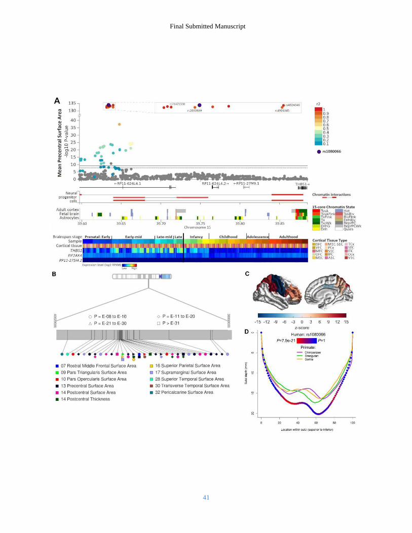
Final Submitted Manuscript
41

Final Submitted Manuscript
42