G ASTROINTESTINAL B LOCK P ATHOLOGY LECTURE 2014 Dr. Maha Arafah Dr. Ahmed Al Humaidi...
39
GASTROINTESTINAL BLOCK PATHOLOGY LECTURE 2014 Dr. Maha Arafah Dr. Ahmed Al Humaidi MalignantTumors of Intestine
-
Upload
emily-lindsey -
Category
Documents
-
view
217 -
download
0
Transcript of G ASTROINTESTINAL B LOCK P ATHOLOGY LECTURE 2014 Dr. Maha Arafah Dr. Ahmed Al Humaidi...

GASTROINTESTINAL BLOCK PATHOLOGY LECTURE 2014
Dr. Maha Arafah
Dr. Ahmed Al Humaidi
MalignantTumors of Intestine

Colon cancer:
Describe the epidemiology of colon cancer.
Compare the pathology (gross and microscopic features) and clinical features of right-sided colonic adenocarcinoma and left-sided colorectal adenocarcinoma.
Describe the relationship between prognosis and the various stages of cancer of the colon and rectum as noted in the TNM (tumor-nodes-metastasis) classification and staging system.
Describe the relationship between carcinoembryonic antigen (CEA) and recurrence following resection of the primary tumor.

TUMORS OF THE SMALL AND LARGE INTESTINES
PolypsCarcinoma Carcinoid tumorLymphoma

Adenocarcinoma of the colon is the most common malignancy of the GI tract and is a major cause of morbidity and mortality worldwide. In contrast, the small intestine, which accounts for 75% of the overall length of the GI tract, is an uncommon site for benign and malignant tumors. Among malignant small intestinal tumors, adenocarcinomas and well-differentiated neuroendocrine (carcinoid) tumors have roughly equal incidence, followed by lymphomas and sarcomas.
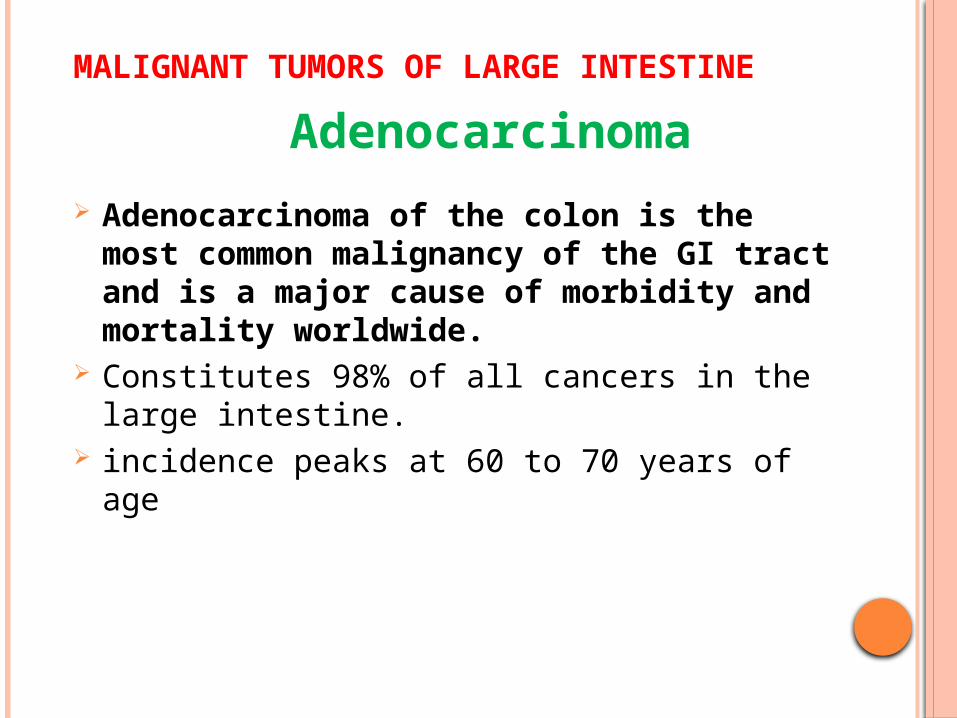
MALIGNANT TUMORS OF LARGE INTESTINE
Adenocarcinoma of the colon is the most common malignancy of the GI tract and is a major cause of morbidity and mortality worldwide.
Constitutes 98% of all cancers in the large intestine.
incidence peaks at 60 to 70 years of age
Adenocarcinoma

MALIGNANT TUMORS OF LARGE INTESTINE
Predisposing factors1. IBD, adenomas, polyposis syndrome.2. Diet appears to play an important role in the risk
for colon cancer:- Low fibre diet.- High fat content.
Adenocarcinoma
- Alcohol - Reduced intake of vit A, C &
E.- It is theorized that reduced fiber content leads to decreased stool bulk and altered composition of the intestinal microbiota. This change may increase synthesis of potentially toxic oxidative by-products of bacterial metabolism, which would be expected to remain in contact with the colonic mucosa for longer periods of time as a result of reduced stool bulk. High fat intake also enhances hepatic synthesis of cholesterol and bile acids, which can be converted into carcinogens by intestinal bacteria.

Several epidemiologic studies suggest that aspirin or other NSAIDs have a protective effect. This is consistent with studies showing that some NSAIDs cause polyp regression in FAP patients in whom the rectum was left in place after colectomy.

ADENOCARCINOMA OF LARGE INTESTINE
Carcinogenesis Two pathogenetically distinct pathways
for the development of colon cancer, both seem to result from accumulation of multiple mutations: 1- The APC/B-catenin pathway ( 85 % )2- The DNA mismatch repair genes pathway

ADENOCARCINOMA OF LARGE INTESTINE
Carcinogenesis 1- The APC/B-catenin pathway ( 85 % )
chromosomal instability that results in stepwise accumulation of mutations in a series of oncogenes and tumor suppressor genes.
adenoma-carcinoma sequence

FAMILIAL ADENOMATOUS POLYPOSIS
Hereditary mutation of the APC gene is the cause of familial adenomatous polyposis (FAP), where affected individuals carry an almost 100% risk of developing colon cancer by age 40 years.

MALIGNANT TUMORS OF LARGE INTESTINEADENOCARCINOMA
Carcinogenesis
2- The DNA mismatch repair genes pathway(These are referred to as MSI high, or MSI-H, tumors:)10% to 15% of sporadic cases.There is accumulation of mutations (as in the
APC/B-catenin schema) Five DNA mismatch repair genes (MSH2,
MSH6, MLH1, PMS1, AND PMS2) give rise to the hereditary non polyposis colon
carcinoma (HNPCC) syndrome.
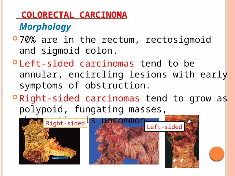
COLORECTAL CARCINOMA Morphology
70% are in the rectum, rectosigmoid and sigmoid colon.
Left-sided carcinomas tend to be annular, encircling lesions with early symptoms of obstruction.
Right-sided carcinomas tend to grow as polypoid, fungating masses, obstruction is uncommon.
Left-sidedRight-sided

COLORECTAL CARCINOMA MORPHOLOGY
Adenocarcinoma Mucinous adenocarcinoma secret abundant
mucin that may dissect through cleavage planes in the wall.

SIGNS AND SYMPTOMS
If located closer to the anus: change in bowel habit, feeling of incomplete defecation, PR bleeding
A tumor that is large enough to fill the entire lumen of the bowel may cause bowel obstruction
Right-sided lesions are more likely to bleed while left-sided tumors are usually detected later and could present with bowel obstruction.

Serum levels of carcinoembryonic antigen (CEA) are related to tumor size and extent of spread. They are helpful in monitoring for recurrence of tumor after resection.
COLORECTAL CARCINOMA

TNM STAGING OF COLON CANCERS IS USED FOR STAGING


COLORECTAL CARCINOMA
The two most important prognostic factors are depth of invasion and the presence or absence of lymph node metastases.


MALIGNANT SMALL INTESTINAL NEOPLASMS
In descending order of frequency: CarcinoidAdenocarcinomasLymphomasLeiomyosarcomas.

SMALL INTESTINAL NEOPLASMS
Carcinoid Tumors Neoplasms arising from endocrine cells found
along the length of GIT mucosa. The peak incidence: sixth decade, but they may
appear at any age. They compose less than 2% of colorectal
malignancies almost half of small intestinal malignant tumors:
60 to 80% appendix and terminal ileum 10 to 20% rectum.

CARCINOID TUMORS BEHAVIOR
Aggressive behavior correlates with:1. Site of origin:
Appendiceal and rectal carcinoids infrequently metastasize, even though they may show extensive local spread
90% of ileal, gastric, and colonic carcinoids that have penetrated halfway through the muscle wall have spread to lymph nodes and distant sites at the time of diagnosis, especially those larger than 2 cm in diameter.
2. Depth of local penetration3. Size of the tumor

SMALL INTESTINAL NEOPLASMS Carcinoid Tumors
Morphology A solid, yellow-tan appearance The cells are monotonously similar, having a scant,
pink granular cytoplasm and a round-to-oval stippled nucleus.
Ultrastructral features: neurosecretory electron dense bodies in the cytoplasm

SMALL INTESTINAL NEOPLASMS
Carcinoid TumorClinical features
Asymptomatic May cause obstruction, intussusception or
bleeding. May elaborate hormones: Zollinger-Ellison,
Cushing’s carcinoid or other syndromes.

SMALL INTESTINAL NEOPLASMS
Carcinoid tumor
Carcinoid syndrome
1% of carcinoid tumor & in 20% of those of widespread metastasis
Paroxymal flushing, episodes of asthma-like wheezing, right-sided heart failure, attacks of watery diarrhea, abdominal pain,
The principal chemical mediator is serotonin The syndrome is classically associated with
ileal carcinoids with hepatic metastases.
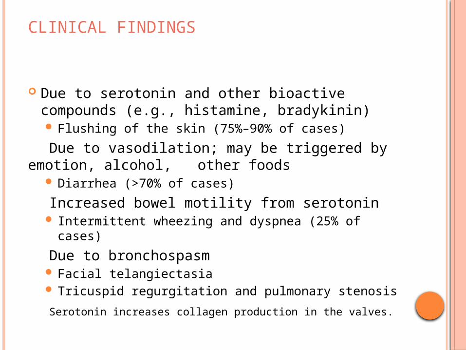
CLINICAL FINDINGS
Due to serotonin and other bioactive compounds (e.g., histamine, bradykinin) Flushing of the skin (75%–90% of cases)
Due to vasodilation; may be triggered by emotion, alcohol, other foods
Diarrhea (>70% of cases)
Increased bowel motility from serotonin Intermittent wheezing and dyspnea (25% of cases)
Due to bronchospasm Facial telangiectasia Tricuspid regurgitation and pulmonary stenosis
Serotonin increases collagen production in the valves.

SEROTONIN AND DIARRHEA
Patients with carcinoid syndrome often suffer from diarrhea, which has both a secretory and a motor component. The secretory component of carcinoid diarrhea is attributable to excessive serotonergic stimulation of submucosal secretomotor neurons; the motor component includes faster small bowel and colon transit and an exaggerated tonic response of the colon to ingestion of a meal

SMALL INTESTINAL NEOPLASMS
Lymphoma Most often low-grade lymphomas arising in
mucosal-associated lymphoid tissue (MALT) lymphoma or high-grade non-Hodgkin's lymphomas of B cell type.
May occur in any part of the intestine; The ileocecal region is a favored site for
Burkitt's lymphoma.


Left colon, carcinoma - Gross, mucosal surface
This specimen from the left colon shows an annular, encircling, and constricting cancer. The margins of the cancer are heaped-up and firm, and the mid-region is ulcerated.
Left-sided colon cancers come to attention by producing occult bleeding and changes in bowel habits (i.e., constipation and cramping in the left lower quadrant).

The carcinoma is composed of irregular glands infiltrating the muscularis propria, serosa, and mesentery. The TNM classification is based on the extent of invasion, number of lymph nodes involved, and extent of metastatic involvement. The deeper tumor extends into the muscularis propria, and as lymph nodes become involved, the prognosis worsens.
T—extent of invasion N—number of lymph nodes involved M—extent of metastatic involvement

Assuming this patient did not have lymph node metastasis, what stage is this carcinoma? The TNM stage for the current case would be T3N0MX.
T3—extends through the muscularis propria N0—no lymph node involvement MX—extent of metastatic involvement
unknown

malignant glands of an adenocarcinoma of the colon infiltrating the muscularis propria.
What is the mode of spread of this cancer? Colonic carcinomas spread by local extension to adjacent structures. The favored sites of metastases are regional lymph nodes, liver, lungs, and bones.

Cecal adenocarcinoma
Tumors in the proximal colon tend to grow as polypoid, fungating, ulcerating masses. Obstruction is uncommon. About 25% of colon carcinomas are located in the cecum or ascending colon. Note the adjacent pedunculated adenomatous polyp. Most colon cancers develop from adenomatous polyps (the adenoma-carcinoma sequence).
Cecal and right colon cancers most often come to clinical attention by the appearance of fatigue, weakness, and iron-deficiency anemia.


DIVERTICULOSIS