Fuzzy Image Segmentation with Fuzzy Labelled …...Fuzzy Image Segmentation with Fuzzy Labelled...
6
Fuzzy Image Segmentation with Fuzzy Labelled Neural Gas Cornelia Br¨ uß 1a , Felix Bollenbeck 1a , Frank-Michael Schleif 2 , Winfriede Weschke 1b , Thomas Villmann 3 , and Udo Seiffert 1a 1- Leibniz Institute of Plant Genetics and Crop Plant Research - (a) Pattern Recognition Group, (b) Gene Expression Group Corrensstraße 3, D-06466 Gatersleben - Germany 2- Bruker Daltonik GmbH Permoserstraße 15, D-04318 Leipzig - Germany 3- University Leipzig - Clinic for Psychotherapy Karl-Tauchnitz-Straße 25, D-04107 Leipzig - Germany Abstract. Processing biological data often requires handling of uncer- tain and sometimes inconsistent information. Particularly when coping with image segmentation tasks against biomedical background, a clear de- scription of for example tissue borders is often hard to obtain. On the other hand, there are only a few promising segmentation algorithms being able to process fuzzy input data. This paper describes one novel alterna- tive applying the recently introduced Fuzzy Labelled Neural Gas (FLNG) as subsequent classification step to a biologically relevant fuzzy labelling with underlying image feature extraction. 1 Introduction Biomedical data is often characterised by uncertain and possibly inconsistent information. This holds even more, among others, in the field of biomedical image processing. In the framework described by the present paper, automatic high-throughput segmentation of cross-section images is a crucial step of a rather complex processing chain and the prerequisite of a subsequent three-dimensional modelling. The objects used in the present paper for fuzzy labelling of biological struc- tures are serial transverse sections of barley grains at different developmental stages. Developing barley grains consist of three genetically different tissue types: the diploid maternal tissues, the filial triploid endosperm, and the diploid embryo. Because of their functionality, cells of a fully differentiated tissue show differences in cell shape and water content and accumulate different compounds. Based on those characteristics, scientists experienced in histology are able to identify and to label differentiated tissues within a given section of a developing grain (segmentation). However, differentiating cells lack these characteristics. Because differentia- tion occurs along gradients, especially borders between different tissue types of developing grains often consist of differentiating cells, which cannot be identi- fied as belonging to one or the other tissue type. Positions of those ”un-sharp” ESANN'2006 proceedings - European Symposium on Artificial Neural Networks Bruges (Belgium), 26-28 April 2006, d-side publi., ISBN 2-930307-06-4. 563
Transcript of Fuzzy Image Segmentation with Fuzzy Labelled …...Fuzzy Image Segmentation with Fuzzy Labelled...
Fuzzy Image Segmentationwith Fuzzy Labelled Neural Gas
Cornelia Bruß1a, Felix Bollenbeck1a, Frank-Michael Schleif2, Winfriede Weschke1b,Thomas Villmann3, and Udo Seiffert1a
1- Leibniz Institute of Plant Genetics and Crop Plant Research -(a) Pattern Recognition Group, (b) Gene Expression Group
Corrensstraße 3, D-06466 Gatersleben - Germany
2- Bruker Daltonik GmbHPermoserstraße 15, D-04318 Leipzig - Germany
3- University Leipzig - Clinic for PsychotherapyKarl-Tauchnitz-Straße 25, D-04107 Leipzig - Germany
Abstract. Processing biological data often requires handling of uncer-tain and sometimes inconsistent information. Particularly when copingwith image segmentation tasks against biomedical background, a clear de-scription of for example tissue borders is often hard to obtain. On theother hand, there are only a few promising segmentation algorithms beingable to process fuzzy input data. This paper describes one novel alterna-tive applying the recently introduced Fuzzy Labelled Neural Gas (FLNG)as subsequent classification step to a biologically relevant fuzzy labellingwith underlying image feature extraction.
1 Introduction
Biomedical data is often characterised by uncertain and possibly inconsistentinformation. This holds even more, among others, in the field of biomedicalimage processing. In the framework described by the present paper, automatichigh-throughput segmentation of cross-section images is a crucial step of a rathercomplex processing chain and the prerequisite of a subsequent three-dimensionalmodelling.
The objects used in the present paper for fuzzy labelling of biological struc-tures are serial transverse sections of barley grains at different developmentalstages. Developing barley grains consist of three genetically different tissuetypes: the diploid maternal tissues, the filial triploid endosperm, and the diploidembryo. Because of their functionality, cells of a fully differentiated tissue showdifferences in cell shape and water content and accumulate different compounds.Based on those characteristics, scientists experienced in histology are able toidentify and to label differentiated tissues within a given section of a developinggrain (segmentation).
However, differentiating cells lack these characteristics. Because differentia-tion occurs along gradients, especially borders between different tissue types ofdeveloping grains often consist of differentiating cells, which cannot be identi-fied as belonging to one or the other tissue type. Positions of those ”un-sharp”
ESANN'2006 proceedings - European Symposium on Artificial Neural NetworksBruges (Belgium), 26-28 April 2006, d-side publi., ISBN 2-930307-06-4.
563
borders depend on the tissue type under consideration and, additionally, on thedevelopmental stage.
Seeds are sink tissues, i.e. development requires import of assimilates fromthe photosynthetic active vegetative parts of the plant. Assimilate import isdetermined by the vascular tissues and regulated by the so-called maternal-filialboundary consisting of nucellar projection and endosperm transfer cells. Typeand amount of incoming assimilates change during development and determinein this way differentiation of the filial seed part. The changing assimilate com-position is determined by development-specific changes of the maternal-filialboundary resulting from lasting differentiation processes. Especially cells sur-rounding the vascular bundle and connecting vascular tissues to the nucellarprojection show different shape during different developmental stages. There-fore, unequivocal segmentation of this grain part at a given developmental stageis not possible. Thus, fuzzy processing is highly desirable.
In order to incorporate the required fuzzy image segmentation, there are gen-erally a number of alternatives [1]. However, since (training) examples, manuallylabelled by a biological expert, are generally available and have to be used totransfer the expert knowledge to the automatic solution, the use of supervisedmethods seems more natural than of unsupervised techniques [2]. Furthermore,neither approaches requiring extensive a-priori knowledge about the areas to besegmented [3] nor morphology based solutions [4] are applicable in the case con-sidered in the present paper. Besides some rule-based techniques, or using fuzzyintegrals, or measures of fuzziness (e.g. fuzzy entropy) and image information(e.g. fuzzy divergence) [5], particularly artificial neural network (ANN) basedsolutions offer promising approaches [6, 7].
Here, similar to many other crisp as well as fuzzy segmentation methods1,a two-stage system is applied, where a set of significant features is extractedfrom the images and then clustered (unsupervised), or, as in this case, classified(supervised). Due to their adaptive behaviour, there are numerous applicationsof ANNs in this context. However, this mainly concerns only crisp segmentation,although there are some neural network paradigms accepting fuzzy input data [8,9, 10].
With the recently suggested Fuzzy Labelled Neural Gas (FLNG) [11] also aprototype-based neural network became available for this purpose now. Afterbriefly introducing an interactive editor to obtain expert labelled training datain the next section, Sect. 3 briefly reviews the FLNG algorithm. Then someresults applying the system to biomolecular real-world data are given.
1Whereas the term crisp commonly refers in this context to segmentation with clear andstrict boundaries between different areas, fuzzy means a gradual transition between adjacentareas.
ESANN'2006 proceedings - European Symposium on Artificial Neural NetworksBruges (Belgium), 26-28 April 2006, d-side publi., ISBN 2-930307-06-4.
564
2 Providing fuzzy labelled expert data
2.1 A biologically plausible fuzzy label editor
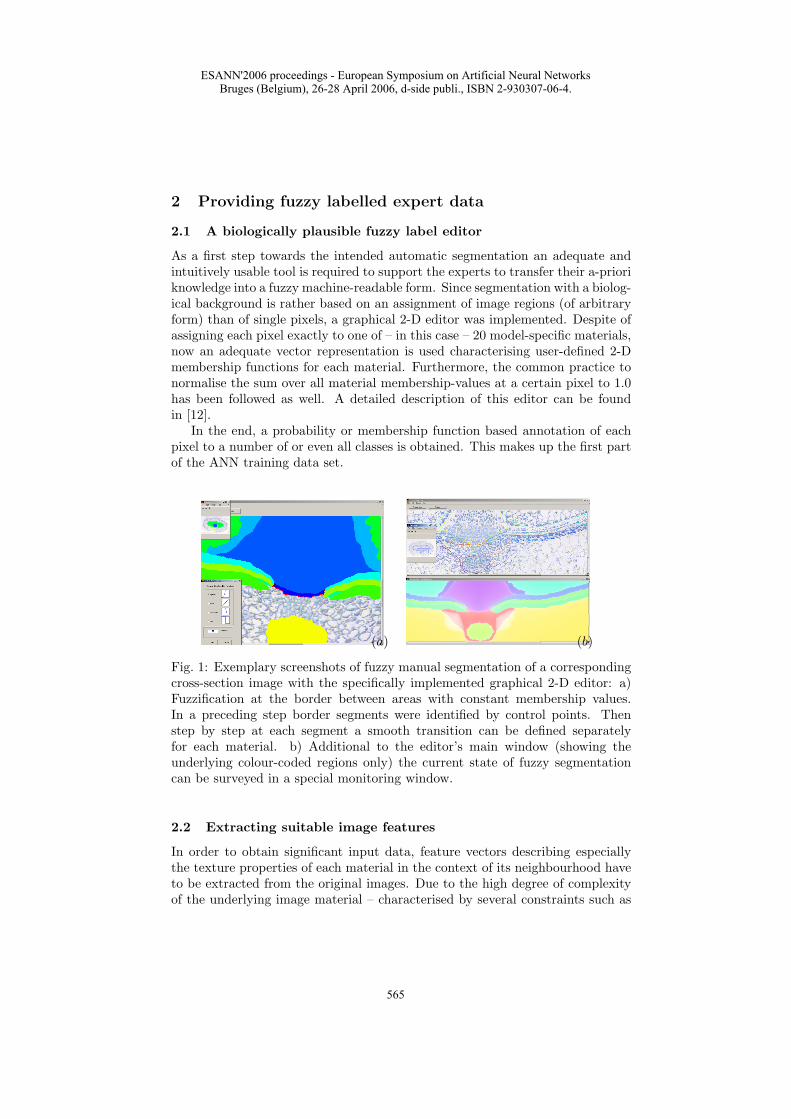
As a first step towards the intended automatic segmentation an adequate andintuitively usable tool is required to support the experts to transfer their a-prioriknowledge into a fuzzy machine-readable form. Since segmentation with a biolog-ical background is rather based on an assignment of image regions (of arbitraryform) than of single pixels, a graphical 2-D editor was implemented. Despite ofassigning each pixel exactly to one of – in this case – 20 model-specific materials,now an adequate vector representation is used characterising user-defined 2-Dmembership functions for each material. Furthermore, the common practice tonormalise the sum over all material membership-values at a certain pixel to 1.0has been followed as well. A detailed description of this editor can be foundin [12].
In the end, a probability or membership function based annotation of eachpixel to a number of or even all classes is obtained. This makes up the first partof the ANN training data set.
(a) (b)
Fig. 1: Exemplary screenshots of fuzzy manual segmentation of a correspondingcross-section image with the specifically implemented graphical 2-D editor: a)Fuzzification at the border between areas with constant membership values.In a preceding step border segments were identified by control points. Thenstep by step at each segment a smooth transition can be defined separatelyfor each material. b) Additional to the editor’s main window (showing theunderlying colour-coded regions only) the current state of fuzzy segmentationcan be surveyed in a special monitoring window.
2.2 Extracting suitable image features
In order to obtain significant input data, feature vectors describing especiallythe texture properties of each material in the context of its neighbourhood haveto be extracted from the original images. Due to the high degree of complexityof the underlying image material – characterised by several constraints such as
ESANN'2006 proceedings - European Symposium on Artificial Neural NetworksBruges (Belgium), 26-28 April 2006, d-side publi., ISBN 2-930307-06-4.
565
incomplete expert knowledge, subjectiveness and often hardly distinguishablematerials – the segmentation was based on a pixel-wise classification.
For the proof of concept the focus was not yet on an exhausting search for theoptimal features for the application of the FLNG classifier to our particular imagedata. Instead, for simplification and comparison reasons an existing feature setwas first utilised, which was formerly successfully used for several crisp ANNclassifiers. This initial feature vector holds 170 properties concerning colour,geometry and symmetry (such as Cartesian and polar coordinates, distance tocentroid, absolute angle to symmetry axis) and particularly texture accordingto varying neighbourhoods (such as Gaussian filters, histogram based features).All features were z-score-transformed to normalise the attributes.
3 Fuzzy classification using FLNG
Fuzzy labelled neural gas (FLNG) is an extension of the well-known prototypebased vector quantization neural gas algorithm [13]. It belongs to gradient de-scent supervised learning schemes [11, 14]. Here, the data v ∈ D ⊆Rd areequipped with class labels, which are fuzzy: for each class k we have the pos-sibilistic assignment xk ∈ [0, 1] collected in the label vector x =(x1, . . . , xNc)as described in Sect 2.1. Nc is the number of possible classes – in the presentcase up to 20. The prototypes wi ∈Rd, i ∈ A, now are featured with fuzzylabels yi =
(yi1, . . . , y
iNc
), too. Further, we assume an arbitrary differentiable,
maybe parameterized, quadratic distance measure ξλ (v,wi) in the data spacewith parameters λ =(λ1, . . . , λm). The cost function of the algorithm is definedas a balanced combination of the cost function ENG of NG and an additionalterm EFL according to the classification accuracy:
EFLNG = (1− β) ENG + βEFL. (1)
Thereby,
ENG =1
2C (σ)
∑
j
∫P (v)hσ (v,wj) ξλ
(v,wj
)dv (2)
is the cost function of NG with a rank based neighborhood function hσ (v,wj)and EFL is defined as
EFL =12
∑
j
∫P (v) gγ (ξλ (v,wi))
(x− yj
)2dv (3)
where gγ
(v,wj
)is a Gaussian kernel describing a neighborhood range in the
data space
gγ
(v,wj
)= exp
(−ξλ (v,wi)
2γ2
). (4)
Note that the kernel gγ depends on the prototype locations, such that EFL isinfluenced by both wi and y. Formal derivation yields
∂EFLNG
∂wk= (1− β)
∂ENG
∂wk+ β
∂EFL
∂wkand
∂EFLNG
∂yk= β
∂EFL
∂yk(5)
ESANN'2006 proceedings - European Symposium on Artificial Neural NetworksBruges (Belgium), 26-28 April 2006, d-side publi., ISBN 2-930307-06-4.
566
which yields
4wi = −εhσ (v,wi)∂ξλ (v,wi)
∂wi+
β
4γ2gγ (v,wi)
∂ξλ (v,wi)∂wi
(x− yi)2 (6)
and4yi = εlβgγ (v,wi) (x− yi) (7)
as learning rules. The respective gradient yields
4λk =∂ξλ
(v,wj
)
∂λk
((1− β)2C (σ)
hσ (v,wj)− β
4γ2gγ
(v,wj
) (x− yj
)2)
(8)
for adaptation. Further details can be found in [11].
4 Results
(a) (b) (c) (d)
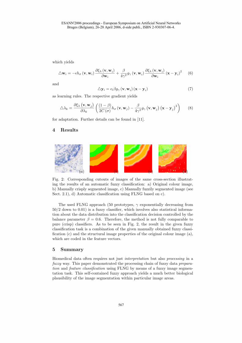
Fig. 2: Corresponding cutouts of images of the same cross-section illustrat-ing the results of an automatic fuzzy classification: a) Original colour image,b) Manually crisply segmented image, c) Manually fuzzily segmented image (seeSect. 2.1), d) Automatic classification using FLNG based on c).
The used FLNG approach (50 prototypes, γ exponentially decreasing from50/2 down to 0.01) is a fuzzy classifier, which involves also statistical informa-tion about the data distribution into the classification decision controlled by thebalance parameter β = 0.6. Therefore, the method is not fully comparable topure (crisp) classifiers. As to be seen in Fig. 2, the result in the given fuzzyclassification task is a combination of the given manually obtained fuzzy classi-fication (c) and the structural image properties of the original colour image (a),which are coded in the feature vectors.
5 Summary
Biomedical data often requires not just interpretation but also processing in afuzzy way. This paper demonstrated the processing chain of fuzzy data prepara-tion and feature classification using FLNG by means of a fuzzy image segmen-tation task. This self-contained fuzzy approach yields a much better biologicalplausibility of the image segmentation within particular image areas.
ESANN'2006 proceedings - European Symposium on Artificial Neural NetworksBruges (Belgium), 26-28 April 2006, d-side publi., ISBN 2-930307-06-4.
567
Acknowledgement
This work was supported by a grant of the German Federal Ministry of Educationand Research (No. 0312706A).
References
[1] E.E. Kerre and M. Nachtegael, editors. Fuzzy Techniques in Image Processing, volume 52of Studies in Fuzziness and Soft Computing. Springer Verlag, Heidelberg, 2000.
[2] H. Caillol, W. Pieczynski, and A. Hillion. Estimation of fuzzy Gaussian mixture andunsupervised statistical image segmentation. IEEE Transactions on Image Processing,6(3):425–440, 1997.
[3] M.A. Ali, L.S. Dooley, and G.C. Karmakar. Fuzzy image segmentation combing ringand elliptic shaped clustering algorithms. In H. Selvaraj, editor, Proceeding of the In-ternational Conference on Information Technology: Coding and Computing (ITCC’05),volume II, pages 118–122. 2005.
[4] I. Bloch, C. Pellot, F. Sureda, and A. Herment. Fuzzy modelling and fuzzy mathemat-ical morphology applied to 3D reconstruction of blood vessels by multi-modality datafusion. In R. Yager, D. Dubois, and H. Prade, editors, Fuzzy Set Methods in InformationEngineering: A Guided Tour of Applications, pages 93–110. John Wiley & Sons, 1996.
[5] M. Grabisch, T. Murofushi, and M. Sugeno. Fuzzy measure of fuzzy events defined byfuzzy integrals. Fuzzy Sets and Systems, 50:293–313, 1992.
[6] A.C.C. Reyes and M.E. Algorri. A combined algorithm for image segmentation usingneural networks and 3D surface reconstruction using dynamic meshes. Rev Mex IngBiomed, 21(3):73–81, 2000.
[7] G. Ou, Y.L. Murphey, and L. Feldkamp. Multiclass pattern classification using neuralnetworks. In Proceeding of the International conference on Pattern Recognition (ICPR2004), volume IV, pages 585–588, 2004.
[8] G.A. Carpenter. Distributed learning, recognition, and prediction by ART and ARTMAPneural networks. Neural Networks, 10:1473–1494, 1997.
[9] L. Cinque, G. L. Foresti, A. Gumina, and S. Levialdi. A modified fuzzy ART for im-age segmentation. In 11th International Conference on Image Analysis and Processing(ICIAP’01), page 102. 2001.
[10] L. Cinque, G. L. Foresti, and L. Lombardi. A clustering fuzzy approach for image seg-mentation. Pattern Recognition, 37:1797–1807, 2004.
[11] T. Villmann, B. Hammer, F.-M. Schleif, and T. Geweniger. Fuzzy Labeled Neural GASfor fuzzy classification. In Marie Cottrell, editor, Proceedings of the Workshop on Self-Organizing Maps WSOM, pages 283–290, 2005.
[12] C. Bruß, F. Bollenbeck, W. Weschke, and U. Seiffert. A graphical 2-D editor for fuzzyimage segmentation. Journal of Integrative Bioinformatics, to appear.
[13] T. Martinetz, S. Berkovich, and K. Schulten. ’Neural-gas’ network for vector quantizationand its application to time-series prediction. IEEE Trans. on Neural Networks, 4(4):558–569, 1993.
[14] B. Hammer, M. Strickert, and T. Villmann. Supervised neural gas with general similaritymeasure. Neural Processing Letters, 21(1):21–44, 2005.
ESANN'2006 proceedings - European Symposium on Artificial Neural NetworksBruges (Belgium), 26-28 April 2006, d-side publi., ISBN 2-930307-06-4.
568