Further Studies of Polymers as Carcinogenic Agents in...
14
Further Studies of Polymers as Carcinogenic Agents The inception of this investigation is an exam ple of serendipity, a word recently popularized to designate the faculty of accidentally making ob servations or discoveries which were not originally sought. In the present instance, a safe antihyper tensive compound was being sought among the quinones to reduce the high blood pressure pro duced by wrapping one or both kidneys of rats with cellophane. After a couple of years, in seven of these rats malignant sarcomas had developed at the site of the wrapping. In several instances, these sarcomas had extended into the peritoneal cavity and had also metastasized. In view of this unexpected finding, a new series of experiments was begun to investigate this phenomenon in van ous directions. It was found that sarcomas could be induced not only by wrapping the kidney in cellophane, but alternatively by imbedding the cellophane subcutaneously in the anterior abdom inal wall. By either of these methods, tumors were induced in approximately 35 per cent of the ani mals (10). Further work soon showed (11, 12) that we were dealing not merely with one more carcinogen, of which hundreds were already known, but that we had chanced upon an entirely new group of carcinogenic substances, the polymer films. In 1941 Turner (13) had observed, also accidentally, that disks of Bakelite implanted subcutaneously in rats produced fibnosarcomas. Subsequent to our original report in 1948, similar results have been obtained by Druckrey and Schmllhl (4, 5), Zol linger (15), Laskin, Robinson, andkWeinniann (7), and Bering'. While, thus, there can be no doubt * This investigation was supported by a research grant, No. C-1620 (C2-3), from the National Cancer Institute, National Institutes of Health, United States Public Health Service. 1 E. A. Bering, Jr., personal communication. Received for publication February 24, 1955. as to the actual facts, the interpretation of these facts as regards the mechanism or mode of action of polymers in inducing tumors is still obscure. The present paper describes the investigations which were pursued in an effort to gain a better understanding of the processes involved in the car cinogenic action of polymers. A study was made of the effects of a number of polymers having dif ferent chemical structures. Also, because the plas tics used in our earlier work were commercial products often containing plasticizers, stabilizers, traces of catalysts, a residual monomer, etc., we have imbedded a number of samples of polymers specially prepared to assure their purity. The pos sible carcinogenic effect of several monomers has also been investigated, and studies have been made on the degradation of polymers within the organism by means of polymers tagged with iso topic carbon. METHODS AND RESULTS IMBEDDING PROCEDURES In most experiments the animal chosen was the Wistar rat, but the Sherman strain was used in earlier tests, and for purposes of comparison ex peniments have also been done on mice of the Longacre, Paris, and C57 strains. In most expeni ments males were used, but, in some, females were employed with similar results. The animals were fed Purina Laboratory Chow and, occasionally, fresh carrots, and had free access to water. The general procedure was to use small squares or circles of film, averaging 1.5 cm. in width, which had been sterilized in Zephiran' (1 : 1000 dilution of the éommercial 12.8 per cent solution) for sev eral hours, and washed in sterile saline. These were inserted subcutaneously one on each side of the abdominal wall, just ventral to the fascia. 2 In the early experiments, sterilization was in 80 per cent alcohol. In a few cases films or powders have been sterilized by heat. 333 . A • 1* in @nimais B. S. OPPENHEIMER, ENIDT OPPENHEIMER, I. DANISHEFSKY, ARTHUR PURDY STOUT, AND FREDERICK R. EIRICH Wrm TUETECHNICAL ASSISTANCE OFMARGARET Wn.inrr.n (Institute of Cancer Research, and the Department of Biochemistry, College of Physicians and Surgeons, Columbia University, New York, N.Y.) on July 10, 2018. © 1955 American Association for Cancer Research. cancerres.aacrjournals.org Downloaded from
Transcript of Further Studies of Polymers as Carcinogenic Agents in...

Further Studies of Polymers as Carcinogenic Agents
The inception of this investigation is an example of serendipity, a word recently popularized todesignate the faculty of accidentally making observations or discoveries which were not originallysought. In the present instance, a safe antihypertensive compound was being sought among thequinones to reduce the high blood pressure produced by wrapping one or both kidneys of ratswith cellophane. After a couple of years, in sevenof these rats malignant sarcomas had developedat the site of the wrapping. In several instances,these sarcomas had extended into the peritonealcavity and had also metastasized. In view of thisunexpected finding, a new series of experimentswas begun to investigate this phenomenon in vanous directions. It was found that sarcomas couldbe induced not only by wrapping the kidney incellophane, but alternatively by imbedding thecellophane subcutaneously in the anterior abdominal wall. By either of these methods, tumors wereinduced in approximately 35 per cent of the animals (10).
Further work soon showed (11, 12) that wewere dealing not merely with one more carcinogen,of which hundreds were already known, but thatwe had chanced upon an entirely new group ofcarcinogenic substances, the polymer films. In1941 Turner (13) had observed, also accidentally,that disks of Bakelite implanted subcutaneouslyin rats produced fibnosarcomas. Subsequent to ouroriginal report in 1948, similar results have beenobtained by Druckrey and Schmllhl (4, 5), Zollinger (15), Laskin, Robinson, andkWeinniann (7),and Bering'. While, thus, there can be no doubt
* This investigation was supported by a research grant,
No. C-1620 (C2-3), from the National Cancer Institute,National Institutes of Health, United States Public HealthService.
1 E. A. Bering, Jr., personal communication.
Received for publication February 24, 1955.
as to the actual facts, the interpretation of thesefacts as regards the mechanism or mode of actionof polymers in inducing tumors is still obscure.
The present paper describes the investigationswhich were pursued in an effort to gain a betterunderstanding of the processes involved in the carcinogenic action of polymers. A study was madeof the effects of a number of polymers having different chemical structures. Also, because the plastics used in our earlier work were commercialproducts often containing plasticizers, stabilizers,traces of catalysts, a residual monomer, etc., wehave imbedded a number of samples of polymersspecially prepared to assure their purity. The possible carcinogenic effect of several monomers hasalso been investigated, and studies have beenmade on the degradation of polymers within theorganism by means of polymers tagged with isotopic carbon.
METHODS AND RESULTSIMBEDDING PROCEDURES
In most experiments the animal chosen was theWistar rat, but the Sherman strain was used inearlier tests, and for purposes of comparison expeniments have also been done on mice of theLongacre, Paris, and C57 strains. In most expeniments males were used, but, in some, females wereemployed with similar results. The animals werefed Purina Laboratory Chow and, occasionally,fresh carrots, and had free access to water.
The general procedure was to use small squaresor circles of film, averaging 1.5 cm. in width, whichhad been sterilized in Zephiran' (1 : 1000 dilutionof the éommercial 12.8 per cent solution) for several hours, and washed in sterile saline. Thesewere inserted subcutaneously one on each side ofthe abdominal wall, just ventral to the fascia.
2 In the early experiments, sterilization was in 80 per centalcohol. In a few cases films or powders have been sterilizedby heat.
333
. A • 1*in @nimais
B. S. OPPENHEIMER,ENIDT OPPENHEIMER,I. DANISHEFSKY,ARTHUR PURDY STOUT, AND FREDERICK R. EIRICH
Wrm TUETECHNICALASSISTANCEOFMARGARETWn.inrr.n
(Institute of Cancer Research, and the Department of Biochemistry, College of Physicians and Surgeons,Columbia University, New York, N.Y.)
on July 10, 2018. © 1955 American Association for Cancer Research. cancerres.aacrjournals.org Downloaded from

Cancer Research334
With few exceptions, each experiment consistedof 50 imbeddings; sometimes the same materialwas imbedded on both sides of 2.5 animals, andsometimes one form of the material was imbeddedon the right side and another on the left. The animals were kept under close observation for severalweeks, until the incisions were completely healed,and subsequently were examined weekly to determine the onset of tumors.
PLASTICS IMBEDDED
Most of the plastics imbedded were obtainedthrough the kindness of various industrial firms.The designation “commercial―implies those manufactured for ordinary industrial purposes, witha more or less unknown content of nonpolymericmaterial. Of the “pure―forms, some have beenspecially made for us by the manufacturers andothers prepared or purified in our own laboratory.
The following are the plastics which were imbedded:
Cellophane A .5—acommercial sausage casing.Cellophane B.—the same material after extrac
tion with alcohol for 3 days.Cellophane C.—Cellophane B after additional
extraction with benzene.Cellophane D.—a special form employed for tis
sue culture work.Cellophane 0.—a Cellophane of the highest pu
rity obtainable which was kept in formalin andwashed just before imbedding.
Dacron, KS.F, Pliojilm, Saran, SilastiC, andT@fion.—allcommercial products.
Nylon.—the purest form obtainable, said to contamnoadditives.
Polyethylene A.—acommercial film.Polyethylene B.—specially prepared for us by
the manufacturers, said to contain no additivesand only a trace of catalyst.
Polyethylene HM (high molecular).—hadall thelow molecular weight fractions removed.
Polymethyl nwthacrylale A.—cast from a cornmercial product.
Polymethyl m€tI&aCrIJlateD.—preparedas followslows : freshly distilled monomer, methyl methacrylate, was sealed in an evacuated glass tube at4 X 10—imm. pressure and kept at 80°C. for 2weeks. The resulting polymer was purified by solution in methyl ethyl ketone and three reprecipitations with methanol. The film was made bycasting from solutions of methyl ethyl ketone. Nocatalysts or additives were employed in this preparation.
“Perspex.―—anEnglish preparation of polymethyl methacrylate, said to be specially pure.
Polyvinyl c/zkri4e A.—a commercial product,known to contain some additives.
Polyvinyl chloride D.—specially prepared for usby ultraviolet polymerization of vinyl chloride.Contains no plasticizers, catalysts, or other additives.
Polystyrene A.—a commercial form.Polystyrene D.—prepared from its monomer,
styrene, by a process similar to that used for polymethyl methacrylate D. It likewise contains nocatalyst or additives.
Ivalon sponge and Vinyon N.—both manufactured for surgical use.
Silk film was made from natural silk fiber bydissolving it in an aqueous solution of lithium bromide, removing the latter by dialysis, and castingon glass.4
As will be seen from the accompanying tables,these films differed greatly both in thickness andflexibility, varying from the delicate pliable membrane of polystyrene, only 0.01 mm. thick, to therigid disk of “Perspex,―with a thickness nearly40 times as great.
Further variations in physical form were introduced by using some of the polymers in the shapeof textile fabrics (fibers), perforated films (625holes per square inch), granules, sponges, and powders. Many of these experiments, and other modiflcations devised to test one theory or another,have been too recently begun for any conclusionsto be drawn.
Rueuvrs or IMBEDDING
The first demonstrable effect of imbedding aplastic film was found to be the encapsulation ofthe film in a sac or pocket of connective tissue ofvarying thickness (Fig. 1). With some films, e.g.,Cellophane, the pocket wall was thick and denseand sometimes even contained calcareous areas;while with other films, such as Pliofilin or polystyrene, the pocket wall was thin and soft. This
encapsulation was evident within 2—3weeks afterimbedding, and was found in animals of all agesexcept in cases where a tumor was induced.
The film was never adherent to the pocket butcould be easily removed, leaving the pocket wallintact. Thin, pliable films were sometimes foundfolded or rolled, but always in a pocket, sometimeswith fibrous membranes between the folds. Atautopsy brittle films would occasionally be foundbroken, with the broken pieces either all in onepocket, or, less often, encapsulated separately.
4 For this film we are indebted to Dr. Peter Alexander of
the Chester Beatty Research Institute, London.a The designations “A,―“B,―“C,―“D,―and “0―are
arbitrary and for convenience of reference only.
on July 10, 2018. © 1955 American Association for Cancer Research. cancerres.aacrjournals.org Downloaded from

OPPENHEIMER ei al.—Polymers as Carcinogenic Agents 335
With perforated films and also with textiles, nodefinite pocket was formed, but the material wasfound enmeshed with connective tissue fiberswhich penetrated through the perforations, orbetween the textile threads, holding the plasticfirmly in place.
In cases where tumors were produced the earlystages could be palpated as a thickening or swelling around the film. The tumors grew rapidly andwere usually large enough for removal (2 cm. ormore in diameter) in 2 or 3 weeks after their firstappearance. Since in most experiments films hadbeen imbedded on both sides of the abdominalwall, the first tumor to appear was remoyed underaseptic conditions and the animal allowed to survive, in order to give an opportunity for growthof a tumor on the opposite side. Sometimes a regrowth of the first tumor occurred at or near thesame site, and in three of these cases metastaticgrowths were found in the lung. In one or two instances in our early experiments, where tumorswere allowed to grow for many months for purposes of gross demonstration, metastases occurredin axillary lymph nodes; but, in general, metastaseswere rarely seen.
The tumors were usually located entirely in thesubcutaneous layers, but were occasionally foundto have penetrated the outer layers of the musclewall, or, very rarely, to have grown through themuscle into the peritoneal cavity. In a few instances, a tumor was found to have ulcerated outwards through the skin.
In most cases the tumor was found to surroundthe film more or less completely, though sometimes a portion of the film would be found projecting from the tumor, or the tumor would havegrown only on one side of the film.
It is noteworthy that when a tumor developedno fibrous capsule could be found, but tumor cellswere seen immediately adjacent to the plastic(Fig. 2), sometimes surrounding it on both sides,sometimes on one side only, which might be eitherthe inner (muscle) or the outer (skin) side. If thetumor had grown on only one side of the film, thena connective tissue wall was present on the otherside. Nothing definite is known as to how the connective tissue capsule disappears from the vicinityof the film. Its cells may be converted into tumorcells, or the capsule may be pushed away by tumor tissue growing between it and the film, or itmay be destroyed by pressure infiltraton. In oneinstance where the capsule had not entirely disappeared, tumor tissue was found on the inner(film) side of the capsular remnant, as well as onits outer side and enmeshed within it. This suggested that, in this particular instance, the cap
sule was in the process of destruction and replacement by infiltration and pressure.
The numbers of malignant tumors obtained bythese imbedding experiments are given in Tables1 and 2.
Since these are all long-term experiments, witha latent period varying from 1 to 2 years after irnbedding before the appearance of tumors, manyof the experiments are still unfinished, and merelythe number of tumors produced to date can be recorded. Many other experiments are in progress,but since in these no tumors have so far been produced no results can be given.
Table 1 shows the completed experiments, withthe number of tumors produced and the percentage production calculated from the number of animals surviving the minimum latent period for tumor appearance. Only tumors arising around, orin direct contact with, the imbedded polymer wereincluded as having been induced by the plastic.Any other tumors appearing in the experimentalanimal were interpreted as “spontaneous,―andthese appeared in about 2 per cent of the animals.
Including the kidney-wrapping experiments(10), our observations show 275 primary malignant tumors induced by plastics. All of these tumors were mesenchymal in origin : the large majority (over 8@5per cent) were fibrosarcomas, butother types, particularly osteogenic sarcomas andrhabdomyosarcomas, were also obtained (Figs.3a to 6). The complete list of types obtained follows:
FibrosarcomaOsteogenic sarcomaRhabdomyosarcomaMesenchymomaLiposarcomaReticulum-cell sarcomaMyxomaPlasmocytomaHistiocytoma (malignant)
The evidence of malignancy was based on histological findings, including the frequency of mitoses, on transplantability, on occasional metastases, and on frequent local recurrence after removalof a primary tumor. The somewhat infrequentoccurrence of metastases from the primary fibrosarcomas may be partly explained by the factthat the tumors were usually removed 2—3weeksafter their first appearance.
The tables show that tumors are induced whether the film is thick or thin, flexible or rigid, andthat a plain film appears to induce more tumorsthan other forms such as perforated films, textiles,or powders. So far we have obtained no tumors
23512
86‘5‘5211
275
on July 10, 2018. © 1955 American Association for Cancer Research. cancerres.aacrjournals.org Downloaded from

336 Cancer Research
with plastics in powder form, but only one of these “linters―(the fibers from which our Cellophane Aexperiments is completed, and little significance was made), surgical cotton, glass cloth, and a numshould be attached to the observation as yet. ber of metal foils. Two natural polymers, keratin
(foetal nails) and a thin collagen film, were also imCONTROLS bedded.
As control experiments we imbedded a variety Of the completed controls, as previously reof nonplastic materials, including glass coverslips, ported (12), linters and surgical cotton producedslips of wood and mica, pellets of paraffin, cotton no tumors, but with the glass coverslip there was
TABLE I
REsuLTsOF1MBEDDINGPlASTIcSSUBCUTANEOUSLYIN RODENTSCompleted Experiments
No.atmsURVIVING
MINIMUM LLTRNT MAlIGNANT
LATIDIT PZRIODR TUMORS
PERIOD (ntTs) No. Per cent
42 495—779 15 85.7sot 245—498 8 22.822@ 869 1 4.544 822—665 20 45.489 890-706 18 46.110 428—521 3 15.822 504—594 4 18.2
DIZCRIPTION 07 FILMThid@neaa
(mm.) Flexibility
0 .04 Flexible0.04 Flexible0 .04 Flexible0 .04 Flexible0 .04 Flexible0 01 Very flexible0 .01 Very flexible
0 .02 Very flexible0 .02 Very flexible0.05 Very soft
Soft, flexible
0.02 Flexible
0 .06 Flexible0.06 Flexible0.08 Soft
0.01 Soft,pliable
0.05 Flexible0 .02 Very soft, pliable0.02 Very soft, pliable0 .02 Very soft, pliable0.15 Rather stifftex
ure0 .07 Flexible
MATERIAL IMBZDDZD
CELLOPHANE:A plainfilmA plainfilmA plainfilmB plainfilmC plainfilmD plainfilmD perforated film
DACRON:Plain filmPerforated filmTextile
IVALON SPONGE
NYLON:PlainfilmPerforated filmTextile
PL1OFILM:Plain
POLYETHYLENE:A plainfilmB plainfilmB plain filmB perforatedfilmB textile
SARAN:Plain film
SILAST1C:Plainfilm
41 830—698 8 19.542 827—651 2 4.888 0 0.0
84 567—857 8 8.8
80 250—581 7 28.8
26 441—651 7 27.081 511—788 2 6.588 0 0.0
46 859—708 8 15.0
80 892—722 10 12.555 885—742 11 20.020t 848—545 8 10.841 407—784 6 14.640 497 1 2.5
84 852—588 8 8.842 0 0.0
KEL-F:Plain film
HM plainfilmPowder
POLYMETHYLMETRACRYLATE:A plainfilm
POLYSTYRENE:A plainfilm
POLYVINYLCHLORIDE:A plainfilmA perforatedfilm
0.14 Rigid,brittle
0.01 Soft, pliable
0 .04 Soft, pliable0.04 Soft, pliable
0 .02 Soft, pliable
0.25 Rubbery
20 581—658 4 20.0
27 859—556 7 25.9
44 189—727 17 88.627 0 0.0
42 800-847 5 11.9
85 800—609 14 40.0
84 480—748 8 28.582 526-728 6 18.7
TEFLON:Plainfilm 0.02 FlexiblePerforated film 0. 02 Flexible
S Except where noted to the @ntrsry, cli animals were Wistar rats.
t Albino(Longacre)micewereused.* Black (C57) mice were used.
on July 10, 2018. © 1955 American Association for Cancer Research. cancerres.aacrjournals.org Downloaded from

OPPENHEIMER et aL—Polyiners as Carthwgeni@ Agents 337
a single fibrosarcoma, appearing in the last sur@viving rat of fifty, 659 days after imbedding. Anexperiment with tin foil and one with a glass textile (fiberglas) have recently terminated, and notumors were obtained; but the other experimentsare still unfinished, and have been in progress tooshort a time to have any significance.
MoNoissats
The possibility that the carcinogenic activityof the polymers may be due to some monomerentrapped in the film, or formed as a result of polymer breakdown, cannot be disregarded. Three ex
induced so by far these materials, but the experiment is not terminated. Pellets which were removed from the animals three or more monthsafter imbedding contained no benzoyl peroxide.This indicates that the material had been cornpletely absorbed, decomposed, or both, within thisperiod. Conclusions which may be drawn fromthese experiments are suggested in the “Discussion.―
Exiummszwrs wrv@TAooEDPOLYMERS
To ascertain whether plastic films, although soinert, undergo any changes in the animal body,
TABLE 2
Ri@suLTsOFIMBEDDINGPLASTIcSSUBCUTANEOUSLYIN RODENTSExperiments in Progress
No. @usoNAN? IVMOSSPRODUORDUP
TO 5/1/55
LLTRST
PZRIOD
(atm)
DxscaniicscOFFILMThickness Flexibility
(mm.)
0.04 Flexible
0. 08 Slightly stiff0.89 Rigid
0.06 Rigid
0.04 Flexible
0. 08 FlexibleIrregular Brittle
0.10 Flexible0.07 Flexible
DAm
IMBZDDRD
10/17/52
2/18/58
5/5/581/28/83
1/14/58
3/26/53
3/6/580/16/58
10/1/534/29/53
12/2/58
IMBZDDRD
CELLOPHANE APerforated film
CELLOPHANE 0Plain film
PERSPEXPOLYMETHYLMETHACRYLATE D
PlainflimPOLYSTYRENE D
Plain filmPOLYVINYLCHLORIDE D
Plain filmSILK FILMVINYON N
P@n filmPOLYETHYLENE-C'4POLYMETHYLMETHACRYLATE.C14POLYSTRYENE-C'4
0 558
6 4188 474
11 447
8 880@
4 588.6 818
2 4407 .872
6 8502 268
0.020.08
FlexibleFlexible
periments were conducted to study the possiblecarcinogenic effect of monomers:
1. Ten rats were painted on the back of the neckwith methyl rnethacrylate 3 times a week for 4months.
2. Ten mice were painted on the back of theneck with a 50 per cent solution of styrene in benzene S times a week for 4 months.
3. Ten mice were painted similarly with a 1 percent beuzene solution of hexamethylene diamine,one of the components of nylon.
In none of these cases was any tumor induced,although some skin irritations resulted.
BERzon@ PzatoxwE
Consideration was also given to the possibilitythat the active carcinogen might be some residualfree radical catalyst in the polymer film. To testthis idea pellets containing 0.5 per cent, 2 per centand 3 per cent benzoyl peroxide were imbedded onboth sides of 30 animals. No tumors have been
metabolic studies were made of rats imbeddedwith polymers tagged with C―.
Radioactive polystyrene was prepared by heatpolymerization of styrene-fl-C'4(C@H5CH = C'4H2)in a manner analogous to that used for polystyrene D. Films of this polymer (55.0 mg.), containing 2.04 X 10' cprn/mg, were imbedded subcutaneously on both sides in the anterior abdominalwall of 2@5male Wistar rats. Tagged polyethylene(—CH2----C'4H2—), having 8.6 X 1O@cpm/mg,and polyrnethyl methacrylate (—CH2C(CH,)-—COOC'4H3), having 8.6 X 1O@cpm/mg, were oh..tamed from their manufacturers, and small pieceswere imbedded in the usualmanner in rats.
The different tissues and the feces were subjected to the Van Slyke wet combustion procedure(14), converted to BaCO@, and counted as such.Respiratory CO2 was trapped in 10 per cent NaOHand converted to BaCO5. Urine samples were concentrated and filtered, and 1 ml. was plated directly. Background yalues for the latter, 2&—82cpm/
on July 10, 2018. © 1955 American Association for Cancer Research. cancerres.aacrjournals.org Downloaded from

338 Cancer Research
ml, were obtained with normal nonradioactiveurine treated in the same manner.
With the polystyrene-imbedded rats, no radioactivity was detectable in the excreta or tissuesfor 21 weeks after the imbedding. At the end ofthis time, however, a small amount of radioactivity was found in the urine (41.3 cpm/24 hr excretion). This low level of urinary radioactivity hascontinued up to the present, i.e., for 40 weeks.The rats imbedded with polyethylene C'4 beganto excrete radioactive material after 26 weeks,and those imbedded with polymethyl methacrylate after 54 weeks. When the film was removed, inany of these cases, the urinary radioactivity disappeared. The urinary radioactivity cannot be dueto any residual monomer in the films, since no radioactive material appeared in the urine imniediately upon imbedding but only after an extendedinterval.
No radioactivity was detectable in the expiredair, in any of the tissues, or even in the tumorswhich have already resulted in some cases (seeTable 2). It would seem that the degradationproduct released from the polymer is very minute and is rapidly removed and excreted. Thisis the same metabolic pattern already demonstrated in previous experiments with styrene (3).The nature of the breakdown products of the polymers is still unknown; experiments to investigatethese are in progress.
DISCUSSION
The carcinogenic polymers enumerated inTables 1 and 2 differ widely in their chemical structure. The simplest one is polyethylene, which isessentially a pure paraffin, . . . —CH2---CH2---.. . . ; there is evidence of branching of the chains
(6), but otherwise it differs from paraffin only inits higher molecular weight, or chain length. Polyvinyl chloride (. . . CHC1—CH2— . . .), Saran(—CC12-—CH5—CHC1---CH2—), Teflon (—CFr-
CF2),VinyonN
and polystyrene
(@H2@—)
all have the polyethylene “backbone―but are substituted at various points. In short, they all arevinyl or acrylic polymers. Nylon
0 0
(—C (Cl2) 4—C—NH (Cl2) 6—NH—. ..),
on the other hand, is a polyamide, while Dacron
0 0II II
(. . . CC@H4—C-—0CH2-—CHI—-0...)
is a polyester; Silastic (. . . (CH,)2—SiO . . .), is asubstituted silicate, Cellophane
CHOH—CHOH
(@
\\ //
CHIOH
a polysaccharide, and Ivalon a cross-linked polyvinyl alcohol
(—CH2----CH----).
The only common denominator in all these sub..stances is that they are polymers, i.e., moleculesof high molecular weight, containing units whichrepeat themselves.
The possibility that the carcinogenic agent inthese experiments is not the actual plastic butsome low molecular weight impurity (plasticizer,additive, or even residual monomer) was seriouslyconsidered. Pure polymers (polystyrene D, polymethyl methacrylate D, polyvinyl chloride D, andCellophane 0) which contain no additives and inwhich the amount of residual monomer, if any, isextremely minute, were therefore imbedded. Theseexperiments are still incomplete, and no final percentages can be given (Table 2). However, the factthat numerous tumors have already been inducedwith these pure plastics does demonstrate that theprimary carcinogen is the macromolecule itself,rather than any additive or impurity which maybe present in the commercial film. Furthermore,the completed results with various forms of Cello
(—CH-—CH2---CH---CH5—.‘.),
@COCH1
(—CF2CFC1— . . .), polymethyl methacry
CH3
(@H2@—. ..),
COOCH5
CH3
(@H@Cl—...)
J(el-Flate
Plioflim
on July 10, 2018. © 1955 American Association for Cancer Research. cancerres.aacrjournals.org Downloaded from

OPPENHEIMER et al.—Polymers as Carthwgenie Agents 339
phane and polyethylene show that there is no relationship between the number of sarcomas produced and the degree of purity of the film. Theabsence of carcinogenicity in the monomers styrene, methyl methacrylate, and hexamethylenediamine rules out the possibility that monomersare the active agents, at least in these particularpolymers. It therefore appears fairly certain thatthe carcinogenic activity of plastics is inherent inthe polymer itself.
The primary difficulty in comprehending thecarcinogenic action of “plastics―is the fact thatthey are insoluble in aqueous systems and chemically rather inert. Any interaction between themand cell components is, therefore, hard to visualize. That their activity is a result of mere mechanical irritation by friction is hardly probable, sincetin foil, cotton linters, and paraffin should alsoproduce irritation; these, however, are not carcinogenic. Moreover, there is no correlation betweencarcinogenicity and the stiffness or rigidity of thefilm, since soft, thin, pliable films often induce tumors to the same extent as the stiff rigid ones whichwould be expected to cause far greater mechanical irritation.
In view of this, it may be very important that
our experiments with tagged molecules have. shown that the polymers are degraded and metab
olized in the body, at least in the rat. The amountof breakdown is extremely minute, and no metabolites have as yet been identified. This re-opensthe possibility of a chemical or physico-chemicalinteraction between the polymer or its degradation products and some basic cell constituent ofthe organism. The carcinogenic activity of thepolymer may thus arise in at least two possibleways. In the first place, it may be the degradation products which are carcinogenic. Since normal polymer breakdown in various aging processes proceeds via a free radical mechanism (9),it is reasonable to assume that some of the biological breakdown products may also be of a freeradical nature. Free radicals are known to effectdepolymerization of nucleic acids (2) and to a certamextentto producetumors(1).A numberofbiological oxidations and enzymatic reactions ap..pear to involve odd electron intermediates (8). Itis possible that polymers are degraded in the presence of these biological free radicals just as theyare in the presence of organic peroxides. Furthermore, the free radical fragments arising from thesedegradations may inhibit enzymatic free radicalprocesses. The carcinogenic activity of polymerswould thus stem from the free radical reactivityof the degradation product.
Secondly, we might visualize the creation of
reactive centers in the polymer itself, as a resultof the degradation. These “activecenters― wouldthen be capable of binding proteins or other basictissue constituents and consequently might impairthe metabolism of the adjacent cell.
On the basis of the long latent period it appearsthat the production of tumors would require thepresence of free radicals in a specific area for anextended period. This may be the reason for theabsence of carcinogenic activity in our imbeddingexperiments with benzoyl peroxide. The latter isrelatively unstable and decomposes in a comparatively short period. Furthermore, tumor production may depend upon a free radical of a very specific nature and stability which is perhaps notcharacteristic of benzoyl peroxide.
In any case, the fact that polymers break downto some extent allows for the possibility of theirchemical interaction with organic constituentsand a resultant carcinogenic activity, whereas,without this evidence, one was reduced to assumlag damage due to metabolic interference by merephysical obstacles, a not very satisfactory hypothesis. Of course, it may possibly be a combinationof physical restriction plus chemical metabolicinterference.
The extreme length of the latent period beforethe appearance of the tumors could be correlatedwith the length of time necessary for polymerbreakdown and the slow rate of release of thebreakdown products, since carcinogenic activitywould then be a cumulative function of this degradation. If this be true, one would expect the latent period to be shortened when more “reactive―plastics, such as polymer hydroperoxides or evenpartially degraded polymers, are imbedded. Studies along these lines will be included in the workof this laboratory in the near future.
SUMMARY1. Malignant tumors were induced in rodents
by subcutaneously imbedding the following polymer films: Cellophane, Dacron, polyethylene,polyvinyl chloride, Silastic, Pliofilm, Nylon, polymethyl methacrylate, polystyrene, Saran, Ivalon,ICel-F, Teflon, and silk.
2. The polymer films were always found encapsulated in a pocket of connective tissue, exceptwhen a tumor was induced.
3. It does not appear that the carcinogenic activity is a result of the presence of impurities, sincetumors were induced by pure polymers as well asby commercial products.
4. The monomers, styrene, methyl methacrylate, and hexamethylene diamine, were not carcinogenic in rodents when painted on the skin.
on July 10, 2018. © 1955 American Association for Cancer Research. cancerres.aacrjournals.org Downloaded from
340 Cancer Research
5. Studies with tagged polymers showed thatthey decomposed at a minute rate when left in theorganism.
. 6. A possible mechanism by which polymers
may exert their carcinogenic activity, based onthe observed degradation, is suggested.
ACKNOWLEDGMENTSWe take pleasure in acknowledging our indebtedness to
many who have given us help and encouragement in thecourse of this work, including Drs. Alexander Haddow andPeter Alexander of the Chester Beatty Research Institute,London, Dr. Arthur L. Walpole of Imperial Chemical Industries, Manchester, England, Dr. William Hueper of theNational Cancer Institute, and Dr. Seymour Lieberman ofColumbia University, as well as to many commercial firmsand their representatives, who have so generouslydonated tous many of the plastic materials we needed. These include theDow Chemical Co. and its affiliates, the DuPont Co., Goodyear Tire & Rubber Co., the Monsanto Co., and Union Carbideand Carbon Co.
REFERENCES1. BRUES, A. M., and BABBON, E. S. G. Biochemistry of
Cancer. Ann. Rev. Biochem.,20:350-66, 1951.2. Bumms, J. A. V., and Coinw&y, B. E. Effect of Oxygen
on the Degradation of NucleicAcidsby X-Rays. .1.Chem.Soc., pp. 3418-26, 1950.
S. D@tNISsnz@saT, I., and Wai.nnt, M. Metabolism ofStyrene. J. Biol. Chem., 211:540-53, 1954.
4. DRucirn, H., and Scnasxm., D. Cancerogene Wirkungvon Kunststoff Folien. Ztschr. Naturforsch., 76:858—61,1952.
5. . Cancerogene Wirkung von anorganiachen midorganiachen polymeren Substanzen bei Rattan. Act.,10:119—25,1954.
6. Fox, J. J., and M@ni, A. E. Investigation of InfraRed Spectra. Determination of C-H Frequencies (—8000cm-') in Paraffins and Olefins, with Some ObservationsOn “Polythenes.― Proc. Roy. Soc., A, 175:208-40, 1940.
7. LasxxN, D. M.; ROBINSON, I. B.; and Wsn@u@iii, J. P.Experimental Production of Sarcomas by Methyl Methacrylate Implants. Proc. Soc. Exper. BioL & Med., 87:829-82, 1954.
8. LEACH, S. J. Mechanism of Enzymic Oxidoreduction.Advances in En.zymology, 15: 1—47,1954.
9. MARK, H. F., and MESROBIAN,R. B. Effect of Oxygen onPhysical and Chemical Properties of Polymers. Ann. Rev.Phys. Chem., 1:825—86, 1950.
10. Oppmmzns@at, B. S.; OPPENHEIMER,B. T.; and@A. P. Sarcomas Induced in Rats by Implanting Cellophane. Proc. Soc. Exper. Biol. & Med., 67:83-84, 1948.
11. . Sarcomas Induced in Rodents by ImbeddingVarious Plastic Films. lUd., 79:866-89, 1952.
12. Oppminsusmn, B. S.; OPPENHEIMER,E. T.; &rovi@,A. P.;and D&xxsnipsxy, I. Malignant Tumors Resulting fromImbedding Plastics in Rodents. Science, 118:806-6, 1958.
18. Tmu@at, F. C. Sarcomas at Sites of Subcutaneously Inn.planted Bakeite Disks. .1. Nat. Cancer Inst., 2:81-83,1941.
14. Viii SLYKE, D. D.; Pi.@zn@, J.; and WEISIOZIR,L R.Reagents for the Van Slyke-Folch Wet Carbon Cornbustion. I. Biol. Chem., 191:200-804, 1951.
15. ZOLLINGER,H. U. Experimentelle Erseugung malignerNierenkapseltumoren bei der Ratte durch Druckreiz(Plastic-Kapsein). Schweiz. Ztschr. Ailgem. Path. Bakt.15:669-71,1952.
FIG. 1.—Connective tissue pocket surrounding polystyreneD film 8 months after imbeddingin male Wistar rat. The filmwas removed before sectioning. X830.
Fio. 2.—Pocket or cleft in a fibrosarcoma induced bypolymethyl methacrylate D in 402 days, Rat No. 2922.The tumor cells completelyline the cleft which contained thefilm (removed before sectioning). X880.
on July 10, 2018. © 1955 American Association for Cancer Research. cancerres.aacrjournals.org Downloaded from

-@ -@
.@: -@@
@ y@‘@@
I@ @--@. . .-.. @p..@t;:;•@ i.,.@@@ ‘
@ .5 ‘@@ .0*@ .@ •
@ P @‘@@@ S
@@ :•;‘.@.;@f/I@,‘:.@ @,@ .c..@ . I 4 .@
@ @)@ ‘@
I •@qfr@ •, fp'. (@@@ 1.'@ ‘ -
@ .@ •.@‘ i'J@, ) ..@
t5@@@ I@
I ‘@
p@ ‘@‘4;
@ ;@@ _i_ .4
I@;@ .@ ,c@,;- i@
@@@ ;@
@ •t@ •@‘•__‘t@
@@ ‘@1
@@ .4,,
j@'@r'@
@ ,@@/@
‘@‘:a@q'*
!*$:@.• i.d@
@# @9. @‘#@‘@.:@
, -@@
@ ‘@
1@,14@
,@ ‘@1
,@
--
@-
, )ç •@,,
.. 1'.'
‘@ ;, 1@@
.@, :@..@d ;;@ @:
.,@
on July 10, 2018. © 1955 American Association for Cancer Research. cancerres.aacrjournals.org Downloaded from

FIG. 3(a).—Fibrosarcoma, induced by Cellophane A in
495 days, Rat No. 51 C, showing hyperchromatism of spindleshaped cells, their arrangement in interlaced bundles, and twoinitotic figures. Stained H & E. X520.
(b).—Thesame tumor, stained Laidlaw silver connectivetissue stain, showing the delicate reticulum fibers wrappedaround each cell. X 520.
on July 10, 2018. © 1955 American Association for Cancer Research. cancerres.aacrjournals.org Downloaded from

.@w ¶@
_‘#@ ‘S
‘ @.:@@‘m:@@ .:.
;@L;@ @.:@ —. e@@@@
@@@@ —, @- mt3'@@@ @..@ ,@@
._@w@ .?s •@ k@r .@ .4' 1@@@@ -@:- £d@1I • @::@ •. -j@I@_ @sb4;@:@ . . .@‘ p
@ ‘@@ .@ %@--,-.@
@@ ,• ., ‘c@1@:@:@ ‘.@
@@@@ .,,@@
m:@@ ;@ ‘@@ .m.,@@@@ ‘@•
,@ @4 ‘_@-“@* 1@@
.—@ a,—.@@ ‘@@@@@ , 9. V / j
@@@ @, - -@
@ ‘ ‘@ @-—E'@@:,-@@ Q :,@@
•@E•@ @1: \**
.@ IL, --@@ @‘@ .‘t ‘ . /j@ @c @r
4',@ :.@•“
:@@
I
.@4 .4 @.
•p@@4 .. I,,1@
@ •1#F ‘@ @, •.*@
I..,@ @::.
-, —@ —
-.
,. ,_-@ (
—,@ i.-,.-@ ‘. U—.
_,.::@e 9@
4'..
.@ u1@1' V
‘A@ ,,@•,
@ @:
-p @‘•?1@'''@ •.@1-‘-4 @-:_@ •@@ 4@44P@ @:@@@ ‘@-‘‘@@ I:;@ @j
.@,1@@(
o@
on July 10, 2018. © 1955 American Association for Cancer Research. cancerres.aacrjournals.org Downloaded from

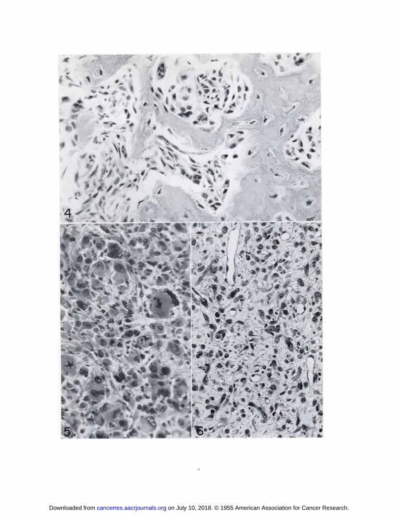
FIG. 4.—Osteogciiic sarcoma, induced by Cellophane C
ill 673 days, Rat No. 406. Stained H & E. X400.
FIG. 5.—Rhabdomyosarcoma, induced by polyvinyl
chloride D in 637 days, Rat No. 3319, showing bizarre giantcells. one of which (right of center) has peripherally arrangedvacuoles characteristic of the so-called “spider-web―cell.Stained II & E. X330.
FIG. 6.—Liposarcoma induced by Saran film in 390 days,
Rat No. 2313. The signet ring forms are partly differentiatedlipoblasts. Stained H & E. X330.
on July 10, 2018. © 1955 American Association for Cancer Research. cancerres.aacrjournals.org Downloaded from
V, ø_@_I,@
•d ‘ ‘Is-@
•@‘
I .-: @e 4
S. @‘
V
a. k'@\ 4
..
‘‘a'_1m'
@1V
•*@@ S
l@'@
@-f_'
0@@,@
†\̃@ @,
@ ‘a.@@ I
I
-@-
‘@
. 4,@ ,*
wS@I
,â€@̃4@41'
. 0
I@, @ta,a,._p_,1@
I' #@@
‘ 4@ A'
El
@1@P@:&@ —
I•
t (
44...@@
0
4...
t'O ‘i@@'
. ,@
on July 10, 2018. © 1955 American Association for Cancer Research. cancerres.aacrjournals.org Downloaded from

1955;15:333-340. Cancer Res B. S. Oppenheimer, Enid T. Oppenheimer, I. Danishefsky, et al. Further Studies of Polymers as Carcinogenic Agents in Animals
Updated version
http://cancerres.aacrjournals.org/content/15/5/333
Access the most recent version of this article at:
E-mail alerts related to this article or journal.Sign up to receive free email-alerts
Subscriptions
Reprints and
To order reprints of this article or to subscribe to the journal, contact the AACR Publications
Permissions
Rightslink site. Click on "Request Permissions" which will take you to the Copyright Clearance Center's (CCC)
.http://cancerres.aacrjournals.org/content/15/5/333To request permission to re-use all or part of this article, use this link
on July 10, 2018. © 1955 American Association for Cancer Research. cancerres.aacrjournals.org Downloaded from