
FUO or FKO? - cdn.doctorsonly.co.il · Primary Biliary Cholangitis 2. ... common cause in the...
41
FUO or FKO? Andrew Beany May 2017 Bnai Zion MC, Haifa
-
Upload
nguyendung -
Category
Documents
-
view
215 -
download
0
Transcript of FUO or FKO? - cdn.doctorsonly.co.il · Primary Biliary Cholangitis 2. ... common cause in the...

FUO or FKO?
Andrew Beany
May 2017
Bnai Zion MC, Haifa

M. G.
• 19 yo female
• Obesity
Fever – 6 monthsWeight lossWeaknessDry coughDiffuse non-specific (abdominal) pain
November 2016

August 2016
Work-up in a different hospital
• HB=9.6 (MCV=69), WBC=7.73, PLT=511
• CRP=240, ESR=110
• Albumin= 3.2
• Folic acid- low
• T. Bil=0.48, ALT=32, AST=37, ALP=152
• LDH=513
• Urinalysis: RBCs
• Radiology
– Chest and sinuses x-ray: Normal
– Abdominal US: Normal
– CT scan: Mild splenomegaly
– PET-CT: Normal
– TTE: Normal except sinus tachycardia
• E.N.T evaluation: Normal
• Normal work-up for infectious diseases
– CMV, EBV, Toxoplasma, Mycoplasma, Brucella, C. burnetii (Q-fever), Rickettsia, Bartonella (cat-scratch), Pertussis, HIV, STD
• Work-up for inflammatory diseases
– ANA, ENA, Anti-P3, Anti-MPO, RF, Anti-GBM, Celiac serology: Normal
– ASLO, C3, C4: mildly high

August 2016
Work-up in a different hospital
• PO Doxycycline 100 mg x 2/day
• Mild symptomatic improvement
• CRP=120

November 2017
Work-up in our hospital
• HB=9.78 (MCV=69), WBC=8.32, PLT=472
• Fe=31, Transferrin=256, Ferritin=203, Vit B12=753, folic ac=6.3
• T.Bil=0.3, ALT=37, AST=45, ALP=154, GGT=162, LDH=313
• CRP=93
• Urinalysis: RBCs
• Work-up for inflammatory diseases– ANA, C3, C4, p/cANCA, CLP, RF,
IgA/G/M, Anti-endomysial, Anti-TTG, LKM Ab, AMA, ASMA, Anti-RNP, Anti-Smith, Anti-RO, Anti-LA, Scl 70 Ab, Anti-centromer, Jo1 Ab- Normal
– ACE=85.4 (UNL: 55)
– ASLO= weakly positive
• Work-up for infectious diseases– Blood and urine cultures, HBsAg,
HBcAb, HCV Ab, EBV IgM, CMV IgM, Brucella, Pertusis, Parapertusis, C. burnetii (Q-fever), Rickettsia, Bartonella(cat-scratch), HIV- Negative
– Quantiferon TB- negative
• Consultations– Ophthalmologist, gynecologist,
pulmonologist (including spirometry): Normal
• Radiology– Chest x-ray: Normal
– TTE: Normal
– Abdominal US: HSM
– CT: HSM, lymphadenopathy


August 2016
Work-up in our hospital
• PO Doxycycline 100 mg x 2/day for 2 weeks
• No improvement in symptoms, CRP, LFT

PDCs, potentially diagnostic clues
(all localizing signs, symptoms, and abnormalities
potentially pointing toward a diagnosis)
Harrison's Principles of Internal Medicine, 19e, 2015
Structured approach to patients with FUO

Liver Biopsy
• Preserved lobular architecture and porto-central ratio
• Marked inflammation in portal areas and liver parenchyma zones 1, 2, 3
• The inflammatory infiltrate is composed of lymphocytes and histiocytes, few plasma cellsand few eosinophils admixed with non-caseating granulomas (sarcoid like)
• In parenchyma, there is no evidence of increased apoptosis, ballooning or Mallory bodies


Liver Biopsy
• Preserved lobular architecture and porto-central ratio
• Marked inflammation in portal areas and liver parenchyma zones 1, 2, 3
• The inflammatory infiltrate is composed of lymphocytes and histiocytes, few plasma cells and few eosinophils admixed with non-caseating granulomas (sarcoid like)
• In parenchyma, there is no evidence of increased apoptosis, ballooning or Mallory bodies
• In the portal areas the inflammatory process expand the portal spaces and involve the interface area with porto-portal and porto-central bridging tendency
• Reticulum and Masson stains show fibrous portal expansion and porto-portal bridging fibrosis
• ZN, PAS, CMV stains does not detect microbiota


Liver Biopsy
• Preserved lobular architecture and porto-central ratio
• Marked inflammation in portal areas and liver parenchyma zones 1, 2, 3
• The inflammatory infiltrate is composed of lymphocytes and histiocytes, few plasma cells and few eosinophils admixed with non-caseating granulomas (sarcoid like)
• In the portal areas the inflammatory process expand the portal spaces and involve the interface area with porto-portal and porto-central bridging tendency
• In parenchyma, there is no evidence of increased apoptosis, ballooning or Mallory bodies
• Reticulum and Masson stains show fibrous portal expansion and porto-portal bridging fibrosis
• ZN, PAS, CMV stains does not detect microbiota
Non-caseating granulomatous hepatitis
DD: AIH, PBC, DILILess probably: sarcoidosis, infectious granulomas

Granulomas
A circumscribed lesion that forms as a result of an inflammatory reaction. It is characterized by a central accumulation of mononuclear cells, primarily macrophages, with a surrounding rim consisting of lymphocytesand fibroblasts
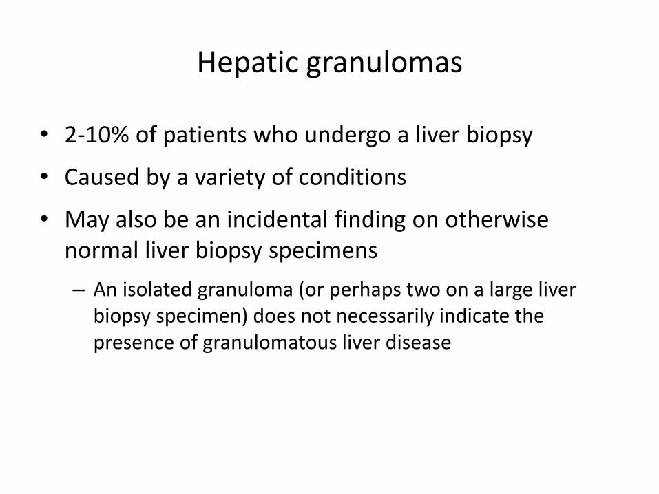
Hepatic granulomas
• 2-10% of patients who undergo a liver biopsy
• Caused by a variety of conditions
• May also be an incidental finding on otherwise normal liver biopsy specimens
– An isolated granuloma (or perhaps two on a large liver biopsy specimen) does not necessarily indicate the presence of granulomatous liver disease

“The pathologist reading the liver biopsy should attempt to determine the location of the granulomas, the presence/absence of necrosis, the type of accompanying infiltrate, any organisms or foreignmaterial in the granuloma, and associated findings”
Sleisenger and Fordtran's Gastrointestinal and Liver Disease, 10th Edition

Histologic variants
• Noncaseating
– Sarcoidosis
• Caseating
– Tuberculosis
• Fibrin-ring
– Hodgkin ly., CMV, HAV, Q fever, allopurinol
• Lipogranulomas
– Mineral oilingestion

Hepatic granulomas
• Can be located throughout the hepatic lobule
• A tendency to be located in specific sites is recognized in some disorders
– Portal or periportal location: sarcoidosis
– Portal location: PBC

Etiology
• What is the most common cause of granulomatous liver disease?
1. Primary Biliary Cholangitis
2. Infectious diseases
3. Sarcoidosis
4. All of the above
“Probably the most common cause in the
developed world is PBC”
Chapter 36, pp. 611
“The most common etiologies in the developing world (and in older studies)
are infectious diseases, especially tuberculosis”
Chapter 36, pp. 611
“Sarcoidosis is the most common etiology”
Chapter 73, pp. 1249
.
Sleisenger and Fordtran's Gastrointestinal and Liver Disease, 10th Edition

Granulomatous liver disease- causes
UpToDate
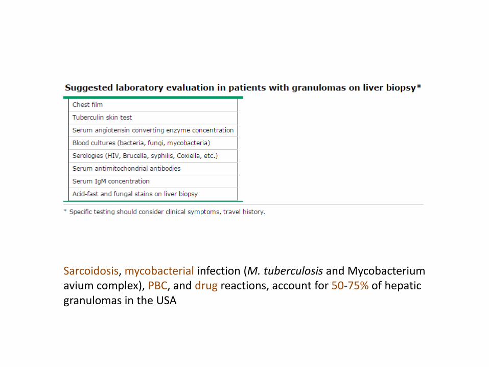
Sarcoidosis, mycobacterial infection (M. tuberculosis and Mycobacterium avium complex), PBC, and drug reactions, account for 50-75% of hepatic granulomas in the USA

Sarcoidosis
• Systemic granulomatous disease of unknown etiology
– Noncaseating epithelioid granulomas
• Prevalnece: 10-20/100.000
• Age 20-60
• Women > men
• Lung disease in > 90% of all cases

Sarcoidosis
• 50-65% have granulomas on liver biopsy
• Symptoms: 5-15% of patients
• Most patients are asymptomatic and have only biochemical abnormalities
– Usually an elevated ALP and GGT
• Rare patients develop cholestatic liver disease, cirrhosis, portal HTN, and/or hepatic vein thrombosis

Sarcoidosis
• No pathognomonic laboratory or histopathologic findings can establish the diagnosis of hepatic sarcoidosis
– Sarcoid granulomas are often located in the portal tract
• Additional steps of identifying characteristic extrahepaticmanifestations, and R/O other causes such as infection, drug-induced granulomas, and malignancy, are essential to making a definitive diagnosis

Trans-Bronchial Biopsy
• Interstitial granulomas, no evidence of inflammation, necrosis, or fibrosis
• Acid fast and silver stain are negative
Non-caseating granulomas in lung
DD: infectious granulomas, sarcoidosis

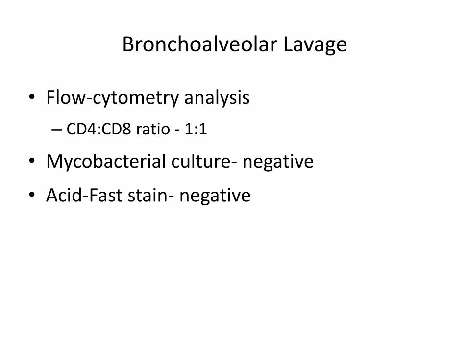
Bronchoalveolar Lavage
• Flow-cytometry analysis
– CD4:CD8 ratio - 1:1
• Mycobacterial culture- negative
• Acid-Fast stain- negative

Management and follow-up
• PO Prednisone 40 mg
• Clinical improvement
• CRP- elevation

Liver Biopsy
• Mycobacterial PCR- negative
• Bacterial DNA PCR- positive
– Nocardia Farcinica
• Fungal DNA PCR- negative

Trans-Bronchial Biopsy
• Mycobacterial PCR- negative
• Bacterial DNA PCR- negative
• Fungal DNA PCR- negative

Nocardia
• Gram-positive, branching rods, aerobic bacteria
• Found worldwide in soil, decaying vegetable matter, and aquatic environments
• Modes of entry: inhalation, ingestion, and direct inoculation through the skin
– Inhalation is the most common route of entry
• The majority of patients are immunocompromised, most often with cell-mediatedabnormalities
– Glucocorticoid therapy, malignancy, organ and hematopoietic stem cell transplantation, and HIV
• Clinical manifestations
– Lungs- the primary site of infection in >2/3 of cases
• Acute, subacute, or chronic
– CNS disease- abscess, ~20% of cases
• Mostly dissemination of infection from a pulmonary or cutaneous site
– Cutaneous disease- mostly by trauma
– Disseminated nocardiosis- two or more noncontiguous sites

Patient re-admitted for further evaluation

Trans-Bronchial Biopsy- 12.2016
• No granulomas
• No Nocardia
– Stains with methenamin silver, PAS, ZN, Gram- negative
– Bacterial PCR- negative
• Mycobacterial PCR- negative

Brocho-alveolar Lavage- 12.2016
• Mycobacterial PCR- negative
• Bacterial DNA PCR- negative
– No Nocardia
• Nocardia culture- negative

Re-evaluation
• Liver biopsy w/o paraffin
– Pan-bacterial PCR- negative
– No Nocardia
• Neutrophil function assessment- normal

Nocardia TB
Other infection
Sarcoidosis
Idiopathic

Idiopathic granulomatous hepatitis
• The cause of hepatic granulomas may remain unclear despite careful evaluation
– 10-36%
• A syndrome of prolonged febrile illness, myalgias, hepatosplenomegaly, and arthralgias of unclear etiology
– Relapsing remitting course
• Laboratory findings are nonspecific. ESR is often markedly elevated

Idiopathic granulomatous hepatitis
• The treatment of symptomatic cases involves immunosuppression
• It is reasonable to treat initially with an empiric course of antituberculousmedications in patients in whom there is a concern about underlying tuberculosis
• If there is no clinical response after 4-8 weeks, empiric corticosteroidsshould be instituted, which usually lead to rapid improvement in symptoms and disappearance of the granulomas
– Prednisone, 20 to 40 mg per daily
– A biochemical response should be noted within several months
– Once symptoms have improved, gradual weaning should be attempted
– The prognosis in patients who respond to corticosteroids is good
– Relapse is common and a repeat course of corticosteroids is often necessary
– Methotrexate, as a steroid-sparing agent

89%
initiate an empirical treatment
(28 days)
50%
Anti-TB
19%
Prednisone
19%
Cyclins
69%
2nd line treatment
(28 days)
16%
Anti-TB
72%
Prednisone
12%
Cyclins

Management and follow-up
• PO Prednisone 30 mg (tapering down)
– Mild clinical improvement
– No improvement in CRP or LFT
• PO Azathioprine 50 mg (steroid-sparing)
• Mild clinical improvement
• Normal LFT
• High CRP and ESR
– MTX- relative CI, d/t liver fibrosis
– Biological agent? (refractory sarcoidosis)
Lost to follow-up


