Fungal Root Microbiome from Healthy and Brittle Leaf ... › wp-content › uploads › ... ·...
18
ORIGINAL RESEARCH published: 28 February 2017 doi: 10.3389/fmicb.2017.00307 Edited by: Dietmar Schlosser, Helmholtz Centre for Environmental Research (UFZ), Germany Reviewed by: Giuseppe Spano, University of Foggia, Italy Maria Lurdes Inacio, Instituto Nacional de Investigação Agrária e Veterinária, Portugal *Correspondence: Lassaad Belbahri [email protected] Specialty section: This article was submitted to Microbiotechnology, Ecotoxicology and Bioremediation, a section of the journal Frontiers in Microbiology Received: 08 January 2017 Accepted: 14 February 2017 Published: 28 February 2017 Citation: Mefteh FB, Daoud A, Chenari Bouket A, Alenezi FN, Luptakova L, Rateb ME, Kadri A, Gharsallah N and Belbahri L (2017) Fungal Root Microbiome from Healthy and Brittle Leaf Diseased Date Palm Trees (Phoenix dactylifera L.) Reveals a Hidden Untapped Arsenal of Antibacterial and Broad Spectrum Antifungal Secondary Metabolites. Front. Microbiol. 8:307. doi: 10.3389/fmicb.2017.00307 Fungal Root Microbiome from Healthy and Brittle Leaf Diseased Date Palm Trees (Phoenix dactylifera L.) Reveals a Hidden Untapped Arsenal of Antibacterial and Broad Spectrum Antifungal Secondary Metabolites Fedia B. Mefteh 1 , Amal Daoud 1 , Ali Chenari Bouket 2,3 , Faizah N. Alenezi 2 , Lenka Luptakova 2,4 , Mostafa E. Rateb 5 , Adel Kadri 1,6 , Neji Gharsallah 1 and Lassaad Belbahri 2,7 * 1 Laboratory of Plant Biotechnology, Faculty of Science, University of Sfax, Sfax, Tunisia, 2 Biotechnology, NextBiotech, Agareb, Tunisia, 3 Graduate School of Life and Environmental Sciences, Osaka Prefecture University, Sakai, Japan, 4 Department of Biology and Genetics, Institute of Biology, Zoology and Radiobiology, University of Veterinary Medicine and Pharmacy, Kosice, Slovakia, 5 School of Science and Sport, University of the West of Scotland, Paisley, UK, 6 College of Science and Arts in Baljurashi, Al Baha University, Al Bahah, Saudi Arabia, 7 Laboratory of Soil Biology, University of Neuchâtel, Neuchâtel, Switzerland In this study, we aimed to explore and compare the composition, metabolic diversity and antimicrobial potential of endophytic fungi colonizing internal tissues of healthy and brittle leaf diseased (BLD) date palm trees (Phoenix dactylifera L.) widely cultivated in arid zones of Tunisia. A total of 52 endophytic fungi were isolated from healthy and BLD roots of date palm trees, identified based on internal transcribed spacer-rDNA sequence analysis and shown to represent 13 species belonging to five genera. About 36.8% of isolates were shared between healthy and diseased root fungal microbiomes, whereas 18.4 and 44.7% of isolates were specific to healthy and BLD root fungal microbiomes, respectively. All isolates were able to produce at least two of the screened enzymes including amylase, cellulase, chitinase, pectinase, protease, laccase and lipase. A preliminary screening of the isolates using disk diffusion method for antibacterial activity against four Gram-positive and three Gram-negative bacteria and antifungal activities against three phytopathogenic fungi indicated that healthy and BLD root fungal microbiomes displayed interesting bioactivities against examined bacteria and broad spectrum bioactivity against fungal pathogens. Some of these endophytic fungi (17 isolates) were fermented and their extracts were evaluated for antimicrobial potential against bacterial and fungal isolates. Results revealed that fungal extracts exhibited antibacterial activities and were responsible for approximately half of antifungal activities against living fungi. These results suggest a strong link between fungal bioactivities and their secondary metabolite arsenal. EtOAc extracts of Geotrichum candidum and Thielaviopsis punctulata originating from BLD microbiome gave best results against Micrococcus luteus and Bacillus subtilis with minimum inhibitory concentration Frontiers in Microbiology | www.frontiersin.org 1 February 2017 | Volume 8 | Article 307
Transcript of Fungal Root Microbiome from Healthy and Brittle Leaf ... › wp-content › uploads › ... ·...

fmicb-08-00307 February 25, 2017 Time: 15:46 # 1
ORIGINAL RESEARCHpublished: 28 February 2017
doi: 10.3389/fmicb.2017.00307
Edited by:Dietmar Schlosser,
Helmholtz Centre for EnvironmentalResearch (UFZ), Germany
Reviewed by:Giuseppe Spano,
University of Foggia, ItalyMaria Lurdes Inacio,
Instituto Nacional de InvestigaçãoAgrária e Veterinária, Portugal
*Correspondence:Lassaad Belbahri
Specialty section:This article was submitted to
Microbiotechnology, Ecotoxicologyand Bioremediation,
a section of the journalFrontiers in Microbiology
Received: 08 January 2017Accepted: 14 February 2017Published: 28 February 2017
Citation:Mefteh FB, Daoud A, Chenari
Bouket A, Alenezi FN, Luptakova L,Rateb ME, Kadri A, Gharsallah N
and Belbahri L (2017) Fungal RootMicrobiome from Healthy and Brittle
Leaf Diseased Date Palm Trees(Phoenix dactylifera L.) Reveals
a Hidden Untapped Arsenalof Antibacterial and Broad Spectrum
Antifungal Secondary Metabolites.Front. Microbiol. 8:307.
doi: 10.3389/fmicb.2017.00307
Fungal Root Microbiome fromHealthy and Brittle Leaf DiseasedDate Palm Trees (Phoenix dactyliferaL.) Reveals a Hidden UntappedArsenal of Antibacterial and BroadSpectrum Antifungal SecondaryMetabolitesFedia B. Mefteh1, Amal Daoud1, Ali Chenari Bouket2,3, Faizah N. Alenezi2,Lenka Luptakova2,4, Mostafa E. Rateb5, Adel Kadri1,6, Neji Gharsallah1 andLassaad Belbahri2,7*
1 Laboratory of Plant Biotechnology, Faculty of Science, University of Sfax, Sfax, Tunisia, 2 Biotechnology, NextBiotech,Agareb, Tunisia, 3 Graduate School of Life and Environmental Sciences, Osaka Prefecture University, Sakai, Japan,4 Department of Biology and Genetics, Institute of Biology, Zoology and Radiobiology, University of Veterinary Medicine andPharmacy, Kosice, Slovakia, 5 School of Science and Sport, University of the West of Scotland, Paisley, UK, 6 College ofScience and Arts in Baljurashi, Al Baha University, Al Bahah, Saudi Arabia, 7 Laboratory of Soil Biology, University ofNeuchâtel, Neuchâtel, Switzerland
In this study, we aimed to explore and compare the composition, metabolic diversityand antimicrobial potential of endophytic fungi colonizing internal tissues of healthy andbrittle leaf diseased (BLD) date palm trees (Phoenix dactylifera L.) widely cultivated inarid zones of Tunisia. A total of 52 endophytic fungi were isolated from healthy andBLD roots of date palm trees, identified based on internal transcribed spacer-rDNAsequence analysis and shown to represent 13 species belonging to five genera. About36.8% of isolates were shared between healthy and diseased root fungal microbiomes,whereas 18.4 and 44.7% of isolates were specific to healthy and BLD root fungalmicrobiomes, respectively. All isolates were able to produce at least two of the screenedenzymes including amylase, cellulase, chitinase, pectinase, protease, laccase andlipase. A preliminary screening of the isolates using disk diffusion method for antibacterialactivity against four Gram-positive and three Gram-negative bacteria and antifungalactivities against three phytopathogenic fungi indicated that healthy and BLD root fungalmicrobiomes displayed interesting bioactivities against examined bacteria and broadspectrum bioactivity against fungal pathogens. Some of these endophytic fungi (17isolates) were fermented and their extracts were evaluated for antimicrobial potentialagainst bacterial and fungal isolates. Results revealed that fungal extracts exhibitedantibacterial activities and were responsible for approximately half of antifungal activitiesagainst living fungi. These results suggest a strong link between fungal bioactivitiesand their secondary metabolite arsenal. EtOAc extracts of Geotrichum candidumand Thielaviopsis punctulata originating from BLD microbiome gave best resultsagainst Micrococcus luteus and Bacillus subtilis with minimum inhibitory concentration
Frontiers in Microbiology | www.frontiersin.org 1 February 2017 | Volume 8 | Article 307
fmicb-08-00307 February 25, 2017 Time: 15:46 # 2
Mefteh et al. Hidden Secondary Metabolites of Phoenix Endophytes
(MIC, 0.78 mg/mL) and minimum bactericidal concentration (6.25 mg/mL). G. candidumgave the best result against Rhizoctonia solani with MIC 0.78 mg/mL and minimumfungicidal concentration (MFC, 6.25 mg/mL). In conclusion, using plant microbiomessubjected to biotic stresses offers new endophytes with different bioactivities thanthose of healthy plants. Therefore, date palm endophytic fungi represent a hiddenuntapped arsenal of antibacterial and broad spectrum antifungal secondary metabolitesand could be considered promising source of bioactive compounds with industrial andpharmaceutical applications.
Keywords: endophytic fungi, secondary metabolites, brittle leaf disease, antimicrobial activity, date palm,enzymes
INTRODUCTION
The recent development of antimicrobial resistance bypathogenic microorganisms intimidates the current treatmentand prevention of ever-increasing range of infections bybacteria and fungi and leads to new microorganisms that cannotbe controlled by the drugs that referred to as “superbugs”(Vargiu et al., 2016). Reports on superbugs such as MRSA(methicillin-resistant Staphylococcus aureus) and NDM-1 (NewDelhi metallo-beta-lactamase 1) strains are rising terrifyinglynowadays and represent a serious threat to public health (Khanand Khan, 2016). The most important causes that favoredantibiotic resistance include, excessive and inappropriateantibiotic use among humans and animals (Kaye, 2016),environmental contamination with antibiotics (Lammie andHughes, 2016), poor hygienic conditions (Senn et al., 2016),global trade and travel (Riddle and Connor, 2016), medicaltourism (Saliba et al., 2016), and a decline in new antibioticdevelopment (Marston et al., 2016).
Therefore, there is an increasing demand for new bioactive,cost-effective and sustainable antimicrobial molecules inmedicine, industry and agriculture that prompted thedevelopment of diverse programs such as screening fornew plants or fungal species with antimicrobial activitiesfrom unexplored ecological niches and habitats (Lugtenberget al., 2016). Historically, plants were considered potentialsource of drugs for treatment of many human diseases. Certainendophytes could also produce the same or similar compoundsas their host plants (Xu et al., 2009). Since the discovery ofthe billion-dollar anticancer drug Taxol from the endophyticfungus of Taxus brevifolia namely Taxomyces andreanae,endophytes have gained increasing interest of mycologists,botanists, pharmacologists, and even plant pathologists (Stierleet al., 1993; Venugopalan and Srivastava, 2015; Gouda et al.,2016; Newman and Cragg, 2016). Recently, considerableknowledge has accumulated about endophytes, defined as acommunity of microorganisms that colonize all plant speciestissues without causing any apparent symptoms (Petrini et al.,1992). Endophytic microorganisms offer many ecologicaland physiological benefits to their host plants includingadaptability to different kinds of stress, growth promotionand resistance against plant pests and pathogens (Jia et al.,2016).
Therefore, several attempts have been made to study thebiology of endophytic microorganisms and to exploit theuntapped potential of their bioactive compounds (Mousa andRaizada, 2013). Consequently, numerous endophytes includingbacteria, fungi, and actinomycetes had been recovered andcharacterized from approximately 300,000 plant species (Strobeland Daisy, 2003). Accordingly, recent studies have shownthat endophytic fungi represent a promising resource ofnovel natural products with biological importance such asantidiabetic, anticancer, antioxidant, antimicrobial, antiviral, andimmunosuppressive activities (Katoch et al., 2015; Chen et al.,2016; Monggoot et al., 2016; Rathna et al., 2016). These bioactivecompounds may be classified as alkaloids, steroids, terpenoids,flavonoids, benzopyranones, quinones, isocumarins, phenolics,tetralones, lactones, peptides, and many other subclasses (Xuet al., 2008; Kaul et al., 2012). The discovery of novelantimicrobial compounds from endophytic fungi constitutes animportant alternative to overcome numerous recent problemssuch as the insufficiency of current antibiotics against humanpathogens, the wider range of infections and the low rate ofnew antimicrobial agent discovery (Strobel et al., 2001; Marstonet al., 2016). Antimicrobial compounds could also be used as foodpreservatives among other biotechnological applications (Haqueet al., 2015).
Different environmental factors in addition to biotic andabiotic stresses are considered the main drivers of plant fungalendophyte diversity and believed to shape their communities.Although endophyte characterization in healthy tissues have beenextensively studied, few reports addressed such communitiesin plants under abiotic or biotic stresses (Douanla-Meli et al.,2013). Additionally, Date palm trees, extensively used in folkmedicine, have been shown to be rich with numerous secondarymetabolites with interesting pharmacological and antimicrobialactivities (Abdennabi et al., 2016). Therefore, endophytes ofthis plant species were the main focus of this study, with adouble objective. First, endophytes from healthy and brittleleaf diseased (BLD) root tissues have been recovered andidentified. Second, endophyte communities have been comparedin terms of metabolic potential by testing enzyme activitiesof amylase, cellulase, chitinase, pectinase, protease, laccaseand lipase and antimicrobial bioactivities against four Gram-positive and three Gram-negative bacterial species and antifungalactivities against three phytopathogenic fungi. Contribution of
Frontiers in Microbiology | www.frontiersin.org 2 February 2017 | Volume 8 | Article 307

fmicb-08-00307 February 25, 2017 Time: 15:46 # 3
Mefteh et al. Hidden Secondary Metabolites of Phoenix Endophytes
secondary metabolites to the bioactivities of endophytes is thenscreened using their EtOAc culture extracts.
MATERIALS AND METHODS
Isolation of Root Fungal Endophytesfrom Healthy and BLD Date Palm TreesRoots of healthy and BLD adult date palm trees variety DegletEnnour were collected from groves located in Nefta Oasis, nearto Algerian-Tunisian border and just at the north of ChottDjerid (Latitude: 33◦ 52′ 23.12′′ N, Longitude: 7◦ 52′ 39.54′′ E).Numerous samples from roots and leaves were collected (n= 20).Plant materials were transferred to the laboratory in sterilebags and stored at 4◦C until processing. Endophytic fungi wereisolated from the internal tissues of date palm roots, as describedby Hallmann et al. (2007). Briefly, samples were washed with tapwater for 30 min and surface sterilized by sequential washes in70% ethanol followed by 3% NaClO for 3 min. Finally, the sterileplant material was washed several times with sterilized distilledwater and cut into small fragments (0.5–1 cm) under sterileconditions using a sterile scalpel. In total, 103 root fragments wereused for fungal isolation. Surface sterilized plant material wasplaced on potato dextrose agar (PDA) media supplemented with100 µg/mL streptomycin for bacterial growth inhibition. Theplates were incubated for 3–5 days at 30◦C until fungal myceliawere observed. Single fungal isolates have been obtained by sub-culturing emergent hyphal tips. Cultures were preserved on PDAslants for subsequent morphological and molecular identificationand biochemical characterization.
Fungal DNA Extraction and AmplificationPure cultures of the isolates on PDA slant vials were selectedfor DNA extraction. Mycelia were excised from 5 daysold plates. The extraction was processed using the DNA-Easy Plant Mini kit (QIAGEN, Basel, Switzerland) followingmanufacturer’s protocol. The quantity and quality of thegenomic DNA were evaluated by a NanoDrop NT-100 UVspectrophotometer (Witec AG, Switzerland) and by visualobservation through 1.5% agarose gel electrophoresis. Themolecular identification of the endophytic fungi isolated fromthe roots of healthy and BLD date palm trees was carried outas described by Paul et al. (2008). The internal transcribedspacer (ITS) of rDNA was amplified by the polymerase chainreaction using primers, ITS1 (TCCGTAGGTGAACCTGCGG)and ITS4 (TCCTCCGCTTATTGATATGC) (White et al., 1990).All reactions were carried out in a total volume of 50 µL,containing 5 µL 10x Ex Taq buffer (20 mM Tris–HCl, pH 8.0,100 mM KCl), 4 µL 2.5 mM dNTP mixture, 0.5 µM of eachprimer, 1.25 units Taq DNA polymerase (Takara Bio, Ohtsu,Japan) and 10 ng DNA. The amplifications were performed usinga master gradient thermal cycler (Eppendorf, Basel, Switzerland)with the following cycling profile: Denaturation step at 95◦C for2 min followed by 30 cycles including denaturation at 94◦Cfor 20 s, annealing at 55◦C for 25 s and extension at 72◦Cfor 15 s and final extension step at 72◦C for 10 min. PCRamplicons were purified with Minelute PCR purification kit
(Qiagen, Basel, Switzerland) according to the manufacturer’sspecifications (Belbahri et al., 2006).
DNA Sequencing and PhylogeneticAnalysisThe purified amplicons were sequenced in both directionsusing the same PCR primers and BigDye R© Terminator v. 3.1cycle sequencing kit. Sequencing reactions were resolved onan ABI 3130 XL available at the iGE3 [Institute of Geneticsand Genomics in Geneva, University of Geneva Medical Center(CMU), Switzerland]. Raw sequence files were manually editedusing SeqManTMII (DNASTAR, Madison, WI, USA) and aconsensus sequence was generated for each sequence. Theconsensus sequence for genomic region was blasted againstthe NCBI’s GenBank sequence database using megablast toidentify their closest species relatives. For the gene region, theretrieved sequences from GenBank together with sequencesgenerated in this study, were aligned using the multiple sequencealignment web-based MAFFT program (Katoh and Toh, 2008).Phylogenetic trees were constructed based on the maximum-likelihood (ML) algorithm (Felsenstein, 1981) using MEGA6(Tamura et al., 2013), with evolutionary distances computedusing the Kimura 2-parameter model (Kimura, 1980). Validityof branches in the resulting trees was evaluated by bootstrapresampling support of the data sets with 1000 replications.
Screening for Extracellular EnzymesScreening for Amylase ActivityAmylase-producing fungi were characterized usingglucose-yeast-peptone (GYP) medium containing (g/L):glucose (1), yeast extract (0.5), peptone (0.5), agar (15), andsupplemented with 0.2% of starch. The pH of the medium wasadjusted to 6.0. Mycelia of each endophytic fungus were placedin the center of the plates and incubated at 30◦C for 72 h. Afterincubation, an iodine solution (I2 = 1 g, KI = 2 g/300 ml) waspoured on the plates. The appearance of clear zones around thecolonies indicated the presence of amylase activity (Saleem andEbrahim, 2014).
Screening for Cellulolytic ActivityCellulolytic activity from endophytic fungi was characterized byinoculating GYP agar supplemented with 0.5% carboxymethylcellulose (CMC). After incubation for 72 h at 30◦C, the plateswere overlaid with 1% red congo and distained with 1 M NaCl.Strains with clear zones around the colonies were mentioned ascellulolytic enzyme producers (Shahriarinour et al., 2011).
Screening for Chitinase ActivityA minimum medium containing (g/L): colloidal chitin (10 g),(NH4)2SO4 (2 g), KH2PO4 (0.7 g), HgSO4.7 H2O (0.5 g), FeSO4.7H2O (0.01 g), agar (15 g) was used for chitinase screening(Jenifer et al., 2014). Colloidal chitin was prepared following theprotocol described by Nagpure and Gupta (2012).
Screening for Pectinase ActivityPectinolytic activity of recovered endophytic fungi wereevaluated on the medium based on the method described by
Frontiers in Microbiology | www.frontiersin.org 3 February 2017 | Volume 8 | Article 307

fmicb-08-00307 February 25, 2017 Time: 15:46 # 4
Mefteh et al. Hidden Secondary Metabolites of Phoenix Endophytes
Nitinkumar and Bhushan (2010). After incubation of inoculatedplates for 72 h at 30◦C, cetrimonium bromide (CTAB) solution(1%) was poured onto colonies for detection of pectinaseproducing fungi.
Screening for Laccase ActivityThe fungal isolates were inoculated on malt extract mediumwith the following composition (g/L): malt extract (30),agar (20) supplemented with 2 mM ABTS (2,2′-azino-di-3-ethylbenzotiazol-6-sulfonate acid) and incubated at 30◦C.Positive laccase activity was recorded by the appearance ofreddish brown halo around the inoculated strains indicating thepresence of laccase activity (Gnanasalomi and Gnanadoss, 2013).
Screening for Lipase ActivityLipase activity was assessed using the protocol described byThota et al. (2012). The endophytic fungi were inoculated onmedium containing (g/L): 8 nutrient broth, 4 sodium chloride,10 agar supplemented after autoclaving with olive oil (16.34 mL),Tween 80 (250 µL) and Rhodamine solution (10 µg/mL). Afterincubation at 30◦C, lipase activity was detected by irradiating theplates with Ultra Violet light at 350 nm.
Screening for Protease ActivityProtease activity was checked using a simple mediumcontaining (g/L): yeast extract (3), casein peptone (5), agar(15) supplemented after autoclave with 250 mL of sterileskimmed milk (Mohanasrinivasan et al., 2012). The fungalisolates were inoculated on the center of plates. After incubationfor 72 h at 30◦C, the appearance of clear zones around themindicated the ability of the isolates to produce and secreteprotease in the medium.
Screening for Secondary MetabolitesProductionAssay of MicroorganismsAmong microorganisms used for antimicrobial assay, sevenwere bacteria including four Gram-positive bacteria (Bacilluscereus ‘Bc’ JN 934390, B. subtilis ‘Bs’ JN 934392, Staphylococcusaureus ‘Sa’ ATCC 6538 and Micrococcus luteus ‘Ml’) and threeGram-negative bacteria (Salmonella enteritidis ‘Se’ ATCC 43972,Escherichia coli ‘Ec’ ATCC 25922, and Klebsiella pneumonia ‘Kp’).Bacterial cultures were prepared in 10 mL of Mueller-Hintonbroth (MHB) (Mueller and Hinton, 1941) and maintained at30◦C with constant shaking (180 rpm). For the studied bacteria,the optical density at 600 nm of overnight cultures was adjustedto 0.1 corresponding to 3.2× 10⁶ CFU/mL.
Antifungal activity was conducted using threephytopathogenic fungi including Rhizoctonia solani ‘Rs,’Fusarium oxysporum ‘Fo’ AB586994 and Pythium catenulatum‘Pc’ AY598675. The examined fungi were inoculated on PDAplates and incubated for 7 days at 30◦C.
In vitro Antimicrobial (Antagonistic)AssayThe endophytic fungi were subjected to antimicrobial assaysthat allows rapid but qualitative selection of the bioactive
isolates. For antibacterial activity, the disk diffusion method wascarried out as described by Ezra et al. (2004). Briefly, plugsfrom 5-days-old pure culture of each endophytic fungi (6 mmdiameter) were cut using sterile Pasteur pipette and placed ontothe periphery of Mueller-Hinton agar (MHA) plates initiallyinoculated with 100 µL of culture of the tested bacteria. Diffusionwas carried out for 2 h at 4◦C. After incubation of platesfor 24 h at 30◦C, the antibacterial activity was expressed bymeasuring the diameters of inhibition zones around the fungalplugs.
The antifungal activity of the isolated fungal endophytes wasassessed by dual culture method according to Alenezi et al.(2016). A dual culture was conducted on PDA plates withplugs from pure culture (10 mm in diameter) from the twopartners, endophytic fungus and tested phytopathogenic fungi.The control plates were inoculated only with the phytopathogenicfungi. The plates were incubated for 72 h at 30◦C and thepercentage of inhibition (IP) of each isolate was recorded.All experiments were conducted in triplicates. The percentageof inhibition was calculated as the following formula (A:radial diameter of phytopathogenic fungus in control plate,B: radial diameter of phytopathogenic fungus in dual cultureplate)
IP = [(A− B)÷ A] × 100
Secondary Screening: Fermentation inLiquid Medium and Antimicrobial ActivityThe endophytic isolates from healthy and BLD date palm rootsthat exhibited inhibitory activity against the largest numberof examined bacteria and fungi were selected for secondaryscreening of bioactive metabolites production (Santos et al.,2015). Endophytic fungi were cultured on PDA plates for 5 daysat 30◦C. Then, three plugs (5 mm × 5 mm) with fungalculture were transferred to 300 mL flasks containing 150 mLof autoclaved potato dextrose broth (PDB). The cultures wereincubated in a shaker at 30◦C, 150 rpm for 21 days untilstationary phase reached. Afterward, the fungal mycelia wereseparated from culture by filtration. The resulting filtrates fromeach endophytic fungus were extracted with equal volume ofEtOAc. The organic solvent was evaporated under reducedpressure using rotary evaporator. The fungal extracts were thendissolved in DMSO or double distilled water to obtain finalconcentration of 100 µg/µL. Finally, the concentrated fungalextracts were passed through 0.2 µm filtration membrane andassayed for their antimicrobial activity by agar diffusion method.Briefly, 100 µL of freshly prepared culture for bacteria or sporesuspension (106 spores/mL) for fungi were seeded onto agarplates surface using a sterile swab cotton. Each well was thenfilled with 60 µL of endophytic fungi extracts. The plates werekept for 2 h at 4◦C to facilitate the diffusion of fungal filtrate.Afterward, they were incubated at 37◦C for 24 h for bacteriaand at 30◦C for 3–5 days for fungal strains. Antimicrobialactivity was evaluated by measuring the diameter of inhibitionzones around the wells. All experiments were carried out induplicate.
Frontiers in Microbiology | www.frontiersin.org 4 February 2017 | Volume 8 | Article 307

fmicb-08-00307 February 25, 2017 Time: 15:46 # 5
Mefteh et al. Hidden Secondary Metabolites of Phoenix Endophytes
Determination of Minimum InhibitoryConcentration (MIC), MinimumBactericidal Concentration (MBC), andMinimum Fungicidal Concentration(MFC) of Endophytic Fungal ExtractsThe minimum bactericidal concentration (MBC) and minimumfungicidal concentration (MFC) were determined using brothmicro-dilution method in a sterile 96 well micro plate, asdescribed by Gulluce et al. (2007). Serial dilution of each fungalextract was prepared to get final concentrations ranging from0.781 to 100 µg/µL. Each well was supplemented with 10 µLof bacterial or fungal suspension, 90 µL of liquid culture brothand 100 µL of fungal extract. The last well containing theabove-cited components without addition of the fungal extractwas considered as positive control. The one containing DMSOwithout extract was the negative control. Plates were incubated at37◦C for 24 h for bacterial strains and for 3 days at 30◦C for fungalstrains. Afterward, 25 µL of MTT (3-(4,5-dimethylthiazol-2-yl)-2,5-diphenyltetrazolium bromide] was added to each well forevaluation of the microorganisms viability. After incubation ofplates for 30 min at 37◦C, the clear wells indicated the inhibitionof cell growth. The MIC is defined as the lowest concentrationof extract that inhibit the growth of microorganisms. The MBCvalues were determined after incubation of plates for 48 h at37◦C as the highest dilution of extract that completely inhibitthe growth of bacteria. MFC was considered as the first wellwith no visible growth of the test fungi. It was determinedby inoculating the PDA plates with 10 µL of the well contentfollowed by incubation for 3–5 days at 30◦C. The MFC valueswere interpreted as the lowest concentration of endophytic fungalextract that inhibits fungal growth.
Statistical AnalysisData were analyzed using IBM SPSS statistics by one-way analysisof variance (ANOVA) and independent-samples T-test. The levelof significance used for all statistical tests is 5% (p < 0.05).
RESULTS
Phylogenetic Affinities of EndophyticFungiEndophytic fungi ITS-rDNA sequence analysis revealed diversetaxonomic affinities among the isolates (Figures 1A,B). In total,52 ascomycetes were obtained in the present study. About 21 ofthem were isolated from healthy date palm roots, the remainingisolates were recovered from BLD roots (Figures 1A,B). Isolatesbelonged to Eurotiomycetes (n = 38), Sordariomycetes (n = 11)and Saccharomycetes (n= 3). Isolates comprised six genera withhighest abundance of Penicillium (26 isolates) and Aspergillus(12 isolates). Among Penicillium species isolated, 12 isolateswere identified as P. bilaiae, nine as P. citrinum, three asP. citreonigrum, one as P. steckii, and one as P. cordubense. The12 Aspergillus species comprised one isolate of A. quadrilineatus,nine as A. flavus, and two as A. niger. Eight Fusarium wereisolated from roots of date palm including six Fusarium sp. and
two F. oxysporum. In Addition, two, three and one isolate ofThielaviopsis punctulata, G. candidum and Thielavia arenaria,respectively, were isolated only from BLD date palm roots(Figures 1B,C).
Screening for Enzyme ProductionAll 52 isolates were evaluated for their enzymatic activity withplate clearing assay for amylase, cellulase, chitinase, pectinase,protease, laccase, and lipase production. The results are presentedin Figure 2.
Amylase ActivityAmylase activity has been studied in either healthy or BLD datepalm roots fungal isolates. The results revealed a relevant capacityof the examined fungi to secrete these enzymes (Figure 2). Mostof amylase producing fungi belonged to Penicillium followed byAspergillus and Fusarium genera (Figures 2A,B). Mean amylaseactivity of all isolates of healthy roots was not significantlydifferent from mean amylase activity of BLD isolates (Figure 2C).
Cellulase ActivityFigure 2A shows that among the recovered endophytic fungi,only 34 isolates displayed cellulase activity (Figures 2A,B).Penicillium species were able to degrade CMC at high percentagefollowed by Aspergillus and Fusarium. Among Geotrichumspecies, only G. candidum TDPEF 19 exhibited cellulase activitywith halo diameter between 15 and 25 mm. Mean cellulaseactivity of all isolates of healthy roots was not significantlydifferent from mean cellulase activity of BLD isolates (Figure 2C).
Chitinase ActivityAll the isolated genera were able to grow on colloidal chitin agarplates reflecting their ability to produce extracellular chitinase(Figure 2A). Among the examined fungi, 25 chitinase-producingisolates belonged to Eurotiomycetes including Penicillium andAspergillus species followed by nine Sordariomycetes and twoGeotrichum species (Figure 2B). Mean chitinase activity of allisolates of healthy roots was not significantly different from meanchitinase activity of BLD isolates (Figure 2C).
Pectinase ActivityThe findings revealed that the highest percentage of theisolated endophytic fungi (84.61%) exhibited pectinase activity(Figure 2A). The ability to degrade pectin substrates weredetected mainly in endophytic fungi belonging to Penicilliumspecies within Thielavia and Thielaviopsis genera (Figure 2B).Almost all of the isolated fungi showed halos diameter between15 and 25 mm. Mean pectinase activity of all isolates of healthyroots were not significantly different from mean pectinase activityof BLD isolates (Figure 2C).
Protease ActivityAs shown in Figure 2A, proteolytic activity was present in59.61% of isolated endophytic fungi. Among the analyzedstrains, 31 fungi showed degradation halos of casein onsolid medium reflecting their ability to produce extracellularproteases. Penicillium, Aspergillus, and Fusarium are amongthe genera able to degrade casein (Figure 2B). Mean protease
Frontiers in Microbiology | www.frontiersin.org 5 February 2017 | Volume 8 | Article 307

fmicb-08-00307 February 25, 2017 Time: 15:46 # 6
Mefteh et al. Hidden Secondary Metabolites of Phoenix Endophytes
FIGURE 1 | (A) Maximum Likelihood phylogenetic tree of fungal endophytes recovered from healthy and brittle leaf diseased (BLD) diseased date palm roots.(B) Fungal species composition in healthy and BLD roots. (C) Venn diagram showing the distribution of endophytic isolates in plants roots (the first number betweenbrackets represents the number of isolates recovered from healthy roots and the second number represents that recovered from roots of LBD roots).
activity of all isolates of healthy roots was not significantlydifferent from mean protease activity of BLD isolates(Figure 2C).
Laccase ActivityHalf of isolated endophytic fungi from healthy and BLD datepalm roots showed positive results for laccase screening on solidmedium (Figure 2A). As shown in Figure 2B, fungal speciesbelonged to Eurotiomycetes class that showed ability to produce
this type of enzyme. Mean laccase activity of all isolates of healthyroots were not significantly different from mean protease activityof BLD isolates (Figure 2C).
Lipase ActivityThe lipolytic activity of the fungal isolates are presented inFigure 2A. After irradiation with UV light of plates, 28endophytic fungal strains showed fluorescent halos aroundtheir mycelia reflecting their ability to produce extracellular
Frontiers in Microbiology | www.frontiersin.org 6 February 2017 | Volume 8 | Article 307

fmicb-08-00307 February 25, 2017 Time: 15:46 # 7
Mefteh et al. Hidden Secondary Metabolites of Phoenix Endophytes
FIGURE 2 | (A) Heat map of enzyme activity of fungal endophytes recovered from both of healthy and BLD date palm roots. (B) Venn diagram showing thedistribution of endophytic fungal isolates in different enzyme activities including amylase, cellulose, chitinase, pectinase, protease, laccase, and lipase (the firstnumber between brackets represents the number of isolates recovered from healthy roots and the second number represents that recovered from BLD roots).(C) Enzyme activity of fungal endophytes isolated from both of healthy and BLD diseased. Data presents mean ± standard error.
lipase. The lipase producing fungi were detected in Penicilliumand within Geotrichum and Thielavia species (Figure 2B).Mean lipase activity of all isolates of healthy roots was notsignificantly different from mean lipase activity of BLD isolates(Figure 2C).
In vitro Antimicrobial (Antagonistic)Assay of Date Palm EndophytesThe isolated date palm healthy and BLD root endophyticfungi were examined against several pathogenic bacteriaand phytopathogenic fungi to assess their antimicrobialactivities. The results are presented in Figures 3–5. All isolatesshowed antimicrobial activity at least against two pathogenicmicroorganisms (Figures 3, 4). For antibacterial activity, theaverage of the inhibition halo diameter was between 9.3 and
26.6 mm. The highest halo diameter was against B. cereusand M. luteus with G. candidum TDPEF 18 and T. punctulataTDPEF 47, respectively (Figure 3A). Only three endophyticfungi exhibited antibacterial activity against all tested bacteriaincluding, Fusarium sp. TDPEF 11 and two T. punctulataspecies TDPEF 47 and TDPEF 50 (Figures 3A,B, 4A,B). TheGram-negative bacteria K. pneumoniae was more sensitivethan the other pathogenic bacteria. Among 52 isolates, 22(42.30%) and 23 (44.23%) displayed antibacterial activity againstboth B. cereus and B. subtilis, respectively (Figures 4A,B).Furthermore, 20 (38.46%) and 27 (51.9%) endophytic fungiwere able to inhibit the Gram-positive bacteria S. aureus andM. luteus, while 19 (36.53%) and 15 (28.84%) isolates couldinhibit S. enteritidis and E. coli, respectively (Figures 3A, 4A).Concerning antifungal activity, the percentage of inhibition
Frontiers in Microbiology | www.frontiersin.org 7 February 2017 | Volume 8 | Article 307

fmicb-08-00307 February 25, 2017 Time: 15:46 # 8
Mefteh et al. Hidden Secondary Metabolites of Phoenix Endophytes
FIGURE 3 | (A) Heat map of antibacterial (Gram-positive bacteria including Bc, Bs, Sa, and Ml) activity against fungal endophytes isolates from healthy and BLDdate palm roots. (B) Venn diagram showing the distribution of endophytic fungal isolates in relation to different Gram-positive bacteria (the first number betweenbrackets represents the number of isolates recovered from healthy roots and the second number represents the number of isolates recovered from of BLD roots).(C) Antibacterial (Gram-positive bacteria) activity against fungal endophytes isolates from healthy and BLD date palm roots. Data presents mean ± standard error.Bars labeled with asterisk are significantly different among the treatments at P < 0.05 using ANOVA analysis.
was between 10.3 and 72 mm (Figure 5A). The best percentageof inhibition was obtained with A. flavus TDPEF 2 againstF. oxysporum. The endophytic isolates were more active againstpathogenic fungi than bacteria, as shown in Figures 5A,B.Only 18 isolates of date palm endophytes were unable toinhibit all the tested fungi. F. oxysporum was the most sensitivephytopathogenic fungus to date palm endophytic isolates.Among 52 endophytic fungi, 39 (75%) displayed antifungalactivity against P. catenulatum (Figure 5B). A high numberof isolates (85%) were able to inhibit the growth of bothphytopathogenic fungi F. oxysporum and R. solani with widespectrum activity (Figures 5A,B). Mean antibacterial activityagainst B. cereus and B. subtilis and antifungal activity againstF. oxysporum of all isolates of healthy roots were significantlydifferent from mean antibacterial activity of BLD isolates that
have higher activities (Figures 3C, 5C). Mean antibacterialactivity against S. aureus and antifungal activity against R. solaniand P. catenulatum show non-significant difference betweenhealthy and BLD endophytic fungi, whereas, healthy datepalm roots endophytes show significantly higher antimicrobialactivities against Ml than BLD derived root endophytes(Figures 3C, 4C, 5C).
Antimicrobial Assay of Date PalmEndophyte ExtractsThe EtOAc extracts of 17 endophytic fungi were evaluatedfor their antibacterial and antifungal activity using agar-welldiffusion method (Figures 6–8). Among the examined fungalextracts, only five originated from the isolates of healthy date
Frontiers in Microbiology | www.frontiersin.org 8 February 2017 | Volume 8 | Article 307

fmicb-08-00307 February 25, 2017 Time: 15:46 # 9
Mefteh et al. Hidden Secondary Metabolites of Phoenix Endophytes
FIGURE 4 | (A) Heat map of antibacterial (Gram-negative bacteria including Se, Ec, and Kp) activity against fungal endophytes isolates from healthy and BLD datepalm roots. (B) Venn diagram showing the distribution of endophytic fungal isolates in relation to different Gram-negative bacteria (the first number between bracketsrepresents the number of isolates recovered from healthy roots and the second number represents that recovered from BLD roots). (C) Antibacterial (Gram-negativebacteria) activity against fungal endophytes isolates from healthy and BLD date palm roots. Data presents mean ± standard error.
palm roots. The 17 isolates submitted to fermentation assay werecomposed of four Aspergillus, two Fusarium, three Geotrichum,two Thielaviopsis, five Penicillium species, and T. arenariaTDPEF46. The inhibitory activity of fungal extracts expressedin term of diameter of inhibition zones were presented inFigures 6A, 7A, 8A. The halo inhibition diameter rangedfrom 12.6 to 28.6 mm for antibacterial activity, while forantifungal activity the inhibition diameter was between 13.6and 30 mm. Among the 17 fungal extracts, 11 showedpromising growth inhibitory activity against B. cereus. A high
number of EtOAc extracts were able to inhibit the growth ofB. subtilis with halo diameter ranging from 14.3 to 28 mm(Figure 6B). Nine extracts of date palm endophytic fungiexhibited significant inhibitory activity against S. enteritidis,E. coli, and K. pneumoniae (Figure 7B). Furthermore, allthe examined extracts displayed antagonistic activity againstF. oxysporum (Figure 8B). The EtOAc extracts of G. candidumand the two strains of T. punctulata exhibited the highest levelsof antagonistic activity thanks to their ability to inhibit thegrowth of six test bacteria and three phytopathogenic fungi.
Frontiers in Microbiology | www.frontiersin.org 9 February 2017 | Volume 8 | Article 307

fmicb-08-00307 February 25, 2017 Time: 15:46 # 10
Mefteh et al. Hidden Secondary Metabolites of Phoenix Endophytes
FIGURE 5 | (A) Heat map of antifungal (fungal isolates including Rs, Fo, and Pc) activity against fungal endophytes isolates from healthy and BLD date palm roots.(B) Venn diagram showing the distribution of endophytic fungal isolates in relation to different fungal isolates (the first number between brackets represents thenumber of isolates recovered from healthy roots and the second number represents that recovered from BLD roots). (C) Antifungal activity against fungal endophytesisolates from healthy and BLD date palm roots. Data presents mean ± standard error. Bars labeled with asterisk are significantly different among the treatments atP < 0.05 using ANOVA analysis.
Concerning antibacterial activity, the highest halo diameterwas recorded with the extract of P. bilaiae TDPEF 25 againstthe Gram-positive bacteria M. luteus (Figure 6B). In addition,the EtOAc extract of G. candidum TDPEF 19 displayed thehighest level of antifungal activity with halo diameter of30 mm against P. catenulatum (Figure 8B). Mean antibacterialactivity against Bs and Ec and antifungal activity against Rsof all isolates of healthy roots were significantly different
from mean lipase activity of BLD isolates that have higheractivities (Figures 6C, 7C, 8C). Mean antibacterial activityagainst Bc, Sa, and Se and antifungal activity against Foand Pc show non-significant difference between healthy andBLD endophytic fungi, whereas, healthy date palm rootsendophytes show significantly higher antimicrobial activitiesagainst Ml and Kp than BLD derived root endophytes(Figures 6C, 7C).
Frontiers in Microbiology | www.frontiersin.org 10 February 2017 | Volume 8 | Article 307

fmicb-08-00307 February 25, 2017 Time: 15:46 # 11
Mefteh et al. Hidden Secondary Metabolites of Phoenix Endophytes
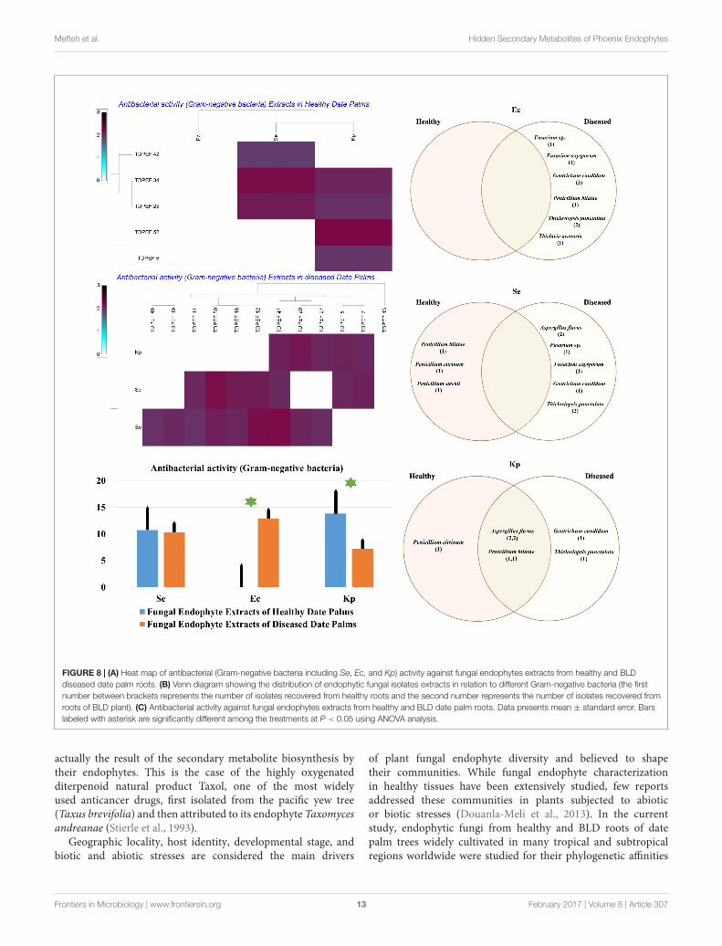
FIGURE 6 | (A) Heat map of antifungal (fungal isolates including Rs, Fo, and Pc) activity against fungal endophytes extracts from healthy and BLD date palm roots.(B) Venn diagram showing the distribution of endophytic fungal isolates extracts in relation to different fungal isolates (the first number between brackets representsthe number of isolates recovered from healthy roots and the second number represents that recovered from BLD roots). (C) Antifungal activity against fungalendophytes extracts from healthy and BLD date palm roots. Data presents mean ± standard error. Bars labeled with asterisk are significantly different among thetreatments at P < 0.05 using ANOVA analysis.
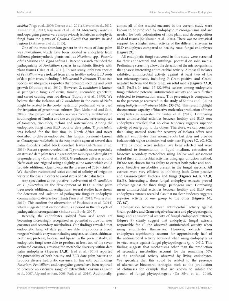
Date Palm Endophytes versusEndophyte Extracts AntimicrobialActivitiesComparison between mean antimicrobial activity against Gram-positive and Gram-negative bacteria and phytopathogenic fungiand antimicrobial activity of fungal endophytes extracts arereported in Figure 9. Results clearly suggest that endophyticroot fungal extracts account for all the observed antimicrobialactivity when using endophytes themselves. However, extracts
from endophytes significantly account approximately forhalf of the antimicrobial activity obtained when usingendophytes in vitro assays against fungal phytopathogens(p < 0.01).
Determination of MIC, MBC, and MFC ofEndophytic Fungal ExtractsThe EtOAc extract of the endophytic fungi G. candidumTDPEF20 and T. punctulata TDPEF47 were subjected to micro-
Frontiers in Microbiology | www.frontiersin.org 11 February 2017 | Volume 8 | Article 307

fmicb-08-00307 February 25, 2017 Time: 15:46 # 12
Mefteh et al. Hidden Secondary Metabolites of Phoenix Endophytes
FIGURE 7 | (A) Heat map of antibacterial (Gram-positive bacteria including Bc, Bs, Sa, and Ml) activity against fungal endophytes extracts from healthy and BLDdate palm roots. (B) Venn diagram showing the distribution of endophytic fungal isolates extracts in relation to different Gram-positive bacteria (the first numberbetween brackets represents the number of isolates recovered from healthy roots and the second number represents that recovered from BLD roots).(C) Antibacterial activity against fungal endophytes extracts from healthy and BLD date palm roots. Data presents mean ± standard error. Bars labeled with asteriskare significantly different among the treatments at P < 0.05 using ANOVA analysis.
dilution plate in order to determine their MIC, MBC, andMFC values. As shown in Table 1, the EtOAc extract ofG. candidum was more active against Bc, Ml, and Rs (MICof 0.56 µg/µL), followed by Bs, Ec, and Fo. In addition,Geotrichum extract exhibited both bacteriostatic and fungistaticeffects (MBC/MIC and MFC/MIC≥4). Concerning Thielaviopsisextract, its MIC values ranged from 0.78 to 6.25 µg/µL.However, Thielaviopsis extract exhibited the best inhibitoryactivity against Bs and Kp. The recorded MBC values indicatedthe bacteriostatic actions of Thielaviopsis extract. It couldpredominantly inhibit completely the fungal growth (MFC/MIC≥4) in exception of Pc. The results obtained in terms of MIC,MBC, and MFC indicated that G. candidum extract displayeda higher bactericidal and fungicidal effect than T. punctulataextract.
DISCUSSION
Endophytes especially fungi produce an impressive myriad ofbioactive molecules and enzymes that cope with pathogeninfection of plants (Newman and Cragg, 2016; Vasundhara et al.,2016). Natural compounds produced by endophytic fungi havebeen shown to interfere with key host and pathogen processesrequired for successful infection of the host and to mitigate theadverse effects of broad variety of animal and plant pathogens(Stierle and Stierle, 2015; Mousa et al., 2016; Tanney et al., 2016).Endophytic fungi have tremendous impacts on their host plantsinducing their tolerance to biotic and abiotic stresses, promotingtheir growth and the production of wide variety of secondarymetabolites (Jia et al., 2016). Therefore, it is believed that somebioactive compounds produced by some medicinal plants are
Frontiers in Microbiology | www.frontiersin.org 12 February 2017 | Volume 8 | Article 307
fmicb-08-00307 February 25, 2017 Time: 15:46 # 13
Mefteh et al. Hidden Secondary Metabolites of Phoenix Endophytes
FIGURE 8 | (A) Heat map of antibacterial (Gram-negative bacteria including Se, Ec, and Kp) activity against fungal endophytes extracts from healthy and BLDdiseased date palm roots. (B) Venn diagram showing the distribution of endophytic fungal isolates extracts in relation to different Gram-negative bacteria (the firstnumber between brackets represents the number of isolates recovered from healthy roots and the second number represents the number of isolates recovered fromroots of BLD plant). (C) Antibacterial activity against fungal endophytes extracts from healthy and BLD date palm roots. Data presents mean ± standard error. Barslabeled with asterisk are significantly different among the treatments at P < 0.05 using ANOVA analysis.
actually the result of the secondary metabolite biosynthesis bytheir endophytes. This is the case of the highly oxygenatedditerpenoid natural product Taxol, one of the most widelyused anticancer drugs, first isolated from the pacific yew tree(Taxus brevifolia) and then attributed to its endophyte Taxomycesandreanae (Stierle et al., 1993).
Geographic locality, host identity, developmental stage, andbiotic and abiotic stresses are considered the main drivers
of plant fungal endophyte diversity and believed to shapetheir communities. While fungal endophyte characterizationin healthy tissues have been extensively studied, few reportsaddressed these communities in plants subjected to abioticor biotic stresses (Douanla-Meli et al., 2013). In the currentstudy, endophytic fungi from healthy and BLD roots of datepalm trees widely cultivated in many tropical and subtropicalregions worldwide were studied for their phylogenetic affinities
Frontiers in Microbiology | www.frontiersin.org 13 February 2017 | Volume 8 | Article 307

fmicb-08-00307 February 25, 2017 Time: 15:46 # 14
Mefteh et al. Hidden Secondary Metabolites of Phoenix Endophytes
FIGURE 9 | Comparison of antibacterial (Gram-positive andGram-negative) and antifungal activity in relation to endophytic fungalisolates and extracts. Data presents mean ± standard error. Bars labeledwith asterisk are significantly different among the treatments at P < 0.05 usingANOVA analysis.
and bioactive potential. Date palm tree was favored becauseof its wide use in folk medicine mainly for its antioxidant,wound healing and antimicrobial activities (Abdennabi et al.,2016). Furthermore, extracts of different parts of date palmtrees including fruits and pollen showed potential inhibitoryactivity against several pathogens (Saleh and Otaibi, 2013;Daoud et al., 2015). In addition, Siala et al. (2016) reportedthe potential of endophytic bacteria isolated from healthy datepalm (P. dactylifera L.) against phytopathogenic fungi namelyF. oxysporum f. sp. albedinis, the causal agent of bayoud disease.Date palms have the ability to survive in arid regions for several
TABLE 1 | Minimum inhibitory, minimum bactericidal and minimumfungicidal concentrations of extracts of G. candidum TDPEF 20 andT. punctulata TDPEF 47 against human bacterial pathogens and fungalphytopathogens.
TDPEF 20 TDPEF 47
Concentration (µ g/µ L)
MIC MBC MIC MBC
Gram-positive bacteria
Bacillus subtilis 1.56 6.25 0.78 6.25
Bacillus cereus 0.78 12.5 1.56 12.5
Staphylococcus aureus 3.12 12.5 − −
Micrococcus luteus 0.78 6.25 1.56 6.25
Gram-negative bacteria
Salmonella enteric serotype enteritidis − − 6.25 50
Escherichia coli 1.56 6.25 3.12 25
Klebsiella pneumoniae 6.25 25 3.12 12.5
Phytopathogen fungi MIC MFC MIC MFC
Rhizoctonia solani 0.78 6.25 3.12 12.5
Fusarium oxysporum 1.56 6.25 6.25 25
Pythium catenulatum 3.12 25 3.12 6.25
years, therefore, their endophytic community may produce widerange of bioactive components during various stages of theirlife cycle. Roots of date palm were chosen for isolation ofendophytic fungi owing to their significant content of endophyticcommunities (Wearn et al., 2012). In addition, previousresearches have revealed that most of endophytes isolated fromfoliar tissues were already known to occur commonly in the rootsof the host plants (Moricca et al., 2012). A total of 52 endophyticfungi was isolated from roots of date palm (P. dactylifera L.).These isolates were identified on the basis of their 18S rDNAsequences analysis and shown to represent 13 species belongingto five genera. Some of isolates (36.8%) were shared betweenhealthy and diseased root fungal microbiomes, whereas 18.4 and44.7% of the isolates were specific to healthy and BLD rootfungal microbiomes, respectively (Figures 1A–C). Shared speciesfound to belong to genera Penicillium, Fusarium, and Aspergillus.Penicillium stecki was specific to healthy tissues, whereas A. niger,A. quadrilineatus, P. citreonigrum, P. cordubense, G. candidum,T. punctulata, and T. arenaria were specific to BLD root fungalmicrobiome (Figure 1C).
Our results disagree with those of Ben Chobba et al. (2013)who recorded the dominance of Alternaria, Pythium, andCurvularia genera in roots of date palm belonging to the samevariety Deglet Ennour. However, many factors such as time ofsampling, age of host plant, soil conditions as well as dynamics ofsoil mycobiota may have influence on endophytes of date palmtrees and can explain therefore this discrepancy. Variations infungal endophytes and their frequency of isolation have beenreported for many host plants (Collado et al., 2001; Nalini et al.,2014). Most of our isolates have been reported as endophytes inother plants, including mangrove, Catharanthus roseus, Moringaoleifera, Paspalum maritimum, Pinus thunbergii, and Coffee
Frontiers in Microbiology | www.frontiersin.org 14 February 2017 | Volume 8 | Article 307
fmicb-08-00307 February 25, 2017 Time: 15:46 # 15
Mefteh et al. Hidden Secondary Metabolites of Phoenix Endophytes
arabica (Vega et al., 2006; Correa et al., 2011; Elavarasi et al., 2012;Kumar et al., 2013; Rajeswari et al., 2016). Moreover, Fusariumand Aspergillus genera were also previously isolated as endophyticfungi from the plants of Opuntia dillenii that survive in aridregions (Ratnaweera et al., 2015).
One of the most abundant genera in the roots of date palmwas Penicillium, which have been isolated as endophyte fromdifferent photosynthetic plants such as Nicotiana spp., Pasaniaedulis Makino and Vigna radiata L. Recent research excluded thepathogenicity of Penicillium species in symbiotic lifestyle withplant tissues (Diaz et al., 2011). In our study, only two speciesof Penicillium were isolated from either healthy and/or BLD rootsof date palm trees, including P. bilaiae and P. citrinum. These twospecies are ubiquitous saprobes that promote seedling and plantgrowth (Mushtaq et al., 2012). However, G. candidum is knownas pathogenic fungus of citrus, tomato, cucumber, grapefruit,and carrot causing sour rot disease (Suprapta et al., 1995). Webelieve that the isolation of G. candidum in the oasis of Neftamight be related to the cooled system of geothermal water usedfor irrigation of greenhouse cultures (Ben Mohamed and Said,2008). The project of greenhouses was recently established insouth regions of Tunisia and the crops produced were composedof tomatoes, cucumber, melons and watermelons. Among theendophytic fungi from BLD roots of date palm, T. punctulatawas isolated for the first time in North Africa and neverdescribed to date as endophyte. This fungus, previously knownas Ceratocystis radicicola, is the responsible agent of several datepalm disorders called black scorched leaves (Al-Naemi et al.,2014). Recent reports revealed that T. punctulata occur especiallyon stressed date palm trees in areas where salinity and drought arepreponderating (Zaid et al., 2002). Greenhouse cultures aroundNefta oasis are irrigated using a slightly saline water, which couldprovide additional clues to explain the presence of T. punctulata.We therefore recommend strict control of salinity of irrigationwater in the oasis in order to avoid stress of date palm trees.
Any speculation about putative involvement of G. candidumor T. punctulata in the development of BLD in date palmtrees needs additional investigations. Several studies have shownthe detection and isolation of pathogenic fungi in endophyticcommunities of diverse host plants (Sun et al., 2012; Wearn et al.,2012). This confirm the observation of Pawłowska et al. (2014)which suggested that endophytism is a period in the life cycle ofpathogenic microorganisms (Schulz and Boyle, 2005).
Recently, the endophytes isolated from arid zones arebecoming increasingly recognized as potential source for newenzymes and secondary metabolites. Our findings revealed thatendophytic fungi of date palm are able to produce a broadrange of valuable enzymes including amylase, cellulase, chitinase,pectinase, protease, laccase, and lipase. In the present study, allendophytic fungi were able to produce at least two of the sevenevaluated enzymes, attesting the metabolic diversity within datepalm endophytes (Figures 2A,B). Jrad et al. (2014) reportedthe potentiality of both healthy and BLD date palm bacteria toproduce diverse hydrolytic enzymes. In line with our findingsFusarium, Penicillium, and Aspergillus genera have been reportedto produce an extensive range of extracellular enzymes (Kwonet al., 2007; Alp and Arikan, 2008; Park et al., 2016). Additionally,
almost all of the assayed enzymes in the current study wereknown to be produced by endophytic microorganisms and areneeded for both colonization of host plant and decompositionof dead tissues (Saikkonen et al., 2004). There was no statisticalsupport for a higher mean activity of the different enzymes inBLD endophytes compared to healthy roots fungal endophytes(Figure 2C).
All endophytic fungi recovered in this study were screenedfor their antibacterial and antifungal potential on solid media.Preliminary screening allows the detection of the microorganismsthat possess interesting antimicrobial activity. Almost all isolatesexhibited antimicrobial activity against at least two of thetest microorganisms, including 7 Gram-positive and Gram-negative bacteria and three fungi, on solid media (Figures 3A,B,4A,B, 5A,B). In total, 17 (32.69%) isolates among endophyticfungi exhibited potential antimicrobial activity and were furthersubjected to fermentation assay. This percentage is comparableto the percentage recovered in the study of Santos et al. (2015)using Indigofera suffruticosa Miller (33.6%). This result highlightthe enormous capacity of bioactive molecules production of theseendophytes as suggested by Santos et al. (2015). Comparingmean antimicrobial activities between healthy and BLD rootendophytes revealed that no clear tendency suggests superioractivity of one group to the others. Therefore, we can concludethat using stressed roots for recovery of isolates offers newdifferent endophytes than normal roots but does not provideisolates with higher antimicrobial activities (Figures 3C, 4C, 5C).
The 17 most active isolates have been selected and weresubmitted to fermentation in liquid medium, extraction ofbioactive secondary metabolites using EtOAc and subsequenttest of their antimicrobial activities using agar diffusion method.EtOAc was chosen for its ability to extract both polar and non-polar bioactive metabolites present in the fungal mycelia. Allextracts were very efficient in inhibiting both Gram-positiveand Gram-negative bacteria and fungi (Figures 6A,B, 7A,B,8A,B). Interestingly, these fungal endophyte extracts provedeffective against the three fungal pathogens used. Comparingmean antimicrobial activities between healthy and BLD rootendophytes extracts revealed also that no clear tendency suggestssuperior activity of one group to the other (Figures 6C,7C, 8C).
Comparison between mean antimicrobial activity againstGram-positive and Gram-negative bacteria and phytopathogenicfungi and antimicrobial activity of fungal endophytes extracts,(Figure 9) clearly suggest that endophytic fungal extractsresponsible for all the observed antimicrobial activity whenusing endophytes themselves. However, extracts fromendophytes significantly account for approximately half ofthe antimicrobial activity obtained when using endophytes asin vitro assays against fungal phytopathogens (p < 0.01). Thisfinding suggests that mechanisms other than the productionof secondary metabolites account for the remaining 50%of the antifungal activity observed by living endophytes.We speculate that this could be related to the presenceof alternative biocontrol strategies such as the productionof chitinases for example that are known to inhibit thegrowth of fungal phytopathogens (Da Silva et al., 2016).
Frontiers in Microbiology | www.frontiersin.org 15 February 2017 | Volume 8 | Article 307

fmicb-08-00307 February 25, 2017 Time: 15:46 # 16
Mefteh et al. Hidden Secondary Metabolites of Phoenix Endophytes
However, more detailed research work is required to confirm thishypothesis.
The two most active isolates G. candidum and T. punctulatawere submitted to the microdilution method to evaluateprecisely their MIC, MBC, and MFC. Extract of G. candidumwas more active against B. cereus, M. luteus, and R. solani(MIC of 0.56 µg/µL). Thielaviopsis extract exhibited the bestinhibitory activity against B. subtilis and K. pneumoniae. Inline with our findings, endophytes from Geotrichum genushave been reported as source of nematicidal, antituberculosis,antifungal, and antimalarial compounds (Kongsaeree et al.,2003; Li et al., 2007). Most of the compounds isolated fromthe endophytes of Geotrichum sp. are almost isocumarin andtriterpenoids. Production of such compounds by endophyticfungi has been reported by recent review (Mousa and Raizada,2013). No previous researches have isolated Thielaviopsis as anendophyte which warrants serious investigation to study its fullantimicrobial potential. Our results indicate that endophyticfungi of healthy and BLD roots of date palm constitute a potentsource of useful antibacterial and antifungal compounds. Thenext step will be to establish a strain collection bank with highthroughput antibacterial and antifungal screening in addition to
performing large scale fermentation of the potential microbialhits to identify the bioactive metabolites responsible for suchactivities in these extracts.
AUTHOR CONTRIBUTIONS
Conceived and designed the experiments: FM, AD, LB, and NG.Performed the experiments: FM, LB, AD, ACB, LL, FA, andMR. Analyzed the data: FM, LB, AD, ACB, LL, MR, and NG.Contributed reagents/materials/analysis tools: LB, LL, FA, andNG. Wrote and enriched the literature: LB, FM, AD, ACB, LL,FA, AK, MR, and NG.
ACKNOWLEDGMENTS
Financial support of the Tunisian Ministry of Higher Educationand Scientific Research is gratefully acknowledged. LL is indebtedto the Ministry of Education, Science, Research and Sport of theSlovak Republic for financial support in the frame of the project“VEGA 1/0061/16.”
REFERENCESAbdennabi, R., Bardaa, S., Mehdi, M., Rateb, M. E., Raab, A., Alenezi, F. N.,
et al. (2016). Phoenix dactylifera L. sap enhances wound healing in Wistar rats:phytochemical and histological assessment. Int. J. Biol. Macromol. 88, 443–450.doi: 10.1016/j.ijbiomac.2016.04.015
Alenezi, F. N., Rekik, I., Belka, M., Ibrahim, A. F., Luptakova, L., Jaspars, M., et al.(2016). Strain-level diversity of secondary metabolism in the biocontrol speciesAneurinibacillus migulanus. Microbiol. Res. 182, 116–124. doi: 10.1016/j.micres.2015.10.007
Al-Naemi, F. A., Nishad, R., and Ahmed, T. A. (2014). First report of Thielaviopsispunctulata causing black scorch disease on date palm in Qatar. Plant Dis. 98,1437. doi: 10.1094/PDIS-04-14-0424-PDN
Alp, S., and Arikan, S. (2008). Investigation of extracellular elastase, acid proteinaseand phospholipase activities as putative virulence factors in clinical isolatesof Aspergillus species. J. Basic Microbiol. 48, 331–337. doi: 10.1002/jobm.200700349
Belbahri, L., Moralejo, E., Calmin, G., Oszako, T., Garcia, J. A., Descals, E., et al.(2006). Phytophthora polonica, a new species isolated from declining Alnusglutinosa stands in Poland. FEMS Microbiol. Lett. 261, 165–174. doi: 10.1111/j.1574-6968.2006.00349.x
Ben Chobba, I., Elleuch, A., Ayadi, I., Khannous, L., Namsi, A., Cerqueira, F., et al.(2013). Fungal diversity in adult date palm (Phoenix dactylifera L.) revealed byculture-dependent and culture-independent approaches. J. Zhejiang Univ. Sci.B 14, 1084–1099. doi: 10.1631/jzus.B1200300
Ben Mohamed, M., and Said, M. (2008). “Geothermal energy development inTunisia: present status and future outlook,” in Proceedings 30th AnniversaryWorkshop, Reykjavik, 12.
Chen, L., Zhang, Q., Jia, M., Ming, Q. L., Yue, W., Rahman, K., et al. (2016).Endophytic fungi with antitumor activities: their occurrence and anticancercompounds. Crit. Rev. Microbiol. 42, 454–473. doi: 10.3109/1040841X.2014.959892
Collado, J., Platas, G., and Pelaez, F. (2001). Identification of an endophyticNodulisporium sp. from Quercus ilex in central Spain as the anamorph ofBiscogniauxia mediterranea by rDNA sequence analysis and effect of differentecological factors on distribution of the fungus. Mycologia 93, 875–886. doi:10.2307/3761753
Correa, M. J. C., Nunes, F. M., Bitencourt, H. R., Borges, F. C., Guilhon, G. M. S. P.,Arruda, M. S. P., et al. (2011). Biotransformation of chalcones by the endophytic
fungus Aspergillus flavus isolated from Paspalum maritimum trin. J. Braz. Chem.Soc. 22, 1333–1338. doi: 10.1590/S0103-50532011000700019
Da Silva, J. A. T., de Medeiros, E. V., da Silva, J. M., Tenorio, D. D., Moreira,K. A., Nascimento, T. C. E. D., et al. (2016). Trichoderma aureoviride URM 5158and Trichoderma hamatum URM 6656 are biocontrol agents that act againstcassava root rot through different mechanisms. J. Phytopathol. 164, 1003–1011.doi: 10.1111/jph.12521
Daoud, A., Drira, M., Bakari, S., Hfaieth, N., Manfgui, K., Kadri, A., et al.(2015). Assessment of polyphenol composition, antioxidant and antimicrobialproperties of various extracts of date palm pollen (DPP) from two Tunisiancultivars. Arabian J. Chem. (in press). doi: 10.1016/j.arabjc.2015.07.014
Diaz, G. A., Yanez, L., and Latorre, B. A. (2011). Low occurrence of patulin-producing strains of Penicillium in grapes and patulin degradation duringwinemaking in Chile. Am. J. Enol. Vitic. 62, 542–546. doi: 10.5344/ajev.2011.11034
Douanla-Meli, C., Langer, E., and Mouafo, F. T. (2013). Fungal endophyte diversityand community patterns in healthy and yellowing leaves of Citrus limon. FungalEcol. 6, 212–222. doi: 10.1016/j.funeco.2013.01.004
Elavarasi, A., Sathiya Rathna, G., and Kalaiselvam, M. (2012). Taxolproducing mangrove endophytic fungi Fusarium oxysporum fromRhizophora annamalayana. Asian Pac. J. Trop. Biomed. 2, S1081–S1085.doi: 10.1016/S2221-1691(12)60365-7
Ezra, D., Hess, W. M., and Strobel, G. A. (2004). New endophytic isolatesof Muscodor albus, a volatile-antibiotic-producing fungus. Microbiology 150,4023–4031. doi: 10.1099/mic.0.27334-0
Felsenstein, J. (1981). Evolutionary trees from DNA sequences: a maximumlikelihood approach. J. Mol. Evol. 17, 368–376. doi: 10.1007/BF01734359
Gnanasalomi, V. D. V., and Gnanadoss, J. J. (2013). Laccases from fungi andtheir applications: recent developments. Asian J. Exp. Biol. Sci. 4, 581–590.doi: 10.1016/j.cbpa.2015.06.009
Gouda, S., Das, G., Sen, S. K., Shin, H. S., and Patra, J. K. (2016). Endophytes:a treasure house of bioactive compounds of medicinal importance. Front.Microbiol. 7:1538. doi: 10.3389/fmicb.2016.01538
Gulluce, M., Sahin, F., Sokmen, M., Ozer, H., Daferera, D., Sokmen, A., et al. (2007).Antimicrobial and antioxidant properties of the essential oils and methanolextract from Mentha longifolia L. ssp. longifolia. Food Chem. 103, 1449–1456.doi: 10.1016/j.foodchem.2006.10.061
Hallmann, J., Berg, G., and Schulz, B. (2007). Isolation Procedures for EndophyticMicroorganisms. Berlin: Springer.
Frontiers in Microbiology | www.frontiersin.org 16 February 2017 | Volume 8 | Article 307

fmicb-08-00307 February 25, 2017 Time: 15:46 # 17
Mefteh et al. Hidden Secondary Metabolites of Phoenix Endophytes
Haque, M. A., Lee, J. H., and Cho, K. M. (2015). Endophytic bacterial diversity inKorean kimchi made of Chinese cabbage leaves and their antimicrobial activityagainst pathogens. Food Control 56, 24–33. doi: 10.1016/j.foodcont.2015.03.006
Jenifer, S., Jeyasree, J., Laveena, D. K., and Manikandan, K. (2014). Purificationand characterization of chitinase from Trichoderma viride n9 and its antifungalactivity against phytopathogenic fungi. World J. Pharm. Pharm. Sci. 3,1604–1611.
Jia, M., Chen, L., Xin, H. L., Zheng, C. J., Rahman, K., Han, T., et al. (2016).A friendly relationship between endophytic fungi and medicinal plants: asystematic review. Front. Microbiol. 7:906. doi: 10.3389/fmicb.2016.00906
Jrad, M., Fendri, I., Ben Choba, I., Drira, N., Kadri, A., and Gharsallah, N. (2014).Enzymatic activities in different strains isolated from healthy and brittle leafdisease affected date palm leaves: study of amylase production conditions.Appl. Biochem. Biotechnol. 175, 2075–2086. doi: 10.1007/s12010-014-1409-x
Katoch, M., Khajuria, A., Sharma, P. R., and Saxena, A. K. (2015).Immunosuppressive potential of Botryosphaeria dothidea, an endophyteisolated from Kigelia Africana. Pharm. Biol. 53, 85–91. doi: 10.3109/13880209.2014.910673
Katoh, K., and Toh, H. (2008). Recent developments in the MAFFT multiplesequence alignment program. Brief. Bioinf. 9, 286–298. doi: 10.1093/bib/bbn013
Kaul, S., Ahmed, M., Zargar, K., Sharma, P., and Dhar, M. K. (2012). Prospectingendophytic fungal assemblage of Digitalis lanata Ehrh. (foxglove) as a novelsource of digoxin: a cardiac glycoside. 3 Biotech 3, 335–340. doi: 10.1007/s13205-012-0106-0
Kaye, D. (2016). Unregulated antibiotic sales may increase risk of “Superbugs” –UK review. Clin. Infect. Dis. 62, 1–2.
Khan, S. N., and Khan, A. U. (2016). Breaking the spell: combating multidrugresistant ‘Superbugs’. Front. Microbiol. 7:174. doi: 10.3389/fmicb.2016.00174
Kimura, M. (1980). A simple method for estimating evolutionary rates of basesubstitutions through comparative studies of nucleotide sequences. J. Mol. Evol.16, 111–120. doi: 10.1007/BF01731581
Kongsaeree, P., Prabpai, S., Sriubolmas, N., Vongvein, C., and Wiyakrutta, S.(2003). Antimalarial Dihydroisocoumarins produced by Geotrichum sp., anendophytic fungus of Crassocephalum crepidioides. J. Nat. Prod. 66, 709–711.doi: 10.1021/np0205598
Kumar, A., Patil, D., Rajamohanan, P. R., and Ahmad, A. (2013). Isolation,purification and characterization of vinblastine and vincristine from endophyticfungus Fusarium oxysporum isolated from Catharanthus roseus. PLoS ONE8:e71805. doi: 10.1371/journal.pone.0071805
Kwon, H. W., Yoon, J. H., Kim, S. H., Hong, S. B., Cheon, Y., and Ko, S. J.(2007). Detection of extracellular enzymes activities in various Fusarium spp.Microbiology 35, 162–165. doi: 10.4489/MYCO.2007.35.3.162
Lammie, S. L., and Hughes, J. M. (2016). Antimicrobial resistance, food safety, andone health: the need for convergence. Annu. Rev. Food Sci. Technol. 7, 287–312.doi: 10.1146/annurev-food-041715-033251
Li, G. H., Yu, Z. F., Li, X., Wang, X. B., Zheng, L. J., and Zhang, K. Q. (2007).Nematicidal metabolites produced by the endophytic fungus Geotrichum sp.AL4. Chem. Biodivers. 4, 1520–1524. doi: 10.1002/cbdv.200790131
Lugtenberg, B. J., Caradus, J. R., and Johnson, L. J. (2016). Fungal endophytesfor sustainable crop production. FEMS Microbiol. Ecol. 92:fiw194. doi: 10.1093/femsec/fiw194
Marston, H. D., Dixon, D. M., Knisely, J. M., Palmore, T. N., and Fauci, A. S. (2016).Antimicrobial resistance. JAMA 316, 1193–1204. doi: 10.1001/jama.2016.11764
Mohanasrinivasan, V., Shankar, V., Elizabeth, R., Soumya, A. R., and Devi, C. S.(2012). Isolation, screening and identification of protease producing fungifrom rhizosphere soil and optimization of pH, incubation time and inducerconcentration for enhanced protease production. Int. J. Pharm. Biol. Sci. 3,784–793.
Monggoot, S., Burawat, J., and Pripdeevech, P. (2016). Antibacterial activitiesof endophytic fungi isolated from Mentha cordifolia leaves and their volatileconstituents. Nat. Prod. Commun. 11, 1349–1351.
Moricca, S., Ginetti, B., and Ragazzi, A. (2012). Species- and organ-specificity inendophytes colonizing healthy and declining Mediterranean oaks. Phytopatol.Mediterr. 51, 587–598.
Mousa, W. K., and Raizada, M. N. (2013). The diversity of antimicrobial secondarymetabolites produced by fungal endophytes: an interdisciplinary perspective.Front. Microbiol. 4:65. doi: 10.3389/fmicb.2013.00065
Mousa, W. K., Schwan, A. L., and Raizada, M. N. (2016). Characterization ofantifungal natural products isolated from endophytic fungi of finger millet(Eleusine coracana). Molecules 21:E1171. doi: 10.3390/molecules21091171
Mueller, J. H., and Hinton, J. (1941). Protein-free medium for primary isolationof gonococcus and meningococcus. Proc. Soc. Exp. Biol. Med. 48, 330–333.doi: 10.3181/00379727-48-13311
Mushtaq, S., Nasim, G., Khokhar, I., and Mukhtar, I. (2012). Effects of Penicilliumextracts on germination vigour in subsequent seedling growth of tomato(Solanum lycopersicum L.). Arch. Phytopathol. Plant Protect. 45, 932–937. doi:10.1080/03235408.2011.603965
Nagpure, A., and Gupta, R. K. (2012). Purification and characterization of anextracellular chitinase from antagonistic Streptomyces violaceusniger. J. BasicMicrobiol. 52, 1–11. doi: 10.1002/jobm.201100648
Nalini, M. S., Sunayana, N., and Prakash, H. S. (2014). Endophytic fungal diversityin medicinal plants of Western Ghats, India. Int. J. Biodivers. 2014:494213.doi: 10.1155/2014/494213
Newman, D. J., and Cragg, G. M. (2016). Natural products as sources of new drugsfrom 1981 to 2014. J. Nat. Prod. 79, 629–661. doi: 10.1021/acs.jnatprod.5b01055
Nitinkumar, P. P., and Bhushan, L. C. (2010). Production and purification ofPectinase by soil isolate Penicillium sp. and search for better agroresidue forits SSF. Recent Res. Sci. Tech. 2, 36–42.
Park, M. S., Lee, S., Oh, S. Y., Cho, G. Y., and Lim, Y. W. (2016). Diversity andenzyme activity of Penicillium species associated with macroalgae in Jeju Island.J. Microbiol. 54, 646–654. doi: 10.1007/s12275-016-6324-0
Paul, B., Mathew, R., Kanak, B., Paul, A., Henry, M., Lefort, F., et al. (2008).Morphology, taxonomy, and phylogenetic analysis of a new species of Pythiumisolated from France. Fungal Divers. 28, 55–63.
Pawłowska, J., Wilk, M., Sliwinska-Wyrzychowska, A., Metrak, M., andWrzosek, M. (2014). The diversity of endophytic fungi in the above-groundtissue of two Lycopodium species in Poland. Symbiosis 63, 87–97. doi: 10.1007/s13199-014-0291-1
Petrini, O., Sieber, T. N., Toti, L., and Viret, O. (1992). Ecology, metaboliteproduction and substrate utilization in endophytic fungi. Nat. Toxins 1, 185–196. doi: 10.1002/nt.2620010306
Rajeswari, S., Umamaheswari, S., Arvind Prasanth, D., and Rajamanikandan,K. C. P. (2016). Bioactive potential of endophytic fungi Aspergillus flavus (SS03)against clinical isolates. Int. J. Pharm. Pharm. Sci. 8, 37–40. doi: 10.22159/ijpps.2016.v8i9.11466
Rathna, J., Yazhini, K. B., Ajilda, A. A. K., Prabu, H. G. M., and Pandian,S. K. (2016). Production of naphthoquinones and phenolics by a novel isolateFusarium solani PSC-R of Palk Bay and their industrial applications. Bioresour.Technol. 213, 289–298. doi: 10.1016/j.biortech.2016.04.050
Ratnaweera, P. B., De Silva, E. D., Williams, D. E., and Andersen, R. J. (2015).Antimicrobial activities of endophytic fungi obtained from the arid zoneinvasive plant Opuntia dillenii and the isolation of equisetin from endophyticFusarium sp. BMC Complement Altern. Med. 15:220. doi: 10.1186/s12906-015-0722-4
Riddle, M. S., and Connor, B. A. (2016). The traveling microbiome. Curr. Infect.Dis. Rep. 18:29. doi: 10.1007/s11908-016-0536-7
Saikkonen, K., Wälli, P., Helander, M., and Faeth, S. H. (2004). Evolution ofendophyte-plant symbioses. Trends Plant Sci. 9, 275–280. doi: 10.1016/j.tplants.2004.04.005
Saleem, A., and Ebrahim, M. K. H. (2014). Production of amylase by fungi isolatedfrom legume seeds collected in Almadinah Almunawwarah, Saudi Arabia.J. Taibah Univ. Sci. 8, 90–97. doi: 10.1016/j.jtusci.2013.09.002
Saleh, F. A., and Otaibi, M. M. (2013). Antibacterial activity of date palm (Phoenixdectylifera L.) fruit at different ripening stages. J. Food Process. Technol. 4:285.doi: 10.4172/2157-7110.1000285
Saliba, V., Washer, P., Pett, P., Kakkar, M., Abbas, S., Raghuvanshi, B., et al.(2016). Comparative analysis of how the media in the United Kingdom andIndia represented the emergence of NDM-I. J. Public Health Policy 37, 1–19.doi: 10.1057/jphp.2015.30
Santos, I. P., Silva, L. C. N., Silva, M. V., Araújo, J. M., Cavalcanti, M. S., andLima, V. L. M. (2015). Antibacterial activity of endophytic fungi from leaves ofIndigofera suffruticosa Miller (Fabaceae). Front. Microbiol. 6:350. doi: 10.3389/fmicb.2015.00350
Schulz, B., and Boyle, C. (2005). The endophytic continuum. Mycol. Res. 109,661–686. doi: 10.1017/S095375620500273X
Frontiers in Microbiology | www.frontiersin.org 17 February 2017 | Volume 8 | Article 307

fmicb-08-00307 February 25, 2017 Time: 15:46 # 18
Mefteh et al. Hidden Secondary Metabolites of Phoenix Endophytes
Senn, L., Clerc, O., Zanetti, G., Basset, P., Prodhom, G., Gordon, N. C.,et al. (2016). The stealthy superbug: the role of asymptomatic entericcarriage in maintaining a long-term hospital outbreak of ST228 methicillin-resistant Staphylococcus aureus. MBio 7, e02039-15. doi: 10.1128/mBio.02039-15
Shahriarinour, M., Wahab, M. N. A., Ariff, A. B., Rosfarizan, M., and Shuhaimi, M.(2011). Screening, isolation and selection of cellulolytic fungi from oil palmempty fruit bunch fibre. Biotechnology 10, 108–113. doi: 10.3923/biotech.2011.108.113
Siala, R., Ben Chobba, I., Vallaeys, T., Triki, M. A., Jrad, M., Cheffi, M., et al.(2016). Analysis of the cultivable endophytic bacterial diversity in the datepalm (Phoenix dactylifera L.) and evaluation of its antagonistic potential againstpathogenic Fusarium species that cause date palm bayound disease. J. Appl.Environ. Microbiol. 4, 93–104. doi: 10.12691/jaem-4-5-2
Stierle, A., Strobel, G. A., and Stierle, D. B. (1993). Taxol and taxane productionby Taxomyces andreanae, an endophytic fungus of Pacific yew. Science 260,214–216. doi: 10.1126/science.8097061
Stierle, A. A., and Stierle, D. B. (2015). Bioactive secondary metabolites producedby the fungal endophytes of conifers. Nat. Prod. Commun. 10, 1671–1682.
Strobel, G., and Daisy, B. (2003). Bioprospecting for microbial endophytes andtheir natural products. Microbiol. Mol. Biol. Rev. 67, 491–502. doi: 10.1128/MMBR.67.4.491-502.2003
Strobel, G. A., Dirksie, E., Sears, J., and Markworth, C. (2001). Volatileantimicrobials from Muscodor albus, a novel endophytic fungus. Microbiology147, 2943–2950. doi: 10.1099/00221287-147-11-2943
Sun, X., Ding, Q., Hyde, K. D., and Guo, L. D. (2012). Community structure andpreference of endophytic fungi of three woody plants in a mixed forest. FungalEcol. 5, 624–632. doi: 10.1016/j.funeco.2012.04.001
Suprapta, D. N., Arai, K., and Iwai, H. (1995). Distribution of Geotrichumcandidum citrus race in citrus groves and non-citrus fields in Japan. Mycoscience36, 277–282. doi: 10.1007/BF02268602
Tamura, K., Stecher, G., Peterson, D., Filipski, A., and Kumar, S. (2013). MEGA6: molecular evolutionary genetics analysis version 6.0. Mol. Biol. Evol. 30,2725–2729. doi: 10.1093/molbev/mst197
Tanney, J. B., Mcmullin, D. R., Green, B. D., Miller, J. D., and Seifert, K. A. (2016).Production of antifungal and anti-insectan metabolites by the Picea endophyteDiaporthe maritima sp. nov. Fungal Biol. 120, 1448–1457. doi: 10.1016/j.funbio.2016.05.007
Thota, P., Bhogavalli, P. K., Vallem, P. R., and Sreerangam, V. (2012). Screeningand identification of potential fungal strains for the production of extracellularlipase from soil. Plant Sci. Fd. 2, 79–84.
Vargiu, A. V., Pos, K. M., Poole, K., and Nikaido, H. (2016). Editorial: badbugs in the XXIst century: resistance mediated by multi-drug efflux pumps ingram-negative bacteria. Front. Microbiol. 7:833. doi: 10.3389/fmicb.2016.00833
Vasundhara, M., Kumar, A., and Reddy, M. S. (2016). Molecular approaches toscreen bioactive compounds from endophytic fungi. Front. Microbiol. 7:1774.doi: 10.3389/fmicb.2016.01774
Vega, F. E., Posada, F., Peterson, S. W., Gianfagna, T. J., and Chaves, F. (2006).Penicillium species endophytic in coffee plants and ochratoxin A production.Mycologia 98, 31–42. doi: 10.3852/mycologia.98.1.31
Venugopalan, A., and Srivastava, S. (2015). Endophytes as in vitro productionplatforms of high value plant secondary metabolites. Biotechnol. Adv. 33,873–887. doi: 10.1016/j.biotechadv.2015.07.004
Wearn, J. A., Sutton, B. C., Morley, N. J., and Gange, A. C. (2012). Species andorgan specificity of fungal endophytes in herbaceous grassland plants. J. Ecol.100, 1085–1092. doi: 10.1111/j.1365-2745.2012.01997.x
White, T. J., Bruns, T., Lee, S., and Taylor, J. (1990). “Amplification and directsequencing of fungal ribosomal RNA genes for phylogenetics,” in PCR Protocols,a Guide to Methods and Applications, eds M. A. Innis, D. H. Gelfand, J. J.Sninsky, and T. J. White (San Diego, CA: Academic Press), 315–322.
Xu, L., Zhou, L., Zhao, J., and Jiang, W. (2008). Recent studies on the antimicrobialcompounds produced by plant endophytic fungi. Nat. Prod. Res. Dev. 20,731–740.
Xu, L. L., Han, L., Wu, J. Z., Zhang, Q. Y., Zhang, H., Huang, B. K., et al. (2009).Comparative research of chemical constituents, antifungal and antitumorproperties of ether extracts of Panax ginseng and its endophytic fungus.Phytomedicine 16, 609–616. doi: 10.1016/j.phymed.2009.03.014
Zaid, A., de Wet, P. F., Djerbi, M., and Oihabi, A. C. (2002). “Diseases and pests ofdate palm,” in Date Palm Cltivation. FAO Plant Production and Protection PaperNo. 156, eds A. Zaid and E. Arias-Jimenez (Rome: FAO), 227–281.
Conflict of Interest Statement: The authors declare that the research wasconducted in the absence of any commercial or financial relationships that couldbe construed as a potential conflict of interest.
Copyright © 2017 Mefteh, Daoud, Chenari Bouket, Alenezi, Luptakova, Rateb, Kadri,Gharsallah and Belbahri. This is an open-access article distributed under the termsof the Creative Commons Attribution License (CC BY). The use, distribution orreproduction in other forums is permitted, provided the original author(s) or licensorare credited and that the original publication in this journal is cited, in accordancewith accepted academic practice. No use, distribution or reproduction is permittedwhich does not comply with these terms.
Frontiers in Microbiology | www.frontiersin.org 18 February 2017 | Volume 8 | Article 307