Fungal Inhibitory Lactic Acid Bacteria - SLU.SEpub.epsilon.slu.se/802/1/avhandlingkstrom.pdf ·...
39
Fungal Inhibitory Lactic Acid Bacteria Characterization and Application of Lactobacillus plantarum MiLAB 393 Katrin Ström Faculty of Natural Resources and Agricultural Sciences Department of Microbiology Uppsala Doctoral thesis Swedish University of Agricultural Sciences Uppsala 2005
Transcript of Fungal Inhibitory Lactic Acid Bacteria - SLU.SEpub.epsilon.slu.se/802/1/avhandlingkstrom.pdf ·...
Fungal Inhibitory
Lactic Acid Bacteria
Characterization and Application of Lactobacillus plantarum MiLAB 393
Katrin Ström Faculty of Natural Resources and Agricultural Sciences
Department of Microbiology Uppsala
Doctoral thesis Swedish University of Agricultural Sciences
Uppsala 2005
Acta Universitatis Agriculturae Sueciae 2005:37 ISSN 1652-6880 ISBN 91-576-7036-6 © 2005 Katrin Ström, Uppsala Tryck: SLU Service/Repro, Uppsala 2005
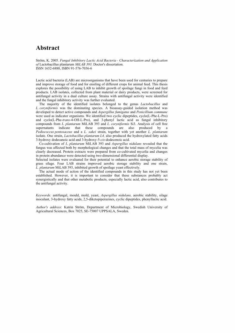
Abstract
Ström, K. 2005. Fungal Inhibitory Lactic Acid Bacteria - Characterization and Application of Lactobacillus plantarum MiLAB 393. Doctor's dissertation. ISSN 1652-6880, ISBN 91-576-7036-6 Lactic acid bacteria (LAB) are microorganisms that have been used for centuries to prepare and improve storage of food and for ensiling of different crops for animal feed. This thesis explores the possibility of using LAB to inhibit growth of spoilage fungi in food and feed products. LAB isolates, collected from plant material or dairy products, were screened for antifungal activity in a dual culture assay. Strains with antifungal activity were identified and the fungal inhibitory activity was further evaluated.
The majority of the identified isolates belonged to the genus Lactobacillus and L. coryniformis was the dominating species. A bioassay-guided isolation method was developed to detect active compounds and Aspergillus fumigatus and Penicillium commune were used as indicator organisms. We identified two cyclic dipeptides, cyclo(L-Phe-L-Pro) and cyclo(L-Phe-trans-4-OH-L-Pro), and 3-phenyl lactic acid as fungal inhibitory compounds from L. plantarum MiLAB 393 and L. coryniformis Si3. Analysis of cell free supernatants indicate that these compounds are also produced by a Pediococcus pentosaceus and a L. sakei strain, together with yet another L. plantarum isolate. One strain, Lactobacillus plantarum L4, also produced the hydroxylated fatty acids 3-hydroxy dodecanoic acid and 3-hydroxy-5-cis-dodecenoic acid.
Co-cultivation of L. plantarum MiLAB 393 and Aspergillus nidulans revealed that the fungus was affected both by morphological changes and that the total mass of mycelia was clearly decreased. Protein extracts were prepared from co-cultivated mycelia and changes in protein abundance were detected using two-dimensional differential display. Selected isolates were evaluated for their potential to enhance aerobic storage stability of grass silage. Four LAB strains improved aerobic storage stability and one strain, L. plantarum MiLAB 393, inhibited growth of spoilage yeast effectively.
The actual mode of action of the identified compounds in this study has not yet been established. However, it is important to consider that these substances probably act synergistically and that other metabolic products, especially lactic acid, also contributes to the antifungal activity. Keywords: antifungal, mould, mold, yeast, Aspergillus nidulans, aerobic stability, silage inoculant, 3-hydroxy fatty acids, 2,5-diketopiperazines, cyclic dipeptides, phenyllactic acid. Author's address: Katrin Ström, Department of Microbiology, Swedish University of Agricultural Sciences, Box 7025, SE-75007 UPPSALA, Sweden.

Till minne av min morfar
Contents
Introduction 7 Fungal spoilage of food and feed 7 Lactic acid bacteria 8
Description of the genera 8 Biopreservation of food and feed 9 Antifungal activity 10 Safety aspects 12
Antifungal compounds-mode of action 12 Target of action 13 Inhibition of fungal growth by weak acids 13
Ensiling of forage crops 14 LAB as silage inoculants 15 Present investigation 16 Aims 16 Isolation and identification of antifungal strains (I & II) 16
Bioassay and screening of antifungal activity 17 Antifungal inhibition spectra 17 Identification of antifungal isolates 18
Identification of antifungal compounds 18
Screening of cell free supernatant 18 Identified compounds 20
Lactobacillus plantarum MiLAB 393 effects on Aspergillus nidulans growth (III) 23
Co-cultivation 24 Growth inhibition and proteome response 24
Antifungal LAB as silage inoculants (IV) 25
Characteristics of selected strains 25 Laboratory scale silage experiment 26 Effects of application of antifungal LAB 27
Concluding remark 29 References 30 Acknowledgments 36
Appendix Papers I-IV This thesis is based on the following papers, wich will be referred to by their Roman numerals.
I. Magnusson, J., Ström, K., Roos, S., Sjögren, J. & Schnürer, J. 2003. Broad and complex antifungal activity among environmental isolates of lactic acid bacteria. FEMS Microbiology Letters 219, 129-135.
II. Ström, K., Sjögren, J., Broberg, A. & Schnürer, J. 2002.
Lactobacillus plantarum MiLAB 393 produces the antifungal cyclic dipeptides cyclo(L-Phe-L-Pro) and cyclo(L-Phe-trans-4-OH-L-Pro) and 3-phenyllactic acid. Applied and Environmental Microbiology 68, 4322-4327.
III. Ström, K., Schnürer, J & Melin, P. 2005.
Co-cultivation of antifungal Lactobacillus plantarum MiLAB 393 and Aspergillus nidulans, evaluation of effects on fungal growth and protein expression. FEMS Microbiology Letters. (In press).
IV. Ström, K., Pauli, T., Ohlsson, C. & Schnürer, J. Lactic Acid Bacteria with Antifungal Properties Can Provide Aerobic Stability and Improve the Quality of Grass Silage. (Manuscript).
Papers are reprinted with permission from the respective publisher
My contributions to the papers included in this thesis has been as follows: I. Took part in planning and performed part of the microbiological laboratory
work. Minor part in writing of the manuscript
II. Did all the microbiological experiments. Planning of experiments and writing of the manuscript was done in collaboration with Jörgen Sjögren. III. Did major part of the laboratory work. Major part in writing the manuscript.
Planning of experiments was done in collaboration with Petter Melin. IV. Did all the microbiological laboratory work and major part of writing the manuscript. Planning of the experiments was done in collaboration with the co-authors.
7
"Imagination is more important than knowledge" Albert Einstein Introduction
In order to survive, all living organisms require nutrients. For humans the taste and appearance of the food plays an important role. Although they are not aware of it, microorganisms such as fungi or bacteria can strongly affect the taste and appearance of food for human consumption. One example of this is fermented foods where both bacteria and fungi can be responsible for the actual fermentation, e.g. fermentation of olives, meat and dairy products by lactic acid bacteria and production of bread, wine and beer by yeasts. On the other hand, many microorganisms are also responsible for spoilage of food products, causing a decrease in nutritional value and potential food poisoning.
This thesis explores the possibility of using one microorganism to inhibit another, more precisely, the use of lactic acid bacteria to inhibit fungal growth. The future perspective is to use these antifungal lactic acid bacteria in food or feed products to decrease the growth of deleterious fungi. Fungal spoilage of food and feed Moulds and yeasts are significant spoilage organisms of food and feed. In addition, the potential production of toxic and carcinogenic mycotoxins by moulds is of particular concern. Besides this problem, fungal spoilage of food also causes significant economic losses. Worldwide, about 5-10 % of the food production is estimated to be spoiled by these organisms (Pitt & Hocking, 1999).
Food and feed are, in general, very diverse microbial growth substrates, due to variation in both intrinsic factors such as pH, nutrients and water activity (aw), and extrinsic factors such as storage temperature and the presence of other microorganisms (Montville & Matthews, 2001). Consequently, fungal spoilage differs depending on the nature of the food, whether it is stored or processed, or if it is perishable food intended for direct consumption. It is important to note that different fungal species have characteristics making them more or less adapted to a particular food system. Some species are able to grow in conditions not suitable for most microorganisms. For example, one of the most common yeasts in fermentative food spoilage, Zygosaccharomyces rouxii, is able to grow at aw as low as 0.62 and at pH values between 1.5 and 10.5 (James & Stratford, 2003; Restaino et al., 1983).
Many mould species are able to utilise most carbon-sources derived from food and many can also utilise nitrate, ammonium or organic nitrogen as a nitrogen source, thus enabling them to grow in wide range of food products (Pitt & Hocking, 1999). Moulds belonging to the species Penicillium, such as P. commune, P. nalgiovense and P. roqueforti commonly spoil various foods as
8
cheese, bread and meat products (Filtenborg, Frisvad & Thrane, 1996; Lund, Filtenborg & Frisvad, 1995). Aspergillus sp. and Penicillium sp. frequently occur as spoiling organisms during storage of grain, whereas Fusarium sp. are the most common spoilage fungi of crops in the field (Filtenborg, Frisvad & Thrane, 1996) (Pitt & Hocking, 1999).
Compared to most filamentous fungi, yeasts are fastidious in their nutrient requirements. However, many foodstuffs are rich substrates containing necessary carbohydrates, amino acids and essential vitamins and minerals, making them ideal growth substrates for many yeast species. Debaromyces hansenii, Kluyveromyces sp., Rhodotorula sp. and several Candida sp. are common causes of deterioration of dairy products, such as cheese and yoghurt (Fröhlich-Wyder, 2003). Yeasts belonging to the species Zygosaccharomyces commonly cause spoilage of acid liquid products with high sugar content, such as juices, jams and sauces (James & Stratford, 2003).
Spoilage caused by many moulds and yeasts can usually be reduced by processing of the food or by conservation. Conservation is achieved either by addition of chemical preservatives or by biopreservation, the use of other microorganisms or their metabolites. In recent years, increasing interest has been shown in biopreservation due to consumer demands for a reduced use of chemicals in food and feed. (Devlieghere, Vermeiren & Debevere, 2004; Ross, Morgan & Hill, 2002; Stiles, 1996). Lactic acid bacteria Description of the genera Lactic acid bacteria (LAB) were first described as milk-souring organisms, due to the sour milk that arose from their production of lactic acid. They are a relatively diverse group of bacteria, but related by a number of typical metabolic and physiological features. Generally the group consists of gram-positive bacteria, cocci or rods, that are non-sporulating, non-respiring and produce lactic acid as the major end product during fermentation of carbohydrates. Historically, the core group comprises the genera Lactobacillus, Leuconostoc, Pediococcus and Streptococcus, but the main LAB genera important in food-technology also include Aerococcus, Carnobacterium, Lactococcus, Oenococcus, Tetragenococcus, Vagococcus and Weisella (Axelsson, 2004; Stiles & Holzapfel, 1997).
LAB can be regarded as homo- or heterofermentative, depending on how they ferment hexoses under non-limited growth conditions. The homofermentative LAB use the glycolysis (Embden-Meyerhof-Parnas) pathway, resulting in lactic acid as the main end product. Heterofermentative LAB use the 6-phosphogluconate/phosphoketolase pathway (6-PG/PK) resulting in lactic acid, carbon dioxide and ethanol (or acetic acid) as the major end products. However, homo- and heterofermentative LAB cannot be distinguished solely on their production of certain fermentation products, since some species are regarded as
9
facultative heterofermenters. Regarding hexose fermentation, these species are homofermenters, but under certain conditions (e.g. if the available carbon source is a pentose), they induce the 6-PG/PK pathway, resulting in heterolactic fermentation (Axelsson, 2004).
LAB are normally found in nutrient-rich environments. Even if they grow in a variety of habitats, they are fastidious and amongst others nutrients they require fermentable carbohydrates, amino acids, fatty acids, salts and vitamins for their growth (Björkroth & Holzapfel, 2003; Hammes & Hertel, 2003). Biopreservation and fermentation of food and feed Since LAB thrive in nutrient-rich environments, they occur naturally in many food and feed systems. They have been used for centuries to prepare and improve storage of food and for ensiling different crops for animal feed. Specific LAB strains are also often included in probiotic products where they are thought to have an beneficial effect on the health of the hosts (animals or humans) (Fuller, 1989). Fermented food has been produced since ancient times, but the important role of microorganisms was not understood until the mid-19th century when the science of microbiology developed (Caplice & Fitzgerald, 1999). Fermentation by LAB is, for example, essential in the production of cheese, yoghurt, fermented sausages and sauerkraut. Today, the primary goal of adding LAB in some products is not preservation in itself, but instead to give a desirable characteristic, such as flavour or texture, to the food. In feed production, fermentation by LAB is probably most important in silage making. The technique has been known for a long time and Egyptian murals dating from 1500 BC demonstrate storage of crop in silos. Today, ensiling is a common method used to preserve forage crops in many parts of the world (McDonald, Hendersoon & Heron, 1991)
Bacteria can protect food (or feed) from microbial spoilage by competitive
growth, by production of antagonistic metabolic products or by formation of other antimicrobial compounds (Schillinger, Geisen & Holzapfel, 1996; Stiles, 1996). Biopreservation refers to the use of a microflora, natural or controlled, or its antibacterial products to extend shelf-life and enhance the safety of foods (Stiles, 1996). The primary preservation effect achieved by LAB is due to the production of lactic acid, which lowers the pH and also directly inhibits many microorganisms (Brul & Coote, 1999). Besides the production of lactic acid, LAB produce other antimicrobial substances, such as acetic acid, hydrogen peroxide, diacetyl, reuterin and bacteriocins, which might play an important role in their preserving capabilities (Caplice & Fitzgerald, 1999; Lindgren & Dobrogosz, 1990).
Since LAB occur naturally in many food systems and they have been a part of the human diet for centuries, they can be regarded as safe organisms to consume. They have a great potential for extended use in biopreservation of both food and feed products.
10
Antifungal activity As mentioned above, LAB can produce a number of antimicrobial substances. Some of them (e.g. lactic acid and reuterin), inhibit both bacteria, yeast and filamentous fungi, whereas others, such as the bacteriocins, only affect closely related bacteria.
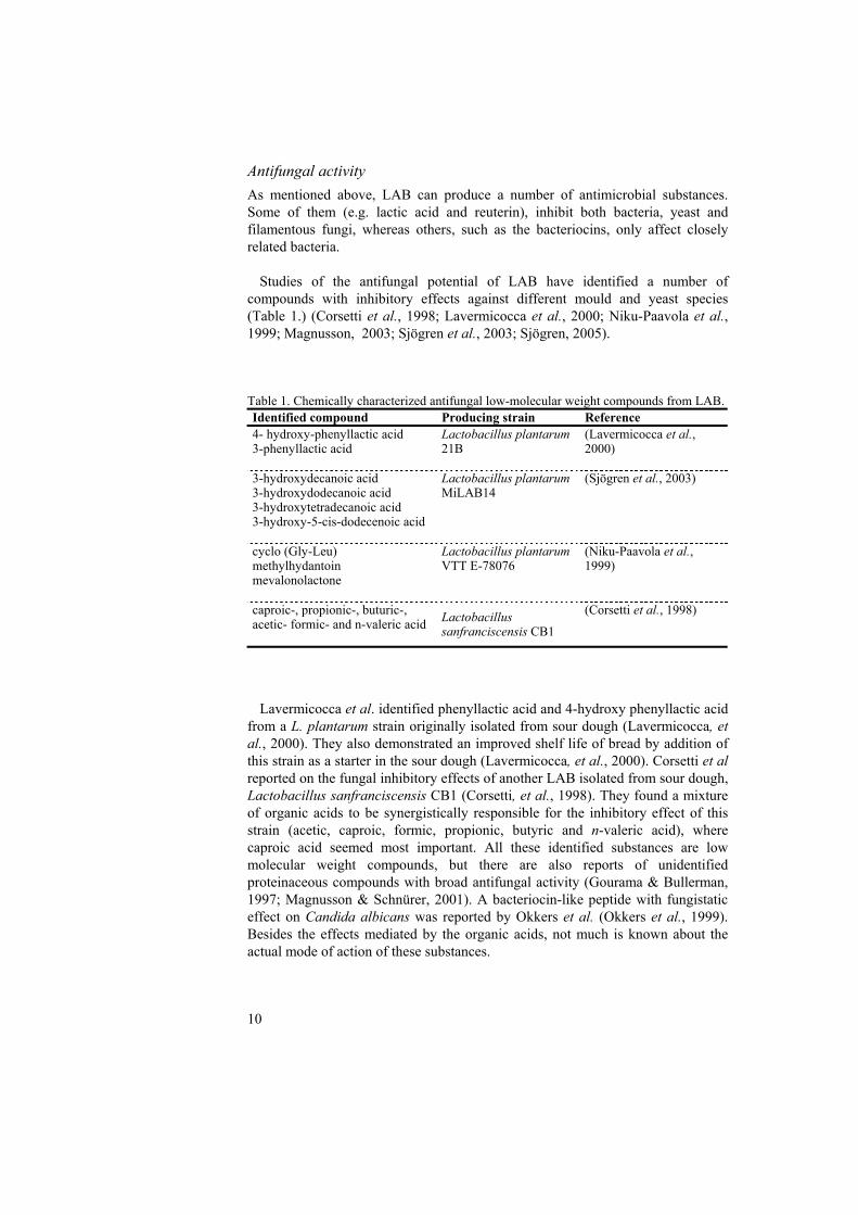
Studies of the antifungal potential of LAB have identified a number of compounds with inhibitory effects against different mould and yeast species (Table 1.) (Corsetti et al., 1998; Lavermicocca et al., 2000; Niku-Paavola et al., 1999; Magnusson, 2003; Sjögren et al., 2003; Sjögren, 2005).
Table 1. Chemically characterized antifungal low-molecular weight compounds from LAB. Identified compound Producing strain Reference 4- hydroxy-phenyllactic acid 3-phenyllactic acid
Lactobacillus plantarum 21B
(Lavermicocca et al., 2000)
3-hydroxydecanoic acid 3-hydroxydodecanoic acid 3-hydroxytetradecanoic acid 3-hydroxy-5-cis-dodecenoic acid
Lactobacillus plantarum MiLAB14
(Sjögren et al., 2003)
cyclo (Gly-Leu) methylhydantoin mevalonolactone
Lactobacillus plantarum VTT E-78076
(Niku-Paavola et al., 1999)
caproic-, propionic-, buturic-, acetic- formic- and n-valeric acid
Lactobacillus sanfranciscensis CB1
(Corsetti et al., 1998)
Lavermicocca et al. identified phenyllactic acid and 4-hydroxy phenyllactic acid
from a L. plantarum strain originally isolated from sour dough (Lavermicocca, et al., 2000). They also demonstrated an improved shelf life of bread by addition of this strain as a starter in the sour dough (Lavermicocca, et al., 2000). Corsetti et al reported on the fungal inhibitory effects of another LAB isolated from sour dough, Lactobacillus sanfranciscensis CB1 (Corsetti, et al., 1998). They found a mixture of organic acids to be synergistically responsible for the inhibitory effect of this strain (acetic, caproic, formic, propionic, butyric and n-valeric acid), where caproic acid seemed most important. All these identified substances are low molecular weight compounds, but there are also reports of unidentified proteinaceous compounds with broad antifungal activity (Gourama & Bullerman, 1997; Magnusson & Schnürer, 2001). A bacteriocin-like peptide with fungistatic effect on Candida albicans was reported by Okkers et al. (Okkers et al., 1999). Besides the effects mediated by the organic acids, not much is known about the actual mode of action of these substances.
11
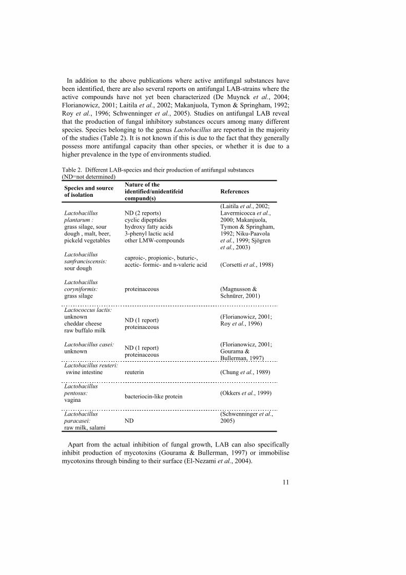
In addition to the above publications where active antifungal substances have been identified, there are also several reports on antifungal LAB-strains where the active compounds have not yet been characterized (De Muynck et al., 2004; Florianowicz, 2001; Laitila et al., 2002; Makanjuola, Tymon & Springham, 1992; Roy et al., 1996; Schwenninger et al., 2005). Studies on antifungal LAB reveal that the production of fungal inhibitory substances occurs among many different species. Species belonging to the genus Lactobacillus are reported in the majority of the studies (Table 2). It is not known if this is due to the fact that they generally possess more antifungal capacity than other species, or whether it is due to a higher prevalence in the type of environments studied.
Table 2. Different LAB-species and their production of antifungal substances (ND=not determined)
Species and source of isolation
Nature of the identified/unidentifeid compund(s)
References
Lactobacillus plantarum : grass silage, sour dough , malt, beer, pickeld vegetables
ND (2 reports) cyclic dipeptides hydroxy fatty acids 3-phenyl lactic acid other LMW-compounds
(Laitila et al., 2002; Lavermicocca et al., 2000; Makanjuola, Tymon & Springham, 1992; Niku-Paavola et al., 1999; Sjögren et al., 2003)
Lactobacillus sanfranciscensis: sour dough
caproic-, propionic-, buturic-, acetic- formic- and n-valeric acid
(Corsetti et al., 1998)
Lactobacillus coryniformis: grass silage
proteinaceous
(Magnusson & Schnürer, 2001)
Lactococcus lactis: unknown cheddar cheese raw buffalo milk
ND (1 report) proteinaceous
(Florianowicz, 2001; Roy et al., 1996)
Lactobacillus casei: unknown
ND (1 report) proteinaceous
(Florianowicz, 2001; Gourama & Bullerman, 1997)
Lactobacillus reuteri: swine intestine
reuterin (Chung et al., 1989)
Lactobacillus pentosus: vagina
bacteriocin-like protein
(Okkers et al., 1999)
Lactobacillus paracasei: raw milk, salami
ND (Schwenninger et al., 2005)
Apart from the actual inhibition of fungal growth, LAB can also specifically
inhibit production of mycotoxins (Gourama & Bullerman, 1997) or immobilise mycotoxins through binding to their surface (El-Nezami et al., 2004).
12
There are now a number of reports on LAB with antifungal properties and their possible use as biopreservatives has recently been reviewed by Schnürer and Magnusson (2005). Antifungal LAB could probably be used in food or feed systems to enhance quality, for example by reducing the use of chemical additives and preventing growth of spoilage yeasts and mycotoxigenic fungi.
Safety aspects As previously mentioned, LAB have been a part of the human diet since ancient times, hence one could argue that they should be completely safe to consume and particularly suitable in biopreservation of food and feed. The regulations concerning addition of LAB to food vary widely internationally, between countries and between international bodies (Feord, 2002; Wessels et al., 2004). LAB in food can be regarded as an additive, ingredient, processing aid or probiotic culture, leading to completely different regulations and regulatory requirements for a novel LAB culture (Wessels, et al., 2004). The important safety features to be considered in application of LAB strains to food and feed are: A. The inability to invade the consumer/host and cause disease B. The absence of transferable antibiotic resistance.
Infections by probiotic strains of LAB have been established from two clinical cases (Saarela et al., 2000) and in a recent review by Cannon et al. (Cannon et al., 2005) over 200 cases of infections with Lactobacillus sp. were reported. The majority of these infections occurred in immunocompromised patients, or in patients undergoing broad-spectrum antibiotic therapy. Although LAB have been safely consumed in many parts of the world since ancient times, it is important to bear in mind that they can cause serious infections, especially in immunocompromised patients.
Due to their structure and physiology LAB are naturally resistant to some antibiotics, but this resistance is commonly non-transferable (Donohue, 2004). However, plasmid-borne antibiotic resistance occurs in LAB (Saarela, et al., 2000) and hence this possibility should be carefully evaluated in strains considered for consumption by humans or animals.
In conclusion, it is obvious that LAB are already a part of the human diet and have been so for a long time. There are no emerging reports on harmful effects from consumption of food containing LAB, but instead beneficial effects have been established for some probiotic strains (Salminen et al., 2004). However, consumer awareness regarding food safety is increasing and a dialogue between researchers, industry, consumers and regulatory authorities concerning novel applications of LAB is necessary.
Antifungal compounds – mode of action Substances that inhibit fungal growth are of importance both in the control of human and animal pathogens, and in the prevention of fungal growth in food and
13
other materials. This section gives an overview of the mode of action of current antifungal antibiotics that are important for fungal inhibition. Many of these substances are reserved for clinical use but some are employed in the control of plant pathogenic fungi. Antibiotics are defined as substances produced by microorganisms that kill or inhibit growth of other microorganisms at low concentrations. This section will not deal with substances produced by LAB since not much is known about their actual mode of action. However, the fungal inhibitory effect of weak acids will be discussed, since this is important in relation to metabolites produced by LAB. Target of action Since fungi are eukaryotic organisms, substances affecting their growth by inhibition of general mechanisms (e.g. protein synthesis) must also be assumed to affect humans or animals. However, some structures are specific for fungi, such as the chitin and β- glucan in the cell wall and ergosterol in the cell membrane. These targets are particularly suitable when selective inhibition is desired. Many antifungal antibiotics have a specific site for their inhibitory action. Common targets are the cell wall, the cell membrane, protein synthesis or cell division. • Cell wall: Inhibition of β- glucan synthesis and inhibition of chitin synthetase are the main mechanisms of compounds that act on the fungal cell wall. Polyoxins and Nikkomycin are inhibitors of chitin synthesis, whereas the compounds belonging to the echinocandin family affect the biosynthesis of β- glucan polysaccharides (Odds, Brown & Gow, 2003). • Cell membrane: Compounds that interfere with the cell membrane in different ways act by binding of sterols with a consequent disruption of the membrane function or by inhibition of the sterol biosynthesis. Polyene compounds (e.g. Amphoteracin B and Nystatin) form complexes by binding to ergosterol, whereas synthetic azoles affect the synthesis of sterol (Groll & Kolve, 2004). • Protein synthesis: Blasticidin inhibits protein synthesis by binding to the ribosomes (Hansen, Moore & Steitz, 2003) while sordarin derivates act by blocking the elongation factor EF2 (Vicente et al., 2003). • Cell division: The mode of action of one of the earliest antifungal agents used clinically, griseofulvin, is not fully understood, but the main action is suggested to be by interference with microtubuli assembly during cell division (Odds, Brown & Gow, 2003). Inhibition of fungal growth by weak acids Microbial growth can be inhibited by the presence of weak organic acids since at a certain concentration, they lower the pH to a level where many microorganisms cannot grow. Major weak organic acids used in food protection are acetic (pKa 4.76), benzoic (pKa 4.19), lactic (pKa 3.86), propionic (pKa 4.87) and sorbic acid (pKa 4.76)
14
Besides the environmental pH-decrease there are other, probably more important, effects of these compounds on fungal growth. The general mechanism behind the inhibition is suggested to be due to the passage of undissociated acid molecules across the cell membrane. Once inside the cell, the higher pH of the cytoplasm will lead to dissociation of the acid. This will generate an accumulation of the anion of the acid together with protons and consequently a decrease of the intracellular pH (pHint). In yeast a lowering of pHint has been shown to inhibit glycolysis (Krebs et al., 1983). Thus, intracellular acidification directly affects growth. Apart from the lowering of pHint, other actions, such as anion accumulation and disordering of cell membrane structure have also been proposed as being responsible for the weak acid inhibition (Piper et al., 2001). Other studies report on the accumulation of trehalose (Cheng, Moghraby & Piper, 1999) and inhibition of macroautophagy (Hazan, Levine & Abeliovich, 2004) and aromatic amino acid uptake (Bauer et al., 2003) in Saccharomyces cerevisiae by weak organic acids.
Comparisons of inhibitory activity by different organic acids have established
that the fungal response is different depending on which weak acids are used (Narendranath, Thomas & Ingledew, 2001; Stratford & Anslow, 1996). According to Stratford and Anslow (1998), the inhibition of S. cerevisiae by sorbate is not only the result of weak acid intracellular acidification, but mainly due to interference with the cell membrane. The responses to weak acids also vary between different organisms (Cheng, Moghraby & Piper, 1999; Lind, Jonsson & Schnürer, 2005).
S. cerevisiae can develop resistance to weak organic acids through the induction
of the plasma membrane proteins Pdr12 and Hsp30 (Piper et al., 1998). In contrast, the preservative resistant yeast Zygosaccharomyces baiili does not seem to respond in the same way. Instead, the resistance to weak organic acids in Z. bailii seems to be caused by an ability to degrade the acids and also to limit their diffusion by changing the cell envelope (Piper, et al., 2001). Weak acid inhibition of filamentous fungi has not received as much attention as effects on yeast. However, Plumridge et al. (Plumridge et al., 2004) showed that sorbic acid causes intracellular acidification and inhibition of conidial germination and mycelial growth in Aspergillus niger.
Most studies of the effects of weak acids on fungal growth have established that
a certain pH is necessary for the inhibitory action, whereby the acid is undissociated, leading to diffusion across the membrane. However, the actual mode of action seems to be different, depending both on the type of organic acid and target organism. In conclusion, this group of compounds shares common features important for inhibition of fungal growth, but some specific actions by different acids also occur. Ensiling of forage crops Preservation of forage crops for animal feed by ensiling is a well-known method. The process is based on fermentation of water-soluble carbohydrates (WSC) by
15
LAB and the exclusion of air. The fermentation lowers the pH due to lactic acid production which inhibits the growth of many spoilage organisms (McDonald, Hendersoon & Heron, 1991). Ensiling occurs naturally and is performed by the epiphytic flora of LAB present on the crop. In successful silage, the fermentation and consequent acidification starts rapidly, achieving an efficient inhibition of unwanted microorganisms.
One problem that occurs in silage, even with an effective fermentation step, is that when air is let in to the system (e.g. during feeding), the growth of aerobic spoilage organisms, such as mould and yeast, can be re-activated (Woolford, 1990). One way to overcome this problem and to make the ensiling process more effective could be to add a LAB-inoculant with suitable antifungal properties. Such an inoculant would supposedly produce antifungal metabolites during the fermentation and prevent the re-activation of fungal microorganisms during feed-out. LAB as silage inoculants Improvement of silage quality by addition of LAB inoculants, alone or in combination with chemical additives, has been examined in several studies (Lindgren, Bromander & Pettersson, 1988; Rooke, Borman & Armstrong, 1990; Weinberg et al., 1993 Kung et al., 2004). LAB have been used as inoculants in silage mainly to improve preservation efficiency or to enhance animal performance (Weinberg & Muck, 1996). The important features of an ideal silage inoculant have been identified as (McDonald, Hendersoon & Heron, 1991; Rees, 1997): • Fast growth and ability to compete with other microorganisms in the crop • Homofermentative – generating rapid production of lactic acid • Acid tolerant, able to produce a final pH of at least 4.0 as rapidly as possible • Ability to grow on material with low aw • Growth or survival at temperatures up to 50°C. • Suitable and stable as a dry formulation (e.g. by freeze drying). Many of these criteria were established when the desired outcome was to improve the actual fermentation. Later it has been suggested that new criteria for silage inoculants should be formulated to address specific needs (e.g. fermentation efficiency, aerobic stability or animal performance (Weinberg & Muck, 1996)). One such specific inoculant could be a LAB possessing antifungal properties, in order to decrease fungal deterioration of silage and to improve aerobic storage stability.
16
Present investigation
Aims The aim of this study was to isolate novel LAB with antifungal properties and to characterize their antifungal activity. Further, the ambition was to prevent fungal deterioration by applying these inhibitory strains in biopreservation. The specific objectives were to: • Isolate and identify LAB-isolates with antifungal activities from natural
environments. I • Elucidate the nature of the antifungal activity and characterize antifungal
compounds. I & II • Evaluate the effect of L. plantarum MiLAB 393 on fungal growth at both
morphological and molecular levels by co-cultivation with Aspergillus nidulans. III
• Elucidate the effects of inoculation with antifungal LAB on aerobic storage
stability of silage. IV Isolation and identification of antifungal strains (I & II) LAB were mainly isolated from plant material or from dairy products since one of the primary goals was to use the antifungal strains for biocontrol in such products. It seemed reasonable that strains already adapted to these environments would be the most suitable to reintroduce in proposed products. Collected plant or dairy material was incubated in small silos under anaerobic conditions to facilitate enrichment of LAB. After ensiling in these small-scale silos, the material was spread on de Man Rogosa Sharp (MRS) agar plates, a medium suitable for growth of most LAB (de Man, Rogosa & Sharp, 1960). Colonies appearing on the plates were isolated and further screened for antifungal activity. All strains were isolated as described above, except P. pentosaceus MiLAB 392 and L. plantarum MiLAB 393. These two strains were both isolated in a silage experiment where the antifungal activity of some other isolates was evaluated. The silage was inoculated with spoilage yeast and moulds and surprisingly the most efficient inhibition of these organisms was displayed in the control silo where no LAB-inoculants had been added. P. pentosaceus MiLAB392 and L. plantarum MiLAB393 were isolated from the control silos and found to have a pronounced antifungal activity in vitro.
17
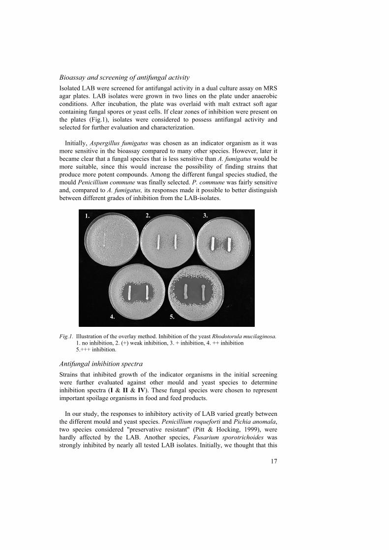
Bioassay and screening of antifungal activity Isolated LAB were screened for antifungal activity in a dual culture assay on MRS agar plates. LAB isolates were grown in two lines on the plate under anaerobic conditions. After incubation, the plate was overlaid with malt extract soft agar containing fungal spores or yeast cells. If clear zones of inhibition were present on the plates (Fig.1), isolates were considered to possess antifungal activity and selected for further evaluation and characterization.
Initially, Aspergillus fumigatus was chosen as an indicator organism as it was more sensitive in the bioassay compared to many other species. However, later it became clear that a fungal species that is less sensitive than A. fumigatus would be more suitable, since this would increase the possibility of finding strains that produce more potent compounds. Among the different fungal species studied, the mould Penicillium commune was finally selected. P. commune was fairly sensitive and, compared to A. fumigatus, its responses made it possible to better distinguish between different grades of inhibition from the LAB-isolates.
Fig.1. Illustration of the overlay method. Inhibition of the yeast Rhodotorula mucilaginosa. 1. no inhibition, 2. (+) weak inhibition, 3. + inhibition, 4. ++ inhibition 5.+++ inhibition. Antifungal inhibition spectra Strains that inhibited growth of the indicator organisms in the initial screening were further evaluated against other mould and yeast species to determine inhibition spectra (I & II & IV). These fungal species were chosen to represent important spoilage organisms in food and feed products.
In our study, the responses to inhibitory activity of LAB varied greatly between the different mould and yeast species. Penicillium roqueforti and Pichia anomala, two species considered "preservative resistant" (Pitt & Hocking, 1999), were hardly affected by the LAB. Another species, Fusarium sporotrichoides was strongly inhibited by nearly all tested LAB isolates. Initially, we thought that this
1. 2. 3.
4. 5.
1. 2. 3.
4. 5.
18
was due to varying sensitivity against the lactic acid produced by the LAB. Since it has been reported that antifungal activity of LAB might be due to the combination of lactic acid production and acetic acid in the MRS-media (Cabo, Braber & Koenraad, 2002) this was evaluated.
The amounts of acetic and lactic acid produced by a number of selected LAB
strains with different inhibition patterns were quantified by HPLC (I). Supernatant from two L. coryniformis strains with different inhibition of A. fumigatus (+ or +++) were included in the analysis. The lactic acid production by these strains did not explain the different inhibition since the strain with only + inhibition in fact produced more lactic acid than the +++ inhibitory strain (95 mM compared to 62 mM). A similar result was obtained when two P. pentosaceus strains, with different inhibition of both A. fumigatus and P. commune, were compared. The acetic acid in the samples corresponded to the amount present in MRS broth. We confirmed that the different responses in A. fumigatus could not be explained by the sensitivity to lactic acid or by the presence of acetic acid in the MRS media. Thus, the conclusion was that other compounds contributed to the antifungal activity.
That the sensitivity to organic acids did not explain the different responses among fungal species was also verified in a recent study of antifungal effects from propionibacteria (Lind, Jonsson & Schnürer, 2005). Here P. roqueforti was found to be more sensitive to the organic acids, propionic-, lactic- and acetic acid, than several other mould and yeast species.
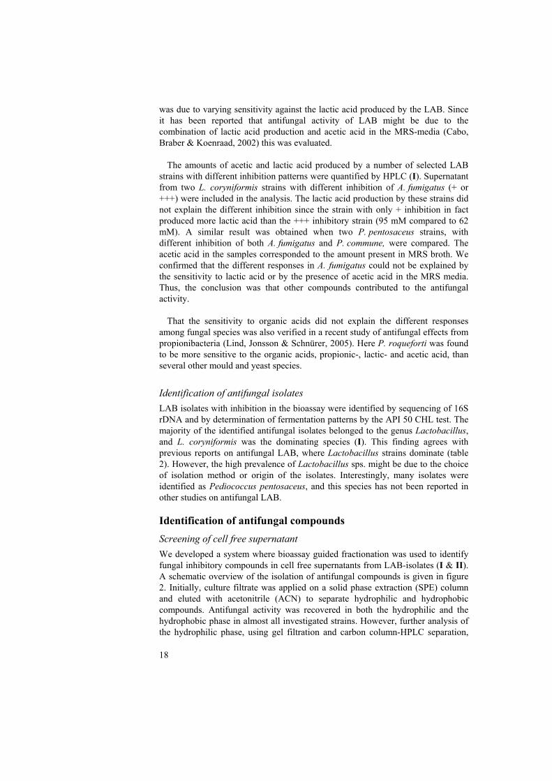
Identification of antifungal isolates LAB isolates with inhibition in the bioassay were identified by sequencing of 16S rDNA and by determination of fermentation patterns by the API 50 CHL test. The majority of the identified antifungal isolates belonged to the genus Lactobacillus, and L. coryniformis was the dominating species (I). This finding agrees with previous reports on antifungal LAB, where Lactobacillus strains dominate (table 2). However, the high prevalence of Lactobacillus sps. might be due to the choice of isolation method or origin of the isolates. Interestingly, many isolates were identified as Pediococcus pentosaceus, and this species has not been reported in other studies on antifungal LAB. Identification of antifungal compounds Screening of cell free supernatant We developed a system where bioassay guided fractionation was used to identify fungal inhibitory compounds in cell free supernatants from LAB-isolates (I & II). A schematic overview of the isolation of antifungal compounds is given in figure 2. Initially, culture filtrate was applied on a solid phase extraction (SPE) column and eluted with acetonitrile (ACN) to separate hydrophilic and hydrophobic compounds. Antifungal activity was recovered in both the hydrophilic and the hydrophobic phase in almost all investigated strains. However, further analysis of the hydrophilic phase, using gel filtration and carbon column-HPLC separation,
19
exclusively detected lactic acid as the active compound. Other active compounds might have been present in the hydrophilic phase, but they could not be separated from the lactic acid. Analysis of the hydrophilic phase was not continued since the majority of the strains also showed activity in the hydrophobic phase.
Fig.2. Schematic overview of the bioassay-guided isolation of antifungal compounds
Cell free supernatant
Bioassay
H2 O
AC
N
C18-HPLCC18-HPLC
1 2 3 4 5 6 7 8 9 10 11 12ABCDEFGH
Bioassay
Structuraldetermination
NMR/MS/GC
1 2 3 4 5 6 7 8 9 10 11 12ABCDEFGH
Hypercarb-HPLC
Hypercarb-HPLC
Bioassay
Bioassay
Gel-filtration
Bioassay
1 2 3 4 5 6 7 8 9 10 11 12ABCDEFGH
Hypercarb-HPLC
Hypercarb-HPLC
Bioassay
?
SPE
20
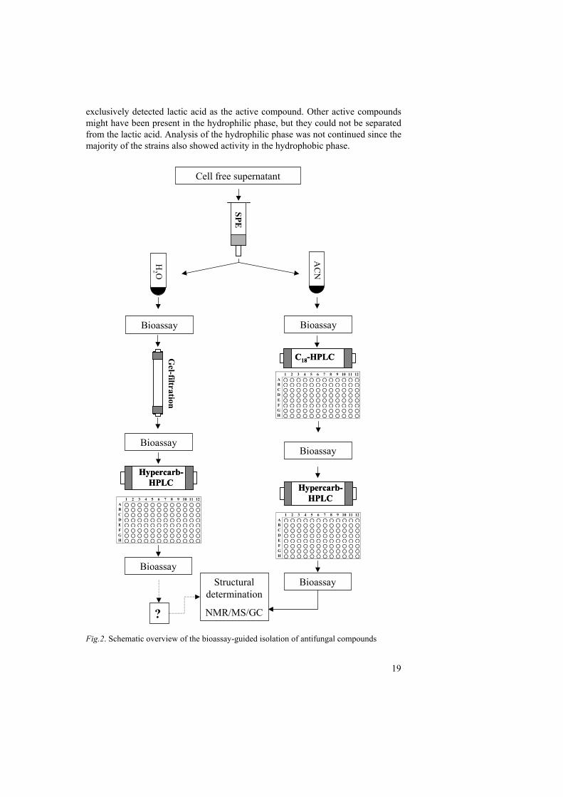
The hydrophobic ACN-phase from the SPE separation was analysed using bioassay-guided separations. Fractions from C18-HPLC separations were collected in 2-ml 96 deep-well plates and fractions were assayed on fungal spores (I & II). The degree of fungal inhibition by the collected fractions was assayed in microtiter wells, and fractions where growth was delayed, partly or totally inhibited (determined by optical density compared to growth of control) were considered as active fractions (Fig 3.).
After bioassay with A. fumigatus or P. commune as indicator organisms, active
fractions were selected for further separation in a second HPLC-run on a porous graphitic carbon column. Active fractions from the second HPLC-run were investigated by mass-spectroscopy (MS), nuclear magnetic resonance (NMR) and gas chromatography (GC) for structural analyses of the compounds (I & II).
Fig.3. Bioassay in microtiter wells, showing total inhibition of A. fumigatus growth in wells A12, B12, D10 and E7.
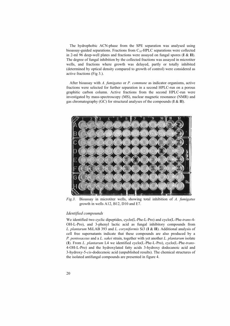
Identified compounds We identified two cyclic dipeptides, cyclo(L-Phe-L-Pro) and cyclo(L-Phe-trans-4-OH-L-Pro), and 3-phenyl lactic acid as fungal inhibitory compounds from L. plantarum MiLAB 393 and L. coryniformis Si3 (I & II). Additional analysis of cell free supernatants indicate that these compounds are also produced by a P. pentosaceus and a L. sakei strain, together with yet another L. plantarum isolate (I). From L. plantarum L4 we identified cyclo(L-Phe-L-Pro), cyclo(L-Phe-trans-4-OH-L-Pro) and the hydroxylated fatty acids 3-hydroxy dodecanoic acid and 3-hydroxy-5-cis-dodecenoic acid (unpublished results). The chemical structures of the isolated antifungal compounds are presented in figure 4.
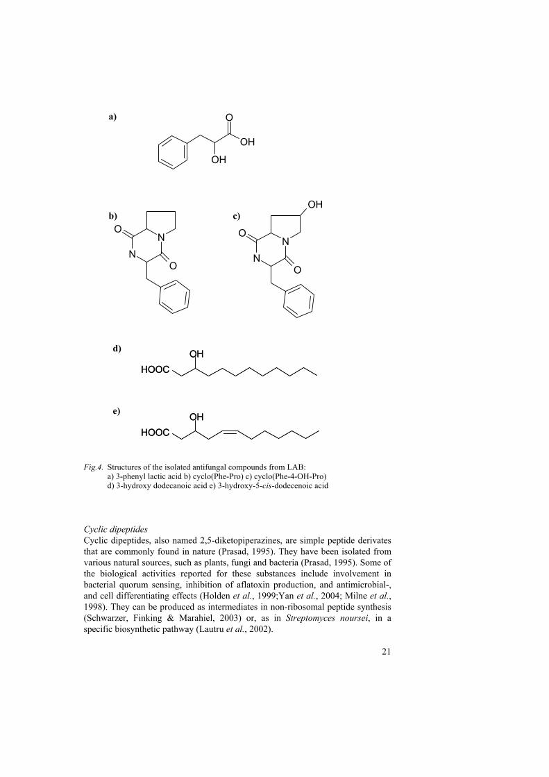
21
Fig.4. Structures of the isolated antifungal compounds from LAB: a) 3-phenyl lactic acid b) cyclo(Phe-Pro) c) cyclo(Phe-4-OH-Pro)
d) 3-hydroxy dodecanoic acid e) 3-hydroxy-5-cis-dodecenoic acid Cyclic dipeptides Cyclic dipeptides, also named 2,5-diketopiperazines, are simple peptide derivates that are commonly found in nature (Prasad, 1995). They have been isolated from various natural sources, such as plants, fungi and bacteria (Prasad, 1995). Some of the biological activities reported for these substances include involvement in bacterial quorum sensing, inhibition of aflatoxin production, and antimicrobial-, and cell differentiating effects (Holden et al., 1999;Yan et al., 2004; Milne et al., 1998). They can be produced as intermediates in non-ribosomal peptide synthesis (Schwarzer, Finking & Marahiel, 2003) or, as in Streptomyces noursei, in a specific biosynthetic pathway (Lautru et al., 2002).
OH
OH
O
NN
O
O
OH
NN
O
O
HOOCOH
HOOCOH
HOOCOH
HOOCOH
a)
b) c)
d)
e)
22
Since we originally purified the cyclic dipeptides from spent MRS-broth, a medium containing several peptides and proteins, we also used a defined growth medium, DM1 (Moretro, Hagen & Axelsson, 1998), to verify that the dipeptidic compounds were indeed produced by the bacteria. Although DM1 only contains free amino acids, both cyclo(L-Phe-L-Pro) and cyclo(L-Phe-trans-4-OH-L-Pro) were produced by MiLAB 393 grown in DM1, but at lower concentrations compared to growth in MRS broth (II).
The minimal inhibitory concentration (MIC) of cyclo(L-Phe-L-Pro) for the
indicator organisms A. fumigatus and P. roqueforti was 20 mg ml-1, but in the presence of another inhibitory compound (3-phenyllactic acid at 5 mg ml-1), the MIC value decreased to 10 mg ml-1. This is a rather high inhibitory concentration compared to other antifungal substances such as amphotericin B and Nikkomycin Z that inhibit fungal growth at concentrations in the µg ml-1 range (Frändberg et al., 2000). Thus, the antifungal activity of the cyclic dipeptides is probably a secondary effect, but it is likely to be of considerable importance for the fungal inhibitory activity of L. plantarum MiLAB 393, L. coryniformis Si3 and other LAB producing these substances.
Another cyclic dipeptide that inhibits fungal growth, Cyclo(L-Leu-L-Pro), is isolated from Achromobacter xylosoxidans (Yan, et al., 2004). It was identified due to its inhibition of aflatoxin production in A. parasiticus. Aflatoxin production was inhibited at a concentration of 1 mg ml-1, whereas higher concentrations were necessary for inhibition of fungal growth. The possible effect of the cyclic dipeptides isolated in the present study on mycotoxin production remains to be elucidated. The antifungal mode of action of the cyclic dipeptides has not been established. Further investigation is needed to evaluate possible inhibitory mechanisms of these compounds. Phenyllactic acid Phenyllactic acid is a small aromatic and acidic compound. It can be formed by LAB and propionic acid bacteria as a product of phenylalanine catabolism, a process important for formation of aroma compounds in cheese production (Helinck et al., 2004; Kieronczyk et al., 2003; Thierry & Maillard, 2002). The production of phenyllactic acid from p-hydroxyphenylpyruvic and phenylpyruvic acid by rumen bacteria has also been demonstrated (Khan et al., 2002). Phenyllactic acid has earlier been reported to be antimicrobial and produced by lactic acid bacteria (I, II, (Lavermicocca, et al., 2000; Valerio et al., 2004)) and by the fungus Geotrichum candidum (Dieuleveux, Lemarinier & Gueguen, 1998). D-3-phenyllactic acid inhibits several bacterial species, supposedly through interference with the bacterial cell wall (Dieuleveux, Lemarinier & Gueguen, 1998). We have observed that fungal growth is inhibited by 3-phenyllactic acid and that it clearly delays fungal growth at concentrations below the MIC-value (II & III). This is in agreement with the findings by Lavermicocca et al. who identified both inhibitory and growth delaying effects of 3-phenyllactic with several fungal species (Lavermicocca, Valerio & Visconti, 2003). How phenyllactic acid affects fungal growth is not yet understood and as with the cyclic
23
dipeptides, further studies are needed to elucidate the mode of action of this substance. However, a possible target for 3-phenyllactic acid could be the enzyme phenylalanine dehydrogenase. This enzyme, identified in bacteria, catalyses deamination of phenylalanine to generate pyridine for nucleotide synthesis. Phenylalanine dehydrogenase is inhibited by phenyllactic acid due to its structural similarities to the ordinary substrate (Brunhuber et al., 2000). Since similar dehydrogenases for other amino acids also exist in fungi, a possible antifungal mechanism for phenyllactic acid could be inhibition of phenylalanine dehydrogenase. 3-hydroxy fatty acids The 3-hydroxy fatty acids (also named β-hydroxy acids) are probably most often recognized as components of lipid A, present in the lipopolysaccharide (LPS) layer in the outer membrane of Gram-negative bacteria (Wilkinson, 1996). However, these substances are also named oxylipins and are widely distributed among various fungal species (Kock et al., 2003). They are mostly found on the surface of sexual spores or vegetative cells (Sebolai et al., 2004) and reported to function as lubricants to facilitate ascospore release (Bareetseng et al., 2004). Other hydroxylated fatty acids, generally with more complex structures, have also been reported as antibacterial (Mundt, Kreitlow & Jansen, 2003), antiviral (Harper et al., 1996) and inhibitors of plant pathogenic fungi (Graner, Hamberg & Meijer, 2003; Hou & Forman, 2000).
In LAB, analysis of cellular fatty acids has identified 3-hydroxyhexadecanoic acid (C16) as a possible taxonomic marker of Leuconostoc sp. (Lee et al., 1996). Sjögren et al. (Sjögren, et al., 2003) reported the isolation of 3-hydroxy fatty acids from L. plantarum and established antifungal MIC values between 5-100 µg ml-1 among different mould and yeast species. The 3-hydroxy fatty acids are therefore much more potent inhibitors than the cyclic dipeptides and phenyllactic acid.
The mechanisms responsible for antifungal activity of the 3-hydroxy fatty acids
are not known. Reports on their presence on the surfaces of fungal spores and yeast cells, as well as involvement in spore release, suggests that they might interfere with spore distribution and spore germination. The findings that non-hydroxylated decanoic acid alters membrane fluidity and lipid composition in yeast (Alexandre, Mathieu & Charpentier, 1996) implies another possible inhibitory mechanism for these compounds. Lactobacillus plantarum MiLAB 393 effects on Aspergillus nidulans growth (III) To further investigate the antifungal activity, a co-cultivation method was developed to evaluate the effects on A. nidulans grown in the presence of L. plantarum MiLAB 393. We selected A. nidulans as a model organism primarily due to the fact that the genome is sequenced and available for searches at Broad Institute of MIT and Harvard (http://www.broad.mit.edu). Further, A. nidulans is easy to culture and our group has earlier used this organism in studies of molecular responses to bacterial metabolites and for generating mutant strains
24
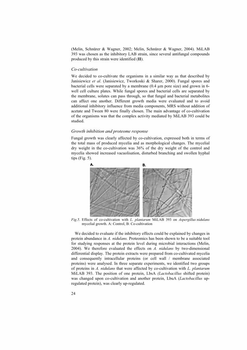
(Melin, Schnürer & Wagner, 2002; Melin, Schnürer & Wagner, 2004). MiLAB 393 was chosen as the inhibitory LAB strain, since several antifungal compounds produced by this strain were identified (II). Co-cultivation We decided to co-cultivate the organisms in a similar way as that described by Janisiewicz et al. (Janisiewicz, Tworkoski & Sharer, 2000). Fungal spores and bacterial cells were separated by a membrane (0.4 µm pore size) and grown in 6-well cell culture plates. While fungal spores and bacterial cells are separated by the membrane, solutes can pass through, so that fungal and bacterial metabolites can affect one another. Different growth media were evaluated and to avoid additional inhibitory influence from media components, MRS without addition of acetate and Tween 80 were finally chosen. The main advantage of co-cultivation of the organisms was that the complex activity mediated by MiLAB 393 could be studied. Growth inhibition and proteome response Fungal growth was clearly affected by co-cultivation, expressed both in terms of the total mass of produced mycelia and as morphological changes. The mycelial dry weight in the co-cultivation was 36% of the dry weight of the control and mycelia showed increased vacuolisation, disturbed branching and swollen hyphal tips (Fig. 5).
Fig.5. Effects of co-cultivation with L. plantarum MiLAB 393 on Aspergillus nidulans mycelial growth. A: Control, B: Co-cultivation
We decided to evaluate if the inhibitory effects could be explained by changes in
protein abundance in A. nidulans. Proteomics has been shown to be a suitable tool for studying responses at the protein level during microbial interactions (Melin, 2004). We therefore evaluated the effects on A. nidulans by two-dimensional differential display. The protein extracts were prepared from co-cultivated mycelia and consequently intracellular proteins (or cell wall / membrane associated proteins) were analysed. In three separate experiments, we identified two groups of proteins in A. nidulans that were affected by co-cultivation with L. plantarum MiLAB 393. The position of one protein, LbsA (Lactobacillus shifted protein) was changed upon co-cultivation and another protein, LbuA (Lactobacillus up-regulated protein), was clearly up-regulated.
A. B.A. B.
25
To further investigate if these protein changes were caused by the inhibitory compounds from L. plantarum MiLAB 393, additional 2D-gel analysis was performed on protein extracts from mycelia grown in the presence of lactic acid, 3-phenyllactic acid or cyclo(L-Phe-L-Pro). The results showed that protein LbsA was not affected by the presence of phenyllactic acid, but a similar shift as found during co-cultivation could be observed when the fungus was grown in the presence of lactic acid. The cyclic dipeptide, cyclo(L-Phe-L-Pro), shifted the protein in the opposite direction compared to the co-cultivation. Protein LbuA was up-regulated by all of the inhibitory substances, but to a varying degree depending on the substance present. The cyclic dipeptide cyclo(L-Phe–L-Pro) had the most pronounced effect, with LbuA up to eight times up-regulated compared to the co-cultivation. N-terminal sequencing of proteins LbuA and LbsA followed by searches in the A. nidulans genome database identified homologue sequences, ID EAA65324 and ID EAA59671.1 respectively, but unfortunately neither of them was annotated. The corresponding sequences were also found in the non-completed genome database of A. fumigatus (at http://www.tigr.org/). Additional BLAST-searches in available databases did not give any results indicating the function of these two proteins. BLAST-searches with the LbsA sequence revealed several hypothetical fungal proteins with high sequence similarity (42-74 % identity), indicating that this protein is present among different fungal species. Protein LbuA is likely to be translocated through the membrane since the first 18 residues appear to be a secretion signal peptide, with a probability of 1.0 according to Signal P 3.0 (Bendtsen et al., 2004). The protein did not hold any transmembrane regions, according to TMHMM Server v. 2.0 (Krogh et al., 2001), and since we isolated this protein from fungal mycelia it is probably re-associated to the cell wall after secretion. Antifungal LAB as silage inoculants (IV) We identified a number of antifungal LAB with broad inhibition spectra (I & II) and these LAB strains were further evaluated by applying them as silage inoculants. The aim was to decrease growth of spoilage moulds and yeasts and to enhance the aerobic storage stability of the silage. Characteristics of selected strains Primarily, strains were selected for their inhibition of selected mould and yeasts in vitro. Since material used for silage making can vary greatly in composition, especially the amount of WSC and dry mass (DM) content (McDonald, Hendersoon & Heron, 1991), the osmotolerance of the selected strains was also evaluated. In this study, the osmotolerance was determined by evaluation of growth on media containing 10% KCl (simulating a DM of 45% and a aw of 0.91). However, using this method, it must be considered that bacterial growth might also be influenced by salt stress due to the high KCl-concentration in the plates.
26
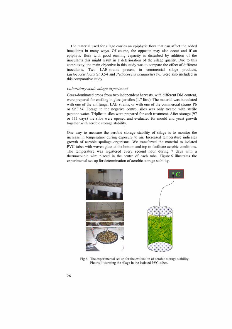
The material used for silage carries an epiphytic flora that can affect the added inoculants in many ways. Of course, the opposite may also occur and if an epiphytic flora with good ensiling capacity is disturbed by addition of the inoculants this might result in a deterioration of the silage quality. Due to this complexity, the main objective in this study was to compare the effect of different inoculants. Two LAB-strains present in commercial silage products, Lactococcis lactis Sr 3.54 and Pediococcus acidilactici P6, were also included in this comparative study. Laboratory scale silage experiment Grass-dominated crops from two independent harvests, with different DM content, were prepared for ensiling in glass jar silos (1.7 litre). The material was inoculated with one of the antifungal LAB strains, or with one of the commercial strains P6 or Sr.3.54. Forage in the negative control silos was only treated with sterile peptone water. Triplicate silos were prepared for each treatment. After storage (97 or 111 days) the silos were opened and evaluated for mould and yeast growth together with aerobic storage stability. One way to measure the aerobic storage stability of silage is to monitor the increase in temperature during exposure to air. Increased temperature indicates growth of aerobic spoilage organisms. We transferred the material to isolated PVC-tubes with woven glass at the bottom and top to facilitate aerobic conditions. The temperature was registered every second hour during 7 days with a thermocouple wire placed in the centre of each tube. Figure 6 illustrates the experimental set-up for determination of aerobic storage stability.
Fig.6. The experimental set-up for the evaluation of aerobic storage stability.
Photos illustrating the silage in the isolated PVC-tubes.
���������������������������������������������������������������������������������������������������
º C
������������������������������������������������������������������������������������������������������
������������������������������������������������������������������������������������������������������
27
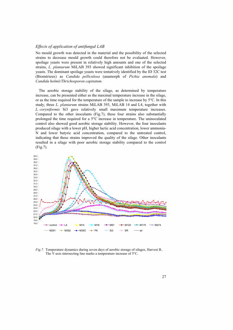
Effects of application of antifungal LAB No mould growth was detected in the material and the possibility of the selected strains to decrease mould growth could therefore not be evaluated. However, spoilage yeasts were present in relatively high amounts and one of the selected strains, L. plantarum MiLAB 393 showed significant inhibition of the spoilage yeasts. The dominant spoilage yeasts were tentatively identified by the ID 32C test (Biomérieux) as Candida pelliculosa (anamorph of Pichia anomala) and Candida holmii/Thrichosporon capitatum. The aerobic storage stability of the silage, as determined by temperature increase, can be presented either as the maximal temperature increase in the silage, or as the time required for the temperature of the sample to increase by 5°C. In this study, three L. plantarum strains MiLAB 393, MiLAB 14 and L4, together with L. coryniformis Si3 gave relatively small maximum temperature increases. Compared to the other inoculants (Fig.7), these four strains also substantially prolonged the time required for a 5°C increase in temperature. The uninoculated control also showed good aerobic storage stability. However, the four inoculants produced silage with a lower pH, higher lactic acid concentration, lower ammonia-N and lower butyric acid concentration, compared to the untreated control, indicating that these strains improved the quality of the silage. Other inoculants resulted in a silage with poor aerobic storage stability compared to the control (Fig.7).
Fig.7. Temperature dynamics during seven days of aerobic storage of silages, Harvest B..
The Y-axis intersecting line marks a temperature increase of 5°C.
18,019,020,021,022,023,024,025,026,027,028,029,030,031,032,033,034,035,036,037,038,039,040,0
control L4 M14 M18 M91 M120 M170 M274
M291 M392 M393 P6 Si3 SR air
28
This study shows that addition of silage inoculants can improve the quality, but that the opposite may also occur. One inoculant, L. plantarum MiLAB 393, inhibited spoilage yeast efficiently and gave good storage stability of the silage. This strain was originally isolated from grass silage due to its inhibition of spoilage moulds and yeasts in such material. In addition, we performed a similar experiment with the same LAB strains in corn silage (unpublished results). The preliminary results from this study show that, compared to the grass experiment, different strains are efficient in enhancing corn silage quality. This indicates that the source of isolation is important for the choice of application and that crop-specific inoculants should be considered.
All LAB strains included in this study were homofermenters (or facultative
heterofermenters) although this was not a selection criterion. Homofermenters are thought to be more suitable as silage inoculants than heterofermenters since they lower the pH more rapidly in the initial fermentation (Weinberg & Muck, 1996). On the other hand, several studies have demonstrated that the heterofermentative L. buchneri is much more efficient in improving aerobic stability of the silage (Filya, 2003b; Kung & Ranjit, 2001). It has been established that inhibition of mould and yeast growth by L. buchneri is due to production of acetic acid (Holzer et al., 2003). A combination of two inoculants could be used to benefit both from enhanced fermentation and increased aerobic stability. A combination of L. buchneri and L. plantarum in corn and sorghum silages did indeed give better initial fermentation, lower protein degradation and fermentation losses compared to silage inoculated with L. buchneri alone (Filya, 2003a). In our study, we selected LAB strains due to their antifungal attributes in an attempt to enhance aerobic stability. Four strains, all homofermenters, gave good aerobic stability, thus indicating that homofermentative LAB with the right properties could be used as inoculants both to enhance fermentation and to improve aerobic storage stability.
As previously mentioned, the material used for silage making can vary greatly in
composition. The growth stage of the crop at the time of harvest together with climate conditions affects biochemical factors as WSC and DM-content (McDonald, Hendersoon & Heron, 1991). Of course the different crops used for ensiling (e.g. grass, corn or cereals) also differ widely in both structure and biochemical composition. Thus, it is almost impossible to find an inoculant that will be successful in all types of silage making. The rational approach would instead be to define specific inoculants that will be efficient in different types of silage making.
29
Concluding remarks
"Have no fear of perfection, you'll never reach it." Salvador Dali
This thesis presents studies where the antifungal properties of LAB have been evaluated. The overall conclusion from this work is that many LAB-species seem able to inhibit fungal growth by means of other activities than lactic acid production. The actual mode of action of the identified compounds has not yet been established and further work on this topic is desirable. However, it must be considered that the substances produced by LAB probably act synergistically and that other metabolic products, especially lactic acid, also contribute to the overall inhibition. We set up a system where bacteria and fungi were co-cultivated in a manner where metabolites, but not cells, could move freely and the effects on fungal protein abundance were evaluated. In future experiments this system could be improved to identify possible targets for antifungal compounds produced by LAB. One future experiment using this method could be to evaluate one of the LAB strains producing the 3-hydroxy fatty acids, since they are more potent inhibitors of fungal growth than the cyclic dipeptides or phenyllactic acid. Saccharomyces cerevisiae may also be used as a fungal target species because of the comprehensive information now available on its genome, proteome and metabolome. Additional experiments in silage should be performed with respect to the application of LAB strains in biocontrol. It would be interesting to evaluate the actual growth of the inoculant in the silage, for example by labelling with green fluorescent protein or some other suitable method. It is also desirable to investigate if the bacteria produce the inhibitory compounds when they grow in the silage material.
Efficient inhibition of spoilage yeast in yoghurt was observed after using a combination of LAB and propionibacteria (Schwenninger & Meile, 2004). These findings indicate that combinations of propionibacteria and LAB could be useful in biopreservation of dairy products. Evaluation of the strains isolated in this work in such an experiment is yet another future perspective. Finally, my conclusion is that the fungal inhibitory activity of LAB is very complex. Hopefully, this thesis has contributed to clarify some aspects of this activity. I also believe that LAB with fungal inhibitory properties can be useful in biopreservation of both food and feed, alone or in combination with other microorganisms.
30
References
Alexandre, H., Mathieu, B. & Charpentier, C. 1996. Alteration in membrane fluidity and lipid composition, and modulation of H+-ATPase activity in Saccharomyces cerevisiae caused by decanoic acid. Microbiology-Uk 142, 469-475.
Axelsson, L.T. 2004. Lactic Acid Bacteria: Classification and Physiology. In Lactic Acid Bacteria - Microbiology and functional aspects. Edited by S. Salminen, A.v. Wright & A. Ouwehand. Marcel Dekker, Inc. 1-66. pp.
Bareetseng, A.S., Kock, J.L.F., Pohl, C.H., Pretorius, E.E., Strauss, C.J., Botes, P.J., van Wyk, P.W.J. & Nigam, S. 2004. Mapping 3-hydroxy oxylipins on ascospores of Eremothecium sinecaudum. Antonie Van Leeuwenhoek International Journal of General and Molecular Microbiology 86, 363-368.
Bauer, B.E., Rossington, D., Mollapour, M., Mamnun, Y., Kuchler, K. & Piper, P.W. 2003. Weak organic acid stress inhibits aromatic amino acid uptake by yeast, causing a strong influence of amino acid auxotrophies on the phenotypes of membrane transporter mutants. European Journal of Biochemistry 270, 3189-3195.
Bendtsen, J.D., Nielsen, H., von Heijne, G. & Brunak, S. 2004. Improved prediction of signal peptides: SignalP 3.0. Journal of Molecular Biology 340, 783-795.
Björkroth, J. & Holzapfel, W. 2003. Genera Leuconostoc, Oenococcus and Weissella. In The Prokaryotes: An Evolving Electronic Resource for the Microbiological Community, http://link.springer-ny.com/link/service/books/10125/. Edited by M. Dworkin. Springer-Verlag, New York.
Brul, S. & Coote, P. 1999. Preservative agents in foods - Mode of action and microbial resistance mechanisms. International Journal of Food Microbiology 50, 1-17.
Brunhuber, N.M.W., Thoden, J.B., Blanchard, J.S. & Vanhooke, J.L. 2000. Rhodococcus L-phenylalanine dehydrogenase: Kinetics, mechanism, and structural basis for catalytic specifity. Biochemistry 39, 9174-9187.
Cabo, M.L., Braber, A.F. & Koenraad, P. 2002. Apparent antifungal activity of several lactic acid bacteria against Penicillium discolor is due to acetic acid in the medium. Journal of Food Protection 65, 1309-1316.
Cannon, J.P., Lee, T.A., Bolanos, J.T. & Danziger, L.H. 2005. Pathogenic relevance of Lactobacillus: a retrospective review of over 200 cases. European Journal of Clinical Microbiology & Infectious Diseases 24, 31-40.
Caplice, E. & Fitzgerald, G.F. 1999. Food fermentations: role of microorganisms in food production and preservation. International Journal of Food Microbiology 50, 131-149.
Cheng, L.L., Moghraby, J. & Piper, P.W. 1999. Weak organic acid treatment causes a trehalose accumulation in low-pH cultures of Saccharomyces, cerevisiae, not displayed by the more preservative-resistant Zygosaccharomyces bailii. FEMS Microbiology Letters 170, 89-95.
Corsetti, A., Gobbetti, M., Rossi, J. & Damiani, P. 1998. Antimould activity of sourdough lactic acid bacteria: identification of a mixture of organic acids produced by Lactobacillus sanfrancisco CB1. Applied Microbiology and Biotechnology 50, 253-256.
de Man, J.C., Rogosa, M. & Sharp, M.E. 1960. A medium for the cultivation of lactobacilli. Journal of Applied Bacteriology 23, 130-135.
De Muynck, C., Leroy, A.I.J., De Maeseneire, S., Arnaut, F., Soetaert, W. & Vandamme, E.J. 2004. Potential of selected lactic acid bacteria to produce food compatible antifungal metabolites. Microbiological Research 159, 339-346.
Devlieghere, F., Vermeiren, L. & Debevere, J. 2004. New preservation technologies: Possibilities and limitations. International Dairy Journal 14, 273-285.
Dieuleveux, V., Lemarinier, S. & Gueguen, M. 1998. Antimicrobial spectrum and target site of D-3-phenyllactic acid. International Journal of Food Microbiology 40, 177-183.
Donohue, D.C. 2004. Saftey of Novel Probiotic Bacteria. In Lactic Acid Bacteria - Microbiology and functional aspects. Edited by S. Salminen, A.v. Wright & A. Ouwehand. Marcel Dekker, Inc. 531-546. pp.
31
El-Nezami, H., Mykkänen, H., Haskard, C., Salminen, S. & Salminen, E. 2004. Lactic acid bacteria as a tool for enhancing food saftey by removal of dietay toxins. In Lactic Acid Bacteria - Microbiology and functional aspects. Edited by S. Salminen, A.v. Wright & A. Ouwehand. Marcel Dekker, Inc. 397-406. pp.
Feord, J. 2002. Lactic acid bacteria in a changing legislative environment. Antonie Van Leeuwenhoek International Journal of General and Molecular Microbiology 82, 353-360.
Filtenborg, O., Frisvad, J.C. & Thrane, U. 1996. Moulds in food spoilage. International Journal of Food Microbiology 33, 85-102.
Filya, I. 2003a. The effect of Lactobacillus buchneri and Lactobacillus plantarum on the fermentation, aerobic stability, and ruminal degradability of low dry matter corn and sorghum silages. Journal of Dairy Science 86, 3575-3581.
Filya, I. 2003b. The effect of Lactobacillus buchneri, with or without homofermentative lactic acid bacteria, on the fermentation, aerobic stability and ruminal degradability of wheat, sorghum and maize silages. Journal of Applied Microbiology 95, 1080-1086.
Florianowicz, T. 2001. Antifungal activity of some microorganisms against Penicillium expansum. European Food Research and Technology 212, 282-286.
Frändberg, E., Petersson, C., Lundgren, L.N. & Schnürer, J. 2000. Streptomyces halstedii K122 produces the antifungal compounds bafilomycin B1 and C1. Canadian Journal of Microbiology 46, 753-758.
Fröhlich-Wyder, M.-T. 2003. Yeasts in dairy products. In Yeasts in food. Edited by T. Boekhout & V. Robert. Behrs Verlag. 209-237. pp.
Fuller, R. 1989. Probiotics in Man and Animals. Journal of Applied Bacteriology 66, 365-378.
Gourama, H. & Bullerman, L.B. 1997. Anti-aflatoxigenic activity of Lactobacillus casei pseudoplantarum. International Journal of Food Microbiology 34, 131-143.
Graner, G., Hamberg, M. & Meijer, J. 2003. Screening of oxylipins for control of oilseed rape (Brassica napus) fungal pathogens. Phytochemistry 63, 89-95.
Groll, A.H. & Kolve, H. 2004. Antifungal agents: In vitro susceptibility testing, pharmacodynamics, and prospects for combination therapy. Eur J Microbiol Infect Dis 23, 256-270.
Hammes, W.P. & Hertel, C. 2003. The Genera Lactobacillus and Carnobacterium. In The Prokaryotes: An Evolving Electronic Resource for the Microbiological Community, http://link.springer-ny.com/link/service/books/10125/. Edited by M. Dworkin. Springer-Verlag, New York.
Hansen, J.L., Moore, P.B. & Steitz, T.A. 2003. Structures of Five Antibiotics Bound at the Peptidyl Transferase Center of the Large Ribosomal Subunit. Journal of Molecular Biology 330, 1061-1075.
Harper, D.R., Gilbert, R.L., Oconnor, T.J., Kinchington, D., Mahmood, N., McIlhinney, R.A.J. & Jeffries, D.J. 1996. Antiviral activity of 2-hydroxy fatty acids. Antiviral Chemistry & Chemotherapy 7, 138-141.
Hazan, R., Levine, A. & Abeliovich, H. 2004. Benzoic acid, a weak organic acid food preservative, exerts specific effects on intracellular membrane trafficking pathways in Saccharomyces cerevisiae. Applied and Environmental Microbiology 70, 4449-4457.
Helinck, S., Le Bars, D., Moreau, D. & Yvon, M. 2004. Ability of thermophilic lactic acid bacteria to produce aroma compounds from amino acids. Applied and Environmental Microbiology 70, 3855-3861.
Holden, M.T.G., Chhabra, S.R., de Nys, R., Stead, P., Bainton, N.J., Hill, P.J., Manefield, M., Kumar, N., Labatte, M., England, D., Rice, S., Givskov, M., Salmond, G.P.C., Stewart, G., Bycroft, B.W., Kjelleberg, S.A. & Williams, P. 1999. Quorum-sensing cross talk: isolation and chemical characterization of cyclic dipeptides from Pseudomonas aeruginosa and other Gram-negative bacteria. Molecular Microbiology 33, 1254-1266.
Holzer, M., Mayrhuber, E., Danner, H. & Braun, R. 2003. The role of Lactobacillus buchneri in forage preservation. Trends in Biotechnology 21, 282-287.
Hou, C.T. & Forman, R.J. 2000. Growth inhibition of plant pathogenic fungi by hydroxy fatty acids. Journal of Industrial Microbiology & Biotechnology 24, 275-276.
32
James, S. & Stratford, M. 2003. Spoilage yeast with emphasis on the genus Zygosaccharomyces. In Yeasts in food. Edited by B. T & R. V. Behrs Verlag. 171-191. pp.
Janisiewicz, W.J., Tworkoski, T.J. & Sharer, C. 2000. Characterizing the mechanism of biological control of postharvest diseases on fruits with a simple method to study competition for nutrients. Phytopathology 90, 1196-1200.
Khan, R.I., Onodera, R., Amin, M.R. & Mohammed, N. 2002. Aromatic amino acid biosynthesis and production of related compounds from p-hydroxyphenylpyruvic acid by rumen bacteria, protozoa and their mixture. Amino Acids 22, 167-177.
Kieronczyk, A., Skeie, S., Langsrud, T. & Yvon, M. 2003. Cooperation between Lactococcus lactis and nonstarter lactobacilli in the formation of cheese aroma from amino acids. Applied and Environmental Microbiology 69, 734-739.
Kock, J.L.F., Strauss, C.J., Pohl, C.H. & Nigam, S. 2003. The distribution of 3-hydroxy oxylipins in fungi. Prostaglandins & Other Lipid Mediators 71, 85-96.
Krebs, H.A., Wiggins, D., Stubbs, M., Sols, A. & Bedoya, F. 1983. Studies on the mechanism of the antifungal action of benzoate Biochem. Journal 214, 657-663
Krogh, A., Larsson, B., von Heijne, G. & Sonnhammer, E.L.L. 2001. Predicting transmembrane protein topology with a hidden Markov model: Application to complete genomes. Journal of Molecular Biology 305, 567-580.
Kung, L., Myers, C.L., Neylon, J.M., Taylor, C.C., Lazartic, J., Mills, J.A. & Whiter, A.G. 2004. The effects of buffered propionic acid-based additives alone or combined with microbial inoculation on the fermentation of high moisture corn and whole-crop barley. Journal of Dairy Science 87, 1310-1316.
Kung, L. & Ranjit, N.K. 2001. The effect of Lactobacillus buchneri and other additives on the fermentation and aerobic stability of barley silage. Journal of Dairy Science 84, 1149-1155.
Laitila, A., Alakomi, H.L., Raaska, L., Mattila-Sandholm, T. & Haikara, A. 2002. Antifungal activities of two Lactobacillus plantarum strains against Fusarium moulds in vitro and in malting of barley. Journal of Applied Microbiology 93, 566-576.
Lautru, S., Gondry, M., Genet, R. & Pernodet, J.L. 2002. The albonoursin gene cluster of S. noursei: Biosynthesis of diketopiperazine metabolites independent of nonribosomal peptide synthetases. Chemistry & Biology 9, 1355-1364.
Lavermicocca, P., Valerio, F., Evidente, A., Lazzaroni, S., Corsetti, A. & Gobbetti, M. 2000. Purification and characterization of novel antifungal compounds from the sourdough Lactobacillus plantarum strain 21B Applied and Environmental Microbiology 66, 4084-90.
Lavermicocca, P., Valerio, F. & Visconti, A. 2003. Antifungal Activity of Phenyllactic Acid against Molds Isolated from Bakery Products. Applied and Environmental Microbiology 69, 634-640.
Lee, J.S., Chun, C.O., Kim, H.J., Joo, Y.J., Lee, H.J., Park, C.S., Ahn, J.S., Park, Y.H. & Mheen, T.I. 1996. Analysis of cellular fatty acid methyl esters (FAMEs) for the identification of Leuconostoc strains isolated from kimchi. Journal of Microbiology 34, 225-228.
Lind, H., Jonsson, H. & Schnürer, J. 2005. Antifungal effect of dairy propionibacteria - contribution of organic acids. International Journal of Food Microbiology 98, 157-165.
Lindgren, S., Bromander, A. & Pettersson, K. 1988. Evaluation of Silage Additives Using Scale-Model Silos. Swedish Journal of Agricultural Research 18, 41-49.
Lindgren, S.E. & Dobrogosz, W.J. 1990. Antagonistic Activities of Lactic Acid Bacteria in Food and Feed Fermentations. FEMS Microbiology Reviews 87, 149-163.
Lund, F., Filtenborg, O. & Frisvad, J.C. 1995. Associated Mycoflora of Cheese. Food Microbiology 12, 173-180.
Magnusson, J. 2003. Antifungal Lactic acid Bacteria, PhD thesis, Swedish University of Agricultural Sciences
Magnusson, J. & Schnürer, J. 2001. Lactobacillus coryniformis subsp. coryniformis strain Si3 produces a broad-spectrum proteinaceous antifungal compound. Applied and Environmental Microbiology 67, 1-5.
33
Makanjuola, D.B., Tymon, A. & Springham, D.G. 1992. Some Effects of Lactic-Acid Bacteria on Laboratory-Scale Yeast Fermentations. Enzyme and Microbial Technology 14, 350-357.
McDonald, P., Hendersoon, N. & Heron, S. 1991. The Biochemistry of Silage second ed. Chalcombe Publications.
Melin, P. 2004. Proteomics as a tool to study microbial interactions. Current Proteomics, 27-34.
Melin, P., Schnürer, J. & Wagner, E.G.H. 2002. Proteome analysis of Aspergillus nidulans reveals proteins associated with the response to the antibiotic concanamycin A, produced by Streptomyces species. Molecular Genetics and Genomics 267, 695-702.
Melin, P., Schnürer, J. & Wagner, E.G.H. 2004. Disruption of the gene encoding the V-ATPase subunit A results in inhibition of normal growth and abolished sporulation in Aspergillus nidulans. Microbiology-Sgm 150, 743-748.
Milne, P.J., Hunt, A.L., Rostoll, K., Van Der Walt, J.J. & Graz, C.J.M. 1998. The biological activity of selected cyclic dipeptides. Journal of Pharmacy and Pharmacology 50, 1331-1337.
Montville, T.J. & Matthews, K.R. 2001. Principles which influence microbial growth, survival and death in foods. In Food Microbiology: Fundamentals and Frontiers. Edited by M.P.D.e. al. ASM Press.
Moretro, T., Hagen, B.F. & Axelsson, L. 1998. A new, completely defined medium for meat lactobacilli. Journal of Applied Microbiology 85, 715-722.
Mundt, S., Kreitlow, S. & Jansen, R. 2003. Fatty acids with antibacterial activity from the cyanobacterium Oscillatoria redekei HUB 051. Journal of Applied Phycology 15, 263-267.
Narendranath, N.V., Thomas, K.C. & Ingledew, W.M. 2001. Acetic acid and lactic acid inhibition of growth of Saccharomyces cerevisiae by different mechanisms. Journal of the American Society of Brewing Chemists 59, 187-194.
Niku-Paavola, M.L., Laitila, A., Mattila-Sandholm, T. & Haikara, A. 1999. New types of antimicrobial compounds produced by Lactobacillus plantarum. Journal of Applied Microbiology 86, 29-35.
Odds, F.C., Brown, A.J.P. & Gow, N.A.R. 2003. Antifungal agents: mechanisms of action. Trends in Microbiology 11, 272-279.
Okkers, D.J., Dicks, L.M., Silvester, M., Joubert, J.J. & Odendaal, H.J. 1999. Characterization of pentocin TV35b, a bacteriocin-like peptide isolated from Lactobacillus pentosus with a fungistatic effect on Candida albicans. Journal of Applied Microbiology 87, 726-734.
Piper, P., Calderon, C.O., Hatzixanthis, K. & Mollapour, M. 2001. Weak acid adaptation: the stress response that confers yeasts with resistance to organic acid food preservatives. Microbiology 147, 2635-2642.
Piper, P., Mahe, Y., Thompson, S., Pandjaitan, R., Holyoak, C., Egner, R., Muhlbauer, M., Coote, P. & Kuchler, K. 1998. The Pdr12 ABC transporter is required for the development of weak organic acid resistance in yeast. Embo Journal 17, 4257-4265.
Pitt, J.J. & Hocking, A.D. 1999. Fungi and food spoilage Second ed. Aspen Publications. pp.
Plumridge, A., Hesse, S.J.A., Watson, A.J., Lowe, K.C., Stratford, M. & Archer, D.B. 2004. The weak acid preservative sorbic acid inhibits conidial germination and mycelial growth of Aspergillus niger through intracellular acidification. Applied and Environmental Microbiology 70, 3506-3511.
Prasad, C. 1995. Bioactive Cyclic Dipeptides. Peptides 16, 151-164. Rees, T. (1997). The development of a novel silage inoculant, PhD thesis, Cranfield
University. Restaino, L., Bills, S., Tscherneff, K. & Lenovich, L.M. 1983. Growth characteristics of
Saccharomyces rouxii isolated from chocolate syrup. Applied and Enviromental Microbiology 45, 1614-1621.
Rooke, J.A., Borman, A.J. & Armstrong, D.G. 1990. The Effect of Inoculation with Lactobacillus plantarum on Fermentation in Laboratory Silos of Herbage Low in Water-Soluble Carbohydrate. Grass and Forage Science 45, 143-152.
34
Ross, R.P., Morgan, S. & Hill, C. 2002. Preservation and fermentation: past, present and future. International Journal of Food Microbiology 79, 3-16.
Roy, U., Batish, V.K., Grover, S. & Neelakantan, S. 1996. Production of antifungal substance by Lactococcus lactis subsp. lactis CHD-28.3. International Journal of Food Microbiology 32, 27-34.
Saarela, M., Mogensen, G., Fonden, R., Matto, J. & Mattila-Sandholm, T. 2000. Probiotic bacteria: safety, functional and technological properties. Journal of Biotechnology 84, 197-215.
Salminen, S., Gobrbach, S., Lee, Y.-K. & Benno, Y. 2004. Human Studies on Probiotics: What is Scientifically Proven Today? In Lactic Acid Bacteria - Microbiology and functional aspects. Edited by S. Salminen, A.v. Wright & A. Ouwehand. Marcel Dekker, Inc. 515-530. pp.
Schillinger, U., Geisen, R. & Holzapfel, W.H. 1996. Potential of antagonistic microorganisms and bacteriocins for the biological preservation of foods. Trends in Food Science & Technology 7, 158-164.
Schwarzer, D., Finking, R. & Marahiel, M.A. 2003. Nonribosomal peptides: from genes to products. Natural Product Reports 20, 275-287.
Schwenninger, S.M. & Meile, L. 2004. A mixed culture of Propionibacterium jensenii and Lactobacillus paracasei subsp. paracasei inhibits food spoilage yeasts. Systematic and Applied Microbiology 27, 229-237.
Schwenninger, S.M., van Ah, U., Niederer, B., Teuber, M. & Meile, L. 2005. Detection of antifungal properties in Lactobacillus paracasei subsp paracasei SM20, SM29, and SM63 and molecular typing of the strains. Journal of Food Protection 68, 111-119.
Sebolai, O.M., Kock, J.L.F., Pohl, C.H., Botes, P.J. & Nigam, S. 2004. Report on the discovery of a novel 3-hydroxy oxylipin cascade in the yeast Saccharomycopsis synnaedendra. Prostaglandins & Other Lipid Mediators 74, 139-146.
Sjögren, J., Magnusson, J., Broberg, A., Schnürer, J. & Kenne, L. 2003. Antifungal 3-hydroxy fatty acids from Lactobacillus plantarum MiLAB 14. Applied and Environmental Microbiology 69, 7554-7557.
Sjögren, J. (2005). Bioassay-guided Isolation and Characterisation of Antifungal Metabolites, PhD thesis, Swedish Agricultural University.
Stiles, M.E. 1996. Biopreservation by lactic acid bacteria. Antonie Van Leeuwenhoek International Journal of General and Molecular Microbiology 70, 331-345.
Stiles, M.E. & Holzapfel, W.H. 1997. Lactic acid bacteria of foods and their current taxonomy. International Journal of Food Microbiology 36, 1-29.
Stratford, M. & Anslow, P.A. 1996. Comparison of the inhibitory action on Saccharomyces cerevisiae of weak-acid preservatives, uncouplers, and medium-chain fatty acids. FEMS Microbiology Letters 142, 53-58.
Thierry, A. & Maillard, M.B. 2002. Production of cheese flavour compounds derived from amino acid catabolism by Propionibacterium freudenreichii. Lait 82, 17-32.
Valerio, F., Lavermicocca, P., Pascale, M. & Visconti, A. 2004. Production of phenyllactic acid by lactic acid bacteria: an approach to the selection of strains contributing to food quality and preservation. FEMS Microbiology Letters 233, 289-295.
Weinberg, Z.G., Ashbell, G., Hen, Y. & Azrieli, A. 1993. The Effect of Applying Lactic Acid Bacteria at Ensiling on the Aerobic Stability of Silages. Journal of Applied Bacteriology 75, 512-518.
Weinberg, Z.G. & Muck, R.E. 1996. New trends and opportunities in the development and use of inoculants for silage. FEMS Microbiology Reviews 19, 53-68.
Wessels, S., Axelsson, L., Bech Hansen, E., De Vuyst, L., Laulund, S., Lahteenmaki, L., Lindgren, S., Mollet, B., Salminen, S. & von Wright, A. 2004. The lactic acid bacteria, the food chain, and their regulation. Trends in Food Science & Technology 15, 498-505.
Vicente, M.F., Basilio, A., Cabello, A. & Pelaez, F. 2003. Microbial natural products as a source of antifungals. Clin Microbiol Infect 9, 15-32.
Wilkinson, S.G. 1996. Bacterial lipopolysaccharides - Themes and variations. Progress in Lipid Research 35, 283-343.
Woolford, M.K. 1990. The Detrimental Effects of Air on Silage. Journal of Applied Bacteriology 68, 101-116.
35
Yan, P.-S., Song, Y., Sakuno, E., Nakajima, H., Nakagawa, H. & Yabe, K. 2004. Cyclo(L-Leucyl-L-Prolyl) Produced by Achromobacter xylosoxidans Inhibits Aflatoxin Production by Aspergillus parasiticus. Applied and Environmental Microbiology 70, 7466-7473.
36
Acknowledgments
The financial support of the Foundation for Strategic Environmental Research (MISTRA), the Swedish Farmers Foundation for Agricultural Research (SLF) and Medipharm AB is gratefully acknowledged. There are a number of people who have supported and encouraged me during my graduate studies. Others have contributed to my personal development and I would like to express my sincere gratitude to:
min huvudhandledare Johan Schnürer. Din entusiasm och kapacitet att initiera nya projekt är verkligen av världsklass! Tack för att du introducerade mig i svamparnas fantastiska värld och för att du har guidat mig under dessa år. Ett speciellt tack också för att du ibland förser mig med diverse "icke vetenskaplig" litteratur som ger näring till mina drömmar om ett liv i extravagans och lyx.
mina biträdande handledare Stefan Roos och Hans Jonsson. Tack för att ni alltid tar er tid att svara på alla möjliga och omöjliga frågor om mjölksyrabakterier, för att ni ständigt låter mig komma in på ert rum och "störa", pratandes både forskning och en massa strunt.
kemisten och West Ham-fantasten ;-) Jörgen Sjögren. Tack för suveräna
kemiska analyser och ett givande samarbete, men också för alla trevliga ölkvällar och "icke vetenskapliga" diskussioner om det som kallas livet.
Lisa Fredlund & Ulrika Ädel Druvefors. Det är mycket som har hänt sedan
jästsvamparna hade sex i mikroskopet på CBS-kursen (sorry om jag skämde ut er då ;-). Lisa, kanske kan vi tipsa varandra om möjliga lösningar på den där ekvationen framöver. Ullis, jag har saknat din närvaro & ditt skratt oerhört mycket den senaste tiden.
Inger Ohlsson, tack för laborativ hjälp den senaste tiden, men framförallt tack
för att du alltid är så oerhört omtänksam. Vad skulle Lifo-gruppen vara utan dig?! min medförfattare Petter Melin, för att du introducerade mig i A. nidulans
molekylära värld, also för att du har gett mig ett så vackert smeknamn ;-) nuvarande och tidigare medlemmar i Lifo-gruppen, Jesper, Anders E, Helena,
Torun, Klara, Cissi, Johan D, Åsa S, Jonas F, Matilda, Marianne, Xin-Mei, Volkmar, Jennifer m.fl. för allt kul vi har gjort (och kommer att göra) tillsammans. Klara tackas speciellt för att jag numera slipper "jympa" ensam på lab och Anders E tackas speciellt för att du tar hand om Buster ;-) och för alla otaliga gånger du erbjudit skjuts hem från glada fester! Jonas F, tack för en fantastiskt fin bild till avhandlingens framsida.
37
alla nuvarande och före detta medarbetare på mikrobiologen, det är ni som gör mikron till en trevlig arbetsplats. Tack Karin J och Fryk för att ni "lurade" Johan att anställa mig. Tack Anki och Ingemar för alla sötsaker ni försett mig med under åren. Speciellt tack också till Sture, Anki och Barbro för allt administrativt arbete. Anna S – tack för påskägget!
Fikamaffian. Medlemmarna i denna konstellation har varierat under åren men
framförallt vill jag tacka: Magnus L och Pelle, det blev inte riktigt lika kul här när ni flyttade till BMC. Karin "nu blommar löken" Enwall, Åsa K, Jonas L, Klara, Mia U, Lotta J, Lotta L, Petter, Ullis, Sara, Lisa, Anders E och alla andra som deltar aktivt i de så OERHÖRT viktiga fikarumsdiskussionerna, det som inte diskuteras i detta rum är inte värt att diskuteras! Jag förvånas varje dag över hur mycket det finns att lära sig…
alla medlemmar i MAaF-programmet, Jan S, Kristof, Elna, Anders B, Anton,
Lennart K mfl. Tack för alla trevliga skidturer, biljardspel och bastubad under diverse möten runt om i landet.
mina medförfattare, Thomas P & Christer O, för att ni har introducerat mig i ensilagets komplexa värld.
UGSBR-människorna, för det mycket lärorika och trevliga år vi spenderade tillsammans, samt för diverse roliga SNiB-träffar runtom i landet.
alla mina tidigare kollegor vid avdelningen för Farmaceutisk Mikrobiologi,
Uppsala Universitet. Det var ni som först introducerade mig till mikrobiologins fantastiska värld. Stort tack för det! Tack Christian, Masood, Karin, Mats och Yvonne för en kul tid tillsammans och för alla trevliga öl-kvällar.
alla mina vänner utanför institutionen för mikrobiologi, för allt skoj vi har gjort tillsammans under många långa år i Uppsala:
Sofia & Per. Jag är lyckligt lottad som har tillgång till er fantastiska vänskap. Tack för att ni alltid ställer upp på alla möjliga och omöjliga sätt!
Christer. Tack för att du övertalade mig att "byta universitet" och tack för att du förser mig med förstklassig jympamusik – lycka till med att konstruera ett eget pass ☺
Niclas P, ser fram mot att få fira tillsammans någon gång i slutet av maj. Vad tror du om att blanda lite goda mjölkdrinkar…
Malin N, vill helst att du skall flytta tillbaka till Uppsala eftersom jag har så oerhört svårt att ta mig den långa sträckan till Stockholm…kanske blir det bättre nu när jag får lite mer fritid!
Mats & Masood, tack för fortsatt aktivitet tillsammans med mig på Uppsalas krogar, då och då!
38
min PUMA-mentor, Leif Busk, för många trevliga diskussioner om karriär och
ledarskap. till alla trevliga människor på Friskis & Svettis, Uppsala, för att jag äntligen har
hittat ett ställe där jag kan göra av med all min överskottsenergi! Speciellt tack till Professor Guss som ofta förgyller mina onsdagspass och särskilt tack till Arne Linder som hjälper mig att komma ihåg mina rörelser i stunder av förvirring!
tjejgänget – Anna, Sara & Åse. Jag ser alltid fram mot våra träffar, att få kunna
diskutera allt mellan himmel och jord med er, från den senaste sminknyheten till de innersta känslorna.
Familjen Lindahl: Bosse & Harriet, Per, Carina, Johanna, Erik och Viktor,
för att ni har välkomnat mig till er familj. Speciellt tack till Bosse & Harriet för att ni ställde upp som barnvakt när jag jobbade som mest med avhandlingen.
min familj hemma i Östergötland:
mamma och pappa, för att ni ALLTID stöttar mig, vad jag än "hittar på". Jag hoppas att jag kan bli en lika bra förälder till mina barn som ni alltid har varit för mig. Jag älskar er!
mormor, Jan & Camilla. För att ni hjälper mig att koppla av från forskningen när jag behöver det som mest.
Emil, min fantastiskt underbara, glada och mycket envisa Lill-sork. Att få vara din mamma är det bästa som hänt mig. Jag hoppas att du en dag kan förstå hur mycket jag älskar dig.
Anders. Min Stor-sork ☺, för allt du har gjort för mig den senaste hektiska tiden, all omtanke i form av tulpanbuketter (= många plus i kanten), alla tröstande ord och all kärlek. Tack för att du har lärt mig allt som är värt att veta om gäddfiske ;-) för att du så ofta får mig att skratta och för att du är en sådan fantastisk pappa till Emil. Du är min bästa vän och jag älskar dig ♥. G.W kan slänga sig i väggen…

39


![A Modern Chemistry & Applications · Lactic acid (2-hydroxypropionic acid) is the chiral molecule that L-lactic acid and D-lactic acid exist as two enantiomers [9,10]. Lactic acid](https://static.fdocuments.net/doc/165x107/5e13c1b9c13fb547163a4725/a-modern-chemistry-applications-lactic-acid-2-hydroxypropionic-acid-is-the.jpg)