![SPECTRALIS - INNOVA · Fundus Autofluorescence in the Abca4[-]/[-] Mouse Model of Stargardt Disease - Correlation With Accumulation of A2E, Retinal Function, and Histology doi: 10.1167/iovs.13-11688](https://static.fdocuments.net/doc/165x107/5ec1d3ad12d1a659545b86a4/spectralis-innova-fundus-autofluorescence-in-the-abca4-mouse-model-of-stargardt.jpg)
Fundus Autofluorescence - Take the Test · • Fundus autofluorescence (FAF) imaging is an in vivo...
24
www.usa.canon.com/cusa/healthcare/products/eyecare
Transcript of Fundus Autofluorescence - Take the Test · • Fundus autofluorescence (FAF) imaging is an in vivo...

www.usa.canon.com/cusa/healthcare/products/eyecare

• Fundus autofluorescence (FAF) imaging is an in vivo
imaging method for metabolic mapping of naturally or
pathologically occurring fluorophores of the ocular
fundus.
• FAF provides information about the well-being of the
retinal pigment epithelium (RPE).
• Although the retina has many fluorophores, the
fluorescence is derived mainly from the lipofuscin.
• Lipofuscin is an ocular pigment and by-product of
intracellular metabolism in the photoreceptors and RPE.
• Excess lipofuscin creates autofluorescence.
What is Fundus Autofluorescence?

Case #1 Color
Image Taken with Canon FAF Camera

Case #1 FAF
Image Taken with Canon FAF Camera

Female, 65 years
VA 1.5 OS
• The colored image shows presence of a few hard drusen superior to the macula
and in the fovea.
• The FAF shows some dark spots indicating minor RPE damage which are only
partly correlated to the drusen seen in the colored image.
Case #1 Explanation

Case #2 Color
Image Taken with Canon FAF Camera

Case #2 FAF
Image Taken with Canon FAF Camera

Female, 70 years
VA 1.2 OD
• The colored image shows multiple hard drusen in the macula and fovea.
• The FAF shows irregular AF mainly superior to the papillae indicating ongoing
RPE changes.
Case #2 Explanation

Case #3 Color
Image Taken with Canon FAF Camera

Case #3 FAF
Image Taken with Canon FAF Camera

Case #3 Explanation
Female, 67 years
VA 0.5 OD
• Patient previously treated for neovascular AMD in right eye.
• The colored image shows soft drusen and RPE changes.
• FAF image shows a lack of AF in large areas due to pigment epithelial atrophy
(dark areas) and AF irregularities near the atrophic patches (bright areas).
• The FAF illustrates the severity of the changes in relation to VA.

Case #4 Color
Image Taken with Canon FAF Camera

Case #4 FAF
Image Taken with Canon FAF Camera
Case #4 Explanation
Female
VA 1.0 OD
• The colored image shows soft drusen and RPE changes.
• FAF image shows AF irregularities indicating ongoing RPE changes
(bright spots) and damage (dark spots).
Case #5 Color
Image Taken with Canon FAF Camera

Case #5 Color
Image Taken with Canon FAF Camera

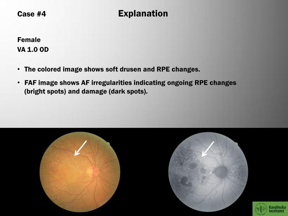
Case #5 Explanation
Male, 60 years
VA 1.2 OS
• The colored image shows hard drusen as well as a few soft drusen in the
macula. The FAF appears normal.

Case #6 Color
Image Taken with Canon FAF Camera

Case #6 FAF
Image Taken with Canon FAF Camera

Male
VA 1.0 OD
• The colored image shows discrete macular changes with some atrophic
areas.
• The FAF shows variations in AF pattern in the macula and around the papillae
indicating ongoing RPE changes (bright dots) and RPE damage (dark areas).
Case #6 Explanation

Case #7 cOLOR
Image Taken with Canon FAF Camera
Case #7 FAF
Image Taken with Canon FAF Camera

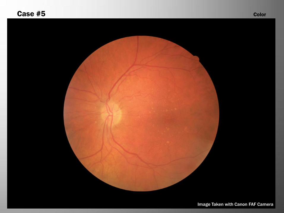
Female, 58 years
VA 1.0 OD
• Choroidal malignant melanoma with overlying orange pigment.
• Corresponding FAF-picture shows areas of both hypo- and hyperfluorescence
where the melanoma is located. The area of increased
• AF corresponds to the areas of orange pigment.
Case #7 Explanation
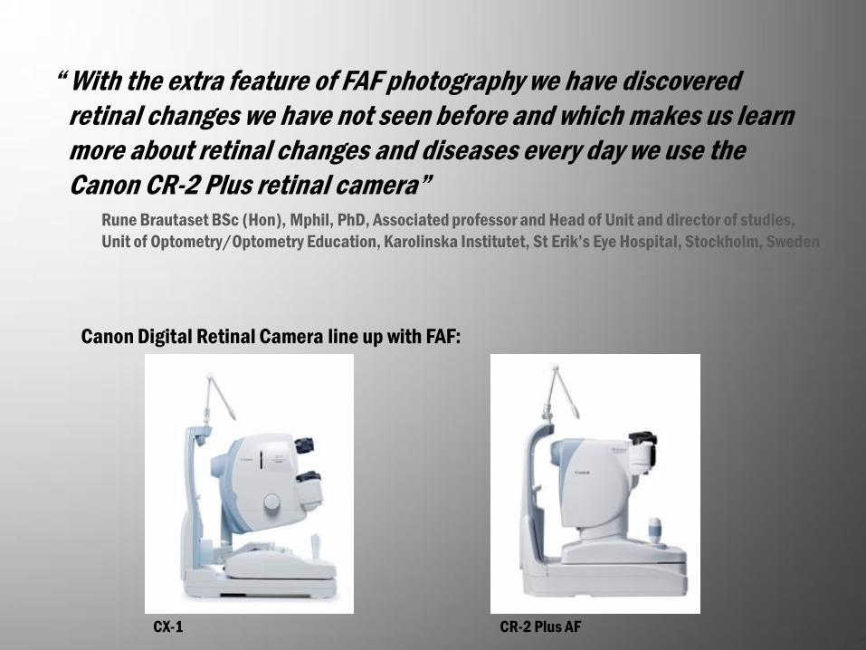
Canon Digital Retinal Camera line up with FAF:
“ With the extra feature of FAF photography we have discovered
retinal changes we have not seen before and which makes us learn
more about retinal changes and diseases every day we use the
Canon CR-2 Plus retinal camera” Rune Brautaset BSc (Hon), Mphil, PhD, Associated professor and Head of Unit and director of studies,
Unit of Optometry/Optometry Education, Karolinska Institutet, St Erik’s Eye Hospital, Stockholm, Sweden
CR-2 Plus AF CX-1