Fundamentals of Tuberculosis Handout - Global TB...
26
Fundamentals of Tuberculosis Handout Introduction This resource is intended for the health care worker with little or no background in tuberculosis (TB). It provides basic information about TB, including its etiology, mode of transmission, diagnosis and treatment. Overview of Contents Etiology .............................................................................................................................................. 2 Transmission ...................................................................................................................................... 2 Figure 1: Transmission of TB ................................................................................................ 2 Pathogenesis ....................................................................................................................................... 4 Figure 2: The Lungs and Alveoli ........................................................................................... 4 Latent TB Infection vs. TB Disease ................................................................................................... 5 Table 1: Latent TB Infection vs. TB Disease ......................................................................... 5 Figure 3: Pathogenesis of Latent TB Infection and Disease .................................................. 6 Risk Factors for Developing TB Disease ........................................................................................... 7 Figure 4: Risk of Developing TB Disease ............................................................................. 8 Sites of TB Disease ............................................................................................................................ 8 Classification System for TB ............................................................................................................. 9 Table 2: Classification System for TB ................................................................................... 9 Diagnosis of Latent TB Infection ..................................................................................................... 10 Figure 5: The Booster Phenomenon ..................................................................................... 14 Figure 6: Two-Step Testing.................................................................................................. 15 Treatment of Latent TB Infection .................................................................................................... 16 Diagnosis of TB Disease .................................................................................................................. 17 Treatment of TB Disease.................................................................................................................. 22 Glossary............................................................................................................................................ 24 - 1 -
Transcript of Fundamentals of Tuberculosis Handout - Global TB...

Fundamentals of Tuberculosis Handout
Introduction
This resource is intended for the health care worker with little or no background in tuberculosis
(TB). It provides basic information about TB, including its etiology, mode of transmission,
diagnosis and treatment.
Overview of Contents
Etiology .............................................................................................................................................. 2
Transmission ...................................................................................................................................... 2
Figure 1: Transmission of TB ................................................................................................ 2
Pathogenesis ....................................................................................................................................... 4
Figure 2: The Lungs and Alveoli ........................................................................................... 4
Latent TB Infection vs. TB Disease ................................................................................................... 5
Table 1: Latent TB Infection vs. TB Disease......................................................................... 5
Figure 3: Pathogenesis of Latent TB Infection and Disease .................................................. 6
Risk Factors for Developing TB Disease........................................................................................... 7
Figure 4: Risk of Developing TB Disease ............................................................................. 8
Sites of TB Disease ............................................................................................................................ 8
Classification System for TB ............................................................................................................. 9
Table 2: Classification System for TB ................................................................................... 9
Diagnosis of Latent TB Infection..................................................................................................... 10
Figure 5: The Booster Phenomenon..................................................................................... 14
Figure 6: Two-Step Testing.................................................................................................. 15
Treatment of Latent TB Infection .................................................................................................... 16
Diagnosis of TB Disease .................................................................................................................. 17
Treatment of TB Disease.................................................................................................................. 22
Glossary............................................................................................................................................ 24
- 1 -

Etiology TB is caused by a bacterium called Mycobacterium tuberculosis (often abbreviated M. tuberculosis). M. tuberculosis organisms are sometimes called tubercle bacilli. Transmission TB is spread from person to person through the air. When a person with infectious TB disease (TB that can be spread) coughs or sneezes, tiny particles containing M. tuberculosis may be expelled into the air. These particles, called droplet nuclei, are about 1 to 5 microns in diameter (less than 1/5000 of an inch). Droplet nuclei can remain suspended in the air for several hours, depending on the environment. If another person inhales air than contains these droplet nuclei, transmission may occur. Transmission is the spread of an organism, such as M. tuberculosis, from one person to another (Figure 1).
Figure 1. Transmission of TB. TB is spread from person to person through the air. The dots in the air represent droplet nuclei containing tubercle bacilli.
- 2 -

Not everyone who is exposed to TB becomes infected. The probability that TB will be transmitted depends on three factors:
• The infectiousness of the TB patient • The type of environment in which the exposure occurred • The length of the exposure
The infectiousness of a TB patient is directly related to the number of tubercle bacilli that is expelled into the air. Patients who expel many tubercle bacilli are more infectious than patients who expel few or no bacilli. Patients are more likely to be infectious if they:
• Have TB of the lungs or larynx • Have a cavity in the lung • Are coughing or undergoing cough-inducing procedures • Have acid-fast bacilli (AFB) on the sputum smear • Are not receiving adequate treatment
Usually, only people with pulmonary or laryngeal TB are infectious. This is because these people may be coughing and expelling tubercle bacilli into the air. People with extrapulmonary TB, that is, TB in parts of the body other than the lungs, are generally not infectious. Patients who have a cavity in the lung may be expelling tubercle bacilli if they are coughing, especially if they do not cover their mouth when they cough, or if they have a cough that produces a lot of sputum. The presence of tubercle bacilli on a sputum smear also indicates that the patient may be expelling tubercle bacilli. Patients who have had no treatment, have recently started treatment, or have not been receiving adequate treatment are much more likely to be infectious. Infectiousness appears to decline very rapidly after adequate treatment is started, but how quickly it declines varies from patient to patient. TB can be spread in many places, including homes or work sites. Groups who are at high risk for TB exposure include:
• Close contacts of a person with infectious TB • Foreign-born persons from areas where TB is common • Persons who work or reside in high-risk congregate settings • Persons who inject drugs • Locally identified high-burden groups, such as farm workers or homeless persons
TB can also be transmitted in other facilities or institutions with people who are at high risk for TB such as, such as hospitals, homeless shelters, correctional facilities, nursing homes, and residential homes for those with HIV. TB is most likely transmitted when health care workers, co-workers (i.e., administrative personnel), and patients come in contact with individuals who have unsuspected TB disease, who are not receiving adequate treatment, and who have not been isolated from others. People with TB disease are most likely to transmit TB before the disease has been diagnosed and treatment has started. TB patients who are receiving treatment are less likely to be infectious. TB is not spread by casual contact but by close, repeated contact or prolonged contact with an infectious individual. People with TB disease are most likely to spread it to the people they spend time with every day, including family members, friends, and co-workers.
- 3 -
Pathogenesis When a person inhales air that contains droplet nuclei, most of the larger droplets become lodged in the upper respiratory tract (the nose and throat), where infection is unlikely to develop. However, the droplet nuclei may reach the small air sacs of the lung (the alveoli), where infection begins (Figure 2). The following section describes the pathogenesis of TB (the way TB infection and disease develop in the body). Figure 2. The lungs and alveoli.
At first, the tubercle bacilli multiply in the alveoli and a small number enter the bloodstream and spread throughout the body. Bacilli may reach any part of the body, including areas where TB disease is more likely to develop. These areas include the upper portions of the lungs, as well as the kidneys, the brain, and bone. Within 2 to 10 weeks, however, the body’s immune system usually intervenes, halting multiplication and preventing further spread. The immune system is the system of cells and tissues that protect the body from foreign substances.
- 4 -

Latent TB Infection vs. TB Disease Latent TB Infection To be infected with TB means that tubercle bacilli are in the body but are being kept in a dormant or latent state by the body’s immune system. The immune system does this by producing special immune cells that surround the tubercle bacilli. The cells form a hard shell that keeps the bacilli contained and under control. Because of the potential for the bacilli to become active, multiply, and lead to TB disease, individuals infected with M. tuberculosis are said to have latent TB infection (LTBI). TB infection is detected by the tuberculin skin test. Most people with LTBI have a positive reaction to the tuberculin skin test. (The tuberculin skin test is discussed in more detail later.) People who have LTBI, but not TB disease are NOT infectious – in other words, they cannot spread the infection to others. These people usually have a normal chest x-ray. It is important to remember that LTBI is not considered a case of TB. Major similarities and differences between LTBI and TB disease are shown in Table 1.
Table 1: LTBI vs. TB Disease LTBI TB Disease (in the lungs)
Tubercle bacilli Tuberculin skin test or QuantiFERON®-TB Gold test result usually positive Chest x-ray usually normal Chest x-ray usually abnormal
Sputum smears and cultures negative Sputum smears and cultures positive No symptoms Symptoms such as cough, fever, weight loss Not infectious Often infectious before treatment
Not a case of TB A case of TB
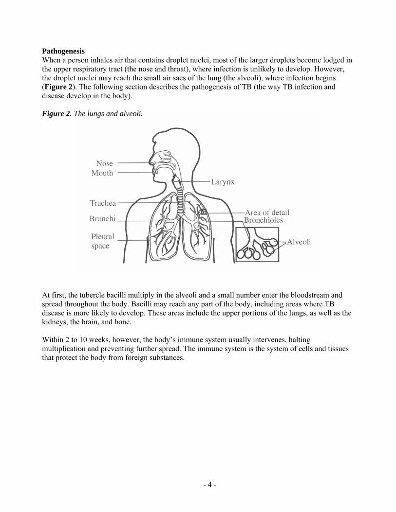
TB Disease Some people with LTBI develop TB disease. TB disease develops when the immune system cannot keep the tubercle bacilli under control and the bacilli begin to multiply rapidly. The risk that TB disease will develop is higher for some people than for others. The pathogenesis of LTBI and disease is shown in Figure 3. TB disease can develop very soon or many years after infection. In the United States, about 5% of all people who have recently been infected with M. tuberculosis will develop TB disease 1 to 2 years after infection. Another 5% will develop disease later in their lives. In other words, about 10% of all people who have LTBI will develop disease at some point. The remaining 90% will stay infected, but free of disease, for the rest of their lives. Because about half the risk of developing TB disease is concentrated in the first 2 years after infection, it is important to detect new infection early. People with LTBI can be given treatment to prevent them from getting TB disease. Thus, detecting new infection early helps prevent new cases of TB disease.
- 5 -
Figure 3. [Pathogenesis of LTBI and TB disease. Droplet nuclei containing tubercle bacilli are inhaled, enter the lungs, and travel to the alveoli.
Tubercle bacilli multiply in the alveoli.
A small number of tubercle bacilli enter the bloodstream and spread throughout the body. The bacilli may reach any part of the body, including areas where TB disease is more likely to develop (such as the lungs, kidneys, brain, or bone). Within 2 to 10 weeks, the immune system produces special immune cells that surround the tubercle bacilli. The cells form a hard shell that keeps the bacilli contained and under control (LTBI).
If the immune system cannot keep the bacilli under control, the bacilli begin to multiply rapidly (TB disease). This process can occur in different places in the body such as the lungs, kidneys, brain, or bone (see diagram in box 3).
- 6 -

Risk Factors for the Developing TB Disease Certain medical conditions increase the risk that LTBI will progress to TB disease. The risk may be about 3 times higher (as with diabetes) to more than 100 times higher (as with HIV infection) for people who have these conditions than for those who do not. Some of these conditions are:
• HIV infection, • Chest x-ray findings consistent with prior TB (in a person inadequately treated) • Low body weight (10% or more below ideal) • Recent infection (within the past 2 years) • Silicosis • Diabetes mellitus • Chronic renal failure/hemodialysis • Prolonged therapy with corticosteroids and other immunosuppressive agents • Certain types of cancer (e.g., leukemia, Hodgkin’s disease, or cancer of the head and neck) • Certain intestinal conditions (e.g., gastrectomy, jejunoileal bypass) • Solid organ transplant
HIV infection is the greatest risk factor of progression from TB infection to TB disease. When the immune system is weakened, the body may not be able to control the multiplication and spread of tubercle bacilli. For this reason, people who are infected with both M. tuberculosis and HIV, as with other infections, are much more likely to develop TB disease than people who are infected only with M. tuberculosis. Studies suggest that the risk of developing TB disease is 7% to 10% each year for people who are infected with both M. tuberculosis and HIV, whereas it is 10% over a lifetime for people infected only with M. tuberculosis.
- 7 -

Figure 4. Risk of Developing TB Disease.
Figure 4 shows the risk of developing TB disease for three different groups of people. For people with LTBI and no risk factors, the risk is about 10% over a lifetime. For people with LTBI and diabetes, the risk is 3 times as high, or about 30% over a lifetime. For people with LTBI and HIV infection, the risk is about 7% to 10% PER YEAR, a very high risk over a lifetime. In an HIV-infected person, TB disease can develop in either of two ways. First, a person who has LTBI can become infected with HIV and then develop TB disease as the immune system is weakened. Second, a person who has HIV infection can become infected with M. tuberculosis and then rapidly develop TB disease. Sites of TB Disease TB disease can occur in different places in the body. Pulmonary TB is TB that occurs in the lungs. About 85% of TB cases are pulmonary. Most patients with pulmonary TB have a cough and an abnormal chest x-ray, and they should be considered infectious until they meet all of the following criteria:
• Adequate treatment for 2 to 3 weeks • Symptoms improvement • Three consecutive negative sputum smears from sputum collected on different days
Extrapulmonary TB occurs in places other than the lungs, such as the larynx, the lymph nodes, the brain, the kidneys, or the bones and joints. Extrapulmonary TB occurs more often in people who are infected with HIV than in people who are not infected with HIV. In HIV-infected people, extrapulmonary TB is often accompanied by pulmonary TB. Most types of extrapulmonary TB are not considered infectious. Miliary TB occurs when tubercle bacilli enter the bloodstream and are carried to all parts of the body, where they grow and cause disease in multiple sites. This condition, which is rare but serious, is called miliary TB because the chest x-ray has the appearance of millet seeds scattered throughout the lung.
- 8 -

Classification System for TB Many systems have been used to classify people who have TB. The current classification system (Table 2) is based on the pathogenesis of TB. Many health departments and private health care providers use this system when describing patients. Thus, it is important to be familiar with this system. In particular, you should be aware that any patient with a classification of 3 or 5 should be receiving treatment for TB, and the suspected case or verified case should be reported to the health department.
Table 2: Classification System for TB Class Type Description
0 No exposure to TB Not infected
No history of exposure, negative reaction to the tuberculin skin test
1 Exposure to TB No evidence of infection
History of exposure, negative reaction to a tuberculin skin test given at least 10 weeks after exposure
2 Latent TB Infection No TB disease
Positive reaction to the tuberculin skin test, negative sputum smears and cultures (if done), no clinical or x-ray evidence of TB disease
3 Current TB disease
Positive sputum culture for M. tuberculosis (if done), or a positive reaction to the tuberculin skin test and clinical or x-ray evidence of current TB disease
4 Previous TB disease (not current)
Medical history of TB disease, or abnormal but stable x-ray findings for a person who has a positive reaction to the tuberculin skin test, negative sputum smears and cultures (if done), and no clinical x-ray evidence of current TB disease
5 TB suspected Signs and symptoms of TB disease, but evaluation not complete
- 9 -

Diagnosis of Latent TB Infection
The Tuberculin Skin Test The tuberculin skin test is used to determine whether a person has LTBI. In this test, a substance called tuberculin is injected into the skin. Tuberculin is protein derived from tubercle bacilli that have been killed by heating. In most people who have LTBI, the immune system will recognize the tuberculin because it is similar to the tubercle bacilli that caused the infection. This recognition generally will cause a reaction to the tuberculin skin test. Tuberculin is used for diagnosing LTBI; it is not a vaccine. Tuberculin testing is useful for:
• Examining a person who is not sick but who may have LTBI, for example, a person who has been exposed to someone with TB. In fact, the tuberculin skin test is the only way to diagnose LTBI before the infection progresses to TB disease
• Screening at-risk groups of people for LTBI • Examining a person who has symptoms of TB disease
Different types of tuberculin tests are available, such as the Mantoux tuberculin skin test and the multiple-puncture test. The Mantoux tuberculin skin test is the preferred test because it is the most accurate. Multiple puncture tests are not recommended. Mantoux Tuberculin Skin Test The Mantoux skin test is given using a needle and syringe to inject 0.1 mL of 5 tuberculin units (TU) of liquid tuberculin between the layers of the skin (intradermally), usually on the forearm. A tuberculin unit is a standard strength of tuberculin. The tuberculin used in the Mantoux skin test is also known as purified protein derivative or PPD. For this reason, the tuberculin skin test is sometimes called a PPD skin test. With the Mantoux tuberculin skin test, the patient’s arm is examined 48 to 72 hours after the tuberculin is injected. Most people with LTBI have a positive reaction to the tuberculin. The result is an area of induration (swelling that can be felt) around the site of injection. The transverse diameter of the induration is measured across the forearm; erythema (redness) or bruising around the indurated area is not measured. QuantiFERON®-TB Gold Test (QFT-G) The QuantiFERON®-TB gold test (QFT-G) is a whole-blood test for detecting LTBI. The test measures the patient’s immune reactivity to M. tuberculosis. Blood samples are mixed with antigens. If the patient is infected with M. tuberculosis, the blood cells will recognize the tuberculin and release interferon-gamma (IFN-γ) in response. As with the tuberculin skin test, follow-up medical evaluation should be conducted on persons with positive test results to rule out TB disease. QFT-G results are interpreted in a manner similar to that used for interpreting positive cut-off values for the tuberculin skin test. However, QFT-G results are usually available within 24 hours. Therefore, test results can be obtained with a single patient visit, and there is no need for the patient to return for test interpretation. Interpretation of QFT-G results is influenced by the patient’s estimated risk for TB infection.
- 10 -

Classification of TST Reactions Whether a reaction to the Mantoux tuberculin skin test is classified as positive depends on the size of induration and the person’s risk factors for TB. ≥5 mm of induration is considered a positive reaction in:
• HIV-infected persons • Close contacts of a person with infectious TB • Persons who have chest x-ray findings consistent with prior TB • Organ transplant recipients • Persons who are immunosuppressed for other reasons (e.g., taking the equivalent of ≥15
mg/day of prednisone for 1 month or more ≥10 mm of induration is considered a positive reaction in:
• Recent immigrants (within last 5 years) from a high-prevalence country • Injection drug users (with unknown or HIV negative status) • Residents or employees of high-risk congregate settings (for example, nursing homes or
correctional facilities) • Mycobacteriology laboratory personnel • Children <4 years of age, or children or adolescents exposed to adults at high risk • People with other high-risk conditions such as diabetes
≥15 mm of induration is considered a positive reaction:
• Persons with no known risk factors for TB In most cases, people who have a very small reaction or no reaction to the tuberculin skin test probably do not have LTBI. For people who may be exposed to TB on the job (such as health care workers and staff of nursing homes or correctional facilities), the classification of the skin test reaction as positive or negative depends on the:
• Size of the induration • Employee’s individual risk factors for TB • Risk of exposure to TB in the person’s job
Therefore, in facilities where the risk of exposure to TB is very low, 15 or more millimeters of induration may be considered a positive reaction for employees with no other risk factors for TB. In patient care facilities, 10 or more millimeters of induration may be considered a positive reaction for employees with no other risk factors for TB. Most people who have a positive skin test reaction will have a positive reaction if they are tested later in their lives, regardless of whether they receive treatment. This is because the tuberculin skin test detects the immune response to tuberculin, not the presence of tubercle bacilli in the body. Additionally, it is important to note that a false-positive or a false-negative reaction may occur when the tuberculin skin test is given incorrectly or the results are not measure properly. The following is a description of false-positive and false-negative reactions.
- 11 -

False-Positive Reaction The skin test is a valuable tool, but it is not perfect. Several factors can affect the skin test reaction. Two of these factors are infection with nontuberculous mycobacteria (mycobacteria other than M. tuberculosis) and bacille Calmette-Guérin (BCG) vaccination. BCG is a vaccine for TB disease that is used in many countries. However, it is rarely used in the United States because studies have shown that it is not completely effective and does not confer life-long immunity. People who are infected with nontuberculous mycobacteria or who have been vaccinated with BCG may have a positive reaction to the tuberculin skin test even if they do not have LTBI. This is called a false-positive reaction. With the tuberculin skin test, there is NO RELIABLE WAY to distinguish a positive tuberculin reaction caused by vaccine with BCG from a reaction caused by a true TB infection.1 However, the reaction is more likely to be due to LTBI if any of the following are true:
• The reaction is large • The person was vaccinated a long time ago • The person comes from an area of the world where TB is common • The person has been exposed to someone with infectious TB disease
People who have positive reaction should be further evaluated for TB disease, regardless of whether they were vaccinated with BCG. False-Negative Reactions Some people have a negative reaction to the tuberculin skin test even though they have LTBI. These are called false-negative reactions. A false-negative reaction may be due to:
• Anergy (inability to mount an immune response) • Recent TB infection (within the past 10 weeks) • Very young age (younger than 6 months old)
The most common cause of false-negative reaction is anergy, the inability to react to the skin test because of a weakened immune system. While HIV infection is a main cause of anergy, many conditions, such as immunosuppressive therapy, or severe TB disease itself, can weaken the immune system and cause anergy. Another cause of false-negative reaction is recent TB infection (infection within the past 10 weeks). It takes 2 to 10 weeks after TB infection for the body’s immune system to be able to react to tuberculin. Therefore, after TB has been transmitted, it takes 2 to 10 weeks before TB infection can be detected by the tuberculin skin test. For this reason, close contacts of someone with infectious TB disease with a negative reaction to the tuberculin skin test should be retested 8-10 weeks after the last time they were in contact with the person who has TB disease.
1 The QuantiFERON®-TB Gold test is able to distinguish M. tuberculosis from other mycobacteria and BCG vaccination
- 12 -

A third cause of false-negative reaction is a very young age. Children younger than 6 months old may have a false-negative reaction to the tuberculin skin test because their immune systems are not yet fully developed. A false-positive or a false-negative reaction may also occur when the tuberculin skin test is given incorrectly or the results are not measured properly. Any patient with symptoms of TB should be evaluated for TB disease, regardless of his/her skin test reaction. In fact, people with symptoms of TB should be evaluated for TB disease right away, at the same time that the tuberculin skin test is given. The symptoms of pulmonary TB disease include coughing up sputum (phlegm from deep in the lungs) or blood. The general symptoms of TB disease (pulmonary or extrapulmonary) include weight loss, fatigue, malaise, fever, and night sweats. The diagnosis of TB disease is discussed in more detail later. Targeted Skin Testing Programs Targeted tuberculin skin testing identifies persons at high risk for the development of TB disease who would benefit from treatment of LTBI, if detected. Therefore, all testing activities should be accompanied by a plan for the necessary follow-up medical evaluation and treatment. Persons at high risk for TB (i.e., risk substantially greater than that of the general U.S. population) have either been infected recently with M. tuberculosis or have clinical conditions that are associated with an increased risk of progression from LTBI to TB disease. Therefore, targeted tuberculin testing programs should be conducted only among groups at high risk and discouraged in those at low risk. Many residential facilities, health care facilities, and other high-risk congregate settings have targeted skin testing programs. This means that employees and residents are periodically given tuberculin skin test in order to:
• Identify people who have LTBI and possibly TB disease, so that they can be given
treatment as needed • Determine whether TB is transmitted in the facility
Two-Step Testing In a targeted skin testing program, employees or residents are skin tested when they start their job or enter the facility. This is called the baseline skin test. If they have a negative skin test reaction, they may be retested at regular intervals thereafter, depending on the risk classification of the setting. Employees or residents of health care or correctional facilities whose skin test reaction converts from negative to positive between testing intervals have probably become recently infected with M. tuberculosis. These skin test conversions may indicate that TB is being transmitted in the facility. People with skin test conversions are at high risk of developing TB disease because they were infected with M. tuberculosis relatively recently (within the past 2 years). In order to detect TB transmission and identify people who have skin test conversions, accurate information must be obtained from every employee’s baseline skin test, as well as from additional skin tests. One factor that can affect the accuracy of the baseline skin test is the booster phenomenon. The booster phenomenon happens because, in some people who have LTBI, the ability to react to tuberculin lessens over time. When these people are skin tested many years after they become infected with M. tuberculosis, they may have a negative reaction. However, if they are tested again within 1 year, they may have a positive reaction. This reaction occurs because the first skin test has “jogged the memory” of the immune system, boosting its ability to react to tuberculin. It may appear that these people were infected between the first and second skin tests (recent TB infection).
- 13 -

However, the second, positive skin test reaction is actually a boosted reaction, due to TB infection that occurred a long time ago. The booster phenomenon occurs mainly among older adults and is illustrated in Figure 5.
Figure 5. The booster phenomenon.
A person becomes infected with M. tuberculosis
Over time, a person’s ability to react to tuberculin lessens.
The person receives a tuberculin skin test
The person has a negative reaction because of a decreased ability to react to tuberculin. However, this skin test “jogs the memory” of the immune system to recognize and react to tuberculin
Up to 1 year later (For this example, we assume the person was NOT exposed to TB during this time).
The person is skin tested again
The person has a positive reaction. This is a boosted reaction due to TB infection that occurred a long time ago, not during the interval between the two skin tests
The booster phenomenon can present a problem in a skin testing program. To avoid misinterpretation, a strategy has been developed for telling the difference between boosted reactions and reactions caused by recent infection. This strategy, called two-step testing, means that if a person has a negative reaction to an initial skin test, he/she is given a second test 1 to 3 weeks later:
• If the reaction to the second test is positive, it probably is a boosted reaction (due to TB infection that occurred a long time ago)
• If the reaction to the second test is negative, the person is considered uninfected. In this person, a positive reaction to a skin test given later will probably be due to recent infection
Two-step testing is often used for employees who will be required to have TB skin testing at frequent intervals. In particular, two-step testing is often used in health care facilities or in other facilities where personnel may be at high risk for TB exposure. The procedure for two-step testing is shown in Figure 6.
- 14 -

Figure 6. Two-step testing.
- 15 -

Treatment for Latent TB Infection Treatment for latent TB infection (formerly known as preventive therapy or chemoprophylaxis) is recommended to prevent progression from LTBI to TB disease. High-risk persons should be evaluated for LTBI if they have a positive skin test reaction, regardless of their age.
Sometimes treatment is given to people who have a negative skin test reaction, such as high-risk contacts and children who have been exposed to TB. All patients being considered for treatment of LTBI should receive a medical evaluation to:
• Exclude the possibility of TB disease • Determine whether they have ever been treated for infectious TB • Identify any medical problems that may complicate therapy or require careful
monitoring The usual treatment of LTBI infection is isoniazid (INH) given daily for 9 months for all persons. Patients should be evaluated every month for signs of hepatitis and other adverse reactions to INH. They should also be educated about the symptoms caused by adverse reactions to INH and instructed to seek medical attention immediately if symptoms occur. In addition, people at greatest risk for hepatitis should have liver function tests before starting INH therapy. An alternate regimen is rifampin (RIF) for 4 months.
- 16 -

Diagnosis of TB Disease Before clinicians can diagnose TB disease in a patient, they must think of the possibility of this disease when they see a patient with symptoms of TB or abnormal chest x-ray findings. Because TB is not as common as it was many years ago, many clinicians do not consider the possibility of TB when making diagnoses for patients who have symptoms of TB. When this happens, the diagnosis of TB may be delayed or even overlooked, and the patient will remain ill and possibly infectious. Anyone with symptoms of TB should be evaluated for TB disease. In addition, anyone found to have a positive tuberculin skin test reaction should be evaluated for TB disease. Overview There are four steps in diagnosing TB disease; the medical history, the tuberculin skin test, the chest x-ray and the bacteriologic examination. A discussion of what is involved in each step follows. 1. The Medical History
a. Exposure to TB: One important part of the medical history is asking a patient about his/her exposure to TB. Patients should be asked whether they have spent time with someone who has infectious TB or someone with TB-like symptoms. Some people may have been exposed to TB in the distant past, when they were children. Others may have been exposed more recently. Anyone who has been exposed to TB may have LTBI. Many people become infected with M. tuberculosis without knowing that they were exposed. The risk of being exposed to TB is higher for some occupations (for example, certain health care workers) and in some residential facilities (for example, nursing homes or correctional facilities).
b. Symptoms of TB disease: Another important part of the medical history is checking for symptoms of TB disease. Although, people with TB disease may or may not have symptoms, most patients with TB disease have one or more symptoms that led them to seek medical care. Occasionally, TB is discovered during a medical examination for an unrelated condition (for example, when a patient is given a chest x-ray before undergoing surgery). Pulmonary TB disease usually causes one or more of the following symptoms: • Cough • Pain in the chest while breathing or coughing • Cough with sputum (phlegm from deep in the lungs) or blood The general symptoms of TB disease (pulmonary or extrapulmonary) include: • Weight loss • Fatigue • Malaise • Fever • Night sweats
- 17 -

The symptoms of extrapulmonary TB depend on the part of the body that is affected by the disease. For example, TB of the spine may cause pain in the back; TB of the kidney may cause blood in the urine. All of these symptoms may be the result of other diseases, but they should prompt the clinician to suspect TB disease.
c. Previous TB infection or TB disease: During the medical history, the clinician should ask
the patient whether he/she has ever been diagnosed with or treated for TB infection or disease.
• Patients known to have a positive skin test reaction probably have LTBI. If they were
infected within the past 2 years, they are at high risk for TB disease. • Patients who have had TB disease before should be asked when they had the disease and
how it was treated. If the regimen prescribed was inadequate or if the patient did not follow the recommended treatment, TB may re-occur, and it may be resistant to one or more of the drugs used in the previous treatment regimen.
d. Risk factors for developing TB disease: A fourth part of the medical history is checking for
risk factors for developing TB disease. The following conditions, also listed on page 54, appear to increase the risk that LTBI will progress to TB disease:
• HIV infection • Chest x-ray findings consistent with prior TB (in a person inadequately treated) • Low body weight (10% or more below ideal) • Recent infection (within the past 2 years) • Silicosis • Diabetes mellitus • Chronic renal failure/hemodialysis • Prolonged therapy with corticosteroids and other immunosuppressive agents • Certain types of cancer (e.g., leukemia, Hodgkin’s disease, or cancer of the head and
neck) • Certain intestinal conditions (e.g., gastrectomy, jejunoileal bypass) • Solid organ transplant
Clinicians should determine whether patients have any of these conditions. In particular, HIV infection greatly increases the risk that LTBI will progress to TB disease. A physical examination is an essential part of the evaluation of any patient. It cannot confirm or rule out TB disease, but it can provide valuable information about the patient’s overall condition and other factors that may affect how TB disease is treated if it is diagnosed.
- 18 -

2. The Tuberculin Skin Test
Patients with symptoms of TB disease are often given a tuberculin skin test to detect exposure to and infection with TB. However, as many as 20% of the patients found to have TB disease have a negative tuberculin skin test reaction. For this reason, patients with symptoms of TB disease should always be evaluated for TB disease, regardless of their skin test results. Furthermore, for patients with symptoms of TB disease, clinicians should not wait for tuberculin skin test results (48 to 72 hours) before starting other diagnostic tests. A tuberculin skin test is not necessary for patients known to have had a previous positive skin test reaction.
3. The Chest X-ray The chest x-ray is useful for diagnosing TB disease. About 85% of TB patients have pulmonary TB. Usually, when a person has TB disease in the lungs, the chest x-ray appears abnormal. It may show infiltrates (collections of fluid and cells in the tissues of the lung) or cavities (hollow spaces within the lung that may contain many tubercle bacilli). However, the results of a chest x-ray cannot confirm that a person has TB disease. A variety of illnesses may produce abnormalities whose appearance on a chest x-ray resembles TB. Although an abnormality on a chest x-ray may lead a clinician to suspect TB, only a bacteriologic culture that is positive for M. tuberculosis proves that a patient has TB disease. Moreover, a chest x-ray cannot detect LTBI.
4. The Bacteriologic Examination
The next step in diagnosing TB disease is the bacteriologic examination. This is done in a laboratory that specifically deals with M. tuberculosis and other mycobacteria (a mycobacteriology laboratory). There are four parts to a bacteriologic examination.
a. Obtaining a specimen b. Examining the specimen under a microscope c. Culturing the specimen d. Conducting drug susceptibility testing
a. Obtaining a specimen: Specimens that will be sent to the laboratory can be obtained in several
ways. Usually, patients who are suspected of having pulmonary TB disease simply cough up sputum (phlegm from deep in the lungs) into a sterile container for processing and examination. This is the easiest and most cost-effective procedure.
If a patient cannot cough up sputum on his/her own, other techniques can be used to obtain a specimen. An induced sputum sample can be obtained easily by having the patient inhale a saline (salt water) mist, which causes the patient to cough deeply. Induced specimens are often clear and watery, so they should be labeled “induced specimen” so that they will not be confused with saliva. (Laboratories will not accept saliva as a specimen.)
Another procedure, bronchoscopy, can be used to obtain pulmonary secretions or lung tissue. In this procedure, an instrument called a bronchoscope is passed through the mouth directly into the diseased portion of the lung, and some sputum or lung tissue is removed.
- 19 -

b. Examining the specimen under a microscope: Before the specimen is examined under a microscope, it is smeared onto a glass slide and stained with a dye. This is called a smear. Then laboratory personnel use the microscope to look for acid-fast bacilli (AFB) on the smear. AFB are mycobacteria that stay stained after they have been washed in an acid solution. Tubercle bacilli are one kind of AFB.
When AFB are seen in a smear, they are counted. There is a system for reporting the number of AFB that are seen at a certain magnification. According to the number of AFB seen, the smears are classified as 4+, 3+, 2+, or 1+. Smears that are classified as 4+ and 3+ are considered strongly positive; 2+ and 1+ smears are considered moderately positive. If very few AFB are seen, the smear is classified by the actual number of AFB seen (no plus sign). For example, if only 4 AFB were seen in the entire smear, the smear is classified as “4 AFB seen.” Smears classified in this way are considered weakly positive. Finally, if no AFB are seen, the smear is called negative. But a negative smear does not rule out the possibility of TB because there can be AFB that were not seen in the smear.
It takes only a few hours to prepare and examine a smear. Therefore, the results of the smear examination should be available to the clinician within 1 day.
The results of the smear examination can be used to help determine the infectiousness (contagiousness) of the patient. Patients who have many tubercle bacilli in their sputum have a positive smear. Patients who have positive smears are considered infectious because they can cough many tubercle bacilli into the air. However, because AFB are not always tubercle bacilli, patients who have positive smears do not necessarily have TB. Furthermore, as mentioned previously, patients who have negative smears may have TB.
c. Culturing the specimen: Culturing the specimen means growing mycobacteria on media
(substances that contain nutrients) in the laboratory. When mycobacteria have formed colonies (groups), they can be identified. All specimens should be cultured, regardless of whether the smear is positive or negative.
Culturing the specimen is necessary to determine whether it contains M. tuberculosis and to confirm a diagnosis of TB disease. (However, in some cases, patients are diagnosed with TB disease on the basis of their signs and symptoms, even if their specimen does not contain M. tuberculosis.)
The first procedure in culturing the specimen is to detect the growth of the mycobacteria, which grow very slowly. This can take from 4 days to 8 weeks, depending on the type of medium used.
The second procedure is to identify which type of mycobacteria is growing. When M. tuberculosis is identified, the patient is said to have a positive culture for M. tuberculosis. A positive culture for M. tuberculosis, also called M. tuberculosis isolate, confirms the diagnosis of TB disease.
When M. tuberculosis is NOT identified, the patient is said to have a negative culture for M. tuberculosis. A negative culture does not necessarily rule out diagnosis of TB disease; as mentioned earlier, some patients with negative cultures are diagnosed with TB disease on the basis of their signs and symptoms.
- 20 -
d. Conducting drug susceptibility testing: Drug susceptibility tests, the final part of the
bacteriologic examination, are done to determine which drugs will kill the tubercle bacilli that are causing TB disease in a particular patient. Tubercle bacilli that are killed by a particular drug are said to be susceptible to that drug, whereas those that can grow even in the presence of a particular drug are said to be resistant to that drug. The drug susceptibility pattern of a strain of tubercle bacilli is the list of drugs to which the strain is susceptible and to which it is resistant.
The results of drug susceptibility tests can help clinicians choose the appropriate drugs for each patient. This is very important. Patients with TB disease who are treated with drugs to which their strain of TB is resistant may not be cured. In fact, their strain of TB may become resistant to additional drugs.
Drug susceptibility tests should be done when a patient is first found to have a positive culture for M. tuberculosis (that is, the first isolate of M. tuberculosis). In addition, drug susceptibility tests should be repeated if a patient has positive culture for M. tuberculosis after 2 months of treatment or if a patient does not seem to be getting better. That way, the clinician can find out whether the patient’s strain of TB has become resistant to certain drugs. If necessary, the clinician may change the drugs used for treating the patient.
- 21 -

Treatment of TB Disease TB disease must be treated for at least 6 months and in some cases, treatment lasts even longer if sputum smears and cultures do not convert to negative. The initial phase for treating TB disease should include four first-line drugs: isoniazid, rifampin, pyrazinamide, and ethambutol, unless otherwise indicated. When the drug susceptibility results are available, usually in about 8 weeks, clinicians may change the regimen. Treatment is continued with at least two drugs to which the bacilli are susceptible for the duration of the regimen. Using only one drug to treat TB disease can create a population of tubercle bacilli that is resistant to that drug. Drug resistance can also develop when patients do not take treatments as prescribed. Thus, to prevent relapse and drug resistance, clinicians must prescribe an adequate regimen and make sure that patients adhere to treatment. The best way to ensure that patients adhere to treatment is to use directly observed therapy (DOT), which is health care worker or trained outreach worker observation of the ingestion of prescribed medication. Monitoring All patients being treated for TB disease should be educated about the signs of adverse reactions to the drugs they are taking and instructed to seek medical attention immediately if they have any of these symptoms. Patients should be seen by a clinician at least monthly during treatment, and evaluated for possible adverse reactions. In addition, before starting treatment, patients should have baseline tests to help clinicians detect any abnormalities that may complicate treatment. Patients who are not receiving DOT should be carefully monitored for adherence to treatment. The only way to ensure adherence to treatment is to use DOT. If DOT cannot be provided for the entire treatment period, a clinician should work closely with the patient to establish a routine for regular medication taking. It is also crucial for the clinician to work with patients in identifying barriers to adherence and brainstorm ways to overcome these obstacles. Clinical and bacteriologic evaluations should be performed during treatment to determine whether a patient is responding to the treatment regimen. Patients should be carefully reevaluated if their:
• Symptoms do not improve during the first 2 months of treatment • Symptoms worsen after improving initially • Culture results have not become negative after 2 months of treatment • Culture results become positive after being negative
In certain situations, clinicians may also use chest x-rays to monitor a patient’s response to treatment. The treatment of TB can be complicated, especially in patients who fail to respond to treatment, relapse, have drug-resistant TB, or have adverse reactions to medications. Clinicians who do not have experience with these situations should consult a TB expert.
- 22 -

Infectiousness The infectiousness of a TB patient is directly related to the number of tubercle bacilli that he/she expels into the air. Patients who expel many tubercle bacilli are more infectious than patients who expel few or no bacilli. Patients are more likely to be infectious if they:
• Have TB of the lungs or larynx • Have a cavity in the lung • Are coughing or undergoing cough-inducing procedures • Have AFB on the sputum smear • Are not receiving adequate treatment
Infectiousness appears to decline very rapidly after adequate treatment is started, but how quickly it declines varies from patient to patient. Patients who have been receiving adequate treatment for 2 to 3 weeks, whose symptoms have improved, and who have 3 consecutive negative sputum smears from sputum collected on different days can be considered noninfectious. TB can be spread in many places, such as homes, schools, or health care facilities. In a health care facility, TB is most likely to be transmitted when health care workers and patients come in contact with patients who have suspected TB disease, who are not receiving adequate treatment, and who have not been isolated from others. All health care facilities should take measures to prevent the spread of TB. People with TB disease are most likely to transmit TB before the disease has been diagnosed and treatment has started. TB patients who are receiving treatment are less likely to be infectious. TB patients who may be infectious should be instructed to cover their mouths and noses with tissues when coughing and sneezing. If a patient is suspected to have TB, he/she should be immediately placed in an environment in which transmission to others is unlikely. In many cases, hospitalization of a patient until non-infectious can be warranted. A patient should be kept from work or school at least until he/she is non-infectious and well enough to resume normal activities. All of these measures should be taken without compromising the dignity and comfort of the patient.
- 23 -

Glossary acid-fast bacilli (AFB) – mycobacteria that stay stained even after they have been washed in an acid solution; may be detected under a microscope in a stained smear; number in smear determines classification as positive or negative alveoli – the small air sacs of the lung that are at the end of the airway; when droplet nuclei reach these air sacs, TB infection begins anergy – the inability to react to a skin test because of a weakened immune system, often caused by HIV infection or severe illness bacille Calmette-Guérin (BCG) – a vaccine for TB disease that is used in many countries but rarely used in the United States; may cause a false-positive reaction to the tuberculin skin test baseline skin test – the tuberculin skin test given to employees or residents in certain facilities when they start their job or enter the facility booster phenomenon – a phenomenon in which people (especially older adults) who are skin tested many years after becoming infected with M. tuberculosis may have negative reaction to an initial skin test, followed by a positive reaction to a skin test given up to 1 year later; this happens because the first skin test boosts the immune response. Two-step testing is used in programs in which routine skin testing is done to determine the difference between boosted reactions and reactions caused by recent infection. bronchoscopy – a procedure used to obtain pulmonary secretions or lung tissue with an instrument called a bronchoscope; used only when patients cannot cough up sputum on their own and an induced specimen cannot be obtained close contacts – people who spend frequent and prolonged time with someone who has infectious TB disease colonies – groups of mycobacteria that have grown in a culture culture – organisms grown on media (substances containing nutrients) so that they can be identified; a positive culture for M. tuberculosis contains tubercle bacilli, whereas a negative culture contains no detectable tubercle bacilli droplet nuclei – very small droplets (1 to 5 microns in diameter) that may be expelled when a person who has infectious TB coughs or sneezes; these droplets can remain suspended in the air for several hours, depending on the environment drug-resistant – able to grow in the presence of a particular drug drug-susceptible – able to be killed by a particular drug
- 24 -

erythema – redness around the site of the injection when a Mantoux tuberculin skin test is done; erythema is not considered when the reaction size is measured because redness does not indicate that a person has TB infection extrapulmonary TB – TB disease that occurs in places other than the lungs, such as lymph nodes, pleura, brain, kidneys, or bones; most types of extrapulmonary TB are not infectious false-negative reaction – a negative reaction to the tuberculin skin test in a person who has TB infection; may be caused by anergy, recent infection (within the past 10 weeks), or very young age (younger than 6 months old) false-positive reaction – a positive reaction to the tuberculin skin test in a person who does not have TB infection; may be caused by infection with nontuberculous mycobacteria or by vaccination with BCG immunosuppressive therapy – therapy that suppresses, or weakens, the immune system induced sputum – sputum that is obtained by having the patient inhale a saline (salt water) mist, causing the patient to cough deeply; this procedure is used to help patients cough up sputum if they cannot do so on their own induration – swelling that can be felt around the site of injection after a Mantoux tuberculin skin test is done; the reaction size is the diameter of the indurated area (excluding any redness or brusing); measured across the forearm infectious – capable of spreading infection; a person who has infectious TB disease expels droplets containing M. tuberculosis into the air when he/she coughs or sneezes infiltrate – a collection of fluid and cells in the tissues of the lung; visible on a chest x-ray in people with pulmonary TB disease malaise – a feeling of general discomfort or illness Mantoux tuberculin skin test – the preferred method of testing for TB infection; done by using a needle and syringe to inject 0.1 mL of 5 tuberculin units of liquid tuberculin between the layers of the skin (intradermally), usually on the forearm; the reaction to this test, a small swollen area (induration), is measured 48 to 72 hours after the injection and is classified as positive or negative depending on the size of the reaction and the patient’s risk factors for TB media – substances containing special nutrients for growing cultures of bacteria found in specimens miliary TB – TB disease that occurs when tubercle bacilli enter the bloodstream and are carried to all parts of the body, where they grow and cause disease in multiple sites; the chest x-ray of patients with miliary TB often looks like millet seeds scattered throughout the lung multiple puncture test – tuberculin skin test done by puncturing the skin of the forearm with a set of short prongs or tines to inject tuberculin (for example, Tine test); although easy to give and
- 25 -

- 26 -
convenient, these tests are not accurate and should not be used to determine whether a person has TB infection nontuberculous mycobacteria – mycobacteria that do not cause TB disease and are not usually spread from person to person; one example is M. avium complex pathogenesis – how an infection or disease develops in the body pulmonary TB – TB disease that occurs in the lungs (about 85% of all U.S. cases), typically causing a cough and an abnormal chest x-ray; pulmonary TB is usually infectious if untreated purified protein derivative (PPD) – the type of tuberculin used in the Mantoux tuberculin skin test quantiFERON-TB gold test (QFT-G) – a blood test used as an aid in diagnosing latent TB infection and TB disease; this test requires only a single patient encounter, and the result can be ready within 1 day; QFT-G appears to be capable of distinguishing between the sensitization caused by M. tuberculosis infection and that caused by BCG vaccination silicosis – a lung disease caused by inhaling silica dust, which is used in the production of glass and ceramics; occurs most often in mining and foundry workers skin test conversion – a change in a skin test reaction from negative to positive between screening intervals smear – a specimen that has been spread onto a glass slide, stained, washed in an acid solution, and then placed under the microscope for examination; used to detect AFB in a specimen sputum – phlegm from deep in the lungs, collected in a sterile container for processing and examination transmission – the spread of an organism, such as M. tuberculosis, from one person to another; depends on the contagiousness of the patient, the type of environment, and the length of exposure tubercle bacilli – another name for Mycobacterium tuberculosis organisms, which cause TB disease tuberculin – protein derived from tubercle bacilli that have been killed by heating; used to determine whether a person has TB infection. Tuberculin is not a vaccine and cannot cause TB tuberculin skin test – a test used to detect TB infection two-step testing – a strategy used in TB screening programs to distinguish a boosted reaction (caused by TB infection that occurred many years before the skin test) from a reaction caused by recent infection. If a person has a negative reaction to an initial skin test, a second test is give 1 to 3 weeks later; a positive reaction to the second test probably represents a boosted reaction, not recent infection. Two-step testing is used in many TB screening programs for skin testing employees when they start their job.