Fundamental to Advanced Radiotherapy Treatment Technique
8
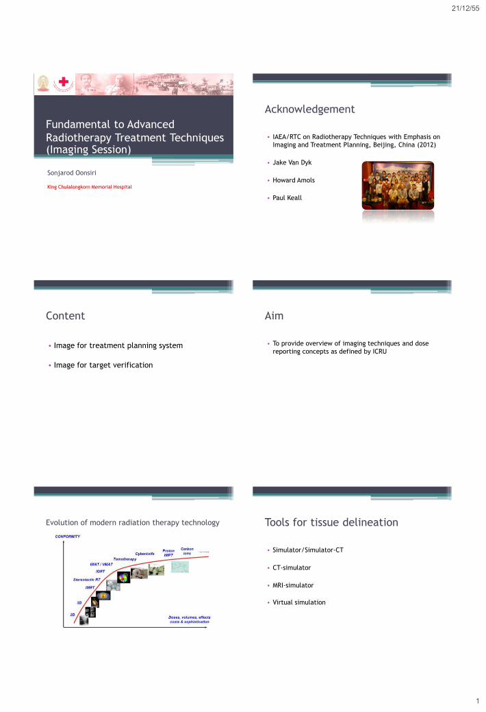
21/12/55 1 Fundamental to Advanced Radiotherapy Treatment Techniques (Imaging Session) Sonjarod Oonsiri King Chulalongkorn Memorial Hospital Acknowledgement • IAEA/RTC on Radiotherapy Techniques with Emphasis on Imaging and Treatment Planning, Beijing, China (2012) • Jake Van Dyk • Howard Amols • Paul Keall Content • Image for treatment planning system • Image for target verification Aim • To provide overview of imaging techniques and dose reporting concepts as defined by ICRU Evolution of modern radiation therapy technology Tools for tissue delineation • Simulator/Simulator-CT • CT-simulator • MRI-simulator • Virtual simulation
Transcript of Fundamental to Advanced Radiotherapy Treatment Technique
21/12/55
1
Fundamental to Advanced
Radiotherapy Treatment Techniques (Imaging Session)
Sonjarod Oonsiri
King Chulalongkorn Memorial Hospital
Acknowledgement
• IAEA/RTC on Radiotherapy Techniques with Emphasis on
Imaging and Treatment Planning, Beijing, China (2012)
• Jake Van Dyk
• Howard Amols
• Paul Keall
Content
• Image for treatment planning system
• Image for target verification
Aim
• To provide overview of imaging techniques and dose
reporting concepts as defined by ICRU
Evolution of modern radiation therapy technology Tools for tissue delineation
• Simulator/Simulator-CT
• CT-simulator
• MRI-simulator
• Virtual simulation
21/12/55
2
Simulator
• Advantage
▫ Reproduces treatment geometry
▫ Real-time breathing motion on fluoroscopy
• Limitation
▫ Limited external contour and
internal structure information
▫ No tissue density information
▫ Not useful for dose calculation
Simulator-CT
• Simulator with CT scanning mode
• CT option added ▫ Use image intensifier or flat panel detector
• Provide ▫ Internal/external contours
▫ Possibly tissue density information
▫ Poor image quality ; Inaccurate definition of target volume
http://www.varian.com
CT-simulator
• High resolution : Diagnostic images
• 3D reconstruction capabilities
(tumor/normal tissue localization)
• CT number/Electron density data
• High accuracy and precision of CT
number
Advantage of CT
• Widely available
• Good geometric accuracy
• Visualization of bony anatomy for comparison with radiograph
• Potential for rapid scanning
• With contrast, many tumors and nodes visualized reasonably well
4D-CT
• Tumour and organs can move up to 3
cm with respiration
• 4D-CT has applications in motion
inclusive, gated and tumour tracking
treatments
Keall et al, Aust Phys Engs Sci Med, 2002 Keall et al, Med Phys, 2006

21/12/55
3
CT-simulation QA
• AAPM Report No.39 (1993) ▫ Specification and Acceptance Testing of
Computed Tomography Scanners
• AAPM Task Group No.66 (2003) ▫ Quality Assurance for Computed
Tomography Simulators and the Computed Tomography Simulation Process
• IAEA Human Health Series No.19 (2012) ▫ Quality Assurance Programme for
Computed Tomography : Diagnostic and Therapy Applications
Limitation of CT
Brain stem? Lesion?
Brain tumor King Chulalongkorn Memorial Hospital, Bangkok, Thailand
MRI-simulator
• Provide superior image quality for
soft-tissue delineation over CT
• Use careful patient positioning and
marking
• Determine the geometric
consistency between the CT and
MRI data sets
Fraass et al, IJROBP, 1987
Chen L et al, IJROBP, 2004
Eilertsen K et al, Acta Oncologica, 2008
Jonsson JH et al, Radiation Oncology, 2010
Accessory for MRI simulator
GE MRI Oncology Package http://www.lap-laser.com
MRI for TPS
• Advantage
▫ Better soft tissue contrast : Better definition of target and
critical structures
▫ Potential for functional MRI imaging can be used to localize
cancer
• Disadvantage
▫ Distortion
▫ Poor definition of bones
▫ Loss of electron density information

21/12/55
4
MRI dose calculation
Chen L, IJROBP, 2004 Jonsson JH, Radiation Oncology, 2010
• The mass densities and electron density recommended in ICRU 46 (1992)
• The absolute dose agreement for PTV was within 2% between CT-based and MR-based
MRI in brachytherapy
• MRI is superior to CT for the
discrimination of GTV and
adjacent normal tissue
• In practice delineation seems
to be more accurate when using MRI
Potter R et al, Radiotherapy and Oncology, 2006
PET-CT
• Combines PET and CT in single gantry
system
▫ PET : Metabolic and functional imaging
▫ CT : Anatomic imaging
• PET-CT combination adds accuracy in
detecting and classifying tumors
• PET-CT planning changes cancer
patient management
▫ Target volume definition
▫ Tumor staging
Consistency ….
• How do we make sure that radiation oncology
professionals use consistent procedures and
terminology?
1978 1993
1999
Change over time
Purdy et al, Sem Rad Oncol, 2004
1978 1993 1999
21/12/55
5
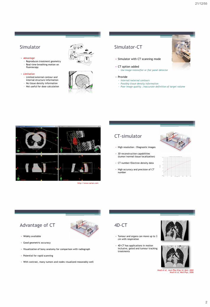
ICRU 83 ; 2010
• Irradiation techniques have advanced
▫ 3D-CRT to IMRT
▫ More availability of CT
▫ Additional imaging : MRI, PET, functional imaging
▫ Improved conformity
▫ More detailed dose-volume information on TPS
▫ Use of dose-volume constraints
▫ Automated optimization, IMRT
ICRU Report 83, 2010
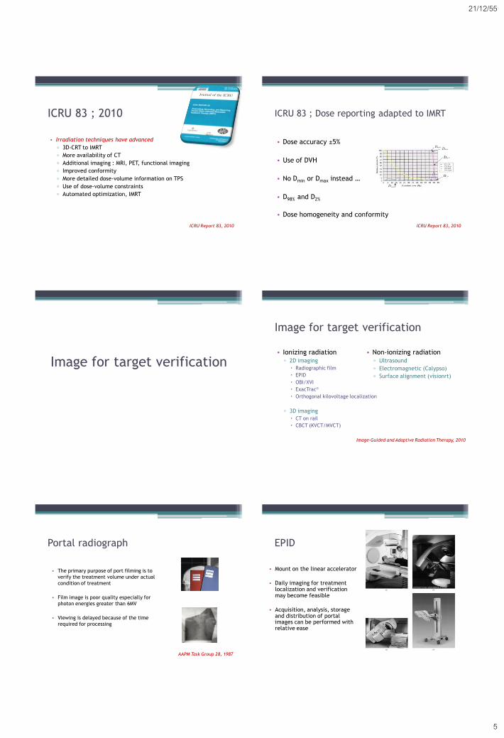
ICRU 83 ; Dose reporting adapted to IMRT
• Dose accuracy ±5%
• Use of DVH
• No Dmin or Dmax instead …
• D98% and D2%
• Dose homogeneity and conformity
ICRU Report 83, 2010
Image for target verification
Image for target verification
• Ionizing radiation ▫ 2D imaging
Radiographic film
EPID
OBI/XVI
ExacTrac
Orthogonal kilovoltage localization
▫ 3D imaging
CT on rail
CBCT (KVCT/MVCT)
Image-Guided and Adaptive Radiation Therapy, 2010
• Non-ionizing radiation ▫ Ultrasound
▫ Electromagnetic (Calypso)
▫ Surface alignment (visionrt)
Portal radiograph
• The primary purpose of port filming is to
verify the treatment volume under actual
condition of treatment
• Film image is poor quality especially for
photon energies greater than 6MV
• Viewing is delayed because of the time
required for processing
AAPM Task Group 28, 1987
EPID
• Mount on the linear accelerator
• Daily imaging for treatment localization and verification may become feasible
• Acquisition, analysis, storage and distribution of portal images can be performed with relative ease

21/12/55
6
CT on rail
Kuriyama K et al, IJROBP, 2003
Ma CM et al, Medical Dosimetry, 2006
On-board imager system
kV Imaging
King Chulalongkorn Memorial Hospital, Bangkok, Thailand
Cone beam CT 4D-CBCT
Sonke et al, Med Phys, 2005
CT CBCT 4D-CBCT

21/12/55
7
ExacTrac
http://www.novalis-radiosurgery.com http://www.visionrt.com
Ultrasound
http://www.elekta.com
Real time tracking of target motion
http://www.calypsomedical.com
MRI guided external beam radiotherapy
Viewray Utrecht University
Alberta University Australian MRI-Linac Program
In-room MRI
• Integrated mobile MRI scanner (IMRIS) with linear accelerator
(TrueBEAM)
▫ Princess Margaret Hospital (Canada)
http://www.variantruebeam.com http://www.imris.com

21/12/55
8
IGRT QA Recommendation
• AAPM Task Group No.75 (2007)
▫ The Management of Imaging Dose during
Image-Guided Radiotherapy
• Jean-Pierre Bissonnette et al (2008)
▫ A Quality Assurance Program for Image
Quality of CBCT Guidance in Radiation
therapy
• AAPM Task Group 142 Report (2009)
▫ Quality Assurance of Medical Accelerators
Future trend
• Knowledge based segmentation
▫ Automatic contouring based on pre-
contoured from database created by renowned radiation oncologist
▫ Reduce structure segmentation time
from hours to minutes
• Deformable registration
▫ Algorithm employs millions of degree of
freedom for the registration
▫ More accuracy for image registration
http://www.varian.com
http://www.mimsoftware.com
Summary
• Appropriate imaging is crucial for accurate target volume and
normal tissue delineation
• In treatment room imaging is important for reducing margins
• ICRU reports help to provide …
▫ Consistency in target volume definition
▫ Consistency in dose prescription
▫ Consistency in reporting
Tumor dose
Normal tissue dose