
Fundamental techniques in cell culture laboratory handbook
64
1 Design and Equipment for the Cell Culture Laboratory Contents 1 1 1 sigma.com/cellculture 1.0 Introduction ............................................. 4 2.0 Design and Equipment for the Cell Culture Laboratory........................... 4 2.1 Laboratory Design ...................................... 4 2.2 Microbiological Safety Cabinets .................. 5 2.3 Centrifuges ................................................ 6 2.4 Incubators .................................................. 6 2.5 Work Surfaces and Flooring ....................... 6 2.6 Plasticware and Consumables .................... 7 2.7 Care and Maintenance of Laboratory Areas ........................................ 7 3.0 Safety Aspects of Cell Culture ................ 7 3.1 Risk Assessment ......................................... 7 3.2 Biohazards ................................................. 9 3.3 Genetically Modified Organisms ................. 9 3.4 Disinfection .............................................. 10 3.5 Waste Disposal ......................................... 11 4.0 Sourcing of Cell Lines............................ 12 5.0 Cell Types & Culture Characteristics ..... 13 5.1 Primary Cultures....................................... 13 5.2 Continuous Cultures ................................ 13 5.3 Culture Morphology................................. 13 5.4 Phases of Cell Growth .............................. 14 5.5 In Vitro Age of a Cell Culture ................... 15 6.0 The Cell Environment............................ 15 6.1 Basic Constituents of Media ..................... 16 6.2 Inorganic Salts.......................................... 17 6.3 Buffering Systems .................................... 17 6.4 Carbohydrates ......................................... 17 6.5 Amino Acids ............................................ 17 6.6 Vitamins................................................... 18 6.7 Proteins and Peptides ............................... 18 6.8 Fatty Acids and Lipids ............................... 18 6.9 Trace Elements ......................................... 18 6.10 Preparation of Media ............................... 18 6.11 Serum ...................................................... 19 6.12 Guidelines for Serum Use ......................... 19 6.13 Origin of Serum ....................................... 20 7.0 Cryopreservation and Storage of Cell Lines ................................................ 21 7.1 Cryopreservation ...................................... 21 7.2 Ultra-low Temperature Storage ................ 22 7.3 Inventory Control ..................................... 23 7.4 Safety Considerations............................... 23 8.0 Good Cell Banking Practices ................. 24 9.0 Quality Control Considerations............ 26 9.1 Introduction ............................................. 26 9.2 Reagents and Materials ............................ 27 9.3 Provenance and Integrity of Cell Lines ................................................. 27 9.4 Avoidance of Microbial Contamination..... 27 9.5 Environmental Monitoring........................ 29 9.6 Aseptic Technique and Contamination Control...................... 29 9.7 What to do in the Event of Contamination..................................... 30 10.0 Authentication of Cell Lines ................. 31 11.0 Alternative Cell Culture Systems ......... 31 11.1 Cell Culture Scale-up Systems................... 31 11.2 Scale-up Solutions .................................... 32 11.3 Roller Bottle Culture ................................. 32 11.4 Multilayer Vessels ..................................... 32 11.5 Disposable Solutions for Suspension Cells .... 34 11.6 Spinner Flask Culture ............................... 34 11.7 Other Scale-up Options ............................ 34
-
Upload
trinhtuyen -
Category
Documents
-
view
228 -
download
2
Transcript of Fundamental techniques in cell culture laboratory handbook

1
Design and Equipment for the Cell Culture LaboratoryContents
111
sigma.com/cellculture
1.0 Introduction ............................................. 4
2.0 Design and Equipment for the
Cell Culture Laboratory ........................... 4
2.1 Laboratory Design ...................................... 4
2.2 Microbiological Safety Cabinets .................. 5
2.3 Centrifuges ................................................ 6
2.4 Incubators .................................................. 6
2.5 Work Surfaces and Flooring ....................... 6
2.6 Plasticware and Consumables .................... 7
2.7 Care and Maintenance of
Laboratory Areas ........................................ 7
3.0 Safety Aspects of Cell Culture ................ 7
3.1 Risk Assessment ......................................... 7
3.2 Biohazards ................................................. 9
3.3 Genetically Modifi ed Organisms ................. 9
3.4 Disinfection .............................................. 10
3.5 Waste Disposal ......................................... 11
4.0 Sourcing of Cell Lines ............................ 12
5.0 Cell Types & Culture Characteristics ..... 13
5.1 Primary Cultures ....................................... 13
5.2 Continuous Cultures ................................ 13
5.3 Culture Morphology ................................. 13
5.4 Phases of Cell Growth .............................. 14
5.5 In Vitro Age of a Cell Culture ................... 15
6.0 The Cell Environment ............................ 15
6.1 Basic Constituents of Media ..................... 16
6.2 Inorganic Salts .......................................... 17
6.3 Buffering Systems .................................... 17
6.4 Carbohydrates ......................................... 17
6.5 Amino Acids ............................................ 17
6.6 Vitamins ................................................... 18
6.7 Proteins and Peptides ............................... 18
6.8 Fatty Acids and Lipids ............................... 18
6.9 Trace Elements ......................................... 18
6.10 Preparation of Media ............................... 18
6.11 Serum ...................................................... 19
6.12 Guidelines for Serum Use ......................... 19
6.13 Origin of Serum ....................................... 20
7.0 Cryopreservation and Storage of
Cell Lines ................................................ 21
7.1 Cryopreservation ...................................... 21
7.2 Ultra-low Temperature Storage ................ 22
7.3 Inventory Control ..................................... 23
7.4 Safety Considerations ............................... 23
8.0 Good Cell Banking Practices ................. 24
9.0 Quality Control Considerations ............ 26
9.1 Introduction ............................................. 26
9.2 Reagents and Materials ............................ 27
9.3 Provenance and Integrity of Cell Lines ................................................. 27
9.4 Avoidance of Microbial Contamination ..... 27
9.5 Environmental Monitoring ........................ 29
9.6 Aseptic Technique and Contamination Control ...................... 29
9.7 What to do in the Event of Contamination ..................................... 30
10.0 Authentication of Cell Lines ................. 31
11.0 Alternative Cell Culture Systems ......... 31
11.1 Cell Culture Scale-up Systems................... 31
11.2 Scale-up Solutions .................................... 32
11.3 Roller Bottle Culture ................................. 32
11.4 Multilayer Vessels ..................................... 32
11.5 Disposable Solutions for Suspension Cells .... 34
11.6 Spinner Flask Culture ............................... 34
11.7 Other Scale-up Options ............................ 34

www.hpacultures.org.uk
2
Fundamental Techniques in Cell Culture
Contents
Cell Lines Available from ECACC ....................... 35
12.0 Cell Culture Protocols ............................ 36
12.1 Basic Do’s and Don’ts of Cell Culture ........ 36
12.2 Protocol 1 – Aseptic Technique and Good Cell Culture Practice ................................. 37
12.3 Protocol 2 – Resuscitation of Frozen Cell Lines ................................................. 39
12.4 Protocol 3 – Subculture of Adherent Cell Lines ...42
12.5 Protocol 4 – Subculture of Semi-Adherent Cell Lines ................................................. 45
12.6 Protocol 5 – Subculture of Suspension Cell Lines ........................... 47
12.7 Protocol 6 – Cell Quantifi cation ............... 49
12.8 Protocol 7 – Cryopreservation of Cell Lines ............................................. 52
12.9 Protocol 8 – Testing for Bacteria and Fungi ................................................ 54
12.10 Protocol 9 – Detection of Mycoplasma by Culture .................................................... 56
12.11 Protocol 10 – Testing for Mycoplasma by Indirect DNA Stain .................................... 58
Tables
Table 1 Commonly used cell lines of each culture type ...................................... 14
Table 2 Different types of culture medium and their uses ......................................... 16
Table 3 Comparison of ultra-low temperature storage methods for cell lines ............ 22
Table 4 “Half-Way House” Solutions to Scale-up ........................................... 33
Table 5 Cell Culture Reagents availablefrom Sigma-Aldrich ....................... 65
Figures
Figure 1 Diagram of Microbiological Safety Cabinet Airfl ow Patterns ..................... 5
Figure 2 Examples of Cell Morphology ........... 13
Figure 3 Schematic Representation of a Tiered Cell Banking System ......................... 25
Figure 4 Bioreactor ........................................ 31
Figure 5 Hyperfl ask & T Flask .......................... 31
Figure 6 Shake Flasks ..................................... 31
Figure 7 Roller Deck ....................................... 32
Figure 8 Roller Bottles ..................................... 32
Figure 9 Spinner Flasks ................................... 32
Figure 10 Flow Scheme for Bacteria and Fungi Testing .............................................. 54
Figure 11 Flow Scheme for Detection of Mycoplasma by Culture .................... 56
Figure 12 Typical ‘fried egg’ colonies, Mycoplasma pneumoniae...................................... 57
Figure 13 Flow Scheme for Detection of Mycoplasma by Indirect DNA Stain .... 58
Figure 14 Testing for Mycoplasma by Indirect DNA Stain ........................................ 60
(a) Hoechst Positive Culture
(b) Hoechst Negative Culture

The European Collection of Cell Cultures (ECACC) was established in 1984 as a cell culture collection to service the research community and provide an International Depository Authority recognised patent depository for Europe. Over the last three decades ECACC has expanded and diversifi ed to become one of the premier collections of authenticated cell cultures in the world and this remains the core of ECACC’s business. The collections currently hold over 40,000 cell lines representing 45 different species, 50 tissue types, 300 HLA types, 450 monoclonal antibodies and at least 800 genetic disorders.
The development and maintenance of such a diverse collection has inevitably produced a high level of specialist knowledge and this, combined with the support of the Health Protection Agency, has enabled ECACC to position itself as a centre of expertise in all aspects of cell culture. ECACC has developed a comprehensive range of cell culture services and diversifi ed into new product areas such as high quality genomic DNA extracted from cell lines.
ECACC is one of the four collections which constitute the Health Protection Agency Culture Collections (HPA Culture Collections). A s As has been the case since its inception, ECACC continues to operate out of the Porton Down site, which is now the Centre for Emergency Preparedness and Response (CEPR), Health Protection Agency, UK.
*See page 35 for more information on the cell lines available.
HPA Culture CollectionsA strategic business unit within the Health Protection Agency.
www.hpacultures.org.uk Tel: +44 (0) 1980 612512 email: [email protected]
The European Collection of Cell Cultures (ECACC)
Visit www.hpacultures.org.uk for more information!
ll Cell Lines and Hybridomasll Primary Cellsll Neuron Culture Kitsll HepaRG® Cellsll DNA & RNA Productsll Bacteria, Plasmids, Transposons ll Mycoplasmasll Fungill LENTICULE Discsll Viruses
l Cell Culture Management Services
l Contract Cell Culture
l Assay Ready Cells
l Cell Line Identity Verifi cation
l DNA Extraction
l Genetic Support Services
l Contract Freeze-Drying
Products Services
www.hppaaccuulltures.org.u
nes and Hybridomasy Cy Cyn CG®®
& RRiaia,
plplaasaas
CULss
ll CeCeCeSSe
ll CoCo
ll AsAs
ll CeCe
ll DNDN
ll GeGe
ll CoCoCo
CCellellssCuultlture re KiKits® CCeeCellsllssslNAAN Prrr oduoduo ctstsctPPlPPlassmma iddsdsdsidss, T T, Tranranspospoosonsonssonss
smmsmasss
LELEE DDi Discscscsssss
Products and Services available from the HPA Culture Collections:
e informattiioon!
eeentntnt
aatiti
eses
tt
oooonn
ll Mycoplasma Testing
ll Virus Contract Services
ll Patent Deposits
ll Safe Deposits
ll Sterility Testing
ll Training
Introduction
Fundamental Techniques in Cell Culture
www.hpacultures.org.uk
4
1.0 Introduction
Over ten years ago, Sigma® Life Science and the European Collection of Cell Cultures (ECACC) formed a working partnership to bring together the most diverse selection of cell culture products and services available commercially. We did this with researchers like you in mind, to ensure that you have the necessary quality products to further your research goals. We continue to expand upon this partnership, and now are able to offer an even greater array of cell lines, cell culture products, knowledge, and services to the global research community.
The fi eld of cell culture has advanced greatly over the years. For more than 25 years, Sigma and ECACC have both been part of and contributed to that advancement. Early cell culture research focused on discovering methods for culturing a diverse array of cells from many species. Today cell culture methods are vital to broad areas of life science research. With the number of researchers adding cell culture to their repertoire of techniques expanding daily, we believe there are many who can benefi t from Sigma’s and ECACC’s combined knowledge and experience in cell culture.
To that aim, we have assembled this updated laboratory handbook of cell culture techniques. For the researcher new to cell culture, this handbook provides a wealth of information from the sourcing of cell lines, safety and laboratory design to aspects of cryopreservation and quality control. Additionally, a series of 10 detailed protocols are provided, which are routinely used in the ECACC laboratories. For the “expert” cell culturist, it addresses a number of important, yet often overlooked topics in cell culture such as cell line authentication and contamination issues, to help ensure that the results obtained from cell culture experiments are both accurate and reproducible. The handbook is intended as a guide rather than an in-depth text book of cell culture and you are encouraged to consult relevant specialised literature to obtain more detailed information.
2.0 Design and Equipment for the Cell Culture Laboratory
2.1 Laboratory DesignPerhaps one of the most under-rated aspects of tissue culture is the need to design the facility to ensure that good quality material is produced in a safe and effi cient manner. Most tissue culture is undertaken in laboratories that have been adapted for the purpose and in conditions that are not ideal. However, as long as a few basic guidelines are adopted this should not compromise the work.
There are several aspects to the design of good tissue culture facilities. Ideally work should be conducted in a single use facility which, if at all possible, should be separated into an area reserved for handling newly received material (quarantine area) and an area for material which is
sigma.com/cellculture
5
known to be free of contaminants (main tissue culture facility). If this is not possible work should be separated by time with all manipulations on clean material being completed prior to manipulations involving the ‘quarantine material’. Different incubators should also be designated. In addition, the work surfaces should be thoroughly cleaned between activities.
All new material should be handled as ‘quarantine material’ until it has been shown to be free of contaminants such as bacteria, fungi and particularly mycoplasma. Conducting tissue culture in a shared facility requires considerable planning and it is essential that a good technique is used throughout to minimise the risk of contamination occurring.
For most cell lines the laboratory should be designated to at least Category 2 based on the Advisory Committee on Dangerous Pathogens (ACDP) guidelines (ACDP, 1995)†. However, the precise category required is dependent upon the cell line and nature of the work proposed. The guidelines make recommendations regarding the laboratory environment including lighting, heating, the type of work surfaces and fl ooring and provision of hand washing facilities. In addition, it is recommended that laboratories should be run at air pressures that are negative to corridors to contain any risks within the laboratory.
† Advisory Committee on Dangerous Pathogens (1995) Categorisation of Biological Agents According to Hazard and Categories of Containment, 4th edition, Health & Safety Executive (HSE) books, Sudbury, UK, (www.hse.gov.uk).
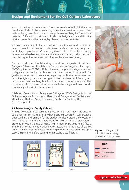
2.2 Microbiological Safety CabinetsA microbiological safety cabinet is probably the most important piece of equipment for cell culture since, when operated correctly, it will provide a clean working environment for the product, whilst protecting the operator from aerosols. In these cabinets operator and/or product protection is provided through the use of HEPA (high effi ciency particulate air) fi lters. The level of containment provided varies according to the class of cabinet used. Cabinets may be ducted to atmosphere or re-circulated through a second HEPA fi lter before passing to atmosphere see fi gure 1.
Class I Class 2 Class 3
known to be free of contaminants (main tissue culture facility) If this is not
Design and Equipment for the Cell Culture Laboratory
Figure 1. Diagram of microbiological safety cabinet airfl ow patterns
Fundamental Techniques in Cell Culture
www.hpacultures.org.uk
6
Environmental monitoring with Tryptose Soya Broth agar settle plates inside the cabinet for a minimum of four hours is a good indicator of how clean a cabinet is (refer to ‘9.5 Environmental Monitoring’). There should be no growth of bacteria or fungi on such plates.
In most cases a class 2 cabinet is adequate for animal cell culture. However, each study must be assessed for its hazard risk and it is possible that additional factors, such as a known virus infection or an uncertain provenance may require a higher level of containment.
2.3 CentrifugesCentrifuges are used routinely in tissue culture as part of the subculture routine for most cell lines and for the preparation of cells for cryopreservation. By their very nature centrifuges produce aerosols and thus it is necessary to minimise this risk. This can be achieved by purchasing models that have sealed buckets. Ideally, the centrifuge should have a clear lid so that the condition of the load can be observed without opening the lid. This will reduce the risk of the operator being exposed to hazardous material if a centrifuge tube has broken during centrifugation. Care should always be taken not to over-fi ll the tubes and to balance them carefully. These simple steps will reduce the risk of aerosols being generated. The centrifuge should be situated where it can be easily accessed for cleaning and maintenance. Centrifuges should be checked frequently for signs of corrosion.A small bench-top centrifuge with controlled braking is suffi cient for most purposes. Cells sediment satisfactorily at 80 – 150 x g. Higher gravitational forces may cause damage and promote agglutination of the cell pellet.
2.4 IncubatorsCell cultures require a strictly controlled environment in which to grow. Specialist incubators are used routinely to provide the correct growth conditions, such as temperature, degree of humidity and CO2 levels in a controlled and stable manner. Generally, they can be set to run at temperatures in the range of 28oC (for insect cell lines) to 37oC (for mammalian cell lines) and set to provide CO2 at the required level (e.g. 5-10%). Some incubators also have the facility to control the O2 levels. Copper-coated incubators are also now available. These are reported to reduce the risk of microbial contamination within the incubator due to the microbial inhibitory activity of copper. The inclusion of a bactericidal agent in the incubator water trays will also reduce the risk of bacterial and fungal growth. However, there is no substitute for regular cleaning.
2.5 Work Surfaces and FlooringIn order to maintain a clean working environment the laboratory surfaces including bench-tops, walls and fl ooring should be smooth and easy to clean. They should also be waterproof and resistant to a variety of chemicals (such as acids, alkalis, solvents and disinfectants). In areas used for the storage of materials in liquid nitrogen, the fl oors should be resistant to cracking if any liquid nitrogen is spilt. Refer to Section 7.4 for safety
Environmental monitoring with Tryptose Soya Broth agar settle plates inside
Design and Equipment for the Cell Culture Laboratory

sigma.com/cellculture
7
considerations on the use of liquid nitrogen. In addition, the fl oors and walls should be continuous with a coved skirting area to make cleaning easier and reduce the potential for dust to accumulate. Windows should be sealed. Work surfaces should be positioned at a comfortable working height.
2.6 Plasticware and ConsumablesAlmost every type of cell culture vessel, together with support consumables such as tubes and pipettes, are commercially available as single use, sterile packs. Suppliers include Sigma-Aldrich, Nunc, Greiner, Bibby Sterilin and Corning. The use of such plasticware is more cost effective than recycling glassware, enables a higher level of quality assurance and removes the need for validation of cleaning and sterilisation procedures. Plastic tissue culture fl asks are usually treated to provide a hydrophilic surface to facilitate attachment of anchorage dependent cells.
2.7 Care and Maintenance of Laboratory AreasIn order to maintain a clean and safe working environment tidiness and cleanliness are key. All spills should be dealt with immediately. Routine cleaning should be undertaken involving the cleaning of all work surfaces both inside and outside of the microbiological safety cabinet, the fl oors and all other pieces of equipment e.g. centrifuges. Humidifi ed incubators are a particular area for concern due to the potential for fungal and bacterial growth in the water trays. This will create a contamination risk that can only be avoided by regular cleaning of the incubator. All major pieces of equipment should be regularly maintained and serviced by qualifi ed engineers, for example:
• Microbiological safety cabinets should be checked every six months to ensure that they are safe to use in terms of product and user protection. These tests confi rm that the airfl ow is correct and that the HEPA fi lters are functioning properly.
• The temperature of an incubator should be regularly checked with a NAMAS (National Accreditation of Measurement and Sampling, UK), or equivalent calibrated thermometer and temperature adjusted as necessary.
• Incubator CO2 and O2 levels should also be regularly checked to ensure the levels are being maintained correctly.
3.0 Safety Aspects of Cell Culture
3.1 Risk AssessmentThe main aim of risk assessment is to prevent injury, protect property and avoid harm to individuals and the environment. In many countries the performance of risk assessment is a legal requirement. For example, this is the case in the UK under the Health and Safety at Work Act, UK
considerations on the use of liquid nitrogen In addition the floors and
Safety Aspects of Cell Culture
T fl asks available from Corning
Fundamental Techniques in Cell Culture
www.hpacultures.org.uk
8
(1974). There are also European Community directives covering Health and Safety at work. You can visit the European Agency for Safety and Health at Work website (www.europe.osha.eu.int) for information on legislation and standards or you should contact your on-site Health and Safety representative. Consequently risk assessments must be undertaken prior to starting any activity. The assessment consists of two elements:
1. Identifying and evaluating the risks.2. Defi ning ways of avoiding or minimising the risk.
For animal cell culture the level of risk is dependent upon the cell line to be used and is based on whether the cell line is likely to cause harm to humans. The different classifi cations are given below:
Low risk - Non human/non primate continuous cell lines and some well characterised human continuous lines.
Medium risk - Poorly characterised mammalian cell lines.
High risk - Primary cells derived from human/primate tissue or blood.
- Cell lines with endogenous pathogens (the precise categorisation is dependent upon the pathogen) – refer to ACDP guidelines, for details†.
- Cell lines used following experimental infection where the categorisation is dependent upon the infecting agent – refer to ACDP guidelines, for details.
†Advisory Committee on Dangerous Pathogens (ACDP) (1995) Categorisation of Biological Agents According to Hazard and Categories of Containment, 4th edition, HSE books, Sudbury, UK. The second supplement to the 1995 document was produced in 2000 - ‘Second supplement to: Categorisation of biological agents according to hazard and categories of containment (Fourth edition, 1995) Second Edition 2000’. Crown copyright 2000, UK. An update to the Approved List of Biological agents was issued in 2004, available at: http://www.hse.gov.uk/pubns/misc208.pdf
Note: The U.S. Department of Health and Human Services (Centers for Disease Control and Prevention) publishes a similar list, in its Biosafety in Microbiological and Biomedical Laboratories (BMBL) document (2007). The U.S. system uses Biological Safety Levels in place of the UK ACDP hazard groups.
A culture collection such as ECACC will recommend a minimum containment level required for a given cell line based upon its risk assessment. For most cell lines the appropriate level of containment is Level 2 requiring a class 2 microbiological safety cabinet. However, this may need to be increased to containment Level 3 depending upon the
(1974). There are also European Community dir
Safety Aspects of Cell Culture

sigma.com/cellculture
9
type of manipulations to be carried out and whether large culture volumes are envisaged. For cell lines derived from patients with HIV or Human T-Lymphotropic Virus (HTLV) Level 3 containment is required.
Containment is the most obvious means of reducing risk. Other less obvious measures include restricting the movement of staff and equipment into and out of laboratories. Good laboratory practice and good bench techniques such as ensuring work areas are uncluttered, reagents are correctly labelled and stored, are also important for reducing risk and making the laboratory a safe environment in which to work. The risk of exposure to aerosols or splashes can be limited by avoiding rapid pipetting, scraping and pouring. In addition, it is recommended that people working in laboratories where primary human material is used are vaccinated against Hepatitis B. Staff training and the use of written standard operating procedures and risk assessments will also reduce the potential for harm. Cell culture training courses covering the basics of tissue culture safety are offered by ECACC.
3.2 BiohazardsViruses pathogenic for humans are one of the most likely biohazards presented by cell cultures. Where infection with an agent pathogenic for humans is known or suspected, the cell culture should be handled at a containment level appropriate for the agent concerned. Other potential biohazards should also be considered. These relate to components of the cell culture medium, other adventitious agents (e.g. contaminating mycoplasmas), and cell products, some of which may be biologically active molecules with pharmacological, immunomodulating or sensitising properties. In addition, the generation and use of modifi ed cells, for example, hybrids, transformed cells and cells containing recombinant DNA can be hazardous. These procedures could potentially result in the appearance of modifi ed or reactivated viruses, novel fusion/hybrid proteins (especially in cross-species hybrids) and the expression of viral or cellular oncogenes.
Laboratory workers should never culture their own cells. In vitro transformation or genetic modifi cation could result in malignant disease or expression of an unusual pharmacologically active protein if they were to be accidentally inoculated into the donor. Therefore, human cells should be obtained from individuals having no association with the experimental work.
Biohazardous waste should be disposed of according to the methods described under ‘3.5 Waste Disposal’.
3.3 Genetically Modifi ed OrganismsThe generation and use of genetically modifi ed organisms (GMOs) should be strictly controlled and regulated. Most countries have regulatory organisations to ensure the risks posed by GMOs are minimised. For example, in the UK all institutions that carry out work using and/or generating GMOs are required by law to have a Genetic Modifi cation
type of manipulations to be carried out and whether large culture volumes
Safety Aspects of Cell Culture

Fundamental Techniques in Cell Culture
www.hpacultures.org.uk
10
Safety Committee (GMSC). Prior to any work commencing proposals for the intended work should go through the committee and , if necessary, be approved by the Health and Safety Executive (HSE). There is a European Directive governing the regulation of GM work. Visit the European Agency for Safety and Health at Work website (www.europe.osha.eu.int) for information on legislation and standards, or contact your on-site Health and Safety representative.
It is the responsibility of the individual cell culture user and institution to ensure compliance with the regulations set by the authorities of the country they are operating in.
3.4 DisinfectionMethods designed for the disinfection/decontamination of culture waste, work surfaces and equipment represent important means for minimising the risk of harm. Always wear appropriate personal protective equipment (PPE) such as gloves and eye protection when using concentrated forms of disinfectants. The selected gloves should protect against the substance being handled and meet the European standard EN374-3. Manufacturers’’ charts will help to identify the best gloves for the work.
The major disinfectants fall into four groups and their relative merits can be summarised as follows:
Hypochlorites (e.g., Sodium Hypochlorite)
• Good general purpose disinfectant
• Active against viruses
• Corrosive against metals and therefore should not be used on metal surfaces e.g. centrifuges
• Readily inactivated by organic matter and therefore should be made fresh daily
• Should be used at 1000ppm for general use surface disinfection, 2500ppm in discard waste pots for disinfecting pipettes, and 10,000ppm for tissue culture waste and spillages
Note: When fumigating a cabinet or room using formaldehyde all the hypochlorites must fi rst be removed as the two chemicals react together to produce carcinogenic products.
Phenolics
Phenolic based disinfectants should never be used as they are notsupported as part of the EU Biocidal Products Directive reviewprogramme.
Alcohol (e.g. Ethanol, Isopropanol)
• Effective concentrations: 70% for ethanol, 60-70% for isopropanol
• Their mode of activity is by dehydration and fi xation
Safety Committee (GMSC). Prior to any work
Safety Aspects of Cell Culture

sigma.com/cellculture
11
• Effective against bacteria. Ethanol is effective against most viruses but not non-enveloped viruses
• Isopropanol is not effective against viruses
Aldehydes (e.g. Formaldehyde)
• Aldehydes are irritants and their use should be limited due to problems of sensitisation
• Should only be used in well ventilated areas.
Formaldehyde is used to fumigate laboratories. The formaldehye is heated in a device so it will vaporise and all exposed surfaces are coated with the disinfectant.
Generally the use of aldehydes for disinfection and fumigation purposes can be hazardous. Check local regulations and with your safety advisor.
3.5 Waste DisposalAny employer has a ‘duty of care’ to dispose of all biological waste safely in accordance with national legislative requirements. Given below is a list of ways in which tissue culture waste can be decontaminated and disposed of safely. One of the most important aspects of the management of all laboratory-generated waste is to dispose of waste regularly and not to allow the amounts to build up. The best approach is ‘little and often’. Different forms of waste require different treatment.
• Tissue culture waste (culture medium) – inactivate for at least 2 hours in a solution of hypochlorite (10,000ppm) prior to disposal to drain with an excess of water.
• Contaminated pipettes should be placed in hypochlorite solution (2500ppm) overnight before disposal by autoclaving and incineration.
• Solid waste such as fl asks, centrifuge tubes, contaminated gloves, tissues, etc., should be placed inside heavy-duty sacks for contaminated waste and incinerated.
• If at all possible waste should be incinerated rather than autoclaved.
• Waste from specially licensed laboratories e.g. those handling genetically modifi ed level 3 (GM3) organisms requires specifi c treatment and tracking.
• Effective against bacteria Ethanol is effective
Safety Aspects of Cell Culture
i Did You Know?
Any employer has a ‘duty of care’ to dispose of all biological waste safely in accordance with national legislativerequirements

Fundamental Techniques in Cell Culture
www.hpacultures.org.uk
12
4.0 Sourcing of Cell Lines
Large numbers of cell lines look identical. Cell lines with very different origins and biological characteristics typically cannot be separated on grounds of morphology or culture characteristics. Infection or contamination of a cell line with an adventitious virus or mycoplasma may signifi cantly change the characteristics of the cells but again such contamination may not be apparent. Cell lines will also change with time in culture, and to add to all these natural hazards it is all too easy to incorrectly label or cross-contaminate different cell lines in a busy cell culture laboratory.
The opportunities for inadvertently introducing error into a cell line are limitless and ever present. It is in the nature of the science that once introduced, an error will be propagated, compounded, consolidated and disseminated.
The integrity and biological characteristics of a cell line have to be actively maintained by a well organised management system based on systematic cell banking supported by testing regimens in a structured quality assured environment. Such a controlled environment will only prevail in a dedicated professionally organised cell culture laboratory or cell bank. A small research laboratory with a high throughput of short-term research students, a minimum of permanent laboratory staff and no formal quality management programme will fi nd it diffi cult to maintain its cell lines unchanged over many years.
For all these reasons it is strongly recommended that new cell lines should only be acquired from a specialist, reputable culture collection such as ECACC. Moreover, if a laboratory believes it already has a certain cell line in its liquid nitrogen store, the identity and purity of such a cell line should be questioned in the absence of a well recorded culture history and recent test data. If there is a doubt, it is straightforward and cost effective to replace such cell stocks with authenticated material from a Culture Collection.
When a Culture Collection acquires a new cell line it will characterise the cell line using techniques such as isoenzyme analysis and DNA profi ling so that the identity of the cell line subsequently can be verifi ed. The Collection will then establish a hierarchy of Master and Working cell banks, cryopreserved in liquid nitrogen, that are demonstrated free from microbial contamination including mycoplasma. Customers are supplied from the authenticated Working Cell Banks (WCB). Replacement WCBs are manufactured from the original Master Cell Bank (MCB) and the new WCB will again be fully tested.
ECACC supplies its cell lines with advice on how to maintain the line. The technical support team can subsequently assist with diffi culties and provide additional technical information about the cell line. Culture Collections exist to ensure that animal cell research is conducted using standardised, authenticated material that ensures the work can be reproduced. An authenticated cell line of known provenance is the very bedrock of any cell based project. See p.35 for more info on cell lines available from ECACC.
4.0 Sourcing of Cell Lines
Sourcing of Cell lines
iDid You Know?
The European Collection of Cell Cultures (ECACC) is one of the world’s largest Biological Resource Centres supplying a diverse range of authenticated cell lines.
sigma.com/cellculture
13
5.0 Cell Types & Culture Characteristics
5.1 Primary CulturesPrimary cultures are derived directly from excised, normal animal tissue and cultures either as an explant culture or following dissociation into a single cell suspension by enzyme digestion. Such cultures are initially heterogeneous but later become dominated by fi broblasts. The preparation of primary cultures is labour intensive and they can be maintained in vitro only for a limited period of time. During their relatively limited lifespan primary cells usually retain many of the differentiated characteristics of the cell in vivo. Important Note: Primary cultures by defi nition have not been passaged, as soon as they are passaged they become a cell line and are no longer primary. ‘Primary’ cells sourced from most suppliers are in fact low-passage cell lines.
5.2 Continuous CulturesContinuous cultures are comprised of a single cell type that can be serially propagated in culture either for a limited number of cell divisions (approximately thirty) or otherwise indefi nitely. Cell lines of a fi nite life are usually diploid and maintain some degree of differentiation. The fact that such cell lines senesce after approximately thirty cycles of division means it is essential to establish a system of Master and Working banks in order to maintain such lines for long periods.
Continuous cell lines that can be propagated indefi nitely generally have this ability because they have been transformed into tumour cells. Tumour cell lines are often derived from actual clinical tumours, but transformation may also be induced using viral oncogenes or by chemical treatments. Transformed cell lines present the advantage of almost limitless availability, but the disadvantage of having retained very little of the original in vivo characteristics.
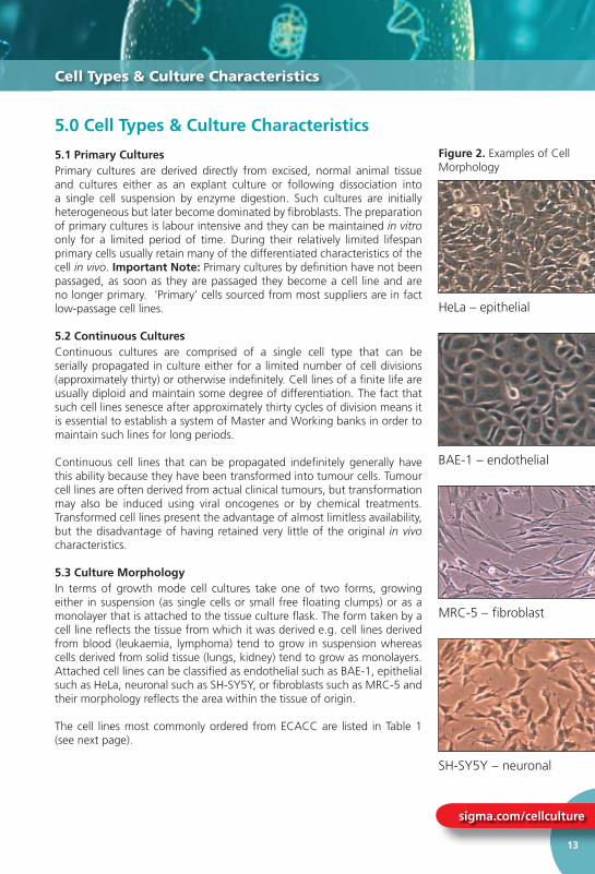
5.3 Culture MorphologyIn terms of growth mode cell cultures take one of two forms, growing either in suspension (as single cells or small free fl oating clumps) or as a monolayer that is attached to the tissue culture fl ask. The form taken by a cell line refl ects the tissue from which it was derived e.g. cell lines derived from blood (leukaemia, lymphoma) tend to grow in suspension whereas cells derived from solid tissue (lungs, kidney) tend to grow as monolayers. Attached cell lines can be classifi ed as endothelial such as BAE-1, epithelial such as HeLa, neuronal such as SH-SY5Y, or fi broblasts such as MRC-5 and their morphology refl ects the area within the tissue of origin.
The cell lines most commonly ordered from ECACC are listed in Table 1 (see next page).
5 0 Cell Types & Culture Characteristics
Cell Types & Culture Characteristics
Figure 2. Examples of Cell Morphology
HeLa – epithelial
BAE-1 – endothelial
MRC-5 – fibroblast
SH-SY5Y – neuronal

Fundamental Techniques in Cell Culture
www.hpacultures.org.uk
14
There are some instances when cell cultures may grow as semi-adherent cells, e.g. B95-8, where there appears to be a mixed population of attached and suspension cells. For these cell lines it is essential that both cell types are subcultured to maintain the heterogeneous nature of the culture.
5.4 Phases of Cell GrowthIt is important to know and record the growth characteristics of the cell line of use before starting any experiments. An alteration in cellular growth can indicate a signifi cant problem within the cell line and if undetected can have detrimental effects on experimental results.
A typical growth curve for cultured cells displays a sigmoid pattern of proliferation. The growth phases associated with normal cells are defi ned as:
Cell Types & Culture Characteristics
Attached Cell Lines
Name Species and tissue of origin Morphology
MRC-5 Human lung Fibroblast
HeLa Human cervix Epithelial
Vero African Green Monkey Kidney Epithelial
NIH 3T3 Mouse embryo Fibroblast
L929 Mouse connective tissue Fibroblast
CHO Chinese Hamster Ovary Fibroblast
BHK-21 Syrian Hamster Kidney Fibroblast
HEK 293 Human Kidney Epithelial
Hep G2 Human Liver Epithelial
BAE-1 Bovine aorta Endothelial
Suspension Cell Lines
Name Species and tissue of origin Morphology
NS0 Mouse myeloma Lymphoblastoid-like
U937 Human Hystiocytic Lymphoma Lymphoblastoid
Namalwa Human Lymphoma Lymphoblastoid
HL60 Human Leukaemia Lymphoblastoid-like
WEHI 231 Mouse B-cell Lymphoma Lymphoblastoid
YAC 1 Mouse Lymphoma Lymphoblastoid
U 266B1 Human Myeloma Lymphoblastoid
SH-SY5Y Human neuroblastoma Neuroblast
Table 1. Commonly used cell lines of each culture type

sigma.com/cellculture
15
1. Lag Phase – at this stage the cells do not divide. During this period the cells adapt to the culture conditions and the length of this phase will depend upon the growth phase of the cell line at the time of subculture and also the seeding density.
2. Logarithmic (Log) Growth Phase – cells actively proliferate and an exponential increase in cell density arises. The cell population is considered to be the most viable at this phase, therefore it is recommended to assess cellular function at this stage. Each cell line will show different cell proliferation kinetics during the log phase and it is therefore the optimal phase for determining the population doubling time.
3. Plateau (or Stationary) Phase – cellular proliferation slows down due to the cell population becoming confl uent. It is at this stage the number of cells in the active cell cycle drops to 0-10% and the cells are most susceptible to injury.
4. Decline Phase – cell death predominates in this phase and there is a reduction in the number of viable cells. Cell death is not due to the reduction in nutrient supplements but the natural path of the cellular cycle.
5.5 In Vitro Age of a Cell Culture Two terms are predominantly used to defi ne the age of a cell culture: (i) passage number - indicates the number of times the cell line has been sub-cultured and (ii) the population doubling (pd) number - indicates the number of cell generations the cell line has undergone i.e. the number of times the cell population has doubled. The in vitro age of a cell culture is particularly useful to know for cell lines with a fi nite lifespan or unstable characteristics that change over time in continuous culture.
6.0 The Cell Environment (including types of culture medium)
In general terms cultured cells require a sterile environment and a supply of nutrients for growth. In addition, the culture environment should be stable in terms of pH and temperature. Over the last 30 years various defi ned basal media types have been developed and are now available commercially. Originally, balanced salt solutions were used to maintain contractility of mammalian heart tissue and Tyrode’s salt solution was designed for use in work with primary mammalian cells. These have since been modifi ed and enriched with amino acids, vitamins, fatty acids and lipids. Consequently media suitable for supporting the growth of a wide range of cell types are now available. The precise media formulations have often been derived by optimising the concentrations of every constituent. Examples of the different media and their uses are given in Table 2 (see next page).
1. Lag Phase – at this stage the cells do not divide. During this period
The Cell Environment (including types of culture medium)
i Did You Know?
The cell population is at its most viable during the log growth phase.
Note: Different cell lines have different timescales for each phase, this graph is provided as a general example of a typical growth curve.

Fundamental Techniques in Cell Culture
www.hpacultures.org.uk
16
6.1 Basic Constituents of Media• Inorganic salts
• Carbohydrates
• Amino Acids
• Vitamins
• Fatty acids and lipids
• Proteins and peptides
• Serum
• Trace Elements
The Cell Environment (including types of culture medium)
Media Type Examples Uses
Balanced salt solutions
PBS, Hanks’ BSS, Earle’s saltsDPBS HBSS EBSS
Form the basis of many complex media
Basal media MEM Primary and diploid culture
DMEM Modification of MEM containing increased level of amino acids and vitamins. Supports a wide range of cell types including hybridomas
GMEM Glasgows modified MEM was defined for BHK-21 cells
Complex media RPMI 1640 Originally derived for human leukaemic cells. It supports a wide range of mammalian cells including hybridomas
Iscoves DMEM Further enriched modification of DMEM which supports high density growth
Leibovitz L-15 Designed for CO2 free environments
TC 100 Graces insect medium Schneider's Insect medium
Designed for culturing insect cells
Serum free media
CHO HEK293
For use in serum free applications
Ham F10 and derivatives Ham F12 DMEM/F12
Note: these media must be supplemented with other factors such as insulin, transferrin and epidermal growth factor. These media are usually HEPES buffered
Insect cells Serum-Free Insect Medium 1 (Cat no. S3777)
Specifically designed for use with Sf9 insect cells
Table 2. Different types of culture medium and their uses

sigma.com/cellculture
17
Each type of constituent performs a specifi c function as outlined below:
6.2 Inorganic SaltsThe inclusion of inorganic salts in media performs several functions. Primarily they help to retain the osmotic balance of the cells and help regulate membrane potential by provision of sodium, potassium and calcium ions. All of these are required in the cell matrix for cell attachment and as enzyme cofactors.
6.3 Buffering SystemsMost cells require pH conditions in the range 7.2-7.4 and close control of pH is essential for optimum culture conditions. There are major variations to this optimum. Fibroblasts prefer a higher pH (7.4-7.7) whereas, continuous transformed cell lines require more acid conditions pH (7.0-7.4).
Regulation of pH is particularly important immediately following cell seeding when a new culture is establishing and is usually achieved by one of two buffering systems; (i) a “natural” buffering system where gaseous CO2 balances with the CO3/HCO3 content of the culture medium and (ii) chemical buffering using a zwitterion called HEPES.
Cultures using natural bicarbonate /CO2 bufferingsystems need to be maintained in an atmosphere of 5-10% CO2 in air usually supplied in a CO2 incubator. Bicarbonate/CO2 is low cost, non-toxic and also provides other chemical benefi ts to the cells.
HEPES has superior buffering capacity in the pH range 7.2-7.4 but is relatively expensive and can be toxic to some cell types at higher concentrations. HEPES buffered cultures do not require a controlled gaseous atmosphere.
Most commercial culture media include phenol red as a pH indicator so that the pH status of the medium is constantly indicated by the colour. Usually the culture medium should be changed/replenished if the colour turns yellow (acid) or purple (alkali).
6.4 CarbohydratesThe main source of energy is derived from carbohydrates generally in the form of sugars. The major sugars used are glucose and galactose, however, some media contain maltose or fructose. The concentration of sugar varies from basal media containing 1g/L to 4.5g/L in some more complex media. Media containing the higher concentration of sugars are able to support the growth of a wider range of cell types.
6.5 Amino AcidsAmino acids are the building blocks of proteins. ‘Essential’ amino acids must be added to culture media as cells are not able to synthesize these themselves. The concentration of amino acids in the culture medium will determine the maximum cell density that can be achieved - once depleted the cells will no longer be able to proliferate.
Each type of constituent performs a specific function as outlined below:
The Cell Environment (including types of culture medium)

Fundamental Techniques in Cell Culture
www.hpacultures.org.uk
18
In relation to cell culture, glutamine, an essential amino acid, is particularly signifi cant. In liquid media or stock solutions glutamine degrades relatively rapidly. Optimal cell performance usually requires supplementation of the media with glutamine prior to use.
Adding supplements of non-essential amino acids to media both stimulates growth and prolongs the viability of the cells in culture.
6.6 VitaminsSerum is an important source of vitamins in cell culture. However, many media are also enriched with vitamins making them consistently more suitable for a wider range of cell lines. Vitamins are precursors for numerous co-factors. Many vitamins, especially B group vitamins, are necessary for cell growth and proliferation and for some lines the presence of B12 is essential. Some media also have increased levels of vitamins A and E. The vitamins commonly used in media include ribofl avin, thiamine and biotin.
6.7 Proteins and PeptidesThese are particularly important in serum free media. The most common proteins and peptides include albumin, transferrin, fi bronectin and fetuin and are used to replace those normally present through the addition of serum to the medium.
6.8 Fatty Acids and LipidsLike proteins and peptides these are important in serum free media since they are normally present in serum e.g. cholesterol and steroids essential for specialised cells.
6.9 Trace ElementsThese include trace elements such as zinc, copper, selenium and tricarboxylic acid intermediates. Selenium is a detoxifi er and helps remove oxygen free radicals.
6.10 Preparation of MediaWhilst all media may be made from the basic ingredients this is time consuming and may predispose to contamination. For convenience most media are available as ready mixed powders or as 10x and 1x liquid media. All commonly used media are listed in the Sigma-Aldrich Life Science Catalogue. If powder or 10x media are purchased it is essential that the water used to reconstitute the powder or dilute the concentrated liquid is free from mineral, organic and microbial contaminants. It must also be pyrogen free and of tissue culture grade. In most cases water prepared by reverse osmosis and resin cartridge purifi cation with a fi nal resistance of 16-18MΩ is suitable. Once prepared, the media should be fi lter sterilised before use.
In relation to cell culture, glutamine, an essential amino acid, is particularly
The Cell Environment (including types of culture medium)
iDid You Know?
In culture medium glutamine degrades rapidly to toxic ammonia at 37oC. Therefore aliquot your medium before use and only warm and use volumes you need that day. Discard opened bottles of prepared medium containing glutamine after 4-6 weeks.
sigma.com/cellculture
19
6.11 SerumSerum is a complex mix of albumins, growth factors and growth inhibitors and is probably one of the most important components of cell culture medium. The most commonly used serum is foetal bovine serum (FBS). Other types of serum are available including newborn calf serum and horse serum. The quality, type and concentration of serum can all affect the growth of cells and it is therefore important to screen batches of serum for their ability to support the growth of cells. In addition, there are other tests that may be used to aid the selection of a batch of serum including cloning effi ciency, plating effi ciency and the preservation of cell characteristics.
Serum is also able to increase the buffering capacity of cultures that can be important for slow growing cells or where the seeding density is low (e.g. cell cloning experiments). It also helps to protect against mechanical damage which may occur in stirred cultures or whilst using a cell scraper.
A further advantage of serum is the wide range of cell types with which it can be used despite the varying requirements of different cultures in terms of growth factors. In addition, serum is able to bind and neutralise toxins. However, serum is subject to batch-to-batch variation that makes standardisation of production protocols diffi cult.
There is also a risk of contamination associated with the use of serum. These risks can be minimised by obtaining serum from a reputable source since suppliers of large quantities of serum perform a battery of quality control tests and supply a certifi cate of analysis with the serum. In particular, serum is screened for the presence of bovine viral diarrhoea virus (BVDV) and mycoplasma. Heat inactivation of serum (incubation at 56°C for 30 minutes) can help to reduce the risk of contamination since some viruses are inactivated by this process. However, the routine use of heat inactivated serum is not an absolute requirement for cell culture. The use of serum also has a cost implication not only in terms of medium formulation but also in downstream processing. A 10% FBS supplement contributes 4.8mg of protein per millilitre of culture fl uid which complicates downstream processing procedures such as protein purifi cation.
6.12 Guidelines for Serum UseFoetal bovine serum (FBS) has been used to prepare a number of biologicals and has an excellent record of safety. The recognition of Bovine Spongiform Encephalopathy (BSE) in 1986 and its subsequent spread into continental Europe along side the announcement of the probable link between BSE and a new variant of Creutzfeldt Jacob disease in Humans stimulated an increased concern about safe sourcing of all bovine materials. In 1993, the Food and Drug Administration (FDA) "recommended against the use of bovine derived materials from cattle which have resided in, or originated from countries where BSE has been diagnosed”.
The current European Union (EU) guidelines on viral safety focus on sourcing, testing and paying particular attention to the potential risk of cross contamination during slaughtering or collection of the starting tissue.
6 11 Serum
The Cell Environment (including types of culture medium)
i Did You Know?
The most commonly used serum is foetal bovine serum. Other types of serum are available including newborn calf serum and horse serum.
Fundamental Techniques in Cell Culture
www.hpacultures.org.uk
20
As far as BSE is concerned, the EU guidance on minimising the risk of BSE transmission via medicinal products, EMEA/410/01 Rev. 2, recommends the main measures to be implemented in order to establish the safety of bovine material. Similarly the focus is on geographical origin, the age of the animals, the breeding and slaughtering conditions, the tissue to be used and the conditions of its processing.
The use of FBS in production processes of medicinal products is acceptable provided good documentation on sourcing, age of the animals and testing for the absence of adventitious agents is submitted. All responsible suppliers of FBS for bio-pharmaceutical applications will provide such documentation.
Regulatory requirements in Europe stress the importance of justifying the use of material of bovine, caprine or ovine origin in the production of pharmaceutical products. Thus, although FBS has been used for many years in the production process of many medicinal products such as viral vaccines and recombinant DNA products, at present there is a justifi ed trend to remove all material of animal origin from manufacturing processes. Sigma-Aldrich has recognised this growing trend and works closely with customers to optimise animal free media formulations to meet each customer’s cell culture requirements. Serum-free cell lines that have been adapted to media that do not contain serum are available from ECACC.
The United States Department of Agriculture (USDA) regulates all products that contain a primary component of animal origin. With specifi c reference to serum the USDA has declared that for materials which fall under their jurisdiction, only biological products manufactured using serum from approved countries of origin will be allowed in to USA.
6.13 Origin of SerumECACC only uses serum of Zone 1 origin, sterile fi ltered and cell culture tested. Zone 1 countries have BSE –free status such as the USA, Canada, Australia and New Zealand. It is essential to check the source country of the serum used and their Zone status. Sera from Mexico and Central American countries may require additional documentation to prove the geographical region of the donor herd to ensure BSE-free status. This is very important if the intended use of the serum is in the production of medicinal or other products being sent to the USA.
Serum from a reputable supplier should have undergone various quality control tests which will be listed in the product information sheet. Most serum products are cell culture tested including growth promotion, cloning effi ciency and plating effi ciency tests.
As far as BSE is concerned, the EU guidance on minimising the risk of BSE
The Cell Environment (including types of culture medium)

sigma.com/cellculture
21
Standard tests performed on serum commonly include tests to determine the presence and/or level of the following:
Sterility Virus Contamination Mycoplasma ContaminationEndotoxin Haemoglobin Total ProteinImmunoglobulin Hormone Testing pH (at room temperature)Osmolality
7.0 Cryopreservation and Storage of Cells
7.1 Cryopreservation of Cell LinesThe aim of cryopreservation is to enable stocks of cells to be stored to prevent the need to have all cell lines in culture at all times. It is invaluable when dealing with cells of limited life span. The other main advantages of cryopreservation are:
• Reduced risk of microbial contamination
• Reduced risk of cross contamination with other cell lines
• Reduced risk of genetic drift and morphological changes
• Work conducted using cells at a consistent passage number (refer to section 8 ‘Good Cell Banking Practices’)
• Reduced costs (consumables and staff time)
There has been a large amount of developmental work undertaken to ensure successful cryopreservation and resuscitation of a wide variety of cell lines of different cell types. The basic principle of successful cryopreservation and resuscitation is a slow freeze and quick thaw. Although the precise requirement may vary with different cell lines as a general guide cells should be cooled at a rate of –1°C to –3°C per minute and thawed quickly by incubation in a 37°C water bath for 3-5 minutes. If this and the additional points given below are followed then most cell lines should be cryopreserved successfully.
1. Cultures should be healthy with a viability of >90% and no signs of microbial contamination.
2. Cultures should be in log phase of growth (this can be achieved by using pre-confl uent cultures i.e. cultures that are below their maximum cell density and by changing the culture medium 24 hours before freezing).
3. A high concentration of serum/protein (>20%) should be used. In many cases serum is used at 90%.
Did You Know?
Cal zzril irit inim nummodit laore commy nos dit nonse conullaorper.
Standard tests performed on serum commonly include tests to determine
Cryopreservation and Storage of Cells

Fundamental Techniques in Cell Culture
www.hpacultures.org.uk
22
4. Use a cryoprotectant such as dimethyl sulphoxide (DMSO) or glycerol to help protect the cells from rupture by the formation of ice crystals. The most commonly used cryoprotectant is DMSO at a fi nal concentration of 10%, however, this is not appropriate for all cell lines e.g. where DMSO is used to induce differentiation. In such cases an alternative such as glycerol should be used (refer to ECACC data sheet for details of the correct cryoprotectant). Sigma also offers ready made cell freezing media containing DMSO, glycerol and serum-free formulations containing DMSO.
7.2 Ultra-low Temperature Storage of Cell LinesFollowing controlled rate freezing in the presence of cryoprotectants, cell lines can be cryopreserved in a suspended state for indefi nite periods provided a temperature of less than -135°C is maintained. Such ultra-low temperatures can only be attained by specialised electric freezers or more usually by immersion in liquid or vapour phase nitrogen. The advantages and disadvantages can be summarised as follows:
Storage in liquid phase nitrogen allows the lowest possible storage temperature to be maintained with absolute consistency, but requires the use of large volumes (depth) of liquid nitrogen which is a potential hazard.There have also been documented cases of cross contamination by virus pathogens via the liquid nitrogen medium. For these reasons ultra-low temperature storage is most commonly in vapour phase nitrogen.
For vapour phase nitrogen storage, the ampoules are positioned above a shallow reservoir of liquid nitrogen, the depth of which must be carefully maintained. A vertical temperature gradient will exist through the vapour phase, the extremes of which will depend on the liquid levels maintained, the design of the vessel, and the frequency with which it is opened. Temperature variations in the upper regions of a vapour phase storage vessel can be extreme if regular maintenance is not carried out. Modern designs of liquid nitrogen storage vessels are increasingly offering improved vapour phase storage technology.
4. Use a cryoprotectant such as dimethyl sulpho
Cryopreservation and Storage of Cells
Method Advantages Disadvantages
Electric (-135oC) Freezer
• Ease of Maintenance• Steady temperature• Low running costs
• Requires liquid nitrogen back-up• Mechanically complex• Storage temperatures high relative to liquid nitrogen
Liquid Phase Nitrogen
• Steady ultra-low (-196oC) temperature• Simplicity and mechanical reliability
• Requires regular supply of liquid nitrogen• High running costs• Risk of cross-contamination via the liquid nitrogen
Vapour Phase Nitrogen
• No risk of cross- contamination from liquid nitrogen• Low temperatures achieved• Simplicity and reliability
• Requires regular supply of liquid nitrogen• High running costs• Temperature fluctuations between -135oC and -190oC
Table 3. Comparison of ultra-low temperature storage methods for cell lines.
sigma.com/cellculture
23
Loss of entire cell stocks through inadequate storage maintenance is distressingly common. All liquid nitrogen storage vessels should minimally include alarms that warn of low liquid nitrogen levels and should also be constantly temperature monitored and alarmed.. This is particularly true of vapour phase storage systems. The bulk liquid nitrogen storage vessel should not be allowed to become less than half full before it is re-supplied. This will ensure that at least one liquid nitrogen delivery can be missed without catastrophic consequences. It is highly recommended that valuable cell stocks should be backed up by storage at a second site. ECACC offers a Safe Deposit Service for this purpose.
7.3 Inventory ControlAll ultra-low temperature storage vessels should include a racking / inventory system designed to organise the contents for ease of location and retrieval. This should be supported by accurate record keeping and inventory control incorporating the following:
• Each ampoule should be individually labelled, using “wrap around”, liquid nitrogen resistant labels with identity, lot number and date of freezing
• The location of each ampoule should be recorded ideally on an electronic database or spreadsheet, but also on a paper storage plan
• There should be a control system to ensure that no ampoule can be deposited or withdrawn without updating the records
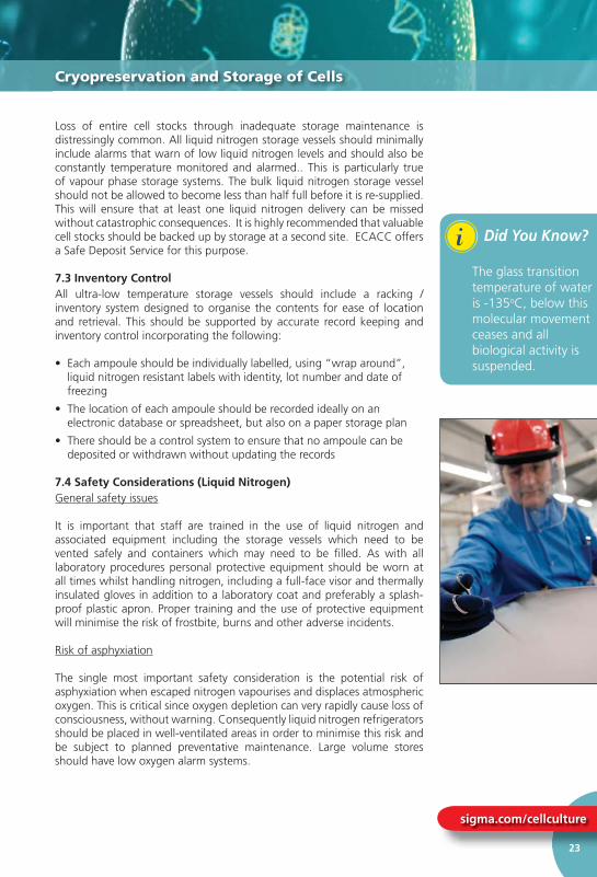
7.4 Safety Considerations (Liquid Nitrogen)General safety issues
It is important that staff are trained in the use of liquid nitrogen and associated equipment including the storage vessels which need to be vented safely and containers which may need to be fi lled. As with all laboratory procedures personal protective equipment should be worn at all times whilst handling nitrogen, including a full-face visor and thermally insulated gloves in addition to a laboratory coat and preferably a splash-proof plastic apron. Proper training and the use of protective equipment will minimise the risk of frostbite, burns and other adverse incidents.
Risk of asphyxiation
The single most important safety consideration is the potential risk of asphyxiation when escaped nitrogen vapourises and displaces atmospheric oxygen. This is critical since oxygen depletion can very rapidly cause loss of consciousness, without warning. Consequently liquid nitrogen refrigerators should be placed in well-ventilated areas in order to minimise this risk and be subject to planned preventative maintenance. Large volume stores should have low oxygen alarm systems.
Loss of entire cell stocks through inadequate storage maintenance is
Cryopreservation and Storage of Cells
i Did You Know?
The glass transition temperature of water is -135oC, below this molecular movement ceases and all biological activity is suspended.

Fundamental Techniques in Cell Culture
www.hpacultures.org.uk
24
Precautions for Dedicated Liquid Nitrogen Storage Areas
• Use oxygen alarms set to 18% oxygen (v/v)
• Staff training – staff should be trained to evacuate the area immediately on hearing the alarm and not return until the oxygen is back to normal levels (~ 20% v/v)
• Staff should work in pairs when handling liquid nitrogen
• Prohibit the use of nitrogen outside of normal working hours
• Mechanical ventilation systems should be installed if at all possible
8.0 Good Cell Banking Practices
It is bad practice to maintain a cell line in continuous or extended culture for the following reasons:
• Risk of microbial contamination
• Loss of characteristics of interest (e.g. surface antigen or monoclonal antibody expression)
• Genetic drift particularly in cells known to have an unstable karyotype (e.g. CHO, BHK 21)
• Loss of cell line due to exceeding fi nite life-span e.g. human diploid cells such as MRC-5
• Risk of cross contamination with other cell lines
• Increased consumables and staff costs
All of these potential risk factors may be minimised by the implementation of a cell banking system as described below. This type of system is known as a tiered banking system or Master Cell Banking system (refer to Figure 3). On initial arrival into the laboratory a new cell culture should be regarded as a potential source of contamination e.g. harbouring bacteria, fungi and mycoplasma and should be handled under quarantine conditions until proven negative for such microbial contaminants. Following initial expansion 3-5 ampoules should be frozen as a Token Stock before a Master Cell Bank is prepared. One of the Token Stock ampoules should then be thawed and expanded to produce a Master Cell Bank of 10-20 ampoules depending upon the anticipated level of use.
Ampoules of this Master Cell Bank (2-3) should be allocated for quality control comprising confi rmation that the cell count and viability of the bank is acceptable and that the bank is free of bacteria, fungi and mycoplasma. Additional tests (such as viral screening and authenticity testing) may also be required. Once these tests have been completed satisfactorily an ampoule from the Master Cell Bank should be thawed and cultured to produce a Working Cell Bank. The size of this bank will again depend on the envisaged level of demand. Quality control tests (cell count and viability and the absence of microbial contaminants) are again required prior to using the cultures for routine experimentation or production. It is
Precautions for Dedicated Liquid Nitrogen Storaq g
Good Cell Banking Practices

sigma.com/cellculture
25
Good Cell Banking Practices
New Cell Line(Handled under
Quarantine Conditions)
Token freezeof 3-5 ampoules andinitial mycoplasma
test
Resuscitate one ampouleand expand in culture
Abandon bankingprocedure
Cryopreservation ofMaster Bank
(10-100 ampoules)
Resuscitate one ampouleand expand in culture
Cryopreservation ofWorking Bank
(20-200 ampoules)
Release for use Repeat Banking
Repeat Banking
Quality ControlTests
(i) Cell count & viability(ii) Microbial QC including
mycoplasma(iii) Authentication
Quality ControlTests
(i) Cell count & viability(ii) Microbial QC including
mycoplasma(iii) Authentication
Fail
Fail
Fail
Pass
Pass
Pass
QUARANTINEFACILITY
MAIN CELL CULTUREFACILITY
Figure 3. Schematic Representation of a Tiered Cell Banking System:
Did You Know?
It is bad practice to maintain a cell line in continuous or extended culture.
i

Fundamental Techniques in Cell Culture
www.hpacultures.org.uk
26
also important at this stage to confi rm that the Master and Working Cell Banks are genetically identical by DNA profi ling techniques.
Implementation of this banking system ensures:
• Material is of a consistent quality
• Experiments are performed using cultures in the same range of passage numbers
• Cells are only in culture when required
• The original cell line characteristics are retained
Notes1. The number of ampoules prepared for Master and Working Cell Banks
depends upon the forecast demand for their use.
2. The number of ampoules sampled for quality control is dependent upon the size of bank. Ideally 5-10% of the bank should be tested before use.
3. Ampoules from the Working Cell Bank should be used sequentially keeping cells in culture for not more than a predetermined number of cell doublings. This number will be least in the case of cell lines having a fi nite life-span (e.g. diploid lines).
4. The Working Cell Bank should be replenished from an ampoule of the Master Cell Bank. This should be done in suffi cient time to allow the quality control to be completed.
5. A new Master Cell Bank should be prepared before the number of original Master stock drops below fi ve ampoules.
6. The panel of quality control tests performed depends upon the use intended e.g. regulatory authorities may require additional tests such as viral screening and karyotypic studies.
9.0 Quality Control Considerations
9.1 IntroductionQuality is important in all aspects of tissue culture. The quality of materials used (cell lines, media and other reagents) will affect the quality of the cultures and the subsequent scientifi c data and products derived from them. The main areas of quality control that are of concern for tissue culture are:
• The quality of the reagents and materials
• The provenance and integrity of the cell lines
• The avoidance of microbial contamination
also important at this stage to confi rm that th
Quality Control Considerations
sigma.com/cellculture
27
9.2 Reagents and MaterialsPotential sources of contamination are reagents and materials, in particular bovine serum which has been identifi ed as a source of bovine viral diarrhoea virus (BVDV). Porcine trypsin is also a potential source of Mycoplasma hyorhinis. Good quality reagents and materials are available from numerous manufacturers of tissue culture media and supplements. In addition, manufacturers such as Sigma will carry out a range of quality control tests including screening for mycoplasma and BVDV and supply a Certifi cate of Analysis with their products. These state the product and lot numbers and form a vital part of record keeping and tracking of reagents used in the production of cell stocks. It is advisable to further test key reagents such as foetal bovine serum to ensure that they are ‘fi t for purpose’ due to batch-to-batch variation.
Manufacturers of sterile plastic ware (fl asks, centrifuge tubes, pipettes) designed for tissue culture use also supply Certifi cates of Analysis for each batch produced, which should be kept for future reference.
9.3 Provenance and Integrity of Cell LinesThe sourcing of cell lines can have an important effect on quality; freshly imported cell lines are a major source of contamination. The advantages of obtaining cell lines from a recognised source such as a culture collection are that the cultures will be:
• Contaminant free
• Fully characterised and authenticated in terms of DNA profi le and species of origin
• Supplied with a detailed data sheet
Once cell lines have been obtained from a reputable source it is important to implement master and working cell banking procedures and the associated quality control steps such as routine testing for microbial contaminants and confi rming the identity of cultures.
9.4 Avoidance of Microbial ContaminationPotential sources of contamination include other cell lines, laboratory conditions and staff poorly trained in core areas such as aseptic techniques and good laboratory practice. Thus the use of cells and reagents of known origin and quality alone is not suffi cient to guarantee quality of product (cell stock or culture products); it is necessary to demonstrate quality throughout the production process and also in the fi nal product. Routine screening aids the early detection of contamination since all manipulations are a potential source of contamination.
The three main types of microbial contaminants in tissue culture are:
• Bacteria and Fungi
• Mycoplasma
• Viruses
9 2 Reagents and Materials
Quality Control Considerations

Fundamental Techniques in Cell Culture
www.hpacultures.org.uk
28
Bacterial and Fungal ContaminationBacterial contamination is generally visible to the naked eye and detected by a sudden increase in turbidity and colour change of the culture medium as the result of a change in pH. The cell culture may survive for a short time but the cells will eventually die. Daily microscopic observation of cultures will ensure early detection of contamination and enable appropriate action to be taken as soon as the fi rst signs of contamination become apparent. In addition, specifi c tests for the detection of bacteria and fungi should be used as part of a routine and regular quality control screening procedure (see Protocol 8 on page 54).
Mycoplasma ContaminationMycoplasmas are the smallest free-living self-replicating prokaryotes. They lack a cell wall and lack the ability to synthesize one. They are 0.3µm in diameter and can be observed as fi lamentous or coccal forms. There are 5 major species that are tissue culture contaminants, namely M. hyorhinis, M. arginini, M. orale, M. fermentans and Acholeplasma laidlawii.
The effects of mycoplasma infection are more insidious than those of bacteria and fungi, inducing several long term effects in cell cultures. These include:
• Altered growth rate
• Morphological changes
• Chromosome aberrations
• Alterations in amino acid and nucleic acid metabolism
However, despite these well-documented effects the presence of mycoplasma is often not tested for with the consequence that in such laboratories the majority of cell lines are positive for mycoplasma. Mycoplasma contamination is diffi cult to detect requiring the use of specialist techniques (see Protocol 9 on page 56 - Isolation by Culture and Protocol 10 on page 58 – Detection by DNA Staining). In the past only specialist laboratories, such as culture collections, have performed these tests. However, a variety of commercial kits are now available although the performance characteristics of these kits can be extremely variable. A combination of these should be used as part of a routine and regular quality control screening procedure. ECACC tests cultures for the presence of mycoplasma on a routine basis and offers a mycoplasma testing service.
Viral ContaminationSome cell lines contain endogenous viruses and secrete virus particles or express viral antigens on their surface (e.g. Epstein-Barr Virus (EBV) transformed lines). These cell lines are not considered contaminated. However, bovine serum is a potential source of bovine viral diarrhoea virus (BVDV) contamination. Use of infected serum will lead to contamination of cell lines with the virus. Contamination of cell lines with BVDV may cause slight changes in growth rate but since this virus is non-cytopathic
Bacterial and Fungal Contaminationg
Quality Control Considerations
iDid You Know?
Mycoplasmas are the smallest free-living self-replicating prokaryotes. They lack a cell wall and do not have the ability to synthesize one.

sigma.com/cellculture
29
macroscopic and microscopic changes in the culture will not be detected. Suppliers of bovine serum are aware of this and screen sera accordingly and generally serum is sold as BVDV tested.
9.5 Environmental MonitoringIt is good practice to monitor the laboratory environment where cell cultures and their products are prepared. Class 2 microbiology safety cabinets, fi tted with HEPA fi lters, should be tested every 6 months to ensure that they are working effi ciently, i.e. the level of airfl ow across the fi lter should be tested. However, it is also advisable to monitor the level of ingress of contaminants into the cabinet by periodically placing open settle plates (Tryptone Soya Bean Agar bacteriological culture plates) on the cabinet work surfaces. In addition, settle plates should be used to assess airborne microbial burden at selected points around the laboratory. Plates should be left open for a period of 4 hours. After this time they should be covered, placed in sealed boxes and incubated at 32°C and 22°C for up to 7 days. At the end of this period the plates should be examined for the presence of microbial growth. The position of each plate in the cabinet should be recorded and results stored for trend analysis.
Acceptable limits should be defi ned in terms of “alert” levels and “action” levels, the actual values being dependent on the containment classifi cation of the work area, the criticality of the work and the levels of cleanliness that can be achieved under normal operating conditions.
9.6 Aseptic Technique and Contamination Control
Personal HygieneWhen entering the laboratory it is important to wash hands since this will remove dry skin and loosely adherent microorganisms which could potentially contaminate cell cultures. Gowns and surgical gloves must be worn. Gloves should be frequently swabbed with 70% (v/v) sterile isopropanol. Other personal protective equipment includes head caps and face masks, but these are not always necessary, particularly when a class 2 microbiological safety cabinet is being used. Long hair should be tied back to remove obstruction and reduce the risk of contamination.
Working within the Microbiological Safety CabinetWhen working within the cabinet the operator should remember that the air-fl ow does not make the environment sterile but keeps it clean. Before any practical procedure is conducted the cabinet should be stocked with all the materials required for the experiment. In doing so the operator restricts the number of times that their hand/arm is removed from the cabinet into a non-clean environment. When stocking up the cabinet it is essential that a clutter free condition is maintained. Each item within the cabinet should be positioned to minimise movement and traffi c over the area where cell culture operations are performed. Both the rear and front of the cabinet should be cleared to achieve maximum airfl ow. Flasks and dishes should be the last items to enter the cabinet. All items that enter the cabinet must be sprayed with 70% (v/v) sterile isopropanol to prevent dust and particulate
macroscopic and microscopic changes in the culture will not be detected
Quality Control Considerations
Did You Know?
The main source of microbial contamination in the cell culture laboratory is the operator.
i

Fundamental Techniques in Cell Culture
www.hpacultures.org.uk
30
from entering the cabinet. Twenty minutes should elapse before any tops or containers are opened to allow the airfl ow to purge the work area of particulates that may have been introduced.
Pipetting and Prevention of AerosolsDisposable plastic pipettes (1ml, 2ml, 5ml, 10ml and 25ml) are the easiest forms to use for cell culture. Microbial and cellular contamination can arise through pipetting errors such as spillage of material. Adherence to the following guidance can minimise contamination and safety risks associated with pipetting:
• Never mouth pipette
• Use automatic pipette aids, with one pipette aid designated to each cabinet. Ensure the pipette fi ts into the pipette aid without any force. To avoid contamination disinfect the pipette aid regularly and ensure that fi lters are changed regularly (weekly).
• Use plugged pipettes when transferring medium
• Avoid drawing liquid into the pipette plug. Use an individual pipette once only.
• To avoid generating aerosols do not create bubbles in the medium or pipette. Aerosols can spread contaminating microorganisms and by introducing cells into the air increases the potential risk of cross- contamination.
• Clean spills that arise immediately with 70% (v/v) sterile isopropanol.
9.7 What to do in the Event of ContaminationOne hugely under-estimated problem in tissue culture is the routine use of antibiotics. Continuous use of antibiotics is unnecessary and can lead to the development of resistant strains that are diffi cult to eradicate and may require the use of more exotic antibiotics that may be toxic to the cell cultures. In addition, the use of antibiotics may mask a low level of contamination.
Once a contamination has been detected, whether it is due to bacteria, fungi or mycoplasma, the recommended course of action is to discard the culture and continue the work with earlier stocks that are known to be free of contaminants or obtain fresh stocks from a recognised source.
Viral infections are virtually impossible to remove from cultures since they do not respond to antibiotic treatment. Also, as they are intra-cellular parasites it is not possible to remove them by centrifugation or other separation techniques. If virus free stocks or a virus free alternative is not available, then a thorough risk assessment should be undertaken prior to continuing work with the infected cell line.
from entering the cabinet. Twenty minutes sho
Quality Control Considerations

sigma.com/cellculture
31
10.0 Authentication of Cell Lines
Whatever the scope of work to be carried out it is important to know that the work is being conducted using the correct reagents. This is no less important for cell cultures, since if cell cultures are not what they are reported to be then work can be invalidated and resources wasted. There is now considerable evidence of widespread cross-contamination and misidentifi cation of cell lines, in particular with HeLa cells. More than 18 cell lines have been recognised as HeLa contaminants and the list is increasing. To minimise the risk of working with contaminated cell lines it is advisable to obtain cells from a recognised source such as a culture collection that will have confi rmed the identity and purity of the cells as part of the banking process.
Tests used to authenticate cell cultures include isoenzyme analysis, karyotyping/cytogenetic analysis and, more recently, molecular techniques of DNA profi ling. While most of these techniques are generalised tests and are applicable to all cell lines additional specifi c tests may also be required to confi rm the presence of a product or antigen of interest. ECACC offers a cell line identity verifi cation service.
11.0 Alternative Culture Systems
11.1 Cell Culture Scale-up SystemsMost tissue culture is performed on a small scale where relatively small numbers of cells are required for experiments. At this scale cells are usually grown in T fl asks (see image on page 7) ranging from 25cm2 to 175cm2. Typical cell yields from a T175 fl ask range from 1x10⁷ for an attached line to 1x10⁸ for a suspension line. However, exact yields will vary depending on the cell line. It is not practical to produce much larger quantities of cells using standard T fl asks, due to the amount of time required for repeated passaging of the cells, demand on incubator space and cost.
When considering scaling up a cell culture process there are a whole range of parameters to consider which will need to be developed and optimised if scale-up is to be successful. These include problems associated with nutrient depletion, gaseous exchange, particularly oxygen depletion, and the build up of toxic by-products such as ammonia and lactic acid. To optimise such a process for quantities beyond 1L volumes it is best left to expert process development scientists. Note, in these cases often a “scale-down” approach is adopted to allow many parameters to be evaluated on many replicates.
However, there are many commercially available systems that attempt to provide a "half-way house" solution to scale-up which do not necessarily require expert process development services. A selected list of some of the systems available along with a brief summary of their potential yields, advantages and disadvantages is provided in Table 4 on page 33.
10 0 Authentication of Cell Lines
Authentication of Cell Lines/Alternative Culture Systems
Figure 4. Biostat® A plus bioreactor from Sartorius Stedim Biotech
Figure 5. Hyperfl ask (left) & T175 Flask (right) from Corning
Figure 6. Shake fl asks in a Certomat CT plus from Sartorius Stedim Biotech

Fundamental Techniques in Cell Culture
www.hpacultures.org.uk
32
11.2 Scale-up SolutionsThe image in Figure 4 shows a BIOSTAT® A Plus autoclavable bioreactor from Sartorius Stedim that can be used for cell culture with a working volume of 1L to 5L. Please refer to the following sub-sections for a variety of alternative scale-up solutions.
A word of caution – although the systems listed in Table 4 are often described as “off-the-shelf” solutions to scale-up they are not universally applicable to all cell types and often require a period of familiarisation and optimisation.
11.3 Roller Bottle CultureThis is the method most commonly used for initial scale-up of attached cells also known as anchorage dependent cell lines. Roller bottles are cylindrical vessels that revolve slowly (between 5 and 300 revolutions per hour) which bathe the cells that are attached to the inner surface with medium. Roller bottles are available typically with surface areas of 1050cm2. The size of some of the roller bottles presents problems since they are diffi cult to handle in the confi ned space of a microbiological safety cabinet. Roller bottles with expanded inner surfaces have become available which has made handling large surface area bottles more manageable, but repeated manipulations and subculture with roller bottles should be avoided if possible. A further problem with roller bottles is with the attachment of cells since some cells lines do not attach evenly. This is a particular problem with epithelial cells. This may be partially overcome by optimising the speed of rotation, generally by decreasing the speed, during the period of attachment for cells with low attachment effi ciency. The RC40 from Cellon provides a semi-automated multiple roller bottle platform.
11.4 Multilayer VesselsA variety of disposable multi-layer vessels are now available for simple and rapid scale-up of anchorage dependent cells with little or no process optimisation. These include triple layer fl asks which are useful for maximising incubator space, however it is vessels such as CellStacks and Hyperfl asks that offer the greatest advantages. CellStacks™ provide multiples of 1, 2, 5, 10 and 40 layers, each layer offering 636cm2 (therefore a 10 layer Cellstack™ provides 6,360cm2 for cell growth in a single vessel). Cell stacks are in effect giant cell culture fl asks but with two vented caps for fi lling, harvest and gas exchange rather than a single cap. Some familiarisation, validation of cell growth and care and attention to manual handling is required as these vessels can only be practically employed if pouring techniques are used for fi lling and harvesting (although fi lling connectors can be exchanged for the caps) but in essence most cell growth in T fl asks can be directly translated to CellStacks™. Forty layer CellStacks™ are too large to be handled manually and require specialised trolleys for manipulations. Originally designed for robotic systems Hyperfl asks™ are another multilayer system, however, rather more revolutionary in their design. Consisting of 10 multiple “fl asklets” each with the same approximate footprint as a T175 fl ask the HyperFlask™ is entirely fi lled with medium and cell inoculum. Gaseous exchange in this case is achieved by diffusion of gases directly through the thin surfaces of the fl asklets.
11.2 Scale-up Solutions
Alternative Culture Systems
Figure 8. Roller Bottles
Figure 7. Roller Deck
Figure 9. Spinner Flasks
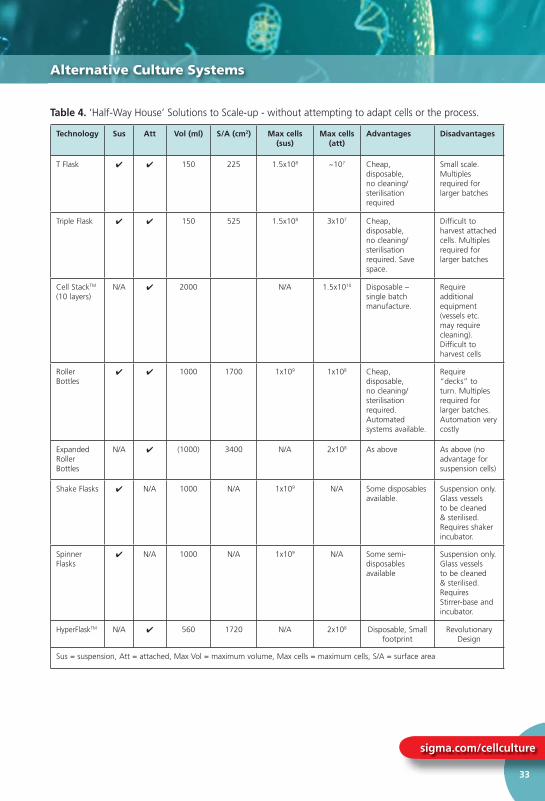
sigma.com/cellculture
33
Alternative Culture Systems
Technology Sus Att Vol (ml) S/A (cm2) Max cells (sus)
Max cells (att)
Advantages Disadvantages
T Flask ✔ ✔ 150 225 1.5x10⁸ ~107 Cheap, disposable, no cleaning/sterilisation required
Small scale. Multiples required for larger batches
Triple Flask ✔ ✔ 150 525 1.5x10⁸ 3x10⁷ Cheap, disposable, no cleaning/sterilisation required. Save space.
Difficult to harvest attached cells. Multiples required for larger batches
Cell StackTM (10 layers)
N/A ✔ 2000 N/A 1.5x10¹⁰ Disposable – single batch manufacture.
Require additional equipment (vessels etc. may require cleaning). Difficult to harvest cells
Roller Bottles
✔ ✔ 1000 1700 1x10⁹ 1x10⁸ Cheap, disposable, no cleaning/sterilisation required. Automated systems available.
Require “decks” to turn. Multiples required for larger batches. Automation very costly
Expanded Roller Bottles
N/A ✔ (1000) 3400 N/A 2x10⁸ As above As above (no advantage for suspension cells)
Shake Flasks ✔ N/A 1000 N/A 1x10⁹ N/A Some disposables available.
Suspension only. Glass vessels to be cleaned & sterilised. Requires shaker incubator.
Spinner Flasks
✔ N/A 1000 N/A 1x10⁹ N/A Some semi-disposables available
Suspension only. Glass vessels to be cleaned & sterilised. Requires Stirrer-base and incubator.
HyperFlaskTM N/A ✔ 560 1720 N/A 2x10⁸ Disposable, Small footprint
Revolutionary Design
Sus = suspension, Att = attached, Max Vol = maximum volume, Max cells = maximum cells, S/A = surface area
Table 4. ‘Half-Way House’ Solutions to Scale-up - without attempting to adapt cells or the process.

www.hpacultures.org.uk
34
Fundamental Techniques in Cell Culture
Authentication of Cell Lines/Alternative Culture Systems
11.5 Disposable solutions for anchorage independent (suspension) cellsIn the last few years many new disposable systems for growth of cells in suspension have emerged. These include disposable stirrer vessels, single use disposable bioreactors and Wave/Cultibag (GE Healthcare®/Sartorius Stedim®) technology. The Wave™ /Cultibag™ systems allow cell growth in large (10s of litres) sterile, disposable bags which are fi lled with cells, medium and an air/gas headspace and then gently agitated on a temperature controlled rocking platform. These bags are either “off the shelf” or custom made with most of the connectors for seed, harvest and sampling built in. Recent advances in disposable sensors has now also meant that pH and dissolved oxygen sensors can be built into the bags making them effi cient bioreactors suitable for GMP production and seed vessels for larger bioreactors.
11.6 Spinner Flask CultureThis is the method of choice for suspension lines including hybridomas and attached lines that have been adapted to growth in suspension e.g. HeLa S3. Spinner fl asks are either plastic or glass bottles with a central magnetic stirrer shaft and side arms for the addition and removal of cells and medium, and gassing with CO2 enriched air. Inoculated spinner fl asks are placed on a stirrer and incubated under the culture conditions appropriate for the cell line. Cultures should be stirred at 100-250 revolutions per minute.
11.7 Other Scale-up OptionsThe next stage of scale-up for both suspension and attached cell lines is the bioreactor that is used for large culture volumes (in the range 100-10,000 litres). For suspension cell lines the cells are kept in suspension by either a propeller in the base of the chamber vessel or by air bubbling through the culture vessel. However, both of these methods of agitation give rise to mechanical stresses. A further problem with suspension lines is that the density obtained is relatively low e.g. 2x10⁶ cells/ml.
For attached cell lines the cell densities obtained are increased by the addition of micro-carrier beads. These small beads are 30-100µm in diameter and can be made of dextran, cellulose, gelatin, glass or silica, and increase the surface area available for cell attachment considerably. The range of micro-carriers available means that it is possible to grow most cell types in this system. A recent advance has been the development of porous micro-carriers which has increased the surface area available for cell attachment by a further 10-100 fold. The surface area on 2g of beads is equivalent to 15 small roller bottles.
Footnote: Information on commercial scale-up systems is valid on the date of last revision,
but such systems may be improved or supplanted subsequently.
Biostat® CultiBag RM from Sartorius Stedim Biotech

HPA Culture CollectionsA strategic business unit within the Health Protection Agency.
www.hpacultures.org.uk Tel: +44 (0) 1980 612512 email: [email protected]
Cell Lines available from ECACC
General CollectionMore than 1200 cell lines originating from over 45 different species and a wide variety of tissues. Over 600 human cell lines including many different cancer and drug resistant cell lines are available.
Primary Cells & Media CollectionA range of normal primary cells from a diverse selection of human and animal tissues. These cells are usually not more than 6 passages from the primary culture. They are more representative of the main functional component of the tissue from which they are derived when compared to continuous (tumour or artifi cially immortalised) cell lines. The recommended culture media is also available to order.
Hybridoma CollectionOver 400 monoclonal antibody-secreting hybridomas with a diverse range of antigen specifi cities.
Chromosomal Abnormality CollectionApproximately 3000 cell lines (the majority are B-lymphoblastoid cell lines) from individuals with defi ned chromosomal abnormalities.
HLA-Typed Collection A specialised collection of over 430 reference B-lymphoblastoid cell lines, originating from laboratories all over the world, for which Human Leukocyte Antigen (HLA) typing data is available.
Human Random Control Collection A collection of more than 700 B-lymphoblastoid cell lines derived from randomly selected Caucasian blood donors whose parents and grandparents were born in the UK or Ireland.
Quality Control & AuthenticationAll cell lines supplied by ECACC undergo full quality control and authentication procedures. These include testing for mycoplasma by culture isolation, Hoescht DNA staining and PCR, together with culture testing for contaminant bacteria, yeast and fungi. Authentication procedures used include species verifi cation by isoenzyme analysis and identity verifi cation by DNA profi ling. Classical DNA fi ngerprinting using multi-locus probes is carried out for non-human cell lines. Human cell lines are analysed by PCR of short tandem repeat sequences within chromosomal microsatellite DNA (STR-PCR). Certifi cates of Analysis are available for each cell line lot no. supplied which report the cell count, identity of the cell line and the absence of bacteria, fungi and mycoplasma.
Supply FormatsCell lines can be supplied as frozen or growing cultures. Genomic DNA and RNA extracted from the cell lines are also available.
Visit www.hpacultures.org.uk to view the collections.
ECACC cell lines can be ordered via
ECACC has a range of cell line collections grouped according to the type of cell line and the data available. We have an active program to acquire new cell lines and therefore it is always worth checking our website for new lines. ECACC is accredited to ISO 9001:2008. The collections include:

Fundamental Techniques in Cell Culture
www.hpacultures.org.uk
36
12.0 Cell Culture Protocols
12.1 Basic Techniques - The “Do’s and Don’ts” of Cell Culture
Given below are a few of the essential "do’s and don’ts" of cell culture. Some of these are mandatory e.g. use of personal protective equipment (PPE). Many of them are common sense and apply to all laboratory areas. However, some of them are specifi c to tissue culture.
The Do’s1. Use personal protective equipment (PPE), (laboratory coat/gown,
gloves and eye protection) at all times. In addition, thermally insulated gloves, full-face visor and splash-proof apron should be worn when handling liquid nitrogen.
2. Use disposable head caps to cover hair.
3. Wear dedicated PPE for the tissue culture facility and keep separate from PPE worn in the general laboratory environment. The use of different coloured gowns or laboratory coats makes this easier to enforce.
4. Keep all work surfaces free of clutter.
5. Correctly label reagents including fl asks, medium and ampoules with contents and date of preparation.
6. Only handle one cell line at a time. This common-sense point will reduce the possibility of cross contamination by mislabelling etc. It will also reduce the spread of bacteria and mycoplasma by the generation of aerosols across numerous opened media bottles and fl asks in the cabinet.
7. Clean the work surfaces with a suitable disinfectant (e.g. 70% isopropanol) between operations and allow a minimum of 15 minutes between handling different cell lines.
8. Maintain separate bottles of media for each cell line in culture.
9. Examine cultures and media daily for evidence of gross bacterial or fungal contamination. This includes medium that has been purchased commercially.
12.0 Cell Culture Protocols
Cell Culture Protocols

sigma.com/cellculture
37
10. Quality control all media and reagents prior to use.
11. Keep cardboard packaging to a minimum in all cell culture areas.
12. Ensure that incubators, cabinets, centrifuges and microscopes are cleaned and serviced at regular intervals.
13. Test cells for the presence of mycoplasma on a regular basis.
The Don’ts1. Do not use antibiotics continuously in culture medium as this can
lead to the appearance of antibiotic resistant strains and may mask underlying contamination.
2. Do not allow waste to accumulate particularly within the microbiological safety cabinet or in the incubators.
3. Do not have too many people in the lab at any one time.
4. Do not handle cells from unauthenticated sources in the main cell culture suite. They should be handled in quarantine until quality control checks are complete.
5. Avoid keeping cell lines continually in culture without returning to frozen stock.
6. Avoid cell cultures from becoming fully confl uent. Always sub-culture at 70-80% confl uency or as advised on the ECACC cell culture data sheet.
7. Do not allow media to go out of date. The shelf life is only 4 - 6 weeks at +4°C once glutamine and serum are added.
8. Avoid water baths from becoming dirty by regular cleaning.
9. Do not allow essential equipment to become out of calibration. Ensure microbiological safety cabinets are tested regularly.
12.2 Protocol 1 - Aseptic Technique and Good Cell Culture Practice
AimTo ensure all cell culture procedures are performed to a standard that will prevent contamination from bacteria, fungi and mycoplasma and cross contamination with other cell lines.
Materials• 70% (v/v) isopropanol in sterile water
• Sodium Hypochlorite (also known as ‘Chloros’)
• Appropriate disinfectant
Equipment• Personal protective equipment (sterile gloves, laboratory coat, safety
visor, overshoes, head cap)
• Microbiological safety cabinet at appropriate containment level
10. Quality control all media and reagents prior to use.
Cell Culture Protocols

Fundamental Techniques in Cell Culture
www.hpacultures.org.uk
38
Procedure1. Sanitise the cabinet using 70% isopropanol before commencing
work.
2. Sanitise gloves by spraying them with 70% isopropanol and allowing to air dry for 30 seconds before commencing work.
3. Put all materials and equipment into the cabinet prior to starting work. Equipment in the cabinet or that which will be taken into the cabinet during cell culture procedures (media bottles, pipette tip boxes, pipette aids) should be wiped with tissue soaked with 70% isopropanol prior to use.
4. Whilst working do not contaminate gloves by touching anything outside the cabinet (especially face and hair). If gloves become contaminated re-spray with 70% isopropanol as above before proceeding.
5. Discard gloves after handling contaminated cultures and at the end of all cell culture procedures.
6. Movement within and immediately outside the cabinet must not be rapid. Slow movement will allow the air within the cabinet to circulate properly.
7. Speech, sneezing and coughing must be directed away from the cabinet so as not to disrupt the airfl ows.
8. After completing work disinfect all equipment and material before removing from the cabinet. Spray the work surfaces inside the cabinet with 70% isopropanol and wipe dry with tissue. Dispose of tissue by incineration.
9. Liquid cell culture waste should be discarded in sodium hypochlorite (10,000 ppm) and must be kept in the cabinet for a minimum of two hours (preferably overnight) prior to discarding to the drain with copious amounts of water.
10. Periodically clean the cabinet surfaces with a disinfectant or fumigate the cabinet according to the manufacturers instructions. However, you must ensure that it is safe to fumigate your own laboratory environment due to the generation of gaseous formaldehyde, consult your on-site Health and Safety Advisor.
Procedure
Cell Culture Protocols

sigma.com/cellculture
39
12.3 Protocol 2 - Resuscitation of Frozen Cell Lines
AimMany cultures obtained from a culture collection, such as ECACC, will arrive frozen and in order to use the cells they must be thawed and put into culture. It is vital to thaw cells correctly in order to maintain the viability of the culture and enable the culture to recover more quickly. Some cryoprotectants, such as DMSO, are toxic above 4°C therefore it is essential that cultures are thawed quickly and diluted in culture medium to minimise the toxic effects.
Materials• Media– pre-warmed to the appropriate temperature (refer to the
ECACC Cell Line Data Sheet for the correct medium and temperature.)
• 70% (v/v) isopropanol in sterile water
• DMSO • Tryan blue (vital stain)
Equipment• Personal protective equipment (sterile gloves, laboratory coat, safety
visor)
• Container to transport frozen ampoules e.g. box of dry ice or liquid nitrogen Dewar.
• Waterbath set to appropriate temperature
• Microbiological safety cabinet at appropriate containment level
• Incubator
• Pre-labelled fl asks
• Inverted phase contrast microscope
12.3 Protocol 2 - Resuscitation of Frozen Cell Lines
Cell Culture Protocols
Check cell line data sheet
Prepare flasks with pre-warmed media
Collect cells
Allow to thaw
Pipette cells into pre-warmed growth medium, dilute if required
Incubate at appropriate temperature
Examine cells after 24 hours

Fundamental Techniques in Cell Culture
www.hpacultures.org.uk
40
• Haemocytometer
• Centrifuge
• Marker Pen
• Pipettes
• Ampoule Rack
• Tissue
Procedure1. Read the cell line data sheet to establish specifi c requirements for your
cell line.
2. Prepare the fl asks - label with cell line name, passage number and date.
3. Collect an ampoule of cells from liquid nitrogen storage wearing appropriate personal protective equipment and transfer to the laboratory in a container of liquid nitrogen or on dry ice. It is important to handle the ampoules with care: on rare occasions ampoules may explode on warming due to expansion of trapped residual liquid nitrogen.
4. In a microbiological safety cabinet, hold a tissue soaked in 70% alcohol around the cap of the frozen ampoule and turn the cap a quarter turn to release any residual liquid nitrogen that may be trapped. Re-tighten the cap. Quickly transfer the ampoule to a 37oC waterbath until only one or two small ice crystals, if any, remain (1-2 minutes). It is important to thaw rapidly to minimise any damage to the cell membranes. Note: Do not totally immerse the ampoule as this may increase the risk of contamination.
5. Wipe ampoule with a tissue soaked in 70% alcohol prior to opening.
6. Pipette the whole content of the ampoule into a sterile tube (e.g. 15 ml capacity). Then slowly add 5ml pre-warmed medium that has already been supplemented with the appropriate constituents. Determine the viable cell density (see Protocol 6 – Cell Quantifi cation on page 49). Transfer the appropriate volume of cell suspension to a fl ask to achieve the cell seeding density recommend on the cell line data sheet.
For adherent cell lines: Adjust the volume of the medium, and if necessary the fl ask size, to achieve the cell seeding density recommended on the cell line data sheet. A pre-centrifugation step to remove cryoprotectant is not normally necessary as the fi rst media change will remove residual cryoprotectant. If it is, then this will be specifi ed on the data sheet. If the cells are to be used immediately (e.g. for a cell based assay), rather than subcultured, it may be advisable to perform a pre-centrifugation step to remove cryoprotectant. For suspension cell lines: A pre-centrifugation step to remove cryoprotectant is recommended i.e. pellet the cells by centrifugation at 150 x g for 5 minutes and resuspend the cell pellet in fresh medium using the appropriate volume to achieve the correct seeding density.
• Haemocytometer
Cell Culture Protocols

sigma.com/cellculture
41
7. Incubate at the temperature and CO2 level recommended on the data sheet. If a CO2 fed incubator is used the fl ask should have a vented cap to allow gaseous exchange.
8. Examine cells microscopically (phase contrast) after 24 hours and sub-culture as necessary.
Key Points1. Most text books recommend washing the thawed cells in media to
remove the cryoprotectant. This is only necessary if the cryoprotectant is known to have an adverse effect on the particular cell type. For example, some cell types are known to differentiate in the presence of DMSO. In such cases the cells should be washed in media before being added to their fi nal culture fl asks.
2. The addition of the thawed cell suspension to culture medium effectively dilutes the cryoprotectant (e.g. DMSO) reducing the toxicity of the cryoprotectant. That is why it is important to add the thawed cell suspension to a larger volume of culture medium immediately after the ampoule has thawed; do not allow thawed ampoules to sit at room temperature for long periods.
3. Do not use an incubator or the palm of your hand to thaw cell cultures since the rate of thawing achieved is too slow resulting in a loss of viability. Use a water bath as described in the protocol above.
4. If a CO2 incubator is not available gas the fl asks for 1-2 minutes with 5% CO2 in 95% air fi ltered through a 0.2µm fi lter.
5. For most cultures it is best practice to subculture before confl uence is reached so that the cells are harvested during their log phase of growth and are at optimum viability ready for seeding into new fl asks. Furthermore there are some specifi c cell types that must be subcultured before confl uence is reached in order to maintain their characteristics e.g. the contact inhibition of NIH 3T3 cells is lost if they are allowed to reach confl uence repeatedly.
6. Some hybridomas may be slow to recover post resuscitation therefore start in 20% (v/v) FBS and 10% (v/v) hybridoma enhancement supplement in the appropriate medium.
7 Incubate at the temperature and CO level recommended on the data
Cell Culture Protocols
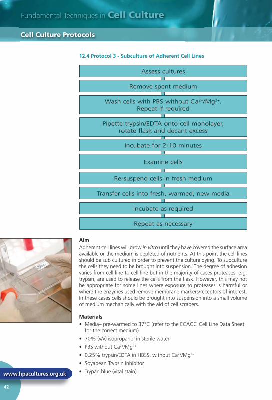
Fundamental Techniques in Cell Culture
www.hpacultures.org.uk
42
12.4 Protocol 3 - Subculture of Adherent Cell Lines
AimAdherent cell lines will grow in vitro until they have covered the surface area available or the medium is depleted of nutrients. At this point the cell lines should be sub cultured in order to prevent the culture dying. To subculture the cells they need to be brought into suspension. The degree of adhesion varies from cell line to cell line but in the majority of cases proteases, e.g. trypsin, are used to release the cells from the fl ask. However, this may not be appropriate for some lines where exposure to proteases is harmful or where the enzymes used remove membrane markers/receptors of interest. In these cases cells should be brought into suspension into a small volume of medium mechanically with the aid of cell scrapers.
Materials• Media– pre-warmed to 37°C (refer to the ECACC Cell Line Data Sheet
for the correct medium)
• 70% (v/v) isopropanol in sterile water
• PBS without Ca2+/Mg2+
• 0.25% trypsin/EDTA in HBSS, without Ca2+/Mg2+
• Soyabean Trypsin Inhibitor
• Trypan blue (vital stain)
12.4 Protocol 3 - Subculture of Adherent Ce
Cell Culture Protocols
Assess cultures
Remove spent medium
Wash cells with PBS without Ca2+/Mg2+. Repeat if required
Pipette trypsin/EDTA onto cell monolayer, rotate flask and decant excess
Incubate for 2-10 minutes
Examine cells
Re-suspend cells in fresh medium
Transfer cells into fresh, warmed, new media
Incubate as required
Repeat as necessary

sigma.com/cellculture
43
Equipment• Personal protective equipment (sterile gloves, laboratory coat, safety
visor)
• Waterbath set to appropriate temperature
• Microbiological safety cabinet at appropriate containment level
• Incubator
• Pre-labelled fl asks
• Inverted phase contrast microscope
• Centrifuge
• Haemocytometer
• Marker Pen
• Pipettes
• Ampoule rack
• Tissue
Procedure1. View cultures using an inverted microscope to assess the degree
of confl uency and confi rm the absence of bacterial and fungal contaminants.
2. Remove spent medium.
3. Wash the cell monolayer with PBS without Ca2+/Mg2+ using a volume equivalent to half the volume of culture medium. Repeat this wash step if the cells are known to adhere strongly.
4. Pipette trypsin/EDTA onto the washed cell monolayer using 1ml per 25cm2 of surface area. Rotate fl ask to cover the monolayer with trypsin. Decant the excess trypsin.
5. Return fl ask to the incubator and leave for 2-10 minutes.
6. Examine the cells using an inverted microscope to ensure that all the cells are detached and fl oating. The side of the fl asks may be gently tapped to release any remaining attached cells.
7. Resuspend the cells in a small volume of fresh serum-containing medium to inactivate the trypsin. Remove 100-200µl and perform a cell count (see Protocol 6 - Cell Quantifi cation). In the case of cells cultured in serum-free media, use a trypsin inhibitor e.g. soyabean trypsin inhibitor to inactivate the trypsin.
8. Transfer the required number of cells to a new labelled fl ask containing pre-warmed medium (refer to ECACC Cell Line Data Sheet for the required seeding density).
9. Incubate as appropriate for the cell line.
10. Repeat this process as demanded by the growth characteristics of the cell line.
Equipment
Cell Culture Protocols
Did You Know?
Serum-free media will not inactivate trypsin. A trypsin inhibitor, e.g. soyabean trypsin inhibitor, must be used.
i

Fundamental Techniques in Cell Culture
www.hpacultures.org.uk
44
Key Points1. Some cultures whilst growing as attached lines adhere only lightly to
the fl ask, thus it is important to ensure that the culture medium is retained and the fl asks are handled with care to prevent the cells detaching prematurely.
2. Although most cells will detach in the presence of trypsin alone the EDTA is added to enhance the activity of the enzyme.
3. Trypsin is inactivated in the presence of serum. Therefore, it is essential to remove all traces of serum from the culture medium by washing the monolayer of cells with PBS without Ca2+/Mg2+.
4. Cells should only be exposed to trypsin/EDTA long enough to detach cells. Prolonged exposure could damage cell surface receptors.
5. Trypsin should be neutralised with serum prior to seeding cells into new fl asks otherwise cells will not attach.
6. Trypsin may also be neutralised by the addition of soyabean trypsin inhibitor, where an equal volume of inhibitor at a concentration of 1mg/ml is added to the trypsinised cells. The cells are then centrifuged, resuspended in fresh culture medium and counted as above. This is especially necessary for serum-free cell cultures.
7. If a CO2 incubator is not available gas the fl asks for 1-2min with 5% CO2 in 95% air fi ltered through a 0.2µm fi lter.
8. If the cells harvested are at too low a cell density to re-seed at the appropriate cell density into fresh fl asks it may be necessary to centrifuge the cells e.g. 5 mins at 150 x g, and resuspend in a smaller volume of medium.
Key Points
Cell Culture Protocols

sigma.com/cellculture
45
12.5 Protocol 4 - Subculture of Semi-Adherent Cell Lines
AimSome cultures grow as a mixed population (e.g. B95-8 - marmoset) where a proportion of cells do not attach to the tissue culture fl ask and remain in suspension. Therefore to maintain this heterogeneity both the attached cells and the cells in suspension must be subcultured.
Materials• Media– pre-warmed to 37°C (refer to the ECACC Cell Line Data Sheet
for the correct medium)
• 70% (v/v) isopropanol in sterile water
• PBS without Ca2+/Mg2+.
• 0.25% trypsin/EDTA in HBSS, without Ca2+/Mg2+.
• Soyabean Trypsin Inhibitor
• Trypan blue (vital stain)
Equipment• Personal protective equipment (sterile gloves, laboratory coat, safety
visor)
• Waterbath set to appropriate temperature
12 5 Protocol 4 - Subculture of Semi-Adherent Cell Lines
Cell Culture Protocols
Assess cultures
Decant spent medium
Wash cells with 1-2mls PBS without Ca2+/Mg2+.
Pipette trypsin/EDTA onto cell monolayer, rotate flask and decant excess
Incubate for time taken for cells to round up
Examine cells
Centrifuge
Decant and re-suspend cells
Pipette into fresh medium and inoculate into new flask
Repeat as necessary
Fundamental Techniques in Cell Culture
www.hpacultures.org.uk
46
• Microbiological safety cabinet at the appropriate containment level
• Centrifuge
• Inverted phase contrast microscope
• Incubator
• Haemocytometer
• Pre-labelled fl asks
• Marker Pen
• Pipettes
Procedure1. View cultures using an inverted phase contrast microscope to assess
the degree of confl uency and confi rm the absence of bacterial and fungal contaminants. Give the fl ask a gentle knock fi rst, this may dislodge the cells from the fl ask and remove the need for a trypsinisation step with the subsequent loss of some cells due to the washes.
2. Decant medium containing non-adherent cells into a sterile centrifuge tube and retain.
3. Wash any remaining attached cells with PBS without Ca2+/Mg2+ using 1-2ml for each 25cm2 of surface area. Retain the washings.
4. Pipette trypsin/EDTA onto the washed cell monolayer using 1ml per 25cm2 of surface area. Rotate fl ask to cover the monolayer with trypsin. Decant the excess trypsin.
5. Return fl ask to incubator and leave for 2-10 minutes.
6. Examine the cells using an inverted microscope to ensure that all the cells are detached and fl oating. The side of the fl asks may be gently tapped to release any remaining attached cells (see Protocol 6 - Cell Quantifi cation).
7. Transfer the cells into the centrifuge tube containing the retained spent medium and cells.
8. Centrifuge the entire cell suspension at 150 x g for 5 minutes.
9. Remove the supernatant and re-suspend the cell pellet in a small volume (10-20ml) of fresh culture medium. Count the cells.
10. Pipette the required number of cells into a new labelled fl ask and dilute to the required volume using fresh medium (refer to ECACC Cell Line Data Sheet for the required seeding density).
11. Repeat this process every 2-3 days as necessary.
Key Points1. Although most cells will detach in the presence of trypsin alone the
inclusion of EDTA is used to enhance the activity of the enzyme.
2. Trypsin is inactivated in the presence of serum. Therefore, it is essential to remove all traces of serum from the culture medium by washing the monolayer of cells with PBS without Ca2+/Mg2+. Repeated warming to 37°C also inactivates trypsin.
• Microbiological safety cabinet at the appropr
Cell Culture Protocols

sigma.com/cellculture
47
3. Cells should only be exposed to trypsin/EDTA long enough to detach cells. Prolonged exposure could damage cell surface receptors. In general, a shorter time of exposure to trypsin is required for semi- adherent cell lines in comparison to adherent cell lines.
4. Trypsin should be neutralised with serum prior to seeding cells into new fl asks otherwise cells will not attach.
5. Trypsin may also be neutralised by the addition of Soyabean Trypsin Inhibitor, where an equal volume of inhibitor at a concentration of 1mg/ml is added to the trypsinised cells. The cells are then centrifuged, resuspended in fresh culture medium and counted as above.
6. If a CO2 incubator is not available gas the fl asks for 1-2 minutes with 5% CO2 in 95% air fi ltered through a 0.2µm fi lter.
12.6 Protocol 5 - Subculture of Suspension Cell Lines
AimIn general terms cultures derived from blood (e.g. lymphocytes) grow in suspension. Cells may grow as single cells or in clumps (e.g. EBV transformed lymphoblastoid cell lines). For these types of cell lines subculture by dilution is relatively easy. However, for cell lines that grow in clumps it may be necessary to bring the cells into a single cell suspension by centrifugation and re-suspension by pipetting in a smaller volume before counting.
Materials • Media– pre-warmed to 37°C (refer to the ECACC Cell Line Data Sheet
for the correct medium)
• 70% (v/v) isopropanol in sterile water
• Trypan blue (vital stain)
3 Cells should only be exposed to trypsin/EDTA long enough to detach
Cell Culture Protocols
View cultures and assess condition
Centrifuge 150 x g for 5 minutes (pH of medium must be acidic)
Add 10-20% conditioned media to fresh media
Take sample of cells
Calculate cells/ml and reseed according to recommended density
Repeat every 2-3 days
Fundamental Techniques in Cell Culture
www.hpacultures.org.uk
48
Equipment• Personal protective equipment (sterile gloves, laboratory coat, safety
visor)
• Waterbath set to 37°C
• Microbiological safety cabinet at appropriate containment level
• Centrifuge
• Incubator
• Inverted phase contrast microscope
• Haemocytometer
• Pre-labelled fl asks
• Marker Pen• Pipettes
Procedure1. View cultures using an inverted phase contrast microscope. Cells
growing in exponential growth phase should be bright, round and refractile. Hybridomas may be very sticky and require a gentle knock to the fl ask to detach the cells. EBV transformed cells can grow in very large clumps that are very diffi cult to count and the centre of the large clumps may be non-viable.
2. Do not centrifuge to subculture unless the pH of the medium is acidic (phenol red = yellow) which indicates the cells have overgrown and may not recover. If this is so, centrifuge at 150 x g for 5 minutes, re-seed at a slightly higher cell density and add 10-20% of conditioned medium (supernatant) to the fresh media.
3. Take a small sample (100-200µl) of the cells from the cell suspension and count the cells (Protocol 6 - Cell Quantifi cation). Calculate cells/ml and re-seed the desired number of cells into freshly prepared fl asks, without centrifugation, just by diluting the cells. Refer to the data sheet supplied with the cell line for the recommended seeding density.
4. Repeat this every 2-3 days.
Key Points1. If the cell line is a hybridoma or another cell line that produces a
substance (e.g. recombinant protein or growth factor) of interest retain the spent media for analysis.
Equipment
Cell Culture Protocols
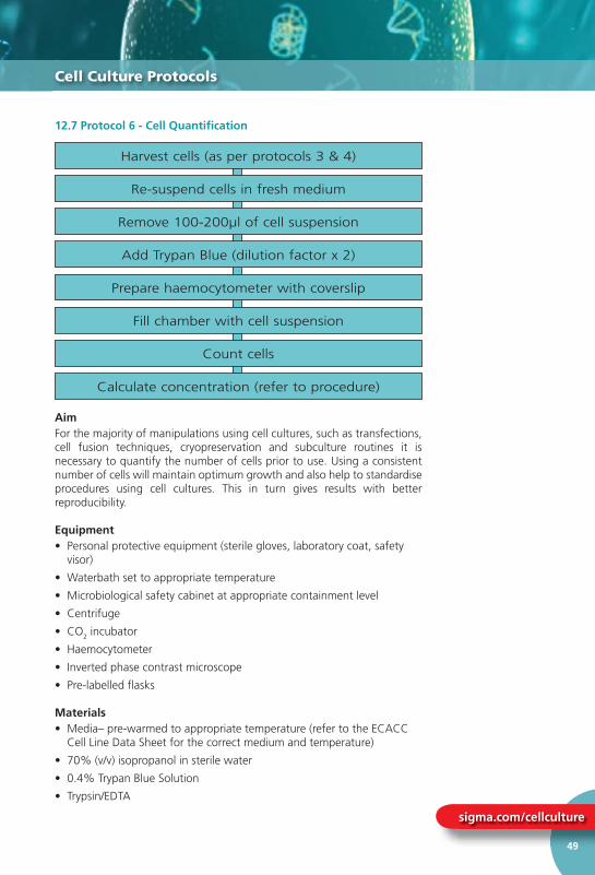
sigma.com/cellculture
49
12.7 Protocol 6 - Cell Quantifi cation
AimFor the majority of manipulations using cell cultures, such as transfections, cell fusion techniques, cryopreservation and subculture routines it is necessary to quantify the number of cells prior to use. Using a consistent number of cells will maintain optimum growth and also help to standardise procedures using cell cultures. This in turn gives results with better reproducibility.
Equipment• Personal protective equipment (sterile gloves, laboratory coat, safety
visor)
• Waterbath set to appropriate temperature
• Microbiological safety cabinet at appropriate containment level
• Centrifuge
• CO2 incubator
• Haemocytometer
• Inverted phase contrast microscope
• Pre-labelled fl asks
Materials• Media– pre-warmed to appropriate temperature (refer to the ECACC
Cell Line Data Sheet for the correct medium and temperature)
• 70% (v/v) isopropanol in sterile water
• 0.4% Trypan Blue Solution
• Trypsin/EDTA
12 7 Protocol 6 - Cell Quantification
Cell Culture Protocols
Harvest cells (as per protocols 3 & 4)
Re-suspend cells in fresh medium
Remove 100-200µl of cell suspension
Add Trypan Blue (dilution factor x 2)
Prepare haemocytometer with coverslip
Fill chamber with cell suspension
Count cells
Calculate concentration (refer to procedure)
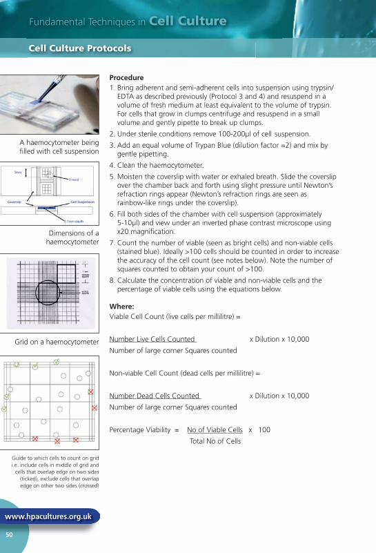
Fundamental Techniques in Cell Culture
www.hpacultures.org.uk
50
Procedure1. Bring adherent and semi-adherent cells into suspension using trypsin/
EDTA as described previously (Protocol 3 and 4) and resuspend in a volume of fresh medium at least equivalent to the volume of trypsin. For cells that grow in clumps centrifuge and resuspend in a small volume and gently pipette to break up clumps.
2. Under sterile conditions remove 100-200µl of cell suspension.
3. Add an equal volume of Trypan Blue (dilution factor =2) and mix by gentle pipetting.
4. Clean the haemocytometer.
5. Moisten the coverslip with water or exhaled breath. Slide the coverslip over the chamber back and forth using slight pressure until Newton’s refraction rings appear (Newton’s refraction rings are seen as rainbow-like rings under the coverslip).
6. Fill both sides of the chamber with cell suspension (approximately 5-10µl) and view under an inverted phase contrast microscope using x20 magnifi cation.
7. Count the number of viable (seen as bright cells) and non-viable cells (stained blue). Ideally >100 cells should be counted in order to increase the accuracy of the cell count (see notes below). Note the number of squares counted to obtain your count of >100.
8. Calculate the concentration of viable and non-viable cells and the percentage of viable cells using the equations below.
Where:Viable Cell Count (live cells per millilitre) =
Number Live Cells Counted x Dilution x 10,000
Number of large corner Squares counted
Non-viable Cell Count (dead cells per millilitre) =
Number Dead Cells Counted x Dilution x 10,000
Number of large corner Squares counted
Percentage Viability = No of Viable Cells x 100
Total No of Cells
Procedure
Cell Culture Protocols
A haemocytometer being fi lled with cell suspension
Dimensions of a haemocytometer
Grid on a haemocytometer
Guide to which cells to count on grid i.e. include cells in middle of grid and
cells that overlap edge on two sides (ticked), exclude cells that overlap edge on other two sides (crossed)

sigma.com/cellculture
51
Key Points1. Trypan Blue is toxic and is a potential carcinogen. Protective clothing,
gloves and face/eye protection should be worn. Do not breathe the vapour.
2. The central area of the counting chamber is 1mm2. This area is subdivided into 25 smaller squares (1/25mm2). Each of these is surrounded by triple lines and is then further divided into 16 (1/400mm2). The depth of the chamber is 0.1mm.
3. The correction factor of 104 converts 0.1mm3 to 1ml (0.1mm3 = 1mm2 x 0.1mm)
4. There are several sources of inaccuracy:
• The presence of air bubbles and debris in the chamber.
• Overfi lling the chamber such that sample runs into the channels or the other chamber
• Incomplete fi lling of the chamber.
• Cells not evenly distributed throughout the chamber.
• Too few cells to count. This can be overcome by centrifuging the cells, re-suspending in a smaller volume and recounting.
• Too many cells to count. This can be overcome by using a higher dilution factor in trypan blue e.g. 1:10
5. The use of a haemocytometer can be time consuming, susceptible to subjective judgements by the operator and some cell types, such as those that form clusters, are particularly diffi cult to count using this method. Technology which counts cell nuclei, such as the NucleoCounter (Chemometec), is available offering an alternative cell quantifi cation method. Unlike other cell quantifi cation methods, automated nuclei counters eliminate manual counting and do not rely on the retention of physical and/or morphological properties of the cells. Low volumes, e.g. 200µl, of prepared samples are drawn into a cassette which is inserted into the nuclei counter providing a cell count in approximately thirty seconds.
Key Points
Cell Culture Protocols
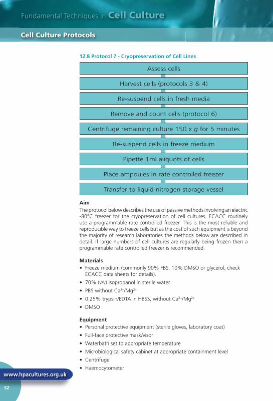
Fundamental Techniques in Cell Culture
www.hpacultures.org.uk
52
12.8 Protocol 7 - Cryopreservation of Cell Lines
AimThe protocol below describes the use of passive methods involving an electric -80°C freezer for the cryopreservation of cell cultures. ECACC routinely use a programmable rate controlled freezer. This is the most reliable and reproducible way to freeze cells but as the cost of such equipment is beyond the majority of research laboratories the methods below are described in detail. If large numbers of cell cultures are regularly being frozen then a programmable rate controlled freezer is recommended.
Materials• Freeze medium (commonly 90% FBS, 10% DMSO or glycerol, check
ECACC data sheets for details).
• 70% (v/v) isopropanol in sterile water
• PBS without Ca2+/Mg2+
• 0.25% trypsin/EDTA in HBSS, without Ca2+/Mg2+
• DMSO
Equipment• Personal protective equipment (sterile gloves, laboratory coat)
• Full-face protective mask/visor
• Waterbath set to appropriate temperature
• Microbiological safety cabinet at appropriate containment level
• Centrifuge
• Haemocytometer
12.8 Protocol 7 - Cryopreservation of Cell L
Cell Culture Protocols
Assess cells
Harvest cells (protocols 3 & 4)
Re-suspend cells in fresh media
Remove and count cells (protocol 6)
Centrifuge remaining culture 150 x g for 5 minutes
Re-suspend cells in freeze medium
Pipette 1ml aliquots of cells
Place ampoules in rate controlled freezer
Transfer to liquid nitrogen storage vessel
sigma.com/cellculture
53
• Pre-labelled ampoules/cryotubes
• Cell Freezing Device
Procedure1. View cultures using an inverted microscope to assess the degree of cell
density and confi rm the absence of bacterial and fungal contaminants. Harvest cells in the log phase of growth. For adherent cell lines harvest cells as close to 80 - 90% confl uency as possible.
2. Bring adherent and semi adherent cells into suspension using trypsin/EDTA as described previously (Protocol 3 and 4 – Subculture of adherent/attached and semi-adherent cell lines) and re-suspend in a volume of fresh medium at least equivalent to the volume of trypsin. Suspension cell lines can be used directly.
3. Remove a small aliquot of cells (100-200µl) and perform a cell count (Protocol 6 – Cell Quantifi cation). Ideally, the cell viability should be in excess of 90% in order to achieve a good recovery after freezing.
4. Centrifuge the remaining culture at 150 x g for 5 minutes.
5. Re-suspend cells at a concentration of 2-4x10⁶ cells per ml in freeze medium.
6. Pipette 1ml aliquots of cells into cyroprotective ampoules that have been labelled with the cell line name, passage number, cell concentration and date.
7. Place ampoules inside a passive freezer e.g. Nalgene Mr Frosty box Sigma cat no. C1562. Fill freezer with isopropyl alcohol and place at -80oC overnight.
8. Frozen ampoules should be transferred to the vapour phase of a liquid nitrogen storage vessel and the locations recorded.
Key Points1. The most commonly used cryoprotectant is dimethyl sulphoxide
(DMSO), however, this is not appropriate for all cell lines e.g. HL60 where DMSO is used to induce differentiation. In such cases an alternative such as glycerol should be used (refer to ECACC data sheet for details of the correct cryoprotectant).
2. ECACC freeze medium recommended above has been shown to be a good universal medium for most cell types. Another commonly used freeze medium formulation is: 70% basal medium, 20% FBS, 10% DMSO but this may not be suitable for all cell types. Check if it works for your cells before using on a regular basis.
3. It is essential that cultures are healthy and in the log phase of growth. This can be achieved by using pre-confl uent cultures (cultures that are below their maximum cell density) and by changing the culture medium 24 hours before freezing.
• Pre-labelled ampoules/cryotubes
Cell Culture Protocols

Fundamental Techniques in Cell Culture
www.hpacultures.org.uk
54
4. The rate of cooling may vary but as a general guide a rate of between –1°C and –3°C per minute will prove suitable for the majority of cell cultures.
5. An alternative to the Mr Frosty system is the Taylor Wharton passive freezer where ampoules are held in liquid nitrogen vapour in the neck of a Dewar. The system allows the ampoules to be gradually lowered thereby reducing the temperature. Rate controlled freezers are also available and are particularly useful if large numbers of ampoules are frozen on a regular basis.
6. As a last resort, if no other devices are available, ampoules may be placed inside a well insulated box (such as a polystyrene box with sides that are at least 1cm thick) and placed at –80°C overnight. It is important to ensure that the box remains upright throughout the freezing process. Once frozen, ampoules should be transferred to the vapour phase of a liquid nitrogen storage vessel and the locations recorded.
7. If using a freezing method involving a -80°C freezer it is important to have an allocated section for cell line freezing so that samples are not inadvertently removed. If this happens at a crucial part of the freezing process then viability and recovery rates will be adversely affected.
12.9 Protocol 8 - Testing for Bacteria and Fungi
Figure 10. Flow Scheme for Bacteria and Fungi Testing
AimIn cases of gross contamination the naked eye may identify the presence of bacteria and fungi. However, in order to detect low-level infections the method above is recommended.
4. The rate of cooling may vary but as a genera
Cell Culture Protocols
Fluid Thioglycollate Medium• Inoculate 2 broths with 1.5ml sample• Inoculate 2 broths with 100 cfu of each
positive control organism• Leave 2 broths un-inoculated as negative
controls
Incubate half the broths of each type at 32oC and the other half at 22oC for 14 days
Examine the broths for turbidity on day 14
Tryptone Soya Broth• Inoculate 2 broths with 1.5ml sample• Inoculate 2 broths with 100 cfu of positive
control organism• Leave 2 broths un-inoculated as negative
controls
sigma.com/cellculture
55
Materials• Aerobic nutrient broth e.g. Soyabean Casein Digest (Tryptone Soya
Broth,TSB) (15ml aliquots)
• Anaerobic nutrient broth Fluid Thioglycollate Medium (20ml aliquots) (TGM)
• Positive control organisms e.g.
Bacillus subtilis NCTC*
Candida albicans NCTC*
Clostridium sporogenes NCTC*
* Available from the National Collection of Type Cultures (NCTC)
Equipment• Personal protective equipment (latex medical gloves, laboratory coat,
safety glasses)
• Waterbath set to 37°C
• Microbiological safety cabinet at appropriate containment level
• Incubator set at 32°C
• Incubator set at 22°C
Procedure1. Culture cell line in the absence of antibiotics for 2 passages prior to
testing.
2. Bring attached cells into suspension with the use of a cell scraper. Suspension cell lines may be tested directly.
3. Inoculate 2 x aerobic broths and 2 x anaerobic broths with 1.5ml test sample.
4. For each of the positive control organisms inoculate 2 x aerobic broths and 2 x anaerobic broths (e.g. 0.1ml control at 100 cfu per broth).
7. Leave 2 aerobic and 2 anaerobic broths un-inoculated as negative controls.
8. Incubate broths as follows:
• Incubate one broth of each pair at 32°C the other at 22°C for 14 days
9. Examine Test and Control broths for turbidity after 14 days.
Criteria for a Valid ResultAll positive control broths show evidence of bacteria and fungi within 14 days of incubation and the negative control broths show no evidence of bacteria and fungi.
Materials
Cell Culture Protocols

Fundamental Techniques in Cell Culture
www.hpacultures.org.uk
56
Cell Culture Protocols
Criteria for a Positive ResultTest broths containing bacteria or fungi show turbidity.
Criteria for a Negative ResultTest broths should be clear and show no evidence of turbidity.
Notes1. Control organisms (Bacillus subtilis, Clostridium sporogenes and
Candida albicans) are available from the National Collection of Type Cultures (NCTC), UK.
2. This test procedure should be carried out in a microbiology laboratory away from the cell culture laboratory.
12.10 Protocol 9 - Testing for Mycoplasma by Culture IsolationFigure 11. Flow Scheme for Detection of Mycoplasma by Culture
AimDetection of mycoplasma by culture is the reference method of detection and has a theoretical level of detection of 1 colony-forming unit (cfu). However, there are some strains of mycoplasma that are non-cultivable (certain strains of Mycoplasma hyorhinis). The method is suitable for the detection of mycoplasma in both cell cultures and cell culture reagents and results are obtained within 4 weeks. Mycoplasma colonies observed on agar plates have a ‘fried egg’ appearance (see fi gure 12).
Agar plates• Inoculate 2 plates with 0.1ml test sample• Inoculate 1 plates with 100 cfu of each control organism• Leave 1 plate un-inoculated as negative control
Incubate anaerobically 14 days at 37°C Incubate aerobically
at 37°C
Observe all plates for Mycoplasma colonies
Label plates and broths
Subculture 0.1ml of broth onto Agarbetween days 3 - 7 and 10 -14
Incubate plates anaerobically14 days at 37°C
Observe all plates forMycoplasma colonies
Broths• Inoculate 1 broth with 0.2ml test sample• Inoculate 1 broths with 100 cfu of each control organism• Leave 1 broth un-inoculated as negative control
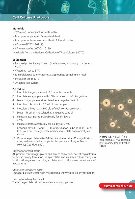
sigma.com/cellculture
57
Cell Culture Protocols
Materials• 70% (v/v) isopropanol in sterile water
• Mycoplasma plates (in 5cm petri dishes)
• Mycoplasma horse serum broths (in 1.8ml aliquots)
• M. orale (NCTC* 10112)
• M. pneumoniae (NCTC* 10119)
*Available from the National Collection of Type Cultures (NCTC)
Equipment• Personal protective equipment (sterile gloves, laboratory coat, safety
visor)
• Waterbath set to 37°C
• Microbiological safety cabinet at appropriate containment level
• Incubator set at 37°C
• Anaerobic jar system
Procedure1. Inoculate 2 agar plates with 0.1ml of test sample.
2. Inoculate an agar plate with 100 cfu of each control organism.
4. Leave 1 agar plate un-inoculated as a negative control.
5. Inoculate 1 broth with 0.2 ml of test sample.
6. Inoculate a broth with 100 cfu of each control organism.
7. Leave 1 broth un-inoculated as a negative control.
8. Incubate agar plates anaerobically for 14 days at 37°C.
9. Incubate broths aerobically for 14 days at 37°C.
10. Between days 3 - 7 and 10 - 14 of incubation, subculture 0.1 ml of test broth onto an agar plate and incubate plate anaerobically as above.
11. Observe agar plates after 14 days incubation at x400 magnifi cation using an inverted microscope for the presence of mycoplasma colonies (see Figure 12).
Criteria for a Valid ResultAll positive control agar plates and broths show evidence of mycoplasma by typical colony formation on agar plates and usually a colour change in broths. All negative control agar plates and broths show no evidence of mycoplasma.
Criteria for a Positive ResultTest agar plates infected with mycoplasma show typical colony formation.
Criteria for a Negative ResultThe test agar plates show no evidence of mycoplasma.
Figure 12. Typical “fried egg colonies” Mycoplasma pneumoniae (magnifi cation x400)

Fundamental Techniques in Cell Culture
www.hpacultures.org.uk
58
Cell Culture Protocols
Notes1. Mycoplasma colonies have a typical colony formation commonly
described as “fried egg” (see fi gure 14) due to the opaque granular central zone of growth penetrating the agar surrounded by a fl at translucent peripheral zone on the surface.
4. Mycoplasma pneumoniae is a potential pathogen and must be handled in a class 2 microbiological safety cabinet operating to ACDP Category 2 Conditions.
5. This test procedure should be carried out in a microbiology laboratory away from the cell culture laboratory.
6. ECACC recommends that samples are tested for mycoplasma using at least two detection methods (e.g. indirect DNA stain and culture isolation) for a more reliable result. This is due to the varying detection sensitivities of the methods for different species of mycoplasma.
7. ECACC also offers a mycoplasma screening PCR assay which can return a result within 24 hours.
12.11 Protocol 10 - Testing for Mycoplasma by Indirect DNA Stain (Hoechst 33258 stain)Figure 13. Flow Scheme for Detection of Mycoplasma by Indirect DNA Stain
Prepare culture dishes and indicator cells
• Inoculate 2 dishes of indicator cells with test sample
• Inoculate 2 dishes of indicator cells with 100 cfu of positive control organisms
• Leave 2 dishes un-inoculated as negative controls
Incubate for 3-5 days at 37°C in 5% CO2
Observe plates for bacterial and/or fungal contamination. Discard if microbially contaminated
Fix samples and allow to dry for 30-120 mins
Add Hoechst stain for 5 mins
Mount
Observe using UV Epi-fluorescence microscope (x1000)
sigma.com/cellculture
59
Cell Culture Protocols
AimDNA staining methods such as indirect Hoechst staining techniques are quick with results available within 72 hours which compares favourably with 4 weeks for detection by culture. Staining of cultures directly with a DNA stain can provide results in 24 hours, however, in a much-reduced sensitivity (~106cfu/ml). This may be improved by co-culturing the test cell line in the presence of an indicator cell line such as Vero. This enrichment step results in a sensitivity of 104 cfu/ml of culture and is the preferred method used by ECACC. This step also improves sensitivity by increasing the surface area upon which mycoplasma can adhere. As with detection by culture, DNA staining methods are suitable for the detection of mycoplasma from cell cultures or cell culture reagents.
Materials• Media– pre-warmed to 37°C (refer to the ECACC Cell Line Data Sheet
for the correct medium)
• Methanol
• Acetic Acid Glacial
• Hoechst 33258 stain solution
• Indicator cells e.g. Vero cells (ECACC catalogue no. 84113001)
• Mycoplasma hyorhinis NCTC10112
Equipment• Personal protective equipment (sterile gloves, laboratory coat, safety
visor)
• Waterbath set to 37°C
• Microbiological safety cabinet of appropriate containment level
• CO2 Incubator set at 37°C
• Microscope (UV Epi-Fluorescence.)
• Tissue culture dishes
• Multi-dish 24 well
• Microscope slides and 22mm coverslips
• Aluminium foil
Procedure
1. Place sterile coverslips in tissue culture dishes/plates e.g. 12 well plates.
2. Inoculate indicator cells into the prepared dishes e.g. 2 x 10⁴ Vero cells per well of 12 well plate.
3. Incubate at 37°C in 5% CO2 for 2 – 24 hrs to allow the cells to adhere to the cover slips.
4. Bring attached test cell lines into suspension using a cell scraper. Suspension cell lines may be tested directly.
5. Remove 1ml of culture supernatant from duplicate dishes and add

Fundamental Techniques in Cell Culture
www.hpacultures.org.uk
60
Cell Culture Protocols
1ml of test sample to each. Inoculate 2 dishes with 100 cfu of each positive control organism.
6. Leave duplicate tissue culture dishes un-inoculated as negative controls.
7. Incubate dishes at 37°C in 5% CO2 for 1-3 days.
8. After 3 - 5 days fi x cells to cover-slip by adding a minimum of 2 ml of freshly prepared Carnoy’s fi xative (1:3 glacial acetic acid: absolute methanol) to each dish and leave for 3 to 5 minutes. Then decant to toxic waste bottle. Repeat once more. Add a minimum of 2ml Hoechst stain 0.4µg/nl. Leave for 5 minutes shielded from direct light e.g. by covering with aluminium foil.
9. Decant used and unused stain to toxic waste.
10. Add 1 drop of mountant to a pre-labelled microscope slide and place coverslip (cell side down) onto slide.
11. Keep slide covered with aluminium foil, allowing it to set for at least 15 minutes at 37°C or for 30 minutes at room temperature.
12. Observe slide under UV Epi-Fluorescence at x1000.
Criteria for a Valid ResultNegative controls show no evidence of mycoplasma infectionPositive controls show evidence of mycoplasma infectionVero cells clearly seen as fl uorescing nuclei.
Criteria for a Positive ResultSamples infected with mycoplasma are seen as fl uorescing nuclei plus extra-nuclear fl uorescence of mycoplasma DNA (small cocci or fi laments).
Criteria for a Negative ResultUninfected samples are seen as fl uorescing nuclei against a dark background. There should be no evidence of mycoplasma i.e. extra-nuclear fl uorescence of mycoplasma DNA.
Notes1. DNA stains such as Hoechst stain bind specifi cally to DNA. In all
cultures cell nuclei will fl uoresce. In theory uncontaminated cultures will show only fl uorescent nuclei whereas mycoplasma positive cultures contain small cocci or fi laments which may or may not be adsorbed onto the cells (see fi gure 16).
However, please note that any extraneous DNA will also fl uoresce e.g. cell debris - which is sometimes mistaken for mycoplasma contimation.
2. Hoechst stain is toxic and should be handled and discarded with care.
3. Culture dishes should be placed in a sealed box or cultured in large petri dishes to reduce evaporation.
6. In some instances results may be diffi cult to interpret for the following reasons:
(b) Hoechst Negative Culture
Figure 14. Testing for Mycoplasma by
Indirect DNA Stain
(a) Hoechst Positive Culture (magnifi cation x 1000)

sigma.com/cellculture
61
Cell Culture Protocols
• Bacterial/yeast/fungal contamination
• Too much debris in the background (as in the case of hybridoma cell lines)
• Broken nuclei as cells are all dead
• Too few or no live cells
7. Although this procedure recommends the use of positive controls, this may not necessarily be feasible or desirable in a cell culture facility with limited resources. If positive controls are to be used they should be done so in a separate laboratory from the main tissue culture facility. If this is not possible then positive control slides can be purchased from ECACC.
If positive controls are not being used then it is strongly recommended that you get an independent testing laboratory to periodically test your cell lines.
8. ECACC recommends that samples are tested for mycoplasma using at least two detection methods (e.g. indirect DNA stain and culture isolation) for a more reliable result. This is due to the varying detection sensitivities of the methods for different species of mycoplasma.
ECACC offers a mycoplasma testing service where three different methods of detection are available i.e. PCR, indirect DNA stain and culture isolation.

• Largest range of cell culture vessels available on the market
www.corning.com/lifesciences
• Corning products can be ordered through

WORKING IN PARTNERSHIP
For further technical or
product information
please contact ECACC or
SIGMA-ALDRICH
F
p
p
S
Tel: +44 (0) 1980 612512 Fax: +44 (0) 1980 611315
email: [email protected] web: www.hpacultures.org.uk
Health Protection Agency Culture Collections, Centre for Emergency Preparedness and Response, Porton Down, Salisbury, SP4 0JG, United Kingdom
HPA Culture CollectionsA strategic business unit within the Health Protection Agency.


