Fundamental Neuroscience for Basic and clinical Applications CHAPTER 11 Ali Al-Wadei, R2 Neurology (...
97
Fundamental Neuroscience for Basic and clinical Applications CHAPTER 11 Ali Al-Wadei, R2 Neurology ( Peds ) January 14, 2009
-
Upload
jacob-grant -
Category
Documents
-
view
216 -
download
0
Transcript of Fundamental Neuroscience for Basic and clinical Applications CHAPTER 11 Ali Al-Wadei, R2 Neurology (...

Fundamental Neuroscience for
Basic and clinical Applications
CHAPTER 11Ali Al-Wadei, R2Neurology ( Peds )January 14, 2009

THE MEDULLA OBLONGATA

Outline ________________________________________________________________________
• Introduction– Development– Basal and Alar Plates
____________________________________________________________________
• External Features – Anterior, Lateral & Posterior Medulla
• Vasculature• Internal Anatomy of the Medulla
– Ascending Pathways Summary / Descending Pathways Summary____________________________________________________________________
• Caudal Medulla: Levels of the Motor (Pyramidal) & Sensory Decussations
• Midmedullary Level• Rostral Medulla and Pons-Medulla Junction____________________________________________________________________
• Internal Vasculature of the Medulla and Medullary Syndromes• Tonsillar Herniation_________________________________________________________________________

The Medulla Oblongata Myelencephalon
• Caudal Brainstem. • Foramen Magnum - Pons. • Cavity of the Medulla consists of
1. a Narrow, caudal part = continuation of the central canal of the cervical spinal cord,
2. a Flared, rostral portion, which is the medullary part of the fourth ventricle.
• The blood supply from the vertebral arteries.

WHY MEDULLA IS IMPORTANT??
Though its size is modest(0.5% of total brain weight)
1. All the tracts passing to or from the spinal cord traverse the medulla,
2. 7/12 cranial nerves (VI to XII) are associated with the medulla or the pons-medullary junction.
3. Also, the medullary reticular formation contains cell groups that influence heart rate and respiration.

Development
• The basic structural plan of the medulla is an elaboration of that seen in the spinal cord.
1

1

The Obex
• The obex (from the Latin for barrier) is the point in the caudal medulla posteriorly b/w fourth ventricle & the central canal of the spinal cord.
• The decussation of sensory fibers happens at this point.
2

2

Development
• The Basal and Alar plates give rise to specific nuclei
• each brainstem level is characterized by appearance of specific structures
3

Basal plate
• the basal plate give rise to 1. hypoglossal nucleus ([GSE] cells)
2. dorsal motor vagal nucleus ( [GVE] cells)
3. inferior salivatory nucleus ( [GVE] cells)
4. nucleus ambiguus ( [SVE] cells).
3

Alar plate
• Gives rise to:1. the vestibular / cochlear nuclei [SSA]
2. the solitary nucleus [GVA] [SVA])
3. the spinal trigeminal nucleus [GSA]
• caudal to the obex give rise to the gracile and cuneate nuclei.
• Rostral to the obex, form the nuclei of the inferior olivary complex.
3

• ascending and descending fibers are traversing the medulla.
• the pyramids prominent bundle of axons ventrally.
3

3

External Features
Anterior Medulla • characterized by an
1. anterior median fissure, two laterally adjacent longitudinal ridges
2. the pyramids, corticospinal fibers3. the olive (inferior olivary eminence)4. the preolivary sulcus (shallow groove b/w the
pyramid and the olive.
5. hypoglossal nerve (XII) Rootlets exit the medulla via preolivary sulcus
6. The abducens nerve (VI) emerges at the pons-medullary junction, generally in line with XII rootlets.
4

6th
4
12th

Lateral Medulla shallow trough
• On the lateral aspect of the medulla, a, the
1. postolivary sulcus, is located between
2. Restiform body and
3. OLIVE = large eminence formed by the underlying inferior olivary nucleus
5

Lateral Medulla shallow trough
• emerge from the pons-medulla jun.(CP angle) as clinically regarded posterolaterally
1. 7th VII (Facial nerve)2. 8th VIII (Vestibulocochlear nerve)
• Indeed, a vestibular schwannoma (incorrectly, referred to as an acoustic neuroma) is a tumor of the vestibular portion of the 8th CN at the CP angle.
• emerge from the postolivary sulcus Laterally3. 9th IX (glossopharyngeal), 9th 4. 10th X (vagus), 10th 5. 11th XI (accessory) 11th the so-called medullary part of
11th is made up from cells in the upper cervical spinal cord, ascend through the foramen magnum, exit skull via the jugular foramen along with the 9th & 10th
5

5

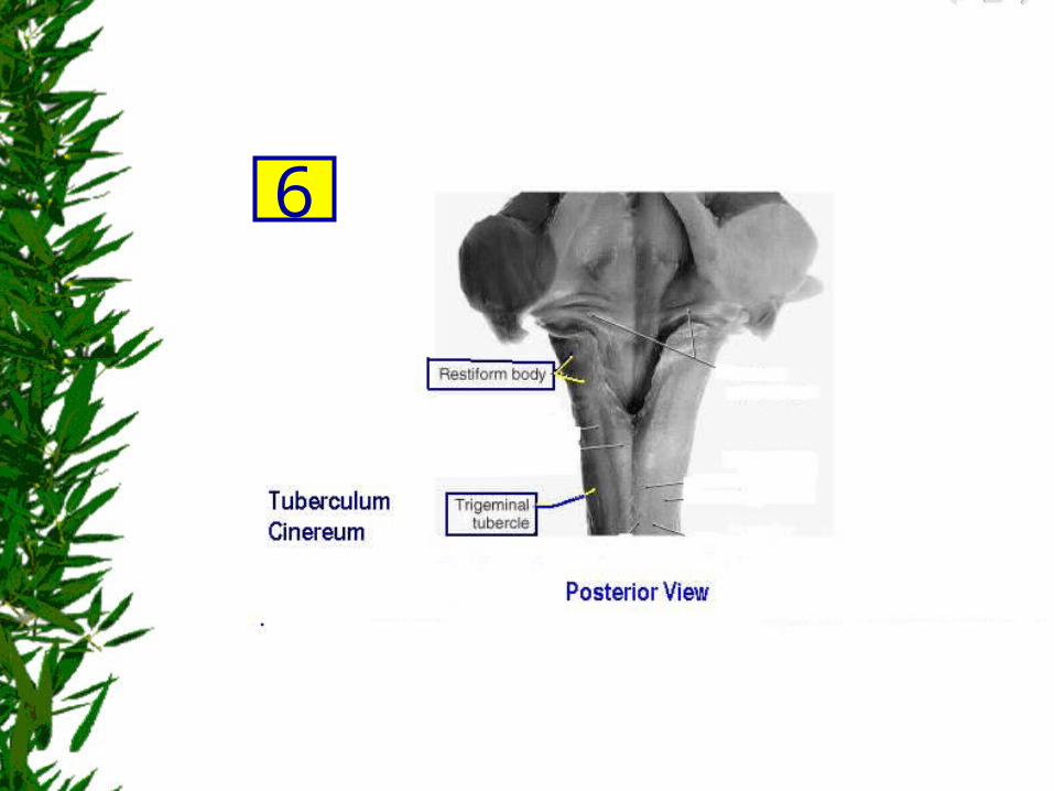
Lateral Medulla
• Fibers of the spinal trigeminal nucleus and tract with superficial location and form – the trigeminal tubercle (tuberculum cinereum)
• Rostral to the obex, these fibers are deeper & internal to a progressively enlarging restiform body.
6
6

Posterior Medulla
• characterized by 1. Gracile & Cuneate fasciculi 2. Gracile & Cuneate tubercles formed by
underlying nuclei.
• the restiform body– prominent elevation Rostrolateral to
the gracile and cuneate tubercles– join the juxtarestiform body In the base
of the cerebellum, to form the inferior cerebellar peduncle.
7

7

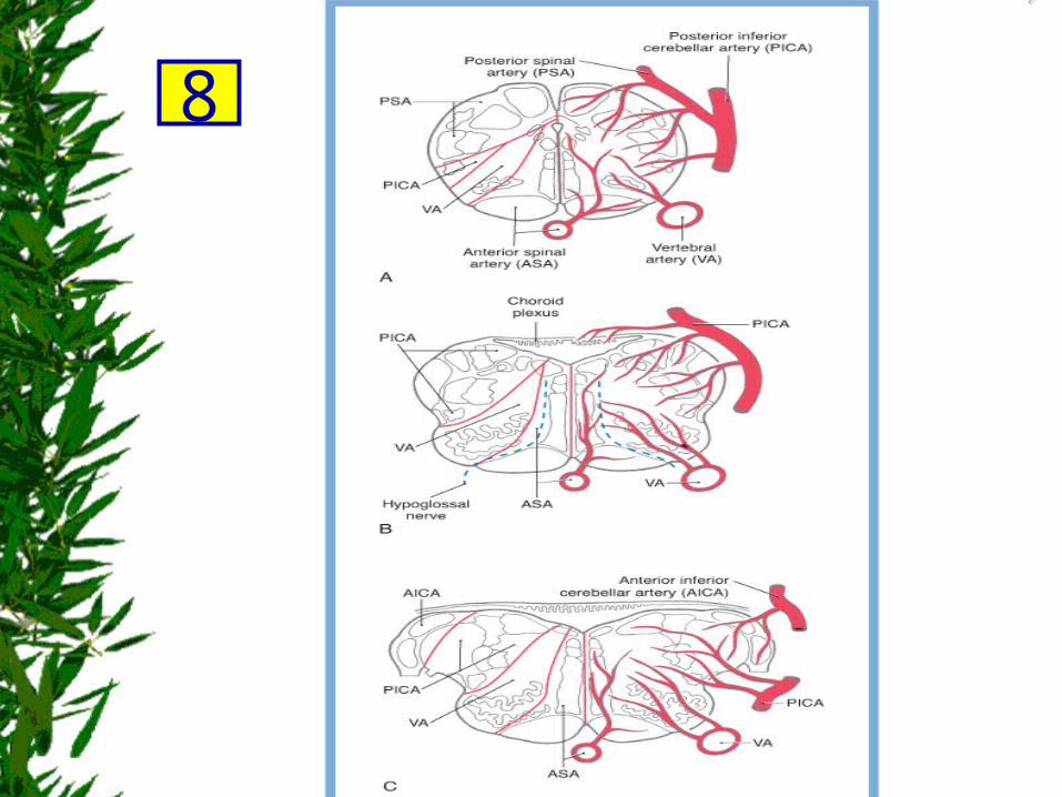
Vasculature
• In general, the entire blood supply arises from branches of the vertebral arteries.
• The exceptions are 1. the choroid plexus out of the foramen of
Luschka
2. and the adjacent cochlear nuclei
served by
Bran. from AICA (bran. from basilar artery)
8

Vasculature
• Medially by – (ASA) anterior spinal artery
• Anterolaterally by – (VA) vertebral small branches from the
• Posterolaterally by– (PSA) posterior spinal artery Caudal to the obex– (PICA) posterior inferior cerebellar artery rostral
to the obex
8
8

Internal Anatomy of the Medulla Ascending Pathways Summary
1. the spinal cord GM (anterolateral system, posterior and anterior spinocerebellar tracts, and so on)
2. posterior root ganglion cells (gracile and cuneate fasciculi) continue into the medulla
anterolateral system spinoreticular fibers,
spinomesencephalic
spinothalamic fibers convey pain and temperature input.
Posterior columns (synapse in the medulla), but medial lemniscus continues rostrally to carry the tactile and vibratory information.
9

9

Descending Pathways Summary
• From1. the cerebral cortex ( corticospinal ) 2. the midbrain (rubrospinal, tectobulbospinal ), 3. the pons ( reticulospinal, vestibulospinal ) 4. The medulla (At this level, the MLF)
10

10
Caudal Medulla: Level of
the Motor Decussation (Pyramidal Decussation) 1/6
• ~ 90% of corticospinal fibers cross the anterior midline to form – the contralateral lateral corticospinal tract of
the cord
11

11

11 Caud. Med. Motor decuss
Caudal Medulla: Level of
the Motor Decussation (Pyramidal Decussation) 2/6
Posteriorly, at this level the Gracile and Cuneate
• nuclei first appear in their respective fasciculi
fasciculi = the posterior (or dorsal) columns, their nuclei = the posterior column nuclei.
12

12
Caud. Med. Motor decuss

Caudal Medulla: Level of
the Motor Decussation (Pyramidal Decussation) 3/6
Laterally, at this level the spinal trigeminal tract
• visible trigeminal tubercle or tuberculum cinereum) is located on the lat. medullary surface. Internal to the spinal trigeminal tract is the spinal trigeminal
nucleus, pars caudalis (Fig. 11-6).
• composed of primary sensory fibers that enter the brain mainly in the trigeminal nerve.
• This tract also receives fibers that originate from cranial nerves VII, IX, and X. (5,7,9,10)
• terminate on the spinal trigeminal nucleus, which, in turn, projects to the contralateral thalamus as the ventral trigeminothalamic tract.
13

13
Caud. Med. Motor decuss

Caudal Medulla: Level of
the Motor Decussation (Pyramidal Decussation) 4/6
Laterally, at this level
1. the anterolateral system ( spinoret./spinomesen./spinothal.) 2. rubrospinal tractare found medial to the superficially located 3. Anterior spinocerebellar tracts 4. Posterior spinocerebellar tracts (Fig. 11-6).
It is important to emphasize that – anterolateral system fibers (conveying pain and temperature input from
the contralateral body) – and spinal trigeminal tract fibers (conveying pain and temperature from
the ipsilateral face) • are located adjacent to each other throughout
the lateral medulla.
14

Anterolateral system
Rubrospinal tract
Anterior spinocerebellar Posterior
spinocerebellar
14Caud. Med. Motor decuss

Caudal Medulla: Level of
the Motor Decussation (Pyramidal Decussation) 5/6
The anterior medulla contains 1. the most rostral part of the accessory nucleus (cranial
nerve XI), 2. the medial motor cell column of C1, 3. and the medial longitudinal fasciculus and
tectobulbospinal system.
• 1 & 2 are seen at this level but do not extend into the medulla.
• At this level, the tectospinal fibers in the tectobulbospinal system are incorporated into the MLF & displaced laterally by the motor decussation compared with more rostral levels.
15

15Caud. Med. Motor decuss

Caudal Medulla: Level of
the Motor Decussation (Pyramidal Decussation) 6/6
• The central gray surrounds the central canal of the medulla and contains
– the caudal extremes of
1. the hypoglossal (XII)
2. and dorsal motor vagal nuclei (X)
16

16Caud. Med. Motor decuss

Caudal Medulla: Level of
the Sensory Decussation1/11
Cells of the posterior column nuclei (gracile and cuneate nuclei)
give rise to axons that swing anteromedially, as internal arcuate fibers,
to cross the midline immediately rostral to the motor decussation (Fig. 11-5).
This crossing of fibers at the midline constitutes the sensory decussation,
so named because it is the point at which a major ascending sensory pathway
(posterior column-medial lemniscus) crosses the midline.
17

17

17Caud. Med. Sens. decuss

Caudal Medulla: Level of
the Sensory Decussation2/11
At this levelThe posterior columns = gracile and cuneate fasciculi
are largely replaced by the gracile and cuneate nuclei
Fibers conveying tactile and vibratory sensations
from lower (the gracile n.) and upper (cuneate n.) levels of the body terminate, respectively, in these nuclei.
The axons of these cells, in turn, form the internal arcuate fibers,
which cross the midline as the sensory decussation to form
the medial lemniscus on the contralateral side
18

Caudal Medulla: Level of
the Sensory Decussation3/11
The posterior columns = gracile and cuneate fasciculi
nuclei
internal arcuate fibers, (sensory decussation)
the medial lemniscus on the contralateral side
18

18Caud. Med. Sens. decuss

Caudal Medulla: Level of
the Sensory Decussation4/11
At this level
• Information from lower extremities (gracile cell axons) is conveyed in the anterior part of the medial lemniscus
• information from the upper extremities (cuneate cell axons) is conveyed in the posterior part of the medial lemniscus (see Fig. 12-13).
• GMLA vs CLUP :– GMLA = Gracille Medial in cord senses Lower extr. Being
Ant. In medial lemn.– CLUP = Cuneate Lateral in cord senses Upper extr. Being
Post. In medial lemn.
19

19

Caudal Medulla: Level of
the Sensory DecussationLateral 5/11
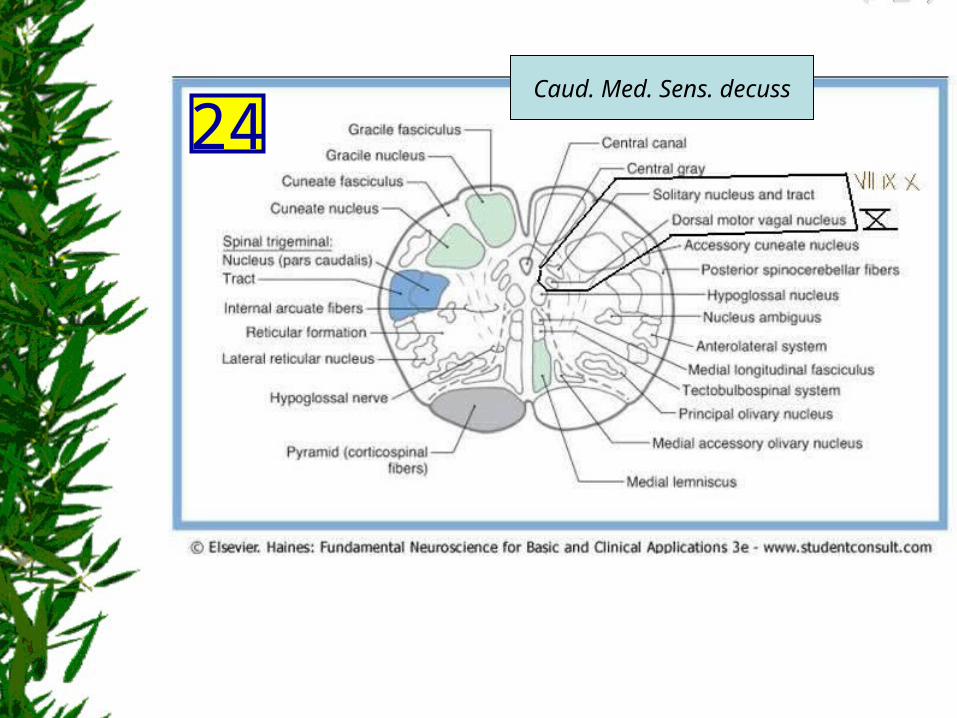
• The spinal trigeminal tract and nucleus (pars caudalis) maintain their position in the lateral medulla.
• caudalis = located caudal to the level of the obex.
20

20 Caud. Med. Sens. decuss
Caudal Medulla: Level of
the Sensory DecussationLateral 6/11
• Just medial to the spinal trigeminal nucleus, a small column of motor neurons, the nucleus ambiguus, appears (Fig. 11-8).
• The axons of these SVE cells travel in the glossopharyngeal (IX) and vagus (X) nerves. 9 & 10
• Fibers of the anterolateral system and rubrospinal tract are located in the anterolateral medulla (Fig. 11-8).
• The lateral reticular nucleus, a distinct cell group adjacent to the anterolateral system, receives spinal input and projects to the cerebellum.
21

21 Caud. Med. Sens. decuss

Caudal Medulla: Level of
the Sensory DecussationAnterior 7/11
• Structures at this level include 1. the pyramid,
2. fibers of the hypoglossal nerve,
3. the caudal end of the inferior olivary complex 1. principal,
2. medial accessory,
3. posterior accessory nuclei.
22

Caudal Medulla: Level of
the Sensory DecussationAnterior 8/11
Hypoglossal (GSE) XII motor neurons innervate
the ipsilateral half of the tongue. These course anterolaterally
along the lateral edge of the medial lemniscus and pyramid.
The inferior olivary nuclei – become larger at more rostral levels, – receive input from a variety of areas – project primarily to the cerebellum.
22

22 Caud. Med. Sens. decuss

Caudal Medulla: Level of
the Sensory DecussationAnterior 9/11
• Internal to the pyramid, and along the midline from anterior to posterior, are 1. the medial lemniscus, 2. tectobulbospinal fibers, 3. medial longitudinal fasciculus (MLF)
• At this level, MLF are characteristically found – adjacent to the midline – anterior to structures of the central gray.
23

23 Caud. Med. Sens. decuss

Caudal Medulla: Level of
the Sensory Decussation10/11
Vagus (GVE )Xthe dorsal motor vagal nucleus
Provide preganglionic parasympathetic fibers to
visceromotor ganglia (autonomic ganglia), the postganglionic fibers of which innervate viscera in the thorax
and abdomen.
The solitary tract and nucleus (7,9,10) receive GVA and SVA (taste) input from cranial nerves
VII, IX, and X.
24
24Caud. Med. Sens. decuss

Caudal Medulla: Level of
the Sensory Decussation11/11
• The 4th ventricle flares open at the level of the obex.
• The area postrema is an emetic (vomiting) center located in the wall of the ventricle at this level.
• Especially noticeable changes are enlargement of
the inferior olivary complex and restiform body.
25

25
OBEX

Midmedullary Level= Rostral to the obex
1/4
dorsally• 4th ventricle medial floor structures
1. the hypoglossal nucleus2. dorsal motor vagal nucleus 3. the vestibular nuclei ( lateral to the sulcus
limitans) medial and inferior (or spinal) They receive input from cranial nerve VIII and interconnect with areas of the brain concerned with balance and eye movement.
4. The solitary tract and nucleus characteristic position immediately inferior to the vestibular nuclei.
26

26
Mid-Medulla

Midmedullary Level2/4
Laterally• Restiform Body
– a prominent elevation on the posterolateral aspect of the medulla
– contains posterior spinocerebellar, cuneocerebellar, olivocerebellar, reticulocerebellar, and other cerebellar afferents.
– join the juxtarestiform body In the base of the
cerebellum, to form the inferior cerebellar peduncle.
• spinal trigeminal tract and nucleus (pars interpolaris / rostral to the obex) are internal to the restiform body
27

27 Mid-Medulla
Midmedullary Level3/4
Laterally
• Other structures are those seen more caudally. 1. the nucleus ambiguus (9 & 10)2. the lateral reticular nucleus 3. the anterolateral system, 4. anterior spinocerebellar tract, 5. rubrospinal tract
• the nucleus ambiguus contribute axons to cranial nerves IX and X, which innervate pharyngeal and laryngeal muscles, including those of the vocal folds.
28

28
Mid-Medulla

Midmedullary Level4/4
Anterolaterally
• the inferior olivary complex – prominent at midmedullary levels – composed of a
1. principal olivary nucleus (large, saccular) 2. medial accessory olivary nuclei (diminutive)3. posterior accessory olivary nuclei (diminutive)
– receive input from a variety of CNS nuclei– project primarily to the contralateral cerebellum (as
olivocerebellar fibers) through the restiform body.
29

29
Mid-Medulla
Rostral Medulla and Pons-Medulla Junction mid medulla structures +
• In the floor of the 4th ventricle, 1. the prepositus (hypoglossal) nucleus 2. the inferior salivatory nucleus
• Replacing hypoglossal and dorsal motor vagal nuclei
1. The prepositus nucleus is – small, flattened cell group– easily distinguished from the hypoglossal nucleus.
2. The GVE cells of the inferior salivatory nucleus are located immediately inferior to
3. the medial vestibular nucleus 4. and medial to the solitary tract and nucleus.
30

Rostral Medulla and Pons-Medulla Junction mid medulla structures +
5. The medial and inferior (or spinal) vestibular nuclei are prominent at this level and are joined, in this plane of section, by
6. the posterior and anterior cochlear nuclei
7. the restiform body with 8. the spinal trigeminal tract and the pars oralis
Medial to it (rostral to the level of the hypoglossal nucleus)
30

30Upper-Medulla

Rostral Medulla and Pons-Medulla Junction
• Facial motor nucleus (SVE cells) appears anterolaterally
• trapezoid body and superior olivary nucleus (auditory information) appear adjacent to the facial nucleus and the spinal trigeminal tract and nucleus.
31

Rostral Medulla and Pons-Medulla Junction
• The inferior olivary complex disappears, and the central tegmental tract, one source of input to the inferior olive, appears about where the latter cell group was located (Fig. 11-14).
• medial lemniscus shift anterolaterally posteroanterior orientation At the pons-medulla junction, oriented obliquely At the mid pons, it is horizontal
31

31Upper-Medulla

Reticular and Raphe Nuclei
• Reticulum = Latin word "little net" denotes mesh-like structures, diffuse and ill defined
• Raphe is a Greek word for "suture" or "seam." Thus, bilaterally symmetrical cell groups located directly adjacent to the midline.
32
Reticular and Raphe Nuclei
• Medullary RF function in the control of heart rate and respiration.
Consequently,
a sudden onset of central apnea,
indicating damage to these respiratory areas,
is often a prime early sign of medullary compression.
32

Reticular and Raphe Nuclei
• Raphespinal fibers – are especially important for the inhibition of pain
transmission in the posterior horn of the spinal cord.
• Serotonin is the principal neurotransmitter • ? cholecystokinin-containing cells • ? enkephalin
32

32

Internal Vasculature of the Medulla and Medullary Syndromes
• from branches of the – VA
1. ASA ( all Ant.med. struct.)
2. PICA (Rostral post.lat.) 3. & its branch PSA (Caudal post) .
33
ASA & the medial medullary syndrome.
• ASA serves Medial structures of the medulla at all levels, including 1. the pyramid, 2. medial lemniscus, 3. hypoglossal nucleus and roots
• Occlusion of these to one side of the medial medulla may result in a pattern of deficits characteristic of the medial medullary syndrome.
33

ASA & the medial medullary syndrome.
• also known as the Dejerine syndrome. • The deficits and corresponding structures
damaged in this syndrome include a 1. contralateral hemiparesis (pyramidal and
corticospinal damage),
2. contralateral loss of proprioception and vibratory sense (medial lemniscus),
3. deviation of the tongue to the ipsilateral side when protruded (hypoglossal root or nucleus injury).
33

PSA & Posterior medullary syndrome
• The posterior medulla caudal to the obex served by branches of the PSA
• Major structures in this area include 1. the posterior column (gracile and cuneate) nuclei 2. and the spinal trigeminal tract and nucleus.
• Although rare, may produce 1. an ipsilateral loss of proprioception and vibratory sense on the
body (posterior columns and nuclei) 2. ipsilateral loss of pain and temperature sensation from the
face (spinal trigeminal tract).
33

33

PICA and Lateral medullary syndrome
• PICA serves the entire posterolateral medulla Rostral to the obex
• Included in the territory served by this vessel are
1. Anterolateral system, 2. Spinal trigeminal tract and nucleus, 3. Vestibular nuclei,4. Solitary tract and nucleus, 5. Nucleus ambiguus.
34
PICA and Lateral medullary syndrome
• Vascular insufficiency of PICA (or blockage of one VA) gives rise to a characteristic set of sensory and motor deficits commonly called
1. lateral medullary syndrome, 2. PICA syndrome, or 3. Wallenberg syndrome.
34
PICA and Lateral medullary syndrome
• The deficits
1. contralateral loss of pain and temperature sensation from the body (anterolateral system),
2. ipsilateral loss of pain and temperature sensation from the face (spinal trigeminal tract and nucleus),
3. some vertigo and nystagmus (vestibular nuclei), 4. loss of taste from the ipsilateral half of the tongue (solitary tract
and nucleus), and5. hoarseness and dysphagia (nucleus ambiguus or roots of
cranial nerves IX and X) . 6. ? Horner syndrome (injury to hypothalamospinal fibers
descending through the lateral medulla).
34

34
Tonsillar Herniation 1/3
• it may have a profoundly negative impact on the medulla.
• The causes vary, examples include 1. posterior fossa mass: tumor / hrg2. posterior fossa surgery. 3. lumbar puncture in a pt with a mass lesion.

Tonsillar Herniation 2/3
• cerebellar tonsil downward extrusion into, and through, the foramen magnum rapid ICP.
• two mechanisms. 1. mechanical injury ( compression) 2. Vascular injury (infarction)

Tonsillar Herniation 3/3
• The major concern in acute herniation is – damage to the ventrolateral reticular area of the medulla, which
contains neurons that influence and control heart rate and respiration.
• Acute Manifestations: 1. a sudden change in heart rate and respiration (Cheyne-Stokes with
intermittent apnea), 2. increase in blood pressure (hypertension), 3. hyperventilation, 4. rapidly decreasing levels of consciousness, 5. and death.
– chronic Manifestations:– slowly the patient suffers minimal neurologic consequences.

Synopsis of Clinical Points 1
• 7/12 cranial nerves exit from the medulla or medulla-pons junction; tumors in this confined space usually affect these nerves .
• Lesions of the motor (pyramidal) decussation may result in bilateral weakness of the extremities .
• Deficits in the medial medullary (or Dejerine) syndrome reflect
damage to the structures in this area; these deficits are: 1. ipsilateral deviation of the tongue, 2. contralateral weakness of the extremities, 3. and contralateral loss of proprioception/position sense .
• Occlusion of the anterior spinal artery or its penetrating branches may result in different patterns of deficits .

Synopsis of Clinical Points 2
• the lateral medullary (or Wallenberg) syndrome Has characteristic Deficits.
1. an alternating hemianesthesia, 2. vertigo, 3. nystagmus, 4. dysarthria, 5. and dysphagia .
may be seen following occlusion of 1. the vertebral artery 2. its major branch, the PICA .
• Tentorial herniation may result from:1. rapidly expanding lesion in the fourth ventricle 2. lumbar puncture in a pt with a supratentorial mass .
• Tonsillar herniation compresses the medulla damages cardiac and respiratory centers . Cheyne-Stokes respiration and apnea .

THANK YOU