FunctionsFunctions Support Surrounding Tissue Protect Vital Organs and Soft Tissue Provide an...
83
-
Upload
dinah-burns -
Category
Documents
-
view
216 -
download
0
Transcript of FunctionsFunctions Support Surrounding Tissue Protect Vital Organs and Soft Tissue Provide an...


FunctionsFunctionsFunctionsFunctions
• Support Surrounding Tissue
• Protect Vital Organs and Soft Tissue
• Provide an Attachment Place for Muscle– Bones act as levers to produce
movement

FunctionsFunctions
• Produce Red Blood Cells–Bone Marrow
• Storage of Minerals
–Calcium & Phosphorus
• Fat Storage


Haversian Canal
Lacuna with Osteocyte
Canaliculi

Microscopic Anatomy of Compact BoneMicroscopic Anatomy of Compact Bone
MatrixMatrix– 25% water, 25% collagen fibers, 50%
mineral salts
CellsCells• Osteogenic cells in periosteum
• Osteoblasts
– Secrete collagen fibers and build matrix
– With time, they become

Microscopic Anatomy of Compact BoneMicroscopic Anatomy of Compact Bone
Cells (continued)Cells (continued)
• Osteocytes that maintain bone
• Osteoclasts digest bone matrix for normal bone turnover
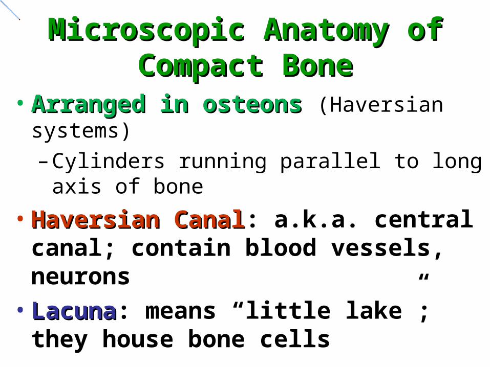
Microscopic Anatomy of Microscopic Anatomy of Compact BoneCompact Bone
• Arranged in osteons Arranged in osteons (Haversian systems)
– Cylinders running parallel to long axis of bone
• Haversian CanalHaversian Canal: a.k.a. central canal; contain blood vessels, neurons
• LacunaLacuna: means “little lake”; they house bone cells
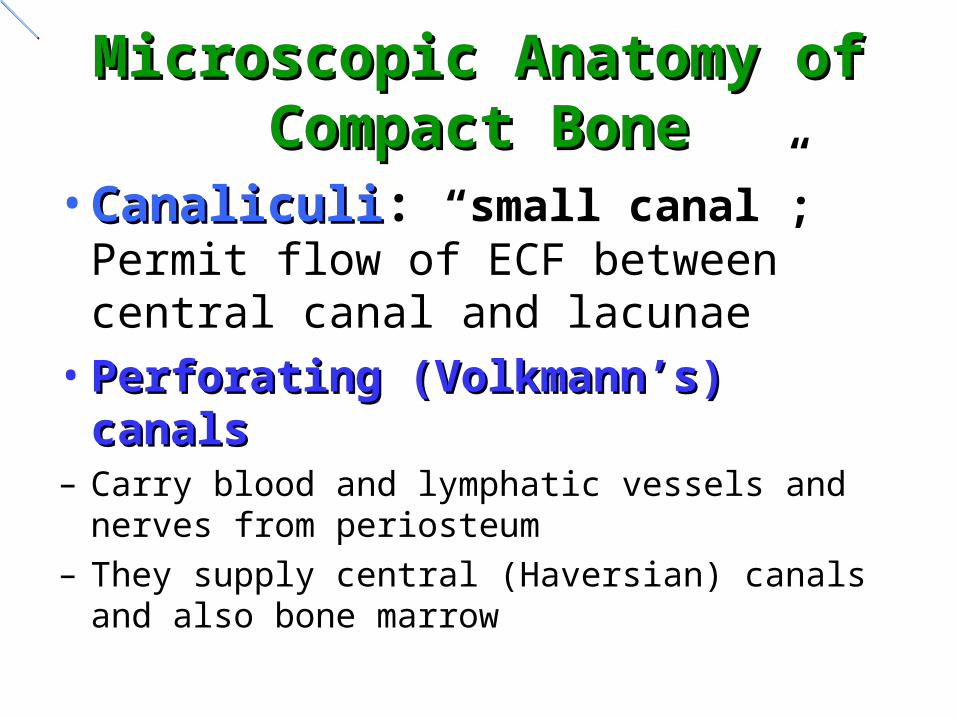
Microscopic Anatomy of Microscopic Anatomy of Compact BoneCompact Bone
• CanaliculiCanaliculi: “small canal”; Permit flow of ECF between central canal and lacunae
• Perforating (Volkmann’s) canalsPerforating (Volkmann’s) canals– Carry blood and lymphatic vessels and nerves
from periosteum – They supply central (Haversian) canals and also
bone marrow


Spongy BoneSpongy Bone
• Not arranged in osteons
• Irregular latticework of trabeculae – These contain lacunae with osteocytes and
canaliculi
• Spaces between trabeculae may contain red bone marrow
• Spongy bone is lighter than compact bone, so reduces weight of skeleton


Classification of BonesClassification of BonesClassification of BonesClassification of Bones
1.1. Long BonesLong Bones
a. Examples: femur, radius, ulna
b. Diaphysis: Shaft; composed of compact bone
c. Epiphysis: End of long bone; composed of spongy bone;

Classification of BonesClassification of BonesClassification of BonesClassification of Bones
2. Short BonesShort Bones
a. Cube-shaped; spongy bone surrounded by a thin layer of compact bone
b. Includes carpals and tarsals

Classification of BonesClassification of BonesClassification of BonesClassification of Bones
3. Flat BonesFlat Bonesa. Spongy bone within two plate-like
coverings of compact boneb. Protect; provide surface area for muscles;
ribs, sternum, skull bones4. Irregular BonesIrregular Bones
a. Include vertebrae, scapula

Classification of BonesClassification of BonesClassification of BonesClassification of Bones
5. Sesamoid BonesSesamoid Bones• small, flat, and shaped somewhat like a
sesame seed
• Develop inside tendons and are most commonly found near joints at the knees, the hands, and the feet
• Example: patella

Classification of BonesClassification of BonesClassification of BonesClassification of Bones
6. Sutural (Sutural (Wormian)Wormian) BonesBones• small, flat, irregularly shaped bones
between the flat bones of the skull
• individual variations in the number, shape, and position
• range in size from a grain of sand to the size of a quarter


Anatomy of A Long BoneAnatomy of A Long BoneAnatomy of A Long BoneAnatomy of A Long Bone
1. DiaphysisDiaphysis
a. Made of compact bone; covered by a tough membrane, the periosteum
b. Central Cavity called the Medullary cavity
1) In children: filled with red marrow
2) Adults: marrow replaced by fat
3) Inner lining = endosteum

Anatomy of A Long BoneAnatomy of A Long BoneAnatomy of A Long BoneAnatomy of A Long Bone
2. EpiphysisEpiphysis (Plural: epiphyses)
a. The end(s) of long bone
b. Made of a thin layer of compact bone on the outside
c. Spongy bone on the inside

Anatomy of A Long BoneAnatomy of A Long Bone
d. Enlarged for muscle attachment, joint formation
e. Site of hemopoiesis in adults (red marrow)
f. Articular cartilage covers surface (hyaline)

Anatomy of A Long BoneAnatomy of A Long BoneAnatomy of A Long BoneAnatomy of A Long Bone
3. Epiphyseal DiskEpiphyseal Disk (Line)
a. Flat plate of hyaline cartilage
b. Site of longitudinal bone growth
(length-wise)
c. At the end of growth, cartilage is replaced by bone; process is
called fusion

Anatomy of A Long BoneAnatomy of A Long Boned. Doctors can predict growth from x-rays
4. PeriosteumPeriosteum: Membrane covering the diaphysis
a. Essential in bone growth, repair, and nutrition
b. Contains C.T., osteogenic cells and osteoblasts

Typical Long Bone StructureArticular cartilageEpiphyseal lines
Spongy bone
Spongy bone (red marrow)
Epiphysis (proximal)
Diaphysis
Epiphysis (distal)
Compact bone
Medullary cavity
Yellow marrow
Periosteum

Structure of Long Bone
Figure 6.3a


Copyright 2010, John Wiley & Sons, Inc.
Bone Dynamics and TissueInteractions Animation
Bone Dynamics and Tissue
You must be connected to the internet to run this animation.

Bone FormationBone Formation• Known as ossification
• Timeline– Initial bone development in embryo and fetus– Growth of bone into adulthood– Remodeling: replacement of old bone– Repair if fractures occur
• Mesenchyme (early connective tissue) model – This initial “skeleton” model will be replaced by
bone tissue beginning at 6 weeks of embryonic life

Bone FormationBone Formation• Two different methods of ossification each
result in similar bone tissue– Intramembranous: bone forms within sheets of
mesenchyme that resemble membranes • Only a few bones form by this process: flat bones of the
skull, lower jawbone (mandible), and part of clavicle (collarbone)
– Endochondrial: mesenchyme forms hyaline cartilage which then develops into bone
• All other bones form by this process

Intramembranous OssificationIntramembranous OssificationFour steps1.Development of ossification center
Mesenchyme cells osteogenic osteoblastsOsteoblasts secrete organic matrix
2.Calcification: cells become osteocytes In lacunae they extend cytoplasmic processes to each otherDeposit calcium & other mineral salts
3. Formation of trabeculae (spongy bone)Blood vessels grow in and red marrow is formed
4.Periosteum covering the bone forms from mesenchyme

Endochondrial OssificationEndochondrial Ossification• Six Steps
1. Formation of cartilage model of the “bone” • As mesenchyme cells develop into chondroblasts
2. Growth of cartilage model• Cartilage “bone” grows as chondroblasts secrete
cartilage matrix• Chondrocytes increase in size, matrix around them
calcifies• Chondrocytes die as they are cut off from nutrients,
leaving small spaces (lacunae)

Endochondrial OssificationEndochondrial Ossification3. Primary ossification center
– Perichondrium sends nutrient artery inwards into disintegrating cartilage
– Osteogenic cells in perichondrium become osteoblasts that deposit bony matrix over remnants of calcified cartilage spongy bone forms in center of the model
– As perichondrium starts to form bone, the membrane is called periosteum

Endochondrial OssificationEndochondrial Ossification4. Medullary (marrow) cavity
– Spongy bone in center of the model grows toward ends of model
– Octeoclasts break down some of new spongy bone forming a cavity (marrow) through most of diaphysis
– Most of the wall of the diaphysis is replaced by a collar of compact bone

Endochondrial OssificationEndochondrial Ossification5. Secondary ossification center
– Similar to step 3 except that nutrient arteries enter ends (epiphyses) of bones and osteoblasts deposit bony matrix spongy bone forms in epiphyses from center outwards
– Occurs about time of birth
6. Articular cartilage and epiphyseal cartilage– Hyaline cartilage at ends of epiphyses becomes
articular cartilage– Epiphyseal (growth) plate of cartilage remains
between epiphysis and diaphysis until bone growth ceases

1
Articular cartilage
Spongy bone
Epiphyseal plate
Secondaryossificationcenter
Nutrientartery and vein
Uncalcifiedmatrix
Epiphysealartery andvein
Formation of articular cartilageand epiphyseal plate
Development of secondaryossification center
Development ofcartilage model
Development ofprimary ossificationcenter
Development ofthe medullarycavity
Growth ofcartilage model
2 3 4
5 6
Hyalinecartilage
Uncalcifiedmatrix
CalcifiedmatrixPeriosteum(covering compact bone)
Uncalcifiedmatrix
Calcifiedmatrix
Medullarycavity
Nutrientartery and vein
Nutrientartery
Perichondrium
Proximalepiphysis
Distalepiphysis
Diaphysis
Periosteum
Primaryossificationcenter
Spongybone

Growth In LengthGrowth In Length• Chondrocytes divide and grow more cartilage
on epiphyseal side of the epiphyseal plate • Chondrocytes on the diaphyseal side die and
are replaced by bone• Therefore bone grows from diaphyseal side
towards epiphyseal side• Growth in length stops between 18-25 years;
cartilage in epiphyseal plate is completely replaced by bone (epiphyseal line)

Growth In WidthGrowth In Width• As bones grow in length, they must also grow
in thickness (width)– Perichondrial osteoblasts osteoblasts lay
down additional lamellae of compact bone– Simultaneously, osteoclasts in the endosteum
destroy interior bone to increase width of the marrow
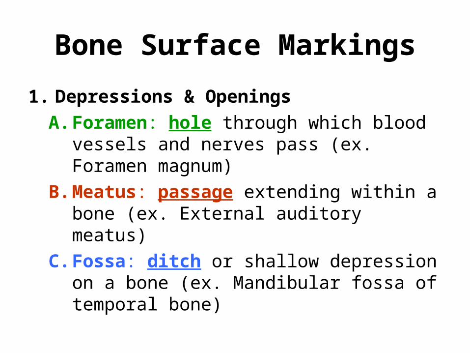
Bone Surface Markings
1. Depressions & Openings
A. Foramen: hole through which blood vessels and nerves pass (ex. Foramen magnum)
B. Meatus: passage extending within a bone (ex. External auditory meatus)
C. Fossa: ditch or shallow depression on a bone (ex. Mandibular fossa of temporal bone)

2. Processes that form joints
A. Condyle: large rounded prominence forming a joint
B. Head: rounded projection that forms a joint (ex. Head of femur)
C. Facet: smooth, flat surface (ex. Facet of vertebrae)

3. Processes to which tendons & ligaments attach
A. Tuberosity: large rounded projection with a rough surface (ex. Deltoid tuberosity of humerus)
B. Spinous process: sharp, slender projection (on vertebrae)
C. Crest: Prominent ridge (ex. illiac crest of pelvic bone)
D. Trochanter: large, blunt projection (only on femur)


Bone Quizzes
1. Functions, microscopic anatomy, classification
2. Long bone anatomy, bone “markings”
3. Bone homeostasis, articulations
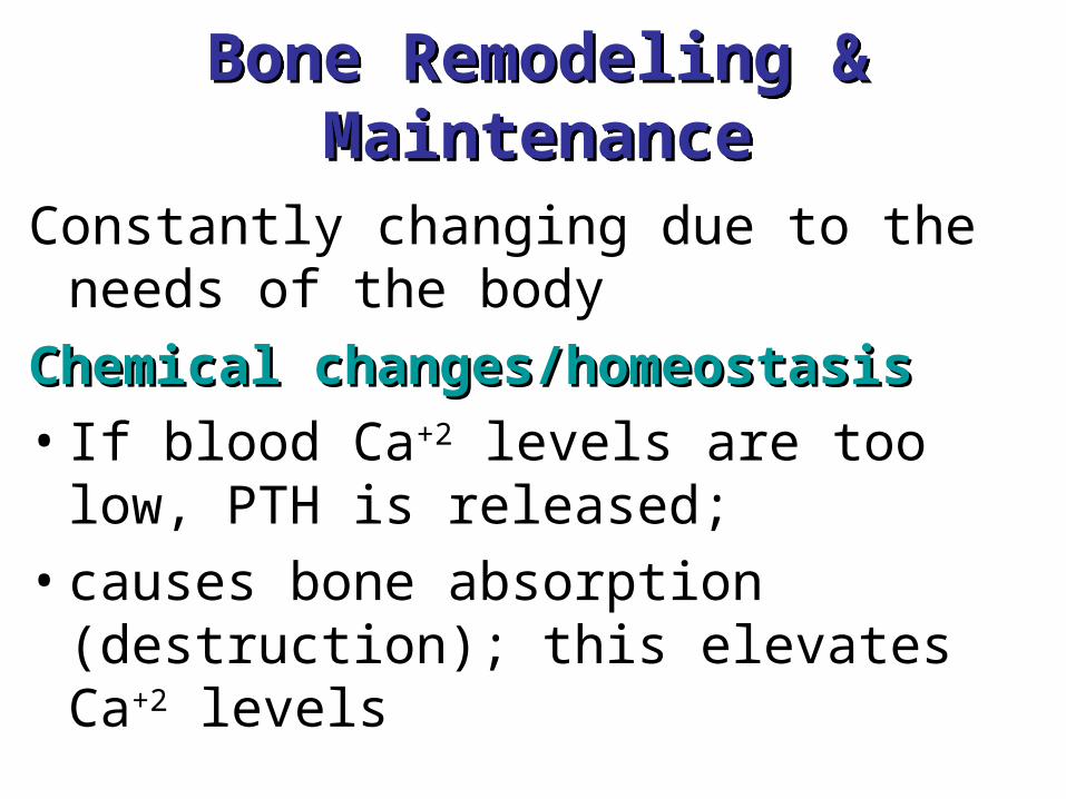
Bone Remodeling & MaintenanceBone Remodeling & Maintenance
Constantly changing due to the needs of the body
Chemical changes/homeostasisChemical changes/homeostasis
• If blood Ca+2 levels are too low, PTH is released;
• causes bone absorption (destruction); this elevates Ca+2 levels

Bone Remodeling & MaintenanceBone Remodeling & Maintenance
• PTH, parathyroid hormone is produced by the parathyroid gland
• If blood calcium levels are too high,
calcitonin is released; causes Ca+2 to be deposited onto bone;
• Ca+2 levels in the blood then drop

Bone Remodeling & MaintenanceBone Remodeling & MaintenanceBone Remodeling & MaintenanceBone Remodeling & Maintenance
• Calcitonin is a hormone produced by the thyroid gland
Mechanical StressesMechanical Stresses
• Gravity
• Muscle Tension (from exercise)

Homeostasis of Bone Tissue
Vitamin D1. Stimulates absorption of calcium in
small intestine2. Promotes reabsorption of Calcium by
the kidneys3. Inihibits PTH production

Importance of Ionic Calcium in the Body
• Calcium is necessary for:
–Transmission of nerve impulses
–Muscle contraction
–Blood clotting
–Secretion by glands and nerve cells
–Cell division

ArticulationsArticulationsArticulationsArticulationsArticulations & JointsArticulations & Joints; Functions:Functions:
• Hold bones together (includes ligaments)
• The structure of a joint determines the type of movement that may occur
• Each joint reflects some compromise between strength and mobility.

ArticulationsArticulations
Types of JointsTypes of Joints
• SynarthrosesSynarthroses: “without joint”; joints that allow no movement– Bones connected by fibrous or cartilage
tissue; STRENGTH, no mobility
– There are 4 types; we need to know only one
– Example: skull sutures


ArticulationsArticulationsArticulationsArticulations
AmphiarthrosesAmphiarthroses: allow slight movement; compromise between mobility and strength
• Bones are connected by collagen fibers or cartilage
• At a syndesmosis (desmos, a band or ligament), bones are connected by a ligament; tibia/fibula
• At a symphysis, the bones are separated by a pad of fibrocartilage; pubic symphysis


ArticulationsArticulations
DiarthrosesDiarthroses: freely moveable joints; also called synovial joints
• surrounded by a fibrous articular capsule, and a synovial membrane lines the cavity
• Synovial joints may have a variety of accessory structures, including pads of cartilage or fat, ligaments, tendons, and bursas

ArticulationsArticulations
Diarthroses (continued)Diarthroses (continued)
• Nonaxial jointsNonaxial joints: allow only sliding movement to occur between bones:
• Sometimes called gliding joints
–ankle, wrist

ArticulationsArticulations
Diarthroses (continued)Diarthroses (continued)
• Uniaxial jointsUniaxial joints: allow mvt in one plane–hinge type: elbow– pivot type: radius/ulna
• Biaxial joints:Biaxial joints: allow mvt in two planes–knuckles

Articulations
Diarthroses (continued)Diarthroses (continued)
• Multiaxial:Multiaxial: allow movement in all planes–ball & socket: shoulder and hip

ArticulationsArticulations
All diarthrotic joints have 4 distinguishing characteristics:
(1) articular cartilage
(2) fibrous articular (joint) capsule
(3) joint cavity
(4) reinforcing ligaments


• SkullSkull–Cranial bones (8) and facial bones
(14)
• Vertebral ColumnVertebral Column–5 regions–4 curves
• Bony ThoraxBony Thorax–Ribs and sternum


• Air-filled, mucous-membrane lined Air-filled, mucous-membrane lined cavities within certain bonescavities within certain bones
• 4 pairs found in the4 pairs found in the ethmoid, frontal, ethmoid, frontal, maxillae, and sphenoid bonesmaxillae, and sphenoid bones
• Lighten the boneLighten the bone
• Warm the airWarm the air
• Provide resonance in speechProvide resonance in speech


Upper Limb:Upper Limb:• Pectoral (shoulder) GirdlePectoral (shoulder) Girdle
–Scapula and clavicleScapula and clavicle
• Bones of the arms and handsBones of the arms and hands


Lower Limbs:Lower Limbs:• Pelvic (hip) GirdlePelvic (hip) Girdle
–Os coxae (ileum, ischium, Os coxae (ileum, ischium, pubis)pubis)
• Bones of legs and feetBones of legs and feet


Comparison of Male and Female Pelves
Table 7.4.2