Functions of the Nervous System 1. Sensory input Monitor changes occurring inside and outside the...
25
Functions of the Nervous Functions of the Nervous System System 1. Sensory input Monitor changes occurring inside and outside the body 2. Integration Processes and interprets sensory input and decide what to be done Mainly occurs in brain and spinal cord 3. Motor output A response to integrated stimuli by activating effector organs – muscles and glands
-
Upload
rodger-bond -
Category
Documents
-
view
247 -
download
0
Transcript of Functions of the Nervous System 1. Sensory input Monitor changes occurring inside and outside the...

Functions of the Nervous SystemFunctions of the Nervous System
1. Sensory input
Monitor changes occurring inside and outside the body
2. Integration
Processes and interprets sensory input and decide what to be done
Mainly occurs in brain and spinal cord
3. Motor output
A response to integrated stimuli by activating effector organs – muscles and glands

Functions of the Nervous SystemFunctions of the Nervous System
An example of how the nervous system uses all three functions together is when you are driving and see a red light ahead (sensory input), your nervous system integrates this information (red light means “stop”, and your foot goes for the brake pedal (motor output)

Organization of the Nervous SystemOrganization of the Nervous System Classified based on structure and function
Structure
Brain
Spinal cord
Nerves
Function
Sensory
Eyes, ears, nose
Motor
Muscles and glands

Structural ClassificationStructural Classification
Central nervous system (CNS)
Brain and spinal cord
Integrating and command center
Peripheral nervous system (PNS)
Cranial nerves and spinal nerves
Communication lines between the CNS and the rest of the body
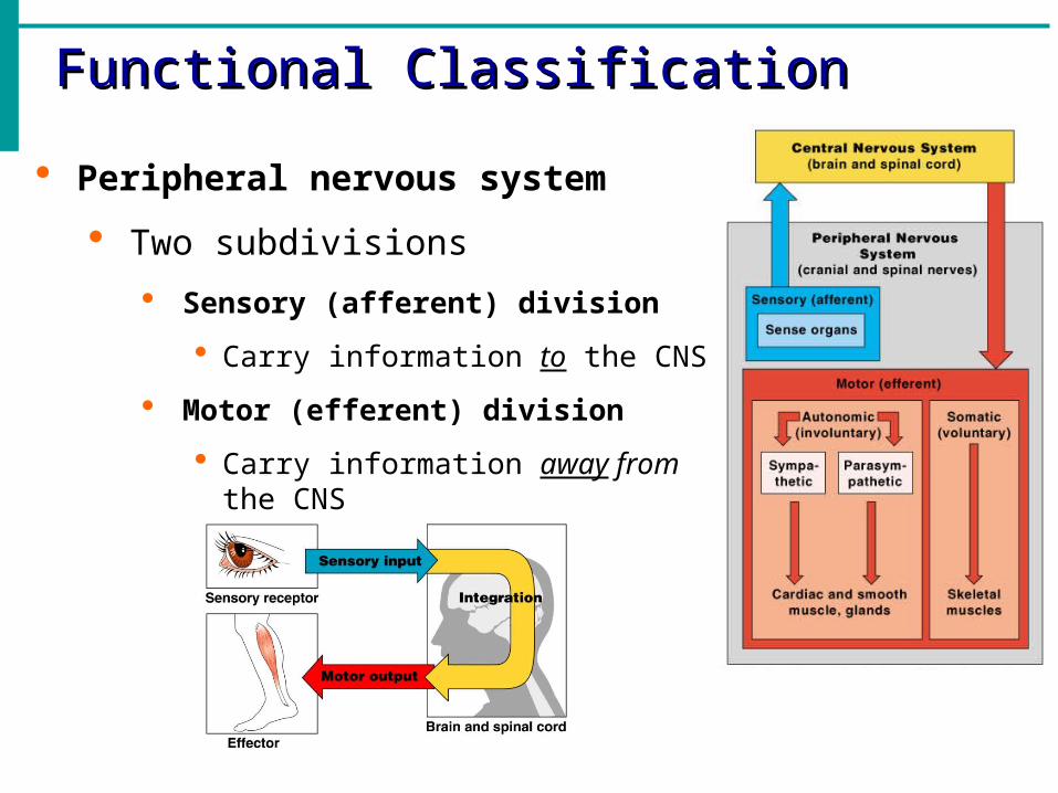
Functional ClassificationFunctional Classification
Peripheral nervous system
Two subdivisions
Sensory (afferent) division
Carry information to the CNS
Motor (efferent) division
Carry information away from the CNS

Functional ClassificationFunctional Classification
Motor (efferent) division Two subdivisions
Somatic nervous system Voluntary and conscious control Skeletal muscles
Autonomic nervous system Involuntary and subconscious Smooth muscles, cardiac muscles and glands

Functional ClassificationFunctional Classification Autonomic Nervous System
Two subdivisions Sympathetic
Fight or flight Parasympathetic
Rest and digest

Nervous TissueNervous Tissue
Two principal types of cells
Support cells (neuroglia) Support, insulate and protect the
neurons
Cannot transmit nerve impulses
Continue to divide
4 in the CNS and 2 in the PNS
Neurons
Structural units of the nervous system
Transmit electrical impulses from one area of the body to another area using neurotransmitters
Cannot divide = amitotic

Support Cells (Neuroglia) in CNSSupport Cells (Neuroglia) in CNS
Astrocytes Abundant, star-shaped cells
Anchor and brace neurons
Form barrier and make exchanges between capillaries and neurons
Control the chemical environment of the brain by cleaning up potassium ions and neurotransmitters
Microglia Small ovoid cells with
thorny processes
Spider-like phagocytes
Dispose of debris

Support Cells (Neuroglia) in CNSSupport Cells (Neuroglia) in CNS
Ependymal cells Line central cavities of the
brain and spinal cord
Cells are squamous and columnar shaped and ciliated
Circulate cerebrospinal fluid
Oligodendrocytes
Produce myelin sheath (fatty insulation) around nerve fibers

Support Cells (Neuroglia) in PNSSupport Cells (Neuroglia) in PNS
Satellite cells Surround neuron cell bodies located in the PNS
Protection and cushioning neurons
Similar to astrocytes in CNS
Schwann cells Surround and form myelin sheath in the PNS
Help with regeneration of damaged peripheral nerve fibers

NeuronsNeurons
Structural unit of the nervous system
Highly specialized cells that conduct messages in the form of impulses from one part of the body to another
Special characteristics
Extreme longevity (about 100 years)
Amitotic (cannot divide)
High metabolic rate
Vary in structure but all have a cell body and one or more slender processes
Plasma membrane is the site of electrical signaling

NeuronsNeurons Major regions of neurons
Cell body (soma)
Nucleus and metabolic center
Most are located in the CNS
Clusters of cell bodies in the CNS are called nuclei
Lie along the nerves in the PNS are called ganglia
Processes (dendrites and axons) Arm like processes extend from the body of all neurons
Brain and spinal cord contain both cell bodies and their processes
PNS consists mainly of neuron processes
Bundles of neuron processes are called tracts (CNS) or nerves (PNS)
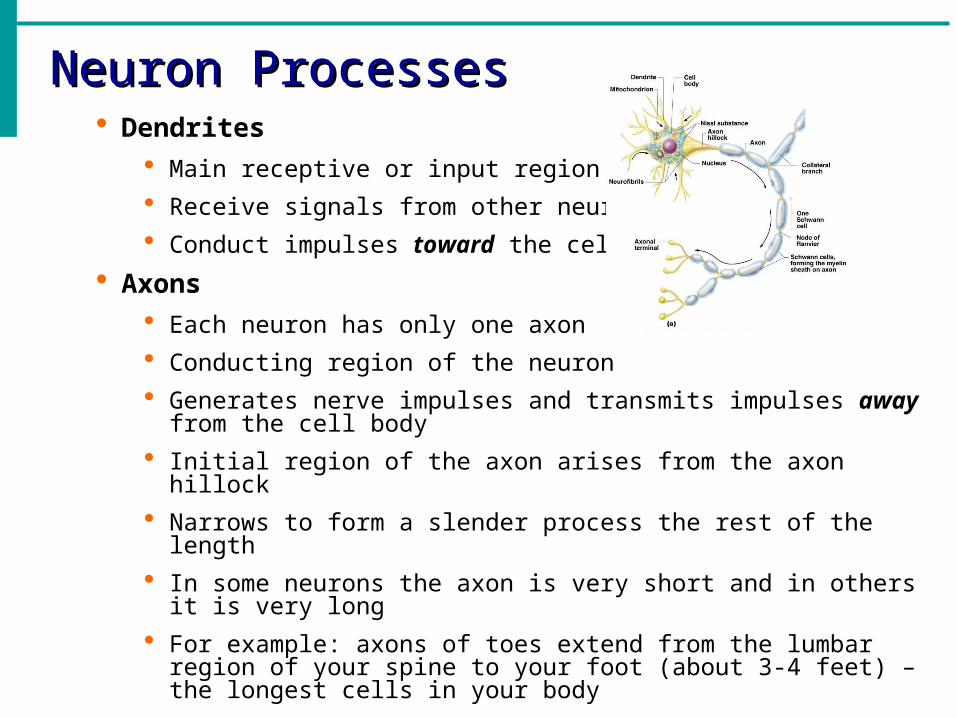
Neuron ProcessesNeuron Processes Dendrites
Main receptive or input region
Receive signals from other neurons
Conduct impulses toward the cell body
Axons Each neuron has only one axon
Conducting region of the neuron
Generates nerve impulses and transmits impulses away from the cell body
Initial region of the axon arises from the axon hillock
Narrows to form a slender process the rest of the length
In some neurons the axon is very short and in others it is very long
For example: axons of toes extend from the lumbar region of your spine to your foot (about 3-4 feet) – the longest cells in your body

Axons and axonal terminalsAxons and axonal terminals
Axons can branch many times but all end in axon terminals
Axonal terminals
Knoblike distal endings also called the secretory region
When impulses reaches the terminal it causes neurotransmitters to be released
Neurotransmitters either excite or inhibit neurons
Neurons never touch other neurons
Separated from the next neuron by a gap
Synaptic cleft
Gap between adjacent neurons

Axon CoveringsAxon Coverings Many axons are covered with a whitish, fatty,
segmented myelin sheath
Myelin
Covering that protects and insulates the axons and increases the speed of transmission of nerve impulses
Myelin sheath
A tight coil of wrapped membranes encloses the axon

Axon CoveringsAxon Coverings Myelin sheaths
Peripheral nervous system
Formed by Schwann cells which wrap themselves around the axon in jelly-roll fashion
Central nervous system
Formed by oligodendrocytes
Nodes of Ranvier
Adjacent Schwann cells and oligodendrocytes do not touch each other so there are gaps in the myelin sheath
Myelinated fibers conduct nerve impulses rapidly
Unmyelinated fibers conduct impulses slowly

Axon CoveringsAxon Coverings
Multiple Sclerosis (MS)
Myelin sheath in the CNS are damaged and demyelination occurs
Affects the ability of nerve cells to communicate with each other
Caused by an autoimmune disease where the body’s own immune system attacks and damages the myelin
Electrical signals are slowed or stopped from reaching different parts of the body.
Visual disturbances, problems controlling muscles, speech disturbances, and urinary incontinence
Person loses ability to control his/her muscles
Cause is unknown and there is no cure

Neuron Cell Body LocationsNeuron Cell Body Locations
Most are found in the central nervous system
White matter
Dense collections of myelinated fibers
Gray matter
Unmyelinated fibers and cell bodies
Nuclei
Clusters of cell bodies within the white matter of the central nervous system
Outside the CNS
Ganglia
Collections of cell bodies
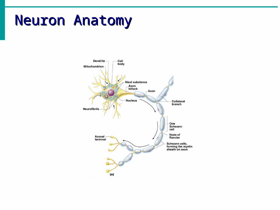
Neuron AnatomyNeuron Anatomy

Neuron ClassificationNeuron Classification
Neurons classified based on their structure and function

Structural Classification of NeuronsStructural Classification of Neurons Multipolar neurons
Three or more processes
One axon and the rest dendrites
Most common neuron type (motor and interneurons)
Major neuron type in CNS
Bipolar neurons
Two processes
An axon and a dendrite from opposite sides of the cell body
Rare, found in special sensory organs
Unipolar neurons
Have a short single process leaving the cell body
Mainly found in ganglia of PNS (sensory neurons)

Functional ClassificationFunctional Classification
Neurons classified based on the direction the nerve impulse travels in relation to the CNS
Sensory neurons
Interneurons
Motor neurons

Functional Classification of NeuronsFunctional Classification of Neurons Sensory (afferent) neurons
Transmit impulses towards the CNS
Sensory receptors in the skin or internal organs
Cutaneous sense organs (Meissner’s and Pacinian Corpuscles)
Proprioceptors (detect stretch or tension)
Motor (efferent) neurons
Transmit impulses away from the CNS
Muscles or glands
Association (interneuron)
Found between motor and sensory neurons
In neural pathways in the CNS
Make up 99% of the neurons of the body

Functional Classification of NeuronsFunctional Classification of Neurons
Amyotrophic lateral sclerosis (ALS) “Lou Gehrig’s Disease”
Disease of the motor neurons in the CNS that control voluntary movements
Motor neurons degenerate or die and can no longer send messages to muscles
Condition gets worse and usually ends in paralysis and death in about 3-5 years
Cause is unknown (about 10% are genetic)
Amyotrophic
Muscle without nourishment
Sclerosis
Hardening of tissue