Functionally Similar WRKY Proteins Regulate Vacuolar … · Functionally Similar WRKY Proteins...
19
Functionally Similar WRKY Proteins Regulate Vacuolar Acidification in Petunia and Hair Development in Arabidopsis Walter Verweij, a,1,2 Cornelis E. Spelt, a,b,1 Mattijs Bliek, a,b Michel de Vries, a,3 Niek Wit, a,4 Marianna Faraco, a,5 Ronald Koes, a,b,6 and Francesca M. Quattrocchio a,b a Department of Molecular and Cell Biology, VU University, 1071 HK Amsterdam, The Netherlands b Swammerdam Institute for Life Sciences, University of Amsterdam, 1012 WX Amsterdam, The Netherlands ORCID IDs: 0000-0001-8310-9068 (W.V.); 0000-0001-8012-2291 (C.E.S.); 0000-0002-0488-4873 (M.B.); 0000-0001-7441-5995 (N.W.); 0000-0002-6537-9926 (M.F.); 0000-0003-3793-5072 (R.K.); 0000-0001-8040-4061 (F.M.Q.) The WD40 proteins ANTHOCYANIN11 (AN11) from petunia (Petunia hybrida) and TRANSPARENT TESTA GLABRA1 (TTG1) from Arabidopsis thaliana and associated basic helix-loop-helix (bHLH) and MYB transcription factors activate a variety of differentiation processes. In petunia petals, AN11 and the bHLH protein AN1 activate, together with the MYB protein AN2, anthocyanin biosynthesis and, together with the MYB protein PH4, distinct genes, such as PH1 and PH5, that acidify the vacuole. To understand how AN1 and AN11 activate anthocyanin biosynthetic and PH genes independently, we isolated PH3. We found that PH3 is a target gene of the AN11-AN1-PH4 complex and encodes a WRKY protein that can bind to AN11 and is required, in a feed-forward loop, together with AN11-AN1-PH4 for transcription of PH5. PH3 is highly similar to TTG2, which regulates hair development, tannin accumulation, and mucilage production in Arabidopsis. Like PH3, TTG2 can bind to petunia AN11 and the Arabidopsis homolog TTG1, complement ph3 in petunia, and reactivate the PH3 target gene PH5. Our findings show that the specificity of WD40-bHLH-MYB complexes is in part determined by interacting proteins, such as PH3 and TTG2, and reveal an unanticipated similarity in the regulatory circuitry that controls petunia vacuolar acidification and Arabidopsis hair development. INTRODUCTION Regulatory complexes consisting of a MYB, basic helix-loop-helix (bHLH), and WD40 protein (MBW complexes) control multiple pathways involved in the differentiation of epidermal cells in angiosperms (reviewed in Broun, 2005; Koes et al., 2005; Ramsay and Glover, 2005; Ishida et al., 2008). The best understood function of this complex involves the biosynthesis of anthocyanin pigments that color many flowers and fruits. Closely related, and in most cases functionally exchangeable, MYB, bHLH, and WD40 proteins collaboratively activate the transcription of genes that encode enzymes of the anthocyanin pathway in a broad range of species, including distantly related dicots like Arabidopsis thaliana (a Rosid) and petunia (Petunia hybrida; an Asterid) and monocots like maize (Zea mays; Koes et al., 2005; Hichri et al., 2011; Petroni and Tonelli, 2011). In addition, related MBW complexes activate numerous other pathways and processes, such as vacuolar acidification, formation of seed mucilage, and the development of hair cells, a process that seems restricted to smaller sets of species (Koes et al., 2005; Ramsay and Glover, 2005; Serna and Martin, 2006). In petunia flowers, the WD40 protein ANTHOCYANIN11 (AN11) and the bHLH factor AN1 activate structural anthocyanin genes in floral tissues, such as petals and anthers, and a few vegetative tissues (Quattrocchio et al., 1993; de Vetten et al., 1997; Spelt et al., 2000). To this end, AN11 and AN1 interact with paralogous and exchangeable MYB proteins, which are encoded by AN2, AN4, and DEEP PURPLE and are expressed in different tissues (Quattrocchio et al., 1999, 2006; Albert et al., 2011). Furthermore, AN1 and AN11 activate, in concert with the MYB protein PH4, a distinct pathway that alters the color of the flower by acidifying the central vacuole in epidermal petal cells, where the antho- cyanins are stored (Spelt et al., 2002; Quattrocchio et al., 2006). To date, seven such PH genes have been identified in petunia via mutants that all show a similar phenotype: petals with a bluish color and an increased pH of petal homogenates (de Vlaming et al., 1983; van Houwelingen et al., 1998). Molecular analysis revealed that ph6 mutants represent specific an1 alleles that lost the ca- pacity to acidify the vacuole but can still drive anthocyanin bio- synthesis (Spelt et al., 2002) and that PH5 and PH1 are direct target genes of the AN11-AN1-PH4 complex that encodes two inter- acting P-ATPase transmembrane transporters that reside in the tonoplast (Verweij et al., 2008; Faraco et al., 2014). The AN1-AN11-PH4 triumvirate is also required for the stability of anthocyanins in the vacuole, as in certain genetic backgrounds 1 These authors contributed equally to this work. 2 Current address: The Genome Analysis Centre, Norwich Research Park, Norwich NR4 7UH, UK. 3 Current address: Swammerdam Institute of Life Sciences, University of Amsterdam, Science Park 904, 1098 XH Amsterdam, The Netherlands. 4 Current address: Protein and Nucleic Acid Chemistry Division, MRC Laboratory of Molecular Biology, Francis Crick Avenue, Cambridge Biomedical Campus, CB2 0QH Cambridge, UK. 5 Current address: Department of Biological and Environmental Sciences and Technologies (DiSTeBA), Università del Salento, 73100 Lecce, Italy. 6 Address correspondence to [email protected]. The author responsible for distribution of materials integral to the findings presented in this article in accordance with the policy described in the Instructions for Authors (www.plantcell.org) is: Ronald Koes (ronald. [email protected]). www.plantcell.org/cgi/doi/10.1105/tpc.15.00608 The Plant Cell, Vol. 28: 786–803, March 2016, www.plantcell.org ã 2016 American Society of Plant Biologists. All rights reserved.
Transcript of Functionally Similar WRKY Proteins Regulate Vacuolar … · Functionally Similar WRKY Proteins...
Functionally Similar WRKY Proteins Regulate VacuolarAcidification in Petunia and Hair Development in Arabidopsis
Walter Verweij,a,1,2 Cornelis E. Spelt,a,b,1 Mattijs Bliek,a,b Michel de Vries,a,3 Niek Wit,a,4 Marianna Faraco,a,5
Ronald Koes,a,b,6 and Francesca M. Quattrocchioa,b
a Department of Molecular and Cell Biology, VU University, 1071 HK Amsterdam, The Netherlandsb Swammerdam Institute for Life Sciences, University of Amsterdam, 1012 WX Amsterdam, The Netherlands
ORCID IDs: 0000-0001-8310-9068 (W.V.); 0000-0001-8012-2291 (C.E.S.); 0000-0002-0488-4873 (M.B.); 0000-0001-7441-5995 (N.W.);0000-0002-6537-9926 (M.F.); 0000-0003-3793-5072 (R.K.); 0000-0001-8040-4061 (F.M.Q.)
The WD40 proteins ANTHOCYANIN11 (AN11) from petunia (Petunia hybrida) and TRANSPARENT TESTA GLABRA1 (TTG1)from Arabidopsis thaliana and associated basic helix-loop-helix (bHLH) and MYB transcription factors activate a variety ofdifferentiation processes. In petunia petals, AN11 and the bHLH protein AN1 activate, together with the MYB protein AN2,anthocyanin biosynthesis and, together with the MYB protein PH4, distinct genes, such as PH1 and PH5, that acidify thevacuole. To understand how AN1 and AN11 activate anthocyanin biosynthetic and PH genes independently, we isolated PH3.We found that PH3 is a target gene of the AN11-AN1-PH4 complex and encodes a WRKY protein that can bind to AN11 and isrequired, in a feed-forward loop, together with AN11-AN1-PH4 for transcription of PH5. PH3 is highly similar to TTG2, whichregulates hair development, tannin accumulation, and mucilage production in Arabidopsis. Like PH3, TTG2 can bind topetunia AN11 and the Arabidopsis homolog TTG1, complement ph3 in petunia, and reactivate the PH3 target gene PH5. Ourfindings show that the specificity of WD40-bHLH-MYB complexes is in part determined by interacting proteins, such as PH3and TTG2, and reveal an unanticipated similarity in the regulatory circuitry that controls petunia vacuolar acidification andArabidopsis hair development.
INTRODUCTION
Regulatory complexes consistingof aMYB,basic helix-loop-helix(bHLH), and WD40 protein (MBW complexes) control multiplepathways involved in the differentiation of epidermal cells inangiosperms (reviewed in Broun, 2005; Koes et al., 2005; Ramsayand Glover, 2005; Ishida et al., 2008). The best understoodfunction of this complex involves the biosynthesis of anthocyaninpigments that colormanyflowersand fruits.Closely related, and inmost cases functionally exchangeable, MYB, bHLH, and WD40proteins collaboratively activate the transcription of genes thatencode enzymes of the anthocyanin pathway in a broad range ofspecies, includingdistantly relateddicots likeArabidopsis thaliana(a Rosid) and petunia (Petunia hybrida; an Asterid) and monocotslike maize (Zeamays; Koes et al., 2005; Hichri et al., 2011; Petroni
and Tonelli, 2011). In addition, related MBW complexes activatenumerous other pathways and processes, such as vacuolaracidification, formation of seedmucilage, and the development ofhair cells, a process that seems restricted to smaller sets ofspecies (Koes et al., 2005; Ramsay and Glover, 2005; Serna andMartin, 2006).In petunia flowers, theWD40 protein ANTHOCYANIN11 (AN11)
and the bHLH factor AN1 activate structural anthocyanin genes infloral tissues, such as petals and anthers, and a few vegetativetissues (Quattrocchio et al., 1993; de Vetten et al., 1997; Speltet al., 2000). To this end, AN11 and AN1 interact with paralogousand exchangeable MYB proteins, which are encoded by AN2,AN4, and DEEP PURPLE and are expressed in different tissues(Quattrocchio et al., 1999, 2006; Albert et al., 2011). Furthermore,AN1 and AN11 activate, in concert with the MYB protein PH4,a distinct pathway that alters the color of the flower by acidifyingthe central vacuole in epidermal petal cells, where the antho-cyanins are stored (Spelt et al., 2002;Quattrocchio et al., 2006). Todate, seven such PH genes have been identified in petunia viamutants that all show a similar phenotype: petals with a bluishcolor andan increasedpHofpetal homogenates (deVlamingetal.,1983; van Houwelingen et al., 1998). Molecular analysis revealedthat ph6 mutants represent specific an1 alleles that lost the ca-pacity to acidify the vacuole but can still drive anthocyanin bio-synthesis (Spelt etal., 2002)and thatPH5andPH1aredirect targetgenes of the AN11-AN1-PH4 complex that encodes two inter-acting P-ATPase transmembrane transporters that reside in thetonoplast (Verweij et al., 2008; Faraco et al., 2014).The AN1-AN11-PH4 triumvirate is also required for the stability
of anthocyanins in the vacuole, as in certain genetic backgrounds
1 These authors contributed equally to this work.2 Current address: The Genome Analysis Centre, Norwich ResearchPark, Norwich NR4 7UH, UK.3Current address: Swammerdam Institute of Life Sciences, University ofAmsterdam, Science Park 904, 1098 XH Amsterdam, The Netherlands.4 Current address: Protein and Nucleic Acid Chemistry Division, MRCLaboratory of Molecular Biology, Francis Crick Avenue, CambridgeBiomedical Campus, CB2 0QH Cambridge, UK.5Current address: Department of Biological and Environmental Sciencesand Technologies (DiSTeBA), Università del Salento, 73100 Lecce, Italy.6 Address correspondence to [email protected] author responsible for distribution of materials integral to the findingspresented in this article in accordance with the policy described in theInstructions for Authors (www.plantcell.org) is: Ronald Koes ([email protected]).www.plantcell.org/cgi/doi/10.1105/tpc.15.00608
The Plant Cell, Vol. 28: 786–803, March 2016, www.plantcell.org ã 2016 American Society of Plant Biologists. All rights reserved.
ph4 and specific an1 alleles (formerly known asph6 alleles) triggerthe complete disappearance of anthocyanins and “fading” of theflowercolor after openingof thebud (deVlaminget al., 1982, 1983;Quattrocchio et al., 2006; Passeri et al., 2016). Fading is nottriggeredby thepHshift alonebecause it isnotseen inph5andph2mutants but seems due to a different vacuolar defect causedby the misregulation of distinct AN1-AN11-PH4 target genes(Quattrocchio et al., 2006). In seeds, AN1 and AN11, pre-sumably togetherwith a yet unidentifiedMYBprotein, influencethe expression of a broad set of genes and are required for thebiosynthesis of proanthocyanidins and to prevent cell divisionsin the seed coat epidermis (Spelt et al., 2002; Zenoni et al.,2011).
InArabidopsis, homologousMBWcomplexes, consistingof theAN11 homolog TRANSPARENTA TESTA GLABRA1 (TTG1;Walker et al., 1999) and a few selected bHLH and MYB proteins,regulate the biosynthesis of (pro)anthocyanidins (tannins) andother processes such as mucilage production in seeds and thedevelopment of hairs on aerial tissues (trichomes) and non-haircells in the root epidermis (reviewed inBroun, 2005; Lepiniec et al.,2006; Ishida et al., 2008). In the seed coat, a complex of TTG1, thebHLH protein TRANSPARENT TESTA8 (TT8), and the MYB pro-tein TT2 is required to induceproanthocyanidin biosynthesis (Nesiet al., 2000, 2001; Baudry et al., 2004). In various aerial tissues,TTG1activates thebiosynthesis of anthocyanins togetherwith thepartially redundant bHLH proteins GLABRA3 (GL3) and EN-HANCER OF GLABRA3 (EGL3) and a MYB protein encoded byeither PRODUCTION OF ANTHOCYANIN PIGMENT1 (PAP1),PAP2, MYB113, or MYB114 (Borevitz et al., 2000; Zhang et al.,2003;Gonzalezetal., 2008). Inepidermal leafandstemcells,TTG1and GL3 or EGL3 interact with the MYB protein GL1 to promotetrichome formation (Payne et al., 2000; Balkunde et al., 2011),whereas interaction of the same factors with the GL1 paralogWEREWOLF specifies the non-hair fate (atrichoblast) of certaincells in the root epidermis (Lee and Schiefelbein, 1999; Bernhardtet al., 2003). Interestingly, gain- and loss-of-function mutations inhomologousWD40andbHLHproteins donot affect the formationof hairs in either petunia or maize, indicating that this role of theMBW complexes is restricted to few species, including Arabi-dopsis. This contrasts with the role of MBW complexes inproanthocyanidin and anthocyanin biosynthesis, which is widelyconserved among angiosperms and apparently more ancient.
It is not well understood how similar MBW complexes canactivate distinct downstream pathways in different tissues andhow they acquired distinct roles in different species during evo-lution. Because in Arabidopsis and petunia the effects of muta-tions of the WD40 and bHLH partners are highly pleiotropic andthose of theMYB partners much less so, the specificity of distinctMBW complexes seems determined at least in part by the MYBpartner (Koes et al., 2005; Ramsay and Glover, 2005). Consistentwith this idea, theWD40andbHLHgenes are expressed in amuchwider domain than their MYB partners. Moreover, the TTG1 ho-molog PALE ALEURONE COLOR1 and the bHLH protein RED,both required for anthocyanin synthesis but not trichome de-velopment in maize, are able to restore trichome differentiation,anthocyanin biosynthesis, tannin accumulation, and mucilageproduction defects inArabidopsis ttg1mutants (Lloyd et al., 1992;Carey et al., 2004). This makes it unlikely that distinct functions of
MBW complexes evolved by alterations in the WD40 and bHLHproteins but rather may have resulted from alterations in partnerproteins, like theMYBpartner or other unknownproteins or in (cis-elements of) downstream genes.Downstream genes of the Arabidopsis TTG1-GL3/EGL3-GL1
complex encode, among other things, a secondary tier of fourtranscription factors that includes the homeodomain proteinGLABROUS2 (GL2) and the WRKY protein TTG2 (Ishida et al.,2007; Zhao et al., 2008; Morohashi and Grotewold, 2009).Inactivation of GL2 disrupts the development of trichomes onaerial tissues and converts non-hair cells (atrichoblasts) in theroot epidermis into hair cells (trichoblasts) (Rerie et al., 1994; DiCristina et al., 1996). Arabidopsis ttg2mutants have defects inproanthocyanidin and mucilage production in seed coats andthe development of trichomes (Johnson et al., 2002). In theroot, TTG2 is specifically expressed in atrichoblasts and ex-pression of a dominant-negative version of TTG2 causes ec-topic root hair formation, indicating that TTG2 has a partiallyredundant role in the specification of atrichoblasts (Ishida et al.,2007).In this study, we describe the cloning of petunia PH3, which is
required for expression ofPH5 andPH1 and vacuolar acidificationin petals (Verweij et al., 2008; Faraco et al., 2014). We show thatPH3acts inpartdownstreamofAN1,AN11, andPH4andencodesaWRKY-type transcription factor that can interactwithAN11.PH3and Arabidopsis TTG2 appear to be orthologs and the encodedproteins are functionally exchangeable in protein interactionassays. Transgenic expression of TTG2 can rescue all defects inph3 mutant. These new findings reveal an unexpected similaritybetween the regulatory circuitry that controls hair formation,proanthocyanidin deposition, and vacuolar acidification in dif-ferent species.
RESULTS
Isolation of Unstable ph3 Alleles
The petunia line W138 contains an unstable an1 allele (an1W138)with a dTPH1 transposon insertion and consequently bears whiteflowers with red (AN1REV) revertant spots resulting from somaticexcisions (Spelt et al., 2000). Because of the high number andactivity of dTPH1 elements in W138, new mutations occur fre-quently among W138 progeny. When screening W138 progeny,we identified a family (V2068) that segregated for a revertantAN1REV allele and a novel flower colormutation, because of whichthe colored (AN1REV) spots in unstable an1W138 petals hada purplish rather than a red color, while in the revertant AN1REV
background, theevenlycoloredcorollawaspurplish insteadof red(Supplemental Figure 1A). Test crosses with the original ph3R49/R49mutant (line R143) showed that the newmutation representedanewph3allele (ph3V2068) thatwas furthermaintained in lineR144.Because neither ph3R49 nor ph3V2068 displayed any sign of so-matic or germinal instability, it was unclear whether these alleleswere tagged by transposon insertions.To isolate unstable ph3 alleles, we performed a targeted
mutagenesis experiment for which the petunia line R144(ph3V2068/V2068) was crossed with PH3+ lines containing highly
The PH3 Gene of Petunia 787
activedTPH1 elements (W138 and derivatives; seeMethods). Asexpected,mostprogenyof thesecrosses (;5400plants in total)hadaPH3+ phenotype, i.e., white flowers with red spots if homozygousfor an1W138, or evenly red colored petals, if containing a revertantAN1REV allele. Three plants had the phenotype expected for a ph3mutant (plants B2267-1, B2299-1, and B2219-1). Plant B2219-1was homozygous for an1W138 and had white flowers with coloredspots, but the spots were purplish rather than red, and within thepurplish sectors occasional red spots could be seen, suggestingthat B2219-1 contained a new ph3 allele that was somaticallyunstable.PlantsB2267-1andB2299-1hadevenlycoloredflowers(AN1REV)withapurplishcolor andoccasional red (revertant) spots,suggesting they contained independent unstable ph3 alleles
(Figure 1A; Supplemental Figure 1B). Test crosses of thesethree plants to homozygous ph3R49 and ph3V2068 mutantsyielded mostly plants with ph3 mutant phenotype, and at lowfrequency germinal PH3 revertants, confirming that they har-bored a new genetically unstable ph3 allele (SupplementalFigure 1).
Effect of ph3 Mutations on the Expression of FlowerPigmentation Genes
Acharacteristic ofphmutants is the reduced acidity of crudepetalhomogenates (de Vlaming et al., 1983). Becausemutation of AN1reduces the acidity of petal extracts in a similar way, we used
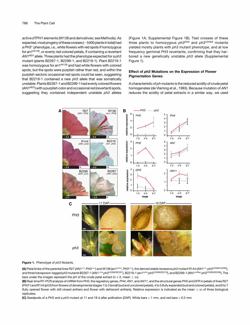
Figure 1. Phenotype of ph3 Mutants.
(A) Petal limbs of the parental lines R27 (AN1+/+ PH3+/+) andW138 (an1m/m,PH3+/+), the derived stable recessive ph3mutant R144 (AN1+/+ ph3V2068/V2068),and three transposon-tagged ph3mutantsB2267-1 (AN1+/m ph3V2068/B2267), B2219-1 (an1m/mph3V2068/B2219), andB2299-1 (AN1m/Rev ph3V2068/B2299). Thebars under the images represent the pH of the crude petal extract (n = 3, mean 6 SD).(B)Real-timeRT-PCR analysis ofmRNA fromPH3, the regulatory genes,PH4,AN1, andAN11, and the structural genesPH5 andDFR in petals of linesR27(PH3+) andR144 (ph3) fromflowersofdevelopmental stages1 to3 (small budanduncoloredpetals), 4 to5 (fully expandedbudandcoloredpetals), and6 to7(fully opened flower with still closed anthers and flower with dehiscent anthers). Relative expression is indicated as the mean 6 SD of three biologicalreplicates.(C) Seedpods of a PH3 and a ph3 mutant at 11 and 18 d after pollination (DAP). White bars = 1 mm, and red bars = 0.2 mm.
788 The Plant Cell
progeny of the primary ph3mutants harboring the new ph3 alleles inan AN1+ or AN1REV background to determine whether these muta-tionsalter thepHofpetal homogenates.We found thatcrudeextractsof ph3V2068/B2267 and ph3V2068/B2219 corollas had a higher pH thanthose of isogenic wild types or germinal PH3 revertants (PH3V2068/B2219-Rev) (Figure1).Petals fromtheph3V2068/B2299heterozygote (plantB2299-1) displayed a smaller pH shift of the petal extract thanph3B2267/V2068orph3B2219/V2068.This indicates thatph3B2299 isaweakallele and may explain why these petals are a mosaic of red andpurplish cells and have a “marbled” appearance (Figure 1A).
The genetic backgrounds of the petunia lines R27 and R143, inwhich theph3 alleles aremaintained, containmutations that block59 hydroxylation (hf1 hf2) and rhamnosylation (rt) of the antho-cyanins and all subsequent modification steps (5-glucosylation,acylation, and methylation) (Supplemental Table 1). In these R27andR143backgrounds,ph3hadonly a small effect on the amountand chemical composition of anthocyanins (Supplemental Figure2). Thin-layer chromatography of hydrolyzed anthocyaninsshowed that PH3 and ph3 petals contain mostly cyanidins,consistent with the hf1 hf2 rt genotype. PH3 petals yielded inaddition a small (trace) amount of peonidins, while these werereduced in ph3 siblings. HPLC analysis identified four major an-thocyanin peaks (P1-P4) in PH3 petals and a minor compound(P5). In ph3 petals, we observed similar amounts of the antho-cyanins in P1 to P4, whereas the minor compound in P5 wasclearly reduced (Supplemental Figure 2).
Real-time RT-PCR analysis of ph3V2068 and PH3+ petals (Figure1B) revealed that ph3 had little or no effect on the expression ofregulatoryanthocyaningenes (AN1andAN11)or theregulatoryPH4gene. However, ph3V2068 almost completely abolished expressionofPH5,which isconsistentwithpreviousresults (Verweijetal.,2008)and can account for the reduced acidity of ph3 petal homogenates.Unexpectedly, expression of DFR, a structural anthocyanin geneencoding DIHYDROFLAVONOL REDUCTASE, was reduced 3- to4-fold, but not abolished, in ph3 petals. DFR expression is appar-ently not rate-limiting for anthocyaninbiosynthesis, sincemutations(an6) that abolish DFR expression are fully recessive (Huits et al.,1994), which may explain why the reduced DFR expression in ph3has little or no effect on the anthocyanin biosynthesis.
Taken together, thesedatashowthatPH3 is, togetherwithAN1,AN11, and PH4, essential for expression of genes involved invacuolar acidification. Although PH3 enhances DFR expression;3- to 4-fold, it is, unlike AN1 and AN11, not essential for DFRexpression or anthocyanin biosynthesis.
Effect of ph3 on Seed Development
Petunia lines with mutations in PH3 or PH5 were reported to befemale sterile (Wiering, 1974;deVlaminget al., 1984).Although theinbredph3andph5 lines thatwereusedby theseauthorshadbeendiscontinued in the 1980s, wewere able to recover and germinateseeds containing the same ph3 and ph5 alleles from F2 crosseswith unrelated lines. With the same ph5 allele in a new geneticbackground, we did not observe sterility of ph5 mutants. In ph5mutants that arose by dTPH1 insertions in theW138 background,we also observed no infertility (Verweij et al., 2008). However, theoriginal ph3R49 allele in the newbackground (line R143), aswell asthe ph3V2068, ph3B2219, and ph3B2267 alleles in the W138
background, all caused complete female sterility. The ph3 mu-tation did not block fertilization or early stages of seed de-velopment, as after pollination of a ph3 flower with PH3 pollen theseedpod enlarged and contained immature seeds at 11 and 18d after pollination (Figure 1C). However, compared with fruits onPH3 plants, those on ph3 plants remained smaller and individualseeds were alsomuch smaller than seeds on a PH3 plant. Around18 d after pollination, when seeds on PH3 plants were firmspheres, seeds on ph3 plants had a collapsed appearance. About4 weeks after pollination, fruits and seeds on PH3 plants weremature, whereas seeds on ph3 plants had completely dis-integrated leaving no other trace than some brownish “dust.”Strikingly, this effect of ph3 was suppressed in an an1 back-ground. In the unstable an1W138 background in which ph3V2068
was isolated, an1W138 ph3V2068 female parents were fully fertile,while the numerous (>10) independentAN1REV ph3V2068 revertantsthat we tested over the years were without exception fully femalesterileanddisplayedtheseedabortionphenotypedescribedabove.
Molecular Isolation of the PH3 Locus
Because most petunia mutants isolated in W138 resulted frominsertions of the high copy number transposon dTPH1 (vanHouwelingen et al., 1998), we assumed that the three new ph3alleles were tagged by dTPH1 insertions and could be used toidentify the locus by transposon display.The test cross of plant B2299-1 (ph3V2068/B2299) to line R144
(ph3V2068/V2068) yielded a family (C2124) that segregated fiveplants with purplish flowers (ph3V2068/V2068), 14 with purplishflowers containing a few red revertant spots (ph3V2068/B2299), andtwo with red flowers (PH3V2068/B2299-REV) that originated fromgerminalPH3 reversions (Supplemental Figure 1C).We visualizeddTPH1 flanking sequences in a subset of these plants by trans-poson display and identified two fragments that were amplifiedfrom all seven unstable mutants (ph3V2068/B2299), but not from anyof the three stable mutants or the two revertants (SupplementalFigure 3A). Sequencing revealed that these two fragments had thesame 8-bp target site duplication and therefore represented theleft and right flanking sequence of the same dTPH1 element.To assess whether this fragment originated from PH3, we used
fragment-specific primers to amplify this dTPH1 insertion in familyC2124. With these primers we were not able to amplify the stableph3V2068 allele, whereas all plants harboring the unstable ph3B2299
allele yieldeda fragment thatwas;300 bp larger than the fragmentamplified from wild-type progenitors (PH3+) or a derived revertant(PH3B2299REV) (Supplemental Figure 3B). Moreover, ph3V2068/B2219
and ph3V2068/B2267 plants also contained an ;300-bp dTPH1 in-sertion in the samegenomic region. Analysis of genomic and cDNAclones revealed that these three insertions disrupted a single gene,consisting of six exons, and that the alleles ph3B2219 and ph3B2267
harbor dTPH1 insertions in exon three, 9 bp apart, while ph3B2299
contains a dTPH1 insertion in the fourth exon (Figure 2A).Most progeny from backcrosses of plants B2219-1 (ph3V2068/
B2219) and B2299-1 (ph3V2068/B2299) to stable ph3R49 (line R143) orph3V2068 mutants (line R144) had purplish (ph3) flowers, whereasa few rare individuals had red flowers indicating that a (germinal)reversionof theph3mutationoccurred(SupplementalFigure1).Theappearance of these revertantPH3B2219Rev andPH3B2299Rev alleles
The PH3 Gene of Petunia 789
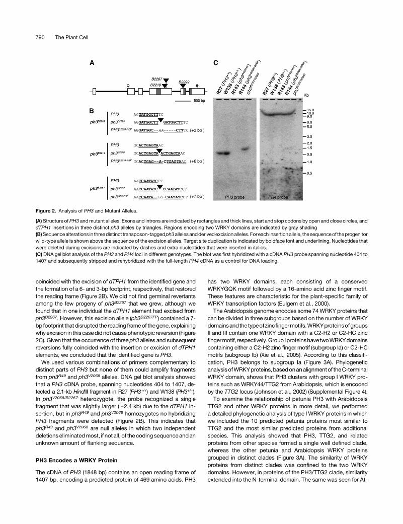
coincided with the excision of dTPH1 from the identified gene andthe formation of a 6- and 3-bp footprint, respectively, that restoredthe reading frame (Figure 2B). We did not find germinal revertantsamong the few progeny of ph3B2267 that we grew, although wefound that in one individual the dTPH1 element had excised fromph3B2267. However, this excision allele (ph3B2267FP) contained a 7-bp footprint that disrupted the reading frameof thegene, explainingwhyexcision in thiscasedidnotcausephenotypic reversion (Figure2C). Given that the occurrence of three ph3 alleles and subsequentreversions fully coincided with the insertion or excision of dTPH1elements, we concluded that the identified gene is PH3.
We used various combinations of primers complementary todistinct parts of PH3 but none of them could amplify fragmentsfrom ph3R49 and ph3V2068 alleles. DNA gel blot analysis showedthat a PH3 cDNA probe, spanning nucleotides 404 to 1407, de-tected a 2.1-kb HindIII fragment in R27 (PH3+/+) and W138 (PH3+/+).In ph3V2068/B2267 heterozygote, the probe recognized a singlefragment that was slightly larger (;2.4 kb) due to the dTPH1 in-sertion, but in ph3R49 and ph3V2068 homozygotes no hybridizingPH3 fragments were detected (Figure 2B). This indicates thatph3R49 and ph3V2068 are null alleles in which two independentdeletionseliminatedmost, if not all, of thecodingsequenceandanunknown amount of flanking sequence.
PH3 Encodes a WRKY Protein
The cDNA of PH3 (1848 bp) contains an open reading frame of1407 bp, encoding a predicted protein of 469 amino acids. PH3
has two WRKY domains, each consisting of a conservedWRKYGQK motif followed by a 16-amino acid zinc finger motif.These features are characteristic for the plant-specific family ofWRKY transcription factors (Eulgem et al., 2000).The Arabidopsis genome encodes some 74WRKY proteins that
can be divided in three subgroups based on the number of WRKYdomainsandthetypeofzincfingermotifs.WRKYproteinsofgroupsII and III contain one WRKY domain with a C2-H2 or C2-HC zincfingermotif, respectively.Group IproteinshavetwoWRKYdomainscontaining either a C2-H2 zinc finger motif (subgroup Ia) or C2-HCmotifs (subgroup Ib) (Xie et al., 2005). According to this classifi-cation, PH3 belongs to subgroup Ia (Figure 3A). PhylogeneticanalysisofWRKYproteins,basedonanalignmentof theC-terminalWRKY domain, shows that PH3 clusters with group I WRKY pro-teins such as WRKY44/TTG2 from Arabidopsis, which is encodedby the TTG2 locus (Johnson et al., 2002) (Supplemental Figure 4).To examine the relationship of petunia PH3 with Arabidopsis
TTG2 and other WRKY proteins in more detail, we performeda detailed phylogenetic analysis of type I WRKY proteins in whichwe included the 10 predicted petunia proteins most similar toTTG2 and the most similar predicted proteins from additionalspecies. This analysis showed that PH3, TTG2, and relatedproteins from other species formed a single well defined clade,whereas the other petunia and Arabidopsis WRKY proteinsgrouped in distinct clades (Figure 3A). The similarity of WRKYproteins from distinct clades was confined to the two WRKYdomains. However, in proteins of the PH3/TTG2 clade, similarityextended into the N-terminal domain. The same was seen for At-
Figure 2. Analysis of PH3 and Mutant Alleles.
(A)Structure ofPH3 andmutant alleles. Exons and introns are indicated by rectangles and thick lines, start and stop codons by open and close circles, anddTPH1 insertions in three distinct ph3 alleles by triangles. Regions encoding two WRKY domains are indicated by gray shading(B)Sequencealterations in threedistinct transposon-taggedph3allelesandderivedexcisionalleles.Foreach insertionallele, thesequenceof theprogenitorwild-type allele is shown above the sequence of the excision alleles. Target site duplication is indicated by boldface font and underlining. Nucleotides thatwere deleted during excisions are indicated by dashes and extra nucleotides that were inserted in italics.(C) DNA gel blot analysis of the PH3 and PH4 loci in different genotypes. The blot was first hybridized with a cDNA PH3 probe spanning nucleotide 404 to1407 and subsequently stripped and rehybridized with the full-length PH4 cDNA as a control for DNA loading.
790 The Plant Cell
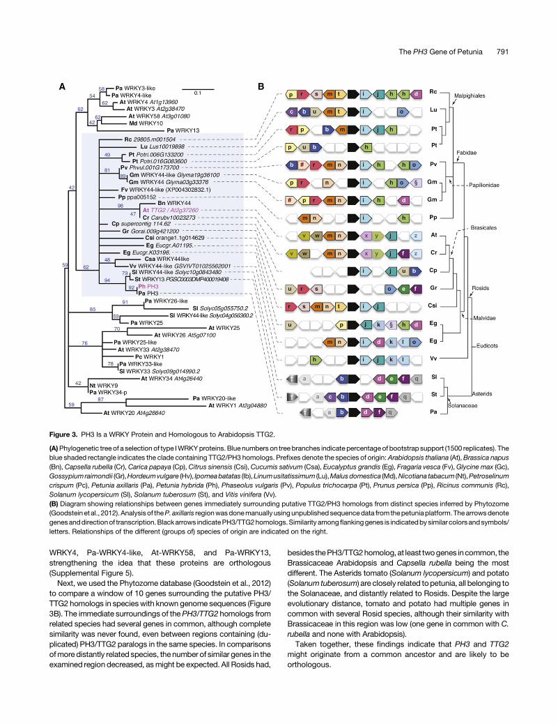
WRKY4, Pa-WRKY4-like, At-WRKY58, and Pa-WRKY13,strengthening the idea that these proteins are orthologous(Supplemental Figure 5).
Next, we used the Phytozome database (Goodstein et al., 2012)to compare a window of 10 genes surrounding the putative PH3/TTG2 homologs in specieswith known genome sequences (Figure3B). The immediate surroundings of thePH3/TTG2 homologs fromrelated species had several genes in common, although completesimilarity was never found, even between regions containing (du-plicated) PH3/TTG2 paralogs in the same species. In comparisonsofmore distantly related species, the number of similar genes in theexamined region decreased, asmight be expected. All Rosids had,
besides thePH3/TTG2homolog,at least twogenes incommon, theBrassicaceae Arabidopsis and Capsella rubella being the mostdifferent. The Asterids tomato (Solanum lycopersicum) and potato(Solanum tuberosum) are closely related topetunia, all belonging tothe Solanaceae, and distantly related to Rosids. Despite the largeevolutionary distance, tomato and potato had multiple genes incommon with several Rosid species, although their similarity withBrassicaceae in this region was low (one gene in common with C.rubella and none with Arabidopsis).Taken together, these findings indicate that PH3 and TTG2
might originate from a common ancestor and are likely to beorthologous.
Figure 3. PH3 Is a WRKY Protein and Homologous to Arabidopsis TTG2.
(A)Phylogenetic tree of a selection of type IWRKYproteins. Blue numbers on tree branches indicate percentageof bootstrap support (1500 replicates). Theblue shaded rectangle indicates the clade containing TTG2/PH3 homologs. Prefixes denote the species of origin:Arabidopsis thaliana (At),Brassica napus(Bn), Capsella rubella (Cr), Carica papaya (Cp), Citrus sinensis (Csi), Cucumis sativum (Csa), Eucalyptus grandis (Eg), Fragaria vesca (Fv),Glycine max (Gc),Gossypiumraimondii (Gr),Hordeumvulgare (Hv), Ipomeabatatas (Ib),Linumusitatissimum (Lu),Malusdomestica (Md),Nicotiana tabacum (Nt),Petroselinumcrispum (Pc), Petunia axillaris (Pa), Petunia hybrida (Ph), Phaseolus vulgaris (Pv), Populus trichocarpa (Pt), Prunus persica (Pp), Ricinus communis (Rc),Solanum lycopersicum (Sl), Solanum tuberosum (St), and Vitis vinifera (Vv).(B) Diagram showing relationships between genes immediately surrounding putative TTG2/PH3 homologs from distinct species inferred by Phytozome(Goodstein et al., 2012). Analysis of theP. axillaris regionwasdonemanually usingunpublished sequencedata from thepetuniaplatform. Thearrowsdenotegenesanddirectionof transcription.Blackarrows indicatePH3/TTG2homologs.Similarity amongflankinggenes is indicatedbysimilar colorsandsymbols/letters. Relationships of the different (groups of) species of origin are indicated on the right.
The PH3 Gene of Petunia 791

Expression Pattern and Genetic Regulation of PH3
We used real-time RT-PCR to analyze the pattern and geneticregulation of PH3mRNA expression in various tissues of petunialine R27. Although R27 is an an4mutant (mutated in a regulator ofanthocyanin biosynthesis in anthers), it contains functional allelesfor all other regulatory AN loci and PH genes that govern thepigmentation of petals. In R27, PH3 was expressed in petals,mostly in the petal limb and to a lesser extent in the petal tube, andovaries, but not in anthers, leaves stems, and sepals (Figure 4A).After pollination, PH3 mRNA expression persisted in the de-veloping seedpod. In situ hybridization on petal sections showedthat PH3 was primarily expressed in the upper epidermis of thecorolla (Figure 4B), similar to AN1, PH4, and the structural genesPH5 and DFR (Quattrocchio et al., 2006; Verweij et al., 2008).However, because thePH3mRNAsignal in the epidermiswas justabove the background, we could not exclude that PH3 was alsoexpressed, albeit at somewhat lower levels, in the mesophyll.
To determine how PH3 expression is regulated, we analyzedPH3mRNA in petals of distinct mutants in theW138 background.We found that PH3mRNAwas expressed at comparable levels inwild-type andph2petalsbutwasclearly reduced in an11,an1, andph4petals especially in later flower stages (Figure 4C). Toexaminethe regulation of PH3 by AN1 in more detail, we used an1 plantsthat contained a transgene in which the p35S promoter drove theexpression of a fusionof AN1and the ligandbinding domain of therat glucocorticoid receptor (p35S:AN1-GR). p35S:AN1-GRcompletely restores DFR, PH1, and PH5 mRNA expression andpigmentation in an1 petals, but only in the presence of dexa-methasone (DEX; Figure 4D), similar to previous results (Speltet al., 2000; Verweij et al., 2008; Faraco et al., 2014). In p35S:AN1-GR petals, PH3mRNA was expressed at a low level at stage 4-5,comparable to isogenic an1 control petals. Application of DEX toan1 p35S:AN1-GR buds increased PH3mRNA expression within2 h,whichwasdue to posttranslational activation of AN1-GR, as itwas not seen in an1 petals lacking the transgene (Figure 4D).Because an1 reduced the expression ofPH3much less than it didDFR or PH5, the fold induction for PH3 upon DEX treatment waslower than for DFR and PH5. In the presence of 50 µM cyclo-heximide, protein biosynthesis in p35S:AN1-GR petals is fullyblocked (Spelt et al., 2000). Under these conditions, DFR ex-pression remained inducible by DEX for a short period (;2 h),suggesting that AN1 activates DFR directly. However, aftera prolonged exposure to cycloheximide and DEX (20 h), DFRexpressiondeclinedagain tobackground levels (Figure4D).This isapparently due to thedecayofAN1-GR,whichappears lessstablethanAN1,andcomplete turnoverofDFRmRNA in20h (Spelt et al.,2000). In the presence of DEX and cycloheximide, PH3 mRNAremainedapproximately constantwithin thefirst 2h,whereasPH3mRNA quickly decayed in the absence of AN1-GR (Figure 4D).These results suggest that the induction of PH3 by AN1-GR wasdirect and did not require biosynthesis of intermediate regulators.
PH3 and TTG2 Are Functionally Homologous
The finding that PH3 and TTG2 are homologous was unexpected,given their very different mutant phenotypes. Arabidopsis ttg2mutants have aberrant trichomes on the leaf surface and reduced
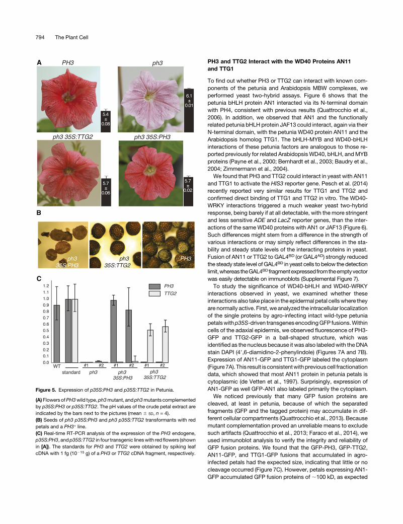
tannin accumulation in the seed coat and lack mucilage pro-duction in germinating seeds (Johnson et al., 2002). Petunia ph3mutants do not show any effect in trichome development, but it isunclear whetherPH3 is involved in tannin accumulation since ph3homozygous seeds abort 2 to 3 weeks after pollination (Figure 1).To examine whether the function of PH3 in petunia and TTG2 inArabidopsis diverged through changes in the encoded proteins orthe downstream network, we performed a mutant complemen-tation study. We cloned the PH3 cDNA or a genomic DNA frag-ment containing the TTG2 coding sequence behind the p35Spromoter and transformed both constructs (p35S:PH3 and p35S:TTG2) into a transformable petunia ph3 mutant that was trans-heterozygous for the deletion allele ph3R49 (deletion allele) andph3B2267FP (frame shift allele) (cf. Figure 2). The petals of thistransformation host contained, compared with the wild type,a slightly reduced amount of (nonfunctional) ph3B2267FP tran-scripts and strongly reduced amount of PH5 mRNA(Supplemental Figure 6) and had the typical purplish color (Figure5A). In 20 out of 21 p35S:PH3 transformants and two out of fourp35S:TTG2 transformants, the transgene rescued the ph3 flowercolor defect and lowered the pH of the crude petal extracts, al-though itdidnot reach thesame level as inPH3flowers (Figure5A).The expression of PH5mRNA was also rescued, consistent withthe flower phenotype, whereas no clear effect was seen on theamountofRNA from theph3B2267endogene (Supplemental Figure6). The latter result suggests that the reduced level of ph3B2267FP
mRNA compared with wild-type PH3 transcripts results fromincreased instability of the mutant mRNA, rather than from re-duced transcription and activity of the PH3 promoter.In addition, p35S:PH3 and p35S:TTG2 also restored female
fertility. Whereas all seeds on ph3 plants die and pulverize duringlate stages of seed development, we could recover viable seedsfrom ph3 p35S:PH3 and p35S:TTG2 female parent plants. Self-fertilization of ph3 p35S:PH3 plants resulted in seed capsulescontaining a low number of seeds, which had an abnormal seedcoat that consisted of smaller cells with a lighter color than wild-type seeds (Figure 5B). However, self-fertilization of ph3 p35S:TTG2 plants resulted in seedpods filled with an almost wild-typenumber of seeds, having a normal seed coat.Real-time RT-PCR analyses showed that in petal limbs of the
two p35S:PH3 petunia lines with the highest transgene expres-sion, the amount PH3 mRNA originating from the transgene wascomparable to that produced by the endogene in wild-type petals(Figure 5C). Because mRNAs from PH3 originated mostly fromepidermal cells, whereas the PH3 RNAs expressed from the 35S:PH3 transgene originated from both epidermal and mesophyllcells, we infer that within epidermal cells the p35S:PH3 transgeneproduces less RNA than the endogenous PH3 gene, which ex-plains the partial complementation of the petal homogenate pH.To compare the amount of TTG2mRNA expressed in the petuniatransformants with PH3mRNA, we used a standard consisting ofleaf cDNA spiked with an equal amount of PH3 and TTG2 cDNA.This revealed that in petal limbs of the two complemented linesp35S:TTG2 was expressed at a similar level as p35S:PH3 incomplemented mutants.Together, these data show that Arabidopsis TTG2 can fully
replace its petunia homolog PH3 in petunia, indicating that bothproteins are functionally very similar.
792 The Plant Cell
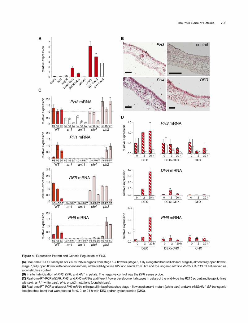
Figure 4. Expression Pattern and Genetic Regulation of PH3.
(A) Real-time RT-PCR analysis of PH3mRNA in organs from stage 5-7 flowers (stage 5, fully elongated bud still closed; stage 6, almost fully open flower;stage 7, fully open flower with dehiscent anthers) of the wild-type line R27 and seeds from R27 and the isogenic an1 line W225.GAPDHmRNA served asa constitutive control.(B) In situ hybridization of PH3, DFR, and AN1 in petals. The negative control was the DFR sense probe.(C)Real-time RT-PCRofDFR,PH3, andPH5mRNAs at different flower developmental stages in petals of thewild-type line R27 (red bar) and isogenic lineswith an1, an11 (white bars), ph4, or ph2 mutations (purplish bars).(D)Real-timeRT-PCRanalysis ofPH3mRNA in the petal limbs of detached stage 4 flowers of an an1mutant (white bars) and an1 p35S:AN1-GR transgenicline (hatched bars) that were treated for 0, 2, or 24 h with DEX and/or cycloheximide (CHX).
The PH3 Gene of Petunia 793
PH3 and TTG2 Interact with the WD40 Proteins AN11and TTG1
To find out whether PH3 or TTG2 can interact with known com-ponents of the petunia and Arabidopsis MBW complexes, weperformed yeast two-hybrid assays. Figure 6 shows that thepetunia bHLH protein AN1 interacted via its N-terminal domainwith PH4, consistent with previous results (Quattrocchio et al.,2006). In addition, we observed that AN1 and the functionallyrelated petunia bHLH protein JAF13 could interact, again via theirN-terminal domain, with the petunia WD40 protein AN11 and theArabidopsis homolog TTG1. The bHLH-MYB and WD40-bHLHinteractions of these petunia factors are analogous to those re-ported previously for related Arabidopsis WD40, bHLH, andMYBproteins (Payne et al., 2000; Bernhardt et al., 2003; Baudry et al.,2004; Zimmermann et al., 2004).We found that PH3 and TTG2 could interact in yeast with AN11
and TTG1 to activate the HIS3 reporter gene. Pesch et al. (2014)recently reported very similar results for TTG1 and TTG2 andconfirmed direct binding of TTG1 and TTG2 in vitro. The WD40-WRKY interactions triggered a much weaker yeast two-hybridresponse, being barely if at all detectable, with the more stringentand less sensitive ADE and LacZ reporter genes, than the inter-actions of the sameWD40 proteins with AN1 or JAF13 (Figure 6).Such differences might stem from a difference in the strength ofvarious interactions or may simply reflect differences in the sta-bility and steady state levels of the interacting proteins in yeast.Fusion of AN11 or TTG2 to GAL4BD (or GAL4AD) strongly reducedthe steady state level of GAL4BD in yeast cells to below the detectionlimit,whereastheGAL4BDfragmentexpressedfromtheemptyvectorwas easily detectable on immunoblots (Supplemental Figure 7).To study the significance of WD40-bHLH and WD40-WRKY
interactions observed in yeast, we examined whether theseinteractions also take place in the epidermal petal cellswhere theyare normally active. First, we analyzed the intracellular localizationof the single proteins by agro-infecting intact wild-type petuniapetalswithp35S-driven transgenesencodingGFP fusions.Withincells of the adaxial epidermis, we observed fluorescence of PH3-GFP and TTG2-GFP in a ball-shaped structure, which wasidentified as the nucleus because it was also labeledwith theDNAstain DAPI (49,6-diamidino-2-phenylindole) (Figures 7A and 7B).Expression of AN11-GFP and TTG1-GFP labeled the cytoplasm(Figure7A). This result is consistentwithpreviouscell fractionationdata, which showed that most AN11 protein in petunia petals iscytoplasmic (de Vetten et al., 1997). Surprisingly, expression ofAN1-GFP as well GFP-AN1 also labeled primarily the cytoplasm.We noticed previously that many GFP fusion proteins are
cleaved, at least in petunia, because of which the separatedfragments (GFP and the tagged protein) may accumulate in dif-ferent cellular compartments (Quattrocchio et al., 2013). Becausemutant complementation proved an unreliable means to excludesuch artifacts (Quattrocchio et al., 2013; Faraco et al., 2014), weused immunoblot analysis to verify the integrity and reliability ofGFP fusion proteins. We found that the GFP-PH3, GFP-TTG2,AN11-GFP, and TTG1-GFP fusions that accumulated in agro-infected petals had the expected size, indicating that little or nocleavage occurred (Figure 7C). However, petals expressing AN1-GFP accumulated GFP fusion proteins of ;100 kD, as expected
Figure 5. Expression of p35S:PH3 and p35S:TTG2 in Petunia.
(A) Flowers ofPH3wild type,ph3mutant, andph3mutants complementedby p35S:PH3 or p35S:TTG2. The pH values of the crude petal extract areindicated by the bars next to the pictures (mean 6 SD, n = 4).(B) Seeds of ph3 p35S:PH3 and ph3 p35S:TTG2 transformants with redpetals and a PH3+ line.(C) Real-time RT-PCR analysis of the expression of the PH3 endogene,p35S:PH3, andp35S:TTG2 in four transgenic lineswith red flowers (shownin [A]). The standards for PH3 and TTG2 were obtained by spiking leafcDNA with 1 fg (10215 g) of a PH3 or TTG2 cDNA fragment, respectively.
794 The Plant Cell

for the full size fusion, as well as two smaller proteins of;70 and;40kD,andpetals expressingGFP-AN1accumulated the full size;100-kD protein alongwith a fragment of;60 kD. These findingsindicate that some of the AN1 proteins with a GFP tag on the Nterminus (GFP-AN1) or on the C terminus (AN1-GFP) are cleavedwithin the AN1 sequence.
To study interactions of theWD40 proteins with AN1, PH3, andTTG2 within petal cells, we used bimolecular fluorescencecomplementation (BiFC). For these experiments, we used petalprotoplasts, becausecotransformationof thesecellswithmultipleconstructs is nearly 100%efficient (i.e., both constructs are foundin nearly all cells that are transformed) and because these retainthe gene expression and protein sorting characteristics (Faracoet al., 2011).We cotransformedpetal protoplastswith a transgeneencoding a fusion of RFP and the Arabidopsis plasmamembraneSNAREproteinSYP122 (Assaadet al., 2004), tomark transformedcells including those in which no BiFC occurred, and different
combinations of transgenes encoding fusions of PH3, TTG2, orAN1 to the N-terminal half of YFP (nYFP) and fusions of AN11 orTTG1 to the C-terminal half of YFP (cYFP). Since the position andrelative orientation of nYFP and cYFP tags in protein pairs canprofoundly affect BiFC efficiency (Bracha-Drori et al., 2004), wecoexpressed in the first experiments multiple combinations of thepetunia protein fusions with different orientations of the cYFP andnYFP tags.Cotransformation of protoplasts with constructs expressing
nYFP-AN1, AN11-cYFP, and cYFP-AN11 or fusions with a dif-ferent arrangement of tags (AN1-cYFP, AN11-nYFP, and nYFP-AN11) caused YFP fluorescence in >90% of the cells expressingthe transformation marker RFP-SYP122 (Supplemental Figure 8).Subsequent experiments showed that YFP fluorescence resultedfrom coexpression of nYFP-AN1 and AN11-cYFP (Figure 8),whereas coexpression of nYFP-AN1 and cYFP-AN11 yielded nodetectable YFP fluorescence (Supplemental Figure 8). No in-teraction was detectable when we replaced nYFP-AN1 with freenYFP or cYFP-AN11 with free cYFP, even though >50% of theprotoplasts expressed the transformation marker RFP-SYP122(Figure 8). This indicates that even though nYFP and cYFP arelikely to localize in the cytoplasm and nucleus of petal cells, likefree GFP (Verweij et al., 2008), they do not cause any noticeablebackgroundfluorescencedue tonYFP-cYFPself-assemblyunderthe conditions used. The finding that the interaction of AN1 andAN11 fusions was orientation dependent, since coexpressioncYFP-AN1 with AN11-nYFP or nYFP-AN11 did not cause YFPfluorescence (Supplemental Figure 8), also indicates that nYFP-cYFP self-assembly did not result in noticeable backgroundfluorescence.Detailed confocal microscopy analysis of some 20 cells
expressing different combinations of AN11 and AN1 fusionsshowed that in all cases the YFP fluorescence localized in thecytoplasm close to the nucleus but was not detectable within thenucleus (Figure 8; Supplemental Figure 8). Moreover, the ob-served interactions were similar, in terms of intensity and in-tracellular localization, in purple protoplasts originating from theepidermis and in colorless protoplasts derived from the meso-phyll, indicating that it occurs independent from other epidermis-specific factors (e.g., other regulators of the anthocyanin and pHpathways). Replacement of AN11-cYFP with the homologousTTG1-cYFP fusion did not obviously alter the frequency, intensity,or intracellular localization of the resulting YFP fluorescence,indicating that Arabidopsis TTG1 and petunia AN11 are inter-changeable regarding their capacity to interact with AN1 (Figure 8).Transformation of petal protoplasts with 35S:nYFP-TTG2 or
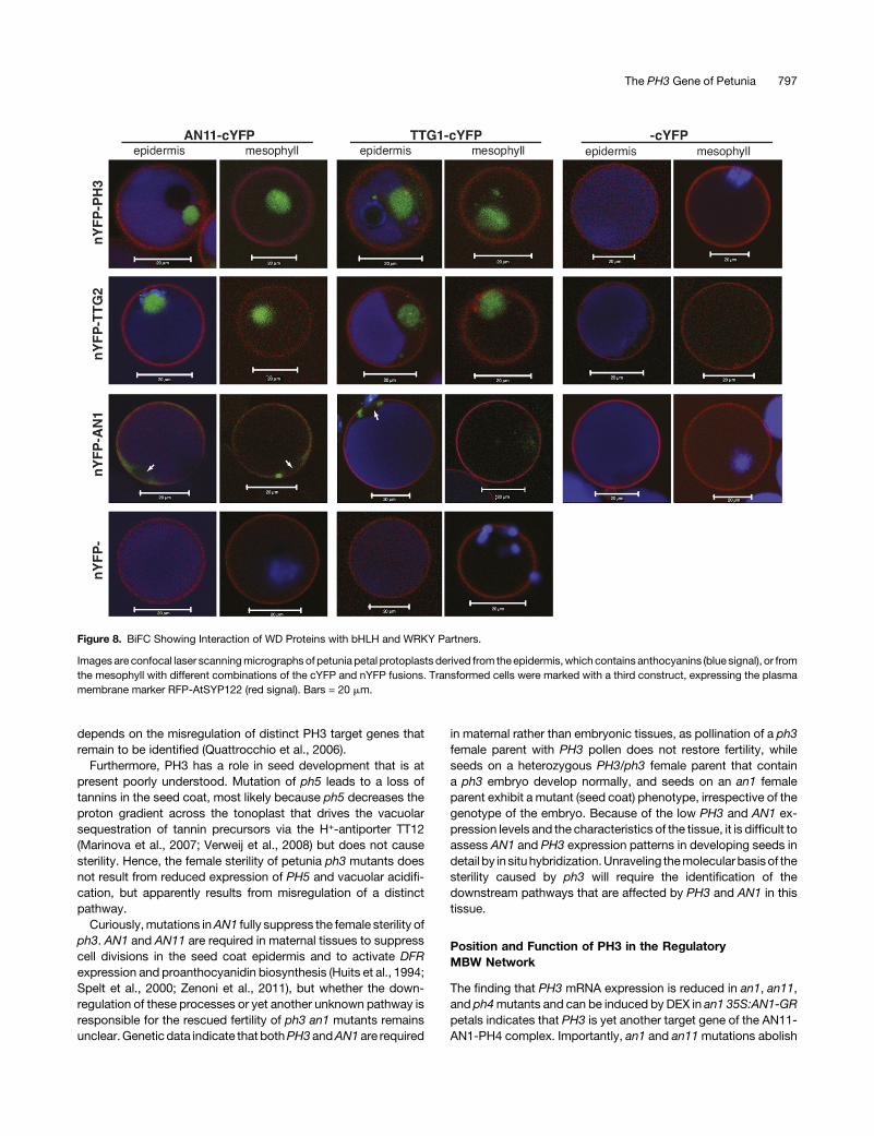
35S:nYFP-PH3 in combination with 35S:AN11-cYFP or 35S:TTG1-cYFP caused a strong BiFC signal in all transformed cellsmarkedbyRFP-SYP122 (Figure8). Even thoughuntaggedcYFP isthought to be present in both cytoplasm and nuclei of petal cells,like free GFP, no BiFC was seen upon coexpression with nYFP-PH3or nYFP-TTG2 (Figure 8), andcoexpression of PH3andAN11fusions with different orientations of cYFP and nYFP tags did notcauseBiFC fluorescence either. This result indicates that theBiFCseen in cells coexpressing nYFP-PH3 or nYFP-TTG2 with AN11-cYFP or TTG1-cYFP results from the interaction of theWRKY andWD40 moieties rather than self-assembly of the cYFP and nYFPtags. Interestingly, the complexes of AN11withPH3or TTG2were
Figure 6. Interaction of PH3 and TTG2 with WD40 Proteins.
Yeast strains expressing the indicated GAL4AD and GAL4BD fusions weregrown on dropout media lacking leucine (2L), tryptophan (2T), histidine(2H), and/or adenine (2A). Activation of the GAL4-regulated HIS and ADEreporter genes is seen asgrowthon –LTHand –LTHAmedia andof the lacZgene as a blue staining after overlaying the cells with X-Gal.
The PH3 Gene of Petunia 795
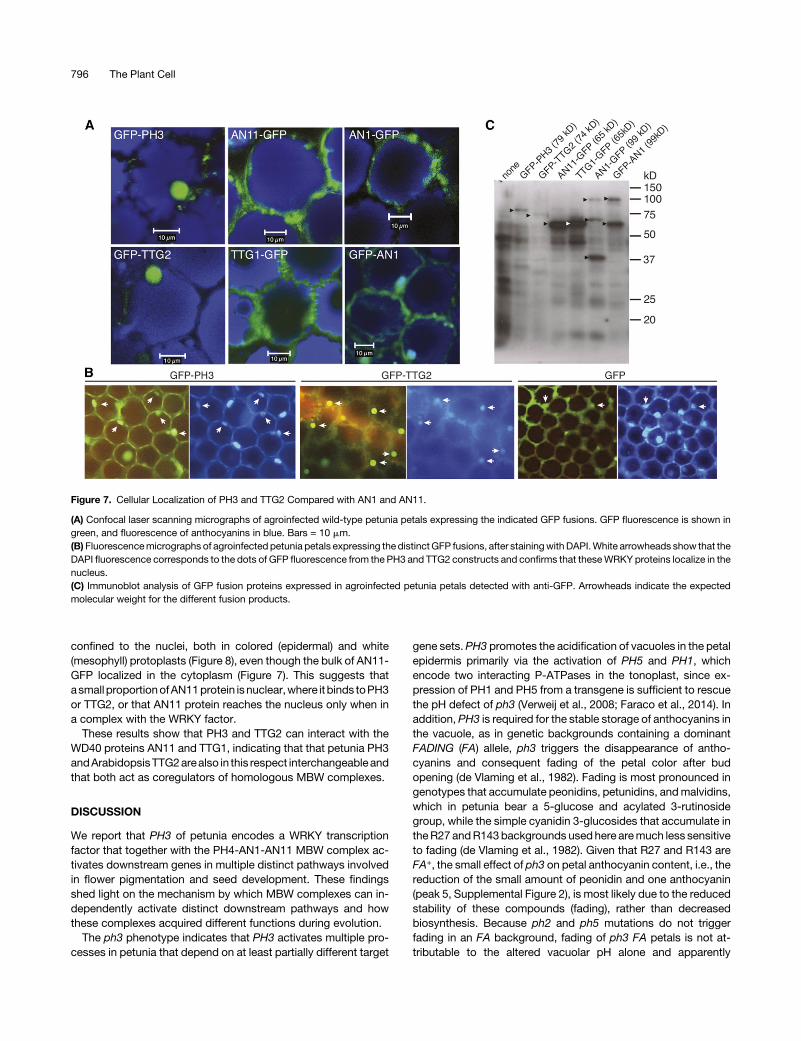
confined to the nuclei, both in colored (epidermal) and white(mesophyll) protoplasts (Figure 8), even though the bulk of AN11-GFP localized in the cytoplasm (Figure 7). This suggests thatasmall proportionofAN11protein isnuclear,where it binds toPH3or TTG2, or that AN11 protein reaches the nucleus only when ina complex with the WRKY factor.
These results show that PH3 and TTG2 can interact with theWD40 proteins AN11 and TTG1, indicating that that petunia PH3andArabidopsisTTG2arealso in this respect interchangeableandthat both act as coregulators of homologous MBW complexes.
DISCUSSION
We report that PH3 of petunia encodes a WRKY transcriptionfactor that together with the PH4-AN1-AN11 MBW complex ac-tivates downstream genes in multiple distinct pathways involvedin flower pigmentation and seed development. These findingsshed light on the mechanism by which MBW complexes can in-dependently activate distinct downstream pathways and howthese complexes acquired different functions during evolution.
The ph3 phenotype indicates that PH3 activates multiple pro-cesses in petunia that depend on at least partially different target
gene sets.PH3 promotes the acidification of vacuoles in the petalepidermis primarily via the activation of PH5 and PH1, whichencode two interacting P-ATPases in the tonoplast, since ex-pression of PH1 and PH5 from a transgene is sufficient to rescuethe pH defect of ph3 (Verweij et al., 2008; Faraco et al., 2014). Inaddition, PH3 is required for the stable storage of anthocyanins inthe vacuole, as in genetic backgrounds containing a dominantFADING (FA) allele, ph3 triggers the disappearance of antho-cyanins and consequent fading of the petal color after budopening (de Vlaming et al., 1982). Fading is most pronounced ingenotypes that accumulate peonidins, petunidins, andmalvidins,which in petunia bear a 5-glucose and acylated 3-rutinosidegroup, while the simple cyanidin 3-glucosides that accumulate intheR27 andR143backgrounds usedhere aremuch less sensitiveto fading (de Vlaming et al., 1982). Given that R27 and R143 areFA+, the small effect of ph3 on petal anthocyanin content, i.e., thereduction of the small amount of peonidin and one anthocyanin(peak 5, Supplemental Figure 2), is most likely due to the reducedstability of these compounds (fading), rather than decreasedbiosynthesis. Because ph2 and ph5 mutations do not triggerfading in an FA background, fading of ph3 FA petals is not at-tributable to the altered vacuolar pH alone and apparently
Figure 7. Cellular Localization of PH3 and TTG2 Compared with AN1 and AN11.
(A) Confocal laser scanning micrographs of agroinfected wild-type petunia petals expressing the indicated GFP fusions. GFP fluorescence is shown ingreen, and fluorescence of anthocyanins in blue. Bars = 10 mm.(B)Fluorescencemicrographs of agroinfected petunia petals expressing the distinctGFP fusions, after stainingwithDAPI.White arrowheads show that theDAPI fluorescence corresponds to the dots of GFP fluorescence from the PH3 and TTG2 constructs and confirms that theseWRKY proteins localize in thenucleus.(C) Immunoblot analysis of GFP fusion proteins expressed in agroinfected petunia petals detected with anti-GFP. Arrowheads indicate the expectedmolecular weight for the different fusion products.
796 The Plant Cell
depends on the misregulation of distinct PH3 target genes thatremain to be identified (Quattrocchio et al., 2006).
Furthermore, PH3 has a role in seed development that is atpresent poorly understood. Mutation of ph5 leads to a loss oftannins in the seed coat, most likely because ph5 decreases theproton gradient across the tonoplast that drives the vacuolarsequestration of tannin precursors via the H+-antiporter TT12(Marinova et al., 2007; Verweij et al., 2008) but does not causesterility. Hence, the female sterility of petunia ph3 mutants doesnot result from reduced expression of PH5 and vacuolar acidifi-cation, but apparently results from misregulation of a distinctpathway.
Curiously,mutations inAN1 fully suppress the female sterility ofph3. AN1 and AN11 are required in maternal tissues to suppresscell divisions in the seed coat epidermis and to activate DFRexpression and proanthocyanidin biosynthesis (Huits et al., 1994;Spelt et al., 2000; Zenoni et al., 2011), but whether the down-regulation of these processes or yet another unknown pathway isresponsible for the rescued fertility of ph3 an1 mutants remainsunclear.Genetic data indicate that bothPH3 andAN1 are required
in maternal rather than embryonic tissues, as pollination of a ph3female parent with PH3 pollen does not restore fertility, whileseeds on a heterozygous PH3/ph3 female parent that containa ph3 embryo develop normally, and seeds on an an1 femaleparent exhibit a mutant (seed coat) phenotype, irrespective of thegenotype of the embryo. Because of the low PH3 and AN1 ex-pression levels and the characteristics of the tissue, it is difficult toassess AN1 and PH3 expression patterns in developing seeds indetail by in situhybridization.Unraveling themolecular basisof thesterility caused by ph3 will require the identification of thedownstream pathways that are affected by PH3 and AN1 in thistissue.
Position and Function of PH3 in the RegulatoryMBW Network
The finding that PH3 mRNA expression is reduced in an1, an11,and ph4mutants and can be induced by DEX in an1 35S:AN1-GRpetals indicates that PH3 is yet another target gene of the AN11-AN1-PH4 complex. Importantly, an1 and an11mutations abolish
Figure 8. BiFC Showing Interaction of WD Proteins with bHLH and WRKY Partners.
Images are confocal laser scanningmicrographs of petunia petal protoplasts derived from theepidermis,which contains anthocyanins (blue signal), or fromthe mesophyll with different combinations of the cYFP and nYFP fusions. Transformed cells were marked with a third construct, expressing the plasmamembrane marker RFP-AtSYP122 (red signal). Bars = 20 mm.
The PH3 Gene of Petunia 797
the expression of downstream genes involved in anthocyaninbiosynthesis (e.g.,DFR) and vacuolar acidification (PH1 and PH5)almost completely, whereas PH3 mRNA expression is reducedonly;3-fold in mature buds and open flowers, with the strongesteffect in later stages. This suggests thatPH3 is activated inparallelby a second pathway that is AN1- and AN11-independent andpredominantly active in younger buds.PH3 expression in seeds isalso partially regulated by AN11, as a PH3-derived EST (PE-TIN034019) was some 3- to 5-fold downregulated in an11 seedscompared with the wild type (Zenoni et al., 2011), and by AN1(Figure 4), whereas DFR and other anthocyanin genes are morestrongly reduced (Huits et al., 1994; Zenoni et al., 2011).
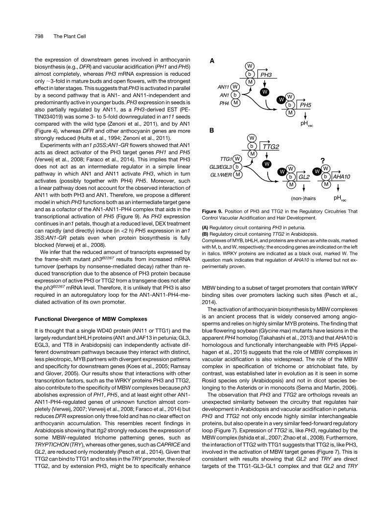
Experiments with an1 p35S:AN1-GR flowers showed that AN1acts as direct activator of the PH3 target genes PH1 and PH5(Verweij et al., 2008; Faraco et al., 2014). This implies that PH3does not act as an intermediate regulator in a simple linearpathway in which AN1 and AN11 activate PH3, which in turnactivates (possibly together with PH4) PH5. Moreover, sucha linear pathway does not account for the observed interaction ofAN11 with both PH3 and AN1. Therefore, we propose a differentmodel inwhichPH3 functions both as an intermediate target geneand as a cofactor of the AN1-AN11-PH4 complex that aids in thetranscriptional activation of PH5 (Figure 9). As PH3 expressioncontinues in an1 petals, though at a reduced level, DEX treatmentcan rapidly (and directly) induce (in <2 h) PH5 expression in an135S:AN1-GR petals even when protein biosynthesis is fullyblocked (Verweij et al., 2008).
We infer that the reduced amount of transcripts expressed bythe frame-shift mutant ph3B2267 results from increased mRNAturnover (perhaps by nonsense-mediated decay) rather than re-duced transcription due to the absence of PH3 protein becauseexpression of active PH3 or TTG2 from a transgene does not alterthe ph3B2267 mRNA level. Therefore, it is unlikely that PH3 is alsorequired in an autoregulatory loop for the AN1-AN11-PH4-me-diated activation of its own promoter.
Functional Divergence of MBW Complexes
It is thought that a single WD40 protein (AN11 or TTG1) and thelargely redundant bHLHproteins (AN1 and JAF13 in petunia; GL3,EGL3, and TT8 in Arabidopsis) can independently activate dif-ferent downstream pathways because they interact with distinct,less pleiotropic, MYBpartners with divergent expression patternsand specificity for downstream genes (Koes et al., 2005; Ramsayand Glover, 2005). Our results show that interactions with othertranscription factors, such as the WRKY proteins PH3 and TTG2,also contribute to the specificity ofMBWcomplexes because ph3abolishes expression of PH1, PH5, and at least eight other AN1-AN11-PH4-regulated genes of unknown function almost com-pletely (Verweij, 2007; Verweij et al., 2008; Faraco et al., 2014) butreducesDFR expression only three fold and has no clear effect onanthocyanin accumulation. This resembles recent findings inArabidopsis showing that ttg2 strongly reduces the expression ofsome MBW-regulated trichome patterning genes, such asTRYPTICHON (TRY ),whereasother genes, suchasCAPRICE andGL2, are reduced only moderately (Pesch et al., 2014). Given thatTTG2canbind toTTG1and tosites in theTRYpromoter, the roleofTTG2, and by extension PH3, might be to specifically enhance
MBW binding to a subset of target promoters that contain WRKYbinding sites over promoters lacking such sites (Pesch et al.,2014).The activation of anthocyanin biosynthesis byMBWcomplexes
is an ancient process that is widely conserved among angio-sperms and relies on highly similar MYB proteins. The finding thatblue flowering soybean (Glycine max) mutants have lesions in theapparentPH4 homolog (Takahashi et al., 2013) and that AHA10 ishomologous and functionally interchangeable with PH5 (Appel-hagen et al., 2015) suggests that the role of MBW complexes invacuolar acidification is also widespread. The role of the MBWcomplex in specification of trichome or atrichoblast fate, bycontrast, was established later in evolution as it is seen in someRosid species only (Arabidopsis) and not in dicot species be-longing to the Asterids or in monocots (Serna and Martin, 2006).The observation that PH3 and TTG2 are orthologs reveals an
unexpected similarity between the circuitry that regulates hairdevelopment in Arabidopsis and vacuolar acidification in petunia.PH3 and TTG2 not only encode highly similar interchangeableproteins, but also operate in a very similar feed-forward regulatoryloop (Figure 7). Expression of TTG2 is, like PH3, regulated by theMBWcomplex (Ishida et al., 2007; Zhao et al., 2008). Furthermore,the interaction of TTG2with TTG1suggests that TTG2 is, likePH3,involved in the activation of MBW target genes (Figure 7). This isconsistent with results showing that GL2 and TRY are directtargets of the TTG1-GL3-GL1 complex and that GL2 and TRY
Figure 9. Position of PH3 and TTG2 in the Regulatory Circuitries ThatControl Vacuolar Acidification and Hair Development.
(A) Regulatory circuit containing PH3 in petunia.(B) Regulatory circuit containing TTG2 in Arabidopsis.Complexes ofMYB, bHLH, and proteins are shown aswhite ovals,markedwithM, b, andW, respectively; the encoding genes are indicated on the leftin italics. WRKY proteins are indicated as a black oval, marked W. Thequestion mark indicates that regulation of AHA10 is inferred but not ex-perimentally proven.
798 The Plant Cell

expression is coregulated by TTG2 (Ishida et al., 2007; Zhao et al.,2008; Morohashi and Grotewold, 2009; Pesch et al., 2014).
Although the modulation of extracellular pH is important for thedevelopment of root hairs (Bibikova et al., 1998), there is no ev-idence that the regulation of vacuolar pH is required for root hairformation. Hence, the pathway(s) influenced by TTG2 in theArabidopsis leaf or root epidermis may have little similarity withthose influencedbyPH3 inpetuniapetals. The roleofTTG2 in seedpigmentation, however, is likely to share similaritieswith the role ofPH3 in petal pigmentation. The lackof tannins in ttg2seeds initiallysuggested that TTG2 is required to activate genes involved intannin biosynthesis, such as BANYULS (BAN) or genes actinglater (Johnson et al., 2002). However, even though BAN expres-sion is dependent on aMBWcomplex, it does not seem to requireTTG2 (Debeaujon et al., 2003). Although we cannot exclude thatTTG2 is required for the activation of genes acting afterBAN in thetannin biosynthetic pathway, we favor a different role for TTG2 intannin accumulation that is more similar to the role of PH3 inpetunia. PH3 promotes vacuolar acidification via activation ofPH5, which encodes a tonoplast H+ pump that is also required fortannin accumulation in the seed (Verweij et al., 2008). Becauseamutation in theArabidopsisPH5homologAHA10alsoabolishestannin accumulation in the seed (Baxter et al., 2005; Appelhagenet al., 2015), just like ttg2, it is likely that TTG2 enables tanninaccumulation, at least in part, by activating AHA10.
Given that proanthocyanidins are found in most angiospermsand more ancestral plants (gymnosperms and ferns), such a rolefor TTG2 and PH3 in driving vacuolar acidification is probably theancestral function, whereas the role of TTG2 and the associatedMBW complex in trichome development was established muchlater,most likely after the separationofRosids andAsterids (Sernaand Martin, 2006). Presumably, this involved the acquisition ofTTG2-responsive elements by downstreamgenes involved in hairdevelopment, butdirect evidence for this ideawill have toawait theidentification of such genes.
METHODS
Plant Material
Genotypes of petunia (Petunia hybrida) lines and details of an1 and ph3alleles usedare shown inSupplemental Tables 1 and2. They all derive fromthe Amsterdam petunia collection and were grown in a greenhouse at VUUniversity (Amsterdam) with supplemental artificial lighting (cycles of 16 hlight and 8 h darkness) at a (minimum) temperature of 22°C or higher.
To isolate dTPH1-tagged ph3 alleles, we crossed R144 (ph3V2068) toPH3+ line W138 or derived lines with unstable mutations in AN11 (lineW137), AN10 (W237), or EXTRAPETALS (EXP) and identified three in-dividuals with an unstable ph3 phenotype among ;5400 progeny. Testcrosses with R143 (ph3R49) confirmed these plants to be ph3mutants andheterozygous for an11, an10, or exp, excluding that they resulted fromcontaminations. ph3 lines were maintained by fertilization of PH3/ph3heterozygotes with pollen from ph3/ph3 homozygous siblings and se-lection of PH3/ph3 heterozygotes (red flowers) and ph3 homozygotes(purplish flowers) from the progeny.
Analysis of Anthocyanins and pH of Crude Petal Extracts
For eachpHmeasurement, the corolla of a freshopen flowerwas ground ina mortar with 6 mL water, and the pH of the extract was immediately
measured with a pH electrode (Checker; Hanna Instruments). Measure-mentswere repeatedon three tofivedifferentflowersand theaveragevalueand SE were calculated. Thin-layer chromatography and HPLC analysis ofanthocyanins was done as described previously (van Houwelingen et al.,1998; Quattrocchio et al., 2006).
DNA and RNA Methodology
Transposon display was performed as described before (Tobeña-Santa-maria et al., 2002) using primers shown in Supplemental Table 3. Gelswereexposed overnight and read by a phosphor imager (Molecular Dynamics).
The PH3 cDNA 39-end was obtained by screening a R27 petal cDNAlibrary (Quattrocchio et al., 2006), and the 59-end was amplified by 59 rapidamplification of cDNA ends (59/39-RACE KIT 2nd generation; Roche) usingPH3-specific primers shown in Supplemental Table 4.
Extractions of DNA and total RNA were performed as described pre-viously (de Vetten et al., 1997). Real-time RT-PCR analysis was performedwith an iTaq Universal Syber Green kit (Bio-Rad) using primers listed inSupplemental Table 5 and an ECO P1180 real-time PCR cycler with Ecoversion 4.0 software (Illumina). Quantitative RT-PCR was performed asdescribed previously (Quattrocchio et al., 1998) using gene-specific pri-mers (Supplemental Table 6) and a reduced number of cycles, to ensurelinear amplification, and detection of amplification products by hybrid-ization of DNA gel blots.
ForDNAgel blot analysis, genomicDNAwasdigestedwithHindIII, size-separated on an agarose gel, blotted to Hybond-N membrane, and hy-bridized overnight at 65°Cwith a 32P-labeled PH3 probe (nucleotides 404 to1407). The blot was washed with 23 SSC + 0.1% SDS at 60°C for 30 min,exposed overnight, and read by a phosphor imager (Molecular Dynamics).Subsequently theblotwasstrippedandrehybridizedwithaPH4cDNAprobe.
RNA in situ hybridization was performed as described before (Soueret al., 1996). Probes were prepared by in vitro transcription using T7polymerase (PH3 and PH4 probes) or SP6 polymerase (DFR probe) ontemplates thatwere PCRamplified fromcloned cDNA fragments in pGEM-Teasywith aT7orSP6promoterprimer andagene-specific forwardprimer(Supplemental Table 7) or in case of the control DFR sense probe, a gene-specific reverse primer. The PH3 probe included nucleotides 404 to 1407(containing both WRKY and zinc finger motifs). Because anthocyaninscould mask the reddish hybridization signal, we used petals in whichanthocyanin biosynthesis was blocked by amutation inAN3, encoding theF39H enzyme (van Houwelingen et al., 1998; Quattrocchio et al., 2006).
Phylogenetic and Synteny Analysis
Sequence alignments were generated with Multiple Sequence Compari-son by Log Expectation (MUSCLE), and phylogenetic trees were con-structed with maximum likelihood (PhyML) after curation with G-blocks toremove badly aligning regions, using online tools at http://phylogeny.lirmm.fr (Dereeper et al., 2008). To generate a phylogenetic tree of WRKYproteins from different subfamilies (Supplemental Figure 4), we usedmanually selected protein fragments spanning the C-terminal WRKYdomain plus 10 upstream amino acids for alignments with MUSCLE andgeneration of a tree with PhyML. The alignments used to generate thethrees are available as Supplemental Data Sets 1 and 2.
Synteny was analyzed using the Web-based Phytozome V9 platform(Goodstein et al., 2012). For petunia, we used unpublished sequence datafrom the petunia platform (www.http://flower.ens-lyon.fr/petuniaplatform/petuniaplatform.html). We compared an annotated 0.95 Mb scaffold(Peaxi162Scf00472), which contained PH3 of Petunia axillaris, and per-formed tBLASTn searches of flanking petunia gene sequences against thegenomesof relatedSolanaceae (tomato [Solanum lycopersicum] andpotato[Solanumtuberosum]) andviceversaby tBLASTnsearchesofgenesflankingthe tomato and potato PH3 homologs to the P. axillaris genome.
The PH3 Gene of Petunia 799
Gene Constructs and Transgenic Lines
The full-length PH3 cDNA coding sequence was amplified from R27 petalcDNA (stage 5-6) with primers #1295 and #2254 and the TTG2 codingsequence from Arabidopsis thaliana Columbia genomic DNA with primers#2224 and #1391 (see Supplemental Table 8 for primer sequences). Bothfragments were digested with XbaI and BamHI and subsequently ligatedinto the pGreen1H vector (XbaI/BamHI) (Hellens et al., 2000).
Plasmids were introduced, together with helper plasmid pJIC, intoAgrobacterium tumefaciens strain AGL0 by electroporation. A single colonywas used for an overnight culture and used to infect leaf discs. Shoot re-generationwasachieved in thepresenceof10mg/mLhygromycin.As lines inthe R27 and R143 background are difficult to transform, we crosseda ph3V2068/B2267FP plant in the R27 background (cf. Supplemental Figure 1E)toaPH3+/R49 individual in theR143backgroundandusedtransheterozygousph3R49/B2267FP progeny as a transformation host.
Yeast Two-Hybrid Analysis
Yeast two-hybrid analysis was done as described (Quattrocchio et al.,2006). Constructs expressing fusion of the GAL4BD or GAL4AD domain tofull-size PH4, AN1, JAF13, or the N-terminal 238 amino acids were de-scribed before (Quattrocchio et al., 2006); plasmids expressing fusions ofPH3, TTG2, and TTG1 were generated in the same way, using cDNAsamplified frompetuniapetals (PH3)orArabidopsis leaves (TTG1andTTG2)with primers listed in Supplemental Table 9.
Transient Expression of GFP Fusions and Split-YFP Constructs
Full-size coding sequences of PH3, TTG2, and TTG1 were amplified withprimers containing attB1 and attB2 sites. These fragments were re-combined with BP clonase in the entry vector pDONR P1-P2 (Invitrogen).Full-sizecodingsequenceofAN11wasamplifiedwithprimerscontainingToposites and cloned in pENTR/D Topo (Invitrogen), and AN1 was amplified withprimers containing NcoI and EcoRI restriction sites and cloned in pENTR4(Invitrogen). All primers are shown inSupplemental Table 10. For expressionofGFP fusions, coding sequences were transferred by Gateway recombinationintopK7FWG2.0orpK7WGF2.0 (Hellensetal.,2000)or, forexpressionofnYFPand cYFP fusions, into plasmids pGWnY (AB626693), pnYGW (AB626694),pGWcY (AB626696), and/or pcYGW (AB626696) (Hino et al., 2011).
Transient transformation of intact petals by agroinfection and trans-formation of petal protoplasts was described previously (Verweij et al., 2008;Faraco et al., 2014). Imaging of DAPI-stained cells was done using a Zeissfluorescencemicroscope; all other fluorescencemicrographswere obtainedusing a confocal laser scanning microscope, as described before (Verweijet al., 2008). InBiFCassays,plasmidsencodingnYFPandcYFP fusionswerecotransformed with a construct (35S:RFP-SYP122) that expresses theplasma membrane marker RFP-SYP122 (Assaad et al., 2004) to labeltransformedcellsandto interpretnegativeBiFCresults.Typically>50%oftheprotoplasts were transformed. For each combination of constructs, severalhundred cells were examined for RFP or BiFC expression by normal fluo-rescencemicroscopy,and5to10cellswere imagedbyconfocalmicroscopy.
Immunoblot Analysis
Immunoblot analysis was performed as described (de Vetten et al., 1997;Verweij et al., 2008) using anti-GAL4BD (Clontech; catalog number 630403,lot number 1107146A) or anti-GFP antibodies (Santa Cruz Biotechnology;catalog number sc-8334, lot number L1803).
Accession Numbers
Sequence data from this article can be found in the GenBank/EMBL datalibraries under accession numbers KU761265 (PH3 gene) and KU641009
(PH3 cDNA). For the protein alignments and phylogenetic analyses weused the following WRKY protein sequences: from Arabidopsis (At):AtWRKY4 (At1g13960). AtWRKY6 (At1g62300), AtWRKY9 (At1g68150),AtWRKY8 (At5g46350), AtWRKY11 (At4g31550), AtWRKY7 (At4g24240),AtWRKY14 (At1g30650), AtWRKY13 (At4g39410), AtWRKY18 (At4g31800),AtWRKY20 (At4g26640), AtWRKY23 (At2g47260), AtWRKY24 (At5g41570),AtWRKY34 (At4g26440), AtWRKY35 (At2g34830), AtWRKY39 (At3g04670),AtWRKY42 (At4g04450), AtWRKY46 (At2g46400), AtWRKY53 (At4g23810),AtWRKY44 (At2g37260), AtWRKY62 (At5g01900), AtWRKY67 (At1g66550),AtWRKY69 (At3g58710), and AtWRKY70 (At3g56400). From parsley (Pc;Petroselinum crispum) we used PcWRKY1 (AAC49527), PcWRKY3(AAD27591.1),PcWRKY4 (AAR98818), fromtobaccoWIZZ (AB042973.1), frombarley SUSIBA2 (AY323206.1), from Ipomoea batatas (Ib) SPF1 (D30038.1),from Solanum lycopersicum (Sl) WRKY44-like (XM_004249754.1), from Pop-ulustrichocarpa (Pt)WRKYprotein (XM_002326290), fromFragariavescaVesca(Fv) WRKY44-like (XM_004302784.1), fromMalus x domestica (Md) WRKY10(HM122713.1), from Cucumis sativus (Cs) WRKY44-like (XM_004137750.1),and from Nicotiana tabacum (Nt) WRKY9 (AB063576).
Supplemental Data
Supplemental Figure 1. Isolation and genetic characterization of ph3mutants.
Supplemental Figure 2. Anthocyanin content of PH3 and ph3 petals.
Supplemental Figure 3. Identification of the PH3 gene.
Supplemental Figure 4. PH3 is a type I WRKY protein with similarityto Arabidopsis TTG2.
Supplemental Figure 5. Alignment of PH3 with homologs from otherspecies and closely related WRKY proteins.
Supplemental Figure 6. Expression p35S:PH3 and p35S:TTG2 trans-genes rescues PH5 mRNA expression in ph3 mutants.
Supplemental Figure 7. Immunoblot analysis of yeast two-hybridstrains.
Supplemental Figure 8. Interaction of AN1 and AN11 in petunia petalprotoplasts.
Supplemental Table 1. Petunia lines and genotypes used.
Supplemental Table 2. AN1, AN2, PH2, PH4, and PH3 alleles used.
Supplemental Table 3. Primers used for transposon display.
Supplemental Table 4. Primers used for RACE amplification of full-length PH3 cDNA.
Supplemental Table 5. Primer number and sequence used for real-time RT-PCR.
Supplemental Table 6. Primers and number of cycles used for RT-PCR analysis.
Supplemental Table 7. Primers used to generate templates forsynthesis of RNA probes.
Supplemental Table 8. Primers used to generate 35S:PH3 and 35S:TTG2.
Supplemental Table 9. Primer number and sequence used for yeasttwo-hybrid cloning.
Supplemental Table 10. Primer number and sequence used for GFPand split-YFP constructs.
Supplemental Data Set 1. Sequence alignment used to generate thephylogenetic tree in Figure 3.
Supplemental Data Set 2. Sequence alignment used to generate thephylogenetic tree in Supplemental Figure 4.
800 The Plant Cell
ACKNOWLEDGMENTS
We thank Pieter Hoogeveen, Martina Meesters, and Daisy Kloos for plantcare, David Smyth for useful exchange of information, Erik Manders,Ronald Breedijk, Linda Joosen, and Dorus Gadella of the Centre forAdvanced Microscopy “van Leeuwenhoek,” Section Molecular Cytology,Swammerdam Institute for Life Sciences, University of Amsterdam for theuse of their facilities and technical assistance, and Tsuyoshi Nakagawa forthe gift of split YFP vectors. This research was supported by the DutchTechnology Foundation STW (Grant VGC.6717), which is part of theNetherlands Organization for Scientific Research (NWO), and which ispartly funded by the Ministry of Economic Affairs, and by an EMBOshort-term fellowship to M.F.
AUTHOR CONTRIBUTIONS
All authors performed experimental work, discussed results, and com-mented on the article. W.V., F.M.Q., and R.K. wrote the article.
ReceivedJuly13, 2015; revisedFebruary8, 2016; acceptedMarch8, 2016;published March 14, 2016.
REFERENCES
Albert, N.W., Lewis, D.H., Zhang, H., Schwinn, K.E., Jameson, P.E.,and Davies, K.M. (2011). Members of an R2R3-MYB transcriptionfactor family in petunia are developmentally and environmentally regu-lated to control complex floral and vegetative pigmentation patterning.Plant J. 65: 771–784.
Appelhagen, I., Nordholt, N., Seidel, T., Spelt, K., Koes, R., Quattrochio,F., Sagasser, M., and Weisshaar, B. (2015). TRANSPARENT TESTA 13is a tonoplast P3A -ATPase required for vacuolar deposition of proan-thocyanidins in Arabidopsis thaliana seeds. Plant J. 82: 840–849.
Assaad, F.F., Qiu, J.L., Youngs, H., Ehrhardt, D., Zimmerli, L.,Kalde, M., Wanner, G., Peck, S.C., Edwards, H., Ramonell, K.,Somerville, C.R., and Thordal-Christensen, H. (2004). The PEN1syntaxin defines a novel cellular compartment upon fungal attack and isrequired for the timely assembly of papillae. Mol. Biol. Cell 15: 5118–5129.
Balkunde, R., Bouyer, D., and Hülskamp, M. (2011). Nuclear trap-ping by GL3 controls intercellular transport and redistribution ofTTG1 protein in Arabidopsis. Development 138: 5039–5048.
Baudry, A., Heim, M.A., Dubreucq, B., Caboche, M., Weisshaar, B.,and Lepiniec, L. (2004). TT2, TT8, and TTG1 synergistically specifythe expression of BANYULS and proanthocyanidin biosynthesis inArabidopsis thaliana. Plant J. 39: 366–380.
Baxter, I.R., Young, J.C., Armstrong, G., Foster, N., Bogenschutz,N., Cordova, T., Peer, W.A., Hazen, S.P., Murphy, A.S., andHarper, J.F. (2005). A plasma membrane H+-ATPase is required forthe formation of proanthocyanidins in the seed coat endothelium ofArabidopsis thaliana. Proc. Natl. Acad. Sci. USA 102: 2649–2654.
Bernhardt, C., Lee, M.M., Gonzalez, A., Zhang, F., Lloyd, A., andSchiefelbein, J. (2003). The bHLH genes GLABRA3 (GL3) andENHANCER OF GLABRA3 (EGL3) specify epidermal cell fate in theArabidopsis root. Development 130: 6431–6439.
Bibikova, T.N., Jacob, T., Dahse, I., and Gilroy, S. (1998). Localizedchanges in apoplastic and cytoplasmic pH are associated with root hairdevelopment in Arabidopsis thaliana. Development 125: 2925–2934.
Borevitz, J.O., Xia, Y., Blount, J., Dixon, R.A., and Lamb, C. (2000).Activation tagging identifies a conserved MYB regulator of phe-nylpropanoid biosynthesis. Plant Cell 12: 2383–2394.
Bracha-Drori, K., Shichrur, K., Katz, A., Oliva, M., Angelovici, R.,Yalovsky, S., and Ohad, N. (2004). Detection of protein-proteininteractions in plants using bimolecular fluorescence complemen-tation. Plant J. 40: 419–427.
Broun, P. (2005). Transcriptional control of flavonoid biosynthesis:a complex network of conserved regulators involved in multiple aspectsof differentiation in Arabidopsis. Curr. Opin. Plant Biol. 8: 272–279.
Carey, C.C., Strahle, J.T., Selinger, D.A., and Chandler, V.L. (2004).Mutations in the pale aleurone color1 regulatory gene of the Zeamays anthocyanin pathway have distinct phenotypes relative to thefunctionally similar TRANSPARENT TESTA GLABRA1 gene in Ara-bidopsis thaliana. Plant Cell 16: 450–464.
de Vetten, N., Quattrocchio, F., Mol, J., and Koes, R. (1997). Thean11 locus controlling flower pigmentation in petunia encodesa novel WD-repeat protein conserved in yeast, plants, and animals.Genes Dev. 11: 1422–1434.
de Vlaming, P., Cornu, A., Farcy, E., Gerats, A.G.M., Maizonnier,D., Wiering, H., and Wijsman, H.J.W. (1984). Petunia hybrida: Ashort description of the action of 91 genes, their origin and theirmap location. Plant Mol. Biol. Rep. 2: 21–42.
de Vlaming, P., Schram, A.W., and Wiering, H. (1983). Genes af-fecting flower colour and pH of flower limb homogenates in Petuniahybrida. Theor. Appl. Genet. 66: 271–278.
de Vlaming, P., van Eekeres, J.E.M., and Wiering, H. (1982). A gene forflower colour fading in Petunia hybrida. Theor. Appl. Genet. 61: 41–46.
Debeaujon, I., Nesi, N., Perez, P., Devic, M., Grandjean, O.,Caboche, M., and Lepiniec, L. (2003). Proanthocyanidin-accu-mulating cells in Arabidopsis testa: regulation of differentiation androle in seed development. Plant Cell 15: 2514–2531.
Dereeper, A., Guignon, V., Blanc, G., Audic, S., Buffet, S.,Chevenet, F., Dufayard, J.F., Guindon, S., Lefort, V., Lescot,M., Claverie, J.M., and Gascuel, O. (2008). Phylogeny.fr: robustphylogenetic analysis for the non-specialist. Nucleic Acids Res. 36:W465–W469.
Di Cristina, M., Sessa, G., Dolan, L., Linstead, P., Baima, S.,Ruberti, I., and Morelli, G. (1996). The Arabidopsis Athb-10(GLABRA2) is an HD-Zip protein required for regulation of root hairdevelopment. Plant J. 10: 393–402.
Eulgem, T., Rushton, P.J., Robatzek, S., and Somssich, I.E. (2000).The WRKY superfamily of plant transcription factors. Trends PlantSci. 5: 199–206.
Faraco, M., Di Sansebastiano, G.P., Spelt, K., Koes, R.E., andQuattrocchio, F.M. (2011). One protoplast is not the other! PlantPhysiol. 156: 474–478.
Faraco, M., et al. (2014). Hyperacidification of vacuoles by thecombined action of two different P-ATPases in the tonoplast de-termines flower color. Cell Reports 6: 32–43.
Gonzalez, A., Zhao, M., Leavitt, J.M., and Lloyd, A.M. (2008).Regulation of the anthocyanin biosynthetic pathway by the TTG1/bHLH/Myb transcriptional complex in Arabidopsis seedlings. PlantJ. 53: 814–827.
Goodstein, D.M., Shu, S., Howson, R., Neupane, R., Hayes, R.D.,Fazo, J., Mitros, T., Dirks, W., Hellsten, U., Putnam, N., andRokhsar, D.S. (2012). Phytozome: a comparative platform for greenplant genomics. Nucleic Acids Res. 40: D1178–D1186.
Hellens, R.P., Edwards, E.A., Leyland, N.R., Bean, S., andMullineaux, P.M. (2000). pGreen: a versatile and flexible binary Tivector for Agrobacterium-mediated plant transformation. Plant Mol.Biol. 42: 819–832.
Hichri, I., Barrieu, F., Bogs, J., Kappel, C., Delrot, S., andLauvergeat, V. (2011). Recent advances in the transcriptional reg-ulation of the flavonoid biosynthetic pathway. J. Exp. Bot. 62: 2465–2483.
The PH3 Gene of Petunia 801
Hino, T., Tanaka, Y., Kawamukai, M., Nishimura, K., Mano, S., andNakagawa, T. (2011). Two Sec13p homologs, AtSec13A and At-Sec13B, redundantly contribute to the formation of COPII transportvesicles in Arabidopsis thaliana. Biosci. Biotechnol. Biochem. 75:1848–1852.
Huits, H.S.M., Gerats, A.G.M., Kreike, M.M., Mol, J.N.M., and Koes,R.E. (1994). Genetic control of dihydroflavonol 4-reductase geneexpression in Petunia hybrida. Plant J. 6: 295–310.
Ishida, T., Hattori, S., Sano, R., Inoue, K., Shirano, Y., Hayashi, H.,Shibata, D., Sato, S., Kato, T., Tabata, S., Okada, K., and Wada,T. (2007). Arabidopsis TRANSPARENT TESTA GLABRA2 is directlyregulated by R2R3 MYB transcription factors and is involved inregulation of GLABRA2 transcription in epidermal differentiation.Plant Cell 19: 2531–2543.
Ishida, T., Kurata, T., Okada, K., and Wada, T. (2008). A geneticregulatory network in the development of trichomes and root hairs.Annu. Rev. Plant Biol. 59: 365–386.
Johnson, C.S., Kolevski, B., and Smyth, D.R. (2002). TRANS-PARENT TESTA GLABRA2, a trichome and seed coat developmentgene of Arabidopsis, encodes a WRKY transcription factor. PlantCell 14: 1359–1375.
Koes, R., Verweij, W., and Quattrocchio, F. (2005). Flavonoids:a colorful model for the regulation and evolution of biochemicalpathways. Trends Plant Sci. 10: 236–242.
Lee, M.M., and Schiefelbein, J. (1999). WEREWOLF, a MYB-relatedprotein in Arabidopsis, is a position-dependent regulator of epi-dermal cell patterning. Cell 99: 473–483.
Lepiniec, L., Debeaujon, I., Routaboul, J.M., Baudry, A., Pourcel,L., Nesi, N., and Caboche, M. (2006). Genetics and biochemistry ofseed flavonoids. Annu. Rev. Plant Biol. 57: 405–430.
Lloyd, A.M., Walbot, V., and Davis, R.W. (1992). Arabidopsis andNicotiana anthocyanin production activated by maize regulators Rand C1. Science 258: 1773–1775.
Marinova, K., Pourcel, L., Weder, B., Schwarz, M., Barron, D.,Routaboul, J.M., Debeaujon, I., and Klein, M. (2007). The Arabi-dopsis MATE transporter TT12 acts as a vacuolar flavonoid/H+-antiporter active in proanthocyanidin-accumulating cells of theseed coat. Plant Cell 19: 2023–2038.
Morohashi, K., and Grotewold, E. (2009). A systems approach re-veals regulatory circuitry for Arabidopsis trichome initiation by theGL3 and GL1 selectors. PLoS Genet. 5: e1000396.
Nesi, N., Debeaujon, I., Jond, C., Pelletier, G., Caboche, M., andLepiniec, L. (2000). The TT8 gene encodes a basic helix-loop-helixdomain protein required for expression of DFR and BAN genes inArabidopsis siliques. Plant Cell 12: 1863–1878.
Nesi, N., Jond, C., Debeaujon, I., Caboche, M., and Lepiniec, L.(2001). The Arabidopsis TT2 gene encodes an R2R3 MYB domainprotein that acts as a key determinant for proanthocyanidin accu-mulation in developing seed. Plant Cell 13: 2099–2114.
Passeri, V., Koes, R., and Quattrocchio, F.M. (2016). New chal-lenges for the design of high value plant products: stabilization ofanthocyanins in plant vacuoles. Front. Plant Sci. 7: 153.
Payne, C.T., Zhang, F., and Lloyd, A.M. (2000). GL3 encodes a bHLHprotein that regulates trichome development in Arabidopsis throughinteraction with GL1 and TTG1. Genetics 156: 1349–1362.
Pesch, M., Dartan, B., Birkenbihl, R., Somssich, I.E., andHülskamp, M. (2014). Arabidopsis TTG2 regulates TRY expres-sion through enhancement of activator complex-triggered activa-tion. Plant Cell 26: 4067–4083.
Petroni, K., and Tonelli, C. (2011). Recent advances on the regulation ofanthocyanin synthesis in reproductive organs. Plant Sci. 181: 219–229.
Quattrocchio, F., Verweij, W., Kroon, A., Spelt, C., Mol, J., andKoes, R. (2006). PH4 of petunia is an R2R3 MYB protein that
activates vacuolar acidification through interactions with basic-he-lix-loop-helix transcription factors of the anthocyanin pathway.Plant Cell 18: 1274–1291.
Quattrocchio, F., Wing, J., van der Woude, K., Souer, E., de Vetten,N., Mol, J., and Koes, R. (1999). Molecular analysis of the antho-cyanin2 gene of petunia and its role in the evolution of flower color.Plant Cell 11: 1433–1444.
Quattrocchio, F., Wing, J.F., Leppen, H., Mol, J., and Koes, R.E.(1993). Regulatory genes controlling anthocyanin pigmentation arefunctionally conserved among plant species and have distinct setsof target genes. Plant Cell 5: 1497–1512.
Quattrocchio, F., Wing, J.F., van der Woude, K., Mol, J.N.M., andKoes, R. (1998). Analysis of bHLH and MYB domain proteins:species-specific regulatory differences are caused by divergentevolution of target anthocyanin genes. Plant J. 13: 475–488.
Quattrocchio, F.M., Spelt, C., and Koes, R. (2013). Transgenes andprotein localization: myths and legends. Trends Plant Sci. 18: 473–476.
Ramsay, N.A., and Glover, B.J. (2005). MYB-bHLH-WD40 proteincomplex and the evolution of cellular diversity. Trends Plant Sci. 10:63–70.
Rerie, W.G., Feldmann, K.A., and Marks, M.D. (1994). The GLABRA2gene encodes a homeo domain protein required for normal trichomedevelopment in Arabidopsis. Genes Dev. 8: 1388–1399.
Serna, L., and Martin, C. (2006). Trichomes: different regulatorynetworks lead to convergent structures. Trends Plant Sci. 11: 274–280.
Souer, E., van Houwelingen, A., Kloos, D., Mol, J., and Koes, R.(1996). The no apical meristem gene of petunia is required for pat-tern formation in embryos and flowers and is expressed at meristemand primordia boundaries. Cell 85: 159–170.
Spelt, C., Quattrocchio, F., Mol, J.N., and Koes, R. (2000). antho-cyanin1 of petunia encodes a basic helix-loop-helix protein thatdirectly activates transcription of structural anthocyanin genes.Plant Cell 12: 1619–1632.
Spelt, C., Quattrocchio, F., Mol, J., and Koes, R. (2002). ANTHO-CYANIN1 of petunia controls pigment synthesis, vacuolar pH, andseed coat development by genetically distinct mechanisms. PlantCell 14: 2121–2135.
Takahashi, R., Yamagishi, N., and Yoshikawa, N. (2013). A MYBtranscription factor controls flower color in soybean. J. Hered. 104:149–153.
Tobeña-Santamaria, R., Bliek, M., Ljung, K., Sandberg, G., Mol,J.N., Souer, E., and Koes, R. (2002). FLOOZY of petunia is a flavinmono-oxygenase-like protein required for the specification of leafand flower architecture. Genes Dev. 16: 753–763.
van Houwelingen, A., Souer, E., Spelt, K., Kloos, D., Mol, J., andKoes, R. (1998). Analysis of flower pigmentation mutants generatedby random transposon mutagenesis in Petunia hybrida. Plant J. 13:39–50.
Verweij, C.W. (2007). Vacuolar Acidification: Mechanism, Regulationand Function in Petunia Flowers. (Amsterdam: VU-University).
Verweij, W., Spelt, C., Di Sansebastiano, G.P., Vermeer, J., Reale,L., Ferranti, F., Koes, R., and Quattrocchio, F. (2008). An H+ P-ATPase on the tonoplast determines vacuolar pH and flower colour.Nat. Cell Biol. 10: 1456–1462.
Walker, A.R., Davison, P.A., Bolognesi-Winfield, A.C., James,C.M., Srinivasan, N., Blundell, T.L., Esch, J.J., Marks, M.D.,and Gray, J.C. (1999). The TRANSPARENT TESTA GLABRA1 lo-cus, which regulates trichome differentiation and anthocyanin bio-synthesis in Arabidopsis, encodes a WD40 repeat protein. Plant Cell11: 1337–1350.
Wiering, H. (1974). Genetics of flower colour in Petunia hybrida Hort.Genen. Phaenen. 17: 117–134.
802 The Plant Cell

Xie, Z., Zhang, Z.L., Zou, X., Huang, J., Ruas, P., Thompson, D.,and Shen, Q.J. (2005). Annotations and functional analyses of therice WRKY gene superfamily reveal positive and negative regulatorsof abscisic acid signaling in aleurone cells. Plant Physiol. 137: 176–189.
Zenoni, S., D’Agostino, N., Tornielli, G.B., Quattrocchio, F.,Chiusano, M.L., Koes, R., Zethof, J., Guzzo, F., Delledonne, M.,Frusciante, L., Gerats, T., and Pezzotti, M. (2011). Revealing im-paired pathways in the an11 mutant by high-throughput charac-terization of Petunia axillaris and Petunia inflata transcriptomes.Plant J. 68: 11–27.
Zhang, F., Gonzalez, A., Zhao, M., Payne, C.T., and Lloyd, A. (2003).A network of redundant bHLH proteins functions in all TTG1-de-pendent pathways of Arabidopsis. Development 130: 4859–4869.
Zhao, M., Morohashi, K., Hatlestad, G., Grotewold, E., and Lloyd,A. (2008). The TTG1-bHLH-MYB complex controls trichome cellfate and patterning through direct targeting of regulatory loci. De-velopment 135: 1991–1999.
Zimmermann, I.M., Heim, M.A., Weisshaar, B., and Uhrig, J.F.(2004). Comprehensive identification of Arabidopsis thaliana MYBtranscription factors interacting with R/B-like BHLH proteins. PlantJ. 40: 22–34.
The PH3 Gene of Petunia 803
DOI 10.1105/tpc.15.00608; originally published online March 14, 2016; 2016;28;786-803Plant Cell
Koes and Francesca M. QuattrocchioWalter Verweij, Cornelis E. Spelt, Mattijs Bliek, Michel de Vries, Niek Wit, Marianna Faraco, Ronald
Development in ArabidopsisFunctionally Similar WRKY Proteins Regulate Vacuolar Acidification in Petunia and Hair
This information is current as of July 26, 2018
Supplemental Data /content/suppl/2016/03/14/tpc.15.00608.DC1.html
References /content/28/3/786.full.html#ref-list-1
This article cites 65 articles, 29 of which can be accessed free at:
Permissions https://www.copyright.com/ccc/openurl.do?sid=pd_hw1532298X&issn=1532298X&WT.mc_id=pd_hw1532298X
eTOCs http://www.plantcell.org/cgi/alerts/ctmain
Sign up for eTOCs at:
CiteTrack Alerts http://www.plantcell.org/cgi/alerts/ctmain
Sign up for CiteTrack Alerts at:
Subscription Information http://www.aspb.org/publications/subscriptions.cfm
is available at:Plant Physiology and The Plant CellSubscription Information for
ADVANCING THE SCIENCE OF PLANT BIOLOGY © American Society of Plant Biologists