Functionalization of nickel nanowires with a … · Functionalization of nickel nanowires with a...
23
Accepted Manuscript Functionalization of nickel nanowires with a fluorophore aiming at new probes for multimodal bioanalysis Paula C. Pinheiro, Célia T. Sousa, João P. Araújo, António J. Guiomar, Tito Trindade PII: S0021-9797(13)00728-5 DOI: http://dx.doi.org/10.1016/j.jcis.2013.07.065 Reference: YJCIS 19010 To appear in: Journal of Colloid and Interface Science Received Date: 6 March 2013 Accepted Date: 29 July 2013 Please cite this article as: P.C. Pinheiro, C.T. Sousa, J.P. Araújo, A.J. Guiomar, T. Trindade, Functionalization of nickel nanowires with a fluorophore aiming at new probes for multimodal bioanalysis, Journal of Colloid and Interface Science (2013), doi: http://dx.doi.org/10.1016/j.jcis.2013.07.065 This is a PDF file of an unedited manuscript that has been accepted for publication. As a service to our customers we are providing this early version of the manuscript. The manuscript will undergo copyediting, typesetting, and review of the resulting proof before it is published in its final form. Please note that during the production process errors may be discovered which could affect the content, and all legal disclaimers that apply to the journal pertain.
Transcript of Functionalization of nickel nanowires with a … · Functionalization of nickel nanowires with a...

Accepted Manuscript
Functionalization of nickel nanowires with a fluorophore aiming at new probes
for multimodal bioanalysis
Paula C. Pinheiro, Célia T. Sousa, João P. Araújo, António J. Guiomar, Tito
Trindade
PII: S0021-9797(13)00728-5
DOI: http://dx.doi.org/10.1016/j.jcis.2013.07.065
Reference: YJCIS 19010
To appear in: Journal of Colloid and Interface Science
Received Date: 6 March 2013
Accepted Date: 29 July 2013
Please cite this article as: P.C. Pinheiro, C.T. Sousa, J.P. Araújo, A.J. Guiomar, T. Trindade, Functionalization of
nickel nanowires with a fluorophore aiming at new probes for multimodal bioanalysis, Journal of Colloid and
Interface Science (2013), doi: http://dx.doi.org/10.1016/j.jcis.2013.07.065
This is a PDF file of an unedited manuscript that has been accepted for publication. As a service to our customers
we are providing this early version of the manuscript. The manuscript will undergo copyediting, typesetting, and
review of the resulting proof before it is published in its final form. Please note that during the production process
errors may be discovered which could affect the content, and all legal disclaimers that apply to the journal pertain.

Functionalization of nickel nanowires with a fluorophore aiming at new probes
for multimodal bioanalysis
Paula C. Pinheiroa, Célia T. Sousab, João P. Araújob, António J. Guiomarc, Tito
Trindadea*
aDepartment of Chemistry-CICECO, Aveiro Institute of Nanotechnology, University
of Aveiro, 3810-193 Aveiro, Portugal
bIFIMUP and IN, Department of Physics, University of Porto,4169-007 Porto,
Portugal
cDepartment of Life Sciences and CIEPQPF, University of Coimbra, 3001-401
Coimbra, Portugal
*Correspondence at the above address
E-mail: [email protected]
Phone: +351 234 370 726
FAX: +351 234 370 084
Abstract
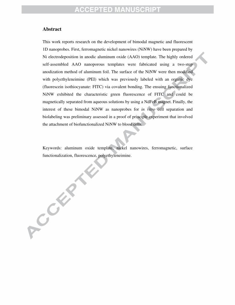
This work reports research on the development of bimodal magnetic and fluorescent
1D nanoprobes. First, ferromagnetic nickel nanowires (NiNW) have been prepared by
Ni electrodeposition in anodic aluminum oxide (AAO) template. The highly ordered
self-assembled AAO nanoporous templates were fabricated using a two-step
anodization method of aluminum foil. The surface of the NiNW were then modified
with polyethyleneimine (PEI) which was previously labeled with an organic dye
(fluorescein isothiocyanate: FITC) via covalent bonding. The ensuing functionalized
NiNW exhibited the characteristic green fluorescence of FITC and could be
magnetically separated from aqueous solutions by using a NdFeB magnet. Finally, the
interest of these bimodal NiNW as nanoprobes for in vitro cell separation and
biolabeling was preliminary assessed in a proof of principle experiment that involved
the attachment of biofunctionalized NiNW to blood cells.
Keywords: aluminum oxide template, nickel nanowires, ferromagnetic, surface
functionalization, fluorescence, polyethyleneimine.
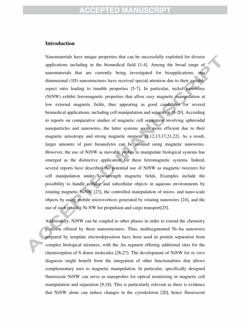
Introduction
Nanomaterials have unique properties that can be successfully exploited for diverse
applications including in the biomedical field [1-4]. Among the broad range of
nanomaterials that are currently being investigated for bioapplications, one
dimensional (1D) nanostructures have received special attention due to their variable
aspect ratio leading to tunable properties [5-7]. In particular, nickel nanowires
(NiNW) exhibit ferromagnetic properties that allow easy magnetic manipulation at
low external magnetic fields, thus appearing as good candidates for several
biomedical applications, including cell manipulation and separation [8-20]. According
to reports on comparative studies of magnetic cell separation involving spheroidal
nanoparticles and nanowires, the latter systems seem more efficient due to their
magnetic anisotropy and strong magnetic moment [8,12,13,17,21,22]. As a result,
larger amounts of pure bioanalytes can be isolated using magnetic nanowires.
However, the use of NiNW as movable probes to manipulate biological systems has
emerged as the distinctive application for these ferromagnetic systems. Indeed,
several reports have described the potential use of NiNW as magnetic tweezers for
cell manipulation under low-strength magnetic fields. Examples include the
possibility to handle cellular and subcellular objects in aqueous environments by
rotating magnetic NiNW [23], the controlled manipulation of micro- and nano-scale
objects by using mobile microvortices generated by rotating nanowires [24], and the
use of such rotating Ni NW for propulsion and cargo transport[25].
Additionally, NiNW can be coupled to other phases in order to extend the chemistry
platform offered by these nanostructures. Thus, multisegmented Ni-Au nanowires
prepared by template electrodeposition have been used in protein separation from
complex biological mixtures, with the Au segment offering additional sites for the
chemisorption of S donor molecules [26,27]. The development of NiNW for in vitro
diagnosis might benefit from the integration of other functionalities that allows
complementary uses to magnetic manipulation. In particular, specifically designed
fluorescent NiNW can serve as nanoprobes for optical monitoring in magnetic cell
manipulation and separation [9,18]. This is particularly relevant as there is evidence
that NiNW alone can induce changes in the cytoskeleton [20], hence fluorescent
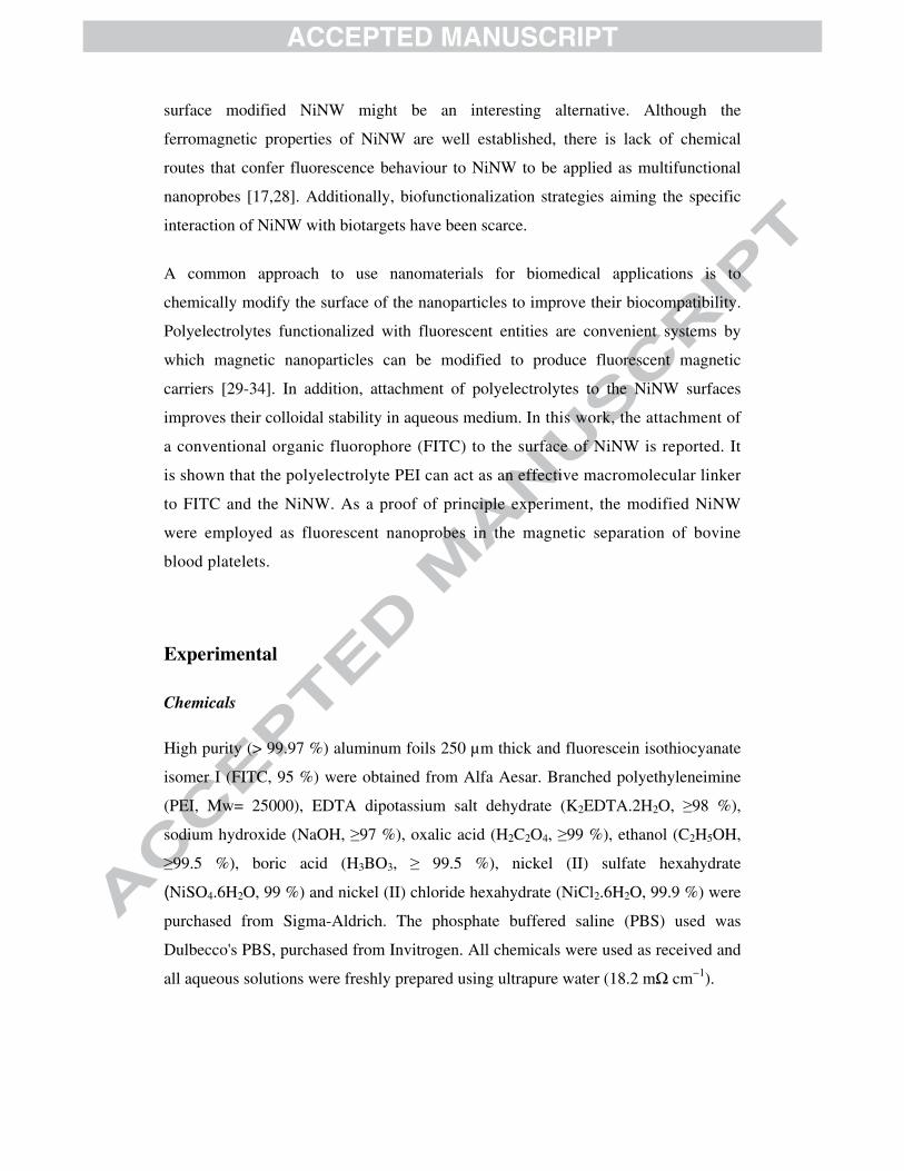
surface modified NiNW might be an interesting alternative. Although the
ferromagnetic properties of NiNW are well established, there is lack of chemical
routes that confer fluorescence behaviour to NiNW to be applied as multifunctional
nanoprobes [17,28]. Additionally, biofunctionalization strategies aiming the specific
interaction of NiNW with biotargets have been scarce.
A common approach to use nanomaterials for biomedical applications is to
chemically modify the surface of the nanoparticles to improve their biocompatibility.
Polyelectrolytes functionalized with fluorescent entities are convenient systems by
which magnetic nanoparticles can be modified to produce fluorescent magnetic
carriers [29-34]. In addition, attachment of polyelectrolytes to the NiNW surfaces
improves their colloidal stability in aqueous medium. In this work, the attachment of
a conventional organic fluorophore (FITC) to the surface of NiNW is reported. It
is shown that the polyelectrolyte PEI can act as an effective macromolecular linker
to FITC and the NiNW. As a proof of principle experiment, the modified NiNW
were employed as fluorescent nanoprobes in the magnetic separation of bovine
blood platelets.
Experimental
Chemicals
High purity (> 99.97 %) aluminum foils 250 µm thick and fluorescein isothiocyanate
isomer I (FITC, 95 %) were obtained from Alfa Aesar. Branched polyethyleneimine
(PEI, Mw= 25000), EDTA dipotassium salt dehydrate (K2EDTA.2H2O, ≥98 %),
sodium hydroxide (NaOH, ≥97 %), oxalic acid (H2C2O4, ≥99 %), ethanol (C2H5OH,
≥99.5 %), boric acid (H3BO3, ≥ 99.5 %), nickel (II) sulfate hexahydrate
(NiSO4.6H2O, 99 %) and nickel (II) chloride hexahydrate (NiCl2.6H2O, 99.9 %) were
purchased from Sigma-Aldrich. The phosphate buffered saline (PBS) used was
Dulbecco's PBS, purchased from Invitrogen. All chemicals were used as received and
all aqueous solutions were freshly prepared using ultrapure water (18.2 mΩ cm−1).

Syntheses
Ni nanowires were fabricated by electrodeposition using anodized aluminum oxide
(AAO) membrane (Figure 1a) with cylindrical nanopores of 35 nm in diameter and 5
µm thick. These dimensions have been selected in order to obtain high aspect ratio
particles whose high coercivity and remanence lead to more efficient cell sorting and
bioseparation tasks [35]. Anodization of aluminum foil was carried out by a two-step
anodization process with 0.3 M oxalic acid as an anodizing solution at 40 V and 4 ºC.
Then, nickel was deposited into the pores by electroplating from an acid bath with
350 g/L NiSO4.6H2O, 45 g/L NiCl2.6H2O and 45 g/L H3BO3 at 40 ºC and pH 4,5 [36].
Finally, the AAO was dissolved in an aqueous solution of 0.2 M H2CrO4 and 0.4 M
H3PO4 at 60 C, releasing the NiNW from the membranes. Once in suspension, the
wires were collected with a magnet, washed with deionized water and in ethanol.
The synthesis of PEI-FITC was based on the reaction between the isothiocyanate
group of FITC and the primary amino group of polyethyleneimine [37]. A reaction
scheme for this process is shown in Figure 1b. The FITC (12.6 mg) was added to
312.6 mg PEI in 30 ml ultra-pure water. The resulting solution was kept in dark
conditions under magnetic stirring and after adjusting its pH to 11. Dialysis of free
FITC was carried out by immersing the dialysis membrane (molecular weight cut-off
12–14 kDa, Medicell International) containing the PEI-FITC solution into 1 L of
distilled water, previously placed in a beaker wrapped in aluminum foil, under
magnetic stirring and at room temperature. This process took one week, during which
the supernatant water has been exchanged daily; the dialysis membrane containing
PEI-FITC was kept in dark conditions except during the few minutes required to
water exchange.
As illustrated in Figure 1c, the NiNW were then functionalized with PEI-FITC.
Hence, 5 mg of the ensuing NiNW were mixed with 5 ml of an aqueous solution of
PEI/FITC at pH= 5.5. This mixture was mechanically stirred (600 rpm) at room
temperature for 1 h. The resulting functionalized nickel nanowires were collected as
powders by using a NdFeB magnet and were thoroughly washed with ultra-pure
water.
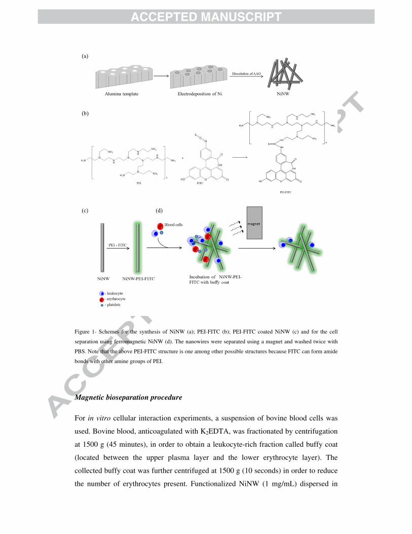
Figure 1- Schemes for the synthesis of NiNW (a); PEI-FITC (b); PEI-FITC coated NiNW (c) and for the cell
separation using ferromagnetic NiNW (d). The nanowires were separated using a magnet and washed twice with
PBS. Note that the above PEI-FITC structure is one among other possible structures because FITC can form amide
bonds with other amine groups of PEI.
Magnetic bioseparation procedure
For in vitro cellular interaction experiments, a suspension of bovine blood cells was
used. Bovine blood, anticoagulated with K2EDTA, was fractionated by centrifugation
at 1500 g (45 minutes), in order to obtain a leukocyte-rich fraction called buffy coat
(located between the upper plasma layer and the lower erythrocyte layer). The
collected buffy coat was further centrifuged at 1500 g (10 seconds) in order to reduce
the number of erythrocytes present. Functionalized NiNW (1 mg/mL) dispersed in

PBS were added to the cell suspension and rolled for 2 h at room temperature. The Ni
nanowires were then separated using a NdFeB magnet and were again resuspended in
PBS and washed twice with PBS (Figure 1d). For the fluorescence microscopy
studies, 50 µL aliquots of each experiment were placed into the wells of a 8-well μ-
Slide (Ibidi, GmbH) and observed with a Zeiss Axiovert 200 inverted fluorescence
microscope, using Zeiss Filter Set 02 (excitation: G 365; beam splitter: FT 395;
emission: LP 420), allowing excitation near the 365/366 nm mercury line and
detection of emission longer than 420 nm.
Characterization of the Ni nanowires
Fourier transform infrared (FTIR) spectra of FITC and PEI-FITC samples were
recorded using a spectrometer Bruker optics tensor 27 coupled to a horizontal
attenuated total reflectance (ATR) cell, using 256 scans at a resolution of 4 cm-1. The
X-ray powder diffraction patterns were recorded using a X-ray diffractometer Philips
X’Pert equipped with a CuKα monochromatic radiation source. Transmission electron
microscopy (TEM) analysis was performed by using a FEI Tecnai T20 microscope
operating at 200 keV. Samples for TEM analysis were prepared by placing an aliquot
of a dilute suspension of Ni NW on a copper grid coated with an amorphous carbon
film. For SEM analysis an aliquot of a dilute nanowires suspension was allowed to air
dry on glass slides and then were coated with evaporated carbon. SEM was performed
using a scanning electron microscope Hitachi SU70 operating at an accelerating
voltage of 25 kV. Zeta potential measurements were performed by using a Zetasizer
Nanoseries instrument of Malvern Instruments. Fluorescence measurements were
performed using a fluorometer FluoroMax3 – HORIBA Jobin Yvon and using quartz
cuvettes. The magnetization measurements were performed using a commercial
Vibrating Sample Magnetometer (VSM; LOT-Oriel EV7), operating at room
temperature, in which an electromagnet provides a magnetic field that reaches the
maximum value of 1.5 T (for a minimum gap of 3 cm) and also allows adjustable
angles between -180º to 180º during the measurements. The nickel samples employed
in the VSM measurements were prepared as follows. The alumina template was first
removed as described above. The powders were then thoroughly washed with water
and dried under a N2 stream. Finally the powders were placed in a diamagnetic sample
holder where the measurements were carried out.

Results and Discussion
The nickel nanowires (NiNW) used in this research have been fabricated by a
template-assisted method, using Ni electrodeposition in anodic aluminum oxide
(AAO) templates [38-41]. The morphological characteristics of NiNW prepared by
this method can be adjusted by the anodization voltage and time of the second step,
during the formation of the membrane in nanoporous AAO [40,41]. The anodizing
rate was set at 2.5 µm/h for a deposition time of two hours to produce NiNW with an
aspect ratio of 140. The resulting NiNW, after the template removal, were analyzed
by vibrating sample magnetometer (VSM) and powder X-ray diffraction (XRD), as
well as, by scanning and transmission electron microscopies (SEM and TEM). Figure
2 shows the SEM and TEM images for a typical NiNW sample obtained in these
conditions.
Figure 2- SEM (top) and TEM (bottom) images of nickel nanowires prepared by electrodeposition in alumina
template.
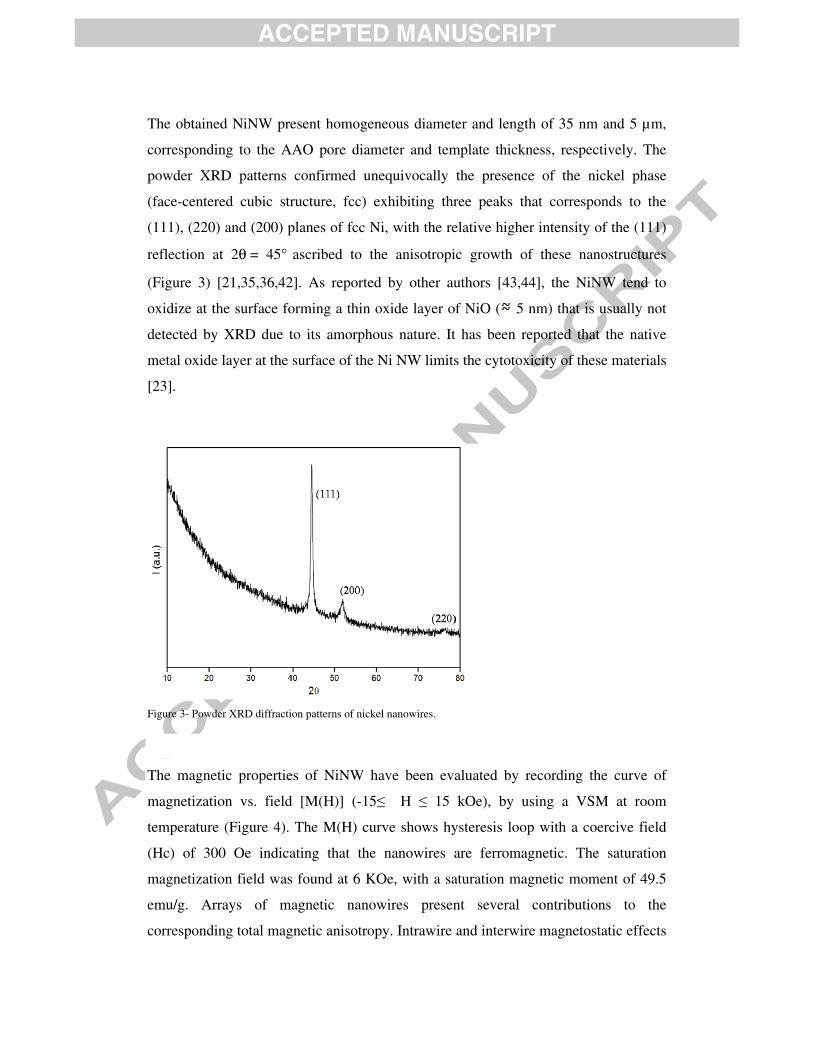
The obtained NiNW present homogeneous diameter and length of 35 nm and 5 µm,
corresponding to the AAO pore diameter and template thickness, respectively. The
powder XRD patterns confirmed unequivocally the presence of the nickel phase
(face-centered cubic structure, fcc) exhibiting three peaks that corresponds to the
(111), (220) and (200) planes of fcc Ni, with the relative higher intensity of the (111)
reflection at 2θ = 45° ascribed to the anisotropic growth of these nanostructures
(Figure 3) [21,35,36,42]. As reported by other authors [43,44], the NiNW tend to
oxidize at the surface forming a thin oxide layer of NiO ( 5 nm) that is usually not
detected by XRD due to its amorphous nature. It has been reported that the native
metal oxide layer at the surface of the Ni NW limits the cytotoxicity of these materials
[23].
Figure 3- Powder XRD diffraction patterns of nickel nanowires.
The magnetic properties of NiNW have been evaluated by recording the curve of
magnetization vs. field [M(H)] (-15≤ H ≤ 15 kOe), by using a VSM at room
temperature (Figure 4). The M(H) curve shows hysteresis loop with a coercive field
(Hc) of 300 Oe indicating that the nanowires are ferromagnetic. The saturation
magnetization field was found at 6 KOe, with a saturation magnetic moment of 49.5
emu/g. Arrays of magnetic nanowires present several contributions to the
corresponding total magnetic anisotropy. Intrawire and interwire magnetostatic effects

are the dominant features of the anisotropy in packed nanowires. These are the key
factors to control the coercive field (Hc) and that influence the magnetization reversal
processes [35]. Figure 4 presents the typical hysteresis loop for dispersed Ni NWs
after alumina template removal. Out of the template, randomly orientated Ni NWs
present a behavior similar to that observed for Ni nanoparticles with a small Hc of
300 Oe and HSat = 6 kOe due to the dipole-dipole interactions (interwire coupling)
that overcomes the shape anisotropy, rotating the magnetic easy axis towards an
intermediate direction for the nanowires [35].
Figure 4- Room temperature M(H) curve for Ni NW powders obtained after alumina template removal and placed
onto a diamagnetic sample holder.
Chemical surface modification methods were carried out in order to upgrade the
NiNW for fluorescent biolabeling, which together with their intrinsic ferromagnetic
characteristics, allows the potential use as multimodal bioprobes. The initial stage
along the NiNW surface functionalization involved the grafting of the fluorophore
FITC onto the polymer chains of the polyelectrolyte PEI. This has been confirmed by
ATR-FTIR spectroscopy, as shown in Figure 5, by comparing the spectrum of PEI-
FITC with that one of FITC, the latter shows the characteristic band corresponding to
the stretch vibration of the NCS group (2038 cm-1) which is absent in the spectrum of
the conjugate. The modification of polyethyleneimine (PEI) with fluorescein
isothiocyanate (FITC) involves reaction between the NCS group of FITC with amine
group (NHR group) of PEI, resulting in a thiourea bond (SC(NR)2). Other bands

indicating the presence of FITC are those observed at 1209 cm-1, 1508 cm-1, 1170 cm-
1, that can be assigned respectively to C-O-C stretching, C=C stretching, and =C-H
bending (Figure 6). The shift of the band at 1618 cm-1 in the PEI-FITC spectrum, in
relation to the band at 1589 cm-1 in the PEI spectrum, may be due to the stretching
vibration of C=C bond of FITC. The deviation of the vibrational bands corresponding
to the C-N and C-C stretching of PEI, at 1115 cm-1 and 1049 cm-1, respectively, to
1026 cm-1 and 966 cm-1, is due to proximity of the FITC xanthene ring. In fact, the
electronic effects of neighboring groups and hydrogen bonds significantly affect the
vibrational modes of a covalent bond [45-47].
Figure 5- FT-Infrared spectra (1800-2200 cm-1 region) for the fluorophore FITC and the modified polyelectrolyte
PEI-FITC.

Figure 6- ATR-FTIR of polyelectrolyte PEI and modified polyelectrolyte PEI-FITC.
The functionalized PEI-FITC polyelectrolyte was used to modify the surface of the
NiNW. Therefore aqueous suspensions of NiNW were treated with solutions of the
functionalized polyelectrolyte and the powders were magnetically separated. In
Figure 7, the fluorescence emission spectra of suspensions of these powders exhibit
the characteristic absorption due to FITC. We note that these suspensions have been
prepared from NiNW that have been magnetically separated and thoroughly washed
with distilled water. Hence, the fluorescence observed for the NiNW samples is due to
FITC that has been grafted onto the PEI, whose macromolecular chains are interacting
with the NiNW surfaces. The bathochromic shift of 16 nm observed in the emission
maximum (Figure 7) between PEI-FITC and the fluorophore FITC has previously
been reported and is related to the changes of the local environment of dye molecules,
such as changes in polarity or polarizability [48,49]. In PEI-FITC, the number of dye
molecules that are in each others vicinity increases causing interactions between
neighboring molecules, which lowers their excited state energy and produces a red
shift in the spectra. These interactions are less favored at the NiNW-PEI-FITC sample

once the polyelectrolyte is adsorbed at the nanowire surface, thus explaining the less
pronounced shift observed in the spectrum of the PEI-FITC modified NiNW sample.
Figure 7- Fluorescence spectra of FITC, PEI conjugated with FITC (PEI-FITC), and functionalized NiNW with
PEI-FITC (NiNW-PEI-FITC) (λex= 494nm).
Additional evidence for the surface modification of the NiNW was obtained by
measuring the zeta potential prior and after treatment with FITC functionalized PEI, a
cationic polyelectrolyte. Hence, after surface modification of the nanowires with PEI-
FITC a positive zeta potential was obtained (ζ= +49.8 mV, pH 6) as compared to ζ= -
33.5 mV, for the non-modified NiNW and at the same pH. The interactions that take
place between the polyelectrolyte and the nanowires surface have predominant
electrostatic character. However, other types of interactions cannot be excluded, such
as hydrogen bonding between surface OH groups (due to nickel oxidation described
above) and the amino groups of PEI [50].
Finally, preliminary experiments were carried out as proof of principle for the
multifunctionality associated to the modified NiNW as magnetic and fluorescent
bioprobes, using a suspension of bovine blood cells. Figure 8 shows the results of
optical microscopy applied to the modified NiNW that have contacted the cell
suspension and were magnetically separated and washed. The optical transmission
image in Figure 8b shows areas containing cells with the morphological
characteristics of leukocytes bound to micrometric nanowire aggregates, which
indicate close interaction between the cells and the modified NiNW. In fluorescence
mode, these areas appear with the typical green fluorescence of FITC (Figure 8c).

Figure 8- Optical microscopy image of (a) buffy coat showing the three types of blood cells; (b) NiNW/PEI/FITC
after contact with buffy coat followed by magnetic separation and washing with PBS (transmission mode); and (c)
NiNW/PEI/FITC after contact with buffy coat (fluorescence mode) (λex= 365 nm).

Previous studies, which employed leukocyte suspensions, have shown that surface
modification with PEI of polyurethane films/filters improved leukocyte removal and
adhesion to surfaces; however, no improvement was found when whole blood was
used [51,52]. Leukocytes have a negative zeta potential [53] and therefore are prone
to interact strongly with the positive surface of the NiNW. However, other types of
interactions are certainly involved, such as hydrogen bonding between groups on the
leukocyte membrane and electronegative nitrogen atoms of PEI [52-54]. Moreover, it
has been widely accepted that surface wettability and hydrophilicity also influence the
adhesion of leukocytes [53,54]. As such, the mechanism of the cell−PEI-NiNW
interaction cannot be anticipated at this stage.
Leukocyte adhesion plays a predominant role in leukocyte depletion, which is applied
clinically to remove these cells from blood. In some situations, such as in blood
transfusion, it is necessary to reduce the concentration of leukocytes in blood, in order
to prevent leukocyte-mediated adverse effects [51,54,55]. Therefore, the materials
described here may contribute to develop additional tools for the depletion of
leukocytes, taking advantage of the ferromagnetic properties of nickel nanowires.
Conclusions
In conclusion, ferromagnetic nickel nanowires prepared on anodic aluminum oxide
template have been surface modified with polyethyleneimine with minimal effects on
their magnetic separation from aqueous solutions. An improvement on this surface
modification strategy comprises the functionalization of the polyelectrolyte with
fluorescein isothiocyanate. In this way, not only the NiNW can be used as
ferromagnetic nanodrivers or nanotweezers for magnetic biomanipulation procedures,
but optical labeling is also possible due to the green fluorescence typical of the
organic dye. Although a detailed study on the interaction of the modified NiNW with
the blood cells in variable conditions was out of the scope of this research, these
results suggest that these materials might be useful in magnetic
separation/manipulation and fluorescent labeling of leukocytes, for which the
functionalized NiNWs seem to have stronger affinity. However, the use of these
systems has yet to be established in real in vitro bioanalysis by evaluating the diverse

variables that could affect their performance. This aspect has great relevance namely
due to the possibility of interactions between the NiNW surfaces and other blood
cells. Future research on these fluorescent ferromagnetic NiNW might bring further
evidence for their potential use in magnetic manipulation of blood cells by taking into
account specific biorecognition issues.
Acknowledgements
We thank Fundação para a Ciência e Tecnologia (NANO/NMed-SD/0140/2007,
ERA-Eula/0003/2009 and Pest-C/CTM/LA0011/2011), FSE and POPH for funding.
We also thank Luísa Cortes (Center for Neurosciences and Cell Biology, Coimbra,
Portugal) for valuable technical assistance with the microscope. Célia T. Sousa is also
thankful to FCT for post-doctoral grant SFRH/BPD/82010/2011.

References
[1] T. Trindade, A. L. Daniel-da-Silva, Nanocomposite Particles for Bio-Applications
– Materials and Bio-Interfaces, Pan Stanforf Publishing: Singapore, 2011.
[2] G. A. Ozin, A. C. Arsenault, L. Cademartiri, Nanochemistry: A Chemical
Approach to nanomaterials; Royal Society of Chemistry: Toronto, 2005.
[3] S. H. Lee, J. H. Sung, T. H. Park, Ann. Biomed. Eng., 40 (2012) 1384.
[4] Q. A. Pankhurst, J. Connolly, S. K. Jones, J. Dobson, J. Phys. D.: Appl. Phys., 36
(2003) R167.
[5] N. Chopra, V. G. Gavalas, B. J. Hinds, L. G. Bachas, Anal. Lett., 40 (2007) 2067.
[6] C. D. Keating, M. Natan, Adv. Mater., 15 (2003) 451.
[7] J. Wang, ChemPhysChem, 10 (2009) 1748.
[8] M. Vazquez, K. Pirota, M. Hernandez-Velez, V. M. Prida, D. Navas, R. Sanz, F.
Batallan, J. Velazquez, J. Appl. Phys., 95 (2004) 6642.
[9] A. Prina-Mello, Z. Diao, J. M. D. Coey, J. Nanobiotechnology, 4 (2006) 1.
[10] M. Tanase, E. J. Felton, D. S. Gray, A. Hultgren, C. S. Chen, D. H. Reich, Lab
Chip, 5 (2005) 598.
[11] D. Choi, A. Fung, H. Moon, D. Ho, Y. Chen, E. Kan, Y. Rheem, B. Yoo, N.
Myung, Biomed. Microdevices, 9 (2007) 143.
[12] N. Gao, X. Yang, Y. T. Tsai, G. M. Chu, H. Wang, E. H. Yang, Proc. of SPIE,
7318 (2009) 73181E-1.
[13] A. Hultgren, M. Tanase, C. S. Chen, G. J. Meyer, D. H. Reich, J. Appl. Phys., 93
(2003) 7554.
[14] A. Hultgren, M. Tanase, C. S. Chen, D. H. Reich, IEEE Tran. Magnetics, 40
(2004) 2988.
[15] D. H. Reich, M. Tanase, A. Hultgren, L. A. Bauer, C. S. Chen, G. J. Meyer, J.
Appl. Phys., 93 (2003) 7275.
[16] L. A. Bauer, N. S. Birenbaum, G. J. Meyer, J. Mater. Chem., 14 (2004) 517.
[17] A. Hultgren, M. Tanase, E. J. Felton, K. Bhadriraju, A. K. Salem, C. S. Chen, D.
H. Reich, Biotechnol. Prog., 21 (2005) 509.
[18] A. O. Fung, V. Kapadia, E. Pierstorff, D. Ho, Y. J. Chen, Phys. Chem. C, 112
(2008) 15085.
[19] N. Gao, H. Wang, E. H. Yang, Nanotechnology, 21 (2010) 1.
[20] F. Johansson, M. Jonsson, K. Alm, M. Kanje, Exp. Cell Res., 316 (2010) 688.
[21] F. Byrne, A. Prina-Mello, A. Whelan, B. Mohamed, A. Davies, Y. Gunko, J. M.
D. Coey, Y. Volkov, J. Magn. Magn. Mater., 321 (2009) 1341.
[22] A. A. Wang, J. Lee, G. Jenikova, A. Mulchandani, N. V. Myung, W. Chen,
Nanotechnology, 17 (2006) 3375.
[23] L. Zhang, T. Petit, K. E. Peyer, B. J. Nelson, Nanomedicine, 7 (2012) 1074.
[24] T. Petit, L. Zhang, K. E. Peyer, B. E. Kratochvil, B. J. Nelson, Nano Lett., 12
(2012) 156.
[25] L. Zhang, T. Petit, Y. Lu, B. E. Kratochvil, K. E. Peyer, R. Pei, J. Lou, B. J.
Nelson, ACS Nano, 4 (2010) 6228.
[26] K. B. Lee, S. Park, C. A. Mirkin, Angew. Chem. Int., 43 (2004) 3048.
[27] N. S. Birenbaum, B. T. Lai, C. S. Chen, D. H. Reich, G. J. Meyer, Langmuir, 19
(2003) 95802.
[28] C. L. Chien, L. Sunb, M. Tanasea, L. A. Bauerc, A. Hultgrena, D. M. Silevitcha,
G. J. Meyerc, P. C. Searsonb, D. H. Reich, J. Magn. Magn. Mater., 249 (2002) 146.
[29] C. Kaewsaneha, P. Opaprakasit, D. Polpanich, S. Smanmoo, P. Tangboriboonrat,
J. Coll. Interf. Sci., 377 (2012) 145.

[30] W. Xu, J. Y. Park, K. Kattel, M. W. Ahmad, B. A. Bony, W. C. Heo, S. Jin, J.
W. Park, Y. Chang, T. J. Kim, J. A. Park, J. Y. Do, K. S. Chae, G. H. L. Lee, RSC
Adv., 2 (2012) 10907.
[31] Y. Okamoto, F. Kitagawa, K. Otsuka, Anal. Chem., 79 (2007) 3041.
[32] Y. Ge, Y. Zhang, S. He, F. Nie, G. Teng, N. Gu, Nanoscale Res. Lett., 4 (2009)
287.
[33] P. Govindaiah, T. Hwang, H. Yoo, Y. S. Kim, S. J. Lee, S. W. Choi, J. H. Kim, J.
Coll. Interf. Sci., 379 (2012) 27.
[34] X. Hong, J. Li, M. Wang, J. Xu, W. Guo, J. Li, Y. Bai, T. Li, Chem. Mater., 16
(2004) 4022.
[35] M. Vazquez, K. Pirota, J. Torrejon, D. Navas, M. Hernandez-Velez, J. Magn.
Magn. Mater., 294 (2005) 174.
[36] M. P. Proença, C.T. Sousa, J. Ventura, M. Vazquez, J.P. Araújo, Electrochim.
Acta, 72 (2012) 215.
[37] G. T. Hermanson, Bioconjugate Techniques; Academic Press: London, 1996.
[38] M. Lai, D. J. Riley, J. Coll. Interf. Sci., 323 (2008) 203.
[39] J. P. O'Sullivan, G. C. Wood, Proc. R. Soc. Mater., 128 (2000) 582.
[40] C. T. Sousa, D. C. Leitão, M. P.Proença, A. Apolinário, J. G. Correia,; J.
Ventura, J. P. Araújo, Nanotechnology, 22 (2011) 1.
[41] W. Lee, R. Ji, U. Gosele, K. Nielsch, Nature Mater., 9 (2006) 741.
[42] G. B. Cheng, G. C. Wei, C. J. Hao, S. T. Li, Electron. Mater. Lett., 5 (2009) 123.
[43] Z. F. Zhou, Y. C. Zhou, Y. Pan, W. X. Lei, C. F. Xu, Scr. Mater., 60 (2009) 512.
[44] L. He, Z. M. Liao, H. C. Wu, X. X. Tian, D. S. Xu, G. L. W. Cross, G. S.
Duesberg, I. V. Shvets, D. P. Yu, Nano Lett., 11 (2011) 4601.

[45] A. Lex, P. Pacher, O. Werzer, A. Track, Q. Shen, R. Schennach, G. Koller, G.
Hlawacek, E. Zojer, R. Resel, M. Ramsey, C. Teichert, W. Kern, G. Trimmel, Chem.
Mater., 20 (2008) 2009.
[46] B. H. Stuart, Infrared Spectroscopy: Fundamentals and applications. Wiley: New
York, 2004.
[47] L. P. Donald, M. L. Gary, S. K. George, R. V. James, Introduction to
spectroscopy. Brooks/Cole: California, 2009.
[48] E. W. Voss Jr., J. C. Croney, D. M. Jameson, J. Protein Chem., 21 (2002) 231.
[49] A. Imhof, M. Megens, J. J. Engelberts, D. T. N. Lang, R. Sprik, W. L. Voss, J.
Phys. Chem. B, 103 (1999) 1408.
[50] J. Gregory, S. Barany, Adv. Colloid Interface Sci., 169 (2011) 1.
[51] A. Bruil, H. A. Oosterom, I. Steneker, B. J. M. Al, T. Beugeling, W. G. Aken, J.
Feijen, J. Biomed. Mater. Res., 27 (1993) 1253.
[52] A. Bruil, J. G. A. Terlingen, T. Beugeling, W. G. Aken, J. Feijen, Biomaterials,
13 (1992), 915.
[53] M. M. B. Ribeiro, M. M. Domingues, J. M. Freire, N. C. Santos, M. A. R. B.
Castanho, Front. Cell. Neurosci., 6 (2012) 1.
[54] A. Bruil, T. Beugeling, J. Feijen, W. G. Aken, Transfus. Med. Rev., 9 (1995)
145.
[55] S. Singh, A. Kumar, Biotechnol. J., 4 (2009) 1140.
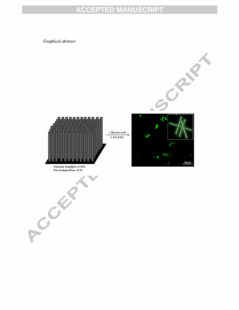
Graphical abstract

Highlights
• Nickel electrodeposition in anodic aluminum oxide template forming nanowires.
• Functionalization of polyethyleneimine with a fluorophore (FITC)
• Ferromagnetic nanowires functionalized with FITC- grafted polyethyleneimine.
• New bimodal magnetic/fluorescent nanoprobes for in vitro experiments.