Functionalizable hydrogel microparticles of tunable size and stiffness for soft-tissue filler...
11
Functionalizable hydrogel microparticles of tunable size and stiffness for soft-tissue filler applications Ka Man Carmen Chan a,b , Randolph H. Li a,b , Joseph W. Chapman a , Eric M. Trac b , James B. Kobler a , Steven M. Zeitels a , Robert Langer b , Sandeep S. Karajanagi a,⇑ a Massachusetts General Hospital, Center for Laryngeal Surgery and Voice Rehabilitation, 70 Blossom Street, Boston, MA 02114, USA b Department of Chemical Engineering, Massachusetts Institute of Technology, 77 Massachusetts Ave, Cambridge, MA 02139, USA article info Article history: Received 4 November 2013 Received in revised form 17 January 2014 Accepted 12 February 2014 Available online 21 February 2014 Keywords: Soft-tissue filler Hydrogel Microparticles Suspension photopolymerization abstract Particle size, stiffness and surface functionality are important in determining the injection site, safety and efficacy of injectable soft-tissue fillers. Methods to produce soft injectable biomaterials with controlled particle characteristics are therefore desirable. Here we report a method based on suspension photopo- lymerization and semi-interpenetrating network (semi-IPN) to synthesize soft, functionalizable, spherical hydrogel microparticles (MP) of independently tunable size and stiffness. MP were prepared using acry- lated forms of polyethylene glycol (PEG), gelatin and hyaluronic acid. Semi-IPN MP of PEG-diacrylate and PEG were used to study the effect of process parameters on particle characteristics. The process param- eters were systematically varied to produce MP with size ranging from 115 to 515 lm and stiffness rang- ing from 190 to 1600 Pa. In vitro studies showed that the MP thus prepared were cytocompatible. The ratio and identity of the polymers used to make the semi-IPN MP were varied to control their stiffness and to introduce amine groups for potential functionalization. Slow-release polymeric particles loaded with Rhodamine or dexamethasone were incorporated in the MP as a proof-of-principle of drug incorpo- ration and release from the MP. This work has implications in preparing injectable biomaterials of natural or synthetic polymers for applications as soft-tissue fillers. Ó 2014 Acta Materialia Inc. Published by Elsevier Ltd. All rights reserved. 1. Introduction Injectable biomaterials are used in medicine to augment lost or compromised tissue in order to restore form and/or function to the affected region and represent a large market, worth billions of dol- lars annually. Examples of such biomaterials include dermal fillers, used to treat facial defects; viscosupplements, used to treat osteo- arthritis of the knee; and biomaterials, used for augmenting the urethral wall to treat urinary incontinence. Novel injectable biomaterials continue to be developed to address this growing injectable-fillers market. Further, in a small subset of patients, adverse events, such as granulomas, swelling, erythema and nod- ules, occur even with US Food and Drug Administration (FDA) approved or cleared injectables [1–5]. There is thus significant interest in developing methods to prepare new and putatively better injectable biomaterials. Most injectable fillers are particulate or break down into partic- ulates upon injection [6]. The size, shape, mechanical properties and surface chemistry of the particulates are known to influence their in vivo performance. For example, silicone particles of less than 70 lm have been shown to migrate away from the periure- thral injection site and cause complications [7]. The migration potential of an implant is important for determining its safety as per the FDA’s guidelines for vocal fold applications [8]. Phagocyto- sis of polymethymethacrylate particles by macrophages is also size dependent [9]. Polymethylmethacrylate particles less than 50 lm in size or irregular in shape induce greater production of tumor necrosis factor alpha, an inflammatory cytokine, than particles that are spherical in shape or are between 50 and 350 lm in size in vivo [10]. Particles with acute angles, such as triangles, also induce a greater foreign body response than spherical particles [11]. Surface chemistry is also known to impact the in vivo response to the injected materials [12–15]. For example, polyurethane functional- ized with zwitterionic or anionic functional groups showed lower in vivo inflammatory reaction than those functionalized with cat- ionic chemistries [16]. Similarly, polypropylene particles coated with carboxylic acid groups were less inflammatory than those coated with amine or hydroxyl groups [14]. Particle characteristics also influence the choice of in vivo application of the filler. For example, the stiffness of a filler needs to be different depending upon its injection location, functional http://dx.doi.org/10.1016/j.actbio.2014.02.021 1742-7061/Ó 2014 Acta Materialia Inc. Published by Elsevier Ltd. All rights reserved. ⇑ Corresponding author. Tel.: +1 518 495 2959; fax: +1 617 724 9949. E-mail address: [email protected] (S.S. Karajanagi). Acta Biomaterialia 10 (2014) 2563–2573 Contents lists available at ScienceDirect Acta Biomaterialia journal homepage: www.elsevier.com/locate/actabiomat
Transcript of Functionalizable hydrogel microparticles of tunable size and stiffness for soft-tissue filler...
Acta Biomaterialia 10 (2014) 2563–2573
Contents lists available at ScienceDirect
Acta Biomaterialia
journal homepage: www.elsevier .com/locate /actabiomat
Functionalizable hydrogel microparticles of tunable size and stiffnessfor soft-tissue filler applications
http://dx.doi.org/10.1016/j.actbio.2014.02.0211742-7061/� 2014 Acta Materialia Inc. Published by Elsevier Ltd. All rights reserved.
⇑ Corresponding author. Tel.: +1 518 495 2959; fax: +1 617 724 9949.E-mail address: [email protected] (S.S. Karajanagi).
Ka Man Carmen Chan a,b, Randolph H. Li a,b, Joseph W. Chapman a, Eric M. Trac b, James B. Kobler a,Steven M. Zeitels a, Robert Langer b, Sandeep S. Karajanagi a,⇑a Massachusetts General Hospital, Center for Laryngeal Surgery and Voice Rehabilitation, 70 Blossom Street, Boston, MA 02114, USAb Department of Chemical Engineering, Massachusetts Institute of Technology, 77 Massachusetts Ave, Cambridge, MA 02139, USA
a r t i c l e i n f o
Article history:Received 4 November 2013Received in revised form 17 January 2014Accepted 12 February 2014Available online 21 February 2014
Keywords:Soft-tissue fillerHydrogelMicroparticlesSuspension photopolymerization
a b s t r a c t
Particle size, stiffness and surface functionality are important in determining the injection site, safety andefficacy of injectable soft-tissue fillers. Methods to produce soft injectable biomaterials with controlledparticle characteristics are therefore desirable. Here we report a method based on suspension photopo-lymerization and semi-interpenetrating network (semi-IPN) to synthesize soft, functionalizable, sphericalhydrogel microparticles (MP) of independently tunable size and stiffness. MP were prepared using acry-lated forms of polyethylene glycol (PEG), gelatin and hyaluronic acid. Semi-IPN MP of PEG-diacrylate andPEG were used to study the effect of process parameters on particle characteristics. The process param-eters were systematically varied to produce MP with size ranging from 115 to 515 lm and stiffness rang-ing from 190 to 1600 Pa. In vitro studies showed that the MP thus prepared were cytocompatible. Theratio and identity of the polymers used to make the semi-IPN MP were varied to control their stiffnessand to introduce amine groups for potential functionalization. Slow-release polymeric particles loadedwith Rhodamine or dexamethasone were incorporated in the MP as a proof-of-principle of drug incorpo-ration and release from the MP. This work has implications in preparing injectable biomaterials of naturalor synthetic polymers for applications as soft-tissue fillers.
� 2014 Acta Materialia Inc. Published by Elsevier Ltd. All rights reserved.
1. Introduction
Injectable biomaterials are used in medicine to augment lost orcompromised tissue in order to restore form and/or function to theaffected region and represent a large market, worth billions of dol-lars annually. Examples of such biomaterials include dermal fillers,used to treat facial defects; viscosupplements, used to treat osteo-arthritis of the knee; and biomaterials, used for augmenting theurethral wall to treat urinary incontinence. Novel injectablebiomaterials continue to be developed to address this growinginjectable-fillers market. Further, in a small subset of patients,adverse events, such as granulomas, swelling, erythema and nod-ules, occur even with US Food and Drug Administration (FDA)approved or cleared injectables [1–5]. There is thus significantinterest in developing methods to prepare new and putativelybetter injectable biomaterials.
Most injectable fillers are particulate or break down into partic-ulates upon injection [6]. The size, shape, mechanical propertiesand surface chemistry of the particulates are known to influence
their in vivo performance. For example, silicone particles of lessthan 70 lm have been shown to migrate away from the periure-thral injection site and cause complications [7]. The migrationpotential of an implant is important for determining its safety asper the FDA’s guidelines for vocal fold applications [8]. Phagocyto-sis of polymethymethacrylate particles by macrophages is also sizedependent [9]. Polymethylmethacrylate particles less than 50 lmin size or irregular in shape induce greater production of tumornecrosis factor alpha, an inflammatory cytokine, than particles thatare spherical in shape or are between 50 and 350 lm in size in vivo[10]. Particles with acute angles, such as triangles, also induce agreater foreign body response than spherical particles [11]. Surfacechemistry is also known to impact the in vivo response to theinjected materials [12–15]. For example, polyurethane functional-ized with zwitterionic or anionic functional groups showed lowerin vivo inflammatory reaction than those functionalized with cat-ionic chemistries [16]. Similarly, polypropylene particles coatedwith carboxylic acid groups were less inflammatory than thosecoated with amine or hydroxyl groups [14].
Particle characteristics also influence the choice of in vivoapplication of the filler. For example, the stiffness of a filler needsto be different depending upon its injection location, functional

2564 K.M.C. Chan et al. / Acta Biomaterialia 10 (2014) 2563–2573
requirements and desired residence time. Fillers used to treat uri-nary incontinence need to be stiff to impart strength, while facialfillers need to be relatively soft. Hyaluronic acid facial fillers witha lower degree of crosslinking, which are therefore softer, have ashorter residence time [17]. Further softer facial fillers are usedin superficial injections around the lips and eyes, while stiffer fill-ers are used to fill deeper wrinkles [18–20]. Also, in the cases ofPerlane� and Restylane�, both manufactured by the same com-pany, the former contains larger particulates and is used to fill dee-per wrinkles, while Restylane�, with smaller particulates, issuggested for use on superficial wrinkles [19].
The size, morphology, stiffness and surface chemistry of bioma-terial particulates contribute to their in vivo application and per-formance. However, current manufacturing technology platformsoffer limited independent control of particle size, shape and sur-face chemistry. Hyaluronic acid (HA) based soft fillers are madeusing top-down methods involving bulk polymerization, followedby screening and homogenization to break down the bulk gel intoirregularly shaped, polydisperse particles [6,18]. For example, Rad-iesse VoiceGel� and Restylane� particulates are of irregular shapesand sizes. Other injectable fillers, such as Zyderm� and Cymetra�,result in particulates of irregular shapes and polydisperse sizesupon injection through a needle. Some fillers, such as Radiesse�
and Deflux�, contain polydisperse yet controlled-size sphericalmicroparticles (MP) of hydroxylapatite or dextranomers to impartstiffness.
Current methods to synthesize hydrogel MP of controlled sizeand shape include microfabrication-based techniques [21–24].While microfabrication techniques provide excellent control oversize and shape, the methods are complex and ‘‘intrinsically moreexpensive’’, and have low throughput and yield [21]. Furthermore,the use of low molecular weight (MW) polymers at high concentra-tions to achieve high conversions often result in MP with relativelyhigh stiffness [25,26]. Developing methods that allow preparationof sufficient quantities of injectable fillers, especially with charac-teristics suitable for use as soft-tissue filler and independent con-trol over its size, mechanical properties and surface chemistry, isdesirable.
Here we report a suspension–polymerization-based method tocreate spherical, controlled-size hydrogel MP with tunable sizeand stiffness using photopolymerization. While some suspensionphotopolymerization-based methods have been reported earlierto prepare spherical microparticles for drug delivery [27–29], useof this method to prepare soft MP with tunable properties foruse as injectable fillers remains relatively unexplored. Polyethyl-ene glycol (PEG) was chosen to synthesize the MP because of itsextensive use in FDA-approved or cleared products and its use inpreparing biocompatible materials. HA and gelatin were chosenbecause of their favorable biocompatibility profiles, history of usein commercial dermal fillers and use in preparing soft, viscoelastichydrogels [17]. Another motivation for using HA and gelatin to pre-pare MP was to demonstrate the generality of our method. Thesemi-interpenetrating technology (semi-IPN), which involvescrosslinking of a polymer in the presence of a non-crosslinkedpolymer, was used to prepare the MP because this technologyallows the preparation of materials with tunable properties byvarying parameters such as the polymers used, their relative MWand their concentrations. Photocrosslinking was chosen becauseit can be carried out in ambient conditions, is efficient and affordsprecise spatiotemporal control over the crosslinking. The sizedistribution of suspension-polymerized MP can change as the reac-tion proceeds and, unlike chemical or thermal crosslinking, light-based crosslinking provides better control over the terminationof the reaction [30]. Irgacure 2959 (I2959) was chosen as thephotoinitiator (PI) because of its better cytocompatibility relativeto other commercially available photoinitiators and its extensive
use in preparing biomaterials, including those that encapsulatemammalian cells [31–34]. Furthermore, PEG hydrogels preparedusing PEG diacrylate (PEG-DA) and I2959 were shown to be safein humans in a 15-patient clinical trial for articular cartilage repair,thereby making the use of this system attractive [35]. In addition,GelrinC, a CE Mark-approved product in Europe, is also an in situpolymerized PEG-DA and I2959-based hydrogel that was shownto be safe and effective for the treatment of articular cartilage in in-jured knees over a period of 24 months in a multi-center clinicaltrial in Europe [36].
Semi-IPN MP of PEG-DA and PEG were prepared using suspen-sion photopolymerization. The effect of process parameters suchas stirring speed, surfactant concentration, gelation time, PI concen-tration and UV intensity on MP size and yield was investigated. Invitro cytocompatibility of the PEG MP was evaluated using theMTT assay. MP were also prepared using acrylated versions of HAand gelatin. The feasibility of preparing functionalizable MP andMP that encapsulate drug-releasing particles was also explored.
2. Materials and methods
2.1. Materials
PEG and PEG-DA (MW of both: 10 kDa) were purchased from AlfaAesar (Lancaster, UK) and SunBio (Anyang City, South Korea), respec-tively. Dioctyl sulfosuccinate (AOT, sodium salt, 99%), n-hexane, PEGbis(amine) (MW: 3400 Da), trinitrobenzenesulfonic acid (TNBS),sodium dodecyl sulfate (SDS), sodium bicarbonate, hydrochloricacid, dexamethasone, polyvinyl alcohol (PVA), Rhodamine 6G,gelatin (300 g bloom strength, Type A from porcine skin) and meth-acrylic anhydride were from Sigma–Aldrich (St. Louis, MO). Sodiumhyaluronate (MW: 351–600 kDa) and 2-hydroxy-1-[4-(2-hyroxy-ethoxy)phenyl]-2-methyl-1-propanone (Irgacure 2959, PI) were fromLifecore Biomedical (Chaska, MN) and Ciba (Tarrytown, NY), respec-tively. Sulfosuccinimidyl acetate (sulfo-NHS-acetate) was fromThermo Scientific (Rockford, IL). Polyoxyethylene (20) SorbitanMonoeleate (Tween 80) was from VWR (West Chester, PA). Ester-terminated 75:25 poly(DL-lactide-co-glycolide) (PLGA; inherentviscosity: 0.55–0.75 dl g�1) was from Durect Corporation (Cupertino,CA). India ink was from Becton, Dickinson and Company (Cockeysville,MD). The MTT cell proliferation assay kit and NIH/3T3 fibroblastcells were from American Type Culture Collection (Mannassas,VA). Cell culture media, serum, phosphate-buffered saline (PBS),and trypsin were from Invitrogen (Carlsbad, CA). Radiesse VoiceGel�, Restylane� and Zyderm� were from Merz Aesthetics (SanMateo, CA), Medicis Aesthetics (Scottsdale, AZ) and Allergan (Irvine,CA), respectively. All chemicals were used as received.
2.2. Preparation of PEG hydrogel MP
Solutions of PEG and PEG-DA (100 mg ml�1 each) in deionized(DI) water were mixed in a pre-determined volumetric ratio. PIwas added to this solution (0.5 mg ml�1 final concentration) toprepare the precursor solution (1 ml), which was then added toan AOT solution in hexane (9 ml) in a 2 inch wide polypropylenebeaker. This suspension was then stirred at a predetermined speedusing a 1 inch long stir bar on an IKA RET basic ETS-D5 magneticstir plate. While being stirred, the suspension was photopolymer-ized at 72 mW cm�2 (measured at 365 nm at the level of the baseof the beaker) with an Omnicure Series 2000 UV lamp for 260 s.Unless otherwise noted, the AOT concentration was 2 mM andthe stirring speed was 800 rpm. Post-polymerization, thesuspension was centrifuged at 150 rcf for 2 min and the organicsupernatant was removed. The resulting MP were washed threetimes, once with 0.1 vol.% Tween-80 and twice with DI water using
K.M.C. Chan et al. / Acta Biomaterialia 10 (2014) 2563–2573 2565
centrifugation, and finally separated from the solution using a40 lm nylon cell strainer. Excess water was wicked from the MPusing Kimwipes. The volumetric percentage yield was estimatedas volumetric yield = (volume of MP judged visually in a calibratedsyringe/volume of PEG precursor solution) � 100. The gels or MPprepared using a precursor solution containing 50 vol.% PEG-DAare referred to as PEG50, while those prepared using 75% PEG-DAare referred to as PEG75, and so forth.
2.3. Preparation of PEG hydrogel particulates of uncontrolled size
The PEG precursor solution (1 ml) was photopolymerized at2 mW cm�2 for 200 s in a 12-well plate. After polymerization, thehydrogel was incubated in PBS at a 1:9 ratio (v/v) at 37 �C over-night. The swollen hydrogels were added to a syringe and brokeninto smaller particulates by shearing them twice each throughsequentially narrower (16, 18, 20 and 22 gauge) needles.
2.4. Preparation of MP from gelatin, HA and PEG-diamine
Gelatin methacrylate (GelMA) was synthesized using gelatin(0.1 g ml�1) and methacrylic anhydride (1 vol.%) as per a method de-scribed earlier [37]. To prepare MP, a solution of GelMA(100 mg ml�1) and PI (0.5 mg ml�1) was made in DI water at 50 �C.Photopolymerization was carried out as described earlier for PEGMP at 800 rpm and 72 mW cm�2 for 30 s with an AOT concentrationof 1 mM. Post-polymerization, the hexane–AOT was removed andthe GelMA MP formed were stirred in water (15 ml) to preventagglomeration and then washed to remove the residual hexane–AOT, as outlined for PEG MP. HA-methacrylate (HAMA) was synthe-sized using a method described earlier [38]. HAMA MP wereprepared using a method identical to that used for GelMA MP butwith HAMA and AOT concentrations of 10 mg ml�1 and 5 mM,respectively. Semi-IPN MP of HAMA and gelatin were prepared usinga 75:25 (v/v) ratio of HAMA (10 mg ml�1) and gelatin (100 mg ml�1),with the same polymerization parameters as those for GelMA MPbut with an AOT concentration of 2.5 mM. To prepare PEG-diamineMP, PEG was replaced with PEG-diamine (200 mg ml�1) in the pro-cedure described earlier for PEG MP, and a stirring speed of 600 rpmand an AOT concentration of 0.5 mM were used.
2.5. Preparation of NS–MP and MS–MP hybrid particles
PLGA nanospheres (NS) encapsulated with dexamethasone(Dex) were prepared using a method reported earlier, but withdichloromethane and acetone (3:2 volumetric ratio) as the solventfor PLGA and Dex [39]. To prepare Rhodamine-encapsulated PLGAmicrospheres (MS), PLGA (300 mg) and Rhodamine (30 mg) wereco-dissolved in dichloromethane (2.25 ml), added to a PVA solution(10 mg ml�1) and homogenized at 9000 rpm for 5 min with anL4RT-A homogenizer. The organic solvent was removed in a rotaryevaporator and by repeated washing with water. The NS and MSwere lyophilized overnight to obtain dry powders. To prepareNS–MP or MS–MP conjugates, dry PLGA NS with Dex (40 mg ml�1)or PLGA MS with Rhodamine (10 mg ml�1) were suspended in PEGprecursor solution and added to AOT/hexane solution (0.5 mM),photopolymerized and washed as described earlier for PEG MPparticles.
2.6. Determination of loading and release rate of dexamethasone inNS–MP hybrid particles
The hybrid NS–MP were thoroughly washed to remove any NSsticking to the surface of the MP. To quantify the amount of Dexencapsulated, the NS–MP hybrid particles (200 mg) were hydro-lyzed with NaOH (1 N, 200 ll), neutralized with HCl (1 N),
dissolved in acetonitrile (4 ml) and then the Dex concentration inthis solution was determined using HPLC as per previous methods[40]. To determine the Dex release rate, the NS–MP particles(�100 mg) were placed inside a cellulose ester dialysis tubing(MWCO 10 kDa, Spectrum Labs, Rancho Dominguez, CA) and thenplaced in a rotating falcon tubes filled with PBS (50 ml) at 37 �C.The PBS was collected at 6 hours, 1 day and 10 days and replen-ished with fresh PBS, and the Dex in the collected PBS was deter-mined using HPLC.
2.7. Determination and blocking of free amine groups
Free amine groups in PEG-diamine MP were estimated using acolorimetric TNBS assay described elsewhere [41,42]. A calibrationcurve made using PEG-diamine standards ranging from 0 to10 mg ml�1 was used to estimate the amount of free amines. Theamine groups of the PEG-diamine MP were blocked by reactingthe MP with an aqueous sulfo-NHS-acetate solution (25� molarexcess, 1 ml) for 1 h at room temperature. Excess sulfo-NHS-acetate solution was removed by washing twice with DI waterand the remaining free amine groups were determined using theTNBS assay as described above.
2.8. Determination of particle size distribution
Particle size distributions of the MP and MS were determinedusing a Coulter Counter Multisizer III (Beckman Coulter, Inc., Brea,CA) as per the manufacturer’s protocol. The particle size of the NSwas determined using a Zetasizer Nano (Malvern Instruments,Malvern, UK). The particle size was reported as the D90 (weightedby number percentage), where D90 represents the diameter that isequal to or greater than that of 90% of the particles in the sample.The relative span that describes the polydispersity of the particleswas calculated as the relative span = (D90 � D10)/D50, where thedefinitions of D10 and D50 are analogous to that of D90.
2.9. Measurement of viscoelastic properties of MP
The viscoelastic properties of MP were measured using anAR-2000 rheometer (TA Instruments, Inc., New Castle, DE) with aparallel plate geometry at 37 �C. Care was taken to ensure thatno excess liquid was present in the MP. Based on previous reports,a gap height of five times the D90 was used [43,44]. For example,for MP prepared by stirring at 800 and 400 rpm, the gap heightswere 750 and 4000 lm, respectively. To eliminate wall slip, a320 grit sand paper (36 lm) was attached to the upper and lowerplates. The parallel plate diameter was at least 10 times the gapheight. The shear storage modulus and loss modulus of MP weremeasured as a function of frequency from 1 to 10 Hz at a strainof 0.6% at 37 �C and reported at 10 Hz.
2.10. In vitro cytocompatibility
The cytocompatibility of MP was measured using the MTT cellproliferation assay. PEG50 MP (20 mM AOT, 400 rpm) were synthe-sized in a laminar flow hood using a precursor solution filteredthrough a 0.2 lm filter. Fibroblast (NIH/3T3) cells were plated in24-well plates (40,000 cells ml�1) and allowed to attach in Dul-becco’s modified Eagle’s medium with 10 vol.% calf serum and1 vol.% penicillin–streptomycin. Sheared hydrogel particulatesand PEG50 MP were added separately to the cells. To determineif leachates from the MP and the sheared gel are cytotoxic, bothmaterials were incubated in the medium for 3 days at concentra-tions varying from 0 to 50 mg ml�1. Samples were centrifugedand the supernatant medium was collected after 3 days. This med-ium was then used to culture the cells. SDS was used as a positive

2566 K.M.C. Chan et al. / Acta Biomaterialia 10 (2014) 2563–2573
control. After 3 days of culture, cell proliferation in each well wasdetermined as per the manufacturer’s instructions. Since the pur-ple precipitate produced by viable cells was absorbed by the MPand sheared PEG particulates, the solutions were solubilized indetergent overnight and then read at 570 nm, with the referenceread at 670 nm. Normalized viability is reported as ((absorbanceat 570 nm � absorbance at 670 nm) � (absorbance of blank))/(absorbance of negative control – absorbance of blank).
2.11. Fluorescent confocal microscopy
Rhodamine–PLGA MS encapsulated within PEG MP were ex-cited at 526 nm and imaged with an Olympus Fluoview 1000microscope. Images were compiled using ImageJ software.
2.12. Statistics
All data are presented as mean ± standard deviation of at leastthree separate replicates. Student’s t-test was used with a 95% con-fidence interval to determine statistical significance unless other-wise noted.
3. Results
3.1. Preparation of MP
PEG hydrogel MP were prepared using suspension photopoly-merization of PEG-DA in presence of varying quantities of PEG ina hexane–water–AOT system [45]. The MP thus prepared werespherical in shape, as seen in the optical picture of PEG100 MP(Fig. 1A). A dilute solution of India ink was used as the contrastagent to better visualize the MP. While the MP could be injectedthrough a 22 gauge needle without distortion (Fig. S1), whenpassed through a 25 gauge needle, a few particles (possibly largerones) became distorted, though the majority remained unchanged(Fig. 1B). However, no clogging of the 25 gauge needle was ob-served. The regular shape of the PEG100 MP is in contrast to theirregular shape of commercially available injectable fillers, suchas Radiesse� Voice Gel, Restylane� and Zyderm� (Fig. 1D, E andF, respectively). The PEG100 MP were polydisperse in size, withan average diameter of 96 lm, a median diameter of 87 lm, a typ-ical D90 value of 162 lm and a relative span value of 1.47 (Fig. 2).Similar size, shape and injectability data were obtained for MPmade using other PEG-DA:PEG ratios (not shown). MP were alsoprepared using acrylated forms of HA, a biopolymer that has beenused extensively to prepare injectables, and gelatin with D90(span) values of 136 ± 22 lm (1.5 ± 0.2) and 157 ± 4 lm(1.9 ± 0.6), respectively (Figs. 1C and S2). Further semi-IPN MP ofHA–methacrylate and gelatin with D90 (span) value of150 ± 38 lm (2.2 ± 0.2) were also prepared (Fig. S3).
3.2. Effect of process parameters on particle characteristics
The effect of a process parameter on particle size distributionand yield was investigated while keeping other parameters con-stant. The particle size of PEG50 MP decreased with increasing stir-ring speed. For example, increasing the stirring speed from 400 to600 rpm decreased the D90 from 515 ± 50 to 140 ± 33 lm (Fig. 3A).However, increasing the stirring speed beyond 600 rpm did notcause a statistically significant change in size. Similarly, increasingthe stirring speed from 400 to 1000 rpm decreased the volumetricyield from 110 ± 15 to 43 ± 11%. (The volumetric yield can be great-er than 100% because the spherical MP have void spaces whenpacked together.) In addition, MP swell due to absorption of waterduring the washing step. Consequently, the volume of the MPobtained may be higher than the precursor solution volume. The
relative span of the MP decreased from 2.9 ± 0.2 at 400 rpm to2.1 ± 0.7 at 500 rpm, though this difference between the twospeeds was not statistically significant. Between 600 and1000 rpm the relative span ranged from 1.4 to 1.7, with nostatistically significant difference within this stirring speed range.However, this decrease in the relative span at higher speeds wasstatistically significant relative to that at 400 or 500 rpm. Doublingthe PI concentration to 1 mg ml�1 increased the D90 of PEG50 MPfrom 142 ± 25 to 225 ± 22 lm, with no statistically significantchange in the relative span. Similarly, doubling the UV intensityto 144 mW cm�2 increased the D90 to 231 ± 35 lm, with nochange in the relative span. A PI concentration beyond 1 mg ml�1
was not tested because of concerns about potential toxicity [33].Also, the effect of UV intensity on MP characteristics beyond144 mW cm�2 was not tested because higher intensities led toheating and evaporation-related changes in the suspension duringpolymerization. Using other UV exposure times of 30, 65, 130 and520 s did not change the PEG MP size or the relative span under theconditions tested (data not shown).
Increasing the AOT concentration from 0.5 to 5 mM decreased theD90 from 254 ± 16 to 103 ± 21 lm and decreased the volumetricyield from 88 ± 9 to 21 ± 4%, while the relative span remained un-changed at between 1.5 and 1.7 (Fig. 3B). Based on the above generalrelations, MP with D90 as small as 48 and 39 lm were prepared byusing the parameters [AOT] = 50 mM, speed = 750 rpm, UV inten-sity = 365 mW cm�2, UV time = 480 s, [PI] = 1 mg ml�1 and [AOT] =50 mM, speed = 400 rpm, UV = 72 mW cm�2, UV time = 260 s,[PI] = 0.5 mg ml�1, respectively. However, the yield of particles thusobtained was only 3%, hence these conditions were not explored fur-ther. Overall, by varying the process parameters, spherical MP withaverage D90 in the range of 39–515 lm were prepared.
3.3. MP mechanical characterization
The viscoelastic properties of MP can be controlled by varyingthe PEG-DA:PEG ratio and by varying the particle size. Regardlessof the particle size, increasing the PEG-DA:PEG ratio increasedthe shear storage modulus (G0) and shear loss modulus (G00) ofthe MP (Fig. 4A). For MP prepared at 400 rpm, the G0 increased from523 ± 31 to 1599 ± 110 Pa and the G00 increased from 38 ± 3 to111 ± 5 Pa when PEG-DA:PEG ratio (v/v) was increased from 50to 100% while the size stayed the same. Similarly, for MP preparedusing a stirring speed of 800 rpm, the G0 increased from 188 ± 63 to1257 ± 105 Pa and the G00 increased from 15 ± 6 to 100 ± 13 Pa withincreasing PEG-DA:PEG ratio without a change in MP size. For a gi-ven PEG-DA:PEG ratio, the G0 and G00 increased with decreasing par-ticle size. At all PEG-DA:PEG ratios and particle sizes, G00 is at leastone order of magnitude smaller than G0. The G00 was lower than G0
at all frequencies of measurement from 1 to 10 Hz (Fig. S4). Thecomplex viscosity of the MP decreased as the frequency of oscilla-tion was increased, suggesting a shear-thinning behavior for theMP (Fig. 4B). Decreasing the PEG-DA:PEG ratio or decreasingthe particle size decreased the complex viscosity of the MP. All ofthe changes in G0 and G00 described here were statistically significant.
3.4. In vitro cytocompatibility
We tested whether the PEG MP or materials leached from theMP are cytotoxic using NIH/3T3 cells as model mammalian cells.PEG50 particulates of uncontrolled size that were not exposed tohexane or AOT at any step of their synthesis were made by shear-ing a PEG50 gel through needles and used as the control. The sameconcentrations (ranging from 0 to 50 mg ml�1) of PEG50 MP andPEG50 particulates were incubated over NIH/3T3 cells for 3 daysand the cell viability was evaluated using the MTT assay. Morethan 80% of the cells were viable when exposed to PEG50 at

Fig. 1. Optical micrographs of (A) PEG100 MP, (B) PEG100 MP passed through a 25 gauge needle, (C) hyaluronic acid MP, (D) Radiesse� Voice Gel, (E) Restylane� and (F)Zyderm�, with India ink as the contrast agent.
Fig. 2. Particle size distribution of PEG100 MP.
K.M.C. Chan et al. / Acta Biomaterialia 10 (2014) 2563–2573 2567
concentrations ranging from 0.1 to 50 mg ml�1, with the lowestnormalized viability of 80.9 ± 0.8% seen at the highest MP concen-tration (Fig. 5A). In addition, minimal to no toxicity was seen whencells were grown in medium that was incubated for 3 days withjust the MP or particulates (Fig. 5B).
3.5. Introduction of amine groups for potential surfacefunctionalization
Amine groups were introduced in the MP by replacing the PEGin the PEG50 MP with PEG-diamine. The PEG50-amine MP thusprepared had an average of �33 ± 2 free amine groups per nm2
of the MP surface, as estimated using the TNBS assay. If the amines
are assumed to be uniformly distributed throughout the sphericalMP and the outer 5% layer of the MP represents its functionalizablesurface, about 14% of the amines, i.e. 4.7 ± 0.3 free amine groups,are present per nm2 of the MP surface. After reaction withamine-reactive sulfo-NHS-acetate, the amine groups of thePEG50-amine MP decreased to �15%, suggesting that 85 ± 10% ofthe amines are accessible and reactive to small-molecule reactants.
3.6. Encapsulation and release of dexamethasone from MS–MP hybridparticles
Rhodamine was encapsulated in PLGA MS, and the PLGA–Rhodamine MS were then incorporated inside PEG MP. Rhodamine
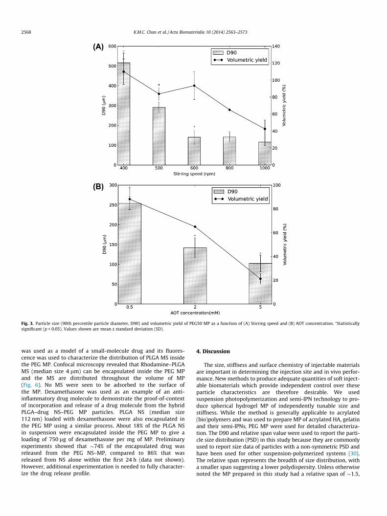
Fig. 3. Particle size (90th percentile particle diameter, D90) and volumetric yield of PEG50 MP as a function of (A) Stirring speed and (B) AOT concentration. ⁄Statisticallysignificant (p < 0.05). Values shown are mean ± standard deviation (SD).
2568 K.M.C. Chan et al. / Acta Biomaterialia 10 (2014) 2563–2573
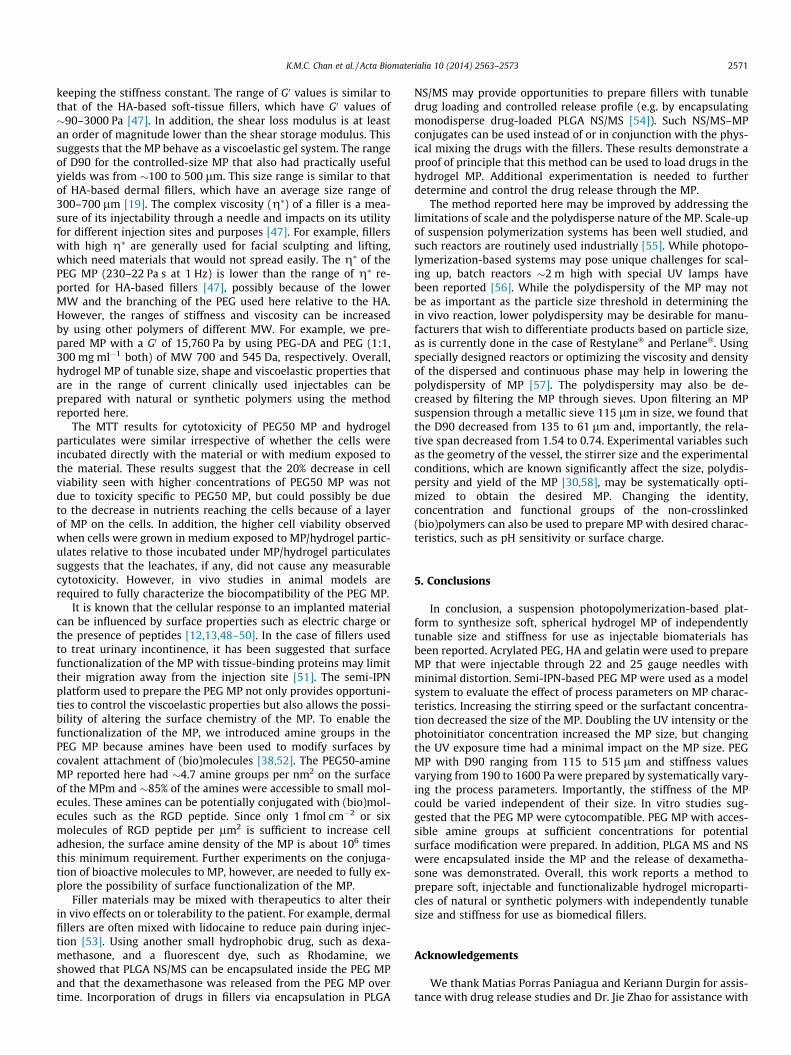
was used as a model of a small-molecule drug and its fluores-cence was used to characterize the distribution of PLGA MS insidethe PEG MP. Confocal microscopy revealed that Rhodamine–PLGAMS (median size 4 lm) can be encapsulated inside the PEG MPand the MS are distributed throughout the volume of MP(Fig. 6). No MS were seen to be adsorbed to the surface ofthe MP. Dexamethasone was used as an example of an anti-inflammatory drug molecule to demonstrate the proof-of-contextof incorporation and release of a drug molecule from the hybridPLGA–drug NS–PEG MP particles. PLGA NS (median size112 nm) loaded with dexamethasone were also encapsulated inthe PEG MP using a similar process. About 18% of the PLGA NSin suspension were encapsulated inside the PEG MP to give aloading of 750 lg of dexamethasone per mg of MP. Preliminaryexperiments showed that �74% of the encapsulated drug wasreleased from the PEG NS–MP, compared to 86% that wasreleased from NS alone within the first 24 h (data not shown).However, additional experimentation is needed to fully character-ize the drug release profile.
4. Discussion
The size, stiffness and surface chemistry of injectable materialsare important in determining the injection site and in vivo perfor-mance. New methods to produce adequate quantities of soft inject-able biomaterials which provide independent control over theseparticle characteristics are therefore desirable. We usedsuspension photopolymerization and semi-IPN technology to pro-duce spherical hydrogel MP of independently tunable size andstiffness. While the method is generally applicable to acrylated(bio)polymers and was used to prepare MP of acrylated HA, gelatinand their semi-IPNs, PEG MP were used for detailed characteriza-tion. The D90 and relative span value were used to report the parti-cle size distribution (PSD) in this study because they are commonlyused to report size data of particles with a non-symmetric PSD andhave been used for other suspension-polymerized systems [30].The relative span represents the breadth of size distribution, witha smaller span suggesting a lower polydispersity. Unless otherwisenoted the MP prepared in this study had a relative span of �1.5,
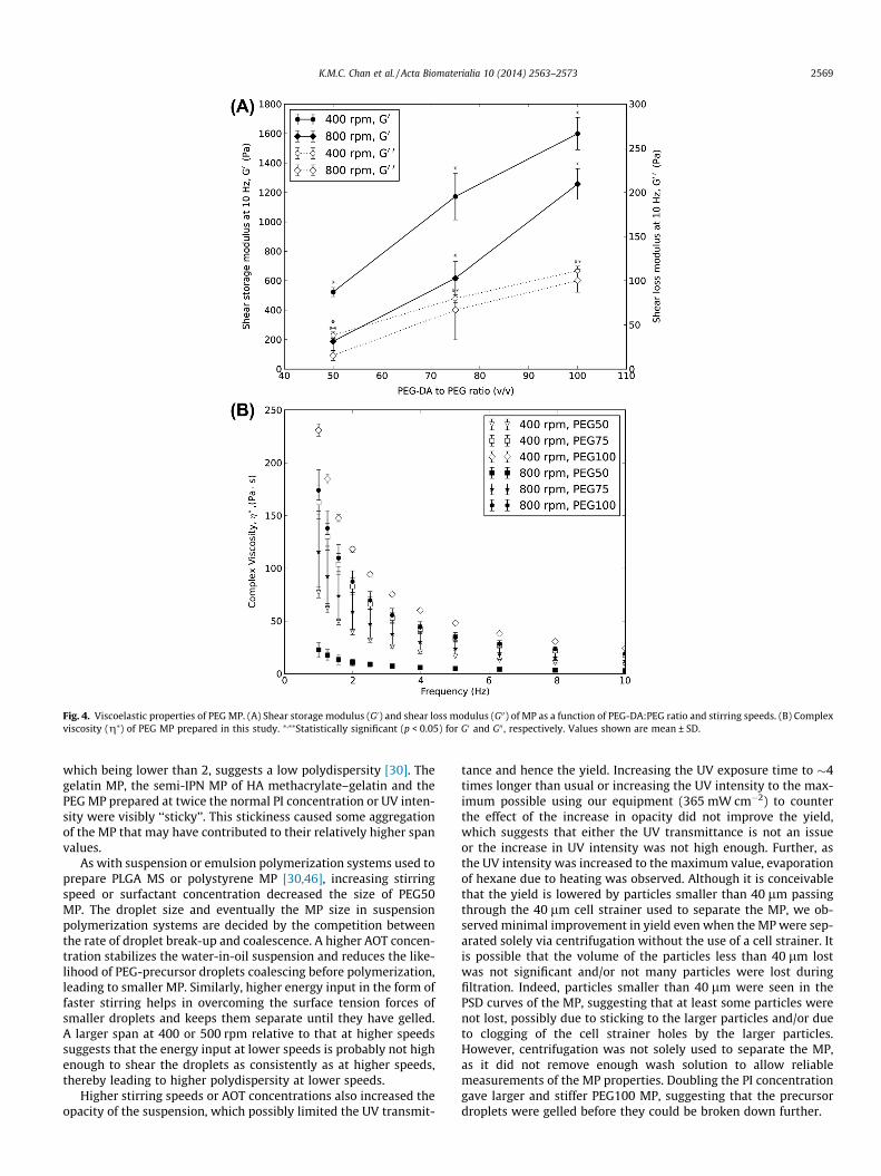
Fig. 4. Viscoelastic properties of PEG MP. (A) Shear storage modulus (G0) and shear loss modulus (G00) of MP as a function of PEG-DA:PEG ratio and stirring speeds. (B) Complexviscosity (g⁄) of PEG MP prepared in this study. ⁄,⁄⁄Statistically significant (p < 0.05) for G0 and G00 , respectively. Values shown are mean ± SD.
K.M.C. Chan et al. / Acta Biomaterialia 10 (2014) 2563–2573 2569
which being lower than 2, suggests a low polydispersity [30]. Thegelatin MP, the semi-IPN MP of HA methacrylate–gelatin and thePEG MP prepared at twice the normal PI concentration or UV inten-sity were visibly ‘‘sticky’’. This stickiness caused some aggregationof the MP that may have contributed to their relatively higher spanvalues.
As with suspension or emulsion polymerization systems used toprepare PLGA MS or polystyrene MP [30,46], increasing stirringspeed or surfactant concentration decreased the size of PEG50MP. The droplet size and eventually the MP size in suspensionpolymerization systems are decided by the competition betweenthe rate of droplet break-up and coalescence. A higher AOT concen-tration stabilizes the water-in-oil suspension and reduces the like-lihood of PEG-precursor droplets coalescing before polymerization,leading to smaller MP. Similarly, higher energy input in the form offaster stirring helps in overcoming the surface tension forces ofsmaller droplets and keeps them separate until they have gelled.A larger span at 400 or 500 rpm relative to that at higher speedssuggests that the energy input at lower speeds is probably not highenough to shear the droplets as consistently as at higher speeds,thereby leading to higher polydispersity at lower speeds.
Higher stirring speeds or AOT concentrations also increased theopacity of the suspension, which possibly limited the UV transmit-
tance and hence the yield. Increasing the UV exposure time to �4times longer than usual or increasing the UV intensity to the max-imum possible using our equipment (365 mW cm�2) to counterthe effect of the increase in opacity did not improve the yield,which suggests that either the UV transmittance is not an issueor the increase in UV intensity was not high enough. Further, asthe UV intensity was increased to the maximum value, evaporationof hexane due to heating was observed. Although it is conceivablethat the yield is lowered by particles smaller than 40 lm passingthrough the 40 lm cell strainer used to separate the MP, we ob-served minimal improvement in yield even when the MP were sep-arated solely via centrifugation without the use of a cell strainer. Itis possible that the volume of the particles less than 40 lm lostwas not significant and/or not many particles were lost duringfiltration. Indeed, particles smaller than 40 lm were seen in thePSD curves of the MP, suggesting that at least some particles werenot lost, possibly due to sticking to the larger particles and/or dueto clogging of the cell strainer holes by the larger particles.However, centrifugation was not solely used to separate the MP,as it did not remove enough wash solution to allow reliablemeasurements of the MP properties. Doubling the PI concentrationgave larger and stiffer PEG100 MP, suggesting that the precursordroplets were gelled before they could be broken down further.
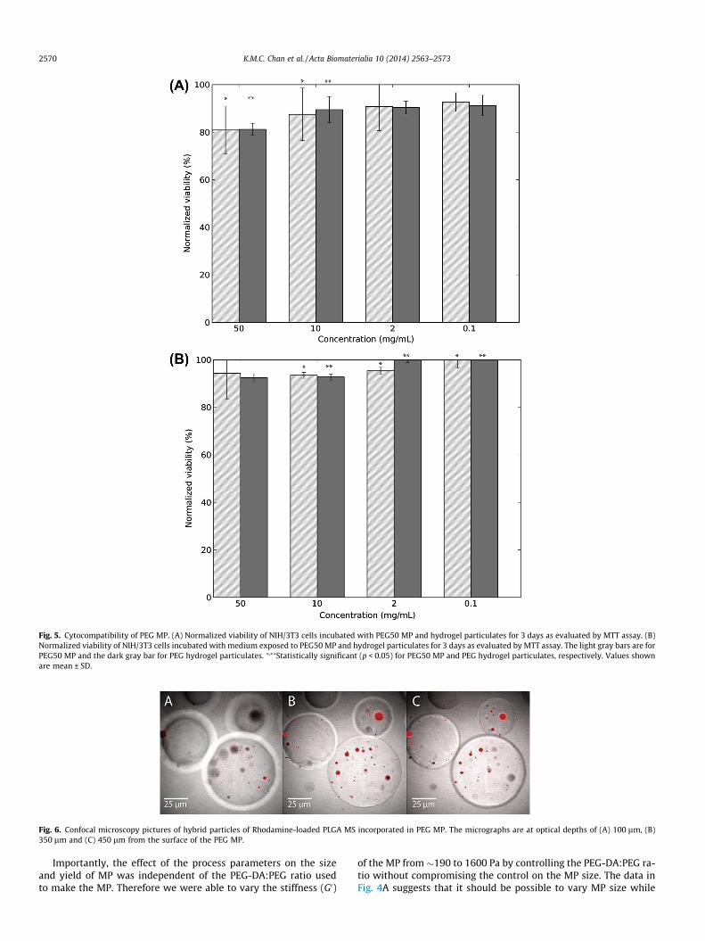
Fig. 5. Cytocompatibility of PEG MP. (A) Normalized viability of NIH/3T3 cells incubated with PEG50 MP and hydrogel particulates for 3 days as evaluated by MTT assay. (B)Normalized viability of NIH/3T3 cells incubated with medium exposed to PEG50 MP and hydrogel particulates for 3 days as evaluated by MTT assay. The light gray bars are forPEG50 MP and the dark gray bar for PEG hydrogel particulates. ⁄,⁄⁄Statistically significant (p < 0.05) for PEG50 MP and PEG hydrogel particulates, respectively. Values shownare mean ± SD.
Fig. 6. Confocal microscopy pictures of hybrid particles of Rhodamine-loaded PLGA MS incorporated in PEG MP. The micrographs are at optical depths of (A) 100 lm, (B)350 lm and (C) 450 lm from the surface of the PEG MP.
2570 K.M.C. Chan et al. / Acta Biomaterialia 10 (2014) 2563–2573
Importantly, the effect of the process parameters on the sizeand yield of MP was independent of the PEG-DA:PEG ratio usedto make the MP. Therefore we were able to vary the stiffness (G0)
of the MP from �190 to 1600 Pa by controlling the PEG-DA:PEG ra-tio without compromising the control on the MP size. The data inFig. 4A suggests that it should be possible to vary MP size while
K.M.C. Chan et al. / Acta Biomaterialia 10 (2014) 2563–2573 2571
keeping the stiffness constant. The range of G0 values is similar tothat of the HA-based soft-tissue fillers, which have G0 values of�90–3000 Pa [47]. In addition, the shear loss modulus is at leastan order of magnitude lower than the shear storage modulus. Thissuggests that the MP behave as a viscoelastic gel system. The rangeof D90 for the controlled-size MP that also had practically usefulyields was from �100 to 500 lm. This size range is similar to thatof HA-based dermal fillers, which have an average size range of300–700 lm [19]. The complex viscosity (g⁄) of a filler is a mea-sure of its injectability through a needle and impacts on its utilityfor different injection sites and purposes [47]. For example, fillerswith high g⁄ are generally used for facial sculpting and lifting,which need materials that would not spread easily. The g⁄ of thePEG MP (230–22 Pa s at 1 Hz) is lower than the range of g⁄ re-ported for HA-based fillers [47], possibly because of the lowerMW and the branching of the PEG used here relative to the HA.However, the ranges of stiffness and viscosity can be increasedby using other polymers of different MW. For example, we pre-pared MP with a G0 of 15,760 Pa by using PEG-DA and PEG (1:1,300 mg ml�1 both) of MW 700 and 545 Da, respectively. Overall,hydrogel MP of tunable size, shape and viscoelastic properties thatare in the range of current clinically used injectables can beprepared with natural or synthetic polymers using the methodreported here.
The MTT results for cytotoxicity of PEG50 MP and hydrogelparticulates were similar irrespective of whether the cells wereincubated directly with the material or with medium exposed tothe material. These results suggest that the 20% decrease in cellviability seen with higher concentrations of PEG50 MP was notdue to toxicity specific to PEG50 MP, but could possibly be dueto the decrease in nutrients reaching the cells because of a layerof MP on the cells. In addition, the higher cell viability observedwhen cells were grown in medium exposed to MP/hydrogel partic-ulates relative to those incubated under MP/hydrogel particulatessuggests that the leachates, if any, did not cause any measurablecytotoxicity. However, in vivo studies in animal models arerequired to fully characterize the biocompatibility of the PEG MP.
It is known that the cellular response to an implanted materialcan be influenced by surface properties such as electric charge orthe presence of peptides [12,13,48–50]. In the case of fillers usedto treat urinary incontinence, it has been suggested that surfacefunctionalization of the MP with tissue-binding proteins may limittheir migration away from the injection site [51]. The semi-IPNplatform used to prepare the PEG MP not only provides opportuni-ties to control the viscoelastic properties but also allows the possi-bility of altering the surface chemistry of the MP. To enable thefunctionalization of the MP, we introduced amine groups in thePEG MP because amines have been used to modify surfaces bycovalent attachment of (bio)molecules [38,52]. The PEG50-amineMP reported here had �4.7 amine groups per nm2 on the surfaceof the MPm and �85% of the amines were accessible to small mol-ecules. These amines can be potentially conjugated with (bio)mol-ecules such as the RGD peptide. Since only 1 fmol cm�2 or sixmolecules of RGD peptide per lm2 is sufficient to increase celladhesion, the surface amine density of the MP is about 106 timesthis minimum requirement. Further experiments on the conjuga-tion of bioactive molecules to MP, however, are needed to fully ex-plore the possibility of surface functionalization of the MP.
Filler materials may be mixed with therapeutics to alter theirin vivo effects on or tolerability to the patient. For example, dermalfillers are often mixed with lidocaine to reduce pain during injec-tion [53]. Using another small hydrophobic drug, such as dexa-methasone, and a fluorescent dye, such as Rhodamine, weshowed that PLGA NS/MS can be encapsulated inside the PEG MPand that the dexamethasone was released from the PEG MP overtime. Incorporation of drugs in fillers via encapsulation in PLGA
NS/MS may provide opportunities to prepare fillers with tunabledrug loading and controlled release profile (e.g. by encapsulatingmonodisperse drug-loaded PLGA NS/MS [54]). Such NS/MS–MPconjugates can be used instead of or in conjunction with the phys-ical mixing the drugs with the fillers. These results demonstrate aproof of principle that this method can be used to load drugs in thehydrogel MP. Additional experimentation is needed to furtherdetermine and control the drug release through the MP.
The method reported here may be improved by addressing thelimitations of scale and the polydisperse nature of the MP. Scale-upof suspension polymerization systems has been well studied, andsuch reactors are routinely used industrially [55]. While photopo-lymerization-based systems may pose unique challenges for scal-ing up, batch reactors �2 m high with special UV lamps havebeen reported [56]. While the polydispersity of the MP may notbe as important as the particle size threshold in determining thein vivo reaction, lower polydispersity may be desirable for manu-facturers that wish to differentiate products based on particle size,as is currently done in the case of Restylane� and Perlane�. Usingspecially designed reactors or optimizing the viscosity and densityof the dispersed and continuous phase may help in lowering thepolydispersity of MP [57]. The polydispersity may also be de-creased by filtering the MP through sieves. Upon filtering an MPsuspension through a metallic sieve 115 lm in size, we found thatthe D90 decreased from 135 to 61 lm and, importantly, the rela-tive span decreased from 1.54 to 0.74. Experimental variables suchas the geometry of the vessel, the stirrer size and the experimentalconditions, which are known significantly affect the size, polydis-persity and yield of the MP [30,58], may be systematically opti-mized to obtain the desired MP. Changing the identity,concentration and functional groups of the non-crosslinked(bio)polymers can also be used to prepare MP with desired charac-teristics, such as pH sensitivity or surface charge.
5. Conclusions
In conclusion, a suspension photopolymerization-based plat-form to synthesize soft, spherical hydrogel MP of independentlytunable size and stiffness for use as injectable biomaterials hasbeen reported. Acrylated PEG, HA and gelatin were used to prepareMP that were injectable through 22 and 25 gauge needles withminimal distortion. Semi-IPN-based PEG MP were used as a modelsystem to evaluate the effect of process parameters on MP charac-teristics. Increasing the stirring speed or the surfactant concentra-tion decreased the size of the MP. Doubling the UV intensity or thephotoinitiator concentration increased the MP size, but changingthe UV exposure time had a minimal impact on the MP size. PEGMP with D90 ranging from 115 to 515 lm and stiffness valuesvarying from 190 to 1600 Pa were prepared by systematically vary-ing the process parameters. Importantly, the stiffness of the MPcould be varied independent of their size. In vitro studies sug-gested that the PEG MP were cytocompatible. PEG MP with acces-sible amine groups at sufficient concentrations for potentialsurface modification were prepared. In addition, PLGA MS and NSwere encapsulated inside the MP and the release of dexametha-sone was demonstrated. Overall, this work reports a method toprepare soft, injectable and functionalizable hydrogel microparti-cles of natural or synthetic polymers with independently tunablesize and stiffness for use as biomedical fillers.
Acknowledgements
We thank Matias Porras Paniagua and Keriann Durgin for assis-tance with drug release studies and Dr. Jie Zhao for assistance with
2572 K.M.C. Chan et al. / Acta Biomaterialia 10 (2014) 2563–2573
confocal microscopy. This work was funded by the Voice HealthInstitute.
Appendix A. Supplementary data
Supplementary data associated with this article can be found, inthe online version, at http://dx.doi.org/10.1016/j.actbio.2014.02.021.
Appendix B. Figures with essential color discrimination
Certain figures in this article, particularly Fig. 6, is difficult tointerpret in black and white. The full color images can be foundin the on-line version, at http://dx.doi.org/10.1016/j.actbio.2014.02.021.
References
[1] Lowe NJ, Maxwell CA, Patnaik R. Adverse reactions to dermal fillers: review.Dermatol Surg 2005;31:1616–25.
[2] Sankar V, McGuff HS. Foreign body reaction to calcium hydroxylapatite afterlip augmentation. J Am Dent Assoc 2007;138:1093–6.
[3] Zielke H, Wolber L, Wiest L, Rzany B. Risk profiles of different injectable fillers:results from the Injectable Filler Safety Study (IFS Study). Dermatol Surg2008;34:326–35.
[4] Herschorn S. Current use of injectable agents for female stress urinaryincontinence. Rev Urol 2005;7(Suppl. 1):S12–21.
[5] Yang JH, Lee SM, Won CH, Chang SE, Lee MW, Choi JH, et al. Foreign bodygranuloma caused by hyaluronic acid/dextranomer microsphere fillerinjection. Int J Dermatol 2012;51:1517–8.
[6] Tezel A, Fredrickson GH. The science of hyaluronic acid dermal fillers. J CosmetLaser Ther 2008;10:35–42.
[7] Henly DR, Barrett DM, Weiland TL, O’Connor MK, Malizia AA, Wein AJ.Particulate silicone for use in periurethral injections: local tissue effects andsearch for migration. J Urol 1995;153:2039–43.
[8] FDA U. Guidance for industry and FDA staff vocal fold medialization devices –premarket notification [510 (k)] Submissions 2004;510.
[9] Morhenn VB, Lemperle G, Gallo RL. Phagocytosis of different particulatedermal filler substances by human macrophages and skin cells. Dermatol Surg2002;28:484–90.
[10] Gelb H, Schumacher HR, Cuckler J, Ducheyne P, Baker DG. In vivo inflammatoryresponse to polymethylmethacrylate particulate debris: effect of size,morphology, and surface area. J Orthop Res 1994;12:83–92.
[11] Matlaga BF, Yasenchak LP, Salthouse TN. Tissue response to implantedpolymers: the significance of sample shape. J Biomed Mater Res1976;10:391–7.
[12] Bellis SL. Advantages of RGD peptides for directing cell association withbiomaterials. Biomaterials 2011;32:4205–10.
[13] Brodbeck WG, Patel J, Voskerician G, Christenson E, Shive MS, Nakayama Y,et al. Biomaterial adherent macrophage apoptosis is increased byhydrophilic and anionic substrates in vivo. Proc Natl Acad Sci U S A2002;99:10287–92.
[14] Kamath S, Bhattacharyya D, Padukudru C, Timmons RB, Tang L. Surfacechemistry influences implant-mediated host tissue responses. J Biomed MaterRes A 2008;86:617–26.
[15] Thevenot P, Hu W, Tang L. Surface chemistry influences implantbiocompatibility. Curr Top Med Chem 2008;8:270–80.
[16] Khandwekar AP, Patil DP, Shouche Y, Doble M. Surface engineering ofpolycaprolactone by biomacromolecules and their blood compatibility. JBiomater Appl 2011;26:227–52.
[17] Fakhari A, Berkland C. Applications and emerging trends of hyaluronic acid intissue engineering, as a dermal filler and in osteoarthritis treatment. ActaBiomater 2013;9:7081–92.
[18] Edsman K, Nord LI, Ohrlund A, Larkner H, Kenne AH. Gel properties ofhyaluronic acid dermal fillers. Dermatol Surg 2012;38:1170–9.
[19] Kablik J, Monheit GD, Yu L, Chang G, Gershkovich J. Comparative physicalproperties of hyaluronic acid dermal fillers. Dermatol Surg 2009;35(Suppl1):302–12.
[20] Baumann L, Blyumin M, Saghari S. Dermal fillers. In: Weisberg E, editor.Cosmetic dermatology: principles and practice. Philadelphia, PA: McGraw-HillProfessional; 2007. p. 191–211.
[21] Helgeson ME, Chapin SC, Doyle PS. Hydrogel microparticles from lithographicprocesses: novel materials for fundamental and applied colloid science. CurrOpin Colloid Interface Sci 2011;16:106–17.
[22] Merkel TJ, Jones SW, Herlihy KP, Kersey FR, Shields AR, Napier M, et al. Usingmechanobiological mimicry of red blood cells to extend circulation times ofhydrogel microparticles. Proc Natl Acad Sci U S A 2011;108:586–91.
[23] Dendukuri D, Pregibon DC, Collins J, Hatton TA, Doyle PS. Continuous-flowlithography for high-throughput microparticle synthesis. Nat Mater2006;5:365–9.
[24] Xu S, Nie Z, Seo M, Lewis P, Kumacheva E, Stone HA, et al. Generation ofmonodisperse particles by using microfluidics: control over size, shape, andcomposition. Angew Chem Int Ed Engl 2005;44:724–8.
[25] Haghgooie R, Toner M, Doyle PS. Squishy non-spherical hydrogelmicroparticles. Macromol Rapid Commun 2010;31:128–34.
[26] Acharya G, Shin CS, McDermott M, Mishra H, Park H, Kwon IC, et al. Thehydrogel template method for fabrication of homogeneous nano/microparticles. J Control Release 2010;141:314–9.
[27] Sahiner N, Jha AK, Nguyen D, Jia XQ. Fabrication and characterization of cross-linkable hydrogel particles based on hyaluronic acid: potential application invocal fold regeneration. J Biomater Sci-Polym Ed 2008;19:223–43.
[28] John G, Morita M. Synthesis and characterization of photo-cross-linkednetworks based on L-lactide/serine copolymers. Macromolecules1999;32:1853–8.
[29] Kumar A, Lahiri SS, Singh H. Development of PEGDMA: MAA based hydrogelmicroparticles for oral insulin delivery. Int J Pharm 2006;323:117–24.
[30] Rodrigo R, Toro CA, Cuellar J. Influence of the geometric factors of theexperimental device used in suspension polymerization on the properties ofpoly(styrene-co-divinylbenzene) microparticles. J Appl Polym Sci2012;124:1431–46.
[31] Bryant SJ, Nuttelman CR, Anseth KS. Cytocompatibility of UV and visible lightphotoinitiating systems on cultured NIH/3T3 fibroblasts in vitro. J Biomater SciPolym Ed 2000;11:439–57.
[32] Fairbanks BD, Schwartz MP, Bowman CN, Anseth KS. Photoinitiatedpolymerization of PEG-diacrylate with lithium phenyl-2,4,6-trimethylbenzoylphosphinate: polymerization rate and cytocompatibility.Biomaterials 2009;30:6702–7.
[33] Williams CG, Malik AN, Kim TK, Manson PN, Elisseeff JH. Variablecytocompatibility of six cell lines with photoinitiators used for polymerizinghydrogels and cell encapsulation. Biomaterials 2005;26:1211–8.
[34] Panda P, Ali S, Lo E, Chung BG, Hatton TA, Khademhosseini A, et al. Stop-flowlithography to generate cell-laden microgel particles. Lab Chip2008;8:1056–61.
[35] Sharma B, Fermanian S, Gibson M, Unterman S, Herzka DA, Cascio B, et al.Human cartilage repair with a photoreactive adhesive-hydrogel composite. SciTransl Med 2013;5.
[36] ICRS 2013 press release. Hydrogel Implant GelrinC Demonstrates ImpressiveRecovery Rates for Patients with Knee Cartilage Damage. http://www.regentis.co.il/files/files/ICRS%202013%20press%20release.pdf.
[37] Van Den Bulcke AI, Bogdanov B, De Rooze N, Schacht EH, Cornelissen M,Berghmans H. Structural and rheological properties of methacrylamidemodified gelatin hydrogels. Biomacromolecules 2000;1:31–8.
[38] Camci-Unal G, Nichol JW, Bae H, Tekin H, Bischoff J, Khademhosseini A.Hydrogel surfaces to promote attachment and spreading of endothelialprogenitor cells. J Tissue Eng Regen Med 2012;7:337–47.
[39] Kim DH, Martin DC. Sustained release of dexamethasone from hydrophilicmatrices using PLGA nanoparticles for neural drug delivery. Biomaterials2006;27:3031–7.
[40] Ju YM, Yu B, West L, Moussy Y, Moussy F. A dexamethasone-loaded PLGAmicrospheres/collagen scaffold composite for implantable glucose sensors. JBiomed Mater Res A 2010;93:200–10.
[41] Habeeb AF. Determination of free amino groups in proteins bytrinitrobenzenesulfonic acid. Anal Biochem 1966;14:328–36.
[42] Snyder SL, Sobocinski PZ. An improved 2,4,6-trinitrobenzenesulfonic acidmethod for the determination of amines. Anal Biochem 1975;64:284–8.
[43] Brown E, Zhang H, Forman NA, Maynor BW, Betts DE, DeSimone JM, et al. Shearthickening in densely packed suspensions of spheres and rods confined to fewlayers. J Rheol 2010;54:1023–46.
[44] Menut P, Seiffert S, Sprakel J, Weitz DA. Does size matter? Elasticity ofcompressed suspensions of colloidal- and granular-scale microgels. SoftMatter 2012;8:156–64.
[45] Saralidze K, Koole LH, Knetsch MLW. Polymeric microspheres for medicalapplications. Materials 2010;3:3537–64.
[46] Wischke C, Schwendeman SP. Principles of encapsulating hydrophobic drugsin PLA/PLGA microparticles. Int J Pharm 2008;364:298–327.
[47] Sundaram H, Voigts B, Beer K, Meland M. Comparison of the rheologicalproperties of viscosity and elasticity in two categories of soft tissue fillers:calcium hydroxylapatite and hyaluronic acid. Dermatol Surg 2010;36(Suppl.3):1859–65.
[48] Barrera DA, Zylstra E, Lansbury PT, Langer R. Synthesis and RGD peptidemodification of a new biodegradable copolymer – poly(lactic acid-co-lysine). JAm Chem Soc 1993;115:11010–1.
[49] Bhatnagar P, Nixon AJ, Kim I, Kameoka J. Protein functionalized microhydrogel features for cell–surface interaction. Biomed Microdevices 2008;10:567–71.
[50] Rimmer S, Johnson C, Zhao B, Collier J, Gilmore L, Sabnis S, et al.Epithelialization of hydrogels achieved by amine functionalization and co-culture with stromal cells. Biomaterials 2007;28:5319–31.
[51] Saralidze K, Knetsch ML, van Hooy-Corstjens CS, Koole LH. Radio-opaque andsurface-functionalized polymer microparticles: potentially safer biomaterialsfor different injection therapies. Biomacromolecules 2006;7:2991–6.
[52] Rowley JA, Madlambayan G, Mooney DJ. Alginate hydrogels as syntheticextracellular matrix materials. Biomaterials 1999;20:45–53.
[53] Alam M, Gladstone H, Kramer EM, Murphy Jr JP, Nouri K, Neuhaus IM,et al. ASDS guidelines of care: injectable fillers. Dermatol Surg 2008;34:S115–48.

K.M.C. Chan et al. / Acta Biomaterialia 10 (2014) 2563–2573 2573
[54] Berkland C, King M, Cox A, Kim K, Pack DW. Precise control of PLG microspheresize provides enhanced control of drug release rate. J Control Release2002;82:137–47.
[55] Brooks BW. Suspension polymerization processes. Chem Eng Technol2010;33:1737–44.
[56] Gao ZQ, Grulke EA, Ray AK. Synthesis of monodisperse polymer microspheresby photopolymerization of microdroplets. Colloid Polym Sci 2007;285:847–54.
[57] Dowding PJ, Goodwin JW, Vincent B. Production of porous suspensionpolymers using a continuous tubular reactor. Colloid Polym Sci2000;278:346–51.
[58] Arshady R. Suspension, emulsion and dispersion polymerization – amethodological survey. Colloid Polym Sci 1992;270:717–32.