Functional Organization of Motor Movements in Epileptic...
43
American Epilepsy Society Annual Meeting Functional Organization of Motor Movements in Epileptic Seizures December 2, 2011 Prakash Kotagal, M.D. Cleveland Clinic Epilepsy Center American Epilepsy Society | Annual Meeting
Transcript of Functional Organization of Motor Movements in Epileptic...
American Epilepsy Society Annual Meeting
Functional Organization of Motor Movements
in Epileptic Seizures
December 2, 2011
Prakash Kotagal, M.D.
Cleveland Clinic Epilepsy Center
American Epilepsy Society | Annual Meeting
American Epilepsy Society Annual Meeting
Disclosures
None related to this presentation
American Epilepsy Society | Annual Meeting
American Epilepsy Society Annual Meeting
Learning Objectives
1. Understand which brain areas are involved in physiological motor movements
2. Explore how seizure manifestations result from involvement of specific brain areas by the ictal discharge
American Epilepsy Society | Annual Meeting

American Epilepsy Society Annual Meeting
Outline
1. Review the brain regions which control motor function
2. Review the semiological features of various types of motor seizures (atonic, hypermotor, myoclonic, epilepsia partialis continua)
3. Examine the contribution of subcortical structures to seizures
American Epilepsy Society | Annual Meeting
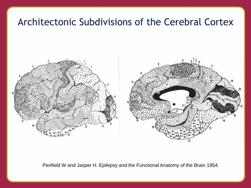
Architectonic Subdivisions of the Cerebral Cortex
Penfield W and Jasper H. Epilepsy and the Functional Anatomy of the Brain 1954.
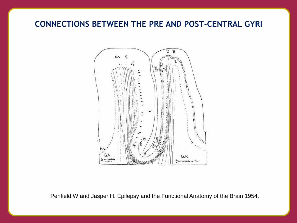
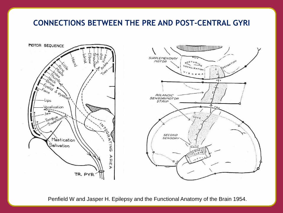
CONNECTIONS BETWEEN THE PRE AND POST-CENTRAL GYRI
Penfield W and Jasper H. Epilepsy and the Functional Anatomy of the Brain 1954.
CONNECTIONS BETWEEN THE PRE AND POST-CENTRAL GYRI
Penfield W and Jasper H. Epilepsy and the Functional Anatomy of the Brain 1954.
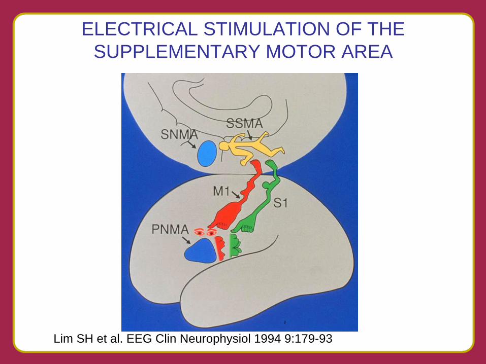
ELECTRICAL STIMULATION OF THE
SUPPLEMENTARY MOTOR AREA
Lim SH et al. EEG Clin Neurophysiol 1994 9:179-93
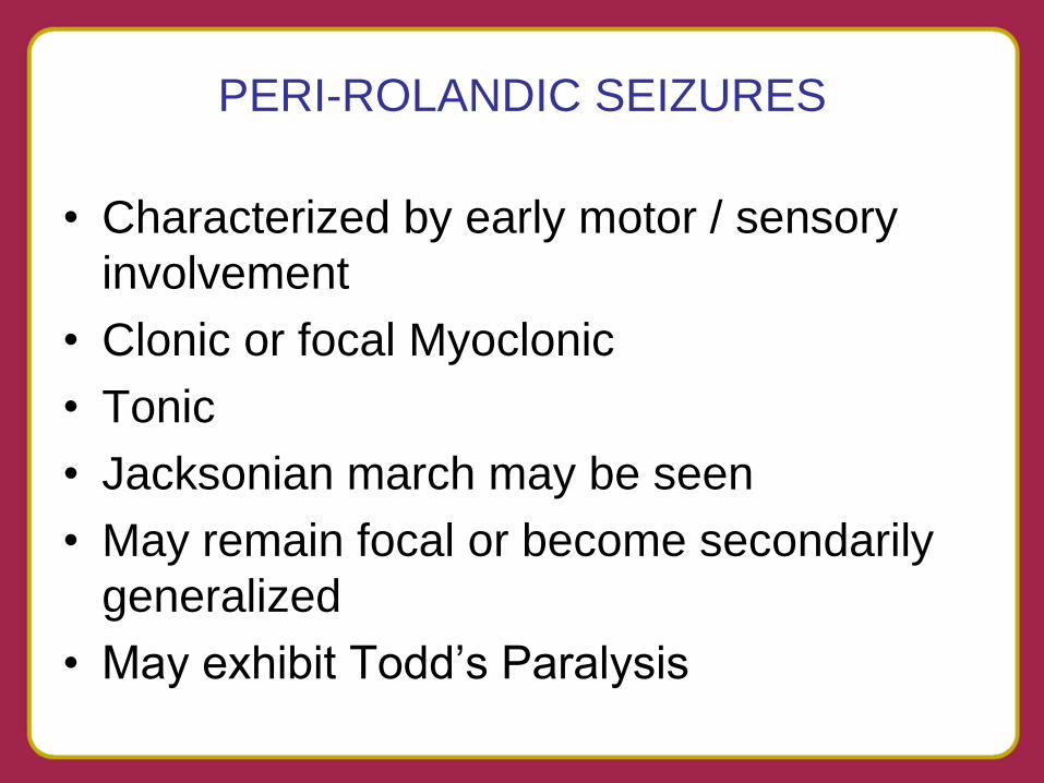
PERI-ROLANDIC SEIZURES
• Characterized by early motor / sensory
involvement
• Clonic or focal Myoclonic
• Tonic
• Jacksonian march may be seen
• May remain focal or become secondarily
generalized
• May exhibit Todd’s Paralysis
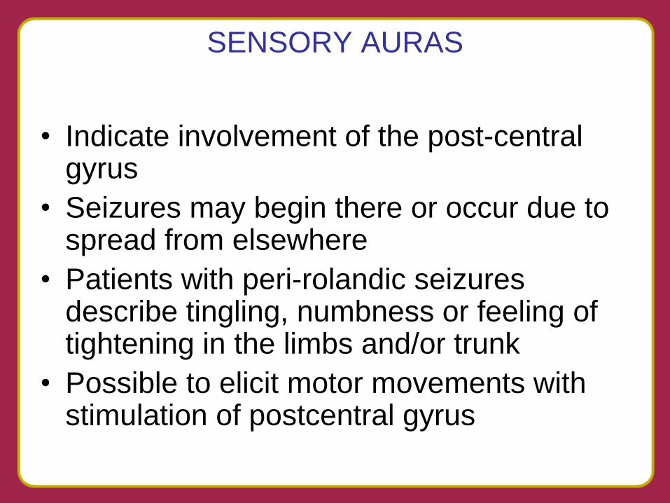
SENSORY AURAS
• Indicate involvement of the post-central gyrus
• Seizures may begin there or occur due to spread from elsewhere
• Patients with peri-rolandic seizures describe tingling, numbness or feeling of tightening in the limbs and/or trunk
• Possible to elicit motor movements with stimulation of postcentral gyrus
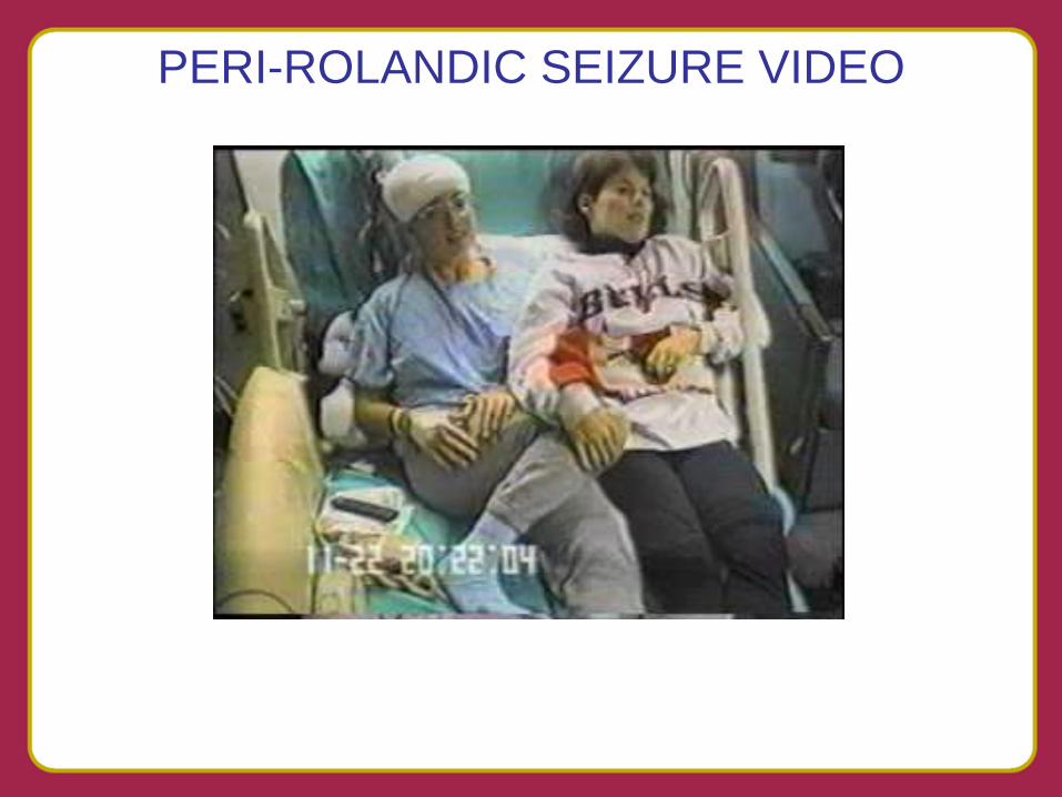
PERI-ROLANDIC SEIZURE VIDEO
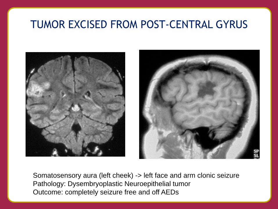
TUMOR EXCISED FROM POST-CENTRAL GYRUS
Somatosensory aura (left cheek) -> left face and arm clonic seizure
Pathology: Dysembryoplastic Neuroepithelial tumor
Outcome: completely seizure free and off AEDs
VERSIVE SEIZURES
• Main feature of seizure is Version
• Version defined as unnatural, extreme and sustained deviation of the eyes and/or head to one side often with neck extension
• Indicate involvement of frontal eye fields
• Version occurs earlier in extratemporal seizures compared to seizures from mesial temporal origin
• Reliably lateralize to contralateral hemisphere
• Ipsilateral head deviation in FLE may precede version
• Lateralizing value uncertain if the seizure does not evolve to GTC seizure
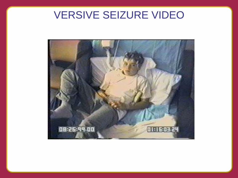
VERSIVE SEIZURE VIDEO
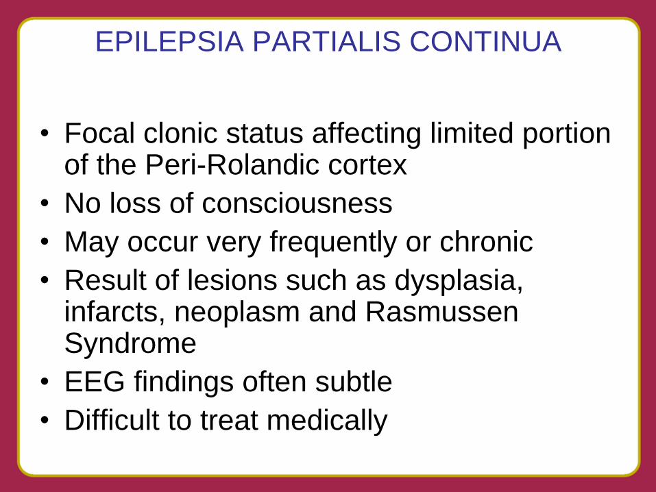
EPILEPSIA PARTIALIS CONTINUA
• Focal clonic status affecting limited portion of the Peri-Rolandic cortex
• No loss of consciousness
• May occur very frequently or chronic
• Result of lesions such as dysplasia, infarcts, neoplasm and Rasmussen Syndrome
• EEG findings often subtle
• Difficult to treat medically
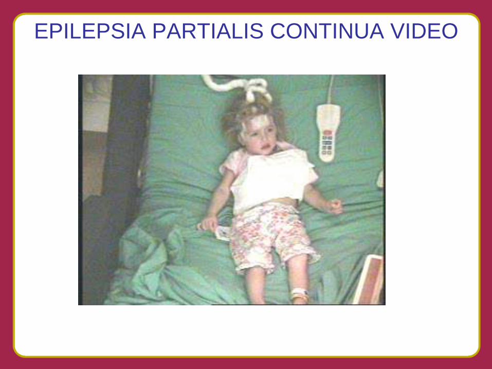
EPILEPSIA PARTIALIS CONTINUA VIDEO
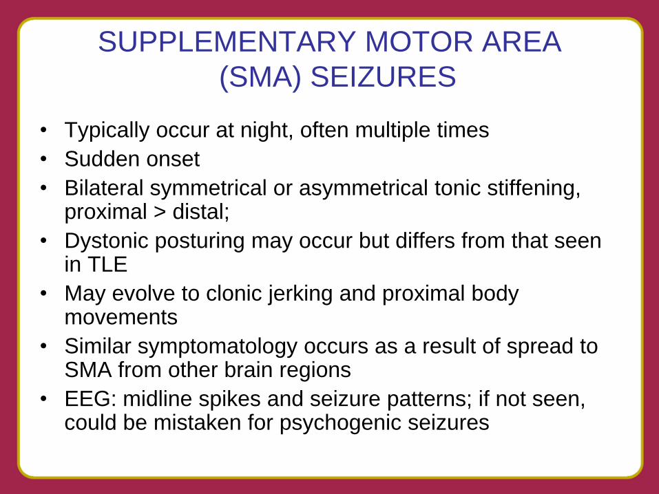
SUPPLEMENTARY MOTOR AREA
(SMA) SEIZURES
• Typically occur at night, often multiple times
• Sudden onset
• Bilateral symmetrical or asymmetrical tonic stiffening, proximal > distal;
• Dystonic posturing may occur but differs from that seen in TLE
• May evolve to clonic jerking and proximal body movements
• Similar symptomatology occurs as a result of spread to SMA from other brain regions
• EEG: midline spikes and seizure patterns; if not seen, could be mistaken for psychogenic seizures
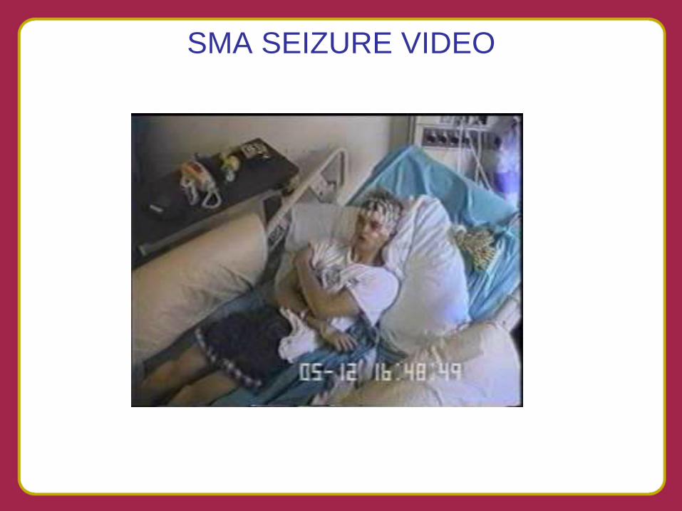
SMA SEIZURE VIDEO
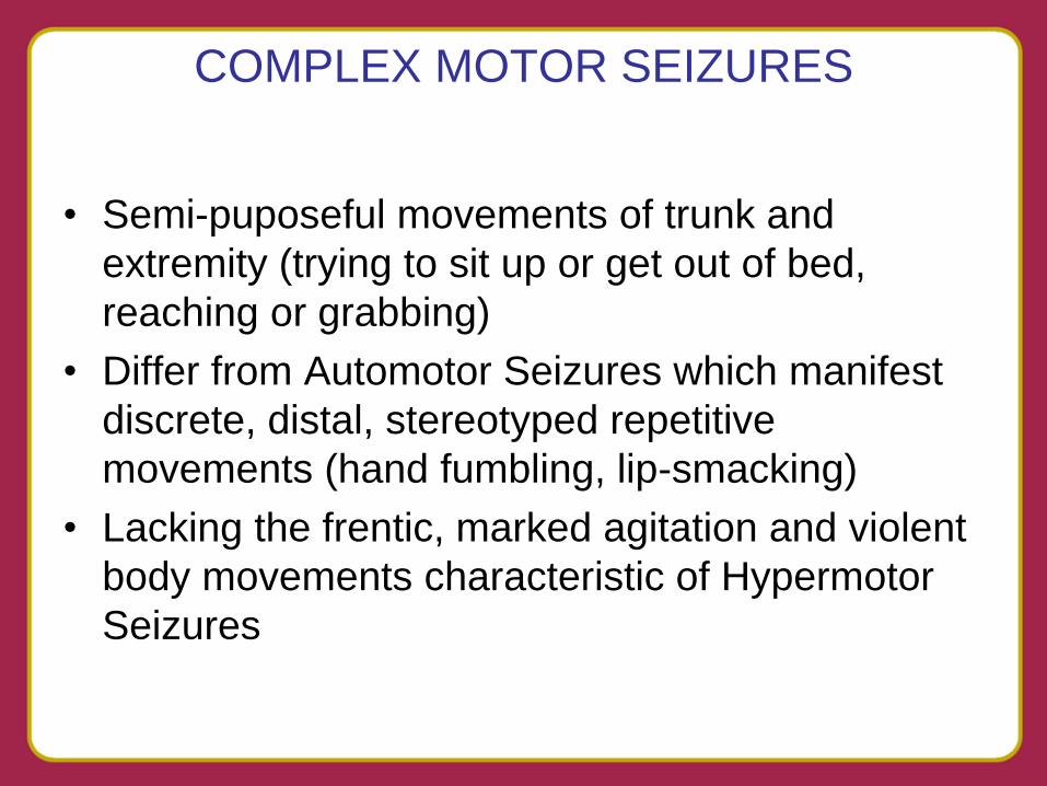
COMPLEX MOTOR SEIZURES
• Semi-puposeful movements of trunk and
extremity (trying to sit up or get out of bed,
reaching or grabbing)
• Differ from Automotor Seizures which manifest
discrete, distal, stereotyped repetitive
movements (hand fumbling, lip-smacking)
• Lacking the frentic, marked agitation and violent
body movements characteristic of Hypermotor
Seizures
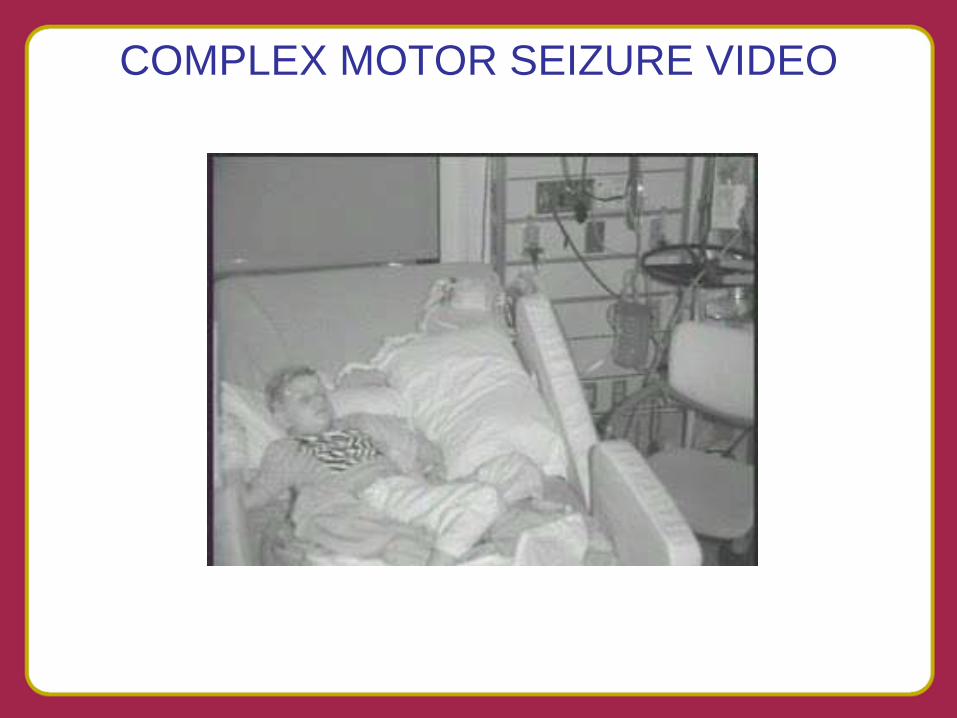
COMPLEX MOTOR SEIZURE VIDEO
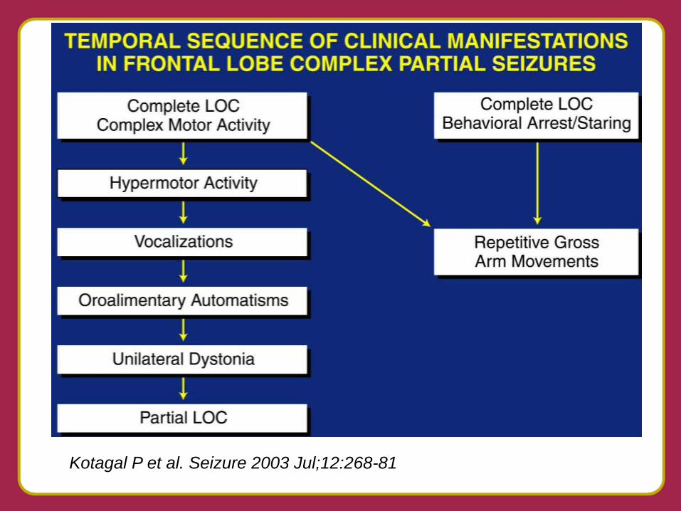
Kotagal P et al. Seizure 2003 Jul;12:268-81
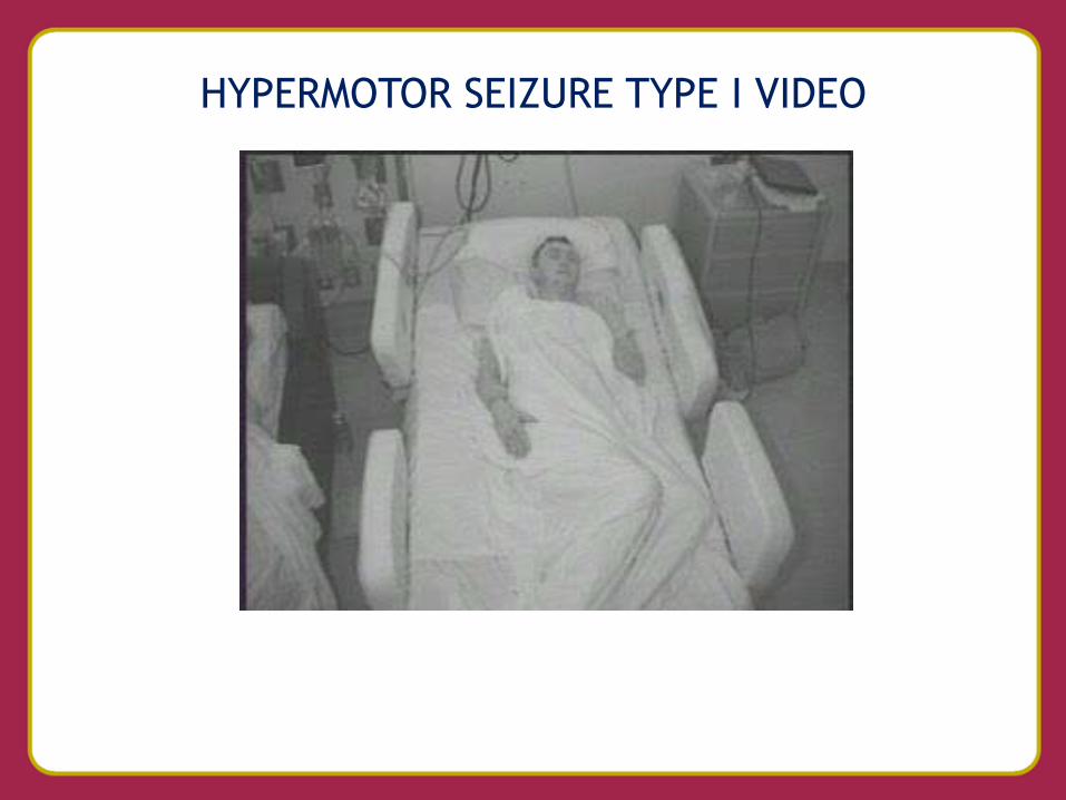
HYPERMOTOR SEIZURE TYPE I VIDEO
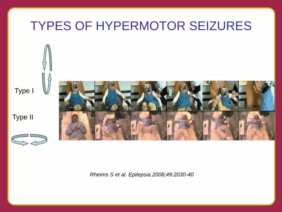
TYPES OF HYPERMOTOR SEIZURES
Rheims S et al. Epilepsia 2008;49:2030-40
Type I
Type II
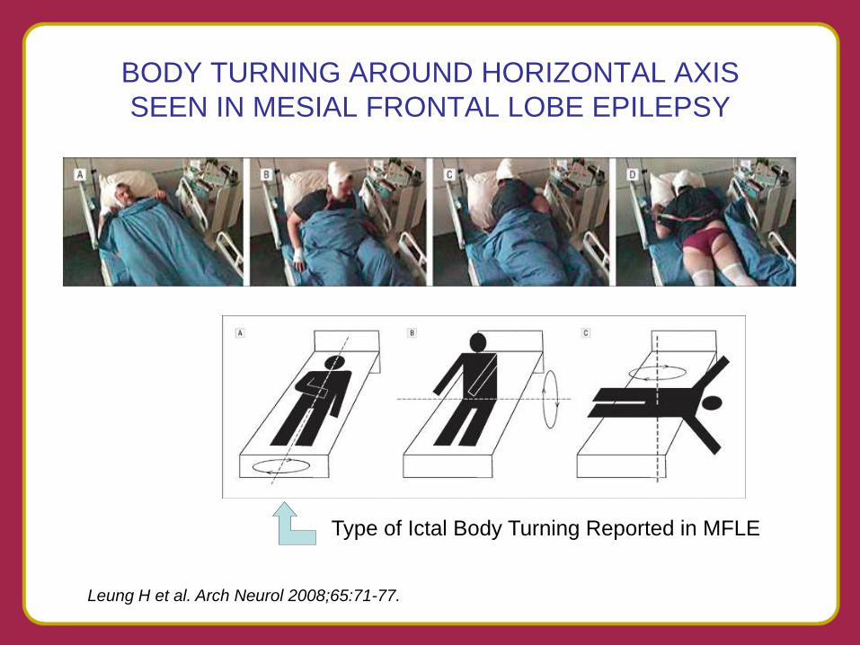
BODY TURNING AROUND HORIZONTAL AXIS
SEEN IN MESIAL FRONTAL LOBE EPILEPSY
Leung H et al. Arch Neurol 2008;65:71-77.
Type of Ictal Body Turning Reported in MFLE
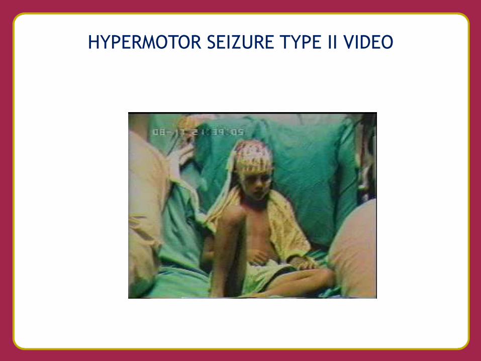
HYPERMOTOR SEIZURE TYPE II VIDEO
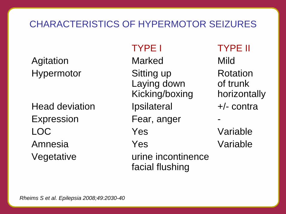
CHARACTERISTICS OF HYPERMOTOR SEIZURES
TYPE I TYPE II
Agitation Marked Mild
Hypermotor Sitting up Rotation Laying down of trunk Kicking/boxing horizontally
Head deviation Ipsilateral +/- contra
Expression Fear, anger -
LOC Yes Variable
Amnesia Yes Variable
Vegetative urine incontinence facial flushing
Rheims S et al. Epilepsia 2008;49:2030-40
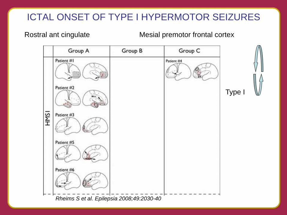
ICTAL ONSET OF TYPE I HYPERMOTOR SEIZURES
Rheims S et al. Epilepsia 2008;49:2030-40
Rostral ant cingulate Mesial premotor frontal cortex
Type I
Rheims S et al. Epilepsia 2008;49:2030-40
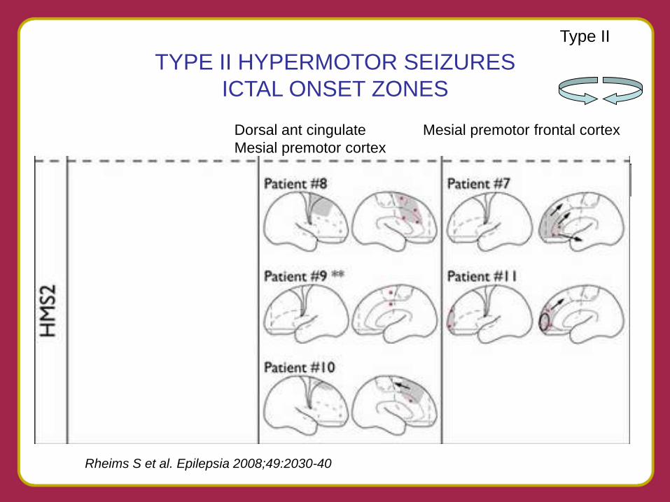
TYPE II HYPERMOTOR SEIZURES
ICTAL ONSET ZONES
Dorsal ant cingulate Mesial premotor frontal cortex
Mesial premotor cortex
Type II
NEGATIVE MOTOR SEIZURES
• Symptomatology: inability to move a body
part
• Difficult to detect unless looked for by
asking patient to elevate the extremity
• Onset from anterior SMA region, where
cortical stimulation produces similar
effects – inhibition of voluntary motor
movements
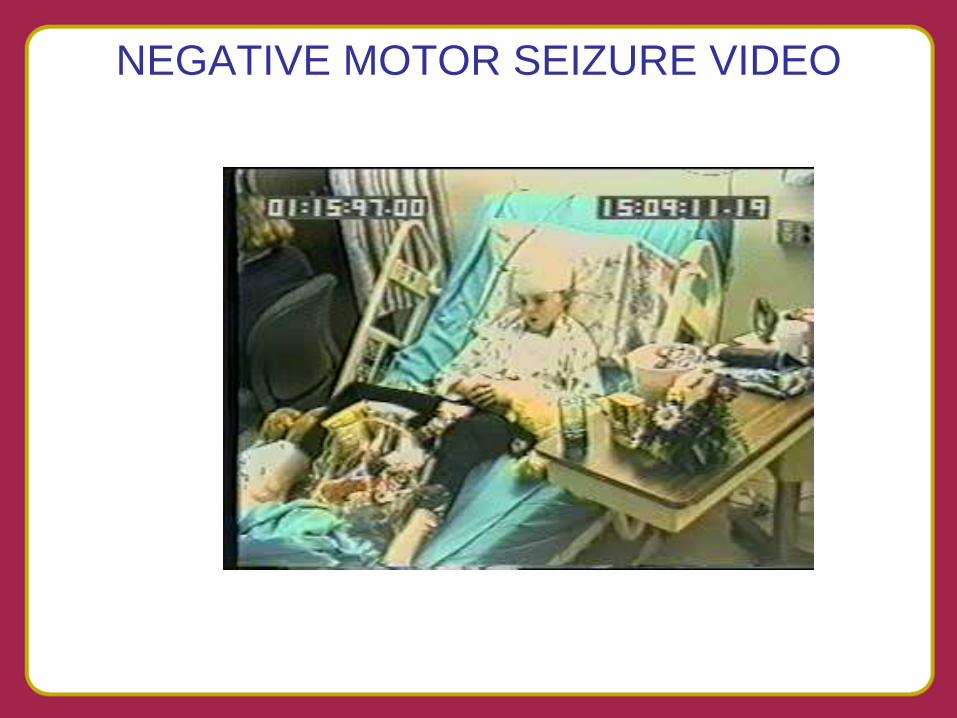
NEGATIVE MOTOR SEIZURE VIDEO

Ikeda A et al. Epilepsia 2009;50:2072-84
Negative Motor Seizures arising
from the Negative Motor Area
Vague aura
Repetitive
vocalization
Inability to speak
or move
Followed by
Left hand clonic
-> GTC seizure
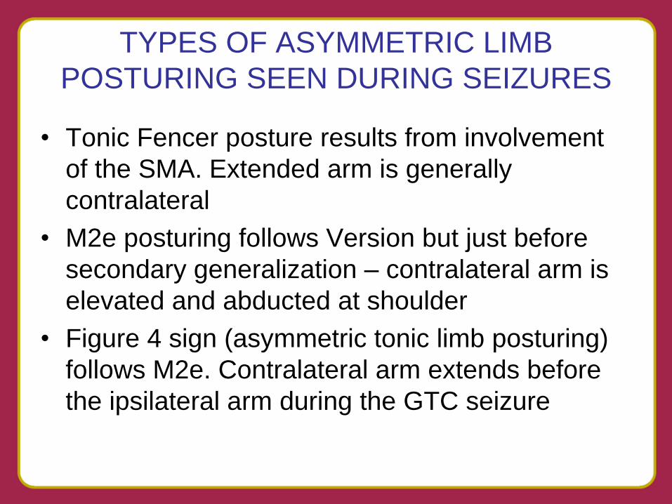
TYPES OF ASYMMETRIC LIMB
POSTURING SEEN DURING SEIZURES
• Tonic Fencer posture results from involvement
of the SMA. Extended arm is generally
contralateral
• M2e posturing follows Version but just before
secondary generalization – contralateral arm is
elevated and abducted at shoulder
• Figure 4 sign (asymmetric tonic limb posturing)
follows M2e. Contralateral arm extends before
the ipsilateral arm during the GTC seizure
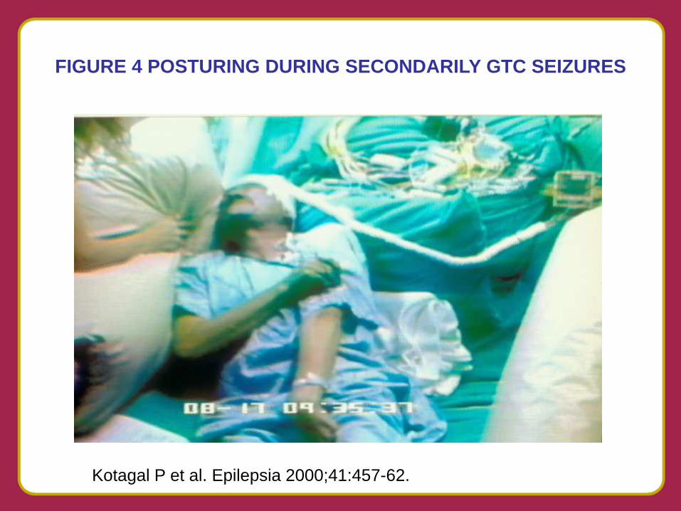
FIGURE 4 POSTURING DURING SECONDARILY GTC SEIZURES
Kotagal P et al. Epilepsia 2000;41:457-62.
American Epilepsy Society Annual Meeting
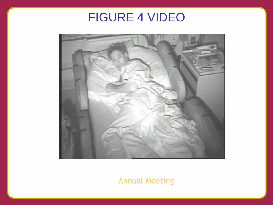
FIGURE 4 VIDEO
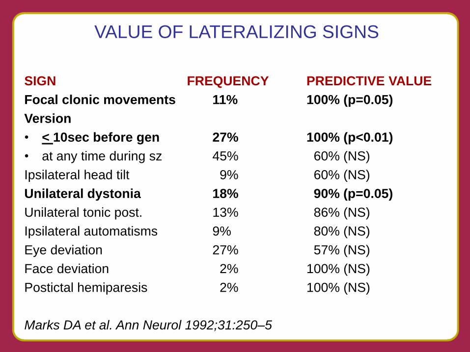
VALUE OF LATERALIZING SIGNS
SIGN FREQUENCY PREDICTIVE VALUE
Focal clonic movements 11% 100% (p=0.05)
Version
• < 10sec before gen 27% 100% (p<0.01)
• at any time during sz 45% 60% (NS)
Ipsilateral head tilt 9% 60% (NS)
Unilateral dystonia 18% 90% (p=0.05)
Unilateral tonic post. 13% 86% (NS)
Ipsilateral automatisms 9% 80% (NS)
Eye deviation 27% 57% (NS)
Face deviation 2% 100% (NS)
Postictal hemiparesis 2% 100% (NS)
Marks DA et al. Ann Neurol 1992;31:250–5
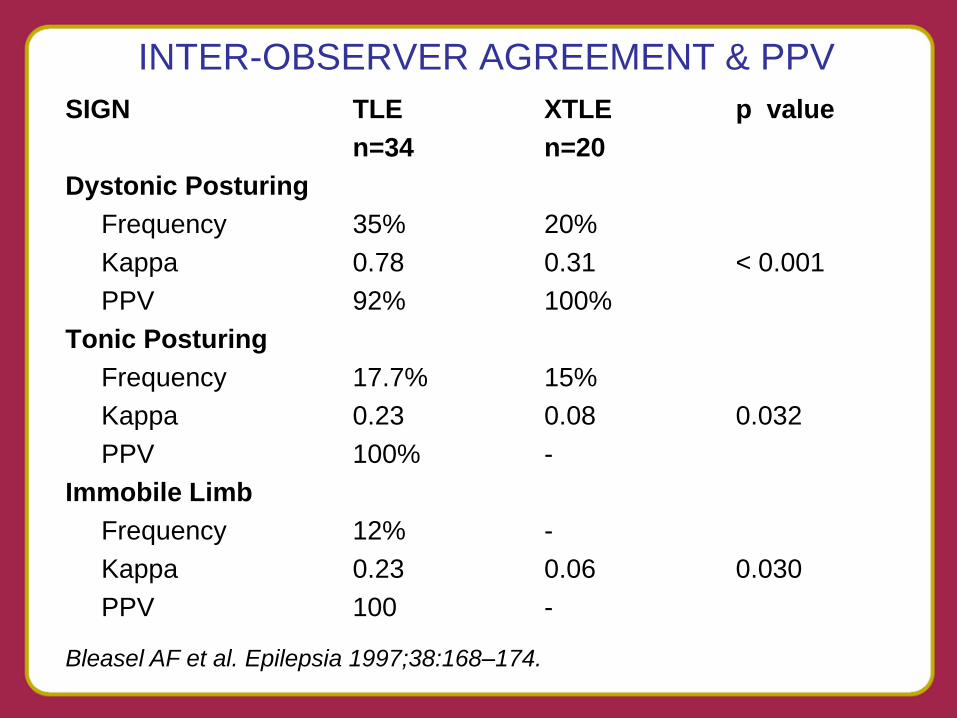
INTER-OBSERVER AGREEMENT & PPV
SIGN TLE XTLE p value
n=34 n=20
Dystonic Posturing
Frequency 35% 20%
Kappa 0.78 0.31 < 0.001
PPV 92% 100%
Tonic Posturing
Frequency 17.7% 15%
Kappa 0.23 0.08 0.032
PPV 100% -
Immobile Limb
Frequency 12% -
Kappa 0.23 0.06 0.030
PPV 100 -
Bleasel AF et al. Epilepsia 1997;38:168–174.
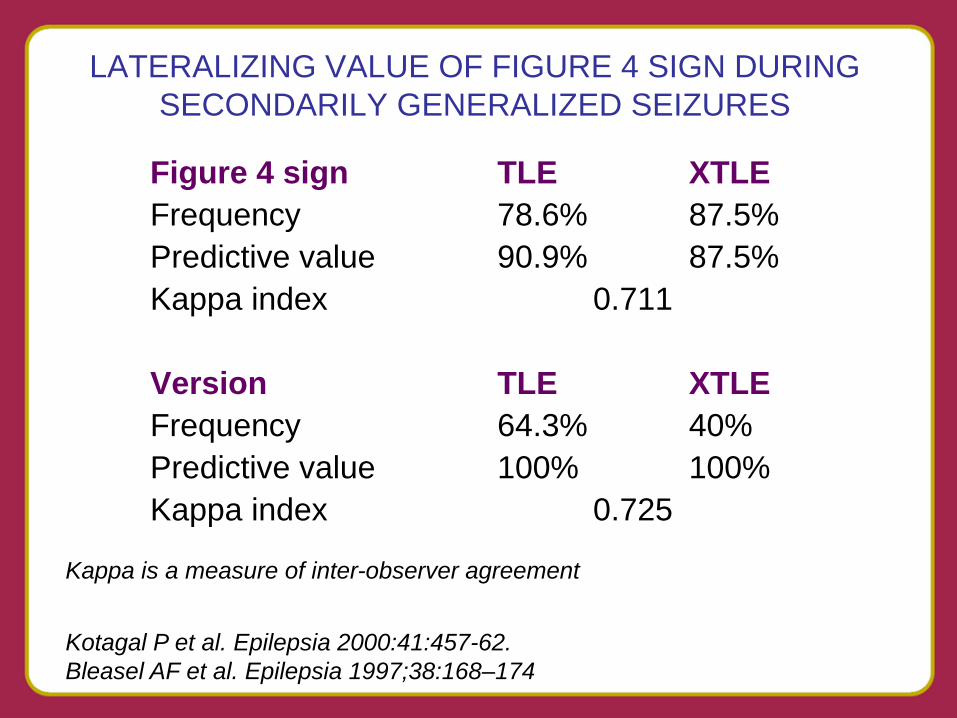
LATERALIZING VALUE OF FIGURE 4 SIGN DURING
SECONDARILY GENERALIZED SEIZURES
Figure 4 sign TLE XTLE
Frequency 78.6% 87.5%
Predictive value 90.9% 87.5%
Kappa index 0.711
Version TLE XTLE
Frequency 64.3% 40%
Predictive value 100% 100%
Kappa index 0.725
Kappa is a measure of inter-observer agreement
Kotagal P et al. Epilepsia 2000:41:457-62.
Bleasel AF et al. Epilepsia 1997;38:168–174
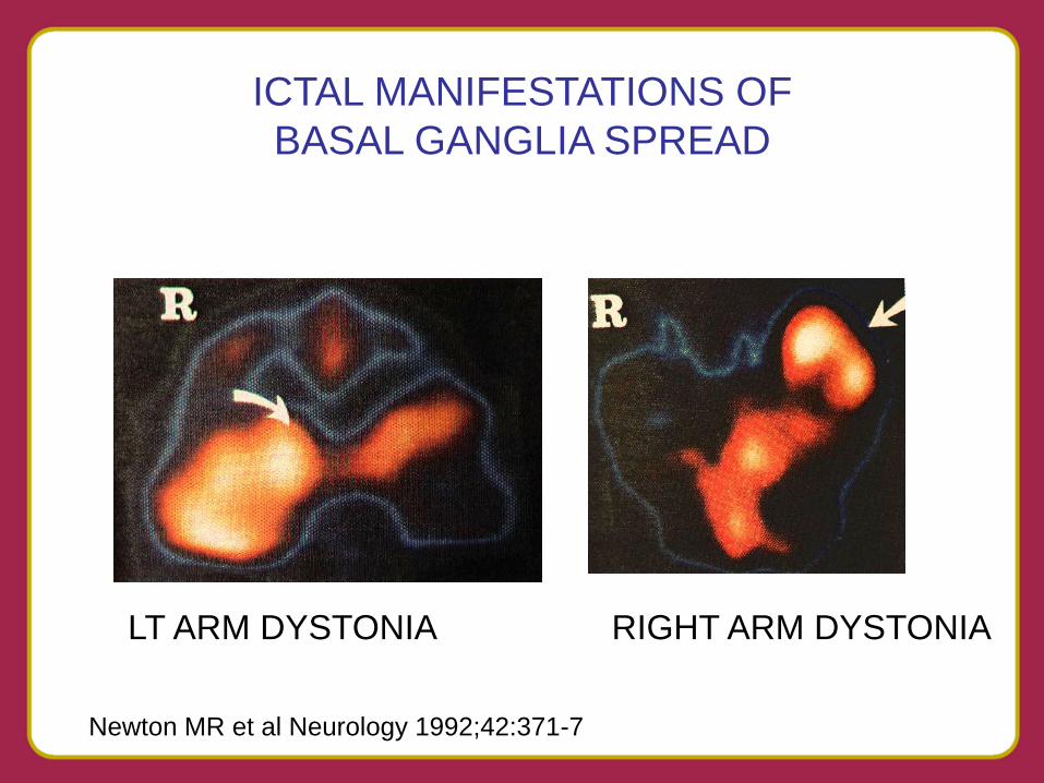
Newton MR et al Neurology 1992;42:371-7
LT ARM DYSTONIA RIGHT ARM DYSTONIA
ICTAL MANIFESTATIONS OF
BASAL GANGLIA SPREAD
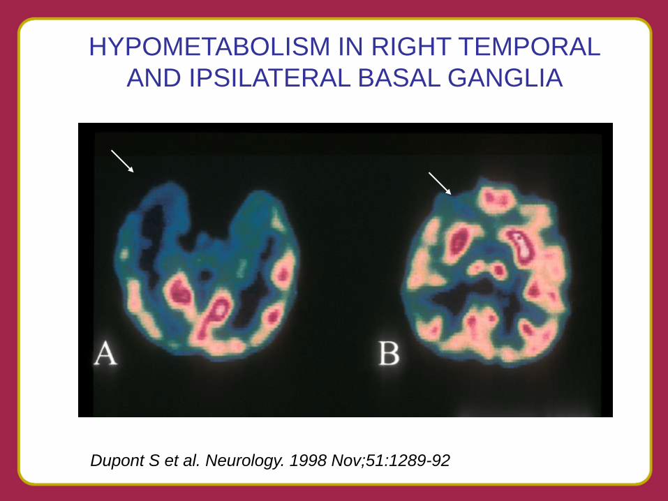
HYPOMETABOLISM IN RIGHT TEMPORAL
AND IPSILATERAL BASAL GANGLIA
Dupont S et al. Neurology. 1998 Nov;51:1289-92
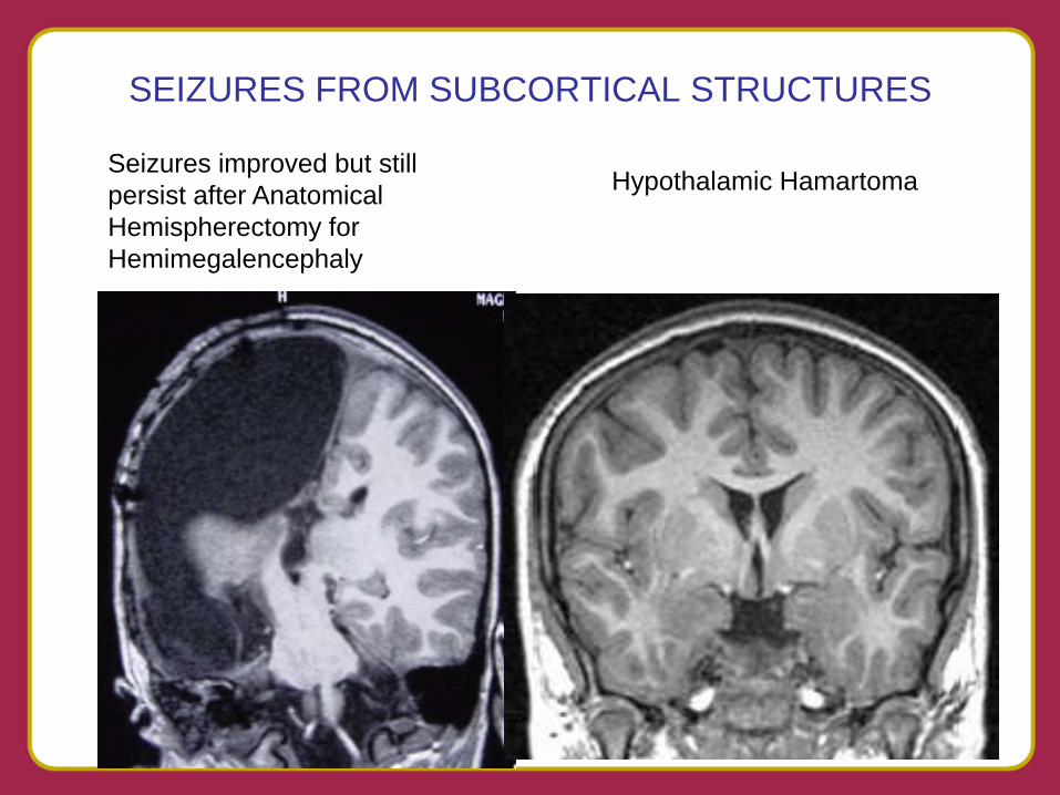
SEIZURES FROM SUBCORTICAL STRUCTURES
Seizures improved but still
persist after Anatomical
Hemispherectomy for
Hemimegalencephaly
Hypothalamic Hamartoma
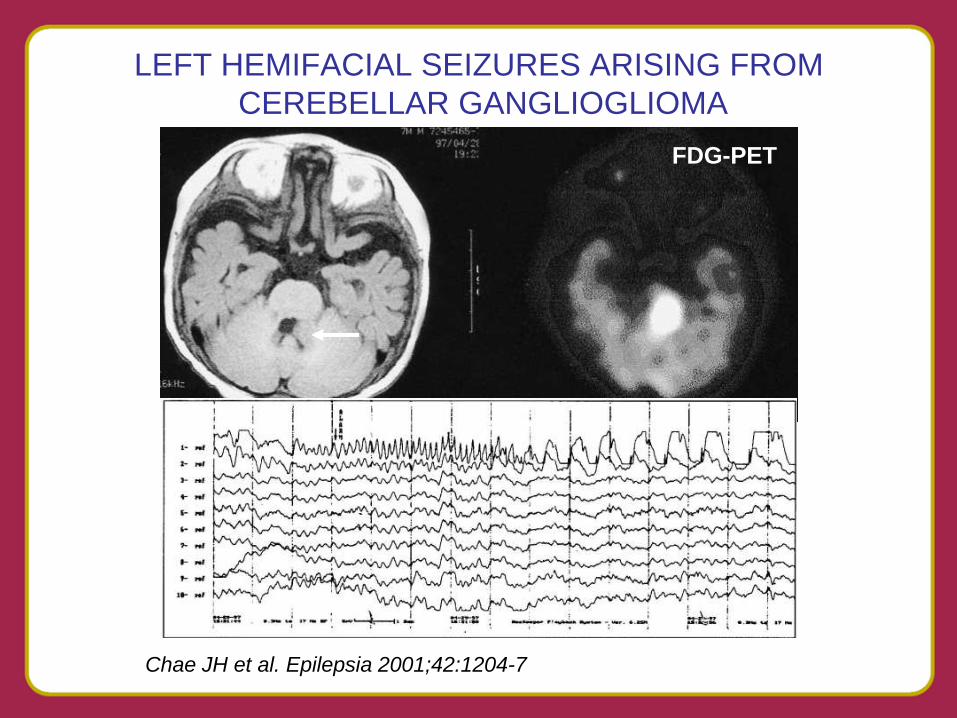
LEFT HEMIFACIAL SEIZURES ARISING FROM
CEREBELLAR GANGLIOGLIOMA
Chae JH et al. Epilepsia 2001;42:1204-7
FDG-PET
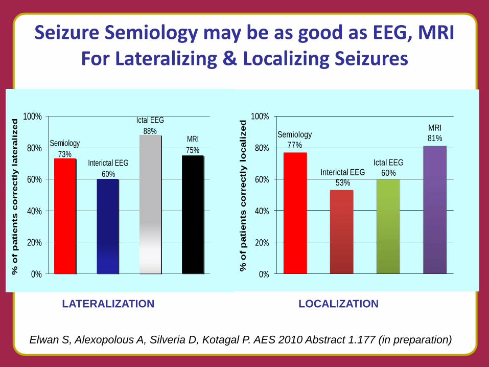
Semiology
73%Interictal EEG
60%
Ictal EEG
88%MRI
75%
0%
20%
40%
60%
80%
100%
% o
f p
ati
en
ts c
orrectl
y l
ate
rali
zed
Semiology
77%
Interictal EEG
53%
Ictal EEG
60%
MRI
81%
0%
20%
40%
60%
80%
100%
% o
f p
ati
en
ts c
orr
ectl
y l
ocali
zed
Seizure Semiology may be as good as EEG, MRI For Lateralizing & Localizing Seizures
Elwan S, Alexopolous A, Silveria D, Kotagal P. AES 2010 Abstract 1.177 (in preparation)
LATERALIZATION LOCALIZATION
American Epilepsy Society Annual Meeting
Impact on Clinical Care and Practice
•Reviewed how motor functions are organized in the cortex •Seen how various seizure symptoms result from ictal involvement of different brain regions •Improved seizure diagnosis based on history, examination and review of seizure semiology •Seizure semiology is an important component of evaluation for epilepsy surgery