Functional morphology of the 'tongue-bite' in the ...glauder/reprints_unzipped/Sanford... ·...
30
JOURNAL OF MORPHOLOGY 202:379-408 (1989) Functional Morphology of the “Tongue-Bite” in the Osteoglossomorph Fish Notopterus CHRISTOPHER P. SANFORD AND GEORGE V. LAUDER Department of Ecology and Evolutionary Biology, Uniuersity of California, Irvine, California 9271 7 ABSTRACT Osteoglossomorph fishes are characterized by having three sets of jaws: a mandibular jaw apparatus (MJA) anteriorly, a pharyngeal jaw apparatus (PJA) posteriorly, and a tongue-bite apparatus (TBA) associated with basihyal and parasphenoid teeth. The TBA is a novel complex feature of the head that character- izes osteoglossomorphfishes and provides a case study in the origin of novel functions and roles in the vertebrate musculoskeletal system. The function of the tongue-bite in the osteoglossomorph fish Notopterus was characterized by using high-speed cinematography and electromyography. The tongue-bite is used during intraoral prey processing to shred and disable prey. Two distinct uses of the TBA were defined on the basis of kinematic and electromyo- graphic profiles: raking and open-mouth chewing. During raking behavior, the prey is held fixed in the MJA, the neurocranium is elevated, and the pectoral girdle is retracted. The adductor mandibulae, hypaxialis, epaxialis, and posterior intermandib- ularis muscles are all highly active, but only very low activity is observed in the sternohyoideus muscle. During open-mouth chewing behavior, the prey is located within the oral cavity, posterior pectoral girdle rotation is less than during raking, and the levator operculi muscle shows relatively high activity. We propose that a shearing action of the basihyal (moved anteroposteriorly by the posterior intermandibularis and hypaxial muscles) with respect to the neurocranium (elevated by epaxial muscles) is the critical aspect of the tongue-bite in Notopterus. The body muscles (epaxials and hypaxials) provide the main power for the tongue- bite. We hypothesize that lack of sternohyoideus activity during intraoral prey processing, posterior pectoral girdle rotation, and a long-fibered posterior interman- dibularis muscle are novel structures associated with the tongue-bite apparatus within osteoglossomorph fishes. Fishes of the teleostean superorder Osteoglos- somorpha provide an interesting case study in the role of novel features in the musculoskeletal system. When such novelties do arise, we cur- rently have little idea of the impact they have on pre-existing functional complexes. For example, if a novel biomechanical system involving new muscles or bony elements arises in the head, what effect does this have on the function of the muscles and bones in surrounding regions? Do these muscles and bones retain their primitive functions, or do muscular or skeletal novelties cause associated changes in the activity and movement patterns of pre-existing muscles and bones? Osteoglossomorphfishes are known commonly as “bony-tongued fishes” because of the pres- ence of an impressive bite between teeth on the hyoid apparatus (the “tongue”) and teeth on the palate and base of the skull. The presence of a tongue-bite apparatus is considered to be a syn- apomorphy for osteoglossomorph fishes (see Lauder and Liem, ’83), a clade composed of approximately 26 genera and 206 species. The complex of bones forming the tongue-bite appa- ratus is anatomically intercalated between the bite of the mandibular jaw apparatus (MJA) anteriorly and the pharyngeal jaw apparatus (PJA) posteriorly (illustrated schematically in Fig. 2). Both the MJA and PJA are primitive features of ray-finned fishes (Lauder, ’80, ’83; Lauder and Liem, ’83; Liem and Sanderson, ’86) and retain their basic primitive configuration in most osteoglossomorphfishes. Thus, in the Osteoglossomorphaa novel mus- culoskeletal system, the tongue-bite apparatus o 1989 ALAN R. LISS, INC.
Transcript of Functional morphology of the 'tongue-bite' in the ...glauder/reprints_unzipped/Sanford... ·...

JOURNAL OF MORPHOLOGY 202:379-408 (1989)
Functional Morphology of the “Tongue-Bite” in the Osteoglossomorph Fish Notopterus
CHRISTOPHER P. SANFORD AND GEORGE V. LAUDER Department of Ecology and Evolutionary Biology, Uniuersity of California, Irvine, California 9271 7
ABSTRACT Osteoglossomorph fishes are characterized by having three sets of jaws: a mandibular jaw apparatus (MJA) anteriorly, a pharyngeal jaw apparatus (PJA) posteriorly, and a tongue-bite apparatus (TBA) associated with basihyal and parasphenoid teeth. The TBA is a novel complex feature of the head that character- izes osteoglossomorph fishes and provides a case study in the origin of novel functions and roles in the vertebrate musculoskeletal system.
The function of the tongue-bite in the osteoglossomorph fish Notopterus was characterized by using high-speed cinematography and electromyography. The tongue-bite is used during intraoral prey processing to shred and disable prey. Two distinct uses of the TBA were defined on the basis of kinematic and electromyo- graphic profiles: raking and open-mouth chewing. During raking behavior, the prey is held fixed in the MJA, the neurocranium is elevated, and the pectoral girdle is retracted. The adductor mandibulae, hypaxialis, epaxialis, and posterior intermandib- ularis muscles are all highly active, but only very low activity is observed in the sternohyoideus muscle. During open-mouth chewing behavior, the prey is located within the oral cavity, posterior pectoral girdle rotation is less than during raking, and the levator operculi muscle shows relatively high activity.
We propose that a shearing action of the basihyal (moved anteroposteriorly by the posterior intermandibularis and hypaxial muscles) with respect to the neurocranium (elevated by epaxial muscles) is the critical aspect of the tongue-bite in Notopterus. The body muscles (epaxials and hypaxials) provide the main power for the tongue- bite. We hypothesize that lack of sternohyoideus activity during intraoral prey processing, posterior pectoral girdle rotation, and a long-fibered posterior interman- dibularis muscle are novel structures associated with the tongue-bite apparatus within osteoglossomorph fishes.
Fishes of the teleostean superorder Osteoglos- somorpha provide an interesting case study in the role of novel features in the musculoskeletal system. When such novelties do arise, we cur- rently have little idea of the impact they have on pre-existing functional complexes. For example, if a novel biomechanical system involving new muscles or bony elements arises in the head, what effect does this have on the function of the muscles and bones in surrounding regions? Do these muscles and bones retain their primitive functions, or do muscular or skeletal novelties cause associated changes in the activity and movement patterns of pre-existing muscles and bones?
Osteoglossomorph fishes are known commonly as “bony-tongued fishes” because of the pres- ence of an impressive bite between teeth on the
hyoid apparatus (the “tongue”) and teeth on the palate and base of the skull. The presence of a tongue-bite apparatus is considered to be a syn- apomorphy for osteoglossomorph fishes (see Lauder and Liem, ’83), a clade composed of approximately 26 genera and 206 species. The complex of bones forming the tongue-bite appa- ratus is anatomically intercalated between the bite of the mandibular jaw apparatus (MJA) anteriorly and the pharyngeal jaw apparatus (PJA) posteriorly (illustrated schematically in Fig. 2). Both the MJA and PJA are primitive features of ray-finned fishes (Lauder, ’80, ’83; Lauder and Liem, ’83; Liem and Sanderson, ’86) and retain their basic primitive configuration in most osteoglossomorph fishes.
Thus, in the Osteoglossomorpha a novel mus- culoskeletal system, the tongue-bite apparatus
o 1989 ALAN R. LISS, INC.

380 C.P. SANFORD A N D G.V. LAUDER
(TBA, Fig. 2), for biting and chewing prey has arisen, and this clade possesses three complete sets of functional jaws in the head. Because 1) the TBA is a synapomorphy of osteoglosso- morph fishes, 2) the TBA is anatomically interca- lated between the MJA and the PJA, and 3) a single set of cranial muscles controls the function of all three jaw systems, the Osteoglossomorpha constitutes an excellent case study of the integra- tion of novel functions in the vertebrate muscu- loskeletal system.
The specific aims of this paper are twofold. First, because the tongue-bite apparatus has not been previously characterized, we provide a func- tional analysis of the TBA by using high-speed films (to document kinematics) and electromyo- graphy (to document patterns of muscle activ- ity). Our focus is on the function of those ele- ments of the TBA that are common to, and thus provide anatomical links between, the three jaw systems in the head, as well as on the prey- processing role of the TBA. Second, we compare the function and activity patterns of muscles controlling the TBA to outgroup taxa in order to provide hypotheses on the evolution of function in the feeding system of primitive ray-finned fishes.
MATERIALS AND METHODS Specimens and morphology
Cinematographic and electromyographic anal- yses were performed on the osteoglossomorph Notopterus chitalu, the "clown knifefish." In contrast to other species of osteoglossomorphs, large individuals of this species could be readily obtained from commercial suppliers. Seven spec- imens were used for kinematic and electromyo- graphic studies and were maintained in aquaria with filtered dechlorinated water a t 24°C. The specimens studied ranged in size from 19 cm to 38 cm (mean = 28.7; s.d. = 6.7). Electromyo- graphic analysis was also conducted on the fol- lowing outgroups (two individuals of each): Lep- isosteus oculatus and Amia calua. These taxa were maintained in cold water aquaria at 20°C.
Anatomical observations on the feeding mech- anism, and especially components of the head associated with the tongue-bite, were made on representatives of the major taxa of lower ray- finned fishes (Lauder and Liem, '83). Anatomi- cal material examined consisted either of speci- mens fixed in formalin and preserved in alcohol (denoted AL) or specimens cleared and stained by using the alizarin red-alcian blue method of Dingerkus and Uhler ('77) (denoted AA). The following specimens were examined Acipenser stellatus, AL, AA; Lepisosteus oculatus, AL, AA; Amia calua, AL, AA, Dorosoma penten-
erne, AA, Hiodon alosoides, AL, AA; Notopterus chitala, AL, AA; Pantodon buchholzi, AA; Scle- ropages formosus, AA; Coregonus lauaretus, AA; Esox americanus, AA; Salmo salar, AA Salueli- nus alpinus, AA. Both the alcohol and cleared and stained material were examined under a Zeiss IVB dissecting microscope with a camera lucida attachment.
Cinematography and electromyography High-speed films of Notopterus feeding on
goldfish (Carassius) were obtained with Kodak 4X Reversal film in a Locam I1 16 mm high- speed camera running at 200 frames per second. Notopterus were trained to feed on goldfish un- der lighting provided by two 600 W Smith- Victor filming lights in a feeding arena with a background grid marked in centimeters. Only those feedings occurring parallel to the film plane were used for analysis, and we made a special effort to obtain films of the chewing behavior associated with the tongue-bite apparatus.
Cranial movements were analyzed frame by frame by using a stop-frame projector, digitizer, and a custom program designed to acquire data from film sequences. A total of 46 chewing cycles from five individuals were filmed; a sample of nine cycles were chosen for detailed analysis. These nine sequences were chosen for analysis because they most clearly showed the knifefish and its prey, exhibited the least transverse move- ment of the fish relative to the film plane, and were the sequences in which prey did not ob- struct the gape or hyoid landmarks. However, the basic pattern of movement was confirmed for all sequences in which landmarks were visi- ble. Each chewing sequence consisted of one to three chews, and each chew lasted approxi- mately 50 film frames (or 250 ms). A total of nearly 800 frames were digitized for this study. Landmarks on the head were chosen that could be reliably located in each frame and that were stable anatomical points (Fig. 1). Several of our landmarks differ from those used in previous research (Lauder, '80; Lauder and Liem, '81; Reilly and Lauder, '89a; ShaRer and Lauder, '88) because prey held in the mouth during chewing obscured many of our standard landmarks. For example, during chewing (as described below) the prey was often held between the jaws and obscured the anterior region of both the mandi- ble and the snout.
From each frame, the following seven vari- ables were measured (Fig. 1). Gape distance (GD) is defined as the distance (in cm) between the nostril and the lower border of the dentary mea- sured along a line perpendicular to the lateral line of the fish. Gape distance accurately reflects

FUNCTIONAL MORPHOLOGY OF NOTOPTERUS 381
Fig. 1. Lateral view of the head and anterior body of Notopterm to illustrate the kinematic variables measured
change in gape that may be achieved both by neurocranial elevation and mandibular depres- sion.
Neurocranial elevation (or head angle, HDA) is measured as the angle (in degrees) between two lines (Fig. I). One line is defined by two points along the lateral line of the knifefish, and the other by two points along the dorsal border of the neurocranium. This angle reflects the ele- vation of the neurocranium on the vertebral col- umn as a result of epaxial muscle contraction: an increase in HDA indicates neurocranial eleva- tion.
Lower jaw angle (LJA) is measured as the angle (in degrees) between two lines, one line being defined by two points along the lower border of the dentary and the other by two points along the lateral line (Fig. I). This angle was chosen to reflect mandibular depression with respect to the body axis independently of cranial motion. Note that depression of the mandible decreases the angle LJA.
Three measurements were made to enable us to triangulate both dorsoventral and anteropos- terior movements of the hyoid relative to the skull (Fig. 1). Orbit-nostril distance (NOD) is the fixed reference line segment and is defined as the distance (in cm) between the nostril and the dorsal midpoint of the orbit. This distance should remain constant during feeding and thus can serve as both an indicator of our digitizing accu- racy and as a measure of the extent to which changes in fish position during chewing might be influencing our measurements. If orbit-nostril distance (NOD) changes substantially during a chewing cycle, then our error is high because of manual digitizing error or movement of the knife- fish. In fact, errors were small relative to the magnitude of changes in our variables. The two
from the high-speed films of feeding behavior. Variables are described in the Materials and Methods section.
sample plots of NOD vs. time presented in Fig- ure 5 exhibit little variation in amplitude.
Orbit-hyoid distance (OHD) is the distance (in cm) between the dorsal midpoint of the orbit and the anteroventral tip of the anterohyal bone. When the hyoid moved dorsally such that the anterohyal moved medial to the opercular series and could not be seen in lateral view, missing values were recorded for this distance. Hyoid- nostril distance (HND) is the distance (in cm) between the anteroventral tip of the anterohyal and the nostril. This distance by itself reflects anterodorsal motion of the hyoid. In conjunction with NOD and OHD, it allows triangulation of hyoid movement to the extent possible in lateral view.
Pectoral girdle distance (PG) is defined as the distance (in cm) from the nostril to the anterior point of the pectoral fin. This distance was cho- sen because preliminary analyses showed that the pectoral girdle undergoes large anteroposte- rior excursions during chewing, and the only anterior landmark available to track such excur- sions was the nostril. Cranial elevation (i.e., change in HDA) confounds this measurement to some extent as elevation of the head will cause an apparent lengthening of PG distance. Thus, care was taken to measure head movement simul- taneously so we could correct for any bias in the measurement of PG distance. However, the an- teroposterior excursion of the nostril along the arc of cranial elevation is small (1-2 mm), and the maximal error in PG due to cranial elevation is only a small component of the large changes seen in PG distance during chewing (about 2 cm).
Electromyograms were obtained by using a procedure similar to that described elsewhere (e.g., Lauder and Shaf€er, '88; Reilly and Lauder,

N A i
lo
Ph
B
Fig. 2. Diagram of the cranial morphology of the osteoglos- somorph fish Notopterm chitala. A: Lateral view of the head and pectoral girdle illustrating cranial osteology and the three sets of jaws: 1) the MJA, mandibular jaw apparatus, 2) the PJA, pharyngeal jaw apparatus, and 3) the TBA, tongue-bite apparatus. The MJA and PJA are primitive features of the feeding mechanism in ray-finned fishes (Lauder and Liem, '83); the TBA is a novelty of osteoglossomorph fishes. B. Lateral view of the head of Notopterm showing the vectors of the major cranial muscles used during feeding. The hypothe- sized motion of the basihyal and parasphenoid teeth during raking behavior (see text) are indicated by the arrows above the basihyal. Note 1) that the posterior intermandibu-
laris muscle (PIM) has long fibers that originate on the hyoid and insert anteriorly near the mandibular symphysis, and 2) that a strong cleithrobranchial ligament (CBL) connects the branchial apparatus to the pectoral girdle. The levator oper- culi muscle (LOP) is not shown here. It arises on the skull, attaches to the opercular series, and acts to depress the lower jaw (Lauder, '80; Liem, '70). Thin black lines indicate the lines of action of major cranial muscles. The body muscles that are an important part of the tongue-bite apparatus, hypaxial (HY) and epaxial (EP) muscles, are shown by heavy black lines with an arrow indicating the major fiber direction in the muscle.

FUNCTIONAL MORPHOLOGY OF NOTOPTERUS 383
'89b). Briefly, knifefish were transferred from aquaria to a large basin filled with water. Approx- imately 500 ml of aquarium water was placed in a beaker in which 3 g of tricaine methane sul- fonate (MS-222) was dissolved. The anesthetic was then slowly added to the basin until the fish was anesthetized. The fish was then placed in a shallow basin filled with half aquarium water and half basin water containing the anesthetic while the electrodes were inserted.
Fine wire steel-alloy bipolar electrodes were implanted percutaneously into head muscles by using hypodermic needles through which the electrodes had been threaded. Generally, six mus- cles were implanted at one time, and electrode placement was based on prior dissection of pre- served specimens. Electrode location was con- firmed by direct visual observation of the inser- tion point: the electrodes were inserted directly into the belly of each muscle and there were no overlying or associated muscles into which the electrode might have strayed. Bared electrode tips were approximately 0.5 mm long and about 1.0 mm apart in the muscle. In order for the electromyographic recordings to be consistent for comparative statistical purposes, electrode placement was always the same for each muscle recorded. On average, the electrode tips formed an angle of approximately 45" with the muscle fiber axis. The electrode wires were gathered together and tied with a single suture (Ethicon silk surgical suture) to the dorsal flank of the body. Recordings were made from six cranial muscles: the levator operculi (LOP), sternohyoi- deus (SH), posterior intermandibularis (PIM), adductor mandibulae (AM), epaxid (EP), and hypaxial (HY) muscles. In several experiments multiple electrodes were placed in different fiber tracts of the hypaxial muscles in order to ascer- tain if the ventral (anatomically distinct) fibers of the hypaxialis showed a different activity pat- tern than the dorsolateral fibers.
During experiments the electrodes were at- tached to Grass P511J preamplifiers (set for 100 to 3,000 Hz half-amplitude bandpass filtering), and electromyograms were recorded on tape by using a Bell and Howell 4020A FM tape re- corder. Amplification of the raw signal was 10,OOO times.
For analysis of the motor pattern of the head muscles, electromyograms were played from the FM tape recorder into a 12-bit analog-to-digital converter at a sampling rate of 2,100 Hz per channel. Electromyograms from fish head mus- cles contain little power above 1,000 Hz, and thus we sampled at a rate greater than twice the
highest-frequency component in the signal. Each burst pattern and sequence to be analyzed was converted into a digital file by using an IBM AT microcomputer and stored on hard disc as a raw data file. These files were then played from disc into a Tektronix 4107 graphics terminal so that electromyograms could be measured by using a custom digitizing program. All electromyograms were digitally rectified (by making all negative values positive) prior to the digitizing of motor pattern variables.
A total of 23 variables were measured from each digital file. For each of the six muscles, the duration of activity (DUR, in ms: LOP-DUR, SH-DUR, PIM-DUR, AM-DUR, EP-DUR, HY- DUR), maximum burst amplitude (AMP, in volts: LOP-AMP, SH-AMP, PIM-AMP, AM- AMP, EP-AMP, HY-AMP), and integrated area of the burst (AREA, in volts x ms: LOP-AREA,
HY-AREA) were measured. In addition, the time of onset of activity in each muscle (in ms) was measured relative to the onset of activity in the levator operculi muscle (LOP-SH, LOP-PIM, LOP-AM, LOP-EP, LOP-HY). The LOP was used as a reference as in previous studies (e.g., Wainwright and Lauder, '86) because it is one of the major mouth-opening muscles in teleost fishes (Lauder, '85; Liem, '70) and shows consis- tent and clear activity patterns. In two individ- ual knifefishes simultaneous electromyographic and kinematic data were obtained with the aid of a pulse synchronizer between the high-speed camera and the FM tape recorder. These se- quences were used to determine the relationship between the pattern of muscle activity and the feeding behaviors defined on the basis of kine- matic measurements.
Electromyograms from chewing behaviors in Notopterus were compared with myograms from the initial strike because several muscles showed very-low-amplitude activity during prey process- ing behavior. Cranial muscle activity during the strike in predaceous fishes is high (Lauder, '80, '83, '85) and provides a control for the possibility that a defective electrode could be causing low- amplitude signals. In all cases high-amplitude activity was observed at the strike, even in those muscles that show relatively low activity during chewing. Thus, for the statistical analyses of chewing behavior presented in this paper, we provide comparative EMG data on the initial strike.
A series of stimulation experiments were also conducted by using the procedures outlined in Wainwright ('89a) and Reilly and Lauder ('89a).
SH-AREA, PIM-AREA, AM-AREA, EP-AREA,
384 C.P. SANFORD AND G.V. LAUDER
Briefly, knifefishes were anesthetized and placed in a shallow tray of water. Monopolar electrodes were implanted into each end of several cranial muscles and a stimulus of 2-5 ms duration and 5-15 V amplitude ranging in frequency from single pulses to 50 Hz was given by using a Grass S44 or S48 stimulator. Bilateral muscle pairs were stimulated simultaneously, and co-contrac- tion of pairs of different muscles could be elic- ited by triggering one stimulator from the other. Observations of the effect of the stimuli on each of the cranial muscles were made visually, and the positions of several of the cranial bones were altered manually to ascertain if the effect of muscle contraction changed with bone position.
Data analysis Statistical analyses of the kinematic and elec-
tromyographic data included the calculation of basic descriptive statistics (Tables 1,2) and one- way analysis of variance and t-tests to determine if mean values for muscle or kinematic variables differed among chewing behaviors. As in previ- ous research, these analyses were used to con- struct simple descriptive "bar-diagrams'' show- ing the timing of muscle activity during different behaviors (Fig. 10; Lauder and ShaEer, '88; Wain- wright, '89b).
A principal components analysis (PCA) was also performed on the electromyographic data using nine of the variables: LOP-AM, LOP-HY, LOP-EP, AM-AREA, EP-AREA, PIM-AREA, HY-AREA, SH-AREA, and LOP-AREA. These variables were chosen because they captured a large amount of the information in the data set (including details of relative onset time and elec- trical energy contained in each muscle burst) and contained the least number of missing val- ues. The PCA provided a summary of the major axes of variation in the electromyographic data set and allowed a comparison of the loadings of
the three types of behaviors (the initial strike and two chewing behaviors) on these axes.
RESULTS Morphology
The cranial anatomy of Notopterus differs little from Papyrocranus afer, which has been described by Greenwood ('71). The cranial oste- ology of Notopterus has been thoroughly de- scribed by Taverne ('78). Other treatments of osteoglossomorph cranial morphology include D'Aubenton ('54), Bridge (1895), Dagot and D'Aubenton ('57), Greenwood ('70, '73), Ker- shaw ('70, '76), Nelson ('68, '69a,b), Omarkhan ('50), Ridewood ('04, '05), and Taverne ('77). Thus, we present only those aspects of cranial morphology that are important for understand- ing the subsequent functional data and interpre- tations.
The mandibular jaw apparatus (Fig. 2A: MJA) is comprised of well-developed teeth distributed around the anterodorsal rim of the dentary, and equally well-developed teeth on the ventral mar- gin of the premaxilla and maxilla. The "bite" of the MJA is thus between the dentary and the premaxilla and maxilla. The pharyngeal jaw ap- paratus (Fig. 2A PJA) is similar to that of other primitive teleostean fishes and is comprised of one set of teeth on the ventral face of the poste- rior infrapharyngo- and epibranchials (Fig. 2A). The opposing set of teeth are located on the dorsal surface of the fifth ceratobranchial.
Intercalated between these two jaw systems is the novel tongue-bite apparatus (Fig. 2 A TBA). The TBA is comprised of a set of well-developed teeth on the dorsal surface of the basihyal oppos- ing a set of teeth on the ventral surface of the neurocranium (Fig. 2A). In addition, teeth are present on lateral palatal bones (the endoptery- goid and/or ectopterygoid, Lauder and Liem, '83; Fink and Weitzman, '82). The basihyal teeth
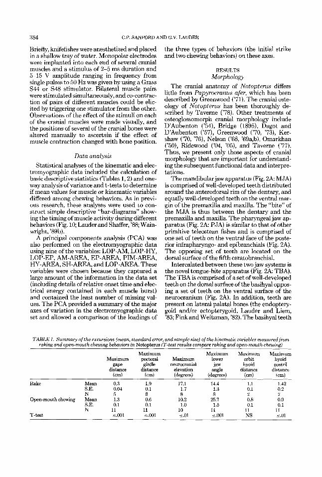
TABLE 1. Summary of the excursions (mean, standard error, and sample size) of the kinematic uariables measured from raking and open-mouth chewing behaviors in Notoptem (T-test results compare raking and open-mouth chewing)
Maximum Maximum Maximum Maximum Maximum pectoral Maximum lower orbit hyoid
gape girdle neurocranial jaw hyoid nos t r i 1 distance distance elevation angle distance distance
(cm) (cm) (degrees) (degrees) fcm) (cm)
S.E. 0.04 0.1 1.7 1.3 0.1 0.2 N 5 8 8 8 2 2
Open-mouth chewing Mean 1.3 0.6 10.2 25.7 0.8 0.9 S.E. 0.1 0.1 1.0 1.5 0.1 0.1 N 11 11 10 11 11 11
T-test 5.001 5.001 5.01 5.001 NS 5.01
Rake Mean 0.3 1.9 17.1 14.4 1.1 1.42

TABL
E 2.
Mea
n ar
eas a
nd a
mol
itude
s for
elec
trom
vozr
awhi
c D
rofil
es of
six
mus
cles
in N
ot~
uter
us~
Uni
ts of
vol
ts x
U
nits
of (
volts
x
x (m
s)
LOP-
AM
P E
P-A
MP
AM
-AM
P SH
-AI"
PI
M-A
MP
HY
-AM
P LO
P-ar
ea
EP-a
rea
AM
-are
a SH
-are
a PI
M-a
rea
HY
-are
a
Stri
ke
Mea
n S.
E.
N
S.E.
N
Rak
ing
Mea
n
Ope
n-m
outh
M
ean
chew
ing
S.E
. N
A
nova
1.99
2.
94
2.31
2.
51
0.36
0.
07
0.33
0.
38
7 7
7 7
0.47
1.
15
2.92
0.
30
0.07
0.22
0.
06
0.03
10
11
10
6
~.
1.60
0.
44
0.67
0.
26
0.17
0.
07
0.15
0.
07
19
13
19
7 <.
MI1
<.
001
<.00
1 <.
001
1.31
0.
44
6 2.22
0.
31
8 1.16
0.
28
6 <.00
1
2.16
0.
40
6 0.91
0.
23
0.44
0.
08
13 <.M
I
11
10.6
7 3.
34
7
11.0
9 15
.28
12.1
1 1.
04
4.03
2.
67
7 7
7 3.
42
8.89
36
.67
1.20
0.
89
3.32
3.
35
0.59
10
11
10
6
~~
11.0
6 1.
44
1.77
0.
34
1.95
0.
25
0.48
0.
12
19
12
19
6 c.
05
<.00
1 <.
001
<.00
1
4.56
9.
03
1.75
3.
10
6 6
11.3
5 4.
14
2.98
1.
71
8 11
4.
38
1.67
1.
66
0.36
6
13
<.Ol
c.
01
~
'AN
OV
A te
st in
dica
tes t
he ov
eral
l lev
el of
slgn
lfica
nce f
or d
iffer
ence
s am
ong
the t
hree
beh
avio
rs. N
refe
n to th
e tot
al n
umbe
r of i
ndin
dud
chew
ing
cycl
es m
easu
red.

386 C.P. SANFORD AND G.V. LAUDER
are, thus, potentially capable of both puncturing prey by pressing against the base of the skull and of shearing actions against the lateral palatal teeth.
An unusual feature of the gill arch apparatus is the presence of large, paired bony elements ventral to the hyobranchial apparatus (Fig. 2A APHB2). The proximal end of each bone is located near hypobranchial2. Greenwood ('71) and Taverne ('78) refer to these bones as ossified or cartilaginous processes of the hypobranchial series. In Notopterus, these bones are clearly distinct ossifications, not attached to any hyo- branchial element, and thus cannot be consid- ered processes. I t is true, however, that in some osteoglossomorph taxa these bones are attached to the second hypobranchial (e.g., in Osteoglos- sum, Pantodon, and Scleropages; see Table 5, and Taverne, '77, '78). Pending a detailed inves- tigation of the homology of this element and of the direction of character polarity within osteo- glossomorphs, we consider this bone in No- topterus to be an autogenous process of hypo- branchial2 (Fig. 2A: APHB2).
Notopterus has awell-developed ligament run- ning from the anterior face of the cleithrum to the proximal end of the autogenous hypobran- chid process (Fig. 2A: CBL). Part of the cleithro- branchial ligament attaches to the second hypo- branchial. Posteroventral movement of the pectoral girdle is thus directly transmitted to the branchial apparatus, causing it to move posterov- entrally also.
The cranial muscles of Notopterus have a very similar arrangement to that described by Greenwood ('71) for Papyrocranus afer. The muscles of primary interest in understanding the mechanics of the tongue-bite are: the adduc- tor mandibulae (AM), epaxial (EP), hypaxial (HY), levator operculi (LOP), posterior inter- mandibularis (PIM), and sternohyoideus (SH) (Fig. 2B). The epaxial muscles insert on the posterodorsal surface of the cranium and act to elevate the neurocranium on the vertebral col- umn. The adductor mandibulae is a large unsub- divided muscle and extends anteroventrally from its origin on the suspensorium to insert on the coronoid process of the mandible (Fig. 2B). The levator operculi takes its origin from the neuroc- ranium and inserts on the medial margin of the operculum. Contraction of this muscle causes the operculum to rotate posterodorsally and the mandible to depress ventrally via the interoper- culomandibular ligament (Lauder, '80; Liem, '70). This muscle, in conjunction with the sternohyoi- deus, is a major depressor of the mandible. The
sternohyoideus (Fig. 2B: SH) originates from the cleithrum and narrows to a tendinous anterior insertion on the anterohyal and ventrohyal. This muscle causes the hyobranchial apparatus to rotate posteroventrally and also causes the man- dible to depress ventrally via the mandibulohy- oid ligament (Lauder, '80). An increase in gape of the mandibular jaws may thus be achieved by 1) activity in the epaxial muscles to lift the cranium, 2) activity in the levator operculi to depress the lower jaw, and 3) activity in the sternohyoideus to depress the lower jaw.
The hypaxial muscles (Fig. 2B: HY) are well- developed and insert along the posterior and ventral margin of the cleithrum. Fibers attach- ing to the most ventral region of the cleithrum are distinct from the fibers of the obliquus inferi- oris subdivision of the hypaxialis, which insert on the posterior flange of the cleithrum (upper HY arrow in Fig. 2B).
The posterior intermandibularis muscle in No- topterus is well-developed and extends from the lateral face of the anterohyal to the anteromedial face of the dentary (Fig. 2B: PIM). Primitively in actinopterygians the fibers of this muscle run transversely between the mandibular rami, and the PIM forms a muscular sling across the ante- rior buccal floor (Lauder, '80). The PIM in No- topterus has a different orientation from this primitive configuration and has long fibers with an anterior-posterior line of action.
Kinematics Feeding in Notopterus was divided into four
distinct behaviors: 1) the initial strike, 2) raking, 3) open-mouth chewing, and 4) swallowing. Both raking and open-mouth chewing involve the tongue-bite and may be classified as intraoral prey processing behaviors. The strike occurs by inertial suction feeding, and this type of prey capture has been described in other actinoptery- gian fishes (e.g., Lauder, '80, '85; Liem, '70); the strike in Notopterus is very similar to that of other generalized ray-finned fishes and is not considered here in detail. Swallowing behavior involves the pharyngeal jaw apparatus and not the muscles associated with the tongue-bite. Only the two behaviors associated with intraoral prey processing, raking and open-mouth chewing, will be discussed in detail.
A series of frames from a high-speed film depicting raking behavior is shown in Figure 3. Raking behavior is performed only on large prey items that are held in the MJA, and commonly between one and four raking cycles occur. An analysis of 46 feedings on prey ranging from 1 cm to 6 cm total length in size showed that no raking

FUNCTIONAL MORPHOLOGY OF NOTOPTERUS 387
of the prey occurred below a prey size of 72 % of knifefish head length. Above this size, raking occurred 71 92 of the time.
During raking, gape distance varies little, un- dergoing maximal excursions of 0.3 cm on aver- age (Fig. 4A,B and Table l), reflecting the fact that the prey is held in the MJA. The pectoral girdle exhibits an excursion of approximately 2 cm during raking (Fig. 4A,B and Table 1). The power stroke of the rake begins when the pecto- ral girdle moves posteriorly and is about one- fourth the duration of the anteriorly directed recovery stroke (Fig. 4). As the pectoral girdle moves posteriorly at the start of raking, the neurocranium is elevated and the lower jaw an- gle generally increases, reflecting closure of the lower jaw on the prey (Fig. 4). Although the lower jaw angle profile is somewhat erratic and shows considerable variation, in general the lower jaw is elevated as the neurocranium is lifted and the pectoral girdle is moved posteriorly. At the end of the raking power stroke gape distance nearly always reaches a local minimum indicat- ing that lower jaw elevation is occurring concom- itantly with cranial elevation to keep the prey fixed between the jaws.
Movement of the hyoid has both a dorsoven- tral component (indicated by the orbit-hyoid distance) and an anteroposterior component (in- dicated by the hyoid-nostril distance) during raking (Fig. 5A); both excursions average be- tween 1 and 2 cm in distance. The raking power stroke involves the rapid movement of the hyoid posteroventrally and the recovery stroke of the hyoid apparatus is slow in comparison to the power stroke. The hyoid apparatus moves a greater distance anteroposteriorly (hyoid-nostril distance) than it does dorsoventrally (orbit- hyoid distance) during raking (Table 1).
Open-mouth chewing, the second intraoral prey processing behavior in Notopterus, occurs only when the prey is located within the buccal cavity. In contrast to raking, the prey is not held in the MJA, and the gape undergoes signifi- cantly larger excursions of nearly 1.3 cm during open-mouth chewing (Fig. 6, Table 1). The angle of the neurocranium shows only relatively low- amplitude irregular fluctuations of about 10.2", significantly less than the average neurocranial elevation during raking (Table 1). The pectoral girdle moves anteroposteriorly during open- mouth chewing, but the maximum change in distance is significantly less than in raking (Ta- ble 1). Posterior movement of the pectoral girdle occurs as the lower jaw is depressed, indicated by decreases in lower jaw angle (Fig. 6). The maxi-
mal change in lower jaw angle is significantly greater than in raking (Table 1) and indicates, in combination with the lack of change in HDA, that the increase in gape during open-mouth chewing is achieved almost entirely by the lower jaw, not by cranial elevation. During open- mouth chewing, hyoid movement has both a dorsoventral component (indicated by the orbit- hyoid distance: Fig. 5B) and an anteroposterior component (indicated by the hyoid-nostril dis- tance: Fig. 5B), and both are less than the excur- sions observed during raking (compare panels A and B in Fig. 5).
Both raking and open-mouth chewing behav- iors are frequently repeated several times, and kinematic patterns often show trends from cycle to cycle. For example, pectoral girdle movement during open-mouth chewing (Fig. 6) shows a steady trend from posterior to anterior, each chewing cycle beginning a t a successively more anterior position as prey reduction occurs. Dur- ing raking, less obvious trends are visible, but neurocranial elevation, lower jaw angle, and gape distance cycles all exhibit some tendency to start subsequent cycles before returning to the initial rest position.
Electromyography The strike has a duration of approximately
110 ms and involves high-level activity in all the cranial muscles monitored during this study (Figs. 7 , 10A, Table 2). The levator operculi, which depresses the lower jaw, is the first muscle to become active (Fig. 10A). Approximately 5 to 10 ms later, all the remaining muscles become active nearly simultaneously (Fig. 1OA).
Following the strike, the prey (in this case the goldfish Carassias auratus) is subjected to exten- sive intraoral processing by the tongue-bite appa- ratus prior to swallowing. An overview of the EMG patterns associated with prey processing is presented in Figure 8. Following the strike and prey capture the prey is often drawn into the buccal cavity. If the prey is large, it is moved anteriorly by a reverse (posterior to anterior) flow of water: water is drawn in the opercular openings, the hyoid is elevated, and the prey is washed anteriorly a small distance as the mouth opens to allow water to flow out. This cycle is repeated several times and is reflected in Figure 8 by the repeated cycles of activity in the levator operculi muscle. As the prey is successively moved forward, the levator operculi shows an increasing amplitude of activity (Fig. 8: LOP). When the prey has been moved sufficiently ante- riorly such that the MJA can bite and hold it, raking begins. The characteristic raking EMG

Figure 3

FUNCTIONAL MORPHOLOGY OF NOTOPTERUS
Fig. 3. Eight frames from a film (taken at 200 frames per second) of a raking sequence in Notopterm chitala. The number of each frame is in the upper left corner of each image. The entire sequence lasts 140 ms. Note that the goldfish (Carassius auratus) is held firmly between the mm- dibular jaws while the pectoral girdle moves posteriorly caw- ing the basihyal teeth to be raked across the prey. Movement of the pectoral girdle is most easily seen by examining the position of the base of the pectoral fin relative to the gill cover. In addition, while the basihyal teeth are moving posteriorly, the neurocranium is moving up, resulting in a shearing action of the two sets of teeth makiig up the tongue-bite. Arrows in frames 1,8,16, and 20 indicate these movements.
389

-4 m _.
3 3 0) m 0
U
f
OL
d
0
N 0
0
w
0..
0
P
0
0
VI
0.-
0
m
0
0
-4
0
0
m
0
0
Low
er J
aw A
ngle
(de
gree
s)
... M
0
0
0
.. .. .. .. .. -c
Ip
Low
er J
aw A
ngle
(de
gree
s)
LI
0
0
0
Neu
rocr
onio
l Ele
v. (
degr
ees)
Neu
rocr
onio
l Ele
v. (
degr
ees)
-
... -
... c
0m
-1
0
00
0
0 -.
0
Pec
t. G
irdle
Dis
t. (
cm) A
L
m m
0
Pec
t. G
irdle
Dis
t. (
crn
)
Gap
e D
ista
nce
(cm
)
Gap
e D
ista
nce
(crn
)

FUNCTIONAL MORPHOLOGY OF NOTOPTERUS
Fig. 4. Graphic representation of the gape distance (in cm), pectoral girdle distance (in cm), neurocranial elevation (in degrees), and lower jaw angle (in degrees), during raking by Notopterm chitala. These variables are plotted as a func- tion of time in ms. Note that as the pectoral girdle distance increases neurocranial elevation increases and that a decrease in the lower jaw angle means that the lower jaw is being depressed. The gape distance profile is erratic and there is relatively little change during individual raking sequences, but there is a substantial posterior motion of the pectoral girdle. Panels A and B Measurements from two different individuals during a series of raking events. Note that over a sequence of repeated rakings of the prey, kinematic variables may exhibit a trend. Mean values for excursions of the kine- matic variables are presented in Table 1.
391

392 C.P. SANFORD AND G.V. LAUDER
A RAKING
.- L + cn 0
2 0 0 100 200 300 400 500 600
$2& 0 100 200 300 400 500 600
+i A
0 100 200 300 400 500 600 1 2
Time (msec)
Fig. 5. Comparisons of hyoid movements during raking (A) and open-mouth chewing (B) behaviors in Notoptems chitala. The variables are measured as a function of time in ms. Nostril-orbit distance is fixed and should not change during feeding. This variable is presented (in the same scale as the other plots, a 3 cm range on the y-axis) to provide an indication of the amount of error present in our data. Varia- tion in nostril-orbit distance includes alI possible sources of error: digitizing errors, difficulty in reliably recognizing land- marks, and movement of the fish out of the film plane.
8 OPEN MOUTH CHEWING
g bb 0.0 0 100 200 300 400 500 600
- 0.51 J
0 ID0 200 300 400 500 600
4.4 L 2.5,:
v1
1.5 q ; ; ; : A I 0.5
0 I D 0 200 300 400 5W 600
Time (msec)
Comparison to other variables in this figure and Figures 4 and 6 shows that the error is small relative to the excursions of head bones measured during feeding. Orbit-hyoid distance is a measure of the dorsoventral motion of the hyoid. Hyoid- nostril distance is a measure of the anteroposterior motion of the hyoid. Note that raking behavior involves both dorsoven- tral and anteroposterior motion of the hyoid. During raking the hyoid moves through much greater distances than it does in open-mouth chewing.

FUNCTIONAL MORPHOLOGY OF NOTOPTERUS
OPEN MOUTH CHEWING 393
2.5 ~
n
0 2.0.. W
0 200 400 600 800 1000
5.9 4 I
5.1 -. 4.8 -. ...
0 ZOO 4bO SO0 800 1000
Time (msec)
Fig. 6. Graphic representation of the gape distance (in cm), pectoral girdle distance (in cm), neurocranial elevation (in degrees), and lower jaw angle (in degrees) during open mouth chewing in Notopterus chitah. These variables are plotted as a function of time in ms. Note that gape distance and lower jaw angle changes significantly in a regular pattern
pattern is then seen with high activity in the epaxialis, adductor mandibulae, and posterior intermandibularis (these muscles were silent while the prey was being moved anteriorly: Fig. 8) and low-level activity in the levator operculi. Figure 8 shows that two raking cycles were per- formed. Then, the prey is usually drawn back into the buccal cavity by one or more brief suc- tion events (indicated by high levels of activity in the levator operculi: Fig. 8: LOP), and the open- mouth chewing behavior begins. This behavior is characterized by relatively high activity in the levator operculi and low-level activity in the ad- ductor mandibulae and epaxialis. This entire process may be repeated several times, espe- cially with large prey, until the prey has been largely dismembered by the action of the tongue-
145 0 200 400 600 800 1000
-- I
I 0 200 400 600 BOO 1000
Time (msec)
(in contrast to raking behavior). There is very little change in neurocranial elevation, which exhibits irregular minor fluctu- ations in angle compared to raking. Pectoral girdle distance excursions are much smaller than those found in raking (also see Table 1). Three cycles of open-mouth chewing are repre- sented.
bite apparatus during raking and open-mouth chewing. Swallowing then begins.
The electromyographic profile of raking behav- ior is quite distinct from that of the strike. The first muscle to become active during raking is the adductor mandibulae (Fig. lOB), which has a significantly greater peak amplitude and inte- grated area than in either the strike or open- mouth chewing (Table 2). Between 20 and 40 ms later the posterior intermandibularis becomes active (Fig. 10B) and this muscle shows a higher level of activity than in either the strike or open- mouth chewing (Table 2). Approximately 5 ms later the levator operculi becomes active (Fig. 10B): this is the primary mouth-opening muscle. During the rake there is only low-level activity of the levator operculi (Figs. 8,9A), with a signifi-

394 C.P. SANFORD AND G.V. LAUDER
Notopterus STRIKE
I0.3mV
LOP --"
EP -- 'i
50msec
Fig. 7. Typical electromyographic (EMG) profile of No- topterus chit& during the strike; the prey is a goldfish (Curassius auratm). Note that all muscles recorded are ac-
tive during the strike and that the first muscle active is the levator operculi (LOP). The amplitude scale for the LOP muscle applies to all channels.
cantly smaller amplitude and integrated area than in either the strike or open-mouth chewing (Table 2). This is not surprising as there is little change in gape distance during raking as de- scribed above. Approximately 50 to 80 ms later the hypaxial, epaxial, and sternohyoideus mus- cles all become active (Fig. 10B). The sternohyoi- deus muscle is active in raking only between 34 and 67 r( of the time (Fig. 10B). In addition, this muscle shows very little activity (Figs. 8, 9A), with a small amplitude and integrated area when compared to the strike (Table 2). During raking both the epaxial and hypaxial muscles show a greater level of activity than during open-mouth chewing, hut less activity than in the strike (Ta- ble 2).
In open-mouth chewing only two muscles are commonly active at moderate to high ampli- tudes and during every sequence: the levator operculi and the adductor mandibulae (Figs. 8, 9B, lOC). As can be seen from the bar-diagram
(Fig. lOC), the epaxial and hypaxial muscles are used between 33 and 67% of the time, whereas the sternohyoideus and posterior intermandibu- laris muscles are used between 0 and 33 % of the time. The first muscle to become active during open-mouth chewing is the levator operculi mus- cle, which depresses the lower jaw and opens the mouth. The levator shows significantly greater activity, both in terms of burst amplitude and integrated area than in raking behavior (Table 2). Approximately 60 to 80 ms later the epaxial and hypaxial muscles become active, closely fol- lowed by the sternohyoideus, posterior interman- dibularis, and adductor mandibulae. During open-mouth chewing the sternohyoideus and posterior intermandibularis muscles have signif- icantly lower integrated areas than during rak- ing behavior (Table 2).
Patterns of EMG variation during the strike, raking, and open-mouth chewing behaviors are summarized in a principal-components analysis

FUNCTIONAL MORPHOLOGY OF NOTOPTERUS 395 POST.+ANTERIOR PREY MOVEMENT RAKING SUCTION OPEN MOUTH CHEWING
EP A h A -
SH A d
PIM r 1
HY 1 1 - 500 msec
Fig. 8. Electromyographic (EMG) profile of Notopterus chitala during oral processing of prey following the strike. (The electromyograms have been rectified and were recorded simultaneously.) This figure provides an overview of a typical prey processing sequence. Following the initial strike (during which all muscles are active at high amplitudes), the prey is moved anteriorly from the buccal cavity to the mandibular jaws. During this time, levator operculi activity gradually increases in intensity. At the onset of the raking behavior, the prey is being held between the mandibular jaws as shown in Figure 3. Once the prey is at the mandibular jaws, the jaws
illustrated in Figure 11, and the loadings of nine EMG variables are presented in Table 3. Compo- nents 1 and 2 together summarize 57% of the variation in the data set and clearly show the three behaviors to be different in multivariate EMG space. High scores on principal-compo- nent 1 reflect greater activity in the adductor mandibulae, posterior intermandibularis, and epaxial muscles, reduced activity in the levator operculi, and shorter relative onset times in the adductor mandibulae and hypaxial muscles. High scores on principal component 2 reflect in- creased activity in the sternohyoideus muscle, reduced activity in the adductor mandibulae, and greater overlap of activity in the levator operculi, epaxial, and hypaxial muscles.
close around the prey by action of the adductor mandibulae. This stabilizes the prey, and there are two raking sequences (identified by the two high-amplitude bursts of activity in the adductor mandibulae (AM) and posterior intermandibularis (PIM)). Following raking, the prey is drawn back into the buccal cavity by water flow created by suction (note the high-amplitude LOP activity). Then, a series of open-mouth chewing sequences is initiated that serve to further macerate the prey. There is little sternohyoideus activity during both open-mouth chewing and raking. The amplitude scale for the LOP muscle applies to aU channels.
DISCUSSION Functional morphology of Notopterus
There are two behaviors that can be identified in Notopterus chitala as being associated with prey processing: raking and open-mouth chew- ing. Both have distinctive kinematic and EMG profiles, and both contribute to the extensive prey destruction observed prior to swallowing. A major feature of the feeding system in No- topterus is, thus, the ability to perform extensive prey manipulation and shredding with the hyo- branchial apparatus.
The major differences between the two prey processing behaviors observed in Notopterus are summarized in Table 4. Both raking and open-

A Notopterus RAKING
10.2rn~
LOP F
EP
AM
SH
PIM
HY ,,.,I
B
LOP
EP
AM
SH
PIM
HY
Notopterus OPEN MOUTH CHEWING
(O.lrnV d
I
I -7,s -
loomsec
Figure 9

FUNCTIONAL MORPHOLOGY OF NOTOPTERUS
Fig. 9. Typical electromyographic (EMG) profile of No- topterus chitala during prey processing behavior. A: Raking behavior, where the prey (a goldfish, Carassius auratus) is held in the mandibular jaws by the action of the adductor mandibulae (closing the jaws). Following this, the basihyal teeth are raked posteriorly acrces the prey and the neurocra- nium elevates, producing a shearing action between the two sets of teeth in the tongue-bite. Note the low activity in the levator operculi and the sternohyoideus muscles that is char- acteristic of raking. This panel shows two consecutive raking cycles. B: Open-mouth chewing behavior, where the prey is located within the buccal cavity and there is strong levator operculi activity. This panel shows three consecutive open- mouth chews. Vertical scale bars next to each channel indi- cate the EMG voltage (in mV). If no vertical scale is shown, the scale is the same as that for the LOP muscle.
397

A STRIKE
I
HYI ----
I 2 c 40 60 80 100 120
TIME (in
B RAKING
EP I I AM - I
I
I . . . . . . . . . . I , . . . , , L
40 - 2 0 0 20 40 60 0 0 100 120 14. l b 0 1 8 C
TIME (in msec)
Figure 10

C FUNCTIONAL MORPHOLOGY OF NOTOPTERUS
OPEN MOUTH CHEWING 399
I E PI
I
I I I
SH
*MI
PlMl
I HYI
I
L
L t -
. -..--.. 20 40 60 eo 100 120 1.10
TIME (in msec)
Fig. 10. Bar-diagrams to illustrate the relative onset of six cranial muscles during feediig in Notopterus chitala. All onsets are measured by using the levator operculi as the reference muscle. The horizontal lines on each side of the bar represent 1 standard error in either onset or offset time. Solid bars indicate that the muscle is active 67-1WC of the time; hatched bars indicate that the muscle is active 33465 of the time; and open bars indicate that the muscle is active 0 4 3 % of the time. A The timing of muscle activity at the initial strike: The levator operculi is active first, and approximately 20 ms later all other cranial muscles recorded become active. -.The timing of muscle activity during rakiig behavior: The
TABLE 3. Loadings of nine variables on principal components (Ws) I to4
Variable PCl PC2 PC3 PC4
LOP-AM AM-area EP-area P1M-area LOP-HY HY-area SH-area LOP-EP LOP-area Variance
explained
- 0.85 0.29 0.01 0.15 0.77 -0.51 0.28 0.05 0.62 0.17 0.28 -0.61 0.59 -0.46 -0.46 -0.21
-0.58 -0.52 0.47 -0.21 0.53 0.23 0.56 0.27 0.41 0.70 0.40 0.08
-0.39 -0.65 0.58 0.03 -0.41 0.39 0.52 0.34 34.9% 22.05 16.9% 10.57
first muscle to become active is the adductor mandibulae, followed by the posterior intermandibularis, then the levator operculi, and finally the epaxial, hypaxial, and sternohyoi- deus muscles, which all become active together. C The timing of muscle activity during open-mouth chewing behav- ior.The levator operculi becomes active well before any other muscles. Approximately 6040 ms later the epaxial and hypaxial muscles become active, followed by the addudor mandibulae, sternohyoideus, and posterior intermandibu- lark, which all become active at approximately the same time. Sample size for each mean ranged from six to 19.
mouth chewing possess uniquely derived fea- tures that permit an unambiguous determina- tion of the behavior from either the kinematic or EMG profiles. Raking of the prey involves little change in gape, low activity in the levator oper- culi, and high activity in the posterior interman- dibularis, epaxialis, and addudor mandibulae. During raking the prey is held firmly in the mandibular jaw apparatus: the dentary and pre- maxilla close around the prey item, stabilizing it, and the basihyal teeth are moved anteriorly, imbedded into the prey, and then raked posteri- orly across the prey by posterior movement of the hyoid and pectoral girdle. The result of this

400 C.P. SANFORD A
TABLE 4. Summary of the kinematic and electromyographic (EMG) characteristics which
distinguish raking from open-mouth chewing prey Drocessine behaviors
Raking
1) Very low LOP activity 2) High AM activity 3) High PIM activity 4) High epaxial activity
5) Hypaxial activity
1) Little change in gape 2) Both antero-posterior
and dorso-ventral motion of hyoid
3) Neurocranial elevation increases as pectoral girdle moves posteriorly
4) Pectoral girdle moves anteroposteriorly
EMG
Kinematics
Open-mouth chewing
1) Moderate LOP activity 2) Low AM activity 3) Low PIM activity 4) Low to no epaxial
5) Hypaxial activity
1) Large increase in gape 2) Motion of hyoid less
activity
than in raking
3) Much less neurocranial elevation
4) Much less antero- posterior motion of the pectoral girdle
behavior is extensive damage to the body wall and musculature of the prey. This raking motion may be repeated several times before the prey is drawn completely into the buccal cavity for fur- ther processing.
In contrast, open-mouth chewing involves sub- stantial changes in gape, moderate levator oper- culi activity, and low activity in the posterior intermandibularis, epaxialis, and adductor man- dibulae (Table 4). Prey are located within the buccal cavity during open-mouth chewing.
A distinctive feature of both prey processing behaviors (in comparison to the initial strike) is the near total lack of activity in the sternohyoi- deus muscle (Figs. 8, 9, Tables 2, 4). Despite consistent high-amplitude sternohyoideus activ- ity at the strike (high SH area is the main vari- able causing strikes to have high scores on PC2 in Fig. 11, thus separating them from raking and open-mouth chewing behaviors in the principal- components analysis), we rarely recorded much activity from the SH during raking or open- mouth chewing. The hypaxial muscles showed consistent moderate levels of activity during both open-mouth chewing and raking (Table 2) and exhibited four to five times the activity of the sternohyoideus.
How does the tongue-bite mechanism func- tion to puncture and shred the prey within the oral cavity? An important feature of raking be- havior is the action of the adductor mandibulae to close the mandibular jaws on the prey item and to thereby stabilize the prey. Approximately 10 ms later, the posterior intermandibularis be- comes active to protract the basihyal. Our mus- cle stimulation experiments showed clearly that
LND G.V. LAUDER
contraction of the PIM strongly protracts and elevates the hyobranchial apparatus and that, no matter what the angle of the lower jaw, stimu- lation of the posterior intermandibularis always protracts the basihyal. Protraction of the hyoid apparatus brings the large basihyal teeth into contact with the prey. Posterior intermandibu- laris activity may also serve to press the prey against the base of the skull as there is a signifi- cant dorsal component to the line of action of this muscle. Once the basihyal teeth have pene- trated the prey, the epaxial and hypaxial mus- cles become active (Fig. IOB). The epaxial mus- cles elevate the neurocranium (Fig. 2B), moving the parasphenoid teeth anterodorsally. The hyp- axial muscles cause the pectoral girdle to move posteriorly (Fig. 4A,B); this in turn causes the basihyal to move posteroventrally over nearly a 2 cm excursion (Figs. 2B, 4A, Table 1). At the same time, the neurocranium elevates (Fig. 4A), producing a shearing action between the two sets of teeth that make up the tongue-bite. The ster- nohyoideus muscle shows little activity during the rake, so we conclude that tension between the pectoral girdle and the basihyal is main- tained by the strong ligamentous and connective tissue links between these two elements (Fig. 2A: CBL). The presence of low-level activity in the sternohyoideus muscle 3 4 6 7 76 of the time (Fig. 10A) might also assist in producing tension be- tween the pectoral girdle and the basihyal. How- ever, there is little doubt from the kinematic results that the main force for pulling the basihyal teeth posteroventrally through the prey comes from the extensive posterior motion of the pecto- ral girdle (Fig. 4).
The hypothesized pattern of motion for the basihyal teeth during raking is illustrated in Fig- ure 2B. We hypothesize that the primary prey reduction stroke of raking is in fact an anteropos- terior shearing caused by relative movement of the neurocranium and hyoid. The basihyal teeth move anterodorsally into the prey as a result of posterior intermandibularis activity, and the re- duction stroke is a posterior shearing of the basihyal teeth against the parasphenoid teeth and lateral palatal teeth. A key conclusion from the kinematic and EMG data is that the main forces driving this shearing arise from the body muscles: epaxialis and hypaxialis.
As discussed above, open-mouth chewing is clearly a distinct prey processing behavior from raking (Table 4, Fig. 11). Open-mouth chewing involves use of the tongue-bite on the prey item within the buccal cavity and contributes to prey reduction and maceration. However, the most dramatic damage to prey items was observed

TAB
LE 5.
Sum
mar
y of
the
anat
omy o
f N
otop
teru
s, ot
her o
steo
glos
som
orph
s, an
d ou
tgro
ups
Bas
ihya
l te
eth
Ster
nohy
oide
us
conn
ectio
n to
uro
hyal
Tend
inou
s ins
ertio
n to v
en-
trohy
al a
nd a
nter
ohya
l In
serti
on to
uroh
yal
Stem
ohyo
ideu
s to
uroh
yal
Tend
inou
s ins
ertio
n to
ven
- tro
hyal
and
ante
rohy
al
Tend
inou
s ins
ertio
n to
uro
-
Tend
inou
s ins
ertio
n to
uro
-
Tend
mou
s ins
ertio
n to
uro
-
hYd
hY+
Ven
tral
scut
es
Cle
ithro
bran
chia
l lig
amen
t
No
visi
ble l
ig.
Run
s fro
m c
leith
. to
back
of
Run
s fro
m c
leith
. to
back
of
BB
2
HB
3
Taxo
n A
PHBZ
PI
M m
uscl
e
IH a
nd P
IM fu
sed
Smal
l sho
rt P
IM
IH a
nd P
IM fu
sed
to be
ver
y
Ver
y lo
ng P
IM
long
Dor
sohy
al
+ V
entro
hyal
U n os s
i fi e d
-
4
Pant
odon
Hio
don
Scle
ropa
ges
Not
opte
rus
+ A
nk to
HB
2 -
Wel
l- de
velo
ped
Ver
y w
ell-
deve
lope
d Po
orly
de
velo
ped
Ver
y deve
lope
d w
ell-
Poor
ly
deve
lope
d W
ell-
deve
lope
d N
one
pres
ent
Poor
ly
deve
lope
d
smal
l
deve
lope
d
Ver
y
P oo
rly
+
+ + An
k to
HBZ
Arti
cula
tes
with
HB
2 -
-
Uno
ssifi
ed
+ +
Run
s fro
m cl
eith
. to
HB
2 an
d A
PHB
2 -
U n o
s s i fi
ed
Run
s fro
m c
leith
. to
back
of
BB
2 R
uns f
rom
cle
ith. t
o ba
ck o
f B
B1 a
nd 2
No
ligam
ent
Run
s fro
m cl
eith
. to
HB
3
Non
e pr
esen
t
Non
e pr
esen
t
No
(GH
) C
oreg
onus
Salm
o
Dor
som
a
Eson
Amia
Lepi
sost
eus
- U
noss
ified
+
I
I
+ hY
+ Te
ndln
ous i
nser
tion
to U
ro-
hYal
Dire
ct c
onne
ctio
n to
vent
ro-
Dire
ct c
onne
ctio
n to
vent
ro-
hyal
(no u
rohy
al)
hYal
Smal
l PIM
Exte
nded
PIM
func
tiona
lly
shor
t
+ +
+

402 C.P. SANFORD AND G.V. LAUDER
+SH-AREA
-LOP-EP
-LOP-HY
- AM-AREA
h x N N - N x
0 0 0
S S
0, S
0
R R
R
R n
-I- EP-AREA
+ PIM-AREA R
PC 1
Fig. 11. Principal-components (PC) analysis of nine elec- tromyographic variables plotted as PC 1 vs. PC 2. These two components account for 35 % and 22% of the overall variance in the data set respectively. This clearly shows that all three behaviors analyzed (S, initial strike; R, raking; 0, open-mouth
- LOP-AM
-LOP-HY
+AM-AREA
I I -2 ' -2 -1 0 1 2
following raking behavior. Posterior movement of the pectoral girdle during open-mouth chew- ing, perhaps because the prey is not being held fixed in the mandibular jaws, appears to inflict less damage than during raking. Open-mouth chewing starts with lower jaw depression result- ing from activity in the levator operculi (Fig. 10C). Between 60 and 80 ms later all the other muscles become active. Then the adductor man- dibulae becomes active, raising the lower jaw and closing the mouth, a t approximately the same time the epaxial and hypaxial muscles (which are the next most frequently used mus- cles (Fig. l0C)) show activity. However, the epax- ialis and hypaxials are much less active and have significantly lower amplitudes and integrated areas than during raking (Table 2). Activity of these two muscles is likely to have the same effect as in raking: the production of a shearing action but of much less intensity. An important point to make, however, is that both the lack of sternohyoideus activity and the presence of pos- terior pectoral girdle movement are features com- mon to both open-mouth chewing and raking, and both behaviors result in extensive shredding and damage to the prey item.
(35%)
chewing) have quite distinct motor patterns. Variables load- ing highly on each axis are indicated next to the axis and the sign of the loading is also given. The meaning of the loadings is discussed in the text.
T h e evolution of the tongue-bite apparatus
The tongue-bite mechanism characterizes os- teoglossomorph fishes. In slightly different mor- phological configurations, a tongue-bite has evolved convergently in several teleost clades (Liem and Greenwood, '81; Nelson, '69b). How- ever, no such mechanism is present in primitive ray-finned fishes (Lauder and Liem, '83).
A salient feature of the tongue-bite is the modification of muscular and skeletal elements of the head such that a novel third set of jaws is present. This third set of jaws (Fig. 2A: TBA) is anatomically intercalated between the anterior mandibular jaw apparatus (MJA) and the poste- rior pharyngeal jaw apparatus (PJA), and yet common muscular elements f u i d o n in all three jaw systems. T'!qkgenetically, the prc sence of the MJA and P J A is prirnii:,cL i'w '9 --finned fishes (Fig. 121, and (at least in nowl ,teleost clades) the tongue-bite has apparently evolved at least twice (Fig. 12: TBA; Lauder and Liem, '83; Fink and Weitzman, '82). The primary role of the MJA is in initial prey capture (Liem, '70), and the PJA plays a critical role during swallow- ing (Lauder, '83). In contrast, the TBA appears

FUNCTIONAL MORPHOLOGY OF NOTOPTER US 403
to be related to prey processing and immobiliza- tion of prey (by disarticulation or evisceration), and perhaps facilitates digestion by ripping the skin and body wall open.
Anatomically, there are several novelties present in Notopterus that might be suspected a priori of being related to the presence of a TBA. Table 5 presents the results of a survey of several possible morphological correlates to intraoral prey processing in selected actinopterygian clades. We investigated the distribution of sev- eral features of Notopterus morphology that might be correlated with the presence of a tongue-bite: ventral scutes, the autogenous pro- cess on hypobranchial 2, the cleithrobranchial ligament, ossification of hyoid elements, the pres- ence of teeth on the basihyal, and the morphol- ogy of the posterior intermandibularis muscle (Table 5). All of these seemed likely candidates for morphological correlates of a tongue-bite ap- paratus (defined by the presence of teeth on both the basihyal and base of the skull) due to their presence in Notopterus.
The presence of basihyal teeth appears to be a primitive character for osteoglossomorphs and possibly also for halecostome fishes (Table 5; Lauder and Liem, '83). Some form of a process from hypobranchial2 appears to be present in at least two other lineages of osteoglossomorphs (Table 5 APHB2), but not in all. In addition, many taxa appear to have ligamentous connec- tions between the cleithrum and the hyobran- chial apparatus.
An important morphological feature of the TBA that Notopterus shares with Scleropages is the elongate PIM muscle (Table 5). This is a key feature of the TBA in Notopterus because the reorientation of the PIM to insert near the mandibular symphysis has resulted in a much longer muscle, presumably with longer fibers. Primitively in actinopterygians the PIM and interhyoideus muscles are the major muscles of the anterior buccal floor; the PIM has short fibers and spans the mandibular rami (Table 5; Lauder, '80). Longer muscles permit greater ex- cursions of attachment sites as the distance of muscle shortening is a function of the number of sarcomeres in series. Thus, we hypothesize that the long PIM of Notopterus is functionally criti- cal to the function of the TBA by permitting extensive anteroposterior movement of the basihyal. The dominant element of the power stroke of the tongue-bite is the extensive retrac- tion of the basihyal powered by the hypaxial muscles. The ability of the basihyal to be pro- tracted over 2 cm and then to be retracted is
dependent on a long PIM muscle. This hypothe- sis suggests that not all osteoglossomorphs will use the raking behavior demonstrated here in Notopterus and suggests caution in extending our results on the function of the TBA in No- topterus to other osteoglossomorphs. However, we predict that other osteoglossomorphs with a TBA and a long PIM muscle (such as Pantodon, Osteoglossum, and Scleropages) may show rak- ing behavior as they also possess PIM muscles modified from the primitive condition (Table 5).
The functional significance of several other morphological features present in Notopterus is still unclear. Neither the remarkable ventral scutes found in Notopterus nor the presence of the greatly elongated autogenous process of hy- pobranchial2 could be related to the function of the TBA. Originally we thought that the pres- ence of the autogenous process in many osteoglos- somorphs might be associated with the use of a tongue-bite. However, this appears not to be the case as we can find no clear functional role for this process, and Hiodon, which possesses very well-developed basihyal teeth and TBA, lacks the process (Table 5 ) .
A major result from our functional analysis is that the sternohyoideus (SH) muscle is virtually inactive during raking and open-mouth chewing behaviors. This result was a surprise because the SH is a major connecting link between the pecto- ral girdle and hyobranchial apparatus and had been shown previously to play a major role in hyobranchial retraction during prey capture by teleost fishes (Lauder, '80, '85; Liem, '78). We had expected prior to conducting the EMG ex- periments to find high-amplitude activity in the SH during raking and open-mouth chewing. Given the lack of SH activity, a question arises as to how the posterior motion of the pectoral girdle is transmitted to the basihyal to achieve the power stroke of raking. In Notopterus there is a well-developed ligament that runs between the pectoral girdle and the basibranchial series (Fig. 2A: CBL). We hypothesize that this liga- ment is a critical feature of raking and open- mouth chewing behaviors and that i t serves to transmit the force produced by the hypaxial muscles during prey processing to the basihyal teeth. Some other taxa also possess a ligament, and Pantodon, which has a tongue-bite, does not possess such a ligament. Thus, this ligament cannot be associated uniquely with the tongue- bite.
The main functional and morphological corre- lates of the tongue bite in Notopterus are 1) the extensive ligament between the pectoral girdle

Figure 10

FUNCTIONAL MORPHOLOGY OF NOTOPTERUS
Fig. 12. Cladogram depicting the interrelationships of several major clades of actinopterygian fishes to show hypoth- esized phylogenetic patterns in the tongue-bite apparatus. The open bars depict characters hypothesized to be primitive for actinopterygians: the presence of an MJA and PJA. Solid bars indicate proposed synapomorphic Characters. In this case the tongue-bite apparatus is suggested to have arisen independently in the osteoglossomorphs and salmonoids (see Fink and Weitzman, '82; Lauder and Liem, '83). Further characters proposed here for notopteroids include the lack of sternohyoideus activity during the tongue-bite, the greatly lengthened posterior intermandibdaris muscle, and the kine- matic pattern at the rake and open-mouth chewing. There are presently no data that would allow us to test the hypothesis that raking and open-mouth chewing prey processing behav- iors are primitive for osteoglossomorphs and are associated with the presence of a tongue-bite morphology.
405

406 C.P. SANFORD AND G.V. LAUDER
A Amia calva 1o.lrnV
LOP I
EP + SH
IH A #. - 100 msec
Lepisosteus oculatus B I0.5mV
RD -+
AD5 rJ,
LE4 - . *
1 0.5mV
SH I' - 500 msec
Fig. 13. Typical electromyographic (EMG) profiles of Amia calua (A) and Lepisosteus oculatus (B) during in- traoral prey processing behavior. Both taxa represent out- groupsto osteoglossomorphs (Fig. 12 Halecomorphi and Gmg- lymcdi). In both outgroups examined there is considerable sternohyoideus (SH) activity during chewing. This suggests
that the lack of sternohyoideus activity during chewing in Notopterus is a derived condition. Data are not yet available to permit a decision on the presence or absence of SH activity during intraoral prey processing in other primitive teleost clades.

FUNCTIONAL MORPHOI
and the hyobranchial region, 2) the presence of a long-fibered PIM muscle, and 3) the lack of activity in the SH muscle. The first two of these three features are neither limited to osteoglosso- morph fishes nor found in all bony-tongued fishes. The pattern of activity of the sternohyoi- deus muscle and the use of raking and open- mouth chewing behaviors by osteoglossomorphs other than Notopterus remain to be determined.
The lack of sternohyoideus activity during prey processing within the oral cavity by No- topterus is a derived feature of the feeding mech- anism. Although outgroup taxa such as Amia and Lepisosteus lack the tongue-bite proper, they do exhibit prey manipulation within the oral cavity following the strike (Lauder, ’80). We recorded electromyograms during intraoral prey processing in these outgroup taxa to determine if sternohyoideus muscle activity was present. As is illustrated in Figure 13, both Amia and Lepi- sosteus show considerable SH activity during intraoral prey manipulation. We hypothesize that lack of sternohyoideus activity during chewing is a derived condition for Notopterus and perhaps for other osteoglossomorphs that use a tongue- bite and raking behavior (Fig. 12).
ACKNOWLEDGMENTS
We thank Steve Reilly, Peter Wainwright, and Miriam Ashley for comments on the manu- script and Heidi Mumford for excellent labora- tory assistance. This research was supported by a NATO/NERC grant to C. Sanford and NSF BSR 85-20305 to G. Lauder.
LITERATURE CITED Bridge, T.W. (1895) On certain features of the skull of Osteo-
glossum jormosum. Proc. Zool. SOC. Lond. 20:302310. Dagot, J., and F. D’Aubenton (1957) Developpement et mor-
phologie de crane d’Heterotis niloticus Ehrenberg. Bull. Inst. Fr. Afr. Noire (A) 19:881-936.
DAubenton, F. (1954) Etude de l’appareil branchiospinal et de l’organse suprabranchia d’Heterotis niloticus Ehren- berg 1927. Bull. Inst. Fr. Afr. Noire (A) 17:1174-1201.
Dingerkus, G., and L.D. Uhler (1977) Enzyme clearing of alcian blue stained whole small vertebrates for demonstra- tion of cartilage. Stain Technol. 52(4):229-232.
Fink, W.L., and S.H. Weitman (1982) Relationships of the stomiiform fishes (Teleostei), with a description of Diplo- phos. B d . Mus. Comp. Zool. 150:31-93.
Greenwood, P.H. (1970) On the genus Lycopteru and its relationships with the family Hiodontidae (Pisces, Osteoglo- ssomorpha). Bull. Br. Mus. Nat. Hist. Zool. 19:257-285.
Greenwood, P.H. (1971) Hyoid and ventral gill arch muscula- ture in osteoglossomorph fishes. Bull. Br. Mus. Nat. Hist. Zool. 221-55.
Greenwood, P.H. (1973) Interrelationships of osteoglossomor- phs. In P.H. Greenwood, R.S. Miles, and C. Patterson (eds): Interrelationships of Fishes. New York Academic Press, pp. 307-332.
Kershaw, D. (1970) The cranial osteology of the “butterfly fish” Pantodon buchholzi. Zool. J . Linn. Soc. Lond. 49:s 19.
,OGY OF NOTOPTERUS 407
Kershaw, D. (1976) A structural and functional interpreta- tion of the cranial anatomy in relation to the feeding of osteoglossoid fishes and a consideration of their phylogeny. Trans. 2001. Soc. Lond. 33:173-252.
Lauder, G.V. (1980) Evolution of the feeding mechanism in primitive actinopterygian fishes: A functional anatomical analysis of Polypterus, Lepisosteus, and Amia. J. Morphol. 163:283-317.
Lauder, G.V. (1983) Functional design and evolution of the pharyngeal jaw apparatus in euteleostean fishes. Zool. J. Linn. Soc. 77:l-38.
Lauder, G.V. (1985) Aquatic feeding in lower vertebrates. In M. Hildebrand, D.M. Bramble, K.F. Liem, and D.B. Wake (eds): Functional Vertebrate Morphology. Cambridge: Har- vard University Press, pp. 210-229.
Lauder, G.V., and K.F. Liem (1981) Prey capture by Lucio- cephulus pulcher: Implications for models of jaw protru- sion in teleost fishes. Environ. Biol. Fish. 6257-268.
Lauder, G.V., and K.F. Liem (1983) The evolution and inter- relationships of the actinopterygian fishes. Bull. M u . Comp. Zool. 150.95197.
Lauder, G.V., and H.B. ShafFer (1988) Ontogeny of functional design in tiger salamanders (Ambystoma tigrinum): Are motor patterns conserved during major morphological trans- formations? J. Morphol. 297r249-268.
Liem, K.F. (1970) Comparative functional anatomy of the Nandidae. Fieldiana Zool. 56:l-166.
Liem, K.F. (1978) Modulatory multiplicity in the functional repertoire of the feeding mechanism in cichlid fishes. I. Piscivores. J. Morphol. 158:323-360.
Liem, K.F., and P.H. Greenwood (1981) A functional ap- proach to the phylogeny of the pharyngognath teleosts. Am. Zool. 21:83-101.
Liem, K.F., and S.L. Sanderson (1986) The pharyngeal jaw apparatus of labrid fishes: A functional morphological per- spective. J. Morphol. 187:143-158.
Nelson, G.J. (1968) Gill arches of some teleostean fishes of the division Osteoglossomorpha. J. Linn. SOC. Zool. 47:261-277.
Nelson, G.J. (1969a) Infraorbital bones and their bearing on the phylogeny and geography of osteoglossomorph fishes. Am. Mus. Novit. 2394:l-37.
Nelson, G.J. (1969b) Gill arches and the phylogeny of fishes, with notes on the classification of vertebrates. Bull. Am. Mus. Nat. Hist. 1413475-552.
Omarkhan, M. (1950) The development of the chondrocra- nium of Notopterus. J. Linn. Soc. Lond. Zool.61.608-624.
Reilly, S.M., and G.V. Lauder (1989a) Kinetics of tongue projection in Ambystoma tigrinum: Quantitative kinemat- ics, muscle function, and evolutionary hypotheses. J. Mor- phol. 199:223-243.
Reilly, S.M., and G.V. Lauder (1989b) Physiological bases of feeding behavior in salamanders: Do motor patterns vary with prey type‘? J. Exp. Biol. 141:343-358.
Ridewood, W.G. (1904) On the cranial osteology of the fami- lies Mormyridae, Notopteridae, and Hyodontidae. J . Linn. SOC. 29:1%217.
Ridewood, W.G. (1905) On the cranial osteology of the fishes of the families Osteoglossidae, Pantodontidae and Phracto- laemidae. J. Linn. Soc. 29:252-282.
Shaf€er, H.B., and G.V. Lauder (1988) The ontogeny of func- tional design: Metamorphosis of feeding behavior in the tiger salamander (Ambystornu tigrinurn). J. Zwl. Lond. 216:437454.
Taverne, L. (1977) Osteologie, Phylogenese et Systematique des Teleosteens Fossiles et Actuels du Super-Ordre des Osteoglossomorphes. Premiere Partie. Osteologie des Genres Hiodon, Eohiodon, Lycoptera, Osteoglossum, Scle- ropuges, Heterotis et Arapaima. Acad. R. Belg. Mem. C1. Sci., Coll. in-8”, 2‘ ser., T. XLII, F. 3,235 pp.
Taverne, L. (1978) Osteologie, Phylogenese et Systematique des Teleosteens Fossiles et Actuels du Super-Ordre des Osteoglossomorphes. Deuxieme Partie. Osteologie des

408 C.P. SANFORD AND G.V. LAUDER
Genres Phareodus, Phareoides, Brychaetus, Musperia, Pantodon. Sineida. Notooterus. Xenomvstlls et Paoyro- cranus. Acad. R. Belg., Mem. Cl. Sc., Cali. in-@, 2" sir., T. XLII, F.6,213pp.
Wainwright, P.C. (1989a) Functional morphology of the pha- rynged jaw apparatus in perciform fishes: An experimental analysis of the Haemulidae. J. Morphol. 200:231-245.
;FS AM APHBS Bb Bh C Cb CBL D E EP HDA HND HY IH IP LE LJA
Abbreviations adductor arcus branchialum 5 muscle anterohyal bone adductor mandibulae muscle autogenous process of hypobranchial2 basibranchial bone and tooth plate basihyal bone and tooth plate cleithrum ceratobranchial cleithrobranchial ligament dentary epibranchial epaxial muscles neurocranial elevation hyoid-nostril distance hypaxial muscles interhyoideus muscle infrapharyngobranchial levator externus muscle lower jaw angle
Wainwright, P.C. (1989b) Prey processing in haemulid fishes: Patterns of variation in pharyngeal jaw muscle activity. J. Exp. Biol. 141:359-375.
Wainwright, P.C., and G.V. Lauder (1986) Feeding biology of sunfishes: Patterns of variation in prey capture. Zool. J. Linn. SOC. Lond. 88t217-228.
LOP M MJA N NOD 0
OHD Ph PHCe PHCi PIM PJA Pm R RD S SH TBA Vh
levator operculi muscle maxilla mandibular jaw apparatus neurocranium nostril-orbit distance open-mouth chewing prey processing behavior orbit-hyoid distance posterohyal pharyngocleithralis externus muscle pharyngocleithralis internus muscle posterior intermandibularis muscle pharyngeal jaw apparatus premaxilla raking prey processing behavior retractor dorsalis muscle initial strike at the prey sternohyoideus muscle tongue-bite apparatus ventrohyal