FUNCTIONAL FOODS, NUTRACEUTICALS AND NATURAL … · FUNCTIONAL FOODS, NUTRACEUTICALS AND NATURAL...
93
F UNCTIONAL F OODS, N UTRACEUTICALS AND N ATURAL P RODUCTS CONCEPTS AND APPLICATIONS Edited by Professor Dhiraj A. Vattem, Ph.D. Texas State University Vatsala Maitin, Ph.D. Texas State University
Transcript of FUNCTIONAL FOODS, NUTRACEUTICALS AND NATURAL … · FUNCTIONAL FOODS, NUTRACEUTICALS AND NATURAL...
FUNCTIONAL FOODS,NUTRACEUTICALS ANDNATURAL PRODUCTS
CONCEPTS AND APPLICATIONS
Edited by
Professor Dhiraj A. Vattem, Ph.D.Texas State University
Vatsala Maitin, Ph.D.Texas State University
stcudorP larutaN dna slacituecartuN ,sdooF lanoitcnuF
hcetSED .cnI ,snoit ac il buP teertS ekuD htroN 934
.A.S.U 20671 ain av lys nneP ,ret sac naL
thgir ypoC © yb 6102 hcetSED .cnI ,snoit ac il buP devres er sthgir llA
a ni derots ,decud orp er eb yam noit ac il bup siht fo trap oN ,snaem yna yb ro mrof yna ni ,det tim snart ro ,met sys laveirt er
,esiw re hto ro ,gni droc er ,gni ypoc ot ohp ,lac i nahc em ,cinort cele.rehsil bup eht fo nois sim rep net tirw roirp eht tuo htiw
aci remA fo setatS detinU eht ni detnirP1 2 3 4 5 6 7 8 9 01
:elt it red nu yrt ne niaMsnoitacilppA dna stpecnoC :stcudorP larutaN dna slacituecartuN ,sdooF lanoitcnuF
A hcetSED koob snoit ac il buP .p :yhp ar go il biB
797 .p xed ni sedulc nI
7377495102 :rebmuN lortnoC ssergnoC fo yrarbiL0-101-59506-1-879 .oN NBSI
xv
Preface
The demand for foods with a positive impact on human health and wellness has exploded globally over the past two decades. This growth is driven by socioeconomic and scientific factors, including increas-es in population, disposable income, life expectancy and healthcare costs. The market for healthier foods is also enhanced by advance-ments in our understanding of dietary bioactive ingredients and their effects on various aspects of human health at a systems and molecu-lar level. This book examines the rapidly growing field of functional foods in the prevention and management of chronic and infectious diseases. It attempts to provide a unified and systematic account of functional foods by illustrating the connections among the different disciplines needed to understand foods and nutrients, mainly: food science, nutrition, pharmacology, toxicology and manufacturing tech-nology. Advances within and among all these fields are critical for the successful development and application of functional foods. Chap-ters in the present volume explore the varied sources, biochemical properties, metabolism, health benefits and safety of bioactive ingre-dients. Special emphasis is given to linking the molecular and chemi-cal structures of biologically active components in foods to their nu-tritional and pharmacological effects on human health and wellness. In addition to discussing scientific and clinical rationales for different sources of functional foods, the book also explains in detail scientific methodologies used to investigate the functionality, effectiveness and safety of bioactive ingredients in food. This text is intended for food, nutrition, medical specialists as well as students, and will give the

Prefacexvi
reader a systematic and in-depth understanding of basic and advanced concepts in functional foods.
DHIRAJ A. VATTEM, PhDVATSALA MAITIN, PhD
1
CHAPTER 1
Functional Foods—History and ConceptsS. ARAI, D.A. VATTEM and H. KUMAGAI
1.1. BACKGROUND
The concept originating in ancient China and transported to Japan long ago—“Medicine and food are isogonics” (Arai 2005)—as well as the doctrine of Hippocrates (460–377 B.C.), “Let food be thy medicine and medicine be thy food,” (Hasler 2001)—has had a resurgence. The advent of up-to-date science and sophisticated technology has made it possible to recognize food as supplying us with more than nutrition. Food can even help reduce the risk of chronic lifestyle-related diseases such as diabetes, dyslipidemia, hypertension, obesity, etc., caused by inadequate metabolic modulation, and cancer, allergies, infection diseases, etc., caused by broken body-protection systems. The recent trend of reconsidering foods and their proper intakes as the first line of defense against these abnormal modalities has grown from our in-creased understanding of physiological rather than nutritional ben-efits of foods. Against this backdrop, the terminology and concept of “functional food” were born in Japan about two dozen years ago (Arai 2005).
S. Arai; Department of Nutritional Science, Tokyo University of Agriculture, 1-1-1 Sakuragaoka, Setagaya-ku, Tokyo 156-8502, Japan. D.A. Vattem; Nutrition Biomedicine and Biotechnology, Texas State University, San Marcos, TX, USA, 78666. H. Kumagai; Department of Chemistry and Life Science, College of Bioresource Sciences, Nihon University, 1866 Kameino, Fujisawa-shi 252-0880, Japan.
FUNCTIONAL FOODS—HISTORY AND CONCEPTS2
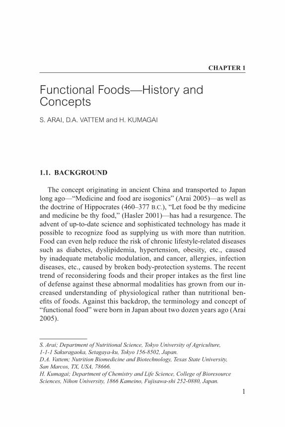
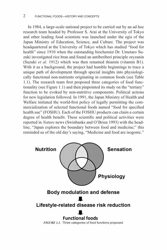
In 1984, a large-scale national project to be carried out by an ad hoc research team headed by Professor S. Arai at the University of Tokyo and other leading food scientists was launched under the egis of the Japan Ministry of Education, Science, and Culture. The project was headquartered at the University of Tokyo which has studied “food for health” since 1910 when the outstanding biochemist Dr. Umetaro Su-zuki investigated rice bran and found an antiberiberi principle oryzanin (Suzuki et al. 1912) which was then renamed thiamin (vitamin B1). With it as a background, the project had humble beginnings to trace a unique path of development through special insights into physiologi-cally functional non-nutrients originating in common foods (see Table 1.1). The research team first proposed three categories of food func-tionality (see Figure 1.1) and then pinpointed its study on the “tertiary” function to be evoked by non-nutritive components. Political actions for new legislation followed. In 1991, the Japan Ministry of Health and Welfare initiated the world-first policy of legally permitting the com-mercialization of selected functional foods named “food for specified health use” (FOSHU). Each of the FOSHU products can claim a certain degree of health benefit. These scientific and political activities were reported in Nature news (Swinbanks and O’Brien 1993) with the head-line, “Japan explores the boundary between food and medicine;” this reminded us of the old day’s saying, “Medicine and food are isogonic.”
FIGURE 1.1. Three categories of food functions proposed.
3
1.2. SCIENTIFIC PERSPECTIVES
What may be most important at present is the evaluation of the func-tion of a food. Since any functional food must be based on scientific principles, its evaluation should essentially depend on data from bio-chemistry, physiology, molecular biology, and most other modern bio-
Scientific Perspectives
TABLE 1.1. Common Non-nutritive Components with Possible Functions for Disease Risk Reduction.
Component Example (origin) Disease
CarotenoidCarotene Lycopene (tomato) Oxidative stressXanthophyll Lutein (vegetable) Oxidative stress
FlavonoidFlavonol Quercetin (vegetable) Oxidative stressFlavone Nobiletin (citrus fruit) Oxidative stressIsoflavone Daidzein (soybean) OsteoporosisFlavanone Naringin (grapefruit) DiabetesCatechin Epigallocatechin gallate Allergy
3- or 4-methyl ester (tea)Anthocyanin Cyanidin (kidney bean) Oxidative stressSimple polyphenol Chlorogenic acid (coffee) CancerPhenyl propanoid Curcumin (turmeric) CancerIsoprenoid Ubiquinone (ubiquitous) Oxidative stressTriterpenoid Soyasaponin (soybean) Oxidative stressPhytosterol β-Sitosterol (soybean) Hypercholesterol-
emiaChromanol derivative Tocotrienol (soybean) Oxidative stressIsothiocyanate Sulforaphane (broccoli) DetoxificationSulfoxide Alliin (garlic) CoagulationVanilloid Capsiate (chili) ObesityAlkaloid Caffeine (coffee) ObesityLignan Sesamin (sesame) HangoverOrganic acid Acetic acid (vinegar) HypertensionAmino acid γ-Aminobutyric acid (rice) HypertensionProtein β-Conglycinin (soybean) ObesityOligopeptide Val-Pro-Pro (sour milk) HypertensionLipid 3-Diacylglycerol (cooking oil) ObesityPolysaccharide Alginic acid (seaweeds) HypercholesteremiaPrebiotics Manno-oligosaccharide (coffee) ObesityProbiotics Bifidobacterium lactis (yogurt) ConstipationSweetening Neoculin (Curculigo latifelia) Diabetes
FUNCTIONAL FOODS—HISTORY AND CONCEPTS4
sciences. A key approach to the development of a functional food is the identification and validation of relevant markers, including biomarkers, which can predict its potential benefits relating to a target function in the body. If a marker represents an event directly involved in the pro-cess, it should be considered as a functional “factor.” However, if a marker represents a correlated event, it should be considered as a func-tion “indicator” (Bellisle et al. 1998; Anonymous 2002).
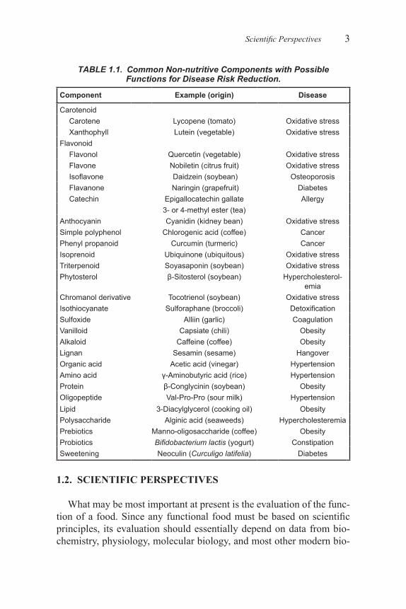
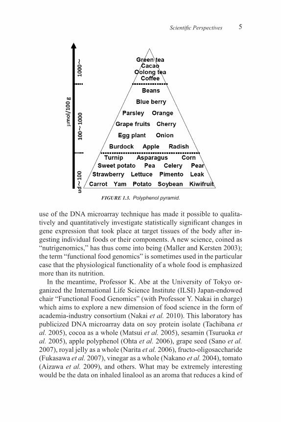
A similar but even lager national project, headed by Professor Arai, started with the title “Non-nutritive functional components in foods: analysis and systematization.” It emphasized the significance of troop-ing a variety of methodologies, with research targets hierarchized as in Figure 1.2 (Arai 2006). The project was successfully undertaken, with its administration operated by Professor M. Uehara at Tokyo University of Agriculture. Scientifically, the research teams investigated hundreds of fruits and vegetables for functional food components and in particu-lar, Professor K. Kanazawa at Kobe University, presented an interesting polyphenol pyramid (see Figure 1.3) (unpublished).
Meanwhile, the genome programs on humans and other representa-tive organisms for scientific and industrial uses were almost completed, enabling us to obtain the genome information by internet services. The
FIGURE 1.2. Systematized methodologies for analysis of the “tertiary” functions of food and hierarchal targets of analysis.
5
use of the DNA microarray technique has made it possible to qualita-tively and quantitatively investigate statistically significant changes in gene expression that took place at target tissues of the body after in-gesting individual foods or their components. A new science, coined as “nutrigenomics,” has thus come into being (Maller and Kersten 2003); the term “functional food genomics” is sometimes used in the particular case that the physiological functionality of a whole food is emphasized more than its nutrition.
In the meantime, Professor K. Abe at the University of Tokyo or-ganized the International Life Science Institute (ILSI) Japan-endowed chair “Functional Food Genomics” (with Professor Y. Nakai in charge) which aims to explore a new dimension of food science in the form of academia-industry consortium (Nakai et al. 2010). This laboratory has publicized DNA microarray data on soy protein isolate (Tachibana et al. 2005), cocoa as a whole (Matsui et al. 2005), sesamin (Tsuruoka et al. 2005), apple polyphenol (Ohta et al. 2006), grape seed (Sano et al. 2007), royal jelly as a whole (Narita et al. 2006), fructo-oligosaccharide (Fukasawa et al. 2007), vinegar as a whole (Nakano et al. 2004), tomato (Aizawa et al. 2009), and others. What may be extremely interesting would be the data on inhaled linalool as an aroma that reduces a kind of
Scientific Perspectives
FIGURE 1.3. Polyphenol pyramid.
6
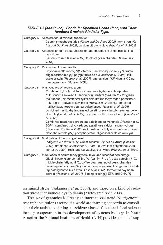
TABLE 1.2. Foods for Specified Health Uses, with Their Numbers Bracketed in Italic Type.
Category 1 Modulation of gastrointestinal conditions1 . Oligosaccharides: lactosucrose [28]; galacto-oligosaccharides [14];
coffee bean manno-oligosaccharides including mannobiose [14]; fructo-oligosaccharides [7]; soybean oligosaccharides [6]; xylo-oligo-saccharides [4]; isomaito-oligosaccharides [4]; lactulose (Hassler et al. 2004); raffinose (Hassler et al. 2004)
2 . Lactic acid bacteria: Lactobacillus casei Shirota [30]; L. acidophilus CK92 and L. helveticus CK60 [7]; Bifidobacterium lactis Bb-12 [7]; L. delbrueckii subsp . bulgaricus 2038 and Streptococcus salivarius subsp . thermophilus 1131[6]; B. breve Yakult [6]; B. longum BB536 [6]; L. casei SP and B. SP (Hassler 2002); B. lactis LKM512 (Has-sler 2002); B. lactis FK120 (Hassler 2002); L. casei NY1301 (Has-sler 2002); L. GG (Hassler 2002); L. casei LC1 (Hassler 2002)
3 . Dietary fibers: indigestible dextran [141]; psyllium husk [22]; wheat bran [4]; gum guaic hydrolysate [6]; agar (Katan and De Roos 2002); polydextrose (Hassler 2002); low-molecular-weight sodium alginate (Hassler 2002); low-molecular-weight sodium alginate and water-soluble corn fiber (Hassler et al. 2004); indigestible dextran and wheat bran (Hassler et al. 2004); dietary fiber from beer yeast (Hassler et al. 2004); indigestible dextran, reduced type (Hassler et al. 2004); indigestible starch (Hassler et al. 2004)
4 . Other components: milk whey fermented by propionic acid bacte-rium (Katan and De Roos 2002); Bacillus subtilis K-2 (Hassler et al. 2004)
5 . Combination of chemically defined components: galacto-oligosac-charide-polydextrose (Hassler et al. 2004)
Category 2 Modulation of serum cholesterol levelChitosan [47]; soybean protein [27]; phospholipid-binding soybean peptides including Cys-Ser-Pro-His-Pro [19]; low-molecular-weight sodium alginate [6]; phytosterol [5]; phytosterol ester (Katan and De Roos 2002); broccoli-cabbage peptide Ser-Mer-Cys-Ser (Hassler 2002); tea catechin [4]
Category 3 Modulation of serum cholesterol level and gastrointestinal conditions Dietary fiber from psyllium husks [19]; low-molecular-weight sodium alginate [9]
Category 4 Modulation of blood pressureSardine peptide including Val-Tyr [65]; lactotripeptides Val-Pro-Pro and Ile-Pro-Pro [12]; dried bonito “katsuobushi” oligopeptides [7]; “wakame” seaweed peptides including Pro-Tyr-Val-Tyr and Ile-Tyr [4]; γ-aminobutyric acid [9]; casein dedocapeptide (Hassler et al. 2004); Ile-Tyr [4]; acetic acid [5]; sesame peptides including Leu-Val-Tyr (Hassler 2002); “nori” seaweed oligopeptides including Ala-Lye-Tyr-Ser-Tyr (Hassler 2002); “tochu” loaf glycoside [4]; royal jelly peptide including Val-Tyr, Ile-Tyr and Ile-Val-Tyr (Hassler 2002); yanlong tea flavonoids including hyperoside and isoquercitrin (Hassler et al. 2004); chlorogenic acid (Hassler 2002)
(continued)
7
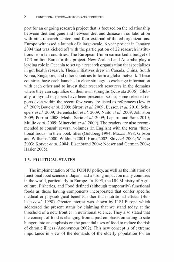
TABLE 1.2 (continued). Foods for Specified Health Uses, with Their Numbers Bracketed in Italic Type.
Category 5 Acceleration of mineral absorptionCasein phosphopeptides (Katan and De Roos 2002); heme iron (Ka-tan and De Roos 2002); calcium citrate-malate (Hassler et al. 2004)
Category 6 Acceleration of mineral absorption and modulation of gastrointestinal conditionsLactosucrose (Hassler 2002); fructo-oligosaccharide (Hassler et al. 2004)
Category 7 Promotion of bone healthSoybean isoflavones [13]; vitamin K as menaquinone-7 [7]; fructo-oligosaccharides [5]; polyglutamic acid (Hassler et al. 2004); milk basic protein (Hassler et al. 2004); and calcium [13] vitamin K-2 as menaquinone-4 (Hassler 2002)
Category 8 Maintenance of healthy teethCombined xylitrol-maltitol-calcium monohydrogen phosphate-“fukuronori” seaweed furanone [23]; maltitol (Hassler 2002); green tea fluorine [7]; combined xylitol-calcium monohydrogen phosphate-“fukuronori” seaweed flavanone (Hassler et al. 2004); combined maltitol-palatinose-green tea polyphenols (Hassler et al. 2004); combined maltitol-hydrogenated palatinose-erythritol-green tea poly-phenols (Hassler et al. 2004); soybean isoflavone-calcium (Hassler et al. 2004);Combined palatinose-green tea palatinose polyphenols (Hassler et al. 2004); combined xylitol-reduced palatinose calcium monohydrogen (Katan and De Roos 2002); milk protein hydrolysate containing casein phohphopeptide [27]; phosphorylated oligosaccharide calcium [6]
Category 9 Modulation of blood sugar levelIndigestible dextrin [136]; wheat albumin [5]; bean extract (Hassler 2002); arabinose (Hassler et al. 2004); guava leaf polyphenol (Has-sler et al. 2004); resistant recrystallized amylose (Hassler et al. 2004)
Category 10 Modulation of serum triacylglycerol level and blood fat percentageGlobin hydrolysate containing Val-Val-Tyr-Pro [14]; tea catechin [15]; middle-chain fatty acid [5]; coffee bean manno-oligosaccharides including mannobiose [20]; oolong tea polymerized polyphenol includ-ing oolong homo-bis-flavan B (Hassler 2002); fermented soy bean extract (Hassler et al. 2004); β-conglycinin [5]; EPA and DHA [4]
restrained stress (Nakamura et al. 2009), and those on a kind of isola-tion stress that induces dyslipidemia (Motoyama et al. 2009).
The use of genomics is already an international trend. Nutrigenomic research institutions around the world are forming consortia to consoli-date their activities aiming at evidence-based functional food science through cooperation in the development of systems biology. In North America, the National Institutes of Health (NIH) provides financial sup-
Scientific Perspectives
FUNCTIONAL FOODS—HISTORY AND CONCEPTS8
port for an ongoing research project that is focused on the relationship between diet and gene and between diet and disease in collaboration with nine research centers and four external affiliated organizations. Europe witnessed a launch of a large-scale, 6 year project in January 2004 that was kicked off with the participation of 22 research institu-tions from ten countries. The European Union earmarked a budget of 17.3 million Euro for this project. New Zealand and Australia play a leading role in Oceania to set up a research organization that specializes in gut health research. These initiatives drew in Canada, China, South Korea, Singapore, and other countries to form a global network. These countries have each launched a clear strategy to exchange information with each other and to invest their research resources in the domains where they can capitalize on their own strengths (Kuwata 2006). Glob-ally, a myriad of papers have been presented so far; some selected re-ports even within the recent few years are listed as references (Jew et al. 2009; Boue et al. 2009; Sirtori et al. 2009; Eusson et al. 2010; Schi-epers et al. 2009; Berendschot et al. 2009; Naito et al. 2009; Johnston 2009; Porrini 2008; Medic-Saric et al. 2009; Laparra and Sanz 2010; Mullie et al. 2009; Minervini et al. 2009). The readers are also recom-mended to consult several volumes (in English) with the term “func-tional foods” in their book titles (Goldberg 1994; Mazza 1998; Gibson and Williams 2000; Wildman 2001; Hurst 2002; Shi et al. 2002; Watson 2003; Korver et al. 2004; Eisenbrand 2004; Neeser and German 2004; Hasler 2005).
1.3. POLITICAL STATES
The implementation of the FOSHU policy, as well as the initiation of functional food science in Japan, had a strong impact on many countries in the world, particularly in Europe. In 1995, the UK Ministry of Agri-culture, Fisheries, and Food defined (although temporarily) functional foods as those having components incorporated that confer specific medical or physiological benefits, other than nutritional effects (Bel-lisle et al. 1998). Greater interest was shown by ILSI Europe which addressed the present status by claiming that we stand today at the threshold of a new frontier in nutritional science. They also stated that the concept of food is changing from a past emphasis on eating to sate hunger, into an emphasis on the potential uses of food to reduce the risk of chronic illness (Anonymous 2002). This new concept is of extreme importance in view of the demands of the elderly population for an
9
improved quality of life, increased life expectancy, and reduced costs for health care. A key approach for the science of functional food to develop in compliance with these demands is, among others, the iden-tification and validation of relevant markers which can predict some potential benefits in the body. It is thus of crucial importance to define the health claim for each functional food by the three different phrases: (1) nutrient function claims, (2) other function claims, and (3) reduc-tion of disease risk claims. The former two would be defined based on genomics data as well as biochemical biomarkers (Bellisle et al. 1998). Health claims should always be scientifically defined in harmonization with global standards. Talking of the FOSHU system, the goal is to evaluate its physiological functionality for formal approval. On the oth-er hand, the enhanced functional claims and the disease risk reduction claims were proposed by both the Codex and EU project in 1999. The structure/function claims were expressed in the Dietary Supplement Health Education Act in the United States in 1994. The nutrient func-tion claim was included in the guidelines adopted by the Codex in 1997. The generic claims primarily include nutrient function claims based on well-established, generally accepted knowledge and could be standard-ized without specific, individual substantiation. Innovative claims such as enhanced function claims or structure/ function claims, however, should be evaluated individually by independent experts in order to protect consumers from false or misleading descriptions. For this sci-entific substantiation, in addition to animal studies and in vitro studies, we also need human intervention studies (Shimizu 2003). The FOSHU system has been making good progress. Up to the present time (June 1, 2010), the Ministry has approved 941 FOSHU products categorized into 10 health claims (see Table 1.2) (Arai et al. 2008). Each product is permitted to claim a certain degree of health benefit as long as the claim does not overestimate its efficacy for disease risk reduction. The legal framework is also expected to stop ill-defined, misleading product advertisements. It is added that a close attention has been nationally and internationally paid to the safety of functional foods. In particular, the ILSI Annual Meeting 2010 in Rio Grande has had a scientific session of “Risk Assessment and Functional Foods,” which comprises topics on the mode of action of functional foods in human relevance framework, their threshold of toxicological concern, and so on.
As discussed above, the name of “functional food” as well as its concept has been globally accepted enthusiastically, and the science is in progress for further development. However, it will be of crucial im-
Political States
FUNCTIONAL FOODS—HISTORY AND CONCEPTS10
portance to add some modification to the current status of functional food science which is going for pharmacology. Food is quite different from medicine in various respects. It is necessary for implementation of functional food research to take into consideration the differences in terms of sensory properties. A new trend has come into being, which emphasizes the significance of studying the “secondary” function (see Figure 1.1). Since this per se is the most representative food attribute, even the science of functional food should consider taste and smell in a large measure. In the meantime, it was found that our body expresses taste receptors in the gut as well as in oral taste buds (Margoskee et al. 2007). Interestingly enough, it is possible that the gut’s sweet taste receptor, T1R2-T1R3, functions to induce insulin secretion shortly af-ter taking even artificial sweeteners and sweet protein (Shimizu-Ibuka et al. 2006). We now need some integrative studies on all the three categories of food functions (see Figure 1.1) and future research along this line will no doubt add a new dimension in the science and policy of functional foods.
1.4. DEFINITIONS AND LIMITATIONS
Currently, there is no general, worldwide definition of functional foods; however, there are multiple definitions developed by various or-ganizations. The International Food Information Council (IFIC) states that functional foods “provide health benefits beyond basic nutrition (Hassler et al. 2004).” The limitation of this definition is that it lacks an explanation of how the food or what part of the food provides health benefits. The definition should differentiate whether the whole food or only food components are beneficial. The International Life Sciences Institute of North America (ILSI) states that functional foods are “foods that, by virtue of physiologically active food components, provide health benefits beyond basic nutrition (Hassler et al. 2004).” This defi-nition is vague, but encompasses the realm of functional foods. There is no delineation between different types of functional foods, such as whole foods and foods that are enhanced with added ingredients. How-ever, this definition provides insight into how the food or what part of the food is beneficial by stating “by virtue of physiologically active food components,” (Hassler et al. 2004), this statement encompasses components of whole foods and components that are added to foods, which have beneficial effects. However, the statement about individual food components fails to demonstrate the role of synergy in providing
11
health benefits. The concept of synergy is important to include in func-tional food definitions because the coordination of the “physiologically active food component” with the other components present in the food is what provides the health benefit.
Health Canada states that functional foods are “similar in appearance to a conventional food, consumed as part of the usual diet, with dem-onstrated physiological benefits, and/or to reduce the risk of chronic disease beyond basic nutritional functions,” (Hassler et al. 2004). One limitation to this definition is the narrowness of stating that a functional food must look similar to a conventional food. If a functional food ap-pears similar to a recognizable food, consumer acceptance is likely in-creased; however, foods that do not appear similar to a conventional food should not be excluded since they may still act as functional foods. In addition, this definition is regional because appearance of conven-tional food differs according to location, culture, and religion. Accord-ing to this definition, two foods may have the exact same physiological function and provide the exact same health benefit; however, if one of these foods has an unconventional appearance, it cannot be classified as a functional food. Another limitation to this definition is that the food must be “consumed as part of the usual diet,” (Hassler et al. 2004). The usual diet of consumers varies from person to person, and a food should not lose classification as a functional food because it is not part of the usual diet of a consumer. If a functional food is part of the usual American diet, but not part of a usual European diet, is the same food not considered functional in Europe?
The Institute of Medicine of the National Academy of Sciences (IOM) states that functional foods are “those in which the concentra-tions of one or more ingredients have been manipulated or modified to enhance their contribution to a healthful diet,” (Hassler et al. 2004). This definition does not differentiate functional foods from any other food; according to this definition, a reduced fat food can be a func-tional food (Katan and De Roos 2002). In addition, this definition only states that functional foods contribute to a healthy diet, not that they have specific functions regarding disease (Katan and De Roos 2002). Moreover, this definition excludes whole foods and foods that have for-eign ingredients added; rather than only stating, “One or more ingredi-ents have been manipulated or modified,” the definition should include whole foods and functional ingredients that have been added to a food (Hassler et al. 2004).
In Japan, the FOSHU organization states that functional foods are
Definitions and Limitations
FUNCTIONAL FOODS—HISTORY AND CONCEPTS12
“processed foods containing ingredients that aid specific body func-tions in addition to being nutritious.” The limitation to this definition is that all unprocessed, whole foods are excluded.
The American Dietetic Association (ADA) states that functional foods include “whole foods and fortified, enriched, or enhanced foods, have a potentially beneficial effect on health when consumed as part of a varied diet and on a regular basis, at effective levels,” (Hassler et al. 2004). The limitation to this definition is that for a food to be functional, it must be “consumed as part of a varied diet,” (Hassler et al. 2004). The statement about a varied diet was likely incorporated to impart the importance of an overall healthy diet; however, a functional food re-tains its functionality as part of any diet. The definition should be for a functional food, not a healthy diet.
The American Council on Science and Health states that functional foods are “whole, fortified, enriched, or enhanced foods that provide health benefits beyond the provision of essential nutrients, when they are consumed at efficacious levels as part of a varied diet on a regular basis,” (Hassler 2002). The limitation of this definition is the same as the limitation of the definition offered by ADA.
1.5. RELEVANCE OF FUNCTIONAL FOODS
The concept of functional foods stems from the traditional paradigm of providing methods to prevent nutritional deficiencies; this paradigm encompasses foods that provide health benefits through fortification with micronutrients. Today, functional foods are designed to promote health by being targeted at specific physiological processes that may lead to disease prevention; this is known as the new paradigm. With the increase in diseases related to excess energy consumption, an ag-ing population, and the increasing cost of health care and pharmaceu-ticals, consumers are turning to food to replace or augment traditional health care for health promotion and disease prevention. With increas-ing consumer interest in self-care of health, functional foods research will continue to expand as will the availability of functional foods to the consumer. The concept of functional foods needs to be incorpo-rated into an overall healthy lifestyle for maximum benefit of disease prevention. Functional foods, an overall healthy diet including fruits, vegetables, unrefined grains, fish, and low-fat dairy products, and foods low in saturated fats and sodium (Katan and De Roos 2002), and ex-ercise, combine to encompass the healthy lifestyle needed for disease
13
prevention. The definition of functional foods expands on one aspect of a healthy lifestyle; this definition is meant to recognize foods and food ingredients, whether natural, added, or modified, that provide disease prevention and health promotion benefits.
1.6. FUNCTIONAL FOOD VERSUS PHARMACEUTICALS
As the population ages, the incidence of chronic diseases associated with caloric excess and the cost of healthcare increases, and consum-ers increasingly desire self-care regarding health and an alternative to pharmaceutical management of disease (Hassler et al. 2004). Lifespan, incidence of obesity, and age are increasing, which results in an insur-gence of chronic diseases such as cardiovascular disease (CVD) and diabetes mellitus. Each of these factors combines and occurs, leading to an increased cost of health care and pharmaceuticals. This increased cost of health care for disease management, coupled with pharmaceuti-cal side effects, and consumer interest in self-care health management and healthy foods result in a demand for functional foods. Compounded to the expense of pharmaceuticals is their limited availability, since they are only available via prescription and their ineffectiveness for some consumers; moreover, consumers desire the preventable approach of functional foods over the symptomatic treatment approach of pharma-ceuticals. The utilization of functional foods for health care encompass-es the use of functional foods to replace or decrease dependence on pharmaceuticals. The use of functional foods rather than pharmaceuti-cals for health benefits will decrease healthcare costs, promote health without inducing pharmaceutical side effects, aid in disease prevention, and provide a method of self-care for consumers. In addition, the use of functional foods for health care has a broad spectrum due to food being necessary for survival, and the flexibility of foods for adaptation to suit specific demographic characteristics.
1.7. IMPACT ON HEALTH CARE AND SOCIETY
Functional foods are currently influencing healthcare through their incorporation into medical treatments, usually alongside pharmaceuti-cals. As the functional food industry expands, their use in healthcare will increase, allowing functional foods to act as a first line of defense against disease rather than pharmaceuticals. In addition, as functional food use for disease prevention continues, the incidence of chronic dis-
Impact on Health Care and Society
FUNCTIONAL FOODS—HISTORY AND CONCEPTS14
ease will decrease leading to a reduction in spending on health care and pharmaceuticals. Overall, the scope of healthcare will shift from treatment to prevention. However, an increase in the use of probiotics may exacerbate antibacterial resistance. Probiotic bacteria such as Lac-tobacillus casei, upon ingestion select for the survival and growth of L. casei, therefore “crowding out” other normal gut microflora. This gives probiotics antibiotic activity; some of the bacteria that are tar-gets of this probiotic will survive, which leads to “super bugs” that are resistant to antibiotics. The use of probiotics and the excess use of pharmaceutical antibiotics compound to exacerbate the problem of antibiotic resistance. Overall, the shift away from pharmaceuticals and toward dietary intervention is necessary for health care in America; this shift will help to remove some of the governmental and societal burden of increasing health care costs, the inability to depend on social security benefits after retirement, and budgeted funds for Medicare.
1.8. FUNCTIONAL FOOD: SOURCES AND CLASSIFICATION
Functional foods are classified by source of origin, including plant, animal, microbial, and miscellaneous (algae, mushrooms, other). Re-gardless of the source of origin, the target of functional foods includes CVD, cancer, immune enhancement, gastrointestinal and women’s health, aging, diabetes mellitus, and stress management.
1.8.1. Plant-Derived Functional Foods
Plant-derived functional foods are separated into primary and sec-ondary metabolites; primary metabolites are plant compounds nec-essary for growth, while secondary metabolites are not essential for growth, but are used for plant survival mechanisms. Primary metabo-lites include plant proteins, beta-glucans, and omega-3 fatty acids. Plant proteins include texturized vegetable protein, soy protein isolate, and amino acids; these proteins act as functional foods by helping to de-crease the amount of meat consumption, which decreases the consump-tion of fat and cholesterol. Beta-glucans, found in oats, act as functional foods by decreasing cholesterol absorption. Omega-3 fatty acids, found in flaxseed, act as a functional food by reducing platelet aggregation. Secondary metabolites include phytoestrogens, antioxidants, vitamins, tocopherols, steroids, gamma-linolenic acid (GLA), and phase II en-
15
zyme inducers. Phytoestrogens, estrogen-like compounds in plants, are found in soybeans and flaxseed and act as functional foods by decreas-ing post-menopausal cancer development. Antioxidants, such as antho-cyanins, act as functional foods by quenching reactive oxygen species. Vitamins, which are abundant in fruits and vegetables, act as functional foods by preventing deficiencies; certain vitamins, such as vitamins C and E, also act as quenchers of reactive oxygen species. Tocopherols, which are vitamin E compounds found in oilseeds, act as quenchers of reactive oxygen species. Steroids are also found in oilseeds and act as functional foods by competing for cholesterol absorption. GLA is a fatty acid involved in the formation of prostaglandins and acts as an in-flammatory modulator (4). Phase II enzyme inducers, found in Brassica vegetables, act as functional foods by glycosylating insoluble toxins to produce soluble compounds that are excreted. Consumption of foods containing phase II enzyme inducers also limits the phase I enzyme detoxification system; the phase I enzyme system produces reactive oxygen species.
1.8.2. Animal-Derived Functional Foods
Zoochemicals, which are animal-derived functional foods, include omega-3 and six fatty acids, conjugated linolenic acid (CLA), small peptides, whey and casein, and glucosamine and chondroitin sulfate. Omega-3 fatty acids include alpha-linolenic, docosahexaenoic (DHA), and eicosapentaenoic (EPA) fatty acids (4). Sources of alpha-linolenic acid include soy and canola oils, walnuts, and flaxseed (4). The main source of EPA and DHA is fatty fish, such as salmon. Omega-6 fatty acids include linolenic, gamma-linolenic, and arachidonic fatty acids (4). Sources of these fatty acids include some vegetable oils, nuts, and whole grains (4). Omega-3 and six fatty acids act as functional foods by enhancing immunity, modulating inflammation, and protecting against neurodegenerative diseases. CLA is a fatty acid present in milk that reportedly acts as a functional food by reducing cancer risks and adi-pose differentiation; however, a fatty liver may develop as a side effect (Hassler 2002). Whey and casein are milk proteins that act as functional foods by being easily digested and absorbed, and help build muscle mass; small peptides function in the same manner. Glucosamine and chondroitin sulfate are required for collagen formation and were stated to act as functional foods by alleviating pain associated with osteoar-thritis; however, this claim has been disproved (4).
Functional Food: Sources and Classification
FUNCTIONAL FOODS—HISTORY AND CONCEPTS16
1.8.3. Microbial Functional Foods
Microbial-derived functional foods include probiotics, prebiotics, symbiotics, and synbiotics. Probiotics are natural microflora that occur in the gut, such as L. casei or numerous Bifidobacter species, which promote health (Hassler 2002). Prebiotics are dietary components that promote growth of probiotic bacteria. Symbiotics contain probiotics and prebiotics combined randomly, while synbiotics contain specific probiotics and prebiotics mixed together to benefit one another. Func-tional foods of microbial origin act by promoting the growth of probi-otic bacteria so that the growth of pathogenic bacteria is limited.
1.8.4. Miscellaneous Functional Foods
Some functional foods are derived from miscellaneous compounds such as algae and mushrooms. Algae function by proving omega-3 fatty acids, which enhance immunity, modulate inflammation, and protect against neurodegenerative diseases. Functional foods derived from mushrooms contain antiviral, antibacterial, and anti-inflammatory properties.
1.9. ACKNOWLEDGMENTS
We wish to express our sincere thanks to Professors K. Abe, Y. Nakai and T. Misaka for pertinent discussion.
1.10. REFERENCES
Aizawa, K., Matsumoto, T., Inakuma, T., Ishijima, T., Nakai, Y., Abe K., and Amano F. 2009. Ad-ministration of tomato and paprika beverages modifies hepatic glucose and lipid metabolism in mice: a DNA microarray analysis. J Agric Food Chem 57:10964–10971.
Anonymous, 2002. ILSI Europe Concise Monograph Series. In: Ashwell M., editor. Concept of Functional Food in Europe. Brussels: ILSI Europe. P. 1–40.
Arai, S., Yasuoka A., and Abe, K. 2008. Functional food science and food for specified health use policy in Japan: state of the art, Current Opinion in Lipidology 19:69–73.
Arai, S. 2005. Perspective functional food science. J Sci Food Agric 85:1603–1605. Arai, S. 2006. Functional food science born in Japan and propagated globally. Science & Technol-
ogy in Japan 25:2–5. Bellisle, F., Diplock, A.T., Hornstra, G., Koletzko, B., Roberfroid, M., Salminen, S., and Saris
W.H.M. 1998. Functional food science in Europe. Brit J Nutr 80:S1–S193. Berendschot, T.T., Plat, J., de Jong, A., and Mensink, R.P. 2009. Long-term plant stanol and sterol
ester-enriched functional food consumption, serum lutoin/zeaxanthin concentration and macu-lar pigment optical density. Br J Nutr 101:1607–1610.
17
Boue, S.M., Cleveland, T.E., Carter-Wientjes, C., Shih, B.Y., Bhatnagar, D., McLachian, J.M., and Burow, M.E. 2009. Phytoalexin-enriched functional foods. J Agric Food Chem 57:2614–2622.
Eisenbrand, G. 2004. Functional Food. In: Safety Aspects. Weinheim: Wiley-Vch Verlag GmbH and Co, KGaA.
Eusson, S., Klungel, O., Garssen, J., Vorhagen, H., van Kranen, H., van Loveren, H., and Rompel-berg, C. 2010. Support of drug therapy using functional foods and dietary supplements: focus on statin therapy. Gr J Nutr 103:1260–1277.
Fukasawa, T., Murashima, K., Matsumoto, I., and Hosono, A. 2007. Identification of marker genes for intestinal immunomodulating effect of a fructooligosaccharide by DNA microarray analy-sis. J Agric Food Chem 55:3174–3179.
Gibson, G.R., and Williams, C.M. 2000. In: Functional Foods: Concept to Product. Boca Raton-Boston-New York-Washington (DC): CRC Press.
Goldberg, I. 1994. In: Functional Foods: Designer Foods, Pharmafoods, Nutraceuticals. New York-London: Chapman & Hall.
Hasler, C., Bloch, A., and Thomson, C. Position of the American Dietetic Association: functional foods. J Am Diet Assoc. 2004; 104(5):814–826.
Hasler, C. Funtional Foods: benefits, concerns and challenges-a position paper from the American Council on Science and Health. J Nutr. 2002; 132:3772–3781.
Hasler, C.M. 2001. Functional Foods. In: Bowman, B.A., Russell, R.M., editors. Present Knowl-edge in Nutrition. Washington (DC): ILSI Press.
Hasler, C.M. 2005. In: Regulation of Functional Foods and Nutraceuticals. Oxford: IFT Press, Blackwell Publishing.
Hurst, W.J. 2002. In: Methods of Analysis for Functional Foods and Nutraceuticals. Boca-Raton-London- NewYork-Washington (DC): CRCress.
Jew, S., AbuMweis, S.S., and Jones, P.J. 2009. Evolution of the human diet: linking our ances-tral diet to modern functional foods as a means of chronic disease prevention. J Med Food 12:925–934.
Johnston, C. 2009. Functional foods as modifiers of cardiovascular disease. Am J Lifestyles Med 3:S539–S543.
Katan, M. and De Roos, N. Promises and problems of functional foods. Crit Rev Food Sci Nutr. 2004; 44:369–377.
Korver, O., Kuhn, M.C., and Richardson, D. 2004. In: The Functional Foods Dossier: Building Solid Health Claims. Wageningen: Foodlink Forum.
Kuwata, T. 2006. Functional foods: comparison of currents state in Japan and other countries. Sci-ence and Technology in Japan 25:21–23.
Laparra, J.M. and Sanz, Y. 2010. Interactions of gut microbiota with functional food components and nutraceuticals. Pharmacol Res 61:219–225.
Maller, M. and Kersten, S. 2003. Nutrigenomics: goal and strategies. Nature Genetics 4:314–322. Margoskee, R.F., Dyer, J., Kokrashvili, Z., Salmon, K.S., Ilegems, E., Daly, K., Maillet, E.L.,
Ninomiya, Y., Mosinger, B., and Shiranzi-Beechey, S.P. 2007. TIR3 and gustducin in gut sense sugars to regulate expression of Na+ glucose cotransporter 1. Proc Natl Acad Sci USA 104:15075–15080.
Matsui, N., Ito, R., Nishimura, E., and Yoshikawa, M. 2005. Ingested cocoa and prevent high-fat diet-induced obesity by regulating the expression on genes for fatty acid metabolism. Nutrition 21:594–601.
Mazza, G. 1998. In: Functional Foods: Biochemical and Processing Aspects. Lancaster-Basel: Techomic Publishing.
Medic-Saric, M., Rastija, V., Bojic, M., and Males, Z. 2009. From functional food to medicinal product: systematic approach in analysis of polyphenolics from propolis and wine. Nutr J 8:33.
Minervini, F., Bilancia, M.F., Siragusa, S., Gobetti, M., and Caponio, F. 2009. Fermented goat’s milk produced with selected multiple starters as a potentially functional food. Food Microbiol 26:559–564.
Motoyama, K., Nakai, Y., Miyashita, T., Fukui, Y., Morita, M., Sanmiya, K., Sasakibara, H.,
References
FUNCTIONAL FOODS—HISTORY AND CONCEPTS18
Matsumoto, I., Abe, K., Yakabe, T., Yajima, N., and Shimoi, K. 2009. Isolation stress for 30 days alters hepatic gene expression profiles especially with reference to lipid metabolism in mice. Physiol Genomics 37:79–87.
Mullie, P., Guelinokx, I., Clarys, P., Degrave, E., Hulenc, M., and Vansant, G. 2009. Cultural, socioeconomic and nutritional determinants of functional food consumption patterns. Eur J Clin Nutr 63:1290–1296.
Naito, Y. and Yoshikawa, T. 2009. Oxidative stress-induced posttranslational modification of pro-teins as a target of functional food. Forum Nutr 61:39–54.
Nakai, Y., Yasuoka, A., Kato, H., and Abe, K. 2010. Genomics applied to nutrients and functional foods in Japan: State of the art, In: Bagehi, D., Lau, F.C., editors. Genomics, Proteomics and Metabolomics in Nutraceuticals and Functional Foods. Hoboken: Blackwell Publishing. p. 127–154.
Nakamura, A., Fujiwara, S., Matsumoto, I., and Abe, K. 2009. Stress repression in restrained rats by (R)-(–)-linalool inhalation and gene expression profiling of their whole blood cells. J Agric Food Chem 57:5480–5485.
Nakano, S., Fukaya, M., and Horiuchi, S. 2004. Enhanced expression of aconitase raises acetic acid resistance in Acetobacter aceti. FEMS Microbiol Lett 235:315–322.
Narita, Y., Nomura, J., Ohta, S., and Inoh, Y. 2006. Royal jelly stimulates bone formation: physi-ologic and nutrigenomic studies with mice and cell lines. Biosci Biotechnol Biochem 70:2508–2514.
Neeser, J.R. and German, J.B. 2004. In: Bioprocess and Biotechnology for Functional Foods and Nutraccuticals. New York–Basel: Marcel Dekker.
Ohta, Y., Sami, M., Kanda, T., and Saito, K. 2006. Gene expression analysis of the anti-obesity effects by apple polyphenols in rats fed a high fat diet or a normal diet. J Oleo Sci 55:305–314.
Porrini, M. 2008. Functional foods: from theory to practice. Int J Vitaminole Nutr Res 78:261–268. Sano, A., Uchida, R., Saito, M., and Shioya, N. 2007. Beneficial effects of grape seed extract on
malondialdehyde-modified LDL. J Nutr Sci Vitaminol 53:174–182. Schiepers, O.J.G., de Groot, R.H.M., van Boxtel, M.P.J., Jolles, J., de Jong, A., Luetjohann, D.,
Plat, J., and Mensink, R.P. 2009. Consuming functional foods enriched with plant sterol or stanol esters for 85 weeks does not effect neurocognitive functioning or mood in statin-treated hypercholesterolemic individuals. J Nutr 139:1368–1373.
Shi, J., Mazza, G., and Le Maguer, M. 2002. In: Functional Foods: Biochemical and Processing Aspects. Boca-Raton-London- NewYork-Washington (DC): CRCress.
Shimizu, T. 2003. Health claims on functional foods: the Japanese regulations and an international comparison, Nutr Res Rev 16:241–252.
Shimizu-Ibuka, A., Morita, Y., Terada, T., Asakura, T., Nakajima, K., Iwata, S., Misaka, T., So-rimachi, H., Arai, S., and Abe, K. 2006. Crystal Structure of Neoculin: Insights into its sweet-ness and Taste-modifying activity. J Mol Biol 359:148–158.
Sirtori, C.R., Galli, C., Anderson, J.W., Sirtori, E., and Arnoldi, A. 2009. Functional foods for dyslipidaemia and cardiovascular risk prevention. Nutr Ros Rev 22:244–261.
Suzuki, U., Shimamura, U., and Okada, S. 1912. Über Oryzanin: ein Bestandteil der Reiskleie und seine physiologische Bedeutung. Biochem Z 43:89–153.
Swinbanks, D. and O’Brien, J. 1993. Japan explores the boundary between food and medicine. Nature 364:180.
Tachibana, N., Matsumoto, I., Fukui, K., Arai, S., and Abe, K. 2005. Intake of soy protein isolate alters hepatic gene expression in rats. J Agric Food Chem 53:4253–4257.
Tsuruoka, N., Kidokoro, A., Matsumoto, I., and Abe, K. 2005. Modulating effect of sesame, a functional lignan in sesame seeds, on the transcriptions levels of lipid-and alcohol-metaboliz-ing enzymes in rat liver: a DNA microarray study. Biosci Biotechnol Biochem 69:179–188.
Watson, D.H. 2003. In: Performance Functional Foods. Boca-Raton-London-New York-Wash-ington (DC): CRCress.
Wildman, R.E.C. 2001. In: Handbook of Functional Foods. Boca Raton-London-New York-Washington (DC): CRC Press.
19
CHAPTER 2
Prebiotics and Probiotics: Concepts and AdvancesA.L. CARVALHO-WELLS and D.M.A. SAULNIER
2.1. INTRODUCTION
Any epithelial surface in the human body which is exposed to the external environment is subject to interaction with exogenous micro-organisms. This occurs particularly on the respiratory, genital, and gut mucosal surfaces. The intestinal mucosa is the main site of interaction with the external environment and therefore is an important intermedi-ary in the maintenance of health. The gut was traditionally thought of as a relatively simple organ comprising an epithelial tube surrounded by a layer of muscle. However recent research has unraveled the diverse functions of the gut and demonstrated the human gastrointestinal tract as a highly dynamic ecosystem, which is central to immune develop-ment, host defense, and human nutrition.
2.1.1. Location and Diversity of the Gut Microbiota
There are four main microhabitats in the gastrointestinal tract: the epithelial surface, the mucus layer which overlays the epithelium, the
A.L. Carvalho-Wells; Hugh Sinclair Unit of Human Nutrition, Institute for Cardiovascular and Metabolic Research (ICMR), Department of Food and Nutritional Sciences, University of Reading, Reading RG6 6AP, UK. D.M.A. Saulnier; Departments of Pathology, Baylor College of Medicine, Houston, TX, USA; and Department of Pathology, Texas Children’s Hospital, Houston, TX, USA. (Current address: Department of Gastrointestinal Microbiology, German Institute of Human Nutrition, Nuthetal, Germany.)
PREBIOTICS AND PROBIOTICS: CONCEPTS AND ADVANCES20
crypts (of the ileum, caecum, and colon), and the intestinal lumen, re-spectively. The levels and diversity of the microbial community within the gastrointestinal tract can differ according to the location as different physicochemical properties and substrate availability dictate the envi-ronmental conditions. Not surprisingly, the stomach mucosa is occu-pied by relatively few organisms due to the high level of acidity which requires a more acid tolerant organism; stomach contents (per gram) have been estimated to contain 103 colony forming units (CFU), reach-ing 104–107 in the small intestine and 1010–1012 in the large intestine where the microbial numbers are highest (Holzapfel et al. 1998). The total area of the mucosal surface of the human gastrointestinal tract is 300 m2 which makes it the largest surface area in the body that inter-acts with the external environment (Bjorksten 2006). The distal large intestine is the area of highest colonization with more than 500 differ-ent species (with some estimates suggesting up to 1,000 species) with potentially up to 100 billion microbial inhabitants (Boyle and Tang 2006). This enormous microbial community has been estimated to be in the region of 1014 microorganisms, the collective gene set of which is termed the “microbiome” and is thought to contain approximately 100 times the number of genes in the human genome (Backhed et al. 2005; Xu and Gordon 2003). Based on a meta-analysis covering several large Sanger-sequencing studies of human gut samples from different populations the most numerically dominant phyla within the intestinal microbial community are the firmicutes and bacteroidetes that comprise more than 90% of the total bacteria. Other bacterial phyla that have been isolated include also actinobacteria, gammaproteobacteria, verru-comicrobia, and betaproteobacteria (Hamady and Knight 2009).
Although the major dominant bacterial groups have been described, there is considerable species variation between individuals. For exam-ple, all humans possess several hundred species within each genus in their gut, however the foremost species within that genus will differ considerably between individuals (Simon and Gorbach 1984). The re-lationship between the human host and microbial symbionts is complex and a focus of modern biology. This has led to collaborative research projects the largest of which is probably the Human Microbiome Proj-ect, which is a worldwide strategy aiming to further delineate the extent of both human and microbial diversity. As it is a collaborative project the techniques used are sophisticated and will result in a vast data set, it is hoped that the project will be able to identify common features of the microbiome at a global level. This will help determine the impact
21
of host genetics on the development and stability of the microbiome by using genetically linked individuals evolutionary lineages (Ley et al. 2008; Turnbaugh et al. 2007).
2.1.2. Techniques Used to Elucidate the Commensal Microbiota
Microbiological techniques traditionally relied on the ability of a bacterium to grow on sterile semi or defined media in order to observe colonies. This enabled preliminary classification of bacteria on the ba-sis of culture phenotype and biochemical characteristics. This was also the case for pioneering work in the area of gut microbiology; however, the majority of bacteria in the gut are anaerobes, therefore they present more of a challenge to grow in standard laboratory conditions. More recently, use of a culture-independent approach has revealed the com-plexity of the resident microbial communities; this was enabled by use of oligonucleotide probes based on 16S rRNA gene sequences. Further classification of bacteria based upon phylogenetic comparisons of the 16S rRNA sequences has been possible using qualitative and quanti-tative techniques. For example, PCR reactions and fluorescent in situ hybridization (FISH) use primers and probes respectively which tar-get bacteria based on their 16S rRNA sequences. It is estimated that these specific probes have enabled identification of up to 80% of the total microbial diversity within the intestinal microflora, which would not have been possible with cultural techniques alone. Recent metage-nomic techniques with high-throughput pyrosequencing have further explored functional genes encoded by the microbiome using annotation schemes such as Kyoto Encyclopaedia of Genes and Genomes (KEGG) pathways or Clusters of Orthologous Groups (COGs) (Turnbaugh et al., 2006, 2007, 2009). Molecular analysis has shown that the aerobic species present reach relatively high cell densities and metabolic activ-ity in the human caecum, in fact 50% of total bacterial ribosomal RNA was found to correspond to these species in this region of the gut. This is in contrast to feces in which only 7% of the total bacterial rRNA from these species is found (Marteau et al. 2001).
2.1.3. Factors Affecting the Composition of the Gut Microbiota
In a healthy gut, there is a balance between potentially harmful, com-mensal and beneficial bacteria. Probably the most important factors in determining the initial colonization pattern are the type of delivery at
Introduction
PREBIOTICS AND PROBIOTICS: CONCEPTS AND ADVANCES22
birth (either vaginal or caesarean section) and the initial diet (whether the newborn is fed mother’s milk or infant formula). The newborn mi-crobiota changes rapidly during the first few weeks and during weaning (Boyle and Tang 2006). Other important factors include the environ-ment, age, gender, and diet (Santosa et al. 2006). Differences in mi-crobiota composition have been found between infants born in differ-ent countries and raised with different diets and even between hospital wards (Adlerberth et al. 1991; Lundequist et al. 1985; Santosa et al. 2006; Simhon et al. 1982). The composition of the adult microbiota is thought to be more stable, however acute effects can disrupt this ho-meostasis for example, during antibiotic treatment, after gastrointes-tinal surgery, exposure to radiation, and in some infectious (diarrheal) disease states (Mombelli and Gismondo 2000). The stability of the mi-crobiota is compromised in the elderly, demonstrating significant vari-ability over time and between individuals; as well as lower species di-versity (Claesson et al., 2011; Makivuokko et al, 2010).
2.2. ROLES OF THE COMMENSAL MICROBIOTA
The presence of the gut microbiota has influenced human evolution in that the human host cannot perform certain vital intestinal functions without them. Germ-free animal models have provided useful insights into the roles of the microbiota and the extent of interaction between the host and the gut microbiota. It can be thought of as a ‘microbial organ’ as the processes performed by this diverse population are extensive; it can communicate with itself (bacteria: bacteria) and with the host (bac-teria: human). Maintenance of homeostasis is an interactive process between the bacteria and host epithelia which influences both intesti-nal physiology and the derivation and distribution of energy. Landmark studies in recent years have shown that the composition of the microbi-ome can have an impact on energy balance and obesity (Turnbaugh and Gordon 2009; Turnbaugh et al. 2006).
2.2.1. Fermentation
A major role of the microbiota is to ferment nondigestible dietary components and endogenous mucus produced by the gut epithelium. This is an example of a symbiotic relationship as the human host bene-fits from a wide array of microbial enzymes which are outside the host’s own biochemical repertoire. This provides the host with a source of
23
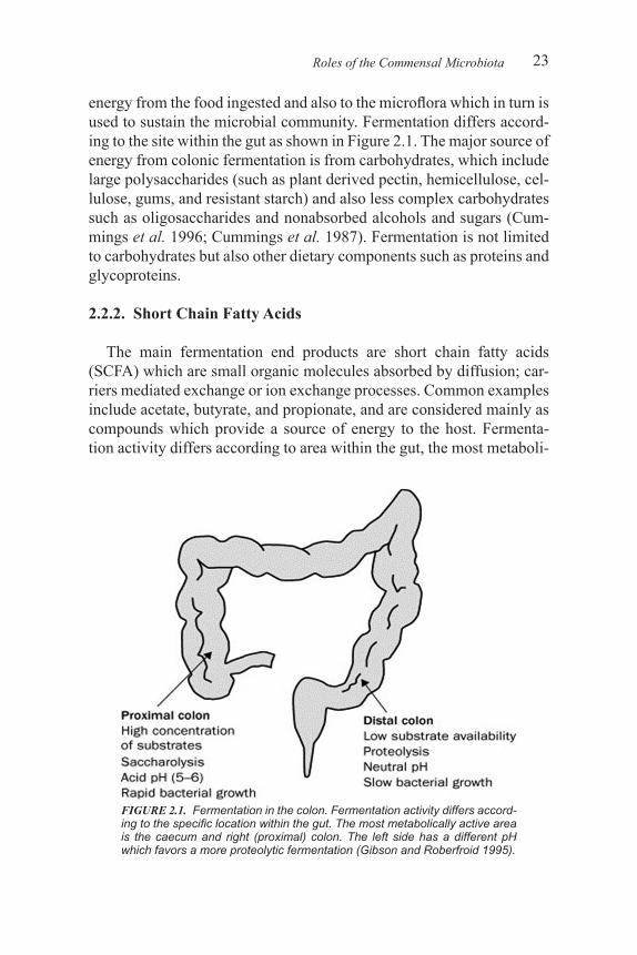
energy from the food ingested and also to the microflora which in turn is used to sustain the microbial community. Fermentation differs accord-ing to the site within the gut as shown in Figure 2.1. The major source of energy from colonic fermentation is from carbohydrates, which include large polysaccharides (such as plant derived pectin, hemicellulose, cel-lulose, gums, and resistant starch) and also less complex carbohydrates such as oligosaccharides and nonabsorbed alcohols and sugars (Cum-mings et al. 1996; Cummings et al. 1987). Fermentation is not limited to carbohydrates but also other dietary components such as proteins and glycoproteins.
2.2.2. Short Chain Fatty Acids
The main fermentation end products are short chain fatty acids (SCFA) which are small organic molecules absorbed by diffusion; car-riers mediated exchange or ion exchange processes. Common examples include acetate, butyrate, and propionate, and are considered mainly as compounds which provide a source of energy to the host. Fermenta-tion activity differs according to area within the gut, the most metaboli-
Roles of the Commensal Microbiota
FIGURE 2.1. Fermentation in the colon. Fermentation activity differs accord-ing to the specific location within the gut. The most metabolically active area is the caecum and right (proximal) colon. The left side has a different pH which favors a more proteolytic fermentation (Gibson and Roberfroid 1995).
PREBIOTICS AND PROBIOTICS: CONCEPTS AND ADVANCES24
cally active area is the caecum and right colon, consequently this is an area of rapid bacterial growth, low pH (5–6) and rapid generation of SCFA as a result of carbohydrate fermentation (Cummings et al. 1987; Macfarlane et al. 1992). Fermentation can also occur with noncarbo-hydrate substrates, therefore proteolytic fermentation (of proteins and peptides) also generates potentially damaging compounds such as am-monia, amines, and phenolic compounds (Macfarlane et al. 1986). In contrast, the left side of the colon has less carbohydrate fermentation, the pH is less acidic, and it is associated with an increase in proteolysis which has been linked to the production of harmful nitrogenous prod-ucts, which accounts for the recommendation to eat a carbohydrate and fiber enriched diet (Guarner and Malagelada 2003). The absorption of ions such as calcium, magnesium, and iron in the caecum is improved in the presence of SCFA (Roberfroid et al. 1995; Younes et al. 2001). The presence of adequate SFCA causes colonic water and sodium ions to be absorbed, therefore producing solid stools therefore having an effect on intestinal transit time, although the mechanisms are unclear (Elsen and Bistrian 1991). SCFA are not only a source of energy for tissues but can also have important effects on host physiology as one SCFA in particular, butyrate has been shown to inhibit the development of colonic cancer cells in vitro (Pool-Zobel and Sauer 2007a). The co-lonic epithelium almost entirely consumes the butyrate that is produced as it is a preferred energy source (Cummings et al. 1987). SCFA have a positive effect on epithelial cell differentiation and proliferation in vivo. Some members of the gut microbiota also produce vitamins such as folate, biotin, and vitamin K-2 (Conly et al. 1994; Hill 1997).
2.3. THE GUT MICROBIOTA AND AUGMENTATION OF HOST DEFENSE
2.3.1. Barrier Function
One role of the commensal flora is to protect against infection from exogenous organisms, of which there is a higher risk within the gastro-intestinal tract. Several mechanisms are thought to contribute to this process, which is collectively termed colonization resistance. The im-portant role of the commensal microbiota in boosting host defense has been confirmed in germ-free animals which demonstrate an increased rate of susceptibility to infections relative to a wild type microflora (Baba et al. 1991; Taguchi et al. 2002). Adhesion is thought to be a
153
CHAPTER 7
Extraction and Purification of Bioactive Ingredients from Natural ProductsG. K. JAYAPRAKASHA and BHIMANAGOUDA S. PATIL
7.1. INTRODUCTION
Bioactive compounds are expected to play an important role as one of the major sources of new drugs in the years to come because of their incomparable structural diversity, the relatively small dimensions (< 2000 Da), and their “drug-like” properties, i.e., their ability to be absorbed and metabolized [1]. Isolation of natural products from higher plants, marine organisms, and microorganisms is critical, using state-of-the-art methodologies. The plant kingdom contains approximately 80–100,000 plant bioactive compounds and the separation and isolation processes are cumbersome and tedious. Isolation of natural products generally combines various separation techniques, which depends on the solubility, volatility, and stability of the compounds to be separated. The choice of different optimization parameters of separation is critical and essential
7.2. EXTRACTION OF BIOACTIVE INGREDIENTS
Isolation of bioactive ingredients from the plant materials is a trial and error excise in which different solvents are tried under a variety of conditions such as time and temperature. The main objectives of the
G. K. Jayaprakasha and Bhimanagouda S. Patil; Vegetable & Fruit Improvement Centre, Department of Horticultural Sciences, Texas A&M University, College Station, TX 77843-2119, USA.
EXTRACTION AND PURIFICATION OF BIOACTIVE INGREDIENTS154
extraction and purification of unique/unstudied bioactive components, exploring the potential of secondary metabolites for chemical finger-printing or metabolomics studies, and bioassay derived identification of bioactives.
Isolation of bioactive compounds involves dissolution of solutes from the plant matrix, diffusion of the compounds into the extractant, and separation of solutes using chromatographic techniques.
7.2.1. Selection of Solvent
In choosing a solvent for the extraction of bioactives, the ability to extract components has to be considered. For instance, ionic solutes can be extracted from aqueous solvents. The general features of the bioac-tive molecule that are helpful to ascertain the isolation process include partition coefficient, acid-base properties, charge, stability, and molecu-lar size. In literature, many basic extraction procedures are available [2]. Solvent choice for the extraction is a critical step. Single solvent is unlikely to extract all groups of bioactive compounds from the natural plant materials. In most of the cases, these methods will be refined to our requirements in terms of plant materials and solutes of our interest. The expected outcome from this extraction process should be high pu-rity product, adequate quantity of bioactive compound, and confirming the stereochemistry of the molecule.
In general, three conventional methods were used for the extraction of bioactive compounds such as solvents, steam, and supercritical flu-ids. On a global level, water extraction is practised while making coffee or tea. Basically, pretreated plant material is extracted with hot water which takes up the flavor, taste, and color of the components. After filtration, the extract is ready for consumption. In case of the isolation of certain bioactive compounds from plant material by means of liquid extraction, some technological problems needs to be resolved [3]. First the plant material has to be pretreated in order to obtain reasonable extraction yields. Another problem is the need for special solvents to be used in the extraction procedure [4]. More recently, attention has been focussed towards the isolation of specific compounds that can be used in the food industry. Of particular interest is the isolation of bioac-tive compounds, aromas, and fragrances from plants and fruits [5,6]. The sequential extractions of bioactives using nonpolar to polar sol-vents are depicted in Figure 7.1. Various polarity solvents are reported as follows: (1) nonpolar solvents (hexane, heptanes, petroleum ether,
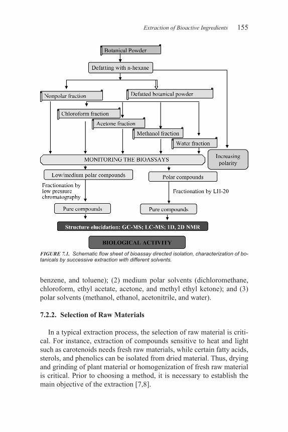
155
benzene, and toluene); (2) medium polar solvents (dichloromethane, chloroform, ethyl acetate, acetone, and methyl ethyl ketone); and (3) polar solvents (methanol, ethanol, acetonitrile, and water).
7.2.2. Selection of Raw Materials
In a typical extraction process, the selection of raw material is criti-cal. For instance, extraction of compounds sensitive to heat and light such as carotenoids needs fresh raw materials, while certain fatty acids, sterols, and phenolics can be isolated from dried material. Thus, drying and grinding of plant material or homogenization of fresh raw material is critical. Prior to choosing a method, it is necessary to establish the main objective of the extraction [7,8].
Extraction of Bioactive Ingredients
FIGURE 7.1. Schematic flow sheet of bioassay directed isolation, characterization of bo-tanicals by successive extraction with different solvents.
EXTRACTION AND PURIFICATION OF BIOACTIVE INGREDIENTS156
7.2.3. Fractionation
After extraction from the plant, the bioactive components have to be separated from the crude mixture. It involves further solvent partition-ing and extensive chromatography by retaining their properties of the desired compound, such as acidity, alkalinity, polarity, stability, molec-ular size, and structure. This may involve simple crystallisation of the compound from the crude extract, e.g., isolation of the glycoside dianel-lin from Dianella caerulea [9] as well as crystallization of limonin from citrus extract [10]. In some cases, the isolation can be assisted by the preparation of suitable derivatives, imparting more easily manageable properties of the desired compound, e.g., isolation of the swainsonine as its triacetate from Rhizoctonia leguminicola and isolimonoic acid was isolated as methyl isolimonate [11]. Finally, purification will be performed to provide compounds of high purity for structural analysis, which may be accompanied by appropriate techniques such as crystal-lisation, sublimation, or distillation [12]. According to Pettit et al., the isolation of bioactive compounds from natural sources is, “always chal-lenging and every step require judgement, improvisation to discover novel components,” [13].
7.3. CONVENTIONAL EXTRACTION TECHNIQUES
Traditionally-used techniques for the extraction of bioactive com-pounds are discontinuous, continuous, and hybrid approaches. The discontinuous techniques include the use of either organic solvents (sometimes assisted by ultrasound) or water, while steam distillation and vacuum distillation are continuous methods. Some methods involv-ing both continuous and discontinuous approaches, such as distillation–extraction; Soxhlet extraction has also been reported [14].
Solvent extraction is a traditional method for extracting bioactive compounds from different plant materials [15–17]. However, less polar components present in most plant tissues may interfere with the subse-quent separation [17,18]. The conventional methods for the extraction of natural products are Soxhlet extraction, cold percolation, hot extrac-tion, and sonication. These methods have certain drawbacks, including long extraction time, use of large amounts of organic solvents, unsatis-factory extraction efficiency, and potential degradation of labile com-pounds. Therefore, the sequential solvent extraction is recommended for efficient separation of bioactive compounds [19]. For this reason,
157
lipophilic compounds are removed with nonpolar organic solvents such as hexane or dichloromethane [17, 20–23]. Meanwhile, the hydrophilic constituents are extracted with polar solvents such as acetone, metha-nol, ethanol, and water [21,24,25].
In some cases, the addition of a polar solvent, such as water, to the sample may increase the recovery of more polar compounds [24,26]. Meanwhile, some bioactive compounds of low or medium polarity can be extracted efficiently with more nonpolar solvents [27–29]. Direct ex-traction of bioactive compounds of a low polarity can also be obtained with polar solvent at high temperature [18,30]. However, the subse-quent cleanup step to separate the low polar bioactive compounds from the polar extract is a challenge [31].
7.3.1. Continuous Techniques
This technique involves steam distillation along with solvent extrac-tion for the isolation of essential oils from plants. This technique has been applied extensively as a step prior to compositional studies of es-sential oils, such as curcuma [32], marjoram [33], grapes [34], soybeans [35], and lavender [36].
7.3.2. Discontinuous Techniques
Solvent extraction has long been used for the isolation of essential oils from natural products. This technique uses either pure organic sol-vents or mixtures. Organic solvent extraction assisted by ultrasound, also known as sonication, is another technique widely used for the iso-lation of essential oils/extracts from plants. Thus, sonication methods based on 20–30 minutes of extraction with methanol–chloroform mix-tures have been used for the isolation of white clove essential oil [37]and cyanogenic glucosides [38].
The use of organic solvent extraction has certain limitations such as solvent residues in the extract, with the subsequent toxicological risk and the long extraction time required in most cases for achieving ef-ficient extractions. In addition, organic solvents have a low selectivity. Thus, apart from the desired substances, high molecular weight, non-volatile components, such as fatty oils, resins, waxes, and coloring mat-ters, are coextracted. The nonfeasibility of automation of the technique is another important drawback to be taken into account.
Water extraction (under ambient conditions, without the application
Conventional Extraction Techniques
251
CHAPTER 11
Mechanism of Neuroprotection by Bioactive CompoundsR.C. STAVINOHA, B.Y. JAMISON, Y. GOMADA, V. MAITIN and D.A. VATTEM
11.1. INTRODUCTION
Alzheimer’s disease (AD) and Parkinson’s disease (PD) are the two most prevalent neurodegenerative diseases worldwide [1]. These dev-astating diseases are characterized by progressive and irreversible neu-rodegeneration of particular neuronal networks in the brain that lead to severe cognitive and behavioral dysfunction [1]. The etiology behind the development of these diseases remains elusive [2]. Although ge-netic susceptibility has been identified in both PD and AD, the majority of cases are sporadic and without certain cause. It is thought that aging, oxidative stress, and environmental factors are involved in the initia-tion of the diseases; however, the molecular mechanisms that underlie the pathogenesis are not clear. The current treatments for AD and PD offer symptomatic relief, but do not affect the underlying neurodegen-eration or natural course of the disease [2]. As such, the development of treatments that can slow or reverse the pathological processes of these diseases is currently a significant focus of research.
11.2. ALZHEIMER’S DISEASE
AD is the most common neurodegenerative disease in the world. At
R.C. Stavinoha, B.Y. Jamison, Y. Gomada, V. Maitin and D.A. Vattem; Nutrition Biomedicine and Biotechnology, Texas State University, San Marcos, TX, USA, 78666.
MECHANISM OF NEUROPROTECTION BY BIOACTIVE COMPOUNDS252
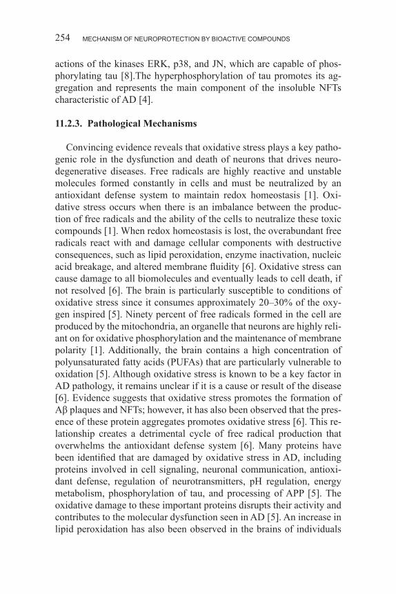
present, the disease affects 24% of the population over age 85.3 In the year 2000, it was estimated that 4.5 million individuals in the United States had AD, a number that is projected to rise to 14 million by 2050 [3]. This disease involves pathological events that affect the anatomy, biology, and function of selective regions of the brain [4]. Specifically, widespread neurodegeneration is observed in the cortex and hippocam-pus [5]. AD is clinically characterized by a progressive loss in cognitive function that typically begins with memory loss, anxiety, and depres-sion. As the disease progresses, the symptoms evolve to severe motor dysfunction, profound cognitive deterioration, and loss of independent function [4]. Pathologically, AD involves a cascade of events that lead to modifications in the metabolism of the amyloid precursor protein (APP) and the tau protein [4]. These changes result in the characteristic development of extracellular amyloid beta-protein (Aβ) deposits and intraneuronal neurofibrillary tangles (NFTs), respectively [4,3]. Con-sequently, common cell signaling pathways are affected and result in neuronal network dysfunction, neuronal loss, neurotransmitter failure, and cell death [4].
11.2.1. Amyloid Beta-Protein Deposits
The APP is a transmembrane protein expressed ubiquitously in cells, indicating that it has a normal biological role; yet, its function is un-known [6]. In cases of AD, the APP is found to be aberrantly processed to yield soluble Aβ oligomers and insoluble Aβ fibrils that aggregate extracellularly in the brain [6]. The metabolism of APP normally oc-curs through a cleavage by α-secretase, a membrane-bound protease that acts on APP within the Aβ domain, therefore it does not produce the Aβ peptide [4,7]. For unknown reasons, in AD, APP is processed in an alternate pathway that involves two subsequent cleavages [7]. In this pathway, referred to as the amyloidgenic pathway, the enzyme α-secretase first cleaves APP, releasing a membrane bound C-terminal fragment consisting of 99 amino acid residues, referred to as C99 [4]. Gamma-secretase subsequently cleaves C99 and releases Aβ, typically containing 40, 42, or 43 amino acids, into the transmembrane domain [4]. The Aβ peptide can oligomerize to form soluble oligomers, but may also converge to form insoluble fibrils in a beta-sheet conformation that are deposited extracellularly [7]. It has recently been found that the sol-uble Aβ oligomers accumulate in synapses and behave as a pathogenic ligand to membrane proteins, which disrupts synaptic transmission and
253
downstream events that are required for memory formation [7]. The insoluble Aβ fibrils that form are deposited as plaques extracellularly and contribute to neurodegeneration in several ways. Aβ plaques are thought to directly contribute to synaptic dysfunction by altering syn-aptic plasticity [4]. The plaques are thought to promote oxidative stress by decreasing antioxidant enzymes, increasing free radical production, and/or causing mitochondrial dysfunction [3]. Additionally, inflamma-tion surrounding the plaque is believed to promote the degeneration of nearby neurons [4]. Aβ plaques are also thought to disrupt cellular func-tions by interacting with cell membranes and promoting oxidation [6]. Furthermore, Aβ plaques form poorly selective channels in the lipid bilayer that disrupts the membrane potential responsible for generating action potentials, which results in an influx of calcium into the cell that promotes apotosis [6].
11.2.2. Neurofibrillary Tangles (NFTs)
NFTs are intraneuronal protein aggregates primarily composed of the hyperphosphorylated cytoskeletal protein, tau. The main function of tau relates to microtubule stability. Tau is intimately involved in main-taining the balance between assembly and disassembly of the micro-tubules, a function that is essential to the stability of the cytoskeleton and to the integrity of neurons [6]. The phosphorylation state of tau modulates the stability of the microtubules and is regulated by various protein kinases and phosphatases. However, in the case of AD, tau is found to be aberrantly hyperphosphorylated, rendering it incapable of microtubule interaction, consequently impacting normal neuronal func-tions, morphology, and viability [6]. Recent evidence suggests that, in addition to being a microtubule stabilizer, tau may regulate neuronal excitability and serve as a master regulator of the trafficking of mol-ecules within the cell that contribute to synaptic function [8]. These ad-ditional functions are also disrupted by the hyperphosphorylation of tau and contribute to the dysfunctional regulation of neuronal signaling and synaptic function common to AD. This is evidenced by a strong correla-tion between the hyperphosphorylated tau and a decrease in presynaptic protein expression [9]. Interestingly, studies have shown that reduction in the levels of tau prevents the neurotoxic effects of Aβ oligomers, sug-gesting that the damage induced by Aβ oligomers on the synapses may be mediated by the aberrant phosphorylation of tau. In addition, both oxidative stress and soluble Aβ oligomers are believed to promote the
Alzheimer’s Disease
MECHANISM OF NEUROPROTECTION BY BIOACTIVE COMPOUNDS254
actions of the kinases ERK, p38, and JN, which are capable of phos-phorylating tau [8].The hyperphosphorylation of tau promotes its ag-gregation and represents the main component of the insoluble NFTs characteristic of AD [4].
11.2.3. Pathological Mechanisms
Convincing evidence reveals that oxidative stress plays a key patho-genic role in the dysfunction and death of neurons that drives neuro-degenerative diseases. Free radicals are highly reactive and unstable molecules formed constantly in cells and must be neutralized by an antioxidant defense system to maintain redox homeostasis [1]. Oxi-dative stress occurs when there is an imbalance between the produc-tion of free radicals and the ability of the cells to neutralize these toxic compounds [1]. When redox homeostasis is lost, the overabundant free radicals react with and damage cellular components with destructive consequences, such as lipid peroxidation, enzyme inactivation, nucleic acid breakage, and altered membrane fluidity [6]. Oxidative stress can cause damage to all biomolecules and eventually leads to cell death, if not resolved [6]. The brain is particularly susceptible to conditions of oxidative stress since it consumes approximately 20–30% of the oxy-gen inspired [5]. Ninety percent of free radicals formed in the cell are produced by the mitochondria, an organelle that neurons are highly reli-ant on for oxidative phosphorylation and the maintenance of membrane polarity [1]. Additionally, the brain contains a high concentration of polyunsaturated fatty acids (PUFAs) that are particularly vulnerable to oxidation [5]. Although oxidative stress is known to be a key factor in AD pathology, it remains unclear if it is a cause or result of the disease [6]. Evidence suggests that oxidative stress promotes the formation of Aβ plaques and NFTs; however, it has also been observed that the pres-ence of these protein aggregates promotes oxidative stress [6]. This re-lationship creates a detrimental cycle of free radical production that overwhelms the antioxidant defense system [6]. Many proteins have been identified that are damaged by oxidative stress in AD, including proteins involved in cell signaling, neuronal communication, antioxi-dant defense, regulation of neurotransmitters, pH regulation, energy metabolism, phosphorylation of tau, and processing of APP [5]. The oxidative damage to these important proteins disrupts their activity and contributes to the molecular dysfunction seen in AD [5]. An increase in lipid peroxidation has also been observed in the brains of individuals
255
with AD [6]. The peroxidation of the particularly susceptible PUFAs in the cell membrane result in alterations in the membrane composition and is thought to contribute to the decreased expression of the nicotinic acetylcholine receptors commonly seen in AD [6]. In addition, byprod-ucts of lipid peroxidation such as HNE and acrolein are capable of co-valently modifying proteins and inhibiting enzyme activity [6]. HNE has also been shown to upregulate the expression of β-secretase, thus promoting the processing of APP to form Aβ peptide [6]. Oxidative stress can also cause modifications to DNA, including strand break-age, cross-linking, base modification, and oxidation of deoxyribose [1]. Oxidative damage to mitochondrial DNA further promotes oxida-tive stress by causing modifications in the electron transport chain that lead to the leakage of electrons [6].
Originally, inflammation was thought to be a consequence of neuro-degeneration, but recent evidence suggests that it may also be a primary pathogenic factor [2]. The persistent formation of plaques and progres-sive neuronal damage that occurs leads to the overactivation of microg-lia, the immune cells of the central nervous system. The overactive mi-croglia release large amounts of proinflammatory and cytotoxic factors, which causes damage to the neurons and further activate microglia, creating a vicious cycle of uncontrolled and prolonged inflammation which drives neurodegeneration [10]. Excitotoxicity is another patho-logical factor common to many neurodegenerative diseases, including AD. Excitotoxicity results from the excessive activation of N-methyl-D-aspartate (NMDA) receptors by glutamate; as a result, calcium levels in the cell increase and triggers apoptosis [11].
11.2.4. Risk Factors
The greatest risk factor for the development of AD is age [3]. It is thought that the increase in oxidative stress that occurs with aging con-stitutes a large part of the risk. Vascular diseases, such as hypertension, hypercholesterolemia, and diabetes, also increase the risk of AD [3]. On top of vascular complications, diabetes poses an additional risk for AD due to the effects of hyperinsulinemia and insulin resistance on amyloid metabolism [3]. This correlation is evidenced by a significantly high incidence of AD development among individuals with diabetes. It has been suggested that hyperinsulinemia and insulin resistance leads to decreased clearance and increased deposition of the Aβ peptide [12]. Additionally, impaired insulin-signaling results in abnormal glucose
Alzheimer’s Disease
MECHANISM OF NEUROPROTECTION BY BIOACTIVE COMPOUNDS256
and energy metabolism, oxidative stress, mitochondrial dysfunction, and hyperphosphorylation of tau via increased activity of glycogen syn-thase kinase 3 [7]. Although the majority of cases of AD are sporadic and without certain cause, approximately 10% of cases can be ascribed to genetic susceptibility [6]. Mutations in APP, presenilin 1, and presenilin 2 located on chromosome 21, 14, and 1, respectively, have been identified to promote the abnormal processing of APP that leads to aggregation [6]. Additionally, inheritance of the ε4 allele of the apolipoprotein E gene on chromosome 19 increases the risk for the development of AD [6].
11.3. PARKINSON’S DISEASE
PD is the second most common neurodegenerative disease that af-fects 0.3% of the population of industrialized countries [2]. The preva-lence increases to 1% of the population over 60 years of age, and to 4% of the population over 80 years of age [2]. PD involves a progressive loss of neurons in the brain manifesting into characteristic motor and
FIGURE 11.1. Possible molecular mechanisms behind development of Alzheimer’s Dis-ease.
257
non-motor symptoms. Motor symptoms include akinesia, bradykinesia, rigidity, resting tremor, postural instability, and gait impairment [11]. Nonmotor symptoms include anosmia, depression, anxiety, sleep disor-ders, gastrointestinal symptoms, autonomic dysfunction, and cognitive impairment [11].
11.3.1. Dopaminergic Pathway and Lewy Bodies
PD is a neurodegenerative disease of the central nervous system hallmarked by the degeneration of dopaminergic neurons, dopamine deficit, and the presence of Lewy bodies in remaining neurons [13]. Al-though there are signs of pathology in multiple neuronal systems in PD, the cardinal symptoms developed are ascribed to the progressive degen-eration of dopaminergic neurons in the substantia nigra pars compacta (SNpc) and subsequent loss of the neurotransmitter dopamine [10]. The SNpc is critical for the control of motor function, since it promotes voluntary movement and inhibits involuntary movement by affecting the basal ganglia loops, a process mediated by dopamine receptor D1 activation and D2 inactivation [10]. Neurodegeneration in this dopa-minergic pathway leads to dopamine depletion, which has shown to alter the proportions of the D1- and D2-receptors [14]. Specifically, the loss of dopamine results in reduced activity of the D1-receptor and in-creased activity of the D2-receptor [14]. These pathological changes in the dopamine receptors alter the balance in the basal ganglia loops and result in the characteristic motor symptoms of the disease [10]. Lewy bodies are found in the cytoplasm of the remaining neurons of the SNpc; however, they are also found in other nondopaminergic neu-rons, including neurons of the amygdala, basal nucleus of Meynert, hippocampus, and brain stem [10]. The presence of Lewy bodies in these nondopaminergic neurons indicates that they play a role in the development of the nonmotor symptoms associated with PD [2]. Alpha-synuclein (α-syn) has been identified as the primary protein aggregate found in Lewy bodies [11]. The exact cause of aggregation is unclear; however, overexpression of the protein, genetic mutations, and oxida-tive stress are thought to contribute [11]. Alpha-syn has been identified as a key factor implicated in PD, because it plays an important role in controlling the function of dopaminergic neurons; thus, the dysfunction of this protein at dopaminergic synapses is thought to contribute to the initiation of neurodegeneration [15]. Additionally, it is suggested that the aggregation of α-syn has a pathogenic role in PD through exerting
Parkinson’s Disease
MECHANISM OF NEUROPROTECTION BY BIOACTIVE COMPOUNDS258
negative effects on the cell membrane and proteasomal functionality, disrupting gene expression regulation, influencing cell signaling and cell death pathways, promoting inflammation, and modifying the stor-age and release of dopamine [11].
11.3.2. Pathological Mechanisms
No single pathogenic event has been found to be the primary fac-tor in the development of PD [11]. Rather, the process appears to be multifactorial involving several mechanisms that act synergistically in a complex manner to foster neurodegeneration [11]. Similar to AD, mechanisms involved in the pathogenesis of PD include oxidative stress, inflammation, and excitotoxicity [11]. Oxidative damage to lip-ids, proteins, and nucleic acids has consistently been observed in the SNpc of individuals with PD [11]. The cause of increased oxidative stress in PD is unclear, but it has been suggested that mitochondrial dysfunction and accelerated dopamine metabolism play a role [10]. The exact cause of mitochondrial dysfunction in PD is also unknown, but results in further production of free radicals, presumably by defects in the electron transport chain that lead to the leakage of electrons [10]. Dopamine metabolism is accelerated in PD, and contributes to oxida-tive stress through the concurrent production of free radicals such as quinones and peroxides [10]. The vulnerability of dopaminergic neu-rons to neurodegeneration in PD compared to other neuronal systems may be linked to unique morphological and physiological properties [10]. The dopaminergic neurons consist of long, narrow, branched pro-jections that have higher basal energy requirements than other neurons, rendering them more susceptible to damage caused by mitochondrial dysfunction [10]. Additionally, dopaminergic neurons are particularly susceptible to oxidative stress due to a high rate of oxygen and cal-cium metabolism, elevated iron concentrations, and reduced levels of antioxidants, specifically glutathione [10]. Inflammation is another mechanism involved in PD, as evidenced by microglia activation in the striatum and elevated levels of proinflammatory cytokines in the cere-brospinal fluid and basal ganglia in individuals with PD [11]. Microglia can become activated in PD in response to toxins, protein aggregates, or damaged neurons and become chronically active as a result of positive feedback from dying neurons, as seen in AD. Excitotoxicity is also im-plicated in PD. Dopaminergic neurons have particularly high levels of receptors for glutamate, the principle excitatory neurotransmitter of the
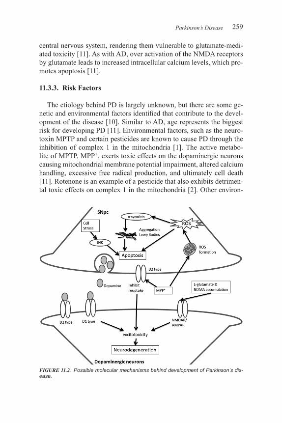
259
central nervous system, rendering them vulnerable to glutamate-medi-ated toxicity [11]. As with AD, over activation of the NMDA receptors by glutamate leads to increased intracellular calcium levels, which pro-motes apoptosis [11].
11.3.3. Risk Factors
The etiology behind PD is largely unknown, but there are some ge-netic and environmental factors identified that contribute to the devel-opment of the disease [10]. Similar to AD, age represents the biggest risk for developing PD [11]. Environmental factors, such as the neuro-toxin MPTP and certain pesticides are known to cause PD through the inhibition of complex 1 in the mitochondria [1]. The active metabo-lite of MPTP, MPP+, exerts toxic effects on the dopaminergic neurons causing mitochondrial membrane potential impairment, altered calcium handling, excessive free radical production, and ultimately cell death [11]. Rotenone is an example of a pesticide that also exhibits detrimen-tal toxic effects on complex 1 in the mitochondria [2]. Other environ-
Parkinson’s Disease
FIGURE 11.2. Possible molecular mechanisms behind development of Parkinson’s dis-ease.
MECHANISM OF NEUROPROTECTION BY BIOACTIVE COMPOUNDS260
mental risk factors include carbon monoxide poisoning, exposure to solvents, and hydrogen sulfide intoxification [2]. Although rare, various genetic mutations have been identified to cause PD in about 5–10% of cases [16]. Specifically, mutations in α-syn, parkin, DJ-1, and LRRK2 are associated with the development of PD [1]. These mutations are believed to cause mitochondrial dysfunction and increased oxidative stress, and thus contribute to the development of PD [1].
A recent study reported that dysfunction of p38 or JNK of MAPK signaling is involved in development of AD, PD, and amyotrophic lat-eral sclerosis (ALS) due to its effects on development and apoptosis [17]. It is reported that overexpression of p38 in microglia, astrocyte and neuron may increase inflammation, excitotoxicity, and synaptic plasticity via upregulation of TNF-α, IL-1β, and nitric oxide [18]. Also, accumulation of amyloid β plaques and overexpression of p38 were reported to involve tau phosphorylation, thus it is suggested that p38 MAPK signaling dysfunction may contribute to PD [18]. Also, acti-vation of p38 MAPK signaling and inflammation via NF-κB was ob-served in a MPTP-treated mice brain, and it is reported upregulation of JNK mediated by ASK1 and MAPK increase neuronal cell death, thus, p38 MAPK is considered to play important role in PD. Also, p38 can phosphorylate GSK-3 along with PI3K/AKT, p38 MAPK is shown to be involved in axonal regeneration [19]. As described, the activity of p38 MAPK regulate many cellular functions, however, during several pathologies, p38 MAPK is shown to be hyperactive, which has results in the progression of neurodegenerative disease, therefore proper regu-lation of MAPK signaling may become a therapeutic target of neuro-degenerative disease. In an AD patient brain, reduced TGF-β signaling are seen and suggested to progress formation of neurofibrillary tangles of aggregates of Aβ, especially Aβ1-42 [20]. It is reported that the reduc-tion of TGF-β signaling reduces trophic support of neuron from glial cell and also reduces anti-inflammatory proteins, thus it is considered to promote the progression of AD [21]. However, overexpression of TGF-β signaling is reported to increase neuronal apoptosis and reduce phagocytosis, thus increases accumulation of aggregated Aβ which may contribute to AD ( Lee et al. 2010). Also, reduction of TGF-β is reported to trigger apoptosis and is suggested to contribute to the development of PD due to increase of ROS formation of microglia [23]. Activation of TGF-β in macroglial cells attributes to inhibit PHOX induced ROS production via prevention of Erk activity, thus activation of TGF-β may play an important role in preventing PD progression through reducing
343
CHAPTER 15
Therapeutic Potential of Green and Black Tea in the Prevention and Treatment of Various DiseasesA. SHEHZAD, M. UL-ISLAM, N. SHAH and Y. SUP LEE
15.1. INTRODUCTION
Tea, derived from Camellia sinensis, is one of the most widely con-sumed beverages in the world. It is cultivated mostly in China and In-dia, as well as parts of some Asian and African countries [1,2]. Tea has received interest from both scientific and consumer communities for its health benefits. Tea is rich in substances with antioxidant prop-erties and contains small quantities of proteins, carbohydrates, amino acids, vitamins, and minerals [3]. Currently, high levels of tea drink-ing throughout the world are attributed to its biological functionality, rather than to habitual tea drinking, which is generally driven by its flavor and stimulant effect. The presence of polyphenolic compounds has given tea impressive therapeutic properties, including antioxidant, anti-inflammatory, antitumor, and metabolic regulatory effects [4].
Although originating from the same plants, tea is categorized into three main types based on their levels of oxidation [5]. Green and black teas are processed differently during manufacturing. To produce green tea, the freshly harvested leaves are steamed and dried to inactivate the
A. Shehzad and Y. Sup Lee; School of Life Sciences, College of Natural Sciences, Kyungpook National University, Daegu 702-701, Korea. M. Ul-Islam; Department of Chemical Engineering, College of Natural Sciences, Kyungpook National University, Daegu 702-701, Korea. N. Shah; Department of Chemistry, Abdul Wali Khan University, Mardan, K.P.K, Pakistan.
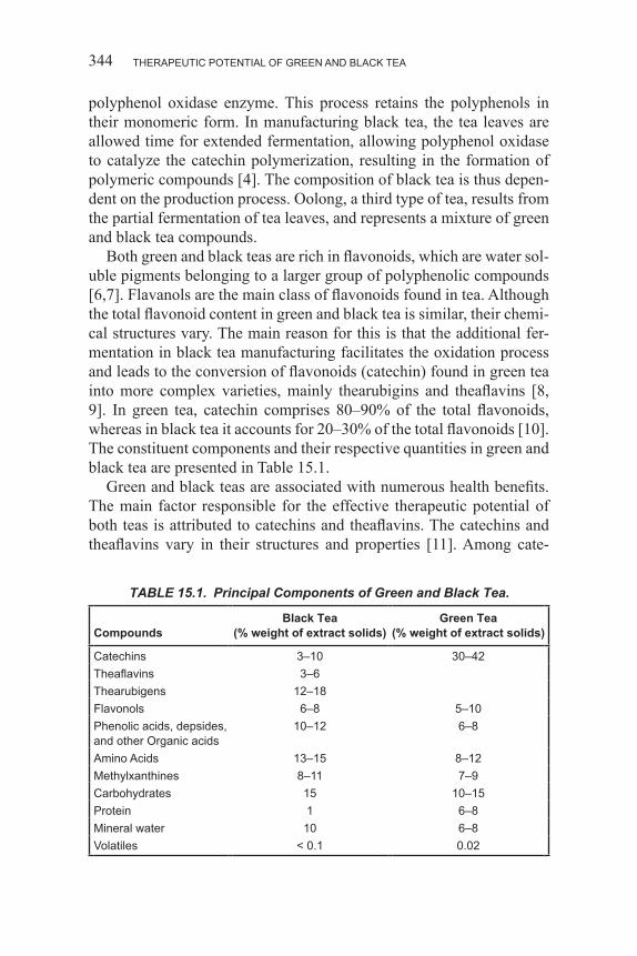
THERAPEUTIC POTENTIAL OF GREEN AND BLACK TEA344
polyphenol oxidase enzyme. This process retains the polyphenols in their monomeric form. In manufacturing black tea, the tea leaves are allowed time for extended fermentation, allowing polyphenol oxidase to catalyze the catechin polymerization, resulting in the formation of polymeric compounds [4]. The composition of black tea is thus depen-dent on the production process. Oolong, a third type of tea, results from the partial fermentation of tea leaves, and represents a mixture of green and black tea compounds.
Both green and black teas are rich in flavonoids, which are water sol-uble pigments belonging to a larger group of polyphenolic compounds [6,7]. Flavanols are the main class of flavonoids found in tea. Although the total flavonoid content in green and black tea is similar, their chemi-cal structures vary. The main reason for this is that the additional fer-mentation in black tea manufacturing facilitates the oxidation process and leads to the conversion of flavonoids (catechin) found in green tea into more complex varieties, mainly thearubigins and theaflavins [8, 9]. In green tea, catechin comprises 80–90% of the total flavonoids, whereas in black tea it accounts for 20–30% of the total flavonoids [10]. The constituent components and their respective quantities in green and black tea are presented in Table 15.1.
Green and black teas are associated with numerous health benefits. The main factor responsible for the effective therapeutic potential of both teas is attributed to catechins and theaflavins. The catechins and theaflavins vary in their structures and properties [11]. Among cate-
TABLE 15.1. Principal Components of Green and Black Tea.
CompoundsBlack Tea
(% weight of extract solids)Green Tea
(% weight of extract solids)
Catechins 3–10 30–42Theaflavins 3–6Thearubigens 12–18Flavonols 6–8 5–10Phenolic acids, depsides, and other Organic acids
10–12 6–8
Amino Acids 13–15 8–12Methylxanthines 8–11 7–9Carbohydrates 15 10–15Protein 1 6–8Mineral water 10 6–8Volatiles < 0 .1 0 .02
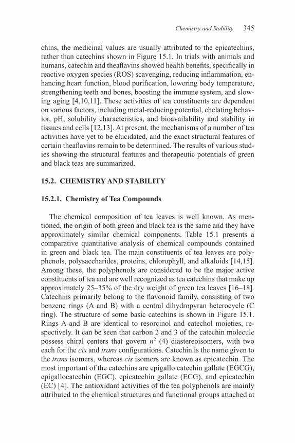
345
chins, the medicinal values are usually attributed to the epicatechins, rather than catechins shown in Figure 15.1. In trials with animals and humans, catechin and theaflavins showed health benefits, specifically in reactive oxygen species (ROS) scavenging, reducing inflammation, en-hancing heart function, blood purification, lowering body temperature, strengthening teeth and bones, boosting the immune system, and slow-ing aging [4,10,11]. These activities of tea constituents are dependent on various factors, including metal-reducing potential, chelating behav-ior, pH, solubility characteristics, and bioavailability and stability in tissues and cells [12,13]. At present, the mechanisms of a number of tea activities have yet to be elucidated, and the exact structural features of certain theaflavins remain to be determined. The results of various stud-ies showing the structural features and therapeutic potentials of green and black teas are summarized.
15.2. CHEMISTRY AND STABILITY
15.2.1. Chemistry of Tea Compounds
The chemical composition of tea leaves is well known. As men-tioned, the origin of both green and black tea is the same and they have approximately similar chemical components. Table 15.1 presents a comparative quantitative analysis of chemical compounds contained in green and black tea. The main constituents of tea leaves are poly-phenols, polysaccharides, proteins, chlorophyll, and alkaloids [14,15]. Among these, the polyphenols are considered to be the major active constituents of tea and are well recognized as tea catechins that make up approximately 25–35% of the dry weight of green tea leaves [16–18]. Catechins primarily belong to the flavonoid family, consisting of two benzene rings (A and B) with a central dihydropyran heterocycle (C ring). The structure of some basic catechins is shown in Figure 15.1. Rings A and B are identical to resorcinol and catechol moieties, re-spectively. It can be seen that carbon 2 and 3 of the catechin molecule possess chiral centers that govern n2 (4) diastereoisomers, with two each for the cis and trans configurations. Catechin is the name given to the trans isomers, whereas cis isomers are known as epicatechin. The most important of the catechins are epigallo catechin gallate (EGCG), epigallocatechin (EGC), epicatechin gallate (ECG), and epicatechin (EC) [4]. The antioxidant activities of the tea polyphenols are mainly attributed to the chemical structures and functional groups attached at
Chemistry and Stability
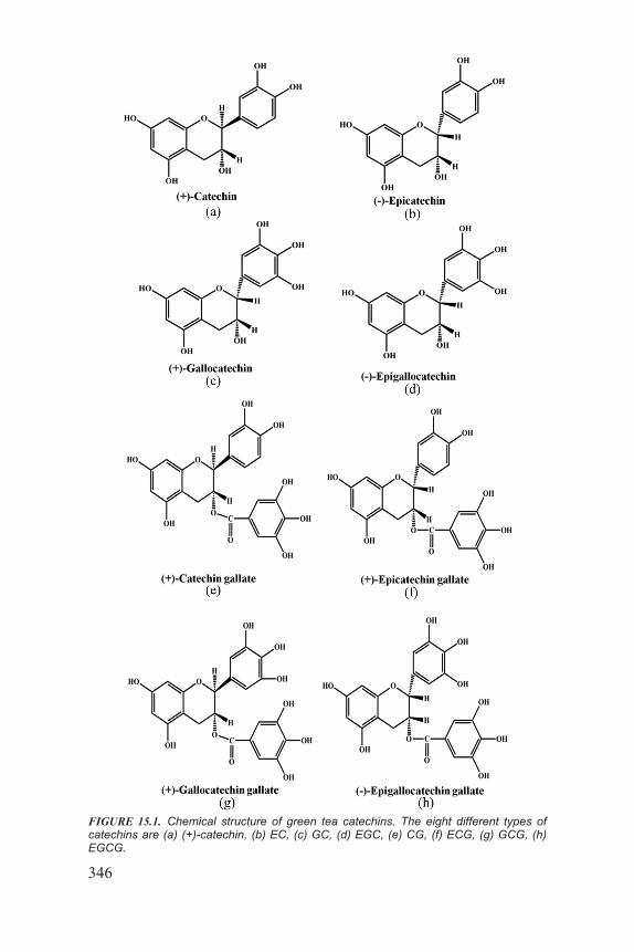
346
FIGURE 15.1. Chemical structure of green tea catechins. The eight different types of catechins are (a) (+)-catechin, (b) EC, (c) GC, (d) EGC, (e) CG, (f) ECG, (g) GCG, (h) EGCG.
347
various positions, including a di- or tri- hydroxyl group in the B-ring, an OH group at 5th and 7th carbon of the A ring, and a gallate group located at the 3 position of the C-ring [19]. The attachment of a gallate group governs additional catechins. All of these are shown in Figure 15.1.
As the manufacturing processes of green and black tea are very dif-ferent, the types of polyphenols contained by both types of tea are also different. The major chemical changes in the black tea constituents take place during its extended fermentation process. It has been suggested that during the fermentation process, the catechins undergo phenolic oxidative coupling reactions and are converted to o-quinones. Further, the continued oxidation process results in the development of polymer-ic compounds, including polyphenols, theaflavins, and thearubigins. It has been suggested that theoflavins are the coupling oxidation products of EC and EGC [20]. The simplified oxidative mechanism of theaflavin production is shown in Figure 15.2. Whereas, Figure 15.3 illustrates the structures of four main theaflavins identified from black tea. These in-clude theaflavin, theaflavin-3-gallate, theaflavin-30-gallate, and theafla-vin- 3,30-digallate [11] .
Chemistry and Stability
FIGURE 15.2. A proposed mechanism of theaflavins formation from catechin oxidation.
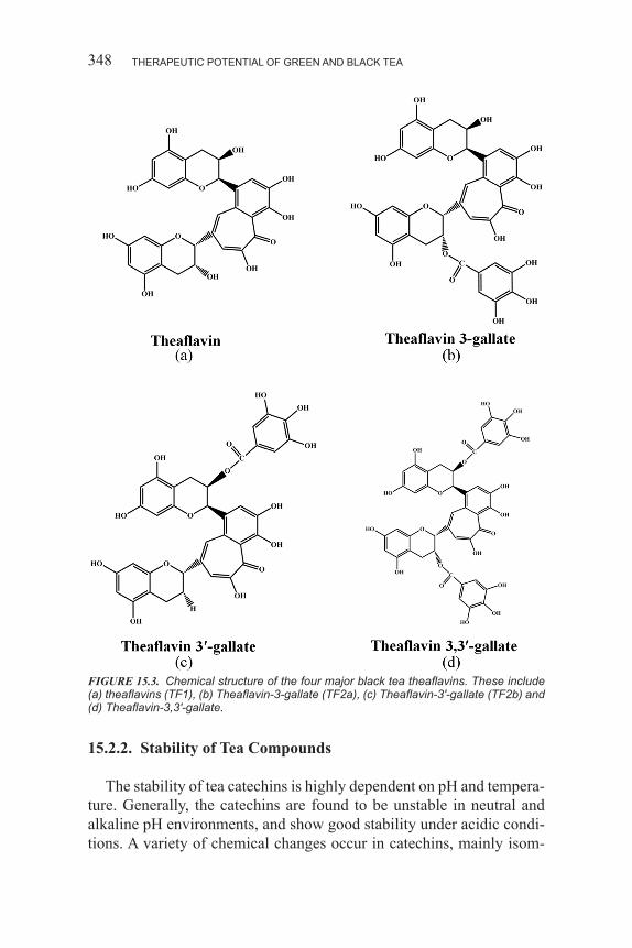
THERAPEUTIC POTENTIAL OF GREEN AND BLACK TEA348
15.2.2. Stability of Tea Compounds
The stability of tea catechins is highly dependent on pH and tempera-ture. Generally, the catechins are found to be unstable in neutral and alkaline pH environments, and show good stability under acidic condi-tions. A variety of chemical changes occur in catechins, mainly isom-
FIGURE 15.3. Chemical structure of the four major black tea theaflavins. These include (a) theaflavins (TF1), (b) Theaflavin-3-gallate (TF2a), (c) Theaflavin-3′-gallate (TF2b) and (d) Theaflavin-3,3′-gallate.
349
erization (epimerization), oxidation, decomposition to small molecules, and polymerization to oligomers or polymers under various conditions. It has commonly been observed that the color of tea changes from green (green tea) to brown upon boiling. The reason for this change is the degradation of molecules.
15.2.2.1. Effect of pH on the Stability of Tea Catechins
pH is very critical factor affecting the stability of catechins. In fact, catechins are extremely stable in acidic solutions (pH < 4), whereas in neutral or alkaline solutions (pH > 6), they are highly unstable [21,22]. It has been observed that green tea catechins were reduced to less than 10% when kept in a Krebs-Ringer buffer at pH 4, whereas they re-mained almost unchanged in water, and only a 15% reduction was ob-served after boiling for 7 hours [21,22]. The complete degradation of EGCG and EGC were reported in sodium phosphate buffer at pH 7.4 for 3 hours, whereas the ECG decreased by 20% and (–)-EC remained unchanged under the same conditions. Although the acidic conditions markedly increase the stability of catechins, the effect differs with the nature of the acids. The stability of green tea catechins was significantly increased with the addition of ascorbic acid, while citric acid did not provide any positive effect on the stability. Under alkaline conditions, the dimerized products of EGCG are converted to theasinensin (A and D rotational isomers) and P-2. Figure 15.2 illustrates the formation of products from EGCG. The proposed mechanism illustrates that EGCG was first dehydrogenated and then decarboxylated under oxidative con-ditions in an alkaline medium [23,24].
The capacity of various catechins for variation differs in similar en-vironments. Among the four catechins examined in alkaline solutions, EGCG and EGC were equally unstable whereas EC and ECG were rela-tively stable. The exact reason for the observed diverse stability is yet to be clarified; however, the existence of an additional OH group at position 5 in EGCG is considered to be one of the factors. The three adjacent OH groups in EGCG and EGC were more susceptible to dev-astation than the two OH groups in EC and ECG [21]. Further studies have also shown the higher semiquinone free radical formation suscep-tibilities of ECG in alkaline media [25].
In addition to degradation or polymerization, the catechin (EC, ECG, EBC, and EGCG) molecules also exhibit small amounts of epimers pro-duced from epimerization. The rate of epimerization varies under cer-
Chemistry and Stability
THERAPEUTIC POTENTIAL OF GREEN AND BLACK TEA350
tain conditions that ultimately affect the relative amounts of the epimers. The rate of epimerization is also largely dependent on temperature, pH, buffer type, and concentrations of metal ions. The rate of epimerization was much higher at pH 6 than the rate in distilled water [26].
15.2.2.2. Effect of Temperature on the Stability of Tea Catechins
The stability of tea catechins is adversely affected at higher tempera-tures. As mentioned earlier, the color of green tea turns brown at high temperatures; an effect mainly attributed to the thermal variations in the chemical structure of catechins. The most predominant change upon heating is the conversion to isometric forms through epimerization. An important study demonstrated that epimerization of catechin was achieved when tea is subjected to a temperature of 40°C for an extended period of time [27]. The study clarified that not only temperature, but also time, are involved in the epimerization process. An example of the epimerization process is the conversion of epigallocatechin gallate to its epimer component gallocatechin gallate. It is also worth men-tioning that the thermal process sometimes causes both epimerization and degradation to occur simultaneously [28]. It has been reported that epimerization of the four tea catechins, (–)-EC, ECG, EGC, EGCG, and 3″-O-methyl- EGCG, increased with increasing time at 90°C [29].
15.2.3. Effects of Oxygen Concentration and Metal Ions on the Stability of Tea Catechins
In addition to conditions (pH and temperature), the stability of tea catechins is also influenced by oxygen concentration and the presence of free radicals and metal ions. The catechins are oxidized more effi-ciently in the presence of higher oxygen levels and at lower antioxidant concentrations [30]. It is more appropriate to say that oxygen concen-tration is the main factor that facilitates the degradation of catechins, even at a suitable temperature and pH. In an N2 atmosphere (low O2 concentration) at 37°C and pH 7.4, EGCG showed only a 5% reduction after 6 hours. This small reduction in EGCG concentration was also attributed to epimerization because no degradation product (dimers) was detected. In contrast, under normal conditions (O2 atmosphere), an EGCG aqueous solution drastically degraded (90%) in 2 hours and completely diminished after 6 hours (no EGCG was left after 6 hours).
Metal ions are very reactive species. They react with catechins to
561
CHAPTER 22
Bioactivity, Bioavailability, and Human Health Effects of Berries’ Bioactive CompoundsM.A. VAZQUEZ-CRUZ, S.N. JIMENEZ-GARCIA, A.A. FEREGRINO-PEREZ and R.G. GUEVARA-GONZALEZ
22.1. BIOACTIVE COMPOUNDS RESPONSIBLE FOR FUNC-TIONAL PROPERTIES OF BERRIES
Berry fruits, such as bilberry (Vaccinium myrtillus), blackberry (Ru-bus fruticosus), blackcurrant (Ribes nigrum), blueberry (Vaccinium corymbosum), chokeberry (Aronia melanocarpa), cranberry (Vaccini-um macrocarpon), grape (Vitis vinifera), raspberry (Rubus idaeus), and strawberry (Fragaria x ananassa) are a particularly rich source of an-tioxidants. Those compounds are mainly represented by vitamin C and polyphenols such as anthocyanins, phenolic acids, flavonols, and tan-nins. They are known as natural antioxidants, and due to their high con-centration and qualitative diversity, berry fruits are increasingly often referred to as natural functional foods. These findings have been con-firmed in the research of Häkkinen and Törrönen (2000), Wang and Lin (2000), and Taruscio et al. (2004). As demonstrated by clinical research, the bioavailability of those naturally occurring compounds significantly exceeds the health benefits carried by their corresponding supplements in pharmaceutical form (Wang et al. 1996). Due to climatic conditions, fresh berries are generally available several months a year, while some
M.A. Vazquez-Cruz, S.N. Jimenez-Garcia, A.A. Feregrino-Perez and R.G. Guevara-Gonzalez; Division de Estudios de Posgrado, C.A. Ingenieria de Biosistemas, Facultad de Ingenieria, Universidad Autonoma de Queretaro, C.U. Cerro de las Campanas S/N, Colonia Las Campanas, C.P. 76010, Santiago de Queretaro, Queretaro, Mexico.
BIOACTIVITY, BIOAVAILABILITY, AND HUMAN HEALTH EFFECTS OF BERRIES562
of the harvested fruit is processed to juice, fruit beverages, frozen prod-ucts, wine, jam, marmalade, and jelly (Da Silva et al. 2007).
Epidemiological studies indicate that persons who consume a diet rich in fruits, vegetables, and whole grains have a reduced risk of chron-ic diseases (Rui 2013). Fruits and vegetables are major sources of phy-tochemicals. Phytocemicals are defined as bioactive non-nutrient plant chemicals in fruits, vegetables, grains, and other plant foods that may provide desirable health benefits beyond basic nutrition to reduce the risk of major chronic diseases (Liu 2004). Among the fruits with this type of phytochemicals or bioactive compounds are berries. Commer-cially, the most important berries include members of the genus Vac-cinium (blueberry, lingonberry, cranberry, bilberry), Rubus (blackberry, black raspberry, red raspberry, arctic raspberry/bramble, cloudberry), Fragaria (strawberry), and Sambucus (elderberry, red elderberry), and are usually consumed in fresh and in processed forms in the human diet (Stoner et al. 2008). The bioactive phytochemicals in berries fall into several structural and chemical classes including phenolic acids (hy-droxycinnamic and hydroxybenzoic acids), flavonoids (anthocyanins, flavanols, flavonols), condensed tannins (proanthocyanins), hydrolyz-able tannins (ellagitannins and gallotannins), stilbenoids, lignans, tri-terpenes, and sterols (Seeram 2006; Tulipani et al. 2008). In addition to the bioactives, berries contain many other constituensts including vitamins, such as A, C, E, and folic acid, and minerals, such as calcium, selenium, and zinc (Kresty et al. 2001). Berries contain high levels of a diverse range of phytochemicals, most of which are phenolic mol-ecules, mainly anthocyanins and ellagitannins that, collectively, are re-sponsible for much of their antioxidant activity (Connor et al. 2002; Wada et al. 2002; Cerda et al. 2005; Aaby et al. 2005), well as its color and flavor (anthocyanins and tannins, respectively). Studies reported that instrumental and sensory qualities of berries are affected by many factors such as cultivars, geographic region, storage conditions, ripe-ness, climate, and others may affect the concentration of phenolic com-pounds and in the antioxidant capacity (Castrejon et al. 2008; Seeram 2008; Zhang et al. 2008)
Berry fruits are good source of phenolics. Phenolics are defined as compounds possessing one or more aromatic rings with one or more hydroxyl groups in the estructures. Generally categorized as phenolic acids, flavonoids, stilbenes, coumarins, and tannins (Seeram 2006). Phenolic compounds are secondary metabolism ubiquitous in all higher plants. These compounds have numerous defense functions in plants,
563
e.g., they act as defense mechanisms against pathogens, parasites, pred-ators, and UV irradiation, and also contribute to the color of plants. They are often induced as a response to various stress conditions, e.g., light, temperature, humidity; and internal factors including genetic dif-ferences, nutrients, hormones, etc., contribute to their synthesis (Strack 1997). Phenolics occur in plant tissues as simple substituted phenols, mainly as glycosides, or as complex polymerized molecules with high molecular weights. The concentration of phenolic compounds varies between each species and varieties of berries, and is also affected by factors such as climate, geographic region, storage conditions, etc. (Kalt et al. 1999; Seeram 2008; Battino et al. 2009; Paredes-Lopez et al. 2010; Kim et al. 2013).
The chemistry of berry phenolics directly influences their bioavail-ability, metabolism, and biological effects in vivo (Manach et al. 2004, 2005). However, in general, the berries contain the following com-pounds: flavonoids such as anthocyanidins (pelargonidin, cyanidin, del-phinidin, peonidin, petunidin, malvidin), flavonols (quercetin, myric-etin, kaempferol), flavonols (catechines) ((+)-catechin, (–)-epicatechin, gallocatechin, epigallocatechin), phenolic acids such as cinnamic ac-ids (caffeic acid, p-coumaric acid, ferulic acid), benzoic acids (proto-catchuic acid, p-hydroxybenzoic acid, gallic acid), lignans such as se-coisolariciresinol, and complex phenolic polymers (polymeric tannins) such as ellagitannins (casuarictin) and proanthocyanidins (procyanidin trimer [flavonols]) (Seeram 2006; Paredes-Lopez et al. 2010).
Anthocyanins are one of the dominant groups of flavonoids in berries (up to 2,000–5,000 mg kg–1 fresh weight [FW]) (Määttä et al. 2001). They are good absorbers of visible light, comprise a large group of wa-ter-soluble pigments, and are responsible for the characteristic orange/red/blue colors of berries such as strawberries, raspberries, blueberries, and red and black currants. Their concentration is usually higher in the epidermis and in the tissue directly under the skin compared to the cen-tral part of the fruit (Paredes-López et al. 2010; Slatnar et al. 2012). For example, anthocyanins represent 44% of phenolic compounds present in strawberries. Anthocyanins comprise aglycones, anthocyanidins, and their glycosides anthocyanins (Viskelis et al. 2009). In Berry fruits, an-thocyanins are found in the form of mono-, di-, or triglycosides, where glycoside residues are usually substituted at C3, or less frequently, at C5 or C7. The most prevalent sugars are glucose, galactose, rhamnose, arabinose, rutinose, sambubiose, and sophorose (Battino et al. 2009). Anthocyanin glycoside residues are often acylated by acids: P-cou-
Bioactive Compounds Responsible for Functional Properties of Berries
BIOACTIVITY, BIOAVAILABILITY, AND HUMAN HEALTH EFFECTS OF BERRIES564
maric acid, caffeic acid, ferulic acid, and by p-hydroxybenzoic acid, malonic acid or acetic acid (Viskelis et al. 2009). Caffeic and ferulic acids are the most common phenolic acids in berries, and they are rarely found free; in general they are esterified with other molecules as car-bohydrates and organic acids. The most common esters of hydroxy-cinnamic acids are chlorogenic acid, which is an ester between caffeic acid and quinic acid, and is a commonly occurring compound in many berries. For example, chokeberry is a good source of hydroxycinnamic acid derivatives. They are represented by caffeic acid derivatives, chlo-rogenic acid (301.85 mg/100 g dry weight), and neochlorogenic acid (290.81 mg/100 g dry weight) (Lohachoompol et al. 2008). In cranber-ry fruit, the hydroxycinnamate esters are present in quantities averaging about 15 mg/100 g of fresh fruit (Pappas and Schaich 2009).
On the other hand, flavonoids and phenolic acids form the building blocks for polymeric tannins, which can be classified into hydrolysable and condensed tannins. Hydrolysable tannins are either gallotannins or ellagitannins, are less frequently encountered, and have been found in strawberries, raspberries, and blackberries (Holt et al. 2008). Berries, especially those of Rosaceae family genus Rubus (red raspberry, arc-tic bramble, and cloudberry), are rich in ellagitannins (Häkkinen et al. 200; Mullen et al. 2002). These berries and strawberries produce only ellagitannins based on stable glucose conformation. In addition to pen-tagalloylglucose, these berries contain dimeric or polymeric ellagitan-nins with amounts of monomers (Haslam 1989). Some berries, such as cranberry, also contain condensed tannins, called proanthocyanidins. Small quantities of tannins were found in honeyberry and blackberry. Lingonberry, strawberry, and cranberry are examples of berries rich in diphenolic compounds called lignans (10–15 mg kg–1 dry weight) (Ma-zur et al. 2000). Some other compounds found in berries is resveratrol (stilbenes), the compound found in grapes mainly, however also found small amounts of trans-resveratrol in bilberry (6.78 µg/g), cowberry (30 µg/g), redcurrant (15.72 µg/g), cranberry (19.29 µg/g), and strawber-ry (3.57 µg/g) (Ehala et al. 2005). The carotenoids are other compounds present in berries. Cloudberry, blueberry, cranberry, and cowberry con-tains 2,840, 2,140, 200, and 140 µg/100 g dry weight, respectively. These berries also contain β-carotene but this pigment prevailed greatly in cloudberry (83% of total carotenoids content). Lutein was the major carotenoid in blueberry (71%). Cranberry contains β-carotene (28%), lutein (23%), and neoxanthin (20%) (Lashmanova et al. 2012). On the other hand, chokeberry also of β-carotene and lutein contains lycopene,
565
β-cryptoxanthin, ζ-carotene, 5,6-epoxylutein, transviolaxanthin, cis-violaxanthin, and neoxanthin (Lashbrooke et al. 2010).
The presented characteristics of various berry fruit species point to vast differences in the type of their bioactive compounds. Such differ-ences are observed with regard to both the content and the qualitative composition of those compounds. The most significant health benefits are ascribed to phenolic compounds and vitamin C. Owing to the rich and diversified compositions of bioactive compounds and their health-promoting properties which result mostly from their antioxidant activ-ity, berry fruits are widely recognized as natural functional products (Szajdek and Borowska 2008).
22.2. ANTIMICROBIAL ACTIVITY OF BERRY BIOACTIVE COMPOUNDS
The development of food products or ingredients with specific health promoting benefits (nutraceuticals or functional foods) is currently the fastest growing and most consumer-driven segment of the food industry (Hussein et al. 2011). On the other hand, food-borne illnesses, the spread of antibiotic-resistant pathogens, and concerns regarding safety of syn-thetic antimicrobial agents have increased consumer demand for the use of plant extracts as natural antimicrobials and antioxidants in foods (Al-Zoreky 2009; Staszewski et al. 2011). Nature offers many different types of antimicrobial compounds that play an important role in the natural defense of all kinds of living organisms (Rodriguez Vaquero, et al. 2007). Plant extracts containing flavonols, other phenolic compounds, and or-ganic acids are potent antioxidants and some of them have shown ad-ditionally good antimicrobial activity, which makes their possible use in food systems reasonable (Choi et al. 2006; Kalogeropoulos et al. 2009).
Fruits and berries contain a variety of phenolic compounds located in plant tissues, often in the surface layer of the plant, fruit, or berry, which is in connection to their main natural function, to protect the plant against environmental stress and pathogens. Berries are rich in phenolic compounds, which are classified into four main groups: flavo-noids, phenolic acids, lignans, and polymeric tannins (Nohynek et al. 2006). Flavonoid anthocyanins are common in bilberry, black and red currant, chokeberry, strawberry, and raspberry, in which they appear as colored substances (Määttä-Riihinen et al. 2004). Ellagitannins are complex phenolic polymers, which are the main phenolic compound in red raspberry and cloudberry.
Antimicrobial Activity of Berry Bioactive Compounds
BIOACTIVITY, BIOAVAILABILITY, AND HUMAN HEALTH EFFECTS OF BERRIES566
Strawberries also contain ellagitannins, although the amounts are lower (Aaby et al. 2005). The most common phenolic acids present in berries are derivatives of either hydroxycinnamic acids or hydroxy-benzoic acids. Lingonberry, strawberry, and cranberry are examples of berries containing lignans (Nohynek et al. 2006).
Probiotic bacteria are widely used in functional foods. Therefore, there is an interest in probiotic bacteria such as Lactobacillus aci-dophilus as well as Bifidobacterium spp. together with plant derived compounds, especially in their application in food technology, for their positive impact on human health (Pascual-Teresa et al. 2010).
Antimicrobial activity of plant phenolics has been intensively stud-ied, and, in addition to controlling invasion and growth of plant patho-gens, their activity against human pathogens has been investigated to characterize and develop new healthy food ingredients, medical com-pounds, and pharmaceuticals (Chung et al. 1998). Berries are an im-portant part of the Nordic diet, but they have also been used as natural antimicrobial pharmaceuticals. Bilberry has been used, for example, for gastrointestinal (GI) disorders, and cranberry has been a well-known treatment in urinary infections. For example, cranberry has been report-ed to control the growth of Listeria monocytogenes (Puupponen-Pimiä et al. 2005; Nohynek et al. 2006) and to possess compounds suppress-ing adhesion and growth of Helicobacter pylori (Vattem et al. 2005) and bacteria causing urinary tract infections (Nohynek et al. 2006).
On the other hand, certain berries rich in tannins have been found to increase bacterial infections. Two different types of polymeric tannins in these berries protect against pathogenic bacteria. Presence of the A-type linkage of cranberry (Vaccinium macrocarpon) proanthocyanidins may enhance both in vitro and urinary bacterial antiadhesion activities (Nile and Park 2013). Additionally, other tannin-containing berries may contribute to this effect, as berry juices of a mixture of cranberries (Vac-cinium oxycoccus) and lingonberries as well as cloudberry juice protect against urinary tract infection (Kontiokari et al. 2003).
Among the berries, cranberries, cloudberries, red raspberries, straw-berries, and bilberries possess clear antimicrobial effects against human pathogens. Berry ellagitannins are strong antimicrobial agents acting as possible antiadherence compounds in preventing the colonization and infection of many pathogens (Puupponen-Pimia et al. 2005). The phe-nolic extract of cloudberry, which is comprised primarily of ellagitan-nins, has the strongest antimicrobial effect, followed by red raspberry and strawberry. Salmonella spp., Staphylococcus spp., Helicobacter
567
spp., and Bacillus spp. are the bacteria that are most sensitive to berry phenolics. Additionally, the growth of Escherichia spp., Clostridium spp., and Campylobacter spp., but not the growth of Lactobacillus spp. and Listeria spp., is inhibited by berry phenolics (Nile and Park 2013). Red raspberry phenolics and its ellagitannin fraction also have powerful antimicrobial properties against the growth of human colonial patho-gens, Klebsiella oxytoca and Proteus mirabilis (Nurmi et al. 2009). Several mechanisms of action in the inhibition of bacteria are involved, such as destabilization of cytoplasmic membrane, permeabilization of plasma membrane, inhibition of extracellular microbial enzymes, di-rect actions on microbial metabolism, and deprivation of the substrates required for microbial growth (Kraft et al. 2008). The berry extracts inhibited the growth primarily of gram-negative bacteria but had no ef-fect on gram-positive bacteria. However, there is very little information about the antimicrobial capacity of phenolics present in berries, except in cranberry. Extracts of the aerial parts of bearberry and lingonberry were active against the gram-negative bacteria E. coli and P. vulgaris. The activity is known to be due to the phenolic glycosides arbutin and metylarbutin.
Cloudberry, raspberry, and strawberry extracts were the strongest in-hibitors of gram-negative bacteria, especially Typhimurium spp. (Chung 1998). Ellagic acid inhibits a range of pathogenic organisms includ-ing Vibrio cholerae, Shigella dysenteriae, and Campylobacter spp. It can be hypothesized that ellagitannins could be one of the components in cloudberries, raspberries, and strawberries causing the inhibition against Salmonella. The antimicrobial effects of berry extracts against gram-negative bacteria decreased in the following order: cloudberry > raspberry > strawberry > lingonberry > blueberry > cranberry > sea buckthorn berry > blackcurrant (Nile and Park 2013).
22.3. EFFECT OF BERRIES CONSUMPTION AGAINST CHRONIC DISEASES DEVELOPMENT
Berries are known as being a good source of vitamin C, dietary fiber, and minerals, and they contain high amounts of phenolic compounds, including anthocyanins, chlorogenic acids, flavonols, and procyanidins, resulting in high antioxidant activity that provides health benefits, and conferring on berries the title of a functional food (Huang et al. 2012). Berry extracts, rich in phenolic compounds and anthocyanins, have a range of biological effects which prevent or reduce the incidence of
Effect of Berries Consumption Against Chronic Diseases Development
BIOACTIVITY, BIOAVAILABILITY, AND HUMAN HEALTH EFFECTS OF BERRIES568
chronic disorders, including cancer, cardiovascular disease, diabetes, inflammation, cancer, Parkinson´s and Alzheimer´s disease, and oth-er pathologies (Knekt et al. 1996, 1997; Wang et al. 2000; Krikorian et al. 2010) These diseases are caused, in part, by the conversion of cellular macromolecules to specific reactive oxygen species (ROS) during normal cellular metabolism (Weisburger 1999). ROS and free radicals are produced in an extensive range of physiological processes. Oxidative stress is an imbalance between the production of ROS and antioxidant defense, and may lead to oxidative damage. This biologi-cal condition may result from an insufficiency of antioxidant defense mechanisms, intense production of ROS, and excessive activation of ROS systems, which are implicated in aging and in the pathology of many chronic disorders (Liu and Hotchkiss 1995; Winterbourn 2008). In order to confront oxidative stress, human bodies have developed mechanisms for maintaining redox homeostasis. These mechanisms in-clude the nonenzymatic and enzymatic antioxidant defenses produced in the body, that is endogenous, and others supplied by the diet namely exogenous ones. The exogenous antioxidants include phenolic acids, flavonoids, stilbenes, and tannins (Seeram 2008; Winterbourn 2008). Phenolic compounds exhibit many biologically significant mechanisms of action, such as scavenging or detoxification of ROS, blocking ROS production, impacting cell cycle, suppression of tumors, modulation of signal transduction, apoptosis, detoxifying enzymes, and metabolism (Liu 2004; Hand et al. 2007). Berries contain high levels of natural polyphenol components that act as potent antioxidants. Berry extracts, rich in polyphenols, have a range of biological effects that can have beneficial outcomes on human health.
Studies indicate that extracts of berries cardioprotective effect (Whitson et al. 2004). For example, mechanisms for the prevention of atherosclerosis by dietary antioxidants in fruits have been proposed. In the LDL oxidation hypothesis, oxidized LDL cholesterol by free radicals has been suggested as the atherogenic factor that contributes to cardiovascular disease (CVD) (Berliner et al. 1997; Witztum and Berliner 1998). When circulating LDLs are present at high levels in blood, they infiltrate the artery wall and increase intimal LDL, which can then be oxidized by free radicals. This oxidized LDL, in the intima is more atherogenic than native LDL and serves as a chemotactic factor in the recruitment of circulating monocytes and macrophages. Dietary phytochemicals served as antioxidants that are incorporated into LDL are themselves oxidized when the LDL is exposed to free radicals; this
569
occurs before any extensive oxidation of the sterol or polyunsaturated fatty acids can occur (Sanchez-Moreno et al. 2000). Therefore, they are very effective inhibitors of low density lipoprotein oxidation, a key step in the development of atherosclerosis as described in the study by Heinonen et al. (1998) from inhibited human LDL and liposome oxidation for phenolic extracts of berries (blackberries, red raspberries, sweet cherries, blueberries, and strawberries), also have beneficial ef-fects on platelet aggregation. At low levels, they provide protection of nitric oxide levels in arterial systems. Nitric oxide is crucial for the maintenance of flexible blood vessels and thereby important in con-trolling blood pressure. The same author indicates that the antioxidant activity of a Berry extract in the LDL oxidation was related to the pres-ence of anthocyanins, but the activity in the liposome oxidation was correlated with the amount of hydroxycinnamates. In complex lipid systems where several different antioxidant and pro-oxidant actions oc-cur simultaneously, it is obviously more difficult to observe the effect of a single factor than in simplified radical scavenging models. In lipid oxidation models, peroxyl radical scavenging and metal inactivation properties are very important mechanistic factors, but the polarity of the compound and the physical state of the lipid system also affect the behavior of antioxidants. In addition, synergism, that is, the ability of antioxidant compounds to reinforce each other, can have a significant effect on the antioxidant response (Frankel 1998).
In addition, dietary phytochemicals can lower C-reactive protein (CRP) dramatically. C-reactive protein, a marker of systemic inflam-mation, has been reported to be a stronger predictor of CVD (Ridker et al. 2002). An Iranian study in Tehran found that both fruits and veg-etables were inversely associated with plasma CRP concentrations (Es-maillzadeh et al. 2006), as did a 2004 study in Massachusetts (Gao et al. 2004). Therefore, the anti-inflammatory activity of phytochemicals may play an important role in the prevention of CVD. In addition, di-etary phytochemicals have been shown to have roles in the regulation of prostaglandin synthesis, reduction of platelet aggregation, regulation of cholesterol synthesis and absorption, and reduction of blood pressure.
On the other hand, berry bioactive have many roles in cancer pre-vention and certain berries are considerably more effective than oth-ers. For example, the most active components in raspberry were the ellagitannins, but these components break down readily and the resul-tant products, including ellagic acid, may be the actual active compo-nents (Ross et al. 2007). Roles in cancer prevention include protection
Effect of Berries Consumption Against Chronic Diseases Development
BIOACTIVITY, BIOAVAILABILITY, AND HUMAN HEALTH EFFECTS OF BERRIES570
against oxidative DNA damage by the scavenging of ROS, inhibition of the formation of carcinogen-induced DNA adducts, enhancement of DNA repair, inhibition of carcinogen-induced tumorigenesis in animals, and modulation of signaling pathways involved with cellular prolifera-tion, inflammation, angiogenesis, modulate cell cycle arrest, and induce apoptosis (programmed cell death). A study by Seeram et al. (2006), showed that blackberry, black raspberry, blueberry, cranberry, red rasp-berry, and strawberry extracts inhibit the growth of human oral (KB and CAL27), breast (MCF-7), colon (HT29, HCt 116), and prostate (LNCaP) cancer cell lines in a dose-dependent manner. The extent of inhibition of cell proliferation was found to vary markedly with both the type of Berry extract and the specific cell line studied. The effects of the different berry extract on cell apoptosis was assessed using HT29 cell that were treated with 200 µg/ml of each Berry extract. Strongest inhibition of cell growth was observed for the raspberry, lowbush blueberry, and cranberry juices. The authors reported that the inhibition of proliferation by the berry juices was independent of caspase-dependent apoptosis but appeared to involve cell-cycle arrest, as evidenced by down-regulation of the expression of cyclin dependent kinases, cdk4, cdk6, cyclin D1, and cyclin D3. Some of the berry juices also significantly inhibited the tumor necrosis factor-induced activation of the COX-2 enzyme expression and activation of the transcription factor, nuclear factor kappa B (NF-κB). The authors concluded that different Berry fruits might act through different mechanisms in their cancer preventive ability (Boivin et al., 2007). Ding et al. (2006) investigated the effects of cyanidin-3-O-glu-coside, isolated from blackberries, on gene expression in JB-6-cells. Cyanidin-3-O-glucoside pretreatment led to dose-dependent decrease in the expression of cyclooxygenase-2 (COX-2) and activities of AP-1, NF-κB, and tumor necrosis factor (TNF)α, when the cells were treated with 12-O-tetradecanolyphorbol-13-acetate or ultraviolet. Finally, and anthocyanin-rich preparation from berries (wild blueberry, bilberry, cranberry, elderberry, raspberry, and strawberry), was found to reduce vascular endothelial growth factor (VEGF) expression in a spontane-ously immortalized human keratinocyte cell line (HaCaT) and inhibit endothelial tube formation in a Matrigel assay (Bagchi et al. 2004).
A study recently investigated whether the regulations of apoptosis and the phase-II enzymes glutathione-S-transferase (GST) and quinone reductase (QR) are potential mechanisms through which blueberry may prevent cancer. Srivastava et al. (2007) showed that anthocyanin-enriched fractions purified from the blueberries induced apoptosis of
715
CHAPTER 27
Emerging Sources for Marine NutraceuticalsE. APOSTOLIDIS and C.M. LEE
27.1. INTRODUCTION: GLOBAL PERSPECTIVE IN MARINE NUTRACEUTICALS
Jacques Cousteau, who is considered a father of ocean exploration, once said, “The future of nutrition is found in the oceans.” While the majority of nutraceutical products in the current market are of botanical origin, marine-based nutraceuticals are gaining attention due to their unique features which are not found in the terrestrial-based resources. In recent years, a series of promising new marine nutraceutical products have been introduced to the nutraceutical and functional foods markets. Currently, functional foods are one of the major consumer trends in the food industry. Consumers continue to seek multiple ways to enhance their health to prevent diseases, and to promote healthy aging by paying more attention to what they are eating and how it benefits their health. Today, consumers are more concerned about their weight, cardio-health, digestive-health, and immunity than ever before. Since health is a major concern of consumers, manufacturers are finding new ways to incor-porate natural and innovative ingredients into food products for health benefits. The sales of functional foods and beverages have been rising with a constant 8% per year since 2003. In 2006, the market of func-
E. Apostolidis; Chemistry and Food Science Department, Framingham State University, Framingham, MA, 01701. C.M. Lee; Department of Nutrition and Food Sciences, University of Rhode Island, Kingston, RI 02881.
EMERGING SOURCES FOR MARINE NUTRACEUTICALS716
tional foods and beverages was $195 billion and is estimated to reach $195 billion by 2013. Functional foods are defined as “those foods that encompass potentially healthful products including any modified food or ingredient that may provide a health benefit beyond the traditional nutrients it contains.” These ingredients vary and include antioxidants (vitamins and phenolic phytochemicals), omega-3 fatty acids, plant ste-rols, and many more. In recent years, a series of promising new marine nutraceutical products have been introduced to the nutraceutical and functional foods markets. This chapter presents an outline of emerging sources for marine nutraceutical development. The main sources and products of primary interest for emerging marine nutraceuticals include phospholipids bound omega-3 fatty acids, micro/macro algal nutrition supplements, fish proteins and peptides, and shellfish derived oligo-molecules of chitin. Also discussed are emerging products, production methods, the ongoing R&D activities, and challenges in marine nutra-ceutical markets, along with future developments in marine nutraceuti-cal industry and research.
27.2. PHOSPHOLIPID-BOUND OMEGA-3 FATTY ACIDS
Fish oils are a rich source of long-chain omega-3 polyunsaturated fatty acids (PUFA), which have attracted much attention in the recent years. Fish oil is produced from various sources of marine fish, i.e., anchovy, menhaden, herring, mackerel, salmon, cod liver, and mussel, and marketed in various forms, most commonly concentrated omega-3 oil in soft gel capsules and microencapsulated powder. Phospholipids of marine origin or phospholipid-bound omega-3 fatty acids are consid-ered a good source of more bioavailable and more stable omega-3 fatty acids when compared to free omega-3 fatty acids. Currently, marine phospholipids are produced from krill and squid in commercial quanti-ties.
27.2.1. Krill Derived Phospholipids
Krill, which means “whale food” in Norwegian, are eucarid crusta-ceans divided into two families. Krill occur in all oceans of the world. They are considered an important trophic connection—near the bot-tom of the food chain—because they feed on phytoplankton and to a lesser extent zooplankton, converting these into a form suitable for many larger animals for whom krill makes up the largest part of their
717
diet (Baker et al. 1990; Brinton 1962). The Bentheuphausiidae fam-ily consists solely of Bentheuphausia amblyops (Brinton 1962), a deep water krill species that differs from members of the Family Euphausi-idae in that Bentheuphausia amblyops is not bioluminescent. The Eu-phausiidae family includes the other 89 known krill species, including one of the most common, Euphausia superba (Brinton 1962), which is the most frequent species associated with krill. Well-known species—mainly because they are subject to commercial krill fishery—include Antarctic krill (Euphausia superba), Pacific krill (Euphausia pacifica), and Northern krill (Meganyctiphanes norvegica) (FAO 2010). The two major krill fisheries are the Antarctic Ocean and the North Pacific Ocean along with the coasts of Japan and Canada (FAO 2010). While about 100,000 tons of krill are harvested annually, this represents only about 0.1% of the estimated existing population (Deutsch 2007). Whole krill are composed of 60–80% protein, 7–26% lipid, and 12–17% ash on a dry weight basis (Grantham 1977; Pierce et al. 1969). Krill oil ex-tracted from Antarctic krill (E. superba) is rich in phospholipid-bound omega-3 fatty acids, mainly EPA and DHA. Krill oil also contains vari-ous potent antioxidants, including vitamins A and E, astaxanthin, and a novel flavonoid (similar to 6,8-di-c-glucosylluteolin but with two or more glucose molecules and one aglycone) (Bunea et al. 2004; Deutsch 2007; Sampalis et al. 2003). Krill oil for dietary commercial applica-tions is extracted by a cold vacuum extraction process that protects the biomass from exposure to heat, light, or oxygen to prevent lipid oxidation and maintain the original nutrients of krill intact (Deutsch 2007).
27.2.2. Squid Byproduct Derived Phospholipids
Squid belongs to marine cephalopods of the order Teuthida, which comprises around 300 species (Clarke et al. 1996). Like all other cephalopods, squid has a distinct head, bilateral symmetry, a mantle, and eight arms arranged in pairs and two longer tentacles (Clarke et al. 1996). According to the FAO, the cephalopod catch for 2002 was 3,173,272 tons of which 2,189,206 tons, or 75.8%, were squid (FAO 2003). In the northeastern United States, approximately 19,100 tons of squid were landed annually since 1998 with steady landings projected (Cardin 2000; NOAA 2008). Due to the high demand for cleaned squid in the United States market, most of harvested squid is processed re-sulting in 40–50% of unutilized byproducts, causing a serious disposal
Phospholipid-bound Omega-3 Fatty Acids
EMERGING SOURCES FOR MARINE NUTRACEUTICALS718
problem. According to Point Judith Fishermen’s Co. (Narragansett, RI) and Sea Fresh USA (North Kingstown, RI), it is estimated that squid processing plants, which are located mainly in Rhode Island, New York, and New Jersey, generate in excess of 4,500 tons of processing byproduct annually. Squid processors pay to dispose of the squid pro-cessing byproduct at a rate of $65–$90/ ton (approximately $100,000/ year per processor). The squid processing byproduct consists mainly of head, fin, and viscera, along with unclaimed mantles and tentacles, and contains approximately 11–14% protein, 2–3% lipids (mostly phospho-lipids), with 11.6% EPA and 24.5% DHA omega-3 fatty acids in oil, 0.6–1.3% ash, and 84–85% moisture (Lian et al 2005). The lipid con-tent of squid byproducts is around 2.5% (De Koning 1993; Lian et al. 2005) and 75% of total lipid is in the form of phospholipids (De Koning 1993; Lian et al. 2005).
27.3. HEALTH BENEFITS
27.3.1. Bioavailability: Nonphospholipid Bound versus Phospholipid Bound Omega-3 Fatty Acids
Omega-3 fatty acids are considered essential fatty acids since they are necessary for human health but the body can’t make them and need to be delivered either through diet or dietary supplements. However, in order to have a physiological and health beneficial effect they need to be first absorbed through the intestinal wall and then from the cells of interest for the specific health benefit. Several studies indicate the beneficial role of phospholipids in enhancing the therapeutic efficacy of some molecules that have poor absorption. Since the 1980s, phos-pholipid complexes began to be a common method to increase bio-availability and absorption of several drugs (Ranade 1989). Silibinin, also known as silybin, is the major active constituent of silymarin, the mixture of flavonolignans extracted from blessed milk thistle (Silybum marianum) and is a molecule that has poor water solubility and bio-availability. It was observed that the silybin-phospholipid complex has significantly higher therapeutic efficacy over the pure molecule in pro-tecting the liver and exerting antioxidant activities (Carini et al. 1992; Comoglio et al. 1995; Conti et al. 1992; Morazzoni et al. 1993; Yanyu et al. 2006). Quercetin is a flavonol, plant-derived flavonoid, used as a nutritional supplement that has been reported to have poor absorp-tion (Hollman et al. 1996, 1997; Manach et al. 1996; Miyazawa et al.
719
1999; Scalbert and Williamson 2000). Recent studies have shown that queretin-phospholipid complex has better therapeutic efficacy than the compound alone in rat liver injury induced by carbon tetrachloride (Maiti et al. 2005) and enhanced absorption in rats (Azuma et al. 2002). Curcumin is a low molecular weight polyphenol derived from the rhi-zomes of turmeric (Curcuma longa) that has a vast array of beneficial pharmacological effects (Bush et al. 2001; Gescher et al. 2001; Ireson et al. 2001; Park et al. 2000; Perkins et al. 2002; Ruby et al. 1995; Shao et al. 2001; Sharma et al. 2001a; 2001b). Despite the promising bio-logical effects of curcumin, poor bioavailability in both rodents and hu-mans was reported by several researchers (Ammon et al. 1991; Ireson et al. 2001; Pan et al. 1999; Wahlström et al. 1978). Recent studies have shown that curcumin phospholipid-complex has better hepatoprotective activity (Maiti et al. 2007) and better bioavailability (Liu et al. 2006) than free curcumin.
Omega-3 fatty acids have been found to be readily absorbed (Naka-mura and Nara 2004) in the small intestine once they are released from lipids, after being hydrolyzed by pancreatic enzymes (Lichtenstein and Jones 2001). However, research studies have shown that the associa-tion between phospholipids and long-chain omega-3 fatty acids highly facilitates the passage of fatty acid molecules through the intestinal wall, increasing bioavailability and ultimately improving the omega-3:omega-6 fatty acid ratio (Cansell et al. 2003; Werner et al. 2004), thus increasing the health beneficial efficacy of omega-3 fatty acids. Recent studies indicate that phospholipid-bound omega-3 fatty acids have better efficacy for premenstrual syndrome management (Sampalis et al. 2003) and hyperlipidemia management (Bunea et al. 2004) when compared to fish oil non phospholipid-bound omega-3 fatty acids. A recent clinical study compared the bioavailability of EPA and DHA be-tween krill oil and menhanden oil (Maki et al. 2009). The daily quantity of EPA provided in the krill (216 mg) and menhanden oil (212 mg) in this study was comparable. However, the DHA present in krill oil (90 mg/d) was approximately half of that provided in the menhanden oil (178 mg/d). At the end of the treatment period (4 weeks), the mean plas-ma EPA concentration was slightly higher in the krill oil group when compared to menhanden oil group (377 versus 293 µmol/ L), whereas the mean plasma DHA concentrations were comparable (476 versus 478 µmol/ L) (Maki et al. 2009). These results suggest that phospholip-id-bound EPA and DHA from krill oil could be better absorbed thank those from menhaden oil. Since the concept of phospholipid-bound
Health Benefits
EMERGING SOURCES FOR MARINE NUTRACEUTICALS720
omega-3 fatty acids is emerging and taking into consideration their positive effect on nutraceutical compound bioavailability, more studies on the effect of the phospholipid complex on omega-3 fatty acid bio-availability are expected in the coming years.
27.3.2. Omega-3 Fatty Acids and Cardiovascular Disease
Cardiovascular disease (CVD) is an abnormal function of the heart or blood vessels. It can cause an increase in risk for heart attack, heart fail-ure, sudden death, stroke, and cardiac rhythm problems, thus resulting in decreased quality of life and decreased life expectancy. The causes of cardiovascular disease range from structural defects to infection, in-flammation, environment, and genetics. One in three American adults has some form of CVD. Heart disease and stroke are the most com-mon cardiovascular diseases and are the first and third leading causes of death for both men and women in the United States (Mayo clinic 2010). CVD is the leading cause of deaths in the Western societies (Delorgeril 2001) and has been linked to the high fat intake, particularly saturat-ed fats, common in Western diets (Dolecek and Granditis 1991). The association between omega-3 fatty acids and cardiovascular disease was first indicated in the 1950s following comparison studies between Greenland Inuits and Danish settlers of Greenland (Sinclair 1956). Both groups were consuming approximately the same amounts of fat (40% of caloric intake), however, the Greenland Inuits had lower incidence of heart disease, despite the higher intake of dietary cholesterol (Sinclair 1956). Later in the 1970s, the low mortality rate from coronary heart disease of Greenland Inuit, was reported to be linked to the high ome-ga-3 fatty acid in the Inuit diet which consisted mainly of fish, seal, and whale (Dyerberg et al. 1975). There are many theories regarding the mechanisms of action of omega-3 fatty acids in cardiovascular disease. It is thought that they have a multifactorial mode of action including an-tiarrhythmic effects, antithrombotic effects, antiatherosclerotic effects, anti-inflammatory effects, improvement in endothelial function, lower-ing of blood pressure, and lowering of triglyceride concentrations (Al-bert et al. 2002; Harris 2007; 2006; Kang and Leaf 2000; Knapp 1997; Morris et al. 1993; Nair et al. 1997). In the recent years, clinical trials have assessed the effects of fish and fish oil on CVD prevention. The diet and reinfarction trial (DART) showed a 29% reduction of mortality rate among 2,033 men with a recent myocardial infarction, linked to the omega-3 fatty acid consumption (Burr et al. 1989). The Gruppo Italiano
721
per lo Studio della Sopravvivenza nell’Infarto Miocardico Prevenzione (GISSI-Prevenzione) trial that lasted for three-and-a-half years showed 30% reduction of risk of cardiovascular disease death and 45% reduc-tion of sudden death incidence among 11,324 patients after myocardial infarction and was linked to omega-3 fatty acid consumption (Grupo Italianoperlo Studio 1999). In the Asian population, patients with a sus-pected myocardial infarction showed a significant reduction in mortality rate from coronary heart disease due to omega-3 fatty acid supplemen-tation (Singh et al. 1997). A clinical trial compared the effect of krill oil derived phospholipid bound omega-3 fatty acids with fish oil and ac-cording to Bunea et al. (2004), krill oil was significantly more effective than fish oil for the reduction of glucose, triglycerides, and LDL levels. Another clinical study with 76 overweight and obese adults compared the effect of krill oil phospholipid-bound omega-3 fatty acids and men-handen oil and according to Maki et al. (2009), krill oil supplementa-tion produced significant elevations in plasma levels of EPA and DHA. The above findings confirm the better potential of phospholipid-bound omega-3 fatty acids for cardiovascular disease prevention, mainly due to better bioavailability.
27.3.3. Omega-3 Fatty Acids and Inflammatory Disease
Inflammation is an important component of the early immunologi-cal responses, while inappropriate or dysfunctional immune respons-es underlie chronic inflammatory and autoimmune diseases. Onset of autoimmune and inflammatory diseases has been linked to omega-3 and omega-6 imbalance (James et al. 2000; Simopoulos 2002; Song 2008),with omega-3 fatty acids exerting anti-inflammatory properties (Shahidi 2007; Simopoulos 2002; Song 2008). Arachidonic acid, an omega-6 fatty acid, is the precursor of eicosoanoids that produce com-pounds that activate macrophages and induce inflammatory response (Simopoulos 2002; Song 2008). On the other hand, EPA, an omega-3 fatty acid, competes with arachidonic acid (Simopoulos 2002) and has been shown to inhibit certain immune functions, such as the produc-tion of ecosanoids and proinflammatory cytokines(Song 2008). Sev-eral scientific findings have indicated the positive effect of omega-3 fatty acids on inflammatory disease management (Shahidi 2007; Song 2008). These findings have shown that omega-3 fatty acids can reduce proinflammatory cytokine production (Din et al. 2004; Hulshof et al. 1999; Kamal-Eldin and Yanishlieva 2002; Newton and Snyder 1997;
Health Benefits
EMERGING SOURCES FOR MARINE NUTRACEUTICALS722
Watanabe and Ackman 1974), manage bronchial inflammation (Holmer 1989), and arthritis (Shahidi 2007). In regards to phospholipid-bound omega-3 fatty acids, a clinical study with 90 patients has shown that krill oil inhibits inflammation and reduces arthritic symptoms within a short period of treatment (Deutsch 2007).
27.3.4. Omega-3 Fatty Acids and Brain Functions
All membranes, whether they are cell surface or form of an intra-cellular organelle, are composed of phospholipid bilayers. Membrane lipids provide flexibility and adaptable structure into which are inserted proteins and glycoproteins such as enzymes, transporter proteins, or re-ceptors (Farquharson et al. 1995). More specific research has shown that phospholipids of the brain have an especially high content of the long chain omega-3 fatty acid, DHA, and that these phospholipids are involved in brain function (Amaducci et al. 1991; Hirata and Axelrod 1980; Salem and Niebylski 1995) The structural predominance of DHA in the brain suggests functional significance and according to Young and Conquer (2008), both DHA and EPA are linked with several as-pects of neural function, including phospholipase A2 (PLA2) activity, inflammation, neurotransmission, membrane fluidity, oxidation, ion channel, enzyme regulation, and gene expression. It has been suggested that under pathological conditions, seen in neuropsychiatric disorders such as schizophrenia, an increase in the activity of PLA2 can results in a decrease in neuronal membrane phospholipid biosynthesis and an in-crease in phospholipid breakdown (Horrobin 1998). Finnen and Lovel (1991) have shown that EPA can inhibit PLA2 and according to Mellor et al. (1996), administration of EPA has shown considerable success in the treatment of schizophrenia and bipolar disorder. Modulation of neurotransmitters, such as serotonin and dopamine, has been shown to be a causative factor for unipolar and bipolar depression (Mundo et al. 2001; Ogilvie et al. 1996; Post et al. 1980). Several studies have shown that supplementation through diet of DHA and EPA contribute towards the increased concentrations of serotonin and dopamine (Austed et al. 2000; Chalon and Delion-Vancassel 1998; Zimmer et al. 2000,1999; Owens and Innis 1999). A clinical study on 70 patients diagnosed with premenstrual syndrome showed that supplementation of phospholipid containing krill oil reduced the emotional symptomatology that charac-terizes premenstrual syndrome (Sampalis et al. 2003). In this study, it was suggested that the effectiveness of krill oil could be based on po-
797
Index
Abiu (Pouteria caimito), 552 Açaí, 526, 534, 545–546 Acerola, 527–529, 534, 543 Acetate, 23, 35, 52, 83, 92, 98, 130, 155, 167,
185, 211, 279, 307, 374, 492, 510, 540, 570, 580
Acetic acid, 3 Acetylcholine, 238, 255, 261–265 Acetylcholine receptors (AchR), 238 Acetylcholinesterase, 262 Acetylsalicylic acid, See Aspirin Acinetobacter spp., 376 Acrolein, 237, 255 Actinomycetes, 27 Activator protein-1 (AP-1), 48, 383, 505,
509–510, 570 Acyl CoA cholesterol transferase (ACAT),
66 Adipogenesis, 378 Adiponectin, 501, 502, 759–761 Adrenergic, 275, 542 Adsorption, 163–164, 167 Advanced glycation end products (AGE’s),
212–215, 466, 528, 543 Aflatoxin, 431 Agar, 6, 724, 727 Agaricus spp.
A. bisporus, 691, 694, 697, 700–701, 703 A. blazei, 685–687, 696, 702
Aging, 12–14, 102, 141–142, 206, 212, 226, 251, 255, 281, 294, 345, 380, 458, 504, 506, 568, 578, 581, 582, 656, 683, 715
Aglycones, 52, 58, 61, 104, 160, 178, 459, 483–485, 563, 577, 584, 596–601, 610, 647, 655, 663–664, 717
Ajoene, 422, 426, 440–442, 444, 448 Akinesia, 257 Alanine amino transferase (ALT), 380, 405,
435 Aldolase, 27, 91–92, 141 Aldosterone, 106 Algae, 14, 16, 72, 723–725, 728, 731 Alginate, 6
Sodium alginate, 6 Alginic acid, 3, 724 Aliphatic, 59, 176, 181, 183, 439, 645,
653–656, 668, 669 Alkaline phosphatase, 316, 405 Alkaloids, 3, 186, 207, 272, 278, 290, 329,
345, 440 Allergy, 3 Allicin (diallyl thiosulfinate), 421–425,
440–441 Alliin, 3, 418, 421, 426, 440, 447 Alliinase, 417, 421, 426 Allium spp.
A. ampeloprasum, 417 A. cepa, 417 A. chinese, 417 A. fistulosum, 417 A. sativum, 417 A. schoenoprasum, 417 A. tuberosum, 417
Index798
Allyl sulfenic acid (2-propene-1-sulfenic acid), 422
Allyl sulfides, 422, 426, 432, 433, 445–446 Disulfide(s), 130, 141, 232, 422, 426–427,
431–432 Trisulfide(s), 422, 426–427, 432–439,
443–444 Alzheimer’s disease (AD), 204, 206, 210, 215,
233–237, 238, 251–267, 278–281, 329– 333, 336, 385–386, 511
Amygdala, 257, 261 α-Amylase, 100, 105–110, 513, 552, 571 Amyloid beta (Aβ), 204, 336, 386
Aggregation, 215 Fibrils, 252–253 Oligomers, 215–253, 279 Plaques, 253–254, 262, 336 β1-42, 206, 215, 234, 238
Amyloid precursor protein (APP), 238, 252–256
Amylopectin, 27 Amylose, 7, 27 Amyotrophic lateral sclerosis (ALS), 260 Anacardium spp.
A. occidentale, 529–531, 543 Ananas spp.
A. comosus, 539, 543 Anethol, 335 Anethole, 335 Angiogenesis, 48, 70–71, 204, 213, 226–227,
314, 458, 469, 503, 506, 537–539, 570, 616
Angiotensin–I converting Enzyme (ACE), 58, 100, 106–111, 700, 734–735, 740
Antheraxanthin, 528, 725 Anthocyanidins, 54, 58–60, 291, 459, 483–
487, 504, 510, 534, 563, 576, 648, 650 Anthocyanins, 3, 15, 49, 54, 58–61, 167,
307, 458, 470, 482, 484, 493–499, 528, 530, 534, 542–547, 561–567, 569–571, 575–578, 580–581, 584–585, 648, 650
Antiaging, 294, 464, 481, 506–507 Antiangiogenic, 62, 464, 469, 473, 539 Antiarrhythmic, 499, 720 Antiatherogenic, 57, 138 Antiatherosclerotic, 449, 499, 584, 720 Antibacterial, 14, 16, 78, 100, 294, 295, 307,
311–312, 317–318, 334–337, 375, 397, 422, 530, 579, 736
Antibiotic, 14, 22, 27–28, 111, 114, 336, 375, 428, 565
Antibiotic associated diarrhea (AAD), 28
Antibiotics, 14, 28–29, 175, 328, 374–375, 600, 602
Anticancer, 50, 62, 78, 81, 101, 175, 335, 382, 402, 406, 428, 439, 463, 481, 505, 510, 537, 579–586, 643, 655
Anticarcinogenic, 291–292, 304, 307, 313–315, 355–356, 406, 428, 431, 583, 623, 644, 651, 653, 662, 668, 736, 781
Anticholelithogenic, 400 Anticoagulant, 127, 472, 697, 727, 740 Antidepressant, 335 Antidiabetic, 377, 401, 447–448, 499, 532,
537, 540, 577, 702–705, 727, 772 Antidiarrheal, 27 Antidyslipidemic, 540 Antifungal, 64, 68, 100, 311–312, 332– 337,
376 Antihypertensive, 304, 471, 542, 734, 736 Anti-inflammatory, 70–71, 76, 78, 81–83,
138, 142, 146–147, 226, 240, 260, 279, 281, 292–293, 304, 307, 329, 332–336, 343, 357–359, 382, 384, 388, 397, 402, 406, 411, 464–465, 473, 481, 499, 510, 532, 547, 569, 576, 584, 586, 613, 622, 643, 656, 687, 694, 720–721, 727, 730, 735, 736
Antimalarial, 77 Antimetastatic, 382, 468, 473, 506, 740 Antimicrobial, 25–27, 60, 64, 78, 90–91, 96,
100–111, 114–115, 209–210, 227, 235, 279–280, 304, 307, 311–313, 317, 319, 329, 332–335, 374–376, 481, 530, 532, 541, 565,–567, 579–580, 586, 656, 694
Antimicrobial peptides (AMP’s), 227 Antimutagenic, 64, 335, 406, 428, 431–432,
586, 657 Antimutagenicity, 687 Antiobesogenic, 759 Antiobiotics
β-lactam, 28 Antioxidant 65–68, 71, 91–95, 99, 100–107,
110–114, 137, 141–147, 202–212, 225–226, 253–258, 276–282, 288–295, 304, 307–319, 329–337, 343, 345, 350–358, 380–384, 397, 399, 401–411, 428, 441, 442, 457, 458, 462, 467, 470–472, 481, 482, 494, 496– 503, 511, 513, 525–532, 534–541, 546–551, 561–569, 572–580, 585–586, 603–604, 610–613, 621, 643, 644–652, 656–658, 664–665, 671, 694–695, 701, 707, 716–718, 723–739
799Index
Antioxidant defense, 104, 137, 254, 282, 288, 404, 498, 568
Antioxidant response element (ARE), 225–226, 511, 652, 686
Antioxidative enzymes, 68 Antiproliferative 50, 60, 62, 69, 314, 356, 383,
411, 434, 439, 440–441, 463, 466–468, 504–506, 571, 579, 584, 655, 728, 735
Antisense oligonucleotide (ASO), 205 Antispasmodic, 335, 397, 532 Antithrombotic, 304, 316, 428, 443, 449, 499,
503–504, 720, 727 Antitumor, 72, 75–79, 81, 104, 207, 212, 304,
343, 355, 397, 406–407, 411, 428, 440, 506, 530, 536–537, 656, 687, 694, 697, 740
Antiviral, 16, 29, 30, 60, 100, 293, 295, 304, 307, 329, 334, 336, 536–537, 543
Anxiety, 252, 257 Apigenin, 49, 50, 209–293, 335–336, 461,
463, 466, 468, 483, 533, 543 Apolipopretin E (apoE), 138, 146, 586 Apolipoprotein A1 (apoA1), 142 Apolipoprotein L1 (apoL1), 142 Apoptosis, 48, 60, 67, 70–71, 76, 79, 83,
102–103, 201–204, 209–212, 227–232, 235, 238–240, 255, 259–260, 279, 288, 314–315, 331, 353, 356, 383, 404, 411, 430, 434, 436, 437, 441–442, 462–463, 482, 503–513, 568, 570–571, 583, 611, 616, 652, 654, 658, 730
Anti-, 70, 229, 277, 314, 353, 442, 445, 481, 509
Apoptotic bodies, 463 Chromatin condensation, 463 DNA fragmentation, 293, 383, 442, 463,
571, 583, 728 Mitochondrial activation, 463 Pro-, 60, 70, 228–229, 230, 353, 356, 439,
445, 509, 652 Apoptosis signal-regulating kinase-1 (ASK-1),
232, 235, 240 Apoptotic protease activating factor 1
(APAF1), 70, 229 Apoptotic signaling, 228–229, 235Appetite, 263, 373, 386, 755–759 Apple, 57, 61 Apricot, 57 Araçá, 545, 548 Araçá–boi, 545, 548–549 Arachidonic acid, 15, 70, 81, 406, 411, 444,
465, 466, 699, 721
Araticum (Annona crassiflora), 552 Aromatase, 50, 68 Aronia spp.
A. melanocarpa, 561 Artemisinin, 76, 77 Arthritis, 38, 51, 337, 358, 374, 382, 385, 406,
411, 706, 722 Ascophyllan, 724, 727 Ascorbic acid, 308, 310, 314, 349, 492, 528,
532, 645 Asparagus, 33 Aspartate amino transferase (AST), 380, 405,
435 Astaxanthin, 717, 723 Asthma, 30, 335, 374, 756, 761, 783 Astringinin, 68 Atherogenesis, 66, 145–146, 357, 402, 750 Atherogenic, 316, 398, 400, 568, 612 Atherosclerosis, 65, 138–139, 147, 315, 319,
355–357, 399, 402, 443, 457, 499,–501, 539, 568, 569, 584, 607–608, 610, 612, 652, 657, 671, 697, 709, 729, 731
Atherosclerotic lesions, 147, 500, 607 Atherosclerotic plaque, 146, 357 Atmospheric pressure chemical ionization
(APCI), 496–497 Auroxanthin, 528 Autophagy, 226, 229–232 Avarol, 78 Avarone, 78 Avicularin, 533, 543 Avocado, 57 2,2′-azino-bis(3-ethylbenzothiazoline-6-
sulphonic acid) (ABTS), 104, 380, 498 Azobis (2–amidinopropane) hydrochloride
(AAPH), 352 Azo–bisisobutyrylnitrile primaquine, 352 Azoxymethane, 434 Bacillus spp., 6, 78, 312–313, 376, 567, 733
B. cereus, 312 B. subtilis, 6, 78, 312
Bacteriocin, 26, 96, 115–16 Bacteriocinogenic, 25, 26 Entericin, 26 Lacticin, 26 Nonlantibiotic, 25 Variacin, 26
Bacuri (Platonia insignis or Scheelea phalerata), 545, 550, 552
Baicalein, 49, 468 Banana, 57
Index800
Basal ganglia, 257, 258, 275 Basil, 159, 161, 208–210, 293–294, 301, 316,
334, 337 B–cell lymphoma 2 proteins (Bcl–2), 229,
437, 440, 445, 508–509, 513 Bak, 70, 229, 437, 440, 508 Bax, 70, 229, 276, 353, 437, 440, 445,
464, 508–509, 513, 571 Bcl-2, 229, 232, 265, 277, 353, 383, 434,
437, 442, 445, 505, 508–509, 513, 652, 728
Bcl-w, 229, 353 Bcl-xL, 229, 277, 353, 437, 442, 509 BH (1–4), 229–230
Beer 6 Benzo(a) pyrene (BAP), 356 Benzoate, 212, 213 Benzoic acid, 63, 99, 291, 305, 482–484,
563 Benzo-γ-pyrone skeleton, 457
See also DiphenylpropanesBerries, 51, 55, 59, 63, 490, 492, 514, 545,
561–586 Betaine, 131, 141 Betulinic acid, 71 Bifidobacterium spp. 3, 6, 16, 27, 30, 33–35,
90, 95, 130 B. adolescentis, 27 B. angulatum, 27 B. bifidum, 27, 32 B. breve, 6, 27, 32
B. breve Yakult, 6 B. catenulatum, 27 B. dentium, 27 B. infantis, 27, 32 B. lactis, 3, 6, 30, 96
B. lactis FK120, 6 B. lactis LKM512, 6
B. longum, 6, 27, 32 B. longum, BB536 6
B. pseudocatenulatum, 27 Biguanides, 377 Bilberries, 60, 561–566, 570, 571, 582–585 Bile acids, 125, 398–400, 408, 708, 738 Bile salts, 409 Binding affinity, 203, 602–603 Bioavailability, 57, 262–265, 270– 282, 304,
345, 353, 361–362, 409, 445, 464, 466, 473, 504, 561, 563, 574, 575–578, 584, 600–602, 658–667, 718–721, 762–763
Biochanin A, 596, 598, 610 Biomarkers, 4, 9, 131–147, 362, 505, 581,
604, 608, 611, 614, 617, 622
Blackberries, 57, 61, 561–564, 570–571, 579, 583
Blackcurrants, 57, 61, 561, 567, 582 Blood glucose, 100, 105, 208–209, 214, 334,
377–381, 386, 401, 410, 448, 502, 537, 542, 571, 658, 696, 701–704, 729, 736, 740, 772–778
Blood pressure, 6, 100, 106, 213–214, 294, 357, 377–379, 386, 449, 470, 482, 502–503, 569, 604, 609, 625, 697,–700, 720, 735–740
Blueberries, 57, 61, 111, 280, 561–571, 577, 580–583
Body mass index (BMI), 50, 132, 738, 752– 757, 760–769, 774–777, 782–784
Bolinaquinone, 78 Bone loss, 359, 606, 618 Bone mineral content (BMC), 618, 625 Bone mineral density (BMD), 606, 618–619,
625 Bone turnover
Bone alkaline phosphatise (BAP), 618–619, 625
Deoxypyridinoline (DPD), 618–619, 625
Osteocalcin, 619 Resorption, 618–619
Borneol, 335 Bradykinesia, 257 Brassica spp., 15, 643–654, 668, 672
B. carinata, 644, 646, 667 B. juncea, 644, 646 B. napus, 644, 646, 650, 655, 667–670 B. nigra, 644, 646 B. oleracea, 644, 646, 648–651, 653, 654,
657, 668–669, 672 B. rapa, 644, 646, 648–649, 668, 671
Broccoli, 3, 6, 291, 644–649, 650–658, 662, 668–669, 671–672
Bromocriptine, 273 Brussels sprouts, 644, 646, 651, 655, 657–658,
671–672 Buriti (Mauritia flexuosa), 545, 550 t-Butylhydroperoxide, 352 Butyl hydroxyl anisole (BHA), 308, 317–318,
536 Butyl hydroxyl toluene (BHT), 308, 309, 310,
317–318, 411 Butyrate, 23–24, 34–35
Cabbage, 6, 59, 167, 644–649, 650–658, 662, 668–669, 672
801Index
Caffeic acid, 64, 65, 99, 100, 161, 208, 293, 310, 313, 335–336, 483–484, 496, 563–564, 576, 666, 726, 730
Caffeine, 3 Cagaita (Eugenia dysenterica), 552 Calbindin, 262 Caloric restriction (CR), 507 Cambuci, 545, 551 Camellia spp.
C. sinensis, 343 Campestrol, 336 Camphene, 336, 337 Camphor, 335 Campylobacter spp., 567 Camu–camu, 545–547 Cancer
Adenoma, 71, 430, 617 Brain, 429 Breast, 54, 62, 68, 81, 204, 205, 210,
355, 356, 429, 430, 440, 442, 467, 504, 505, 506, 509, 605, 613–618, 623–625, 696, 730, 735, 750, 781, 785
Cervical, 383, 510 Colon carcinoma, 83, 210, 463, 537, 654,
728 Colorectal, 59, 71, 429, 430, 440, 605 Endometrial, 430 Gastric, 111, 440, 463 Leukemia, 60, 209, 213, 383, 441, 463–
464, 504–505, 510, 653–654, 727 Melanoma, 71, 81, 213, 383, 467–468, 509 Prostate, 50, 61, 71, 205, 356, 429,
436–439, 468, 504–505, 571, 595, 605, 615, 626, 728
Candida spp., 311, 313, 334, 375–376, 708 Canola, 15, 669 Capsaicin, 91, 101, 532 Capsiate, 3 Carbidopa, 269 Carbon dioxide (CO2), 25, 92, 160 Carbon tetrachloride (CCl4), 402, 433, 435 Carcinogen, 428, 430, 570, 617, 657 Carcinogenesis, 63, 70, 102, 139, 313, 315,
356, 382, 401, 407, 431–434, 462–463, 473, 482, 504, 605, 615, 651–652, 655–656, 661, 728
Carcinogens, 100, 406, 432–433, 466, 652, 655, 657
Cardamom, 373 Cardioprotective, 227, 307, 444, 445, 499,
500, 503, 568, 603, 610, 697
Cardiovascular disease (CVD), 13–14, 47, 50, 58, 71, 91, 100, 106, 145, 280, 288, 290, 294, 315, 319, 329, 331, 335, 354, 357, 358, 360, 381, 397, 399, 401, 443, 458, 469, 470, 473, 482, 497, 513, 568–569, 582, 595, 603, 608, 609, 613, 618, 621, 622, 624, 625, 696–701, 709, 720–721, 729–731, 780, 786
Carnasol, 209 Carnitine, 141 Carnosic acid, 209, 295, 309, 310–311 Carnosol, 295, 309, 310, 313–314, 336 Carnosolic acid, 335γ-Carotene, 542ζ-Carotene, 565 α-Carotene, 71, 528, 530, 725 β-Carotene, 71, 308, 526, 528, 530, 542, 547,
564, 645, 725, 728 Carotenoids, 3, 155, 329, 332, 481,
490–491, 528, 530, 536, 538, 540, 542, 564, 572, 645, 724–725, 728, 739
Carrageenan, 406, 724–725, 727 Carrot, 141, 167, 645 Carvacrol, 161, 210, 294, 309, 310, 313, 315,
335–337, 376 Carvone, 71, 76 Casein, 6, 7, 15, 94 Cashew, 529, 530–531 Caspases, 60, 70, 228–229, 315, 353,
436–437, 441–442, 463–464, 508, 510, 570–571, 652
Catalase (CAT), 27, 68, 91, 104, 146, 202, 206–210, 280, 289, 316, 381, 387, 433–434, 441, 446, 610, 652
Cataract, 214, 335, 403–404 Catechins, 6, 7, 54–57, 292, 344–351, 354,
357, 381, 482, 486–489, 496, 526, 533, 563, 576, 726
Catechol, 268, 274, 345 Catecholamines, 52, 275 Catechol-O-methyl transferase (COMT), 268,
274–275, 277 Cauliflower, 644–646, 649–655, 672 Celery, 49 Cell
Death, 48, 50, 70, 103, 201, 226–231, 239, 240, 252, 254, 258–260, 266, 351–352, 355, 380, 383, 388, 437, 441, 458, 463, 505, 508–509, 570, 658, 701, 728
Index802
Differentiation, 15, 24, 67, 68, 102, 201, 235, 281, 315, 330, 332–333, 359, 440–443, 510, 583, 615, 622, 624, 652–653, 728
Division, 202, 436, 458, 657 Growth 50, 54, 62, 201–204, 234, 439,
443, 458, 465, 468, 505, 508, 537, 570, 616, 728
Membrane, 202, 230, 255, 258, 313–314, 728
Proliferation, 70, 81–83, 102–103, 202–203, 226–227, 236, 314, 356, 382–383, 388, 430, 467, 513, 537, 570–571, 583–584, 616–617, 652, 656, 735, 778
Shrinkage, 315, 463 Cell adhesion molecules (CAMs), 612
E–selectin, 613, 783 Intracellular adhesion molecule-1 (ICAM–
1), 612–613, 625, 783 Platelet endothelial cell adhesion molecule
(PECAM), 500 Vascular adhesion molecule-1 (VCAM-1),
612–613, 622, 626, 783 Cell cycle, 48, 50, 60, 68–71, 81, 202–204,
212, 383, 428, 436–438, 441, 462–463, 482, 505–508, 568, 570, 616, 652–654, 728
G0/G1, 441, 508 G1/S, 69, 508 G2/M, 50, 60, 70, 81, 383, 428, 436, 438,
441 Mitosis, 81 Mitotic phase, 81, 437, 440 S/G2, 508
Cell cycle arrest, 48, 50, 69, 70, 81, 428, 436–438, 570, 653–654, 728
Cerebral cortex, 238–239, 261 Cerrado fruits, 552 Ceruloplasmin, 227 Chemical ionisation (CI), 185 Chemopreventive, 60, 72, 76, 313, 402, 406,
411, 429–430, 507, 513, 537, 653–655, 667
Chemotherapeutic, 76, 81, 216 Cherry, 57, 61, 74, 527, 541 Chicha (Sterculia striata and Naud Acrocomia
aculeata), 552 Chicory, 33, 36 Chili, 3 Chitin, 693, 697, 702, 707, 716, 736, 737 Chitosan, 6, 112–114, 736–741
Chitosan oligosaccharide (COS), 112–113, 737–741
Chlorogenic acid, 3, 6, 65, 210, 335, 528, 564, 666
Chlorophyll, 80, 345 Chocolate, See Cocoa Chokeberry, 561, 564–565, 578 Cholesterol, 6, 14, 15, 62, 66, 95–96, 142,
208, 213–214, 315–317, 328, 356–357, 374, 377, 380, 398–401, 410, 446–448, 472, 481, 498–502, 568–569, 584, 586, 604–608, 622–623, 685, 693–699, 708, 720, 727– 731, 735, 738, 739–741, 759, 763–770, 771, 786–787
Cholesterol esters (CE), 212–213, 759, 763, 779
Metabolism, 66 Serum, 6, 66, 96, 399, 410, 447, 697, 739
Cholinergic pathway, 261–262, 265 Cholinesterase inhibitors (ChEIs), 261–262,
267 Chondroitin sulfate, 15, 727 Chromanol, 3 Chrysin, 49, 470, 471 Cineol, 210, 335 Cinnamaldehyde, 91, 101, 211–215, 279,
374–378, 382–385, 386–388 Cinnamic acids, 59, 63–66, 91, 98, 101,
211–212, 279, 291, 305, 482–484, 563, 650, 666, 726, 730
Cinnamomum spp., 205, 211, 278, 334, 371,–374, 379, 382,–384, 386–388
C. burmannii, 211 C. cassia, 205, 211, 373, 379, 384 C. loureirii, 211 C. pauciflorum, 211 C. tamala, 211 C. zeylanicum, 184, 211, 215, 372, 375,
385 Cinnamon, 182–184, 205, 211–216, 278–280,
327, 334, 371–389 Cinnamyl acetate, 185, 211, 279, 374 Cinnamyl alcohol, 211, 279 Cinnemaldehyde, 212 Cinnzeylanine, 374 Cinnzeylanol, 374 Citrus spp.
C. aurantifolia, 462 C. paradisi, 64 C. reshni, 165 C. reticulata, 462–464 C. sinensis, 165
803Index
Clostridium spp., 567 C. difficile, 28–29, 708 C. perfringens, 313, 376
Cloudberry, 564, 567 Cloves, 157, 373, 447 Coagulation, 3 Cocoa, 5, 56–58, 371, 550 Coffee, 3, 6, 7, 49, 65, 154 Colitis, 28, 30,–32, 335, 337, 388, 727 Collagen, 15, 294, 401, 444, 471–472,
502–503, 612, 700 Collagenase, 68 β-Conglycinin 3 Conjugated dienes (CD), 317 Conjugated linolenic acid (CLA), 15, 138,
749–787 cis-9, trans-11, 138, 749–754, 757–786 trans-8, cis-10, 757–759, 769, 777 trans-10, cis-12, 138, 750–782, 785–787
Constipation, 3, 705 Continuous subcritical water extraction
(CSWE), 158, 161 Coquinho (Butia capitata), 545, 550 Coriander, 327 Corn, 6, 59, 99, 447, 572
Fiber, 6 Coronary Heart Disease (CHD), 143, 472 Correlation spectroscopy (COSY), 177,
188–189, 192 p-Coumaric acid, 59, 64–66, 305, 496, 530,
540, 563, 651, 666 Coumaric acid, 59, 64–66, 99, 305, 483–484,
496, 530, 540, 563, 651, 666, 730 m-Coumaric acid, 64 o-Coumaric acid, 64 Coumarins, 91, 101, 164–167, 334, 562 Countercurrent chromatography (CCC), 166,
167 Cranberries, 112–114, 280, 291, 561–567,
570, 573, 579, 582, 736 C–reactive protein (CRP), 145, 359, 382,
501–502, 569, 622, 780–784 Creatine, 131, 141 Creatine kinase (CK), 141 Creatinine, 131, 380 Crohn’s disease, 30–31, 706 Cruciferous vegetables, 644–661, 666–672 Cryptoxanthin, 528–530, 542, 565 Crystallisation, 156 C–S lyase. See Allinase Cumene hydroperoxide (CHP), 538 Cumin, 159, 327 Cupuassu, 545, 549–550
Curcuma spp. C. longa, 333, 397–407, 719 C. zedoaria, 333
Curcumin, 3, 100, 332–333, 397–408, 719 Curcuminoids, 164, 167, 333, 399, 406 Cyanidin, 3, 58, 60, 484, 499–501, 506, 528,
534, 546–547, 563, 570–571, 577, 650 Cyanogenic glucosides, 157 Cyclin dependent kinases (CDK), 68–69,
570 CDK2, 463 CDK4, 463, 509
Cyclins, 68–69, 508 B1, 436 Cyclin D1, 50, 69, 463, 508–509, 570
Cyclooxygenase (COX), 207, 281, 336, 444, 465, 497, 570
COX–2, 50, 60, 70–71, 212, 281, 336, 465–466, 472, 509–510, 570, 584
Cyclophosphamide, 402 Cyclosporine, 404 Cysteine sulfoxide(s), 417–427, 444 Cytochrome P450 enzymes, 74, 263–265,
337, 600 CYP1A1, 433 CYP1A2, 276–278, 462 CYP2B1, 431 CYP2B2, 431 CYP2D6, 265, 276 CYP2E1, 433 CYP3A2, 431 CYP3A4, 263–265
Cytokines 31, 55, 59, 62, 71, 146, 212, 228, 235–236, 240, 258, 315, 337, 358–359, 388, 501, 512, 612, 721, 727, 778–783
Granulocyte macrophage colony stimulating factor (GM–CSF), 59 IFN-γ, 59, 696, 779 Interleukins, 60, 281
IL-1, 334, 380, 778 IL-1α, 333, 612 IL-1β, 59, 240, 260, 279, 335–337,
386, 779 IL-2, 59, 779–780 IL-4, 335, 779 IL-5, 335 IL-6, 59, 71, 337, 380, 386, 512,
779–783, 784 IL-8, 59, 71, 227, 380, 547, 783–784 IL-10, 59, 388, 780IL-12, 59
Index804
TNF-α, 59, 82, 232, 240, 260, 279–281, 314, 333–335, 380–382, 386, 466, 501, 504, 509–510, 612, 626, 696, 760, 779–783
Cytotoxicity 81, 82, 265, 351, 381, 463, 468, 530, 538
Daidzein (DAI), 3, 54, 104, 291, 483,
596–604, 610–614, 621–622, 625 Daidzin, 596–598, 622 Equol (EQU), 599, 600, 610, 621–622,
625 O-desmethylangolensin (O-DMA),
599–600, 611, 626 Delphinidin, 58, 60, 484, 506, 510, 534, 547,
563, 650 8-hydroxy-2′-deoxyguanosine (8–ODG), 51,
70, 658 Depression, 252, 257, 272, 278, 436, 722 Detoxification, 3, 15, 206, 225, 407, 432, 466,
511, 568, 652, 655–657, 665–666 Phase I, 15, 72, 82, 432, 433, 600, 602,
652 Phase II, 14–15, 79, 225, 407, 432–435,
497, 511–513, 600–602, 652–656, 664, 668
Dextran, 6 Diabetes, 1, 3, 13–14, 38, 60, 91, 95, 100–102,
105–110, 116, 203–205, 208–216, 255, 279, 288, 290, 328, 331–335, 374, 377–381, 386–388, 397–403, 446–449, 466, 469, 503, 512, 528, 533–534, 540–542, 552, 568, 571, 595, 652, 657–658, 701–705, 709, 728–730, 739–741, 750–751, 753, 756–758, 761, 772, 776
Diacetyl, 25, 96, 115 Diacylglycerol, 3 Diallyl disulfide (DADS), 431– 448 Diallyl monosulfide (DAS), 431–437, 442,
446 Diallyl trisulfide (DATS), 432–440, 445–448 Diarrhea, 28–30, 105, 263–265, 282, 373–374,
531–532, 552, 708 Traveler’s, 29 Viral, 30
Dietary fiber ,6, 66, 549–550, 567, 685, 688, 693, 697, 702–707, 727
Dihydropyran, 345 3,4-dihydroxyphenylalanine (DOPA), 239,
268–269 7,12–dimethylbenz[a]anthracene (DMBA),
314, 434, 462
Dimethylhydroxylfuranone, 540 Diode-array detection (DAD), 486, 496 Diosmetin, 466 Diosmin, 49, 464–470 Dioxins, 602, 685 2,2-diphenyl-1-picrylhydrazyl (DPPH), 104,
308–310, 380, 411, 498, 536–538, 547, 551
Diphenylpropanes, 99 Distention, 105 Distillation, 156–159, 426, 492 Distortionless excitation by polarization
transfer (DEPT), 177, 190–193 Diterpenes, 71, 79–80, 293, 304 Dithiin, 422, 426, 444 DNA binding, 509, 612 DNA cross-linking, 255, 401 DNA damage, 51, 70, 102, 314, 360, 411, 432,
462, 570, 583, 613, 657–658 DNA fragmentation, 293, 383, 442, 463, 571,
583, 728 DNA lesion, 79, 314 DNA polymerase, 78, 482, 505 DNA replication, 202 DNA synthesis, 79, 432, 505–506 Docosahexaenoic acid (DHA), 7, 15,
717–722, 729, 762 Donepezil, 262–265 Dopa-decarboxylase inhibitors, 269–270, 274 Dopamine, 204, 239, 257–258, 267–277, 722 Dopamine receptor agonist, 271 Dopamine receptor (D1/D2), 257, 268,
271–273, 277 Dopaminergic neurons, 239–240, 257– 259,
269–272, 281 Dopamine Signaling, 239 Drug resistance, 47, 81 Dyskinesia, 271–273, 278 Dyslipidemia, 1, 7, 410, 696 Dyspepsia, 211, 272, 278, 373
Edema, 83, 406, 464, 470 Eggplant, 99 Ehrlich’s reagent, 178 Eicosanoid, 55, 465 Eicosapentaenoic acid (EPA), 7, 15, 717–722,
762 Elaidic acid, 786 Elastase, 359 Electron impact/ionization (EI), 184–85 Electron ionization-Mass spectrometry
(EI-MS), 184–185
805Index
Electron transport chain (ETC), 255, 258, 287–288
Electrospray ionization (ESI), 137, 486, 496, 513
Elettaria cardamomum, See Cardamom Ellagic acid, 100, 291, 530, 534–536, 548,
551–553, 567–569, 575–576, 579–580, 583
Embryogenesis, 458 Endoplasmic reticulum (ER), 228
ER stress, 228, 231–233, 240 Lumen, 231–232 Membrane, 232 PKR-related ER kinase (PERK), 231–232
Endothelial dysfunction 357, 470–471 Endothelial function, 357–358, 470, 482,
499–500, 503, 609–610, 622, 652, 720, 782
Endothelin-1 (ET-1), 510, 584 Endothelium-dependent relaxation, 55, 503,
510 Endothelium-dependent relaxation (EDR), 503 Entacapone, 274–275 Enterobacter spp., 90, 311, 319
E. amnigenus, 312 E. gergoviae, 312 E. aerogenes, 334, 376
Enterococcus spp., 28, 37, 93, 376, 579 E. faecalis, 93, 312, 376 E. faecium, 28, 93, 96, 376
Enzyme–linked immunosorbent assay (ELISA), 142
Epicatechin (EC), 50, 54–56, 215, 289–291, 345–350, 381, 426, 482–483, 486–489, 496, 528, 551, 563
Epidermal growth factor (EGF), 68, 213, 382 Epigallocatechin (EGC), 3, 54, 207, 291–292,
345–352, 483, 486–488, 496, 528, 563 Epigallocatechin gallate (EGCG), 3, 50, 54,
57, 207, 292, 345–346, 349–361, 486 Epinephrine, 275, 444, 727 Epithiospecifier protein (ESP), 647, 655 Equol, 483, 598–600, 604, 625 Ergocalciferol, 707 Ergogenic, 201 Ergolines, 272 Ergosterol, 694–695, 699, 707 Ergothioneine, 701, 706 Eriocitrin, 295, 308 Eriodictyol, 52 Eritadenine, 685, 694, 699 Erythrocytes, 289, 400, 403, 444, 472, 706, 762
Escherichia spp., 29, 32, 36, 319, 567–579 E. coli, 36, 72, 312, 319, 335, 376, 600
Essential oils (EO), 74–75, 157–162, 176, 184, 293–294, 304, 308–319, 335–337, 371, 374–376, 388, 491, 544
Estrogen, 15, 50, 61–62, 205, 291, 386, 506, 511, 595, 602–603, 613, 618, 622–625, 785
Antiestrogenic, 62, 463, 595, 603, 614 Estradiol, 68, 463, 596, 602 Estrogenic, 60–64, 481, 595, 602–605,
614, 620 Estrogen receptors (ER), 61, 602
ERα, 463, 602–603, 621 ERβ, 602–603, 608, 618, 622–623
Ethnopharmacology, 175 Ethylenediaminetetraacetic acid (EDTA), 127 Eugenia spp.
E. stipitata, 545, 548 E. uniflora, 541–543
Eugenol, 161, 211, 279, 335, 374–376, 383 Eukaryotic translation initiation factor 4E
binding proteins (4E–BPs), 202–203 Euterpe spp.
E. oleracea, 545 Excitotoxicity, 255, 258–260, 265, 511 Exopolysaccharides, 703 Extracellular signal–regulated kinases (Erk),
235, 236, 254, 260, 445, 509, 510, See also Mitogen–activated protein kinase (MAPK)
Extraction, 127–128, 154–162, 166–168, 186, 424–426, 492–497, 581, 608, 685, 717, 725, 730
Exudates, 176
Faecalibacterium prausnitzii, 34 Fatty acids, 7, 15, 34, 137–139, 142, 313,
352, 378, 696, 719–723, 735, 742, 762, 769–783, 786
Fenton reaction, 287–89, 402 Fermentation, 22–24, 27, 34–35, 89–96,
101–116, 129, 344, 347, 354, 495, 552, 574, 597, 671–672, 706
Fermented foods, 90, 93–94 Ferric reducing antioxidant power (FRAP),
381 Fertility, 335, 373, 386, 623 Ferulic acid, 59, 64–66, 291, 307, 313,
335–336, 483, 496, 533, 563–564, 649–651, 666, 670
Fish oil, 138, 142–144, 719–721, 742, 786
Index806
Flame ionization detection (FID), 158, 183 Flammulina spp.
F. velutipes, 688, 691, 701 Flash chromatography (FC), 163–168 Flatulence, 105, 372, 398, 552, 738 Flavan-3-ols. See Flavanols Flavonoid, 3, 6, 48–55, 60, 65, 98–99, 161,
164–166, 178, 181, 186, 208–210, 290–292, 304, 307–313, 332–336, 344, 359, 388, 457–473, 481–486, 496, 512, 526, 530, 533, 536, 542–543, 546, 549, 562–565, 568, 575–576, 586, 610, 648, 650, 656, 663–667, 701
Catechin, 3, 49, 54, 211–212, 291–292, 335–336, 344–358, 362, 459, 483, 488, 493– 494, 551, 726
Flavanols, 49, 50, 54–58, 291, 307, 311, 459, 470, 482–490, 496, 562, 576, 648
Flavanone,3, 7, 52–53, 467 Flavanones, 49, 52–54, 291, 307,
458–459, 470, 483, 648 Flavone, 3, 49–51, 54, 165, 291, 307,
458–459, 461, 467–470, 483, 488, 648
Flavonol, 3, 49–51, 54–57, 291, 307, 344, 458–459, 469–470, 482–483, 486–488, 496, 539, 561–567, 576, 581, 584, 648–649, 663
Isoflavone, 3, 7, 49, 54, 60–62, 67, 104, 145, 291, 307, 459, 470, 595–617, 620–626, 648
Flavonoid skeleton. See Diphenylpropanes Flaxseed, 14, 15, 66–67, 145 Foam cells, 357, 501, 610 Food for Specified Health Uses (FOSHU), 2,
8–11 Forkhead box, class O (FOXO), 20–206 Fourier transformation (FT), 186 Fourier transform infrared spectroscopy
(FTIR), 186 Fractionation, 156 Fragaria spp.
F. ananassa, 561 Free radicals 51, 60, 99–104, 115, 253–254,
258–259, 272, 280, 287–290, 293–295, 309–311, 329–331, 336, 349–352, 380, 402–403, 408, 462, 534, 538, 568, 576, 579, 582, 657, 694. See also Reactive oxygen species (ROS)
French Paradox, 280 Fructans, 33–37
Fructo–oligosaccharide (FOS), 5, 7, 27, 33–35, 130
Fucoidan, 724, 727 Fucosterol, 724, 727 Fucoxanthin, 725, 728–731, 742 Functional foods, 1–16, 116, 132, 301–302,
457, 567, 671–672, 685, 694, 700, 706–709, 716, 724, 740–741
Fungi Aspergillus spp., 311, 334, 376 Fusarium spp., 83, 376 Mycoplasma spp., 376 Penicillium spp., 376
Furcelleran 724
Galacto–oligosaccharides (GOS), 33–37 Galactose, 27, 51, 59, 404, 563, 664, 687 Galactosidases, 33 Galantamine, 262, 265 Gallic acid, 91, 291, 336, 482, 488, 496, 536,
563 Gallocatechin (GC), 54, 350, 486–488, 563 Gamma-aminobutyric acid (GABA), 3, 6,
700 Gamma-linolenic acid (GLA), 14–15 Ganglioside GM1, 36 Garlic, 3, 33, 327, 355, 417–418, 420–434,
441–449, 645 Gas Chromatography (GC), 54, 92, 126,
129, 158, 176–177, 182–186, 195, 346, 484– 488, 581
Gas chromatography–mass spectrometry (GC–MS), 129, 176–177, 184–186, 195
Gastric emptying, 214, 379 Gastritis, 111, 211, 530 Gastrointestinal tract (GI), 19, 20, 24, 31, 34,
95, 104, 292, 373, 566, 577, 661, 664, 699
Gastrointestinal disorders, 89, 90, 94, 327, 652
Microbial load, 20–19 Structure, 19
Gel electrophoresis, 137, 144–146 Difference gel electrophoresis (DIGE),
137 Gene expression, 5, 30, 96, 136, 145, 203,
258, 288, 356, 359, 440, 447, 503–506, 510–512, 570, 582–584, 622, 722
Generally recognized as safe (GRAS), 295, 311, 497, 512, 740–741
Genetic engineering, 572, 667–668
807Index
Genistein (GEN), 54, 62, 67–68, 104, 388, 483, 511, 596–604, 610–615, 620–625
6′-hydroxy-O-desmethylangolensin, 599 Genistin, 596, 610, 622
Genomics, 5–9, 135, 147, 206, 573, 586 Genotoxicity, 430, 652 Gentisic, 99 Geraniol, 71 Ghrelin, 214, 386 Ginger, 332–333, 373, 397, 400, 408–411 Gingerol, 333, 411 Ginkgo, 51 Ginseng, 159, 205 Ginsenosides, 159 Glucagon, 34, 205 Glucagon-like Peptide-1 (GLP-1), 34, 35 β-Glucans, 14, 686–688, 693, 694, 697, 702,
705–708, 724 Ganopoly, 694 Schizophyllan, 694
Glucokinase, 535 Gluconeogenesis, 138, 201–202, 210, 232 Glucophage, 205 Glucosamine, 15, 737–738 Glucose intolerance, 377 Glucose transporter 4 (GLUT-4), 202–203,
214, 378, 445 α-Glucosidase, 100, 105–111, 528, 533,
542–543, 552, 571, 730 β-Glucosidase, 110, 597–598, 663–664 Glucosinolates (GSLs), 645–655, 659–662,
667–672 Glucobrassicanapin (4-pentenyl), 646, 669 Glucobrassicin (3-indolylmethyl),
646–647, 654, 668–669 Glucoerucin (4-methylthiobutyl), 646, 654 Glucohirsutin (8-methylsulfinyloctyl), 646 Glucoiberin (3-methylsulfinilpropyl), 646,
654, 668 Gluconapin (3-butenyl), 646, 668–669Gluconapoleiferin (2-hydroxy-4-pentenyl),
646 Gluconasturtiin (2-phenylethyl), 646, 654 Glucoraphanin (4-methylsulfinylbutyl),
646, 653, 668–671 Neoglucobrassicin (1-methoxy-3-
indolylmethyl), 646 Progoitrin (2-hydroxy-3-butenyl), 646,
655, 669 Sinigrin (2–propenyl) 646, 653–656, 668
Glutamate, 141, 255, 258–259, 265, 266, 693, 698–699
Glutamate–cysteine ligase, 289 Glutamate dehydrogenase, 141 Glutathione (GSH), 68, 104, 141, 146,
208–209, 225, 232, 258, 280–282, 289, 293, ,314–315, 337, 355, 360, 381, 387, 401–403, 418,–421, 431, 432, 435, 438, 441, 444, 446–447, 498, 503, 538, 540, 570, 610, 652, 660–661, 701
Glutathione peroxidase (GPx), 68, 146, 225, 289, 314, 355, 381, 387, 401, 432, 446, 498, 503, 610
Glutathione reductase (GR or GSR), 208, 225, 355, 498
Glutathione-S-transferase (GST), 104, 141, 206, 225, 232, 431–435, 570, 571, 652–653, 661
Glutathione synthetase, 225, 289 Glycated hemoglobin (HbA1c), 145, 378, 379,
774, 775 Glycitein, 596–598, 622
Glycitin, 596–598 Glycogen biosynthesis, 378 Glycogen synthase kinase-3 (GSK-3), 235,
238, 256, 260, 280, 378 Glycolysis, 138–139, 201–202 Glycosides, 49–52, 58–61, 65–66, 388, 459,
472, 484–485, 490, 525, 534–536, 563, 567, 575, 596–598, 649, 657, 662–663
C–glycosides, 459, 536 O–glycosides, 51, 459, 484–486, 536, 663
Glycosylated, 30, 49, 52–54, 483, 650 Glycosylation, 291, 307, 428, 467, 650, 656 Glyzcyrrhiza spp.
G. uralensis. See also LicoriceGoitrogenic, 623 G–protein coupled receptor 41 (Gpr41), 34, 35 Grains, 12, 15, 64–66, 94–95, 99, 207,
290–292, 430, 562, 708 Grapefruit, 3, 52, 64, 165, 528 Grapes, 5, 57–59, 64–68, 99, 130, 141, 157,
167, 280, 291, 481–502, 504, 507–514, 545, 561, 564, 578, 581, 584
Raisins, 68, 481 Red, 130, 167, 498, 578
Grape seed, 5, 499–501 Grape seed extract (GSE), 499–502, 513 Graviola (Annona muricata), 551 Growth factors (GF), 47, 62, 68, 70–71, 83,
201, 213, 235, 382, 465, 509 Guaijaverin, 533, 543 Guava, 7, 526, 531–533, 543, 548 Gut associated lymphoid tissues (GALT), 26
Index808
Haber–Weiss reaction, 289 Heat shock factor-1 (HSF-1), 233–234 Heat shock Protein (HSP), 138, 206, 231–234
HSP-16.2, 206 HSP70, 231, 233, 234, 240 HSP90, 203, 231, 233
Helicobacter pylori, 89, 91, 94, 101–102, 111–114, 116, 294, 334, 383–384, 530, 540, 543, 566, 652
Hemeoxygenase-1 (HO-1), 225, 232, 239 Hemoglobin A1C, 214 Hepatic steatosis, 146–147, 512 Hepatotoxicity, 262, 405 Herbacetin, 66 Herbs, 52, 74–75, 99, 104, 115, 159, 162, 175,
205, 206–211, 292–295, 301–317, 327– 329, 332, 335, 373
Hericium spp. H. erinaceus, 686–688, 700
Hesperetin, 52–54, 291, 461–463, 471 Hesperidin, 52, 209, 291, 309, 461–465,
470–472Hesperitin, 462 Heterofermentation, 91–93
Heterofermenters, 92 Heteronuclear multiple–bond correlation
(HMBC), 177, 193–194 Heteronuclear multiple–quantum correlation
(HMQC), 177, 192–194 Hexanoate, 550–551, 580 Hexokinase, 535 Hexose, 27, 92 High Density Lipoprotein (HDL), 142,
315–316, 379, 399, 410, 447, 586, 604–608, 623–625, 697–699, 729–731, 763–771, 786–787
HDL2, 142 HDL cholesterol, 399, 586, 607–608, 623,
698, 729–731, 767–770 High–pressure liquic chromatography
(HPLC), 163, 176, 195, 484–486, 492, 496, 528, 671, 725
High voltage electrical charge (HVEC), 495 Hippocampus, 238, 252, 257, 261 Hippuric acid, 130 Hispidulin, 210, 293Histone deacetylase (HDAC), 203, 281, 442.
See also Sirtuins Homeostasis, 22, 96, 99–104, 145, 204, 208,
214, 227, 236, 254, 279, 282, 400, 436, 447, 498, 508, 568, 777–778
Iron, 227
Redox, 102, 254, 282, 498, 568Homofermentation, 91–92
Homofermenters, 91 Homogenization, 155, 426 Homorientin, 546 Horminone, 210 Human immunodeficiency virus (HIV), 78,
295, 510, 536, 730 Huntington’s disease, 204–206, 511 Hydrogen peroxide (H2O2) 25, 96, 115, 276,
287–289, 294, 308, 311, 314–316, 351– 353, 405–406, 436, 441, 504, 510, 701
Hydrogen sulfide (H2S), 260, 444–445 Hydrophilic, 157, 168, 528 Hydrophobic, 168, 183, 738 8-hydroxy-2′-deoxyguanosine (8–ODG), 547 3-hydroxy-3-methylglutaryl CoA (HMG-
CoA), 317, 699 Hydroxybenzoic acids, 63, 64, 305, 540,
562–566 Hydroxycinammic Acids (HCA), 64, 65, 305,
482–483, 496, 551, 564–566, 649–651, 666
Hydroxycinnamic alcohols, 66 6-hydroxydopamine (6–OHDA), 352–353Hydroxyflavone, 49 3-hydroxykynurenine (3-HK), 352–353 7-Hydroxylase, 66 Hydroxylation, 74, 307 Hydroxyl radical, 287–289, 310, 352, 498 4-Hydroxynonenal (HNE), 141, 237, 255 3-(2-hydroxyphenyl)-propanoic acid, 384 Hypercholesterolemia, 3, 255, 315–316, 447,
469–697 Hypercholesterolemic, 398–400, 403,
500, 697 Hyperglycemia, 105–106, 109–110, 377, 381,
401, 404, 512, 542 Hyperin, 533–543 Hyperinsulinemia, 255 Hyperlipidemia, 315–316, 399, 469, 686, 719,
738 Hyperlipidemic, 316, 500 Hypertension, 1, 3, 39, 84, 100–107, 110, 116,
236, 255, 267, 275, 315, 373, 445, 469– 470, 503, 542, 686, 696, 698–701, 727, 734–735
Hypertriglyceridemia, 542 Hypochlorous acid, 288, 701 Hypocholesterolemia, 39, 738
Hypocholesterolemic, 316, 398–401, 607–608, 694, 697, 708, 735, 739
809Index
Hypoglycemic, 292, 401, 410, 443, 694, 702–704
Hypotension, 269–272, 275–278 Hypotensive, 542, 694, 698–700 Hypoxia, 204, 226–228, 382 Hypoxia-inducible factor 1 (HIF-1), 226–228,
382 Factor inhibiting HIF (FIH), 227 HIF-1α, 204, 226–228 HIF-1β, 226–228
Hypoxia responsive element (HRE), 226–228
Ilimaquinone, 78 Illudins, 78–79 Immune system, 26, 31, 38, 55, 101, 288,
329–334, 345, 358, 387, 696, 741, 779 Innate immune system, 329–335
Immunity, 15, 16, 31–32, 95, 235, 329, 388, 695–696, 715, 735, 778
Immunoglobulins, 31, 94, 388 IgA, 31 Immunomodulation, 30–31, 39, 333–335 Immunomodulatory, 327, 695 Immunoprotective, 537, 543 Immunostimulants, 332, 683, 687, 695 Indoles
Indole-3-carbinol (I3C), 647, 654–655, 660, 672
Indomethacin, 384 Infectious disease, 77, 100–101, 374–375,
388, 686 Inflammation, 15–16, 31–32, 38, 51, 57,
59, 63, 70–71, 82, 131, 144, 145, 203, 212, 228, 235, 239, 240, 253–255, 258–260, 315, 331, 345, 351, 358–360, 373, 380–381, 403, 411, 464–468, 473, 482, 501–512, 568–570, 584–586, 604, 612–613, 619, 622, 657, 720–722, 779–780
Chronic, 51, 82, 358–360, 657 Inflammatory bowel disease, 30–31, 89, 94,
358, 384 Inflammatory bowel disorders (IBD), 30 Inflammatory bowel syndrome (IBS), 32 Inflammatory diseases, 48, 50, 211, 358, 376,
384, 401, 406, 501, 512, 656, 721 Inflammatory pathways, 138, 146 Inflammatory response, 55, 279, 281,
464–465, 584, 721 Infrared Absorption Spectroscopy, 181 Infrared (IR), 145, 176–177, 181, 186, 195,
378, 405, 776
Inhibitor of kappa kinase (IKK), 294, 509 Insulin, 10, 35, 105, 131, 138, 145, 201–214,
233, 237–238, 255, 279, 374, 377–381, 386, 401, 447–448, 506, 512, 534, 537, 702–705, 739, 750, 772–778, 786–787
Secretagogues, 205 Sensitivity, 778
Insulin–like growth factor receptors (IGFR), 201, 206
Insulin–like growth factors (IGF), 201–205, 227, 233, 237
Insulin like growth factors signaling (ILS), 201–204, 210–211, 216
Insulin receptor substrate (IRS-1), 202–204 Insulin resistance (IR), 131, 138, 145, 203,
214, 255, 374, 378–381, 447, 702–705, 772–778
Insulin signaling, 201–202, 206–208, 211, 238, 279, 506
International Life Sciences Institute (ILSI), 5, 8, 9, 10
Inulin, 33–36 Invasion, 31, 68, 71, 468, 566, 583 Ion–exchange chromatography, 163 β-Ionone, 491 Irofulven, 79 Iron-sulfur center, 288 Ischemia, 405, 445, 473, 499, 658 Ischemic stroke, 53, 357 Isoalliin, 418, 422 Isobaric Tag Relative Absolute Protein
Quantitation (iTRAQ), 137 Isogonic, 2 Isolation, 7, 153–163, 167–168, 737 Isoprenaline, 399 Isoprenoid, 3 Isoprostanes, 145, 611, 782–783 Isorhamnetin, 52, 54, 485–486, 581, 649–650,
657, 663 Isothiocyanates (ITCs), 3, 647, 651–655,
659–662, 668, 672 Allyl isothiocyanate (AITC), 653 Benzyl isothiocyanate (BITC), 653–654 Erucin (4-methylthiobutyl isothiocyanate,
MTBITC), 653–654 Iberin (4-methylsulfinilpropyl
isothiocyanate, MSPITC), 653–654 Phenethyl isothiocyanate (PEITC),
653–654 Sulforaphane (SFN), 652–656, 671–672
Isotope–Coded Affinity Tags (ICAT), 137 Isovitexin, 546
Index810
Jambolan (Jamun), 533–535 Japanese knotweed, 280 Jerusalem artichoke, 33 c-Jun N-terminal kinase (JNK), 71, 233–236,
240, 260, 437, 445, 504, 509–510 Kaempferol, 51–54, 160–161, 291, 313, 335,
461–463, 483–488, 530–536, 542–543, 549–553, 563, 580–581, 649–650, 657, 663
Kale, 291, 644, 649–655, 668, 671–672 Kefir, 93–95, 102–110 Kelch-like ECH-associated protein-1
(Keap-1), 225 Kidney bean, 3 Kiwi, 57 Klebsiella spp.
K. oxytoca, 567 Kovats indices, 176, 183–185 Krill, 716– 723, 732, 737
Oil, 719–723
Lactase phloridzin hydrolase (LPH), 664 Lactic acid bacteria (LAB), 6, 26–27,
35–37, 89–96, 101–106, 111–116, 319, 708
Lactic acid (Lactate), 25–27, 34–35, 89–95, 101–116, 319, 708
Lactitol, 37 Lactobacillus spp., 6, 14, 26–34, 37, 89–95,
130, 311, 566–567, 672, 705 L. acidophilus, 6, 29–32, 95–96, 107–108,
111 L. acidophilus, CK92 6
L. brevis, 93 L. bulgaricus, 107–108, 111 L. casei, 6, 14–16, 32, 93, 95, 111
L. casei, LC1 6 L. casei NY1301, 6 L. casei Shirota, 6
L. curvatus, 93 L. delbrueckii, 6, 32
L. delbrueckii subsp. bulgaricus, 6, 32 L. fermentum, 29, 93 L. gasseri, 93 L. helveticus, 6, 93, 108
L. helveticus CK60, 6 L. johnsonii, 93 L. plantarum, 32, 95, 111 L. rhamnosus, 31, 93
Lactococcus spp. 26, 92–93 L. lactis, 26, 93
Lactose, 27, 33, 89, 94–95 Lactose intolerance, 89, 94–95 Lamiaceae, 208– 210, 287, 292–294,
301–319, 334–336 Laminarin, 724, 727 Lavender, 157, 160 LDL cholesterol, 62, 357, 472, 502, 568, 607,
622, 697, 767–769 LDL/HDL ratio, 316, 763, 768–770 Leeks, 33, 417 Legumes, 49, 99 Lemons, 52, 66, 75, 291 Lentinus spp.
L. edodes, 579, 691, 694, 699–701 Leptin, 386, 759–761 Leucotrienes
Leucotriene B4, 82 Leukocyte, 57, 357 Levodopa, 268–278. See also
3,4-dihydroxyphenylalanine (DOPA) Lewy bodies, 234, 240, 257 Licorice, 167, 205, 373 Liebermann-Burchard Reaction, 178 Lifespan, 202, 205–206, 215, 281, 507, See
also Aging Lignans, 3, 48, 65–68, 291, 562–566
Enterodiol, 66 Enterolactone, 66, 291 Lariciresinol, 66 Matairesinol, 66 Pinoresinol, 65–66 Secoisolariciresinol, 65–66, 563 Syringaresinol, 65–66
Lignins, 65–66, 645, 685 Limonene, 71, 75–76, 374, 547 Limonins, 156, 165, 190–194 Limonoids, 160–161, 164–166, 178 Linalool, 5, 209, 335, 551 Lingonberries, 562, 567, 582 Linoleic acid, 697, 764 α-Linolenic acid (ALA), 15, 697 Lipase, 398, 409, 763 Lipids, 100–104, 125, 131, 162, 237, 258,
288, 313, 331, 351, 379–380, 449, 502, 526, 545, 656–657, 693, 696, 701, 706, 718–719, 722, 727–729, 735, 738–739, 750, 759, 762–763, 770–771, 779–780, 786–787
Biosynthesis, 201, 214 Desaturase, 749, 763 Hydroperoxides (LPO), 289, 307, 311,
381, 472, 499, 538
811Index
Oxidation, 201–202, 307, 318, 513, 569, 717, 732
Peroxidation, 141, 145, 209, 237, 254–255, 281, 316, 352, 358, 380–381, 387, 399–408, 434, 473, 512–513, 538, 584, 604, 610, 611, 697, 701, 781, 786
Peroxides, 360, 399, 403–405, 500 Plasma, 131, 762
Lipogenesis, 35, 139 Lipoic acid, 141 Lipophilic, 157, 459 Lipopolysaccharide (LPS), 54, 212, 239, 386,
466 Lipoxygenase (LOX), 51, 307, 465–466, 472,
538 5–lipoxygenase, 465 Liquid Chromatography (LC), 126–128,
163–164, 167, 177, 182, 496, 581 Liquid chromatography–mass spectrometry
(LC–MS), 126–128, 167, 177, 496 Listeria spp., 567
L. grayi, 335 L. innocua ,312, 319, 335 L. ivanovii, 335, 376 L. monocytogenes, 91, 312, 335, 376, 566,
579, 580 Lisuride, 273 Liver disease, 359, 405 Liver X receptor (LXR), 96 Longevity, 206, 281, 329, 482, 507. See
also Lifespan Loquat, 57 Low–density lipoprotein (LDL), 55, 58,
62–66, 100, 214, 293, 315–317, 356–360, 379–380, 398–402, 410, 445–447, 470–472, 481–482, 498–502, 568–569, 604–612, 622, 626, 657, 697–699, 708, 721, 763–770, 777, 782–786
LDL cholesterol, 62, 357, 472, 502, 568, 607, 622, 697, 767, 769
LDL oxidation, 100, 293, 315, 357, 360, 399, 482, 499, 568, 569, 611
Low–pressure LC (LPLC), 163–166 Lupeol, 71 Lutein, 3, 71, 165, 528, 530, 542, 547, 564,
645, 725 Luteolin, 49, 50, 160, 208–209, 308, 336,
461–463, 468, 483, 488 Luteoxanthin, 528, 547 Lycopene, 3, 71, 542, 564, 728
Lymphocytes B lymphocytes, 465 CD4+ T cells, 333, 388 CD8+ T cells, 333, 359, 382 Natural killer cells (NK), 31, 336, 686,
696, 779 T helper, 333 T lymphocytes (T Cells), 333, 779
Lyophyllum spp. L. decastes, 685, 686, 687
Lysosome, 233, 240
Macrophages, 31, 54, 57–59, 145, 227, 239, 314, 329, 333–336, 357, 402, 411, 464, 472, 500–503, 512, 537, 568, 584, 610–611, 721
Malic acid, 161, 649 Malignant, 70, 75, 428, 505, 661 Malondialdehyde (MDA), 315–317, 380–381,
387, 463, 467, 502, 506, 735 Malpighia spp.
M. emarginata, 527, 534, 543 M. glabra, 527 M. punicifolia, 527
Maltitol, 7, 37 Malvidin, 58, 60, 484, 534, 563, 650 Mammalian target of rampamycin (mTOR),
202–204 Maná-cubiu (Solanum sessiliflorum) 545,
551 Mangaba (Hancornia speciosa), 552 Mangifera spp.
Mangifera indica, 535, 537, 543 Mangiferin, 536–538, 543 Mango, 57, 526, 535–538, 543 Mannitol, 27, 693, 700 Manno-oligosaccharide, 3 Marine, 76–78, 81–83, 153, 164, 175,
715–717, 723–742 Hydrolysates, 731–736 Marine Peptides, 734 Sea sponge, 78, 81–82
Marjoram, 157, 162, 308, 318 Mass Spectrometry (MS), 125–129, 135–137,
146, 158, 167, 176–177, 182–185, 186, 195, 496, 513, 581, 736
Mass spectra, 176–177, 184–186, 195 Mass spectrometers 136, 185
Matrix–assisted laser desorption ionization (MALDI), 137
Matrix metalloproteinases (MMP), 51, 68, 360, 383, 468

Index812
Tissue inhibitor of metalloproteinases (TIMP), 68
Medicinal herbs, 175 Medicinal plants, 76, 175, 377, 541 Medium polar solvents, 155 Medium-pressure LC (MPLC), 163, 166 Medlar, 64 Melatonin, 481, 492 Memantine, 261, 265–267 Membrane
Blebbing 315, 463 Fluidity, 254, 701, 722
Membranes Neuronal, 261
Memory, 58, 142, 252–253, 261, 265–266, 330, 336, 620
Menopausal hot flashes (MHF), 620–621, 626 Metabolic stress, 145 Metabolic syndrome 213–214, 374, 377–379,
502, 538, 751–753, 757, 760, 764–765, 768, 772–773, 777, 778, 780, 782, 787
Metabolism Dopamine, 258, 268 Energy, 38, 103, 139, 226–227, 254–256,
376, 735Iron, 227
Metabolomics, 35, 37, 125–132, 135, 143, 147, 154, 573, 578, 581, 586
Metainflammation, 380 Metallothioneins, 206 Metformin, 205 Methiin, 418 Methionine, 141, 146, 270, 420, 645, 670 Methoxylation, 307 1-Methyl-4-phenyl–pyridinium (MPP+) 240,
259 Microbiome, 20, 21, 22, 37, 38, 39
16S rRNA 21 Actinobacteria, 20 Bacteroidetes, 20 Betaproteobacteria, 20 Colonization, 20–21, 24–25, 566
Resistance, 24 Commensal, 21, 24–26 Firmicutes, 20 Fluorescent in situ hybridization (FISH),
21Gammaproteobacteria, 20 Gut microbiota, 22–26, 31–38, 127, 130,
600–601, 739 Phyla, 20, 683, 691Verrucomicrobia, 20
Microflora, 14–16, 21–24, 291, 578, 600–601, 659–663, 666, 708
Microglia, 239, 255, 258–260, 279–281, 386, 466
Microtubule, 81, 215, 238, 253, 437–440 Microwave–assisted extraction (MAE),
158–159 Milk, 3, 6–7, 15, 22, 93–95, 101–102,
106–115, 596–601, 619, 622, 671, 693, 718, 749, 756–757, 767–768–776, 777, 784–787
Mint, 74, 159 Miso, 596–597, 617 Mitochondria, 102, 139, 203, 228, 238, 254,
259, 315, 437, 512, 729 Cytochrome C, 229, 315, 353, 383, 442 Dysfunction, 139, 253, 256–260
Mitochondrial stress, 228 Mitogen-activated protein kinase (MAPK),
50, 71, 226–227, 232–236, 240, 260, 288, 509
MAPK signaling, 50, 227, 233–236, 260 Mitogenic, 201–202, 382, 510, 613 Mitogens, 70 Mobile phase, 164 Molecular oxygen, 287–289 Molisch test, 178 Monoamine oxidase (MAO), 268
MAO–A, 275 MAO–B, 268, 275–277 MAO–B inhibitors, 276–277
Monocyte chemoattractant protein-1 (MCP-1), 510–512, 612, 626, 783
Monocytes, 145, 335, 357, 503, 568, 604, 612–613, 626, 696
Monolignol, 66 Monoterpenes, 71, 74–76, 162, 176, 211, 293,
304–305, 309, 313, 316, 552 Morphine, 328, 385 Mucin, 27, 30, 35, 398 Mucosa, 19, 20, 36, 375, 409 Mulberries, 280, 490 Multidimensional protein identification
technology (MudPIT), 137 Multidrug-resistant (MDR), 77–78, 529 Mushrooms, 14, 16, 328, 691– 702, 705–709
Almond (Agaricus subrufescens), 702 Basidiomycete, 78 Blushing wood (Agaricus sylvaticus), 705 Hedgehog (Hydnum repandum), 697–699 Jelly ear (Auricularia auricula-judae),
691–693, 704
813Index
King bolete (Boletus edulis), 694 Maitake (Grifola frondosa), 698, 705 Meadow (Agaricus campestris), 703 Morel (Morchella esculenta), 691 Oyster (Pleurotus ostreatus), 328, 691,
699, 705–707 Paddy straw (Volvariella volvacea), 691 Parasol (Marcolepiota procera), 697 Poplar (Agrocybe aegerita), 697 Reishi (Ganoderma lucidum), 691 Shiitake (Lentinus edodes), 685, 691, 694,
699, 706–707 Snow (Tremella fuciformis), 703 Trumpet (Craterellus cornucopioides), 697 Velvet stem (Flammulina velutipes), 691 Winter truffle (Tuber brumale), 691
Mustard, 644, 653, 657, 667 Mutagenesis, 100–102, 406, 430, 530 Mutagenic, 466, 658 Mutagenicity, 431–432 Mutations, 204–206, 209, 256–257, 260, 458,
706 Mutatoxanthin, 528 Mycobacterium spp., 313, 376, 579 Myocardial infarction (MI), 399, 445, 449,
472, 500, 720–721, 772, 780 Myricetin, 51–52, 160, 463, 468, 482–485,
530, 533, 542–543, 563, 576, 581 Myristic acid, 335 Myrosinase (thioglucoside glucohydrolase),
646–647, 659–662, 669, 672
N-acetyl-D-glucosamine, 737 N-acetyl-S-cysteine, 442, 661 NADPH oxidase, 288, 331 NAD(P)H quinone oxidoreductase 1 (NQO1),
225, 472, 511, 652 Naringenin, 52–54, 461–463, 471, 483
Naringin, 3, 165, 291, 462–463, 472, 580 Narirutin, 165, 461 Natural products 91, 101, 153–158, 164–168, 175, 186–187,
190, 206, 292, 302, 327, 328, 332, 355, 375, 464, 468, 530, 658
Nausea, 263–265, 269, 270–272, 373, 408, 738
Necrosis, 82, 103, 232, 281, 288, 353, 380, 466, 501, 570, 612, 626, 658, 696, 778
Negative ion chemical ionisation, 185 Neochrome, 528 Neohesperidin, 461 Neohesperidose, 52, 460–461
Neohesperidoside, 459–461 Neoxanthin, 528, 565, 725, 728 Nephropathy, 214 Nephrotoxicity, 404 Neural networks, 128 Neurodegenerative diseases (NDG), 15–16,
204–216, 234, 235–240, 251, 252–267, 278–282, 328, 336, 385–388, 619, 652
Neurofibrillary tangles (NFTs), 215, 234, 238, 252–254, 260–262
Neuroinflammation, 279, 386 Neuronal injury, 236, 240 Neuropathy, 214, 378 Neuroprotection, 225, 265, 276–282, 511 Neuroprotective, 209, 261, 279–280, 405,
511, 586 Neurotoxicity, 233, 238–240, 385, 405 Neurotransmission, 262, 265, 722 Neurotransmitters, 252, 257–258, 261–262,
266 Neurotrophic, 386 Neutrophils, 330, 336, 359, 465, 500, 537,
778 Nuclear factor kappa-light-chain-enhancer of
activated B cells (NF-κB), 48, 53–71, 138, 203, 212–213, 226–227, 240, 260, 279, 281, 294, 359, 381–383, 386, 445, 503–512, 570, 612
N-g-L-glutamyl-S-sinapyl-L-cysteine, 540 Nicotinic acetylcholine receptors (nAChRs),
238, 255, 265 Niemann–Pick C1–like (NPC1L1), 96 Nitric oxide (NO) 54–57, 63, 260, 281, 294,
314, 333–334, 351–352, 359, 381–382, 386,
402–403, 408, 411, 445, 466, 470–471, 497, 502–504, 569, 604, 609–610, 625–626, 727, 730
Nitric oxide synthase (NOS), 57, 502Endothelial nitric oxide synthase (eNOS),
445–446, 471, 497, 502–504, 511, 610, 625
Inducible nitric oxide synthase (iNOS), 54–58, 281, 333, 466
Nitriles, 181, 647, 651, 655–659, 661 Allyl nitrile, 656 Nitrile crambene, 655 Sulforaphane nitrile, 655
3-Nitrotyrosine (3-NT), 141 N-methyl-D-aspartate (NMDA) 255, 265–266 N-methyl-D-aspartate receptor (NMDAR),
238
Index814
N,N, Dimethyl animobenzaldehyde, See also Ehrlich’s reagent
Nobiletin, 3, 462–463, 467–468, 471 Nomilinic acid, 165 Nonergolines, 272 Nonpolar solvents, 154, 157 Nonsteroidal anti–inflammatory drugs
(NSAID), 51, 70, 293, 384, 735 Aspirin, 207, 383, 385 Ibuprofen, 240, 410
Nonvolatile, 157–158, 175–176, 181, 333
Norepinephrine, 274–275 Normoxia, 227–228 Nosocomial, 28, 37, 375 Nuclear factor-erythroid-2 related factor-2
(Nrf-2), 225–226, 232, 445, 507, 511, 652
Nuclear Magnetic Resonance Spectroscopy (NMR), 125–130, 176–177, 182, 185–191, 195, 581
1H NMR, 125, 187, 188 2D NMR, 186–187 13C NMR, 189–191
Nuclear Overhauser Effect (NOE), 187 Nucleus basalis of Meynert, 261 Nutraceuticals, 71, 162, 175, 484, 538, 565,
573–574, 715–716, 720, 723–731, 740–742
Nutrigenetics, 578, 582, 586 Nutrigenomics, 5, 143, 147, 578, 582–585
Obesity, 1, 3, 13, 22, 38, 135, 328–329, 380–381, 386–388, 501, 512, 542, 552, 696, 702, 728–729, 738
Ocium basilium, 316, 334, See also Basil Olea europaea, See Olives Oleanic acid, 71 Oleic acid, 697 Oleoresin, 333, 410 Oligofructose, See also Fructo-oligosaccharide
(FOS) Oligopeptide, 3 Oligosaccharides, 3–7, 23, 33–37, 112–114,
388, 617, 693, 702, 737 Olives, 64, 138, 146–147, 533, 697, 752–755,
760, 772, 778–783 Oil, 64, 138, 146, 697, 752–755, 760, 772,
778–783 Omega-3 Fatty Acids, 14–16, 388, 716–723,
741–742 Omega-6 Fatty Acids 15
Oncogenes, 70, 458 BRCA1, 615 p21, 50, 69–70, 79, 463, 509 p27, 69, 203, 463, 509 p38, 50, 71, 232–236, 240, 254, 260,
509–510p53, 50, 60, 70, 79, 281, 463–464,
505–510 Onion, 33, 417–418, 421–434, 441–443,
448–449 Orange, 52, 75, 481 Oregano, 99, 162, 208–210, 291, 294, 301,
308–311, 314, 318–319, 334, 337, 736 Organosulfur compounds, 428–429, 443,
446 Orientin, 546 Origanum spp.
O. acutidens, 312 O. compactum, 312 O. rotundifolium, 312 O. vulgare, 208, 334
Ornithine decarboxylase (ODC), 482, 505 Osteoarthritis, 15, 384–385, 411 Osteoclast, 359 Osteoporosis, 3, 358–359, 595, 606, 617, 626,
707 Overhauser enhancement, 177, 187, 190 Oxazolindine-2-thione, 647 Oxidation, 56, 66, 74, 91, 92, 99–104, 115,
138–139, 145–146, 180, 201–202, 210–211, 253–255, 287, 293–295, 307–308, 315–318, 343, 344–360, 378–382, 399–402, 419, 446, 467, 472, 481–482, 498–501, 513, 526, 568–569, 610, 611, 652, 657, 701, 717, 722–723, 729, 732–735, 762– 768
Oxidative damage, 51, 141–142, 209, 254–255, 258, 281, 351–352, 381, 401, 404, 466, 473, 499, 506, 513, 526, 530, 538, 568, 694, 701, 707
Oxidative stress, 3, 48, 66, 99, 102, 137–147, 202–206, 225–240, 251–260, 278–281, 288, 314–315, 351–352, 356, 359, 381, 399–405, 458, 469–470, 498–507, 510–512, 526, 537–538, 547, 568, 571, 656–658, 701, 709
Paclitaxel, 80–81, 382 Palmitic acid, 335 Panax ginseng. See also Ginseng Paraquat
240, 352, 402 Parkin, 240, 260
815Index
Parkinson’s diseases (PD), 204, 230, 234, 239–240, 251, 256–260, 267–272, 276, 278, 279–281, 488
Parsley, 49 Pathogen associated molecular patterns
(PAMPs), 330 Peach, 57 Peanuts, 55, 280, 291, 490 Pelargonidin, 54, 58–60, 506, 528, 543, 563,
650 Peonidin, 58–60, 484, 534, 563, 650 Peppercorn, See Peppers Peppermint, 162 Peppers, 334 Peptides, 6, 15, 24–25, 107, 111, 115–116,
137, 142, 165, 227, 238, 660, 716, 733–735
Perillyl alcohol, 76 Peripheral blood mononuclear cells (PBMC),
143–145, 611, 778–780 Peroxidation, 66, 141, 145, 209, 237, 254–
255, 281, 310, 316, 352, 358, 380,–382, 387, 399–411, 434, 473, 512–513, 538, 584, 604, 610–611, 697, 701, 781, 786
Peroxiredoxins, 146 Peroxisome proliferator-activated receptor
alpha (PPARδ), 445 Peroxisome proliferator-activated receptor
alpha (PPARα), 139, 336, 378, 445 Peroxisome proliferator-activated receptor
beta (PPARβ), 378 Peroxisome proliferator-activated receptor
gamma (PPARγ), 240, 336, 378, 613, 626, 703–704, 728, 757
Peroxynitrite radical, 287, 351–352, 411 Petunidin, 58, 60, 484, 534, 563, 650 P-glycoprotein, 529 Phagocytosis, 31, 260, 329–333
Phagosomes, 331–332 Phenolic phytochemicals, 90–91, 95–106,
110–116, 290–294, 716, 729–730 Phenolics, 24, 47–49, 63–68, 83–84, 90–116,
129–130, 155, 178, 207–212, 274,–280, 290, 291, 292, 293, 294, 304, 305, 307, 308, 309, 310, 311, 313, 314, 316, 329, 334, 335–337, 344, 347, 381, 388, 411, 462, 465, 468, 481–513, 525–540, 547–553, 561, 562–586, 611, 645–650, 656–671, 699, 716, 724–730, 736
Acids, 48, 63, 207, 291, 304, 344 Phenols, 48–49, 354, 411, 482, 493, 496,
563, 724
Phenyl lactic acid, 25 Phenyl propanoid, 3 Phloroglucinol, 724, 729 Phlorotannins, 726, 729–730 Phophatidylinositol-3, 4, 5-trisphosphate
(PIP3), 202 Phophatidylinositol-4, 5-bisphosphate (PIP2),
202 Phosphatase and tensin homolog deleted on
chromosome 10 (PTEN), 203–205, 227
Phosphatidylserine, 314 Phosphodiesterase (PDE), 458, 465, 471–472 Phosphoenolpyruvate carboxykinase
(PEPCK), 202, 205 3′-Phosphoinositide dependent kinase-1
(PDK1), 202–203 Phospholipase, 78, 81, 465–466, 722
Phospholipase A2 (PLA2), 78, 81, 82, 466, 722
Phospholipase-C (PLC) 315Phosphoprotein phosphatase (PP2A), 203,
235 PP2A and its regulatory subunit B56β
(PPTR-1), 203 Phylum 38, 683, 691 Phytoalexin 280, 497 Phytoalexins 68 Phytochemicals 47–48, 90–91, 95–116, 208,
290–294, 329, 334–336, 389, 457, 464, 481–485, 489–514, 526, 562, 568–569, 577, 644–645, 651, 672, 716, 729–730
Phytoestrogens 14–15, 60–62, 68, 332, 497, 511, 595, 602, 614–615, 624–626
Phytosterol 3, 6 β–Sitosterol, 3
PI3K-AKT pathway, 60, 202–205, 225–227, 235–236, 260, 265, 281, 353, 445, 463, 509, 511, 702
V-akt murine thymoma viral oncogene (AKT), 202–205, 226, 227, 235–238, 260, 265, 281
Pineapple, 526, 539–543 Pinene, 161 α-Pinene, 336 β-Pinene, 547 Piper spp.
P. nigrum, 334, 373 Piribedil, 273 Pitanga, 541–543 Plasma cholesterol, 607, 735, 739 Platelet-activating factor (PAF), 66
Index816
Platelet aggregation, 14, 55, 66, 294, 307, 357–358, 443–444, 470–471, 481–482, 497–501, 513, 569, 584, 604, 612–613, 657, 699, 784
Pleurotus spp. P. eryngii, 685–688, 701, 705–706 Plum, 57 Polar solvents, 154–157, 162 Poly ADP ribose polymerase (PARP), 315,
441, 464 Polycyclic aromatic hydrocarbons (PAH), 52 Polydextrose, 6, 37 Polymerization, 81, 238, 344, 349, 488–490,
738 Polyphenols, 3, 4–7, 130, 281, 316, 344,
355, 466, 495, 500–501, 528, 536, 568, 577–581, 649, 662–664, 694, 719
Polyphenol oxidase (PPO), 344 Polysaccharide, 3, 23, 27, 66, 105, 333, 345,
388, 687–688, 693–697, 701, 704, 707–708, 724–730
Poly unsaturated fatty acids (PUFA), 66, 254–255, 716, 756, 761, 776
Trans, 786 Polyuronides, 724, 725 Pomegranate, 57, 291 Postmenopausal, 39, 145, 379, 502, 598, 618,
626, 696, 756–761, 776, 783 Postprandial, 100, 105–106, 110, 201, 214,
542, 552, 608, 704, 736, 740 Pouchitis, 32 Prebiotics, 3, 16, 19, 27, 33–39, 705 Preparative planar chromatography, 163 Presenilin, 256 Pressurized liquid extraction (PLE), 493–494 Principal component analysis (PCA),
128–129, 581 Proanthocyanidins, 49, 54–55, 211, 292,
488–490, 493, 497–501, 512, 563–566, 571
Type-A, 55–56 Type-B, 56, 212, 381, 488, 528
Probiotics, 3, 14–19, 25–39, 89–90, 94–96, 115, 388, 566, 602, 692, 705, 708
Procyanidins, 55–58, 212, 381–382, 488, 494, 501, 512, 528, 563, 567
Programmed cell death (PCD), See Apoptosis Proinflammatory, 31, 70–71, 82, 212, 255,
258, 279–281, 315, 337, 358–359, 388, 466, 507, 510, 584, 652, 701, 721
Prolyl hydroxylases (PHD), 227 (E)-1-Propene-1-sulfenic acid, 422–424
Propionate, 23, 35 Propionic acid, 6 Prostaglandins, 15, 51, 70, 81–82, 444,
465–466, 469 Prostaglandin E2, 82, 778– 779 Prostaglandin, F2a 145 Prostaglandin I2, 444
Prostate specific antigen (PSA), 50, 356, 605, 616, 626
Protein kinase Protein kinase-A (PKA), 386 Protein kinase-B (PKB), 202, 446 Protein kinase-C (PKC), 315, 353, 465,
471, 503 Proteome, 136–137, 142–145, 578 Proteomics, 135–147, 573, 586 Proteus spp., 312, 376, 567
P. vulgaris, 567 Protocatechuic acid, 99 Pseudomonas spp., 312, 319, 334, 376
P. aeruginosa, 312, 335 P. fluorescens, 312 P. pseudoalkaligenes, 312
Psidium spp. P. arboretum, 548 P. cattleianum, 548 P. grandiflorum, 548 P. guajava, 531–533, 548 P. guineensis, 545, 548 P. incanescens, 548
Psyllium husk, 6 Pterostilbene, 68, 490, 506 Pulsed electric field (PEF), 494–496 Pumpkin, 99 Puupehenone, 78 Pyrethrins, 76
Permethrin, 76 Pyrone ring, 457–459
Dihydropyrone, 459
Quercetin, 3, 51–54, 99, 139, 160, 208–209, 313, 411, 461–472, 482–488, 493, 501, 511, 528–544, 548–553, 563, 576, 580, 581, 610, 649, 650, 656, 657, 663, 664, 718
Quince, 57 Quinic acid, 65, 161, 564, 649–651 Quinine, 78, 434, 435 Raffinose, 6, 27 Randomized controlled trials (RCTs),
605–609, 614, 617–620, 625
817Index
Rasagiline, 276, 277 Raspberries, 57–59, 60, 291, 561–571, 576,
579–585 Reactive nitrogen species (RNS), 287, 351,
411 Reactive oxygen species (ROS), 15, 70,
100–103, 146, 207–209, 225, 238–240, 260, 287–289, 315, 345, 351–360, 380–383, 403, 407, 436–437, 442, 469–470, 500–504, 512, 538–541, 546–547, 568–570, 611, 626, 652, 656–657
Quenching, 15, 99, 209, 292, 336, 354, 402, 513, 656
Red currants, 57 Redox signaling, 141, 445 Reproductive health, 335, 624 Resin ducts, 74 Resins, 74, 157, 163, 176 Resorcinol, 345 Resveratrol (RSV), 68–71, 100, 278–282, 291,
482, 490, 496–513, 564 Pallidol (dimer), 68 Piceid (glycoside), 68, 490, 506
Retinoic acid, 71, 441 Retinol, 71 Retinopathy, 214, 378 Reutericyclin, 25 Reuterin, 25 Reversed phase, 165–166 Rheumatoid arthritis, 38, 51, 358, 411, 706 Rhizopus spp.
R. oligosporus, 540, 579 Rhodiola, 102, 108–110, 206 Rice, 2, 3, 61, 379, 572, 684 Rivastigmine, 262–265 RNA interference (RNAi), 669 Ropinirole, 273 Rosemary, 159–162, 208, 209–210, 291, 295,
301, 308–309, 314–318, 334–335 Rosmanol, 209, 295, 310 Rosmarinic acid, 208–209, 293–295, 309–314,
335–337 Rosmarinus officinalis, See RosemaryRotavirus 29, 30, 531–532 Rotenone, 259 Rubixanthin, 542 Rubus spp. R. idaeus, 561 Rutin, 51, 462–463, 470, 528 Rutinose, 52, 459–461, 563 Rye, 66–67
(E)-S-1-Propenyl-L-cysteine sulfoxide, 418, 422–424
S-Adenosylhomocysteine (SAH), 141 S-Adenosylmethionine (SAM), 141, 274 Sadler indices, 185 Sage, 208–210, 291, 293, 308–311, 314, 318,
334–336 Salicylic acid, 139–141, 207 Salidroside, 110 S-(+)-Alk(en)yl-L-cysteine sulfoxide, 417,
420 S-Allylcysteine (SAC), 434, 446 S-Allyl-L-cysteine sulfoxide, 418, 421–427,
444 Salmonella spp., 29, 294, 312, 334, 376,
566–579 S. enteritidis, 312 S. typhi, 312, 334 S. typhimurium, 294, 312, 376
Salvia spp. S. officinalis, 208, 310, 314, 334 S. pisidica, 313 S. tomentosa, 313
Saponins, 159, 165, 329, 607–608, 617 Scavenger receptor, 233, 584 Seaweed, 6, 7, 724–731, 742
Peptides, 6 α-Secretase 252 β-secretase 255 γ-secretase 252 Selective estrogen receptor modulators
(SERM), 603 Selegiline, 276, 277 Selenium 289, 562, 645, 694, 707–709 Seminal vesicles 386 Serotonin 384, 471, 540, 722 Serum glucocorticoid–responsive kinase
(SGK) 203 Sesame, 3, 6, 65–66 Sesamin, 3, 5, 66 Sesquiterpenes, 76–78, 162, 185–186, 211,
293, 304, 549 Sesterterpenes, 81, 82, 83 Shigella spp., 29, 567, 579 Shogaol, 333 Short chain fatty acids (SCFA), 23–25, 34–36,
130, 708 Signaling, 47, 50–54, 61, 68–71, 136–146,
201–214, 225–240, 252–261, 278–282, 288, 294, 329, 353–355, 378–383, 436, 442–446, 458, 472, 482, 499, 505–513, 570, 583, 616, 619, 622, 652, 702
Index818
Silica gel, 163, 164, 725 Silybin, 718 Silymarin, 466, 718 Sinapic acid, 59, 64–65, 307, 313, 649– 651,
666 Sirtuins, 203, 281, 482
SIRT1, 281, 482, 497, 506–512 SIRT2 (Sir2), 203, 507 SIRT3, 281
Size-exclusion chromatography, 163 S-methyl-L-cysteine sulfoxide, 418, 422,
426 Smoking, 383, 502, 547 Solvent extraction, 156–166, 426, 493 Solvent free microwave extraction (SFME),
159 Sonication, 156–159 Sorbitol, 27, 37, 378 Soxhlet extraction, 156–159, 168 Soyasaponin, 3 Soybean 3– 7, 14–15, 37, 60–67, 93–95,
102–113, 157, 165, 291, 388, 538–540, 595–626, 752–756, 765–767, 773–776, 782–783
Soy cheese, 61 Soymilk, 61, 95, 102–115 Soy protein (SP), 5, 6, 14, 62, 163, 183,
525, 596, 605–626 Tempeh, 596–597, 601
Soy foods (SF), 182, 596-626 Soy isoflavones (SI), 595–598, 603-626 Soy protein isolates (SPI), 596–598, 609, 614,
623, 626 Sperm, 387 Spices, 47, 159, 176, 181, 205–211, 292,
301–313, 327–333, 371, 373, 381, 385–387, 398, 408, 411–412
Spinach, 141, 645 Squid, 716–718, 723, 732–737 S-Sinapyl glutathionem 540 S-Sinapyl-L-cysteinen 540 Staphylococcus spp. 311, 566
S. aureus, 91, 294, 312, 313, 334, 335, 375, 530
S. epidermidis, 335 Star anise, 159, 373 Stationary phase, 163–167 Sterol regulatory element binding protein
(SREBP), 139 Stigmasterol, 210 Stilbenes, 48, 68–69, 291, 481–482, 490–491,
506, 562–564, 568, 581, 648
Strawberries, 291, 548, 561–567, 570–573, 580–585
Streptococcus spp., 6, 89, 92–95, 312–313, 334, 376
S. anginosus, 334 S. faecalis, 312 S. intermedius, 334 S. oralis, 334 S. pneumoniae, 313, 376 S. pyogenes, 312 S. salivarius, 32 S. sanguis, 334
Streptozotocin, 403, 410, 447–448, 703, 739 Stress response signaling, 70, 202, 206–209,
231–232 Stroke 53, 315, 357, 512, 604, 618, 700–701, 720, 780
Structure-function, 96, 100–101, 513 Subcritical water extraction (SWE), 493 Sublimation, 156 Substantia nigra pars compacta (SNpc), 204,
239–240, 257–258 Sucrose, 27, 213, 399 Sugar alcohols, 37 Sulfenic acid, 421–424 Sulfonylureas, 205, 377 Sulfoxide, 3 Supercritical fluid extraction (SFE), 158–160,
168, 492–493 Superoxide anion (O2
–) 287–289, 310, 336, 352, 402, 469, 470
Superoxide dismutase (SOD), 51, 68, 104, 141, 146, 202, 206–210, 225, 280–281, 289, 314–315, 381, 434, 503, 697
Cu/Zn-SOD (cytoplasmic), 141, 289 Dismutation, 289 EC–SOD (extracellular), 289 Iron/manganese superoxide dismutase
(SOD–2), 202 Mn–SOD (mitochondrial), 289 Ni–SOD (cytoplasmic), 289
Synbiotics, 130 α–Synuclein, 230, 234, 240, 257, 260 Syringic acid, 130 Syzygium spp.
S. cumini, 533 S. jambolanum, 533 S. aromaticum, See Cloves
Tacrine, 262–263 Tamoxifen, 205 Tandem mass spectrometry (MS/MS), 182,
185, 496, 581
819Index
Tangeretin, 49, 291, 462, 467–468, 471 Tangerines, 52, 291 Tannins, 91, 98–101, 211, 292, 305, 334–336,
481–483, 488, 526, 530, 534–536, 561–568, 584, 648, 726, 729
Condensed, 211, 292, 481, 482, 488, 530, 562, 564
Ellagitannins, 305, 547, 562–569 Gallotannins, 305, 547, 562–564 Hydrolyzable, 305, 536, 562
Tartaric acid, 65, 161 Tau protein, 215, 237–238, 252–256,
260–261, 279, 385 Aggregation, 215, 279, 385 Hyperphosphorylation, 69, 233, 238,
253–256, 261, 436–437 Taxifolin, 467, 546 Taxines, 80 Taxol, 81, 160–161. See also Paclitaxel Taxus spp.
T. baccata. See Yew T. cuspidata. See Yew
Tea, 3, 6, 7, 49–50, 57–58, 99, 154, 167, 207–210, 291–292, 308, 343–362, 371–372, 532–534, 579, 671, 696
Black tea, 167, 344–348, 353–361, 372 Flavonoids, 6 Green tea, 7, 57–58, 99, 207, 292,
343–359, 533, 671, 696 Oolong tea, 344
Terpenes, 73–75, 176, 186, 212, 278, 304, 332–335, 547
Isoprene, 72 Terpenoids, 47–48, 71–74, 79–84, 164, 186,
207–210, 290, 304, 536 Terpineol, 335 Testosterone, 386–387 Tetraterpenes, 71 Texturized vegetable protein (TVP), 597, 601,
626 Theaflavin–3–gallate, 347 Theaflavins, 344–348, 354–358 Thearubigins, 344, 347, 354 Theobroma spp.
T. grandiflorum, 545, 549 Thiazolidinediones (TZD), 205, 377 Thin layer chromatography (TLC), 164 Thiobarbituric acid reactive substances
(TBARS), 141, 209, 309, 317–318, 401, 404, 501
Thiocyanates, 647, 659 Thioredoxin, 141, 146, 225
Thioredoxin reductase (TR), 146, 225 Thiosulfinates, 421–426 Thrombin, 142, 444 Thrombosis, 294, 443, 471, 699 Thromboxane
Thromboxane A2 (TXA2), 444, 466, 471, 503, 612
Thujone, 210, 335 Thyme, 49, 159–161, 208–210, 294, 301,
308–309, 312–319, 334–337 Thymol, 161, 210, 294, 309, 316, 335–337 Thymus spp.
T. algeriensis, 308–309 T. atlanticus, 316 T. capitatus, 308, 312 T. longicaulis, 312 T. mastichina, 312 T. moroderi, 312 T. piperella, 312 T. pulegioides, 312 T. serpyllum, 308 T. sipyleus, 309 T. vulgaris, 208, 308–309, 312, 316, 334 T. zygis, 312, 316
Thyroid hormone, 139, 623, 624 Tocopherols, 14, 15, 309, 310, 314, 356, 498,
645, 701 Tocotrienols, 3 Toll-like receptors (TLR), 212, 235, 330, 333 Tomato, 3, 5, 141 Transcription 47, 57, 71, 139, 202, 212,
225–227, 232–236, 240, 279, 294, 356, 359, 378, 442, 506–511, 570, 585, 602, 612, 652
Transcription factor, 225–226, 232–233, 279, 294, 359, 442, 509, 511, 570, 612, 652
Transcriptomics, 135, 139, 143, 147, 586 Transferrin, 227 Transforming growth factor (TGF)
TGF-α, 227 TGF-β, 53, 203, 226, 236–237, 260–261,
382 TGF-β signaling, 53, 203, 226, 236,
260–261 Trehalose, 693 Triacylglycerol (TAG or TG), 7, 35, 138–139,
146–147, 208–214, 315–316, 377–380, 410, 446–448, 501, 607–608, 626, 697–699, 720–721, 735, 739, 753–770, 774, 782
Trichomes, 74, 304
Index820
Trimethylamine N–oxide (TMAO), 131 Trimethyllysine, 131 Trinitrobenzene sulfonic acid (TNBS),
335 Triterpenes, 71, 293, 562, 694, 703 Triterpenoids, 3, 71–73, 178 Tuberculosis, 77–78
Mycobacterium tuberculosis, 77–78 Plasmodium falciparum, 77
Tubulin, 81, 437–438 Tucumã tucumã (Astrocaryum aculeatum),
551, 552 Tumorigenesis, 213, 295, 355–356, 433, 506,
570, 654 Tumor suppressor, 204, 458, 463, 509, 615 Turmeric, 3, 157, 164, 332–333, 379, 397,
398,–411, 719 Typhimurium spp., 567 Tyrosine kinase, 202, 205, 465, 510 Tyrosol, 110, 482, 507 Ubiquinone, 3 Ulcer, 102, 111, 114, 383 Ulcerative colitis (UC), 30, 31 Ultra–Violet Absorption Spectroscopy, 178 Ultraviolet (UV), 90, 98, 101, 177, 181, 229,
235, 280, 490, 496–499, 510, 563, 648, 695, 707, 728
Ulvans, 725–727 Unfolded protein response (UPR), 231–234 Uric acid, 352, 546 Ursolic acid, 71, 210, 294, 295, 313, 314 Uxi (Endopleura uchi), 551–552 Vaccenic acid, 749, 763, 786 Vaccinium spp.
V. corymbosum, 561 V. macrocarpon, 561, 566 V. myrtillus, 561 V. oxycoccus, 566
Vacuum liquid chromatography (VLC), 163, 164
Vanadium, 704 Vanilloid, 3 Vascular endothelial growth factor (VEGF),
213, 227, 382, 570 VEGF receptor (VEGFR), 213
Vasoconstrictor, 106, 584 Vasodilatation, 609 Vasorelaxation, 470 Very low density lipoprotein (VLDL),
764–770
Vibrio spp. V. parahaemolyticus, 579 V. labrusca, 484
Vinblastine, 382 Vincristine, 382 Vinegar, 3, 5 Viniferin, 68, 490, 581 Violaxanthin, 528, 542, 547, 565, 725 Vitamin C 141, 290, 498–499, 527–529, 532,
536, 540, 546–547, 550, 561, 565–567, 584–585, 645, 671, See also Ascorbic acid
Vitamin E, 15, 141, 209, 318, 498, 499, See also Tocopherols
Vitis spp. V. labrusca, 484 V. vinifera, 64, 68, 484–486, 490–492, 561
Volatile, 76, 160, 175–176, 181–186, 211, 304, 406, 421, 426, 491, 547–551, 580, 581
Walnuts, 15 Wheat, 6, 7, 379, 684, 706–707 Whey, 6, 15 White adipose tissue (WAT), 501, 729 Wine, 49, 59, 68, 99–100, 130, 167, 280, 291,
388, 481–483, 490–513, 562, 575, 581 Red, 59, 99, 130, 167, 280, 388, 498–507,
575 Wogonin, 49 Xanthane oxidase (XO), 352, 402 Xenobiotic, 466, 652, 665 Xenoestrogens, 602 X-ray diffraction, 178 Xylan, 27 Yeasts, 6, 71–72, 313, 670, 708–709
Candida spp., 311–313, 376, 708 Cryptococcus spp., 334 Saccharomyces spp., 28, 281, 313, 670,
692, 707–708 Yew, 80–81 Yogurt, 3, 61, 89–95, 106, 110–111, 579–757,
761, 768–769, 771, 777–787 Zeaxanthin, 542, 645, 725 Zinc, 139, 142, 229, 409, 552, 562
Deficiency, 139 Zingeron, 333 Zinziber spp.
Z. officinale, 333, 373, 397, 408–409 Zoochemicals 15