
Epigenomic and Functional Characterization of Junctophilin ...

Comprehensive Summaries of Uppsala Dissertationsfrom the Faculty of Science and Technology 580
_____________________________ _____________________________
Functional Characterization of the Pointed Cotyledon Subclass
of HDZip Genes in Arabidopsis thaliana
BY
JOHANNES HANSON
ACTA UNIVERSITATIS UPSALIENSISUPPSALA 2000

Dissertation for the Degree of Doctor of Philosophy in Physiological Botany presented at Uppsala University in 2000
ABSTRACT
Hanson, J. 2000. Functional Characterization of the Pointed Cotyledon Subclass of HDZip genes in Arabidopsis thaliana. Acta Universitatis Upsaliensis. Comprehensive Summaries of Uppsala Dissertations from the Faculty of Science and Technology 580. 56 pp. Uppsala. ISBN 91-554-4846-1.
Genes encoding homeodomain leucine zipper, HDZip, transcription factors constitute a large gene family in Arabidopsis thaliana. In this thesis the isolation and characterization of four HDZip genes (ATHB3, -13, -20 and –23) is described. These genes are similar in sequence and form a distinct subclass within the HDZip gene family. Since the genes cause similar alterations in cotyledon shape when expressed constitutively, we refer to the members of this subclass as the pointed cotyledon HDZip genes.
To determine the biological functions of the genes, the phenotypes of plants constitutively expressing the genes have been analysed. Each of the genes specifically inhibits lateral cell expansion in cotyledons and leaves, and thereby causes them to be abnormally narrow. Detailed expression analysis shows that only ATHB23 is expressed in the entire leaf and cotyledon from early stages of development while ATHB20 is predominantly expressed in the root cortex. ATHB13 is expressed in basal parts of mature leaves and floral organs and ATHB3 in root and stem cortex. The ATHB13 protein acts within a signalling pathway that mediates a response to sucrose that specifically regulates the expression of specific sugar-regulated genes. ATHB3 specifically inhibits primary root development without affecting the development of secondary roots when constitutively expressed.
Reduced expression of ATHB3 by antisense suppression results in increased expression of ATHB13, indicating that ATHB3 acts as a repressor of ATHB13 expression in the wild type.
This thesis also reports the isolation of seven new genes of HDZip class I and reviews available functional information on the genes in this class. One conclusion is that HDZip I proteins that are closely related phylogenetically are also functionally related, in most cases. Seven different mutations in HDZip I genes were identified. The lack of phenotypic deviations from wild type of these mutants suggests that these HDZip proteins act in a redundant fashion in the plant.
Johannes Hanson, Department of Physiological Botany, Evolutionary Biology Centre, Villav. 6, SE-752 36, Uppsala, Sweden
© Johannes Hanson 2000
ISSN 1104-232XISBN 91-554-4846-1
Printed in Sweden by University Printers, Uppsala 2000

The homeobox is a 180 base pair sequence motif…
This thesis is based on the following manuscripts, which will be referred to in the text
by their respective Roman numerals:
I Hanson, J., Johannesson, H., and Engström, P. (2000). Sugar dependent alterations in cotyledon and leaf development in transgenic plants expressing the HDZip gene ATHB13. Plant Mol. Biol. In press
II Hanson, J., Regan, S., and Engström, P. (2000). ATHB13 is highly expressed in the vascular tissue at the base of petioles in both Arabidopsis and hybrid aspen. (manuscript)
III Hanson, J., and Engström, P. (2000). Constitutive expression of each of four closely related homeobox genes in transgenic Arabidopsis causes similar pointed cotyledon phenotypes. (manuscript)
IV Hanson, J., and Engström, P. (2000). ATHB3 represses ATHB13 expression, and, when constitutively expressed, specifically affects primary root development in Arabidopsis thaliana. (manuscript)
V Johannesson, H., Hanson, J., Söderman, E., Wang, Y., and Engström, P. (2000) HDZip proteins in Arabidopsis thaliana: a case of functional conservation and redundancy within a family of transcription factors. (manuscript)
Manuscript I has been reprinted with the kind permission of Kluwer Academic Publishers.

5
TABLE OF CONTENTS
ABBREVIATIONS................................................................................................... 6PREFACE ................................................................................................................. 7INTRODUCTION .................................................................................................... 8 Arabidopsis – a useful weed ................................................................................ 8 Root Development ............................................................................................... 9 Post-embryonic development of the root........................................................ 9 The pattern for post-embryonic development of the root is laid down during embryogenesis ........................................................ 10 Initiation and formation of secondary roots ................................................. 12 Cotyledon and leaf development........................................................................ 13 Life history of a leaf ..................................................................................... 13 The control of leaf expansion and leaf shape ............................................... 13 The cotyledon ............................................................................................... 14 Sugar sensing ..................................................................................................... 16 HDZip transcription factors ............................................................................... 19 The pre-history of the HDZip domain.......................................................... 19 HDZip transcription factors are encoded by a large gene family in Arabidopsis .................................................... 20 HDZip proteins act as dimeric transcription factors..................................... 21 HDZip proteins are involved in a wide range of processes in plants ........... 24RESULTS AND DISCUSSION Isolation and characterization of novel HDZip genes........................................ 26 Novel genes distantly related to previously known HDZip I genes ............. 26 Pointed cotyledon-HDZip genes .................................................................. 28 Functional characterization of the poc-HDZip genes ........................................ 30 Constitutive expression of poc-HDZip genes results in pointed cotyledons and serrated leaves .................................................. 30 Poc-HDZip genes are differentially expressed ............................................. 31 ATHB13 affects cotyledon and leaf development in a sucrose dependent manner............................................................... 33 ATHB3 specifically affects primary root development when constitutively expressed................................................................ 35 ATHB3 antisense gene expression increases ATHB13 expression to higher levels ....................................................................................... 36 Lack of phenotypic deviations caused by HDZip I gene mutations indicates functional redundancy within the gene family ....................................... 37 Concluding remarks ........................................................................................... 38SUMMARY IN SWEDISH – POPULÄRVETENSKAPLIG SAMMANFATTNING .................................. 40ACKNOWLEDGEMENTS .................................................................................... 42REFERENCES ....................................................................................................... 44

6
ABBREVIATIONS
2dGlc 2-deoxy glucose35S::ATHB3 line constitutively expressing the gene ATHB335S::POC line constitutively expressing one of the poc-HDZip genes3-O-mGlc 3-O-methyl glucose6dGlc 6-deoxy glucoseABA abscisic acidACC amino-cyclopropane-carboxylic acid (ethylene precursor)AGI Arabidopsis Genome InitiativeATHB Arabidopsis thaliana homeoboxATP adenosine triphosphateBAP 6-benzylaminopurine (cytokinin)bp base pairbZip basic leucine zippercDNA complementary DNADNA deoxyribonucleic acidHDZip homeodomain leucine zipperHXK hexokinaseHXT hexose transporterIAA 3-indoleacetic acid (auxin)mRNA messenger RNAPCR polymerase chain reactionpoc-HDZip pointed cotyledon HDZip RING really interesting new geneRNA ribonucleic acidSAM shoot apical meristemSUT sucrose transporterT-DNA transferred DNA
The following conventions have been followed in this thesis:
Names of genes are written in italicized upper-case letters, e.g. ATHB13
Names of proteins are written in non-italicized upper-case letters, e.g. ATHB13
Names of mutants and mutations are written in italicized lower-case letters,
e.g. athb13-1

7
PREFACE
In 1993, I joined the HDZip group of Peter Engström at the department of physiological
botany, Uppsala University. At that time, only four HDZip genes had been cloned in
the lab and I thought we were going to show that these homeobox genes regulated
similar processes to their animal counterparts. I now know that this was a naive belief
of a young developmental biologist who was unaware of how plants live and develop.
Over the years here in Uppsala I have gradually gained more and more understanding
of plant physiology and development. In the beginning I was very disappointed and
desperately tried to fit plants into my preconceptions of development, based on what
I had learned about the development of the fly Drosophila melanogaster and other
animal models. I have now realized that the cute little plant Arabidopsis thaliana
is not just a simplified fruit fly, but rather an elegant survivor that has evolved its
own fascinating systems to cope with an ever-changing environment. However, this
thesis does not reflect my journey in plant biology. It is just a snapshot of my present
location. I would like to dedicate this snapshot to the species Heliathus tuberosus.
Uppsala, 2000
Johannes Hanson
8
INTRODUCTION
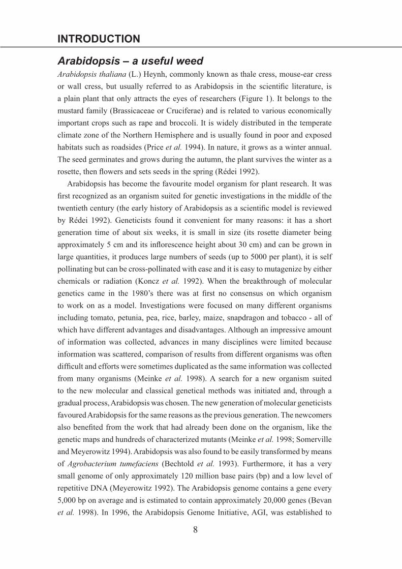
Arabidopsis – a useful weedArabidopsis thaliana (L.) Heynh, commonly known as thale cress, mouse-ear cress
or wall cress, but usually referred to as Arabidopsis in the scientic literature, is
a plain plant that only attracts the eyes of researchers (Figure 1). It belongs to the
mustard family (Brassicaceae or Cruciferae) and is related to various economically
important crops such as rape and broccoli. It is widely distributed in the temperate
climate zone of the Northern Hemisphere and is usually found in poor and exposed
habitats such as roadsides (Price et al. 1994). In nature, it grows as a winter annual.
The seed germinates and grows during the autumn, the plant survives the winter as a
rosette, then owers and sets seeds in the spring (Rédei 1992).
Arabidopsis has become the favourite model organism for plant research. It was
rst recognized as an organism suited for genetic investigations in the middle of the
twentieth century (the early history of Arabidopsis as a scientic model is reviewed
by Rédei 1992). Geneticists found it convenient for many reasons: it has a short
generation time of about six weeks, it is small in size (its rosette diameter being
approximately 5 cm and its inorescence height about 30 cm) and can be grown in
large quantities, it produces large numbers of seeds (up to 5000 per plant), it is self
pollinating but can be cross-pollinated with ease and it is easy to mutagenize by either
chemicals or radiation (Koncz et al. 1992). When the breakthrough of molecular
genetics came in the 1980’s there was at rst no consensus on which organism
to work on as a model. Investigations were focused on many different organisms
including tomato, petunia, pea, rice, barley, maize, snapdragon and tobacco - all of
which have different advantages and disadvantages. Although an impressive amount
of information was collected, advances in many disciplines were limited because
information was scattered, comparison of results from different organisms was often
difcult and efforts were sometimes duplicated as the same information was collected
from many organisms (Meinke et al. 1998). A search for a new organism suited
to the new molecular and classical genetical methods was initiated and, through a
gradual process, Arabidopsis was chosen. The new generation of molecular geneticists
favoured Arabidopsis for the same reasons as the previous generation. The newcomers
also beneted from the work that had already been done on the organism, like the
genetic maps and hundreds of characterized mutants (Meinke et al. 1998; Somerville
and Meyerowitz 1994). Arabidopsis was also found to be easily transformed by means
of Agrobacterium tumefaciens (Bechtold et al. 1993). Furthermore, it has a very
small genome of only approximately 120 million base pairs (bp) and a low level of
repetitive DNA (Meyerowitz 1992). The Arabidopsis genome contains a gene every
5,000 bp on average and is estimated to contain approximately 20,000 genes (Bevan
et al. 1998). In 1996, the Arabidopsis Genome Initiative, AGI, was established to
9
INTRODUCTION
facilitate the sequencing of the whole Arabidopsis genome (Bevan et al. 1997), a
task that will be completed before the end of the year 2000. Thousands of mutants
have been generated by means of insertion mutagenesis. These mutants are easily
screened by means of PCR so, within a few years, knockout mutants for virtually
every Arabidopsis gene will be available to the scientic community (Krysan et al.
1999).
The post-genome era of Arabidopsis research will surely be as successful as
the rst fty years and certainly more and more of the information collected from
Arabidopsis will nd uses in new improvements of more economically important
species.
Root DevelopmentPost-embryonic development of the rootArabidopsis has been shown to be an excellent system for studying post-embryonic
development of the root, as it has a fairly uncomplicated cellular organization (Dolan
et al. 1993) in which the developmental history of every cell has been determined
(Scheres et al. 1994), and its roots are nearly transparent. The Arabidopsis root
consists of a few different cell types organized in a radially symmetrical pattern
along the length axis. The cell types of each layer are continuously produced in les
from progenitor cells, the initials, similar to a conveyor belt, as the root elongates
(Figure 2). Only four cells of the Arabidopsis root meristem, the quiescent centre,
are fully undifferentiated and these cells rarely divide under normal conditions. The
cells around the quiescent centre are the initials for the different cell-types of the
Figure 1
Arabidopsis thaliana var. Ler .
Drawing by Dr. Eva Sundberg.
10
INTRODUCTION
root (the epidermis, cortex, endodermis, etc., see Figure 2). The initials divide to
form one cell that takes on the fate the mother cell and a second cell that is destined
to differentiate into a certain type (epidermis, cortex, endodermis, etc.). Mutations
affecting these processes in different cell types have been identied, for example
the shortroot mutation that blocks the formation of endodermis cells (Scheres et al.
1995). The fate of each initial cell is determined by its position in the root: positional
signals pass from differentiated cells to undifferentiated cells rather than from the
initials (van den Berg et al. 1995). Thus, cells differentiate like their more basal
daughters. The molecular nature of these positional cues is not known but mapping
the plasmodesmata in the root has shown that connections are preferentially made to
cells of the same type in the root (van den Berg et al. 1998 and references therein).
The contacts between the initials and the quiescent centre cells keep the initials in an
undifferentiated state since ablation of the cells in the quiescent centre causes initials
to stop dividing and to differentiate according to cues from the daughter cells in the
same le (van den Berg et al. 1997).
The pattern for post-embryonic development of the root is
laid down during embryogenesisThe cellular organization of the root is laid down during embryogenesis, and the
ontogeny of each cell has been determined through the use of genetically marked
embryonic cells, Figure 2 (Scheres et al. 1994). The rst division of the zygote
generates an apical and a basal cell and the root is formed from derivatives of both
of these cells. The basal cell generates the quiescent centre and the columella root
cap initials. The rest of the root cells, including the initials, are derivatives of the
apical cell (reviewed in Scheres and Heidstra 1999). The apical-basal pattern is laid
down early in embryogenesis as dened by the rst visible developmental deviations
detected in the monopteros, mp, mutant (Berleth and Jurgens 1993). The capacity
of mp mutant plants to make adventitious roots indicates that mp plants can make
largely normal apical structures. All organs, however, display defective vascular
strands and impaired auxin transport (Przemeck et al. 1996). This indicates that MP
promotes axialization and cell le formation, processes that are important for both
embryonic axis formation and vascular system development, rather than specifying
the apical-basal pattern. The determination of cell fates along the apical-basal axis
also involves the HOBBIT, HBT gene, as the hbt mutant is unable to form a root
meristem (Willemsen et al. 1998). From very early development the cells that will
give rise to the quiescent centre and the columella initials (Figure 2) divide aberrantly
in the hbt mutant, consistent with the hypothesis that the gene has a specic function

11
INTRODUCTION
-
AB
C
D
Estele
endod.cortex
epid.
l.r.c.
col.
cot.cot.
Figure 2
Embryonic and post-embryonic root development of Arabidopsis
Simplified schematic drawings of cellular arrangements (transverse sections) during root
development. Cortex and endodermis cells are depicted in dark grey and the stele (internal), columella
and lateral root cap (peripheral) is shown in light grey. The quiescent centre and epidermis of the root
are in white, as are all other cells of the non-root lineage. The figures are adopted from Scheres et al.
1994 and van den Berg et al. 1998, with kind permission from the authors.
A, Early heart stage embryo. B, Late heart stage embryo. C, Seedling. cot., cotyledon. D, Root apex
of seedling. E, Close up of central region in root meristem shown in D. Initial cells for all the different
cell types surrounding the quiescent centre. endod., endodermis; epid., epidermis; l.r.c., lateral root
cap; col., columella. Arrows indicate the direction of daughter cell displacement.

12
INTRODUCTION
in these cells (Willemsen et al. 1998). The radial axis of the root is laid down after
the apical-basal axis, since the rst deviations are detectable in mutants specically
affected in stages of radial pattern formation that appear later in embryogenesis
than those altered in hbt and mp (Scheres et al. 1995). It is reasonable to believe
that embryonic root formation shares extensive similarity to post-embryonic root
development as hbt and mutations affecting the radial pattern of the root also cause
the same deviations in secondary roots (Malamy and Benfey 1997a).
Initiation and formation of secondary rootsThe primary root of the adult Arabidopsis plant is only a minor part of its root system,
which is largely composed of secondary roots formed from the primary root or other
tissue, and further secondary roots formed from them, and so on. The secondary roots
perform and develop like primary roots, but are not initiated during embryogenesis.
Secondary roots are initiated from differentiated tissue and the frequency of initiation
is highly inuenced by environmental conditions (Charlton 1996). This plasticity in
the formation of the root system is one way in which plants overcome their inability
to move away from poor soils and towards nutrients. The lateral roots of Arabidopsis
are initiated from cells in the pericycle (an internal layer adjacent to the stele, Figure
2) of the root. A small number of pericycle cells start dividing and eventually form
the lateral root primordium. The lateral root primordium forms a structure identical
to the root meristem through a series of dened stages of development, and emerges
through the outer cell layers of the root (Laskowski et al. 1995; Malamy and Benfey
1997b). Most mutations identied in Arabidopsis that affect the development of the
primary root also affect the lateral roots. This indicates that the formation of the
lateral root meristem shares many regulatory mechanisms with those of primary root
initiation during embryogenesis (reviewed by Malamy and Benfey 1997a; Scheres
and Heidstra 1999).
The induction of lateral roots is dependent on the hormone auxin, which is
transported from aerial parts of the plant. This has been demonstrated by the
application of both auxin (Torrey 1950) and inhibitors of auxin transport (Reed et
al. 1998). Mutations with reduced sensitivity to auxin exhibit a reduction or loss of
lateral roots (Celenza et al. 1995; Estelle and Sommerville 1987) and the extensive
production of lateral roots in the mutant alf1 (Celenza et al. 1995), which is allelic
to superroot and rooty, has been linked to elevated auxin levels (Boerjan et al.
1995; King et al. 1995). In screens for mutants with altered frequencies of lateral
root initiation in Arabidopsis, only one mutation, alf4-1, has been shown to inhibit
this induction without affecting auxin perception or synthesis (Celenza et al. 1995),
demonstrating the importance of the hormone in the process. However, the initiation

13
INTRODUCTION
of secondary roots in other species, originating from other tissues, may be regulated
by other hormones, as illustrated by the ethylene dependence of adventitious root
formation (i.e. secondary root production from non-root tissue) from stem cuttings of
tomato and petunia (Clark et al. 1999).
Cotyledon and leaf developmentLife history of a leafAll the leaves of a plant originate from a small group of cells in the apical parts of
the shoot, the shoot apical meristem (SAM). This group of cells forms a dome that
undergoes extensive cell division in a highly regular manner (Lyndon 1970). As new
cells are formed the older ones are displaced to the periphery of the dome. These
cells will later form the primordia of leaves (Lyndon and Cunninghame 1986). The
shoot apical meristem continuously produces leaf primordia at its anks in a spiral
pattern. The earliest histological evidence of primordium initiation is a change in the
orientation of cell divisions in the region where the primordia will form (Lyndon
1970). At the molecular level this is correlated with a repression of knox genes in
maize and Arabidopsis. These genes are expressed in the central parts of the SAM and
have been shown to be important for maintaining the undifferentiated state of these
cells (Reiser et al. 2000).
During its initiation or early development the leaf primordium is divided into
discrete domains along the basic axes of the future leaf, in other words, a polarity
is established. The cells in the different positions along these axes/polarities then
develop according to different fates. The phantastica mutation in Antirrhinum majus
abolishes the formation of both the lateral and the dorsoventral axis as the leaves
of the mutant plants have complete radial symmetry (Waites and Hudson 1995).
Regulation of pattern along the proximodistal axis has been extensively studied in
maize where the distal and proximal parts of the leaf differ in many aspects of their
cellular differentiation (Sylvester et al. 1990). At least 15 different gene products
have been shown to specically affect structures along this axis in maize (reviewed
in Sylvester et al. 1996).
The control of leaf expansion and leaf shapeMuch of the diversity of leaf shapes seen in nature is caused by variation in the
amount of expansion within the leaf (Dale 1988). Variations in leaf expansion causing
different shapes of leaves may occur early, as shown in snapdragon, Antirrhinum
majus, where variation in leaf shape along the length of the shoot is due to variation
in the growth of the lamina early in development (Harte and Meinhard 1979a, b). In

14
INTRODUCTION
Arabidopsis, which has leaves with a shape similar to those of snapdragon, the genes
ANGUSTIFOLIA and ROTUNDIFOLIA act after leaf blade formation to control the
length and width of the leaf (Tsuge et al. 1996). The leaves and leaf cells of the
angustifolia (an) mutant are signicantly narrower than wt leaves (Rédei 1962) but
the leaf length is not affected by the mutation (Tsuge et al. 1996). Cell shapes in
plants are determined by the direction-specic inhibition of expansion exerted by
cellulose microbrils, while the driving force is the positive turgor pressure of the
cell (Taiz and Zeiger 1991). This means that inhibition of expansion in one direction
by cell wall microbrils leads to expansion in the other directions. AN affects cell
width, and secondarily leaf width, by this mechanism as an leaves and cells are
signicantly thicker than wt cells (Tsuge et al. 1996). AN also affects the orientation of
microtubules and, thus, most likely the orientation of cellulose microbrils (Tsukaya
et al. 1999). This mechanism also seems to occur in species other than Arabidopsis
since the fat mutant from tobacco, Nicotiana sylvestris, also has thick, narrow leaves
(McHale 1993).
The leaves and cells of the rotundifolia3 (rot3) mutant are shorter than those of the
wt, but the width and thickness of the cells are not altered (Tsuge et al. 1996). This
proves that plants can inhibit leaf cell expansion in one direction without affecting
expansion in the other directions. ROT3 encodes a cytochrome P450 with similarities
to animal steroid hydroxylases (Kim et al. 1998). How it affects the length of the
leaf is not currently known, but the mechanism is distinct from that of AN as the
arrangement of microtubili is not altered in the rot3 mutant (Tsukaya et al. 1999).
Over-expression of ROT3 in transgenic plants causes the development of longer
leaves compared to wt, indicating that the gene can both positively and negatively
regulate leaf length (Kim et al. 1999). Over-expression of the rot3-2 gene (with
encodes a ROT3 protein with an amino acid substitution) causes the rot3 phenotype
(short leaves) to appear in wt plants, indicating that the ROT3-2 protein dominantly
inhibits the wt protein (Kim et al. 1999).
The cotyledon Unlike the leaf, the cotyledon does not originate from a meristem. The cotyledons
are initiated during embryogenesis (Figure 2). The Arabidopsis cotyledons contain
stored reserves (lipids, proteins and starch) that are broken down and mobilized
during germination and early phases of seedling establishment (Manseld and Briarty
1996). Approximately 60 hours post-imbibition the Arabidopsis seedling switches to
auxotrophic growth and starts to photosynthesise. After this transition the cotyledons

15
INTRODUCTION
physiologically act as leaves (Manseld and Briarty 1996). When the rosette leaves
are fully developed, the small cotyledons are effectively shaded from the light and
senesce.
The Arabidopsis mutants extra cotyledon1 and -2 (xtc1 and –2) and altered
meristem programming 1, amp1, frequently have more than two cotyledons (Conway
and Poethig 1997). The position and timing of emergence of these extra cotyledons
indicate that the rst leaves of these mutants develop according to cotyledon fate.
However, these cotyledon-like leaves are initiated prematurely during embryogenesis
by aberrant timing of SAM initiation, and develop like wt leaves if the timing of
their emergence is restored to wt (Conway and Poethig 1997). These observations
indicate that during embryogenesis a factor that suppresses vegetative development
and/or promotes embryo specic development is expressed in a manner allowing it
to affect the prematurely developed leaf primordia. The gene LEAFY COTYLEDON1
(LEC1) is possibly one such factor, as it is expressed in the whole embryo during
early embryogenesis. When ectopically expressed during vegetative development
LEC1 causes the plant to develop according to embryo specic programs, including
the expression of storage proteins and the development of ectopic embryos on
vegetative tissue (Lotan et al. 1998). Mutant lec1 cotyledons develop into leaf-like
organs, which lack storage proteins and oil bodies present in wt cotyledons, while
developing trichomes and vascular characteristics of wt leaves. Mutant lec1 seeds
are intolerant to desiccation and, at a low frequency, germinate viviparously (Meinke
1992). However, the embryonic development of lec1 mutants is not fully transformed
into the vegetative program, possibly indicating that LEC1 acts partly redundantly
with other factors (Lotan et al. 1998).
The development of the cotyledon after the mobilization of storage reserves is
similar to that of leaves, although cells of Arabidopsis cotyledons do not divide after
the seed is mature (Tsukaya et al. 1994). The mechanisms that regulate the shape of
the cotyledon are only partly shared with those of leaves, as an cotyledons develop
an abnormally narrow shape (Tsukaya et al. 1994) while the shape of rot3 cotyledons
is indistinguishable from that of wt cotyledons (Tsuge et al. 1996).

16
INTRODUCTION
Sugar sensingSugars are important molecules in all organisms, both as carriers of stored chemical
energy and as raw materials for the synthesis of other molecules. Sugars are therefore
of great importance during all stages of the plant life cycle and it is not surprising that
changed sugar concentrations affect the expression of a large number of genes (Koch
1996). Continuous sensing of sugar levels occurs at the level of the individual cells
and three major sensing mechanisms/receptors have been suggested to be present
in plants, hexokinase (HXK) sensing, hexose transport (HXT) sensing and sucrose
(SUT) sensing (Smeekens and Rook 1997).
Hexokinase sensing has been shown to control diverse processes and metabolic
pathways in plants such as the sugar induced feedback regulation of photosynthesis
and the mobilization of storage reserves from seeds (Smeekens and Rook 1997). The
sensor is hexokinase (HXK), the rst enzyme of glycolysis, and phosphorylation of
hexoses by HXK induces the enzyme to initiate a signalling cascade (Jang et al.
1997). Two HXK genes have been cloned from Arabidopsis and transgenic plants
with enhanced or reduced expression of these genes have conrmed the importance
of the proteins in sugar signalling (Jang et al. 1997). HXK can phosphorylate
the non-metabolizable sugar analogues 2-deoxy glucose (2dGlc) and mannose, and
consequently start signalling in response to the presence of these molecules. HXK
cannot phosphorylate two other analogues, 3-O-Methyl Glucose (3-O-mGlc) and
6-deoxy glucose (6dGlc), and these sugars are therefore unable to trigger HXK
signalling (Smeekens and Rook 1997). By the use of specic HXK inhibitors Pego
et al. (1999) have shown that the signalling properties of HXK, and not the depletion
of ATP or phosphates, is responsible for the inhibition of germination in response to
these analogues in Arabidopsis.
Sugar analogues that are taken up by the cells but are not phosphorylated by HXK
such as 3-O-mGlc and 6dGlc can affect gene expression, but clearly by a second
mechanism. Genes encoding extracellular invertase and sucrose synthase are induced
by sucrose and glucose in suspension cultures of Chenopodium rubrum cells and this
induction can be mimicked by 6dGlc (Roitsch et al. 1995). The sugar and amino
acid-induced promoter PAT(33B) can also be induced by 6dGluc and 3-O-mGlc in
transgenic Arabidopsis (Martin et al. 1997). The effect of these analogues has been
attributed to Hexose transporter (HXT) signalling in plants (Figure 3), in analogy
with the function of HXT in yeast (Özcan et al. 1996). HXT proteins in yeast, like
HXK in yeast and plants, have dual functions, both transporting hexoses over the
membrane and signalling. However, none of the cloned members of the HXT gene
family from Arabidopsis has yet been proven to be involved in signalling (reviewed
in Buttner and Sauer 2000).

17
INTRODUCTION
Sucrose
HXT
SUT
HXK
Sucrose
Hexose
Hexose
cell membrane
GlycolysisVacuole
Hexose
Nucleus
Figure 3
Sugar signalling receptors in plants
Schematic drawing of simplified plant cell showing the three suggested sugar receptors
(grey) as well as their suggested positions in the cell. The interconversion of sucrose to
hexose is indicated.
Sucrose is an important sugar transport and storage molecule in plants and most
likely also has a signalling function. Several observations indicate that sucrose can
be sensed in plants. The patatin and rolC genes have been shown to be specically
induced by sucrose (Jefferson et al. 1990; Wenzler et al. 1989; Yokoyama et al.
1994), and the expression of a bZip transcription factor gene, ATB2, is specically
repressed by sucrose (Rook et al. 1998). Since sucrose is readily hydrolysed to
glucose and fructose, both inside the cell membrane and in the apoplast, it is difcult
to determine if such effects are directly mediated by the sucrose molecule. However,
in the above cases, combinations of glucose and fructose are less efcient than sucrose
in mediating the response, indicating that sucrose is the specic mediator. A sucrose
transporter protein, SUT (Figure 3) is suggested to be involved in signalling but as
yet there is no experimental support for this hypothesis. A recently cloned gene from
Arabidopsis encodes a protein with sequence similarity to both sucrose transporters
and signal transducing cytoplasmic domains of yeast sugar sensors (Barker et al.
2000). This protein might act as a sucrose sensor in plants as it lacks transport activity,
like yeast sugar sensors (Barker et al. 2000).

18
INTRODUCTION
How the signal is transduced from the receptors to the responses in plants is
presently unknown. However, in yeast many genes and enzymes are repressed by
glucose. This repression has been shown to be controlled by a complex signalling
cascade, in which the protein kinase SNF1 plays a central role (Ronne 1995). SNF1-
like protein kinases have been isolated from many plant species including rye, barley,
tobacco, soybean, potato, Arabidopsis and others (reviewed in Halford and Hardie
1998). Expression of the tobacco SNF1 homologue in yeast causes constitutive
expression of a glucose repressible gene (Muranaka et al. 1994) indicating that there
is functional homology between the yeast and plant proteins and, possibly, in their
signalling cascades. An independent indication of the involvement of SNF1-like
proteins in sugar signal transduction is provided by the PRL1 protein. PRL1, when
mutated, confers sugar hyper-sensitivity to the plant (Nemeth et al. 1998) possibly
by interacting with SNF1-like kinases, as recently shown in vitro (Bhalerao et al.
1999).
Sugar signals are also integrated with signals from other signal transduction
pathways in the plants. Mutations identied on the basis of altered sugar responses
have also been shown to affect hormone-mediated responses. This is a strong
indication of a close connection between the signalling pathways. Crosstalk between
sugar and ethylene (Zhou et al. 1998), abscisic acid (Arenas-Huertero et al. 2000;
Huijser et al. 2000; Laby et al. 2000), and cytokinin, ethylene, abscisic acid and auxin
(Nemeth et al. 1998) signalling pathways has been demonstrated by this method. It
has also been shown that a gene involved in brassinolide biosynthesis is repressed
by sugars (Szekeres et al. 1996). Sugar signals form one among many types of
input monitored by the highly integrated regulatory network controlling the onset of
owering in Arabidopsis. Sucrose availability to the aerial parts of the plants promotes
owering in the dark (Roldan et al. 1999). In the light, high concentrations of sugars
inhibit owering, possibly through repression of the regulatory gene, LEAFY (Otho
et al. 1998). Many more examples of crosstalk between sucrose signalling and other
signalling pathways are likely to be revealed in the future.

19
INTRODUCTION
HDZip transcription factorsThe pre-history of the HDZip domainHomeosis is dened as an anomaly where one part of the body develops into the
likeness of another part (Bateson 1894). Mutations causing this kind of anomaly have
been observed in many organisms including plants and animals and are referred to
as homeotic mutations. When the rst genes corresponding to this type of mutants
in Drosophila were cloned they were found to share a 180 bp sequence motif that
was called the homeobox and the 60 amino acid domain it encoded was named
the homeodomain (McGinnis et al. 1984; Scott and Weiner 1984). Homeodomain
proteins were rst suggested to act as transcription factors, as the amino acid sequence
of the homeodomain has slight similarity to bacterial, viral and yeast transcriptional
regulators of the helix-turn-helix type (Laughon and Scott 1984; Shepherd et al.
1984). This was later proven to be correct experimentally, by demonstrations that the
factors bind DNA, and affect the transcription levels of nearby genes upon binding,
both in vitro (Johnson and Krasnow 1990) and in vivo (Driever and Nüsslein-Volhard
1989). The homeodomain was also found to have a three-dimensional structure similar
to that of helix-turn-helix proteins, when the structure of the ANTENNAPEDIA
homeodomain was resolved (Qian et al. 1989).
Homeobox genes were also found in other segmented animals and were shown to
control processes such as determination of segment identity during embryogenesis.
It was therefore surprising when homeobox genes were found in the non-segmented
animal Ceanorhabditis elegans by screening genomic libraries with a degenerate
nucleotide pool designed to match all possible codons of the most highly conserved
part of previously known homeobox genes (Bürglin et al. 1989). Inspired by Bürglin
et al. three different groups independently took the same approach to search for
homeobox genes in Arabidopsis (Mattsson et al. 1992; Ruberti et al. 1991; Schena
and Davis 1992). All of the groups found homeobox genes, but of a different
type to those present in animals. They found genes encoding proteins having two
separate domains (the homeodomain and the leucine-zipper domain), known from
different types of transcription factors, fused in single proteins. This arrangement
was novel and since then has only been found in plant genes, although the complete
genomes of other species, including Saccharomyces cerevisiae (Goffeau et al. 1996),
Drosophila melanogaster (Adams et al. 2000), Ceanorhabditis elegans (The C.
elegans sequencing consortium 1998), and Homo sapiens have been sequenced
(Clinton 2000). Thus, this arrangement, the homeodomain leucine zipper, HDZip,
domain is most likely plant specic.

20
INTRODUCTION
HDZip transcription factors are encoded by a large gene family
in ArabidopsisAfter isolation of the rst genes encoding Homeodomain leucine zipper (HDZip)
proteins many more were isolated from Arabidopsis on the basis of sequence similarity
(I; III; V; Carabelli et al. 1993; Lee and Chun 1998; Schena and Davis 1994; Söderman
et al. 1994). The HDZip genes of Arabidopsis are grouped into four families HDZip
I – IV (Sessa et al. 1994) based on sequence similarity as well as other sequence
criteria (summarized in Figure 4). The genes of HDZip I and II appear to share a
common origin while members of HDZip III and IV are more distantly related (Chan
et al. 1998) and differ in their arrangement in the homeodomain compared to HDZip
I and II and to the ANTENNAPEDIA class of animal homeodomains (Figure 4).
The HDZip I and II genes are not clustered on the chromosomes like some
animal homeobox genes (Graham et al. 1989). Instead, they are dispersed among
all Arabidopsis chromosomes (Figure 5). There is no clear correlation between
sequence similarity and chromosomal location, which indicates that most of the gene
duplications from which these genes presumably originate are ancient. This is also
HELIX1 HELIX2 HELIX3 LEUCINE ZIPPER
a b c d e f
Class Length of Introns in Extra amino acids mRNA homeodomain* in HDZip domain
HDZip I approx. 1500 bases a or none None
HDZip II approx. 1500 bases c and e None
HDZip III approx. 3300 bases d Four (between helix 2 and 3) and four (between helix 3 and leucine zipper)HDZip IV approx. 3000 bases b and f Seven (between helix 3 and leucine zipper)
* According to the schematic drawing in A
A
B
Figure 4
Summary of differences between the four different classes of HDZip genes in Arabidopsis
A, Schematic drawing of the primary sequence of the HDZip domain. The putative helical
structures are indicated by boxes. Triangles indicate intron positions.
B, Distinguishing characters of the four different classes of HDZip genes in Arabidopsis.
Adopted from Sessa et al. (1994).

21
INTRODUCTION
HAT3ATHB12
ATHB20
mi357
mi456
ATHB1
150
125
100
75
50
25
0cM
ATHB13
ATHB23
mi106
mi443
ATHB54
I III
HAT1ATHB2
ATHB16
ATHB20
mi87
mi123
HAT22
IV
ATHB53ATHB5
ATHB52
HAT2
ATHB33
HAT14
ATHB51mi121
mi184
V
ATHB21ATHB6HAT9
ATHB22
ATHB4ATHB7
mi421
mi79a
IIATHB17
Figure 5
Chromosomal locations of HDZip I and II genes
Chromosomal locations of 26 HDZip I and II genes (V; Sessa et al. 1994). The relative
positions of ten RFLP markers (mi79a to mi443) are indicated (Liu et al. 1996).
true for HDZip IV genes (Tavares et al. 2000). An ancient origin of the gene family
is also supported by the fact that genes encoding HDZip proteins are found in a wide
range of land plant species including: tomato (Meissner and Theres 1995), sunower
(Chan and Gonzalez 1994), soybean (Moon et al. 1996a), resurrection plant (Frank et
al. 1998), poplar (Sterky et al. 1998), carrot (Kawahara et al. 1995; Mattsson 1995),
Phalaenopsis (Nadeau et al. 1996), Pimpinella brachycarpa (Moon et al. 1996b), rice
(Meijer et al. 1997), maize (Ingram et al. 1999) and ferns (Aso et al. 1999).
HDZip proteins act as dimeric transcription factorsMuch of our knowledge of HDZip proteins is deduced from the vast mass of
information concerning the biochemical properties of animal homeodomains and
leucine-zipper proteins. Homeodomains form a globular structure consisting of three
alpha helices. When the factors bind to DNA the third helix is positioned in the
major groove. Basic leucine-zipper, bZip, proteins also bind DNA by an alpha-helical
structure positioned in the major groove, which is enriched with basic residues. This
basic region of bZip proteins extends into a long alpha helix in which every seventh

22
INTRODUCTION
amino acid is a leucine residue. These residues form a hydrophobic face of the helix
that mediates interaction with similar proteins. bZip proteins bind DNA as dimers.
The leucine zipper parts of the protein orientate the basic regions in the major groove
of the DNA. HDZip proteins bind DNA as homeodomain proteins and dimerise as
bZip proteins in accordance with the fact that they consist of a fusion of these two
domains. A simplied model of how HDZip proteins may be oriented when bound to
DNA is shown in Figure 6.
Dimers of two identical HDZip proteins have been shown to bind to a pseudo-
palindromic DNA sequence, CAATNATTG, consisting of two overlapping identical
half-sites (Johannesson et al. 2000; Meijer et al. 2000; Sessa et al. 1993). The homeo-
domains of HDZip proteins bind to DNA in a similar fashion to their monomeric
counterparts from animals as indicated by mutational analysis of ATHB2. Exchanging
residues important for DNA binding of homeodomain proteins in ATHB2 abolishes
its DNA binding capacity (Sessa et al. 1997). The bases in the central position
of the optimal DNA binding site differ between different HDZip proteins, ATHB2
favours a G/C pair while ATHB1 prefers an A/T pair (Sessa et al. 1993; Sessa et
al. 1997). This specicity has been attributed to the amino acids Arg55, Glu46 and
Thr56, and unspecied residues outside helix 3 (Sessa et al. 1997). In contrast to
ATHB1 and ATHB2, the HDZip I proteins ATHB5, ATHB6 and ATHB16 do not show
middle position specicity, and can interact with both sites (Johannesson et al. 2000).
ATHB12 has been demonstrated to interact with a different site (TCAATTAATTGA)
composed of the same two half sites but with a different spacing (Chun and Lee
1999). As ATHB5, -6, -12 and –16 are identical to ATHB1 in the positions in helix 3
that are postulated to be important for specicity (positions 46, 55 and 56) (Sessa et al.
1997), the alternative DNA binding preferences of these proteins must be determined
by residues somewhere else in the proteins. This hypothesis is also supported by the
nding that some HDZip I and II proteins from rice can interact with both types of
site in yeast (Meijer et al. 2000).
The dimerisation specicity of bZip proteins has been extensively studied (Vinson
et al. 1993) and the three-dimensional structures of bZip homo- and hetero- dimers
bound to DNA (Ellenberger et al. 1992; Glover and Harrison 1995) have been
determined. The formation of salt bridges between basic and acidic residues in
different proteins is apparently important (Vinson et al. 1993; Glover and Harrison
1995). Assuming that HDZip proteins form dimers like the bZip proteins, HDZip I
proteins are very similar in the amino acids facing the putative dimerisation surface,
but distinct from HDZip II proteins. In agreement with this notion, all proteins so
far tested can form homodimers in solution (Johannesson et al. 2000; Meijer et al.

23
INTRODUCTION
Figure 6
HDZip domain binding to DNA
Theoretical model of HDZip domain binding to DNA based on the three-dimensional structures
of the Drosophila ENGRAILED homeodomain (Kissinger et al. 1990) and the yeast GCN4
leucine zipper motif (Ellenberger et al. 1992). DNA is shown in white and the HDZip domain
(secondary structure) in grey. (Johansson, K., Johannesson, H. and Söderman, E. unpublished).
2000; Sessa et al. 1993). Some HDZip proteins from rice and resurrection plants are
able to form heterodimers with members of the same class (Frank et al. 1998; Meijer
et al. 2000). For example, ATHB5 forms heterodimers with ATHB6 and ATHB16
but not with ATHB1 in vitro (Johannesson et al. 2000). This indicates that there are
limitations to the promiscuity of dimerisation among the proteins of the two classes.
Members of the homeodomain and bZip transcription factor families have been
shown to either activate or repress (or both) transcription upon DNA binding
(Latchman 1998), and this also applies to different HDZip proteins. ATHB1 activates
transcription from a promoter with HDZip binding sites, CAAT(A/T)ATTG, (Aoyama
et al. 1995) while experiments with ATHB2 indicate that ATHB2 can act as a negative
regulator of transcription (Steindler et al. 1999). A fusion construct between the strong

24
INTRODUCTION
transcriptional activation domain of VP16 from the herpes simplex virus (Dalrymple
et al. 1985) and ATHB2 affects transgenic plants in the same way as a reduction in
the activity of the wt protein (Steindler et al. 1999). This is a strong indication that
ATHB2 acts a transcriptional repressor in wt Arabidopsis. In rice suspension cells
the HDZip proteins Oshox1 and –3 (HDZip II) repress transcription, while Oshox4
and –5 (HDZip I) activate it (Meijer et al. 2000). Possibly, HDZip I proteins may
generally act as activators and HDZip II proteins as repressors. However, the level of
sequence similarity between the different HDZip proteins outside the HDZip domain
is low, indicating that different HDZip I and II proteins use different mechanisms to
affect transcription.
HDZip proteins are involved in a wide range of processes in
plantsThe functions of only three HDZip genes; GL2, IFL1 and ANL2, have been identi-
ed by analysis of mutant phenotypes. The glabra2, gl2, mutation causes disturbed
trichome and root hair development and the mutant also lacks seed coat mucilage
(Di-Cristina et al. 1996; Rerie et al. 1994). The gene INTERFASCICULAR FIBER-
LESS1, IFL1, regulates interfascicular ber differentiation (Zhong and Ye 1999) and
the gene ANTHOCYANINLESS2, ANL2, affects anthocyanin and root development
in Arabidopsis (Kubo et al. 1999). ATHB8 is expressed in procambial cells of the
embryo and adult plants and is suggested to be a regulator of vascular development in
Arabidopsis (Baima et al. 1995). These four genes belong to HDZip classes III (IFL1
and ATHB8) and IV (GL2 and ANL2).
Altered expression levels of the HDZip II gene ATHB2 (also named HAT4) in
Arabidopsis result in phenotypes that suggest it has a role in light signalling (Schena
et al. 1993; Steindler et al. 1997). Elevated levels of ATHB2, by means of transgene
expression, affect cell expansion in cotyledons and hypocotyls, inhibit secondary
growth of the vascular system in roots and inhibit lateral root formation (Steindler et
al. 1999). Some of the phenotypic effects can be reversed by application of the plant
hormone auxin. Reduced levels of ATHB2 expression by antisense suppression cause
reciprocal effects, indicating that the phenotypic effects caused by elevated expression
reect the wt function of the gene (Steindler et al. 1999). ATHB2 expression is
regulated by far-red-rich light (Carabelli et al. 1993) and the effects caused by altered
expression levels of the gene indicate that ATHB2 has a role in shade avoidance
responses (Steindler et al. 1999). ATHB4 expression is regulated in the same manner
as ATHB2, by far-red-rich light (Carabelli et al. 1993), indicating that ATHB4 has a
role similar to that of ATHB2. Two HDZip II genes from resurrection plant, CPHB1

25
INTRODUCTION
and –2, are regulated by drought and may be involved in gene regulation in response
to drought (Frank et al. 1998). Putative homologues of CPHB1 and –2 are present in
the Arabidopsis genome and may be involved in similar responses.
Data on the function of HDZip I genes are sparse. Indirect evidence from
expression studies indicates that the genes ATHB6, ATHB7 and ATHB12 regulate
gene expression in response to both the plant hormone abscisic acid, ABA, and to
environmental conditions known to increase endogenous levels of the hormone, such
as water deciency (Lee and Chun 1998; Söderman et al. 1999; Söderman et al.
1996). The expression of ATHB7 and ATHB12 is specically and strongly induced
by applications of ABA and by treatments known to cause increased endogenous
levels of the hormone. The induced expression of ATHB7 is impaired in the ABA-
insensitive mutant abi1, suggesting that ATHB7 regulates responses in the ABA
signal transduction pathway downstream of the ABI1 gene (Söderman et al. 1996).
ATHB6 is expressed in developing organs and up-regulated by ABA treatments, and is
suggested to have a function related to cell division and/or differentiation (Söderman
et al. 1999). The expression of the tomato HDZip I gene H52, is up-regulated during
pathogen infection. Suppressed expression in transgenic plants results in a miss-
regulation of cell death control and regulation of defence genes, which indicates
that H52 is involved in limiting the spread of programmed cell death after infection
(Mayda et al. 1999). Another tomato HDZip I gene, VaHox1, is expressed in the
phloem during secondary growth and may participate in the regulation of processes
specic to secondary phases of phloem development (Tornero et al. 1996). The
functions of four HDZip I genes, ATHB3, -13, -20 and –23, are discussed in this
thesis.
26
RESULTS AND DISCUSSION
Isolation and characterization of novel HDZip genesNovel genes distantly related to previously known HDZip I
genesIn extensive searches for Arabidopsis HDZip I and II genes in available databases we
found, in total, 26 genes. Seven of these were novel and named ATHB21, -22, -40,
-51, -52, -53 and 54 (V). We think these 26 genes represent all the HDZip I and II
genes in the Arabidopsis genome, as more than 98 % of the estimated Arabidopsis
genome has now been sequenced (September 2000). All novel genes encoded a
HDZip domain similar to those previously isolated. The deduced sequence of all
HDZip domains could be aligned without creating gaps in the alignment, with the
exception of ATHB22 (which aligned after creating a gap in the alignment between
helixes 1 and 2 in the other sequences, see Figure 7). These amino acids may
form a loop in the turn between the two helices, as has been shown for the yeast
homeodomain MATα2, which has an insertion of three residues at a similar position
(Hall and Johnson 1987). The three-dimensional structure of MATα2 (Wolberger et
al. 1991) is very similar to the homeodomains of Drosophila proteins ENGRAILED
and ANTENNAPEDIA, indicating that the extra residues in ATHB22 do not affect
the structure or DNA-binding properties of the domain.
A homeodomain consensus sequence has been dened on the basis of a compilation
of 346 homeodomain sequences from a range of different eukaryotes (Bürglin 1994).
The homeodomains of all HDZip proteins, including the newly identied forms,
are highly similar to this homeodomain consensus sequence (Figure 7). The 26
HDZip domains are also very similar at positions 7, 8, 54, and 61. In addition to the
conserved homeodomain, all HDZip proteins, including the newly identied HDZip
proteins, contain leucine zipper motifs in identical positions, towards the C-terminal
from the homeodomain (Figure 7). The sequence similarity between the proteins was
signicantly lower in the leucine zipper region compared to the homeodomain.
To investigate if the classication suggested by Sessa et al. (1994) was also
applicable to the novel genes, the amino acid sequences of the entire HDZip domain
were phylogenetically analysed. The analysis resulted in four equally parsimonious
trees with similar branching patterns, one of which is depicted in Figure 8. The four
trees all resolve HDZip I and II, indicating that the classication suggested by Sessa
et al. (1994) reects the evolutionary history of the gene family. Related genes in
the tree have the same or similar intron patterns, further supporting the evolutionary
signicance of the depicted tree.

27
RESULTS AND DISCUSSION
AT
HB
3A
TH
B20
AT
HB
13A
TH
B23
AT
HB
5A
TH
B6
AT
HB
16
AT
HB
7A
TH
B12
AT
HB
40A
TH
B21
AT
HB
53A
TH
B51
AT
HB
22A
TH
B54
AT
HB
52
LP
EK
KR
RL
TT
EQ
VH
LL
EK
SF
ET
EN
KL
E-
--
--
--
-P
ER
KT
QL
AK
KL
GL
QP
RQ
VA
VW
FQ
NR
RA
RW
KT
KQ
LE
RD
YD
LL
KS
TY
DQ
LL
SN
YD
SI
VM
DN
DK
L
LG
EK
KK
RL
NL
EQ
VR
AL
EK
SF
EL
GN
KL
E-
--
--
--
-P
ER
KM
QL
AK
AL
GL
QP
RQ
IA
IW
FQ
NR
RA
RW
KT
KQ
LE
RD
YD
SL
KK
QF
DV
LK
SD
ND
SL
LA
HN
KK
L
LG
EK
KK
RL
QL
EQ
VK
AL
EK
SF
EL
GN
KL
E-
--
--
--
-P
ER
KI
QL
AK
AL
GM
QP
RQ
IA
IW
FQ
NR
RA
RW
KT
RQ
LE
RD
YD
SL
KK
QF
ES
LK
SD
NA
SL
LA
YN
KK
L
MG
EK
KR
RL
NM
EQ
VK
TL
EK
NF
EL
GN
KL
E-
--
--
--
-P
ER
KM
QL
AR
AL
GL
QP
RQ
IA
IW
FQ
NR
RA
RW
KT
KQ
LE
KD
YD
TL
KR
QF
DT
LK
AE
ND
LL
QT
HN
QK
L
MG
EK
KR
RL
NM
EQ
LK
AL
EK
DF
EL
GN
KL
E-
--
--
--
-S
DR
KL
EL
AR
AL
GL
QP
RQ
IA
IW
FQ
NR
RA
RS
KT
KQ
LE
KD
YD
ML
KR
QF
ES
LR
DE
NE
VL
QT
QN
QK
L
AA
EK
KR
RL
GV
EQ
VK
AL
EK
NF
EI
DN
KL
E-
--
--
--
-P
ER
KV
KL
AQ
EL
GL
QP
RQ
VA
IW
FQ
NR
RA
RW
KT
KQ
LE
RD
YG
VL
KS
NF
DA
LK
RN
RD
SL
QR
DN
DS
L
LS
EK
KR
RL
SI
NQ
VK
AL
EK
NF
EL
EN
KL
E-
--
--
--
-P
ER
KV
KL
AQ
EL
GL
QP
RQ
VA
VW
FQ
NR
RA
RW
KT
KQ
LE
KD
YG
VL
KT
QY
DS
LR
HN
FD
SL
RR
DN
ES
L
LS
EK
KR
RL
KV
DQ
VK
AL
EK
NF
EL
EN
KL
E-
--
--
--
-P
ER
KT
KL
AQ
EL
GL
QP
RQ
VA
VW
FQ
NR
RA
RW
KT
KQ
LE
KD
YG
VL
KG
QY
DS
LR
HN
FD
SL
RR
DN
DS
L
NK
NN
QR
RF
SD
EQ
IK
SL
EM
MF
ES
ET
RL
E-
--
--
--
-P
RK
KV
QL
AR
EL
GL
QP
RQ
VA
IW
FQ
NK
RA
RW
KS
KQ
LE
TE
YN
IL
RQ
NY
DN
LA
SQ
FE
SL
KK
EK
QA
L
KS
NN
QK
RF
SE
EQ
IK
SL
EL
IF
ES
ET
RL
E-
--
--
--
-P
RK
KV
QV
AR
EL
GL
QP
RQ
VA
IW
FQ
NK
RA
RW
KT
KQ
LE
KE
YN
TL
RA
NY
NN
LA
SQ
FE
IM
KK
EK
QS
L
GL
FR
KR
KL
TD
EQ
VN
ML
EM
SF
GD
EH
KL
E-
--
--
--
-S
ER
KD
RL
AA
EL
GL
DP
RQ
VA
VW
FQ
NR
RA
RW
KN
KR
LE
EE
YN
KL
KN
SH
DN
VV
VD
KC
RL
ES
EV
IQ
L
GW
FR
KR
KL
SD
EQ
VR
ML
EI
SF
ED
DH
KL
E-
--
--
--
-S
ER
KD
RL
AS
EL
GL
DP
RQ
VA
VW
FQ
NR
RA
RW
KN
KR
VE
DE
YT
KL
KN
AY
ET
TV
VE
KC
RL
DS
EV
IH
L
GM
LR
KR
KL
TD
EQ
VN
ML
EY
SF
GN
EH
KL
E-
--
--
--
-S
GR
KE
KI
AG
EL
GL
DP
RQ
VA
VW
FQ
NR
RA
RW
KN
KK
LE
EE
YA
KL
KN
HH
DN
VV
LG
QC
QL
ES
QI
LK
L
EM
IK
KK
RL
TS
GQ
LA
SL
ER
SF
QE
EI
KL
D-
--
--
--
-S
DR
KV
KL
SR
EL
GL
QP
RQ
IA
VW
FQ
NR
RA
RW
KA
KQ
LE
QL
YD
SL
RQ
EY
DV
VS
RE
KQ
ML
HD
EV
KK
L
QE
KK
KK
KM
TS
EQ
LK
FL
ER
SF
QE
EI
KL
NP
DR
KM
KL
NP
DR
KM
KL
SK
EL
GL
QP
RQ
IA
VW
FQ
NR
KA
RW
KN
KQ
LE
HL
YE
SL
RQ
EF
DI
VS
RE
KE
LL
QE
EL
IQ
L
EI
TK
KR
KL
TP
IQ
LR
LL
EE
SF
EE
EK
RL
E-
--
--
--
-P
DR
KL
WL
AE
KL
GL
QP
SQ
VA
VW
FQ
NR
RA
RY
KT
KQ
LE
HD
CD
SL
KA
SY
AK
LK
TD
WD
IL
FV
QN
QT
L
GK
NK
KK
RL
TQ
DQ
VR
QL
EK
CF
TM
NK
KL
E-
--
--
--
-P
DL
KL
QL
SN
QL
GL
PQ
RQ
VA
VW
FQ
NK
RA
RF
KT
QS
LE
VQ
HC
TL
QS
KH
EA
AL
SD
KA
KL
EH
QV
QF
L
HA
T2
HA
T1
HA
T14
AT
HB
4H
AT
3A
TH
B2
HA
T22
HA
T9
TS
RK
KL
RL
SK
DQ
SA
FL
EE
TF
KE
HN
TL
N-
--
--
--
-P
KQ
KL
AL
AK
KL
NL
TA
RQ
VE
VW
FQ
NR
RA
RT
KL
KQ
TE
VD
CE
YL
KR
CV
EK
LT
EE
NR
RL
QK
EA
ME
L
TC
RK
KL
RL
SK
DQ
SA
VL
ED
TF
KE
HN
TL
N-
--
--
--
-P
KQ
KL
AL
AK
KL
GL
TA
RQ
VE
VW
FQ
NR
RA
RT
KL
KQ
TE
VD
CE
YL
KR
CV
EK
LT
EE
NR
RL
EK
EA
AE
L
ST
RK
KL
RL
SK
DQ
SA
FL
ED
SF
KE
HS
TL
N-
--
--
--
-P
KQ
KI
AL
AK
QL
NL
RP
RQ
VE
VW
FQ
NR
RA
RT
KL
KQ
TE
VD
CE
YL
KR
CC
ES
LT
EE
NR
RL
QK
EV
KE
L
GS
RK
KL
RL
SK
DQ
AL
VL
EE
TF
KE
HS
TL
N-
--
--
--
-P
KQ
KL
AL
AK
QL
NL
RA
RQ
VE
VW
FQ
NR
RA
RT
KL
KQ
TE
VD
CE
YL
KR
CC
DN
LT
EE
NR
RL
QK
EV
SE
L
SS
RK
KL
RL
SK
EQ
AL
VL
EE
TF
KE
HS
TL
N-
--
--
--
-P
KQ
KM
AL
AK
QL
NL
RT
RQ
VE
VW
FQ
NR
RQ
RT
KL
KQ
TE
VD
CE
YL
KR
CC
EN
LT
DE
NR
RL
QK
EV
SE
L
NS
RK
KL
RL
SK
DQ
SA
IL
EE
TF
KD
HS
TL
N-
--
--
--
-P
KQ
KQ
AL
AK
QL
GL
RA
RQ
VE
VW
FQ
NR
RA
RT
KL
KQ
TE
VD
CE
FL
RR
CC
EN
LT
EE
NR
RL
QK
EV
TE
L
SA
RK
KL
RL
TK
SA
LL
ED
NF
KL
HS
TL
N-
--
--
--
-P
KQ
KQ
AL
AR
QL
NL
RP
RQ
VE
VW
FQ
NR
RA
RT
KL
KQ
TE
VD
CE
FL
KK
CC
ET
LT
DE
NR
RL
QK
EL
QD
L
SA
RK
KL
RL
TK
SA
LL
EE
SF
KD
HS
TL
N-
--
--
--
-P
KQ
KQ
VL
AR
QL
NL
RP
RQ
VE
VW
FQ
NR
RA
RT
KL
KQ
TE
VD
CE
FL
KK
CC
ET
LA
DE
NI
RL
QK
EI
QE
L
KY
QL
LE
FY
LR
LA
LL
QV
KI
WF
QN
RR
K
AT
HB
17P
PR
KK
LR
LT
RE
QS
RL
LE
DS
FR
QN
HT
LN
--
--
--
--
PK
QK
EV
LA
KH
LM
LR
PR
QI
EV
WF
QN
RR
AR
SK
LK
QT
EM
EC
EY
LK
RW
FG
SL
TE
EN
HR
LH
RE
VE
EL
AT
HB
1
KH
D-c
ons.
Hel
ix1
Hel
ix2
Hel
ix3
1020
3040
5060
7080
Fig
ure
7
The
pri
mar
y se
quen
ce o
f the
HD
Zip
dom
ains
enc
oded
by
26 H
DZ
ip g
enes
from
Ara
bido
psis
.
Cha
rged
res
idue
s (K
, R, E
, D)
are
depi
cted
as
dark
gre
y bo
xes
and
hydr
opho
bic
(F, P
, M, V
, L, I
)
resi
dues
as
light
gre
y bo
xes.
Gap
s ar
e in
dica
ted
by d
ashe
s. A
hom
eodo
mai
n co
nsen
sus
sequ
ence
bas
ed
on a
com
pila
tion
of 3
46 h
omeo
dom
ain
sequ
ence
s is
sho
wn
belo
w th
e al
ignm
ent,
as a
re th
ree
α-he
lices
of th
e ho
meo
dom
ain
(B
ürgl
in 1
994)
.

28
RESULTS AND DISCUSSION
Some groups of functionally related genes form well-separated clades in the tree
(V) e.g. ATHB3, -13, -20 and –23, which cause similar phenotypes when constitutively
expressed (III), and ATHB7/12, both of which are induced by ABA (Lee and Chun
1998; Söderman et al. 1996). The seven novel genes, however, are not closely
assosiated with any previously characterized gene. Hence their function cannot be
predicted on the basis of their phylogenetic relationship with other HDZip proteins.
Pointed cotyledon-HDZip genesThree genes ATHB13, ATHB20 and ATHB23, were isolated on the basis of their
similarity in sequence to the ATHB3 gene (Mattsson et al. 1992). ATHB13 and
ATHB20 were isolated by use of the ATHB3 cDNA as a probe in low stringency
screens of cDNA and genomic libraries (I, III). ATHB23 was rst identied as
an Expressed Sequence Tag or EST (Newman et al. 1994), that showed extensive
sequence similarity to ATHB13 (III). All four genes have now been sequenced by
the Arabidopsis Genome Initiative, AGI, (acc. nr. AL353993, AC013289, AC008261,
AC005508) and the sequences used in our studies are identical to those presented by
the AGI.
A comparison of the proteins encoded by the four genes, ATHB3, ATHB13,
ATHB20 and ATHB23, shows that these genes encode very similar proteins (III),
with a HDZip domain (Figure 7). The four genes belong to HDZip I class of HDZip
proteins (Sessa et al. 1994) as all of them have HDZip I specic amino residues at the
positions that discriminate between HDZip I and II. These four proteins are identical
to each other, but different from all other HDZip I and II proteins in the HDZip
domain at positions 2, 23 and 37, and thus form a subclass of HDZip I proteins. This
is in agreement with phylogenetic analyses of the HDZip domain sequences in which
this subclass forms a separate clade within the HDZip I, Figure 8 (V).
Motifs that are found in each of the four genes but not in other HDZip proteins
are also present outside of the HDZip domain. Two such motifs are located in the
N-terminal part of the proteins: Motif 1, M A F X X X N/G F M X Q X X H E D,
which begins approximately 20 residues from the N terminus and Motif 2, E E X X
S D D G, which is situated directly N-terminal of the homeodomain. The carboxy-
terminal ends of the proteins are also very similar. This motif is 34 to 44 amino acids
long, with stretches of identical amino acids occurring along the whole motif (III).
The two motifs are present in putative homologues of these genes in sunower and
carrot (Chan and Gonzalez 1994; Kawahara et al. 1995). Shared sequence elements
outside the HDZip domain are also found among other HDZip I proteins, but only
in proteins that have very similar HDZip domains e.g. ATHB7/12 and ATHB6/16.

29
RESULTS AND DISCUSSION
ATHB51
ATHB22ATHB40
ATHB53
ATHB21
ATHB12
ATHB7
ATHB1ATHB16
ATHB6
ATHB5
ATHB23 ATHB13ATHB20 ATHB3
ATHB52ATHB54
HAT22HAT9
ATHB2 HAT1
HAT2HAT3
ATHB4HAT14
ATHB17
5 changesFigure 8
Phylogeny of the HDZip I and II domains
Unrooted phylogenetic tree of 26 Arabidopsis HDZip I and II sequences. The analysis was
performed using PAUP (Swofford 2000) based on an amino acid alignment of HDZip I and II
sequences. The presented tree is one of four equally parsimonious trees, which only differ in
the branch-points separating HAT1/HAT2, HAT9/HAT22, HAT3/ATHB4, HAT14 and ATHB2,
respectively.
This indicates that no extensive exon shufing has recently occurred between HDZip
genes and that the phylogenetic analysis based on the sequence of the HDZip domain
reects the evolutionary history of the gene family (V).
Based on the extensive sequence similarity among these four genes as well as the
results from the phylogenetic analysis we conclude that they form a distinct subclass
within HDZip I that we have named pointed cotyledon, or poc-HDZip genes, due to
the phenotypic deviations caused by the constitutive expression of any one of these
genes in transgenic Arabidopsis (III).

30
RESULTS AND DISCUSSION
Functional characterization of the poc-HDZip genesConstitutive expression of poc- HDZip genes results in pointed
cotyledons and serrated leavesTo approach the function of ATHB3, ATHB13, ATHB20 and ATHB23, we expressed
each of the genes in transgenic plants under the control of the strong constitutive 35S
promoter from Cauliower Mosaic Virus (Odell et al. 1985). The transgenic plants
(denoted 35S::ATHB3, 35S::ATHB13, 35S::ATHB20 and 35S::ATHB23, respectively)
showed very similar developmental deviations from wt plants. All genes specically
affected the cotyledon and leaf shape when constitutively expressed (I, III). The
abnormal cotyledons were signicantly narrower than wt cotyledons (Figure 9) due
to the inhibition of lateral cell expansion, as shown by the shape of the epidermal cells
(III). We therefore named the genes belonging to this subclass pointed cotyledon, poc-
HDZip genes. The leaves of the adult 35S::POC plants were also different from wt
control plants in that the leaf margins were markedly serrated (III). The plants most
affected by transgene expression (which also had the highest transgene expression
levels) were severely dwarfed and their reproductive development was delayed (III).
The cotyledons of 35S::POC plants resemble the cotyledons of the angustifolia, an,
mutant (Tsukaya et al. 1994). In contrast to poc-HDZip genes, AN affects leaf and
cotyledon thickness, the number of cell-layers in the cotyledons, the development
of trichomes and the siliques, causing the latter to twist as they develop. The leaf
margins of an plants are not serrated, in contrast to the leaves of 35S::POC plants,
which are similar to the leaves of the serrate, se, mutant (Rédei and Hirono 1964).
Furthermore, se cotyledons are narrow like cotyledons constitutively expressing poc-
HDZip genes. However, in contrast to 35S::POC plants, se plants develop aerial
rosettes on the inorescence, have aberrant phyllotaxy and deviant timing of phase
changes compared with wt (Clarke et al. 1999). As these mutants share similarities
with 35S::poc plants it is possible that the phenotypic deviations in these mutants are
partly mediated by enhanced expression of poc-HDZip genes. This hypothesis has
been partly falsied, since an mutants have been subjected to micro array analysis
and the expression of ATHB3 was not affected by the mutation (Tsukaya et al. 2000).
However, the other poc-HDZip genes were not included in the array.
Leaf length can be regulated independently of the width and thickness dimensions
in Arabidopsis, as illustrated by the short leafed mutant rot3, which has leaves of wt
width and thickness (Tsuge et al. 1996). Poc-HDZip genes, in contrast, affect leaf
width without affecting length or thickness (I, III), unlike the narrow leafed mutant
an, which has leaves that are both abnormally narrow and thick (Tsuge et al. 1996;

31
RESULTS AND DISCUSSION
Figure 9
Pointed cotyledon phenotype
Ten day old seedling constitutively expressing ATHB3, 35S::ATHB3 (right) compared with
wild type, wt (left).
Tsukaya et al. 1994). The phenotypes of constitutive expressers of poc-HDZip genes
thus both support and extend the hypothesis that the dimensions of leaf expansion in
Arabidopsis are independently controlled, as suggested by Tsuge et al. (1996).
Poc-HDZip genes are differentially expressedConstitutively expression of genes in transgenic plants can result in two different
types of effects depending on where the gene is expressed in the wild type, wt. Either
the effect of the transgene expression is present in cells where the endogenous gene
is expressed, denoted effects of over-expression, or in cells where it is not expressed,
denoted effects of ectopic expression or ectopic effects. This distinction is crucial
for interpretation of the phenotypic deviations caused by the transgene, especially
when expressing members of a gene family. If the constitutive expression of a gene
results in ectopic effects, the phenotype caused by the transgene might reect the
activity of another family member. Flowers of plants constitutively expressing the
MADS box genes SPH1 and SPH2 show a phenotype similar to owers from plants
constitutively expressing the very similar MADS box gene AG, namely carpelloid
sepals and staminoid petals (Liljegren et al. 2000). The expression pattern of SPH1
together with the phenotype of double null mutants of the genes clearly shows
that their function is to control fruit dehiscence and not to regulate organ identity
(Liljegren et al. 2000). AG regulates organ identity within the ower, and most likely
SPH1 and –2 take on the functions of AG when ectopically expressed in the ower.
The differentially expressed Arabidopsis RING nger genes SHI, SRS1 and SRS2
also cause the same phenotypic effects when constitutively expressed, indicating that
these genes are also able to functionally substitute for each other when constitutively
expressed (Fridborg 2000).
32
RESULTS AND DISCUSSION
In order to determine which of the above-mentioned situations apply to constitutive
expression of poc-HDZip genes we analysed the expression patterns of the genes using
transgenic Arabidopsis transformed with reporter gene constructs. These constructs
were designed so that the promoters of the poc-HDZip genes directed the expression
of the GUS gene. The location of the promoter-driven expression of the GUS gene
can easily be assayed by histochemical staining of GUS protein activity (Jefferson
et al. 1987). The staining patterns obtained suggest that the poc-HDZip genes
are differentially expressed in Arabidopsis (II, III). Only ATHB23 is expressed in
developing cotyledons and leaves, organs that are affected by the constitutive
expression of the genes. Thus, it is possible that the phenotypic deviations caused
by ATHB23 in leaves and cotyledons might reect the function of the gene in
the wt. The genes ATHB3, -13 and –20 cause ectopic effects in cotyledons and
leaves that constitutively express the transgene. This indicates that these genes
functionally substitute for ATHB23 when expressed in cotyledons and leaves, and
that the pointed cotyledon phenotype is not a reection of their function in the wt. If
so, the experimental demonstration that expression of truncated versions of ATHB13
can cause the same developmental deviations as the full-length protein may indicate
that ATHB23 acts as a transcriptional repressor when inhibiting lateral cell expansion.
The truncated proteins and full-length proteins other than ATHB23 might then exert
their activity in leaves and cotyledons by binding to the ATHB23 binding sites and
impairing the wt activation of the gene or genes.
The expression patterns are informative about the function of the individual genes.
The root and cortex specic expression, respectively, of ATHB20 (III) and ATHB3
(Söderman et al. 1994) strongly suggest that the genes have a specic function
in these cells. ATHB13 is expressed in the vasculature in the basal parts of all
leaf-like organs, cotyledons, rosette and cauline leaves and the ower organs (II).
The expression pattern of ATHB13 is strikingly similar to that of the gene VSP2,
which encodes vegetative storage protein (Utsugi et al. 1998) and ATHB13 may
regulate VSP2 expression in response to sucrose (see below). Other genes that have
some similarities in expression pattern to ATHB13 include the GA1 gene, which is
expressed like ATHB13 in the ower (Silverstone et al. 1997) and the ACS2 gene,
which is expressed like ATHB13 in the silique (Ecker and Theologis 1994). These
genes encode biosynthetic enzymes for the plant hormones, gibberelic acid and
ethylene, respectively. The overlaps in expression patterns, however, are unlikely to
be of biological signicance, as no other data suggest that ATHB13 plays any part in
the regulation of hormone biosynthesis (II).

33
RESULTS AND DISCUSSION
For ATHB13, we also studied its promoter-directed expression in a heterologous
species, hybrid aspen. The staining pattern in hybrid aspen was strikingly similar to
that observed in Arabidopsis (II), indicating that the mechanisms regulating ATHB13
expression are conserved between the two species. The use of aspen also revealed
that it was strongly expressed in the leaf gap area, a feature that was later conrmed
in Arabidopsis upon closer inspection of the staining pattern (II). The functional
signicance of the expression in the leaf gap is difcult to deduce as the leaf gap
area consists of parenchymous tissue dened only by its non-vascular character (Fahn
1990). To our knowledge ATHB13 is the rst gene shown to be specically expressed
in this tissue. The presence of leaf gap-specic gene expression implies a specic
physiological function for the leaf gap tissue, differing from that of the surrounding
cortex.
ATHB13 affects cotyledon and leaf development in a sucrose
dependent mannerConstitutive expression of ATHB13, ATHB3 and ATHB20 results in alterations in
cotyledon and leaf development (I, III). These effects are most likely ectopic as
none of the genes are expressed in these organs (III, Söderman et al. 1994). In
the case of ATHB13, this developmental effect is dependent on the presence of
metabolizable sugars in the medium (I), while ATHB3 and ATHB20 affect cotyledon
development independently of sugar (III). We have determined that the effect of
sugars on 35S::ATHB13 seedlings is independent of the osmotic activity of the
sugars, and is due to their signalling or metabolic properties. The effect on cotyledon
development seen in the 35S::ATHB13 plants is mediated through sucrose specic
signalling, as sucrose causes stronger effects on the cotyledons of 35S::ATHB13
seedlings than glucose, fructose or a combination of these two sugars (I). Further,
results from experiments using sugar analogues capable of activating other sugar
signalling receptors indicate that signals from these receptors are not responsible
for the sugar dependent effects seen in 35S::ATHB13 plants. Enhanced or reduced
hexokinase (HXK) activity, caused by transgene expression, did not affect the
magnitude of the effects on development caused by expression of ATHB13 in
experiments by Jang et al. (1997), further supporting our conclusion that HXK
signalling does not mediate the effect of ATHB13 on development in 35S::ATHB13
plants (I). We also showed that two sugar regulated genes, ATß-Amy and VSP,
encoding ß-amylase and vegetative storage protein, respectively, are signicantly
hyper-regulated in response to sucrose in 35S::ATHB13 seedlings, while general
sucrose sensitivity is unaltered compared to wt seedlings (I). It is possible that

34
RESULTS AND DISCUSSION
ATHB13 directly regulates the expression of these genes, as sites that ATHB13
specically binds in vitro (Johannesson et al. 2000), are present in the promoters of
VSP and ATß-Amy. The expression patterns of the genes also have similarities to that
of ATHB13 (II; Utsugi et al. 1998; Wang et al. 1995), further indicating that ATHB13
might regulate the expression of these genes in vivo. The functions of vegetative
storage proteins and ß-amylase in Arabidopsis have not been resolved, but ATß-Amy
may possibly be expressed in the phloem, and could prevent the accumulation of
highly polymerized polysaccharides at high sucrose concentrations (Wang et al. 1995).
We have interpreted the developmental deviations caused by constitutive expression
of ATHB13 as being due to ectopic effects, possibly it functionally substituting
for ATHB23 (III), and suggest that ATHB13 is a component of a sucrose-specic
signalling pathway, active at points close to the targets of the signal transduction (I).
When constitutive expression of similar transcription factors causes the same
deviation from wt development, the simplest and most likely explanation is that the
factors affect the same set of genes when constitutively expressed. We, therefore,
conclude that ATHB13 activity most likely requires the interaction with a sucrose
dependent factor or a direct modication of ATHB13 by such a factor to be able to
functionally substitute for ATHB23 in cotyledons and leaves. We previously suggested
that a gene downstream of ATHB13 is affected by a sucrose dependent factor (I).
This is now thought unlikely as the sucrose dependent phenotypic deviations caused
by constitutive expression of ATHB13 are identical to those caused by other poc-
HDZip genes (III). The C- and N- terminal domains of ATHB13 are dispensable for
ATHB13 with respect to its effects on the development of cotyledons and leaves since
constitutive expression of genes encoding truncated versions of the ATHB13 protein
cause the same phenotypic deviations from wt development as the full-length protein
(III). That only the HDZip domain is necessary for ATHB13 to affect development
indicates that the putative sucrose dependent factor interacts with this domain. The
HDZip domain of ATHB13 is very similar, but not identical, to that of other poc-
HDZip proteins and to other HDZip I proteins demonstrated to cause other phenotypic
deviations from wt when constitutively expressed, Figure 7 (III). Only two putative
residues, Ala77 and Leu81, are specic to the HDZip domain of ATHB13 and they
may be important in interactions with a sucrose dependent factor. These two residues
are located close to each other in the leucine zipper and oriented away from the
dimerising surface, possibly towards the solution, a location well suited for interaction
with another factor.
SHP1 and –2 functionally substitute for a closely related gene when ectopically
expressed in the ower (Liljegren et al. 2000), in a manner similar to our interpretation
of the way ATHB13 causes abnormal cotyledon development. However, plants

35
RESULTS AND DISCUSSION
constitutively expressing SHP1 and –2 also show phenotypic deviations in the
dehiscence zone, which appear to be indicative of their functions in the plant (Liljegren
et al. 2000). Thus, the specic phenotypes caused by constitutive expression is the
most relevant indicator of the function of a gene, as shown by the effects of SHP1, –2
and ATHB13.
ATHB3 specically affects primary root development when
constitutively expressedBesides affecting cotyledons and leaves, like all other poc-HDZip genes, ATHB3
inhibits primary root development when constitutively expressed (IV). Adventitious
roots are initiated prematurely in 35S::ATHB3 seedlings. Lateral roots in Arabidopsis
are induced by auxin transported from the aerial parts of the plants (Reed et al. 1998).
The process of adventitious root formation in Arabidopsis is similar to that of lateral
roots induced by auxin (King and Stimart 1998). Thus, the premature formation of
adventitious roots in 35S::ATHB3 seedlings might depend on a putative accumulation
of auxin, transported from the aerial parts of the plants, in the basal end of the hypocotyl
when the auxin sink is removed. Surgical removal of the primary root of wt plants
causes premature induction of adventitious roots, as occurs in 35S::ATHB3 seedlings
(IV; Torrey 1950). The growth and development of the premature adventitious roots
of the 35S::ATHB3 seedlings are comparable to primary and adventitious roots of wt
plants (IV). This indicates that initiation of the primary root is the only developmental
anomaly in 35S::ATHB3 seedlings. Hormones like auxins, cytokinins and ethylene
are known to affect the growth of both primary and secondary roots (Meyerowitz
and Sommerville 1994). Adventitious roots and aerial parts of 35S::ATHB3 plants
respond to the hormones IAA, BAP and the ethylene precursor ACC in a wt like
manner (IV), indicating that ATHB3 does not affect the hormone sensitivity of the
plant.
Radicle elongation during germination is a passive process and cell division starts
in the wt root after radicle emergence (Bewley 1997). Thus, two possible ways
in which ATHB3 may affect primary roots can be envisaged. ATHB3 may either
act before germination, and affect the formation of the structures needed for post-
germinative growth of the root, or after germination, specically interfering with the
onset of activity in the meristem of the primary root. Analysis of mutants impaired
specically in root development has shown that most of the processes of embryonic
root development are shared with the development of lateral and adventitious roots
(Malamy and Benfey 1997a; Scheres and Heidstra 1999). Adventitious and lateral
root development is unaffected by constitutive expression of ATHB3. The mutations

36
RESULTS AND DISCUSSION
monopteros, mp, and bodenlos, bdl, specically affect the development of primary
roots (Hamann et al. 1999; Przemeck et al. 1996). Altered root development is
detectable at early stages during embryo development in these two mutants (Berleth
and Jürgens 1993; Hamann et al. 1999). Mutants affecting both primary roots
and lateral roots affect embryo development at later stages (Scheres et al. 1995;
Scheres and Heidstra 1999). It is thus possible that a process during early stages
of embryogenesis is specic for primary roots, and ATHB3 might specically affect
primary root development by interfering with this putative process.
The second possibility is that ATHB3 specically interferes with the onset of
activity in the meristem of the primary root when constitutively expressed. The root
meristem less mutations specically affect cell proliferation in the root meristem
(Cheng et al. 1995). These mutations affect both primary and lateral roots, indicating
that the onset of activity in the meristem of both types of roots is governed by
the same regulatory mechanisms (Cheng et al. 1995). However, the deviations of
35S::ATHB3 plants have not yet been characterized at the cellular level, so we cannot
presently discriminate between the two possibilities.
ATHB3 antisense gene expression induces ATHB13 expres-
sion to higher levelsTrangenic plants designed to have reduced expression of ATHB3 by means of antisense
suppression (35S::antiATHB3 plants) express ATHB13 at higher levels than wt plants
(IV). This effect on ATHB13 expression suggests that ATHB3 acts as a repressor
of ATHB13 expression in the wt. 35S::antiATHB3 plants are indistinguishable from
plants constitutively expressing ATHB13 in that the cotyledons of the seedlings are
signicantly narrower than wt cotyledons, when metabolizable sugars are present in
the media (IV). The phenotypic deviations of 35S::antiATHB3 plants are caused by
increased expression of ATHB13, as the athb13-1 null mutation fully suppresses the
deviations caused in the antisense ATHB3 transgene (IV).
However, expression of the ATHB20 gene is also affected by the antisense
expression, albeit in a different fashion (IV). The antisense ATHB3 mRNA corresponds
to the whole coding sequence, including introns and untranslated parts of the ATHB3
gene. The introns and untranslated regions are not similar to ATHB20, but some parts
of the coding regions are extremely similar. Possibly, the level of sequence similarity
between the genes is sufciently high at some parts of the genes for the antisense
transcript to suppress the expression of ATHB20 (IV). However, we consider this
less likely than the second possibility: that ATHB3 affects the transcription of both
genes in the wt in opposite directions. The effects on transcription of c-MYB and
37
RESULTS AND DISCUSSION
v-MYB proteins binding to DNA depend on both the DNA sequence and the presence
of different co-factors (Ganter et al. 1999). It is conceivable that ATHB3 may have
a similarly variable action in Arabidopsis. The N-terminus of the Oshox1 proteins is
also shown to both repress and activate transcription depending on promoter context
(Meijer et al. 2000). The induction of ATHB13 expression may also be mediated by
the combined effect of reduced activity of both the ATHB3 and ATHB20 proteins.
To resolve this question, null mutants in ATHB3 are needed. We have screened for
such mutants by PCR on DNA from collections of plants harbouring T-DNA and
transposon insertions, but so far we have not been successful (V). Reducing the
activity of ATHB20 by means of mutations or antisense suppression does not affect
the expression of ATHB13, since such plants lack phenotypic deviations from wt
when grown on media with or without sucrose (IV).
Lack of phenotypic deviations caused by HDZip I gene muta-
tions indicates functional redundancy in the gene familyStudying plants with reduced activity of a gene product is the best method for gaining
understanding of the function of a gene. Antisense RNA expression approaches,
although proven to be efcient in some cases, have limitations when suppression
of individual members of a family of closely related genes is wanted. Knock-out
mutations are generally recessive, allowing studies of genes that cause lethal
effects when mutated, which is not possible with constitutively expressed antisense
transcripts. We therefore screened available collections of insertion mutants by means
of PCR, using gene-specic primers directed towards each of the genes in combination
with a primer corresponding to the inserted element (Krysan et al. 1996). In total,
ve insertion mutations were found in HDZip I genes; athb5-1, athb6-1, athb7-1,
athb40-1 and athb51-1, in a collection of T-DNA mutants (V). We also screened a
collection of mutations caused by En-1 transposon insertions, described by Baumann
et al. (1998) and found two mutations, athb13-1 and athb20-1 (I, II).
None of these lines exhibited any visible phenotypic alterations compared to
the wild type, either during seedling growth or as adult plants grown in soil (V).
We also assayed the athb13-1 and athb20-1 mutants for phenotypic deviations on
media containing sucrose, and for abnormal root development on vertical agar plates
supplemented with different hormones (I, III and IV), without detecting any deviations
from wt development. One explanation for the lack of distinguishable phenotypic
effects associated with the mutations in these lines is that HDZip genes might only
be functional under specic physiological conditions. If so, no phenotypic deviations
from wt would be visible unless the mutant plant was subjected to conditions in which

38
RESULTS AND DISCUSSION
the gene is essential. A second explanation could be gene redundancy, as shown in
another family of transcription factors in Arabidopsis (Ferrandiz et al. 2000; Pelaz et
al. 2000).
Most likely poc-HDZip proteins can activate the same set of genes when
constitutively expressed in Arabidopsis (III). ATHB3 and ATHB20 are also expressed
in the same cells in the root (III, Söderman et al. 1994). These data suggest that
ATHB3 may substitute for the missing ATHB20 in the root cortex in athb20-1 plants
and consequently that ATHB20 might be functionally redundant in the root cortex.
Other poc-HDZip genes may possibly functionally substitute for ATHB13 in other
parts of the plant such as the petioles of cotyledons where both ATHB13 and ATHB23
are expressed (III).
Future investigations on HDZip I genes would benet from the availability of
insertion-mutants in all the genes. Double mutants would probably resolve the
problems caused by genetic redundancy within the gene family. The collections of
T-DNA- and transposon-tagged mutations are steadily growing and within the near
future, these tools will be available for the coming generation of HDZip researchers.
Concluding remarksIn this thesis, I have described the characterization of four similar transcription
factor genes and our efforts to reveal their biological functions in the plant. Some
general conclusions can be drawn from the data presented. I want to emphasise the
following.
- Determination of gene function based on the phenotype of plants constitutively
expressing the gene is difficult. However, combined with information on the expression
pattern of the gene, more solid conclusions can be drawn. Constitutively expressed
members of a gene family may cause the same phenotypic effects. This is especially
evident for transcription factors that have their substrate in every cell. However, the
differences (possibly minor) in phenotypic effects caused by the different genes might
give important clues concerning the function of the individual proteins.
- Characterization of the phenotype of null mutants is the safest and most
straightforward method to identify its function. However, mutants may not show
an easily identifiable phenotype – possibly due to gene redundancy, even in the
Arabidopsis genome - one of the smallest genomes among angiosperms.

39
RESULTS AND DISCUSSION
Of the specific results gained from my studies on the genes ATHB3, ATHB13, ATHB20
and ATHB23, I think the following are most relevant to the field of Arabidopsis and
plant research.
- Sucrose signalling is just beginning to be deciphered in plants, and ATHB13 is
one of the first transcription factors shown to be specifically involved in sucrose sig-
nalling.
- The finding that ATHB3 acts as a repressor of ATHB13 expression is a first step
towards identifying the regulatory relations between the different HDZip proteins in
Arabidopsis.
- Cotyledon and leaf width can be regulated independently of thickness in
Arabidopsis, as evident from the phenotypic deviations caused by constitutive
expression of poc-HDZip genes.
These observations, and all the other information gained during my efforts to
understand the function of the four genes studied, have also raised many questions
that I or someone else may try to answer in the future – have fun.

40
SUMMARY IN SWEDISH
Alla levande organismer innehåller gener. Organismens genetiska uppsättning
bestämmer hur den kommer att utvecklas. Detta har varit känt sedan femtiotalet men
riktigt hur det går till är fortfarande inte helt klarlagt. Generna har två funktioner:
dels kan de kopieras till identiska kopior, vilket behövs för att kunna överföra dem
till nästa generation, dels fungerar generna som mallar för bildandet av proteiner.
Proteinerna utför sedan de olika funktioner som organismen behöver, exempelvis
fotosyntes eller sockernedbrytning. Man kan jämföra gener med ett kakrecept som
både skrivs av (kopieras till nästa generation) och bakas efter (gör proteiner).
Det långsiktiga målet med växtfysiologi – nämligen att förstå hur växter fungerar
- har underlättats betydligt sedan man upptäckte metoder att studera gener och deras
funktion. Vi forskare kan nu försöka svara på frågor angående enskilda gener, inte bara
studera hur organismen beter sig som helhet. Vissa funktioner behövs inte överallt i
växten. Exempelvis behövs inte fotosyntes i roten eller på natten och generna för de
proteiner som svarar for detta avläses inte heller i roten eller under natten. Genernas
avläsning regleras av andra proteiner vars enda funktion är att reglera avläsningen
– vi kallar dessa proteiner för transkriptionsfaktorer. Växten backtrav, Arabidopsis
thaliana, har ungefär 20 000 olika gener. Cirka fem procent av dessa gener ger
upphov till transkriptionsfaktorer .
Vi har försökt gå en genväg i vår forskning. Genom att studera
transkriptionsfaktorerna i sig hoppas vi förstå hur de reglerar andra gener och därmed
förstå hur växten fungerar. Jag har valt att studera fyra gener som avläses till fyra
olika transkriptions faktorer (ATHB3, ATHB13, ATHB20 och ATHB23) med hjälp av
två metoder. Först gör jag en s.k. uttrycksanalys d.v.s. jag tar reda på var i växten och
när dessa gener är aktiva och avläses till proteiner. En transkriptionsfaktor som nns i
bladen men ej i rötterna reglerar antagligen bladspecika processer (t. ex. fotosyntes).
Genen ATHB23 är exempelvis aktiv i blad. Detta kallas utrycksanalys. Dessutom kan
jag ta bort eller lägga till gener i växter och får därmed mer eller mindre av proteinerna
i fråga i växten. Jag kan därefter studera de förändrade växternas egenskaper och
på så sätt sluta mig till genernas funktion. Jag har exempelvis ökat aktiviteten av
genen ATHB3 i vissa växter (och därmed fått mer av transkriptions faktorn ATHB3
i växten). I gur 9 syns tydligt att plantan med för mycket ATHB3 (35S::ATHB3)
har smalare blad än den oförändrade vildtypen (wt). Jag kan därför sluta mig till
att proteinet ATHB3 kan påverka växters bladbredd. Genen ATHB23 ger i princip
samma effekt (vilket jag visar i manuskript III). I fallet ATHB23 stämmer resultaten
från båda metoderna mycket bra överens medan resultaten för ATHB3 är lite mer
svårtolkade. En regulator vars funktion är att reglera bladbredd borde vara aktiv i
bladen för att kunna utföra sin funktion.

41
POPULÄRVETENSKAPLIG SAMMANFATTNING
På detta sätt försöker jag förstå de enskilda genernas funktion. Nästa steg är
bland annat att ta reda på vilka gener ATHB23 reglerar - kanske är det en annan
transkriptionsfaktorsgen. Ofta styrs nämligen utvecklingen av former av kedjor av
regulatorer, som bonden på hunden på katten på råttan. Genom att börja med att
studera en av länkarna i kedjan kan vi kanske förstå vilka de andra länkarna är och på
så sätt förstå mer om hur t. ex. bladbredd bestäms i växter.

42
ACKNOWLEDGEMENTS
The creation of this thesis has been a collaborative effort. I would like to thank:
Peter Engström my co-author and supervisor. Without you, I would not know the importance of the clear logical line followed in every good scientific publication. You have also taught me how to dissect natural phenomena into discrete scientifically testable hypothesizes. I would also like to thank you for taking me on as a student and trusting me over the years.
Henrik, besides being the best of friends, you were the only one that kept listening during my worst sugar periods. When I got nice, interesting results, you were the one I showed them to first. From you I have learned the important word “mos” and that there is always some one more “mosig” than myself – an encouraging fact in dark times. I also spent many hours with you, discussing various important scientific matters – never has science been so much fun - schtizz.
Eva Söderman, as well as improving my manuscripts, your advice in the first years improved my laboratory skills and scientific thinking. I will always remember our trips to conferences together.
Jim, my previous room mate. You taught me a lot in the lab in the first years and even more over pizzas late at night. We were always arguing about which of the questions we raised was most important to answer – I do not fully know yet but what I know I learned from you.
Eva Sundberg for giving support during the tough end of my race and for all the effort you spent critically commenting on my manuscripts. Thank you for the drawing (Figure 1). Now, I can admit to you that I do think Ler is nicer than Ws-0.
The HDZip group, Mattias Hjellströn, Yan Wang and Anna Olsson, apart from sharing your results you have contributed valuable suggestions and comments on my work over the years. I wish you all luck with the, sometimes troublesome, HDZips in the future.
Many have contributed to this thesis with their professional skills during my doctoral years: Marie with the staff of Eva B., Afsaneh and Caisa made my life full of N-less sequences. Agneta made endless numbers of transgenic plants. Gunn-Britt made the competent cells used. Caisa supplied me with tons of media, and all the ethanol I needed. Marie Englund (former known as Svensson) taught me how to sequence in the hard days when deciphering 100 nice bp was a success. Charlotta Thornberg helped with RNA preparations and solutions. Anita and Birgitta paid my bills and arranged all the paperwork. Without Stefan and Gary the illustrations presented in this thesis would only exist digitally and not in prints. John for your corrections of this manuscript.

43
ACKNOWLEDGEMENTS
Jens, my fellow computer technician. Together, we have installed Microsoft Office at least 100 times and our discussions during the waiting times have made my work as “avsvarig” most enjoyable. May errors of type 2 never happen again.
I also want to thank Henrik, Mats and Ingela for valuable advice on technical aspects of thesis production.
I would also like to thank Birgitta for your strong and faithful support.
Life as a PhD student has been interesting in many ways. The doktorand gang of Ph.D. students has made lunches, fikas, parties, pub-evenings, dish-washing mornings, melons, clean-up parties, Tupperware™ nights, PCPs, late dinners in the lab, running, aerobic and journal club meetings events that I will remember and miss. Thank you Mats, Henrik, Jens, Katarina, Ingela, Annelie, Sandra, Eva, Yan, Mattias, Anna, Joel and Eva B.
I also like to thank all past and present members of the algal, fungal, and cellscape tribes for making my stay at the department enjoyable.
Tomas, Magnus, Mats and Malin my scientific friends from other fields, you have been listening to my wild plans without laughing (at least, if you did, I didn’t see it) and showed me that science is more than the department of psychological botany.
I also want to thank the grown-up Ph.D. students who now have left the department for teaching me how to behave properly in a laboratory. Thank you Lina, Marie and Anders.
My family, Staffan, Libban, Olof, Maria and Maria for listening and emotional support.
Simon, for forcing me to organize my work.
Jeanette, your support has been invaluable, especially during the writing period. You also convinced me to do more statistical analysis of my data (in agreement with some reviewers) than I would otherwise have done. However, most importantly, you constitutively make my life worth living.

44
REFERENCES Adams, M. D., Celniker, S. E., Holt, R. A., Evans, C. A., Gocayne, J. D., Ama-natides, P. G., Scherer, S. E., Li, P. W., Hoskins, R. A., Galle, R. F., George, R. A., Lewis, S. E., Richards, S., Ashburner, M., Henderson, S. N., Sutton, G. G., Wortman, J. R., Yandell, M. D., Zhang, Q., Chen, L. X., Brandon, R. C., Rogers, Y. H., Blazej, R. G., Champe, M., Pfeiffer, B. D., Wan, K. H., Doyle, C., Baxter, E. G., Helt, G., Nelson, C. R., Gabor Miklos, G. L., Abril, J. F., Agbayani, A., An, H. J., Andrews-Pfannkoch, C., Baldwin, D., Ballew, R. M., Basu, A., Baxendale, J., Bayraktaroglu, L., Beasley, E. M., Beeson, K. Y., Benos, P. V., Berman, B. P., Bhandari, D., Bolshakov, S., Borkova, D., Botchan, M. R., Bouck, J., Brokstein, P., Brottier, P., Burtis, K. C., Busam, D. A., Butler, H., Cadieu, E., Center, A., Chandra, I., Cherry, J. M., Cawley, S., Dahlke, C., Davenport, L. B., Davies, P., de Pablos, B., Delcher, A., Deng, Z., Mays, A. D., Dew, I., Dietz, S. M., Dodson, K., Doup, L. E., Downes, M., Dugan-Rocha, S., Dunkov, B. C., Dunn, P., Durbin, K. J., Evangelista, C. C., Ferraz, C., Ferriera, S., Fleischmann, W., Fosler, C., Gabrielian, A. E., Garg, N. S., Gelbart, W. M., Glasser, K., Glodek, A., Gong, F., Gorrell, J. H., Gu, Z., Guan, P., Harris, M., Harris, N. L., Harvey, D., Heiman, T. J., Hernandez, J. R., Houck, J., Hostin, D., Houston, K. A., Howland, T. J., Wei, M. H., Ibegwam, C., Jalali, M., Kalush, F., Karpen, G. H., Ke, Z., Kennison, J. A., Ketchum, K. A., Kimmel, B. E., Kodira, C. D., Kraft, C., Kravitz, S., Kulp, D., Lai, Z., Lasko, P., Lei, Y., Levitsky, A. A., Li, J., Li, Z., Liang, Y., Lin, X., Liu, X., Mattei, B., McIntosh, T. C., McLeod, M. P., McPherson, D., Merkulov, G., Milshina, N. V., Mobarry, C., Morris, J., Moshrefi, A., Mount, S. M., Moy, M., Murphy, B., Murphy, L., Muzny, D. M., Nelson, D. L., Nelson, D. R., Nelson, K. A., Nixon, K., Nusskern, D. R., Pacleb, J. M., Palazzolo, M., Pittman, G. S., Pan, S., Pollard, J., Puri, V., Reese, M. G., Reinert, K., Remington, K., Saunders, R. D., Scheeler, F., Shen, H., Shue, B. C., Siden-Kiamos, I., Simpson, M., Skupski, M. P., Smith, T., Spier, E., Spradling, A. C., Stapleton, M., Strong, R., Sun, E., Svirskas, R., Tector, C., Turner, R., Venter, E., Wang, A. H., Wang, X., Wang, Z. Y., Wassarman, D. A., Weinstock, G. M., Weissenbach, J., Williams, S. M., Wood-age, T., Worley, K. C., Wu, D., Yang, S., Yao, Q. A., Ye, J., Yeh, R. F., Zaveri, J. S., Zhan, M., Zhang, G., Zhao, Q., Zheng, L., Zheng, X. H., Zhong, F. N., Zhong, W., Zhou, X., Zhu, S., Zhu, X., Smith, H. O., Gibbs, R. A., Myers, E. W., Rubin, G. M., and Venter, J. C. (2000). The genome sequence of Drosophila melanogaster. Science 287, 2185-2195.
Aoyama, T., Dong, C. H., Wu, Y., Carabelli, M., Sessa, G., Ruberti, I., Morelli, G., and Chua, N. H. (1995). Ectopic expression of the Arabidopsis transcriptional activator Athb-1 alters leaf cell fate in tobacco. Plant Cell 7, 1773-1785.
Arenas-Huertero, F., Arroyo, A., Zhou, L., Sheen, J., and Leon, P. (2000). Analy-sis of Arabidopsis glucose insensitive mutants, gin5 and gin6, reveals a central role of the plant hormone ABA in the regulation of plant vegetative development by sugar. Genes Dev 14, 2085-2096.
Aso, K., Kato, M., Banks, J. A., and Hasebe, M. (1999). Characterisation of home-odomain-leucine zipper genes in the fern Ceratopteris richardi and the evolution of the homeodomain-leucine zipper gene family in vascular plants. Mol Biol Evol 15. 544-552.

45
REFERENCES
Baima, S., Nobili, F., Sessa, G., Lucchetti, S., Ruberti, I., and Morelli, G. (1995). The expression of the Athb-8 homeobox gene is restricted to provascular cells in Ara-bidopsis thaliana. Development 121, 4171-4182.
Barker, L., Kuhn, C., Weise, A., Schulz, A., Gebhardt, C., Hirner, B., Hellmann, H., Schulze, W., Ward, J. M., and Frommer, W. B. (2000). SUT2, a putative sucrose sensor in sieve elements. Plant Cell 12, 1153-1164.
Bateson, M. K. (1894). Materials for the study of variation (Cambridge, Cambrigde University Press).
Baumann, E., Lewald, J., Saedler, H., Schulz, B., and Wisman, E. (1998). Success-ful PCR-based reverse genetic screens using a En-1-mutagenised Arabidopsis thal-iana population generated via single seed descent. Theor Appl Genet 97, 729-734.
Bechtold, N., Ellis, J., and Pelletier, G. (1993). In planta Agrobacterium mediated gene transfer by infiltration of adult Arabidopsis thaliana Plants. C R Acad Sci Paris 316, 1194-1199.
Berleth, T., and Jurgens, G. (1993). The role of the monopteros gene in organising the basal body region in the Arabidopsis embryo. Development 118, 575-587.
Bevan, M., Bancroft, I., Bent, E., Love, K., Goodman, H., Dean, C., Bergkamp, R., Dirkse, W., Van Staveren, M., Stiekema, W., Drost, L., Ridley, P., Hudson, S. A., Patel, K., Murphy, G., Piffanelli, P., Wedler, H., Wedler, E., Wambutt, R., Weitzenegger, T., Pohl, T. M., Terryn, N., Gielen, J., Villarroel, R., Chalwatzis, N., and et al. (1998). Analysis of 1.9 Mb of contiguous sequence from chromosome 4 of Arabidopsis thaliana. Nature 391, 485-488.
Bevan, M., Ecker, J., Theologis, S., Federspiel, R., Davis, R., McCombie, D., Martienssen, R., Chen, E., Waterston, B., Wilson, R., Rounsley, S., Venter, C., Tabata, S., Salanoubat, M., Cherry, M., and Meinke, D. (1997). Objective: The complete sequence of a plant genome. Plant Cell 9, 746-748.
Bewley, J. D. (1997). Seed germination and dormancy. Plant Cell 9, 1055-1066.
Bhalerao, R. P., Salchert, K., Bako, L., Okresz, L., Szabados, L., Muranaka, T., Machida, Y., Schell, J., and Koncz, C. (1999). Regulatory interaction of PRL1 WD protein with Arabidopsis SNF1-like protein kinases. Proc Natl Acad Sci U S A 96, 5322-5327.
Boerjan, W., Cervera, M. T., Delarue, M., Beeckman, T., Dewitte, W., Bellini, C., Caboche, M., Van Onckelen, H., Van Montagu, M., and Inze, D. (1995). Super-root, a recessive mutation in Arabidopsis, confers auxin overproduction. Plant Cell 7, 1405-1419.
Bürglin, T. R. (1994). A comprehensive classification of homeobox genes. In Guide-book to the homeobox genes. Duboule, D., ed. (Oxford, England, Oxford University Press), pp. 27-71.
Bürglin, T. R., Finney, M., Coulson, A., and Ruvkun, G. (1989). Caenorhabditis elegans has scores of homoeobox-containing genes. Nature 341, 239-243.

46
REFERENCES
Buttner, M., and Sauer, N. (2000). Monosaccharide transporters in plants: structure, function and physiology. Biochim Biophys Acta 1465, 263-274.
Carabelli, M., Sessa, G., Baima, S., Morelli, G., and Ruberti, I. (1993). The Ara-bidopsis Athb-2 and -4 genes are strongly induced by far-red-rich light. Plant J 4, 469-479.
The C. elegans sequencing consortium (1998). Genome sequence of the nematode C. elegans: A platform for investigating biology. Science 282, 2012-2018.
Celenza, J. L., Jr., Grisafi, P. L., and Fink, G. R. (1995). A pathway for lateral root formation in Arabidopsis thaliana. Genes Dev 9, 2131-2142.
Chan, R. L., Gago, G. M., Palena, C. M., and Gonzalez, D. H. (1998). Home-oboxes in plant development. Biochem Biophys Acta 1442, 1-19.
Chan, R. L., and Gonzalez, D. H. (1994). A cDNA encoding an HD-zip protein from sunflower. Plant Physiol 106, 1687-1688.
Charlton, W. A. (1996). Lateral root initiation. In Plant roots: The hidden half, Second edition, revised and expanded. Waisel, Y., Eshel, A., and Kafkafi, U., eds. (New York, Marcel Dekker, Inc.), pp. 149-174.
Cheng, J. C., Seeley, K. A., and Sung, Z. R. (1995). RML1 and RML2, Arabidopsis genes required for cell proliferation at the root tip. Plant Physiol 107, 365-376.
Chun, J.-Y., and Lee, Y.-H. (1999). Transcription factor gene induced by water defi-cit and abscisic acid isolated from Arabidopsis thaliana. In US Patent & Trademark Office (USA, Korea Kumho Petrochemical Co., Ltd.).
Clark, D. G., Gubrium, E. K., Barret, J. E., Nell, T. A., and Klee, H. J. (1999). Root formation in ethylene-insensitive plants. Plant Physiol 121, 53-59.
Clarke, J. H., Tack, D., Findlay, K., Van Montagu, M., and Van Lijsebettens, M. (1999). The SERRATE locus controls the formation of the early juvenile leaves and phase length in Arabidopsis. Plant J 20, 493-501.
Clinton, B. (2000). President Clinton announces the completion of the first survey of the entire human genome (The White House, Office of press secretary).
Conway, L. J., and Poethig, S. R. (1997). Mutations of Arabidopsis thaliana that transform leaves into cotyledons. Proc Natl Acad Sci U S A 94, 10209-10214.
Dale, J. E. (1988). The control of leaf expansion. Ann Rev Plant Physiol Plant Mol Biol 39, 267-295.
Dalrymple, M. A., McGeoch, D. J., Davison, A. J., and Preston, C. M. (1985). DNA sequence of the herpes simplex virus type 1 gene whose product is responsible for transcriptional activation of immediate early promoters. Nucleic Acids Res 13, 7865-7879.

47
REFERENCES
Di-Cristina, M., Sessa, G., Dolan, L., Linstead, P., Baima, S., Ruberti, I., and Morelli, G. (1996). The Arabidopsis Athb-10 (GLABRA2) is an HD-zip protein required for regulation of root hair development. Plant J 10, 393-402.
Dolan, L., Janmaat, K., Willemsen, V., Linstead, P., Poethig, S., Roberts, K., and Scheres, B. (1993). Cellular organisation of the Arabidopsis thaliana root. Develop-ment 119, 71-84.
Driever, W., and Nüsslein-Volhard, C. (1989). The bicoid protein is a positive regulator of hunchback transcription in the early Drosophila embryo. Nature 337, 138-143.
Ecker, J. R., and Theologis, A. (1994). Ethylene: A Unique Plant Signaling Mol-ecule. In Arabidopsis. Meyerowitz, E. M., and Sommerville, C. R., eds. (New York, Cold Spring Harbor Laboratory Press).
Ellenberger, T. E., Brandl, C. J., Struhl, K., and Harrison, S. C. (1992). The GCN4 basic region leucine zipper binds DNA as a dimer of uninterrupted alpha heli-ces: crystal structure of the protein-DNA complex. Cell 71, 1223-1237.
Estelle, M., and Sommerville, C. (1987). Auxin-resistant mutants of Arabidopsis thaliana with an altered morphology. Mol Gen Genet 206. 200-206.
Fahn, A. (1990). Plant anatomy, 4th edn (Oxford, Pergamon Press pcl.,).
Ferrandiz, C., Gu, Q., Martienssen, R., and Yanofsky, M. F. (2000). Redundant regulation of meristem identity and plant architecture by FRUITFULL, APETALA1 and CAULIFLOWER. Development 127, 725-734.
Frank, W., Phillips, J., Salamini, F., and Bartels, D. (1998). Two dehydration-inducible transcripts from the resurrection plant Craterostigma plantagineum encode interacting homeodomain-leucine zipper proteins. Plant J 15, 413-421.
Fridborg, I. (2000). A genetic approach to the identification of new components regulating development in Arabidopsis thaliana. Comperhensive Summaries of Upp-sala Dissertations from the Faculty of Science and Technology 566.
Ganter, B., Chao, S. T., and Lipsick, J. S. (1999). Transcriptional activation by the myb proteins requires a specific local promoter structure. FEBS Lett 460, 401-410.
Glover, J. N., and Harrison, S. C. (1995). Crystal structure of the heterodimeric bZIP transcription factor c-Fos- c-Jun bound to DNA. Nature 373, 257-261.
Goffeau, A., Barrell, B. G., Bussey, H., Davis, R. W., Dujon, B., Feldmann, H., Galibert, F., Hoheisel, J. D., Jacq, C., Johnston, M., Louis, E. J., Mewes, H. W., Murakami, Y., Philippsen, P., Tettelin, H., and Oliver, S. G. (1996). Life with 6000 genes. Science 274, 546, 563-547.
Graham, A., Papalopulu, N., and Krumlauf, R. (1989). The murine and Drosophila homeobox gene complexes have common features of organization and expression. Cell 57, 367-378.

48
REFERENCES
Halford, N. G., and Hardie, D. G. (1998). SNF1-related protein kinases: global regulators of cabon methabolism in plants? Plant Mol Biol 37, 735-748.
Hall, M. N., and Johnson, A. D. (1987). Homeo domain of the yeast repressor alpha 2 is a sequence-specific DNA-binding domain but is not sufficient for repression. Sci-ence 237, 1007-1012.
Hamann, T., Mayer, U., and Jurgens, G. (1999). The auxin-insensitive bodenlos mutation affects primary root formation and apical-basal patterning in the Arabidop-sis embryo. Development 126, 1387-1395.
Harte, M., and Meinhard, T. (1979a). Phänogenetik der Blattform bei Antirrhinum majus L. II Das allometrische Washstum bei verscheidenen Umweldbedingungen. Biol Zentbl 98, 203-219.
Harte, M., and Meinhard, T. (1979b). Phänogenetik der Blattform bei Antirrhinum majus L. III Das allometrische Washstum bei Blattformmutanten. Biol Zentbl 98, 285-305.
Huijser, C., Kortstee, A., Pego, J., Weisbeek, P., Wisman, E., and Smeekens, S. (2000). The Arabidopsis SUCROSE UNCOUPLED-6 gene is identical to ABSCISIC ACID INSENSITIVE-4: involvement of abscisic acid in sugar responses. Plant J 23, 577-585.
Ingram, G. C., Magnard, J. L., Vergne, P., Dumas, C., and Rogowsky, P. M. (1999). ZmOCL1, an HDGL2 family homeobox gene, is expressed in the outer cell layer throughout maize development. Plant Mol Biol 40, 343-354.
Jang, J. C., Leon, P., Zhou, L., and Sheen, J. (1997). Hexokinase as a sugar sensor in higher plants. Plant Cell 9, 5-19.
Jefferson, R., Goldsbrough, A., and Bevan, M. (1990). Transcriptional regulation of a patatin-1 gene in potato. Plant Mol Biol 14, 995-1006.
Jefferson, R. A., Kavanagh, T. A., and Bevan, M. W. (1987). GUS fusions: beta-glucuronidase as a sensitive and versatile gene fusion marker in higher plants. EMBO J 6, 3901-3907.
Johannesson, H., Wang, Y., and Engström, P. (2000). DNA-binding and dimerisa-tion preferences of Arabidopsis homeodomain-leucine zipper transcription factors in vitro. Plant Mol Biol in press.
Johnson, F. B., and Krasnow, M. A. (1990). Stimulation of transcription by an Ultrabithorax protein in vitro. Genes Dev 4, 1044-1052.
Kawahara, R., Komamine, A., and Fukuda, H. (1995). Isolation and characterisa-tion of homeobox-containing genes of carrot. Plant Mol Biol 27, 155-164.
Kim, G. T., Tsukaya, H., Saito, Y., and Uchimiya, H. (1999). Changes in the shapes of leaves and flowers upon overexpression of cytochrome P450 in Arabidopsis. Proc Natl Acad Sci U S A 96, 9433-9437.

49
REFERENCES
Kim, G. T., Tsukaya, H., and Uchimiya, H. (1998). The ROTUNDIFOLIA3 gene of Arabidopsis thaliana encodes a new member of the cytochrome P-450 family that is required for the regulated polar elongation of leaf cells. Genes Dev 12, 2381-2391.
King, J. J., and Stimart, D. P. (1998). Genetic analysis of variation for auxin-induced adventitious root formation among eighteen ecotypes of Arabidopsis thal-iana L. Heynh. J Hered 89, 481-487.
King, J. J., Stimart, D. P., Fisher, R. H., and Bleecker, A. B. (1995). A mutation altering auxin homeostasis and plant morphology in Arabidopsis. Plant Cell 7, 2023-2037.
Kissinger, C. R., Liu, B. S., Martin-Blanco, E., Kornberg, T. B., and Pabo, C. O. (1990). Crystal structure of an engrailed homeodomain-DNA complex at 2.8 A resolution: a framework for understanding homeodomain-DNA interactions. Cell 63, 579-590.
Koch, K. E. (1996). Carbohydrate-Modulated Gene Expression in Plants. Ann Rev Plant Physiol Plant Mol Biol 47, 509-540.
Koncz, C., Chua, N.-H., and Schell, J., eds. (1992). Methods in Arabidopsis research (Singapore, World scientific publishing co.).
Krysan, P. J., Young, J. C., Tax, F., and Sussman, M. R. (1996). Identification of transferred DNA insertions within Arabidopsis genes involved in signal transduction and ion transport. Proc Natl Acad Sci U S A 93, 8145-8150.
Krysan, P. J., Young, J. C., and Sussman, M. R. (1999). T-DNA as an insertional mutagen in Arabidopsis. Plant Cell 11, 2283-2290.
Kubo, H., Peeters, A. J., Aarts, M. G., Pereira, A., and Koornneef, M. (1999). ANTHOCYANINLESS2, a homeobox gene affecting anthocyanin distribution and root development in Arabidopsis. Plant Cell 11, 1217-1226.
Laby, R. J., Kincaid, M. S., Kim, D., and Gibson, S. I. (2000). The Arabidopsis sugar-insensitive mutants sis4 and sis5 are defective in abscisic acid synthesis and response. Plant J 23, 587-596.
Laskowski, M. J., Williams, M. E., Nusbaum, H. C., and Sussex, I. M. (1995). Formation of lateral root meristems is a two-stage process. Development 121, 3303-3310.
Latchman, D. S. (1998). Eukaryotic transcription factors, third edition edn (London, England, Academic press).
Laughon, A., and Scott, M. P. (1984). Sequence of a Drosophila segmentation gene: protein structure homology with DNA-binding proteins. Nature 310, 25-31.
Lee, Y. H., and Chun, J. Y. (1998). A new homeodomain-leucine zipper gene from Arabidopsis thaliana induced by water stress and abscisic acid treatment. Plant Mol Biol 37, 377-384.

50
REFERENCES
Liljegren, S. J., Ditta, G. S., Eshed, Y., Savidge, B., Bowman, J. L., and Yanofsky, M. F. (2000). SHATTERPROOF MADS-box genes control seed dispersal in Arabi-dopsis. Nature 404, 766-770.
Liu, Y. G., Mitsukawa, N., Lister, C., Dean, C., and Whittier, R. F. (1996). Isola-tion and mapping of a new set of 129 RFLP markers in Arabidopsis thaliana using recombinant inbred lines. Plant J 10, 733-736.
Lotan, T., Ohto, M., Yee, K. M., West, M. A., Lo, R., Kwong, R. W., Yamagishi, K., Fischer, R. L., Goldberg, R. B., and Harada, J. J. (1998). Arabidopsis LEAFY COTYLEDON1 is sufficient to induce embryo development in vegetative cells. Cell 93, 1195-1205.
Lyndon, R. F. (1970). Planes of cell division and growth in the shoot apex of Pisum. Ann Bot 34, 19-28.
Lyndon, R. F., and Cunninghame, M. E. (1986). Control of shoot apical develop-ment via cell division. Symp Soc Exp Biol 40, 233-255.
Malamy, J. E., and Benfey, P. N. (1997a). Down and out in Arabidopsis: the forma-tion of lateral roots. Trends plant sci 2, 390-396.
Malamy, J. E., and Benfey, P. N. (1997b). Organization and cell differentiation in lateral roots of Arabidopsis thaliana. Development 124, 33-44.
Mansfield, S. G., and Briarty, L. G. (1996). The dynamics of seedling and cotyle-don cell development in Arabidopsis thaliana during reserve mobilisation. Int J Plant Sci 157, 280-295.
Martin, T., Hellmann, H., Schmidt, R., Willmitzer, L., and Frommer, W. B. (1997). Identification of mutants in metabolically regulated gene expression. Plant J 11, 53-62.
Mattsson, J. (1995). Isolation and Characterization of homeodomain-leucine zipper genes in plants. Comperhensive Summaries of Uppsala Dissertations from the Fac-ulty of Science and Technology 140.
Mattsson, J., Söderman, E., Svenson, M., Borkird, C., and Engström, P. (1992). A new homeobox-leucine zipper gene from Arabidopsis thaliana. Plant Mol Biol 18, 1019-1022.
Mayda, E., Tornero, P., Conejero, V., and Vera, P. (1999). A tomato homeobox gene (HD-zip) is involved in limiting the spread of programmed cell death. Plant J 20, 591-600.
McGinnis, W., Levine, M. S., Hafen, E., Kuroiwa, A., and Gehring, W. J. (1984). A conserved DNA sequence in homoeotic genes of the Drosophila Antennapedia and bithorax complexes. Nature 308, 428-433.
McHale, N. (1993). LAM-1 and FAT genes control development of the leaf blade in Nicotiana sylvestris. Plant Cell 5, 1029-1038.

51
REFERENCES
Meijer, A. H., de Kam, R. J., d’Erfurth, I., Shen, W., and Hoge, J. H. (2000). HD-Zip proteins of families I and II from rice: interactions and functional properties. Mol Gen Genet 263, 12-21.
Meijer, A. H., Scarpella, E., van-Dijk, E. L., Qin, L., Taal, A. J., Rueb, S., Har-rington, S. E., McCouch, S. R., Schilperoort, R. A., and Hoge, J. H. (1997). Tran-scriptional repression by Oshox1, a novel homeodomain leucine zipper protein from rice. Plant J 11, 263-276.
Meinke, D. W. (1992). A homeotic mutant of Arabidopsis thaliana with leafy coty-ledons. Science 258, 1647-1650.
Meinke, D. W., Cherry, J. M., Dean, C., Rounsley, S. D., and Koornneef, M. (1998). Arabidopsis thaliana: a model plant for genome analysis. Science 282, 662, 679-682.
Meissner, R., and Theres, K. (1995). Isolation and characterization of the tomato homeobox gene THOM1. Planta 195, 541-547.
Meyerowitz, E. M. (1992). Introduction to the Arabidopsis genome. In Methods in Arabidopsis research. Koncz, C., Chua, N.-H., and Schell, J., eds. (Singapore, World scientific publishing co.), pp. 100-118.
Meyerowitz, E. M., and Sommerville, C. R. (1994). Arabidopsis (New York, USA, Cold Spring Harbor Laboratory Press).
Moon, Y. H., Choi, D., Kim, J. C., Han, T. J., Cho, S. H., Kim, W. T., and Lee, K. W. (1996a). Isolation and characterization of a homeodomain leucine zipper gene, Gmh1, from soybean somatic embryo. Molecules Cells 6, 366-373.
Moon, Y. H., Choi, S. B., Kim, J. I., Kim, J. C., Han, T. J., Cho, S. H., and Lee, K. W. (1996b). Isolation and characterization of three homeodomain-leucine zipper genes, Phz1, Phz2 and Phz4 from Pimpinella brachycarpa shoot tips. Mol Cells 6, 697-703.
Muranaka, T., Banno, H., and Machida, Y. (1994). Characterization of tobacco protein kinase NPK5, a homolog of Saccharomyces cerevisiae SNF1 that constitu-tively activates expression of the glucose-repressible SUC2 gene for a secreted inver-tase of S. cerevisiae. Mol Cell Biol 14, 2958-2965.
Nadeau, J. A., Zhang, X. S., Li, J., and O’Neill, S. D. (1996). Ovule development: Identification of stage-specific and tissue-specific cDNAs. Plant Cell 8, 213-239.
Nemeth, K., Salchert, K., Putnoky, P., Bhalerao, R., Koncz-Kalman, Z., Stanko-vic-Stangeland, B., Bako, L., Mathur, J., Okresz, L., Stabel, S., Geigenberger, P., Stitt, M., Redei, G. P., Schell, J., and Koncz, C. (1998). Pleiotropic control of glu-cose and hormone responses by PRL1, a nuclear WD protein, in Arabidopsis. Genes Dev 12, 3059-3073.

52
REFERENCES
Newman, T., de Bruijn, F. J., Green, P., Keegstra, K., Kende, H., McIntosh, L., Ohlrogge, J., Raikhel, N., Somerville, S., Thomashow, M., and et al. (1994). Genes galore: a summary of methods for accessing results from large- scale partial sequenc-ing of anonymous Arabidopsis cDNA clones. Plant Physiol 106, 1241-1255.
Odell, J. T., Nagy, F., and Chua, H.-H. (1985). Identification of DNA sequences Required for Activity of the Cauliflower Mosaic Virus 35S Promoter. Nature 313, 810-812.
Otho, M.-A., Araki, T., and Nakamura, K. (1998). Effects on sugars on floral tran-sition in Arabidopsis thaliana. Paper presented at: 9th International Conference on Arabidopsis Research (Madison, WI, USA).
Pego, J. V., Weisbeek, P. J., and Smeekens, S. C. (1999). Mannose inhibits Arabi-dopsis germination via a hexokinase-mediated step. Plant Physiol 119, 1017-1023.
Pelaz, S., Ditta, G. S., Baumann, E., Wisman, E., and Yanofsky, M. F. (2000). B and C floral organ identity functions require SEPALLATA MADS-box genes. Nature 405, 200-203.
Price, R. A., Palmer, J. D., and Al-Shehbaz, I. A. (1994). Systematic relationships of Arabidopsis: A molecular and morphological perspective. In Arabidopsis. Meye-rowitz, E. M., and Somerville, C. R., eds. (New York, Cold Spring Habor Laboatory Press).
Przemeck, G. K., Mattsson, J., Hardtke, C. S., Sung, Z. R., and Berleth, T. (1996). Studies on the role of the Arabidopsis gene MONOPTEROS in vascular development and plant cell axialization. Planta 200, 229-237.
Qian, Y. Q., Billeter, M., Otting, G., Muller, M., Gehring, W. J., and Wuthrich, K. (1989). The structure of the Antennapedia homeodomain determined by NMR. Cell 59, 573-580.
Rédei, G. P. (1962). Single locus heterosis. Z Vererbungs 93, 164-170.
Rédei, G. P. (1992). A heuristic glace at the past of Arabidopsis genetics. In Meth-ods in Arabidopsis research. Koncz, C., Chua, N.-H., and Schell, J., eds. (Singapore, World scientific publishing co.), pp. 1-15.
Rédei, G. P., and Hirono, Y. (1964). Linkage studies. Arabidopsis Inf Serv 1, 9.
Reed, R. C., Brady, S. R., and Muday, G. K. (1998). Inhibition of auxin movement from the shoot into the root inhibits lateral root development in Arabidopsis. Plant Physiol 118, 1369-1378.
Reiser, L., Sanchez-Baracaldo, P., and Hake, S. (2000). Knots in the family tree: evolutionary relationships and functions of knox homeobox genes. Plant Mol Biol 42, 151-166.
Rerie, W. G., Feldmann, K. A., and Marks, M. D. (1994). The GLABRA2 gene encodes a homeo domain protein required for normal trichome development in Ara-bidopsis. Genes Dev 8, 1388-1399.

53
REFERENCES
Roitsch, T., Bittner, M., and Godt, D. E. (1995). Induction of apoplastic invertase of Chenopodium rubrum by D-glucose and a glucose analog and tissue-specific expres-sion suggest a role in sink-source regulation. Plant Physiol 108, 285-294.
Roldan, M., Gomez-Mena, C., Ruiz-Garcia, L., Salinas, J., and Martinez-Zapa-ter, J. M. (1999). Sucrose availability on the aerial part of the plant promotes mor-phogenesis and flowering of Arabidopsis in the dark. Plant J 20, 581-590.
Ronne, H. (1995). Glucose repression in fungi. Trends Genet 11, 12-17.
Rook, F., Gerrits, N., Kortstee, A., van Kampen, M., Borrias, M., Weisbeek, P., and Smeekens, S. (1998). Sucrose-specific signalling represses translation of the Arabidopsis ATB2 bZIP transcription factor gene. Plant J 15, 253-263.
Ruberti, I., Sessa, G., Lucchetti, S., and Morelli, G. (1991). A novel class of plant proteins containing a homeodomain with a closely linked leucine zipper motif. EMBO J 10, 1787-1791.
Schena, M., and Davis, R. W. (1992). HD-Zip proteins: members of an Arabidopsis homeodomain protein superfamily. Proc Natl Acad Sci U S A 89, 3894-3898.
Schena, M., and Davis, R. W. (1994). Structure of homeobox-leucine zipper genes suggests a model for the evolution of gene families. Proc Natl Acad Sci U S A 91, 8393-8397.
Schena, M., Lloyd, A. M., and Davis, R. W. (1993). The HAT4 gene of Arabidopsis encodes a developmental regulator. Genes Dev 7, 367-379.
Scheres, B., Di Laurenzio, L., Willemsen, V., Hauser, M.-T., Janmaat, K., Weis-beek, P., and Benfey, P. N. (1995). Mutations affecting the radial organisation of the Arabidopsis root display specific defects throughout the embryonic axis. Develop-ment 121, 53-62.
Scheres, B., and Heidstra, R. (1999). Digging out roots: pattern formation, cell divi-sion, and morphogenesis in plants. Curr Top Dev Biol 45, 207-247.
Scheres, B., Wolkenfelt, H., Willemsen, V., Terluow, M., Lawson, E., Dean, C., and Weisbeek, P. (1994). Embryonic origin of the Arabidopsis primary root and mer-istem initials. Development 120. 2475-2487.
Scott, M. P., and Weiner, A. J. (1984). Structural relationships among genes that control development: sequence homology between the Antennapedia, Ultrabithorax, and fushi tarazu loci of Drosophila. Proc Natl Acad Sci U S A 81, 4115-4119.
Sessa, G., Carabelli, M., Ruberti, I., Lucchetti, S., Baima, S., and Morelli, G. (1994). Identification of Distinct Families of HD-Zip Proteins in Arabidopsis thal-iana. In Analysis of Plant Development and Metabolism. Puigdoménech, P., and Coruzzi, G., eds. (Berlin Heidelberg, Springer Verlag), pp. 411-426.
Sessa, G., Morelli, G., and Ruberti, I. (1993). The Athb-1 and -2 HD-Zip domains homodimerize forming complexes of different DNA binding specificities. EMBO J 12, 3507-3517.

54
REFERENCES
Sessa, G., Morelli, G., and Ruberti, I. (1997). DNA-binding specificity of the Homeodomain-leucine Zipper Domain. J Mol Biol 274, 303-309.
Shepherd, J. C., McGinnis, W., Carrasco, A. E., De Robertis, E. M., and Gehring, W. J. (1984). Fly and frog homoeo domains show homologies with yeast mating type regulatory proteins. Nature 310, 70-71.
Silverstone, A. L., Chang, C., Krol, E., and Sun, T. P. (1997). Developmental regu-lation of the gibberellin biosynthetic gene GA1 in Arabidopsis thaliana. Plant J 12, 9-19.
Smeekens, S., and Rook, F. (1997). Sugar Sensing and Sugar Mediated Signal Trans-duction in Plants. Plant Physiol 115, 7-13.
Somerville, C. R., and Meyerowitz, E. M. (1994). In Arabidopsis. Meyerowitz, E. M., and Somerville, C. R., eds. (New York, Cold Spring Harbor Laboatory Press).
Steindler, C., Carabelli, M., Borello, U., Morelli, G., and Ruberti, I. (1997). Phytochrome A, phytochrome B and other phytochrome(s) regulate ATHB-2 gene expression in etiolated and green Arabidopsis plants. Plant Cell and Environment 20, 759-763.
Steindler, C., Matteucci, A., Sessa, G., Weimar, T., Ohgishi, M., Aoyama, T., Morelli, G., and Ruberti, I. (1999). Shade avoidance responses are mediated by the ATHB-2 HD-Zip protein, a negative regulator of gene expression. Development 126, 4235-4245.
Sterky, F., Regan, S., Karlsson, J., Hertzberg, M., Rohde, A., Holmberg, A., Amini, B., Bhalerao, R., Larsson, M., Villarroel, R., Van Montagu, M., Sand-berg, G., Olsson, O., Teeri, T. T., Boerjan, W., Gustafsson, P., Uhlen, M., Sund-berg, B., and Lundeberg, J. (1998). Gene discovery in the wood-forming tissues of poplar: analysis of 5,692 expressed sequence tags. Proc Natl Acad Sci U S A 95, 13330-13335.
Swofford, D. L. (2000). PAUP, Phylogeny Analysis Using Parsimony (Suderland Massachusetts, Sinauer Associates, Inc.).
Sylvester, A. W., Cande, W. Z., and Freeling, M. (1990). Division and differen-tiation during normal and liguleless-1 maize leaf development. Development 110, 985-1000.
Sylvester, A. W., Smith, L., and Freeling, M. (1996). Acquisition of identity in the developing leaf. Annu Rev Cell Dev Biol 12, 257-304.
Szekeres, M., Nemeth, K., Koncz-Kalman, Z., Mathur, J., Kauschmann, A., Alt-mann, T., Redei, G. P., Nagy, F., Schell, J., and Koncz, C. (1996). Brassinosteroids rescue the deficiency of CYP90, a cytochrome P450, controlling cell elongation and de-etiolation in Arabidopsis. Cell 85, 171-182.
Söderman, E., Hjellström, M., Fahleson, J., and Engström, P. (1999). The HD-Zip gene ATHB6 in Arabidopsis is expressed in developing leaves, roots and carpels and up-regulated by water deficit conditions. Plant Mol Biol 40, 1073-1083.

55
REFERENCES
Söderman, E., Mattsson, J., and Engström, P. (1996). The Arabidopsis homeobox gene ATHB-7 is induced by water deficit and by abscisic acid. Plant J 10, 375-381.
Söderman, E., Mattsson, J., Svenson, M., Borkird, C., and Engström, P. (1994). Expression patterns of novel genes encoding homeodomain leucine-zipper proteins in Arabidopsis thaliana. Plant Mol Biol 26, 145-154.
Taiz, L., and Zeiger, E. (1991). Plant physiology (Redwood city, CA., The Benjamin/Cummings Publishing Company, Inc.).
Tavares, R., Aubourg, S., Lecharny, A., and Kreis, M. (2000). Organization and structural evolution of four multigene families in Arabidopsis thaliana: AtLCAD, AtLGT, AtMYST and AtHD-GL2. Plant Mol Biol 42, 703-717.
Tornero, P., Conejero, V., and Vera, P. (1996). Phloem-specific expression of a plant homeobox gene during secondary phases of vascular development. Plant J 9, 639-648.
Torrey, J. G. (1950). The induction of lateral roots by indoleacetic acid and root decapitation. Am J Bot 37. 257-264.
Tsuge, T., Tsukaya, H., and Uchimiya, H. (1996). Two independent and polarized processes of cell elongation regulate leaf blade expansion in Arabidopsis thaliana (L.) Heynh. Development 122, 1589-1600.
Tsukaya, H., Kim, G. T., Tsuge, T., and Uchimiya, H. (1999). AN gene regulates leaf-cell morphology via regution of arrangement oc cortical microtubules. Paper presented at: 10th International Conference on Arabidopsis reseach (Melburne, Aus-tralia).
Tsukaya, H., Nitasaka, E., and Kim, G.-T. (2000). Molecular cloning and microar-ray analysis of the ANGUSTIFOLIA gene that regulates polar elongation of leaf cells. Paper presented at: 11th International Conference on Arabidopsis Research (Madison WI, USA).
Tsukaya, H., Tsuge, T., and Uchimiya, H. (1994). The cotyledon: A superior system for studies of leaf development. Planta 195, 309-312.
Utsugi, S., Sakamoto, W., Murata, M., and Motoyoshi, F. (1998). Arabidopsis thaliana vegetative storage protein (VSP) genes: gene organisation and tissue-specific expression. Plant Mol Biol 38, 565-576.
van den Berg, C., Weisbeek, P., and Scheres, B. (1998). Cell fate and cell differen-tiation status in the Arabidopsis root. Planta 205, 483-491.
van den Berg, C., Willemsen, V., Hage, W., Weisbeek, P., and Scheres, B. (1995). Cell fate in the Arabidopsis root meristem determined by directional signalling. Nature 378, 62-65.
van den Berg, C., Willemsen, V., Hendriks, G., Weisbeek, P., and Scheres, B. (1997). Short-range control of cell differentiation in the Arabidopsis root meristem. Nature 390, 287-289.

56
REFERENCES
Vinson, C. R., Hai, T., and Boyd, S. M. (1993). Dimerization specificity of the leu-cine zipper-containing bZIP motif on DNA binding: prediction and rational design. Genes Dev 7, 1047-1058.
Waites, R., and Hudson, A. (1995). PHANTASTICA, a gene required for dorsoven-trality of leaves in Antirrhinum majus. Development 121, 2143-2154.
Wang, Q., Monroe, J., and Sjolund, R. D. (1995). Identification and characteriza-tion of a phloem-specific beta-amylase. Plant Physiol 109, 743-750.
Wenzler, H., Mignery, G., Fisher, L., and Park, W. (1989). Sucrose-regulated expression of a chimeric potato tuber gene in leaves of transgenic tobacco plants. Plant Mol Biol 13, 347-354.
Willemsen, V., Wolkenfelt, H., de Vrieze, G., Weisbeek, P., and Scheres, B. (1998). The HOBBIT gene is required for formation of the root meristem in the Arabidopsis embryo. Development 125, 521-531.
Wolberger, C., Vershon, A. K., Liu, B., Johnson, A. D., and Pabo, C. O. (1991). Crystal structure of a MAT alpha 2 homeodomain-operator complex suggests a gen-eral model for homeodomain-DNA interactions. Cell 67, 517-528.
Yokoyama, R., Hirose, T., Fujii, N., Aspuria, E. T., Kato, A., and Uchimiya, H. (1994). The rolC promoter of Agrobacterium rhizogenes Ri plasmid is activated by sucrose in transgenic tobacco plants. Mol Gen Genet 244, 15-22.
Zhong, R., and Ye, Z. H. (1999). IFL1, a gene regulating interfascicular fiber differ-entiation in Arabidopsis, encodes a homeodomain-leucine zipper protein. Plant Cell 11, 2139-2152.
Zhou, L., Jang, J.-C., Jones, T. L., and Sheen, J. (1998). Glucose and ethylene signal transduction crosstalk revealed by an Arabidopsis glucose insensitive mutant. Proc Natl Acad Sci U S A 95, 10294-10299.
Özcan, S., Dover, J., Rosenwald, A., Wolfl, S., and Johnston, M. (1996). Two Glu-cose trasnsporters in Saccharomyces cerevisiae are glucose sensors that generate a signal for induction of gene expression. Proc Natl Acad Sci U S A 93, 1-5.

57
REFERENCES


