Functional Anatomy of the spine Lumbo-sacral region.
25
Functional Anatomy of the spine Lumbo-sacral region
-
Upload
sharyl-lawrence -
Category
Documents
-
view
230 -
download
3
Transcript of Functional Anatomy of the spine Lumbo-sacral region.

Functional Anatomy of the spine
Lumbo-sacral region

Learning Outcomes
• On completion of this session you will be able to:– Identify the regions of the spinal column– Describe and observe the movements of the spinal
column.– Surface mark and palpate common bony landmarks
of the spinal column.– Identify the major muscles acting on the spine– Use appropriate anatomical terminology– Act in a professional manner with peers.

Function of the spine:
• Supports the thoracic cage• Provides attachment of muscles from the
pectoral and pelvic girdles.• Anchorage for powerful trunk muscles• Protects spinal cord• Shock absorber via the intervertebral disc and
the spinal curves• Flexibility of movement.
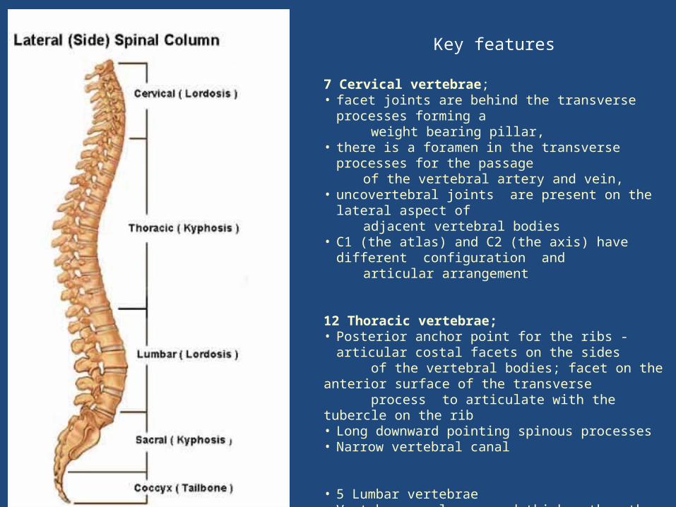
7 Cervical vertebrae; • facet joints are behind the transverse processes forming a weight bearing pillar,• there is a foramen in the transverse processes for the passage of the vertebral artery and vein, • uncovertebral joints are present on the lateral aspect of adjacent vertebral bodies• C1 (the atlas) and C2 (the axis) have different configuration and
articular arrangement
12 Thoracic vertebrae;• Posterior anchor point for the ribs - articular costal facets on
the sides of the vertebral bodies; facet on the anterior surface of the transverse process to articulate with the tubercle on the rib• Long downward pointing spinous processes• Narrow vertebral canal
• 5 Lumbar vertebrae• Vertebra are larger and thicker than the other two regions• Mamillary process is present on the posterior edge of the
superior articular process
Key features

Stability
• Intrinsic stability– Shape of the bones and joints– Ligament support
• Extrinsic stability– Muscular support
• To maintain an erect posture there should be relatively little muscle activity required and therefore less energy used.
• Muscle support is necessary for dynamic movement.
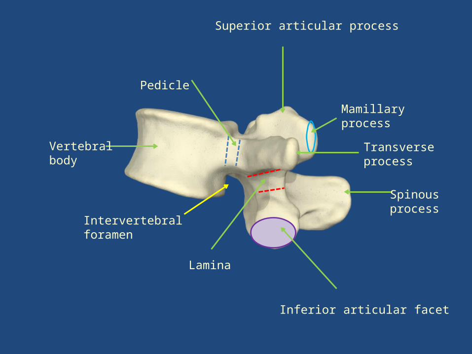
Vertebralbody
Pedicle
Mamillary process
Spinous process
Transverse process
Lamina
Inferior articular facet
Intervertebralforamen
Superior articular process

The joints
• Facet joint – Synovial plane joint– Oriented vertically – allow flexion and extension
some side flexion minimal rotation
• Inter-body joint– Secondary cartilaginous joint
• Together they form the “motion segment”

Motion segment.
• Consists of 2 adjacent vertebral bodies and the disc between them

Facet joint(Zygapophyseal)
Synovial plane jointsOrientation of the facetsdetermines the movement
Inter body joint(Intervertebral disc)
Cartilaginous joint
Superior facet
Inferior facet

Annulus fibrosus
• Conccentic layers of collagen
• Outer fibres are attached to the outer margins of adjacent vertebral bodies
• Adjacent layers run in opposite directions
• Encapsulates the nucleus pulposus
Nucleus Pulposus
• Semi fluid gel (gradually changes with age)
• Deforms under pressure (deformation resisted by the annulus fibrosus
End plates • Separates the disc from the vertebral body• Prevents the nucleus bulging into cancellous bone of the vertebral body• Common site of failure• Essential for nutrition of the discThe intervertebral disc

Functions of IV Disc
• Mobility of the spinal column
• Shock absorption
• Resisting forces (compression, torsion and shear)
• Constantly changing in response to load

Ligaments
• Vertebral column is supported by 2 longitudinal ligaments extending its full length.– Anterior longitudinal ligament– Posterior longitudinal ligaments.
• Between the adjacent vertebra of the motion segment there are additional ligaments:– Supraspinous ligament– Interspinous ligament– Intertransverse ligament – Ligamentum flavum

Inter transverse ligament
Supraspinous ligament
Interspinousligament
Ligamentum flavum
Superior facet
Inferior facet
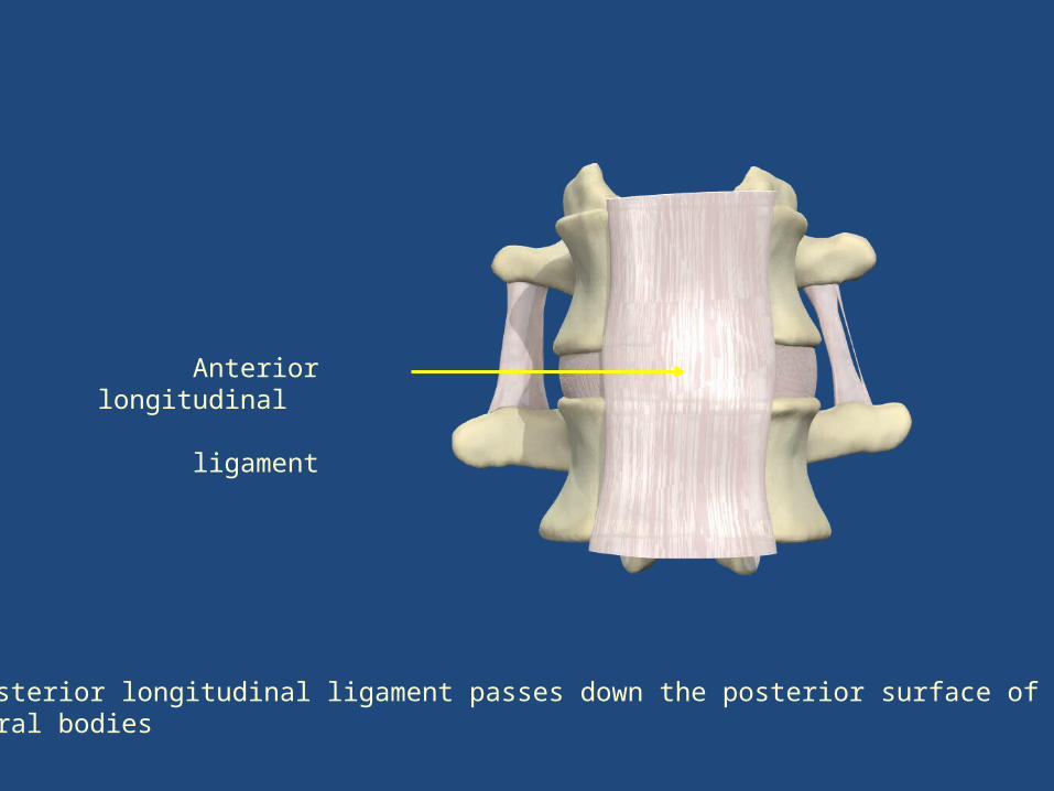
Anterior longitudinal ligament
The posterior longitudinal ligament passes down the posterior surface of thevertebral bodies

Iliolumbar ligament
Lateral lumbosacral ligament
Anterior Sacroiliac ligament
Sacrospinous ligament
Anterior longitudinal Ligament
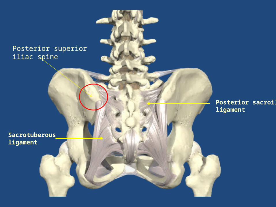
Posterior sacroiliac ligament
Sacrotuberous ligament
Posterior superior iliac spine

Muscles of the trunk
Muscles extending the trunk
Quadratus Lumborum
Multifidus
Erector spinae
Muscles flexing the trunk
Rectus abdominis
Psoas major/minor
Internal oblique*
External oblique*
* Also rotate the trunk
Working unilaterally the muscles produce side flexion

Quadratus lumborum Deep to erector spinaeFibres run upwards and medially
From the distal attachment on the posterior part of the iliac crest (and iliolumbar ligament) To the medial border of the inferior borderOf the 12th rib
Medial border of the muscle attaches to the lateral part of the anterior surface of the Transverse process of all lumbar vertebra
MultifidusDeep to erector spinae
Lies in the ‘gutter’ between the spinous and transverse processes of the vertebrae at all spinal levels
From the sacrum from mamillary process in the lumbar spine, the transverse processes in the thoracic region and the articular processes in the cervical region fibres pass upwards and medially to attach to the spinous processes
Three layers: the deepest passes to the spinous process immediately above; the middle fibres to the second or third above and the superficial fibres Third or fourth above

Erector SpinaeRuns the length of the vertebral columnFrom the sacrum iliac crest and the spinous processes of L5 to T11
Divides into three columns: Medial column (spinalis) from the spinous processes L2– T11 to the spinous processes of the thoracic spine
Intermediate column (longissimus) - longest and thickest to transverse processes and adjacent ribs
Lateral column (iliocostalis) to the inferior border of the lower 6 ribs

Psoas Major (and minor)
Mostly within the abdominal cavity with Relationship to the major vessels
From the anterior surface of the vertebral Bodies and intervening disc and anterior Surface of the transverse process T12 – L5
Passes through the pelvis (blending with Iliacus) under inguinal ligament the to attach to the lesser trochanter

Enclosed within the rectus sheath
From the symphysis pubis and the pubic crest
To the xiphoid process and Costal cartilages of ribs 5 6 and 7
Separated by the linea alba
Tendinous intersections

Oblique muscles
From lateral 2/3 of the inguinal ligament and anterior 2/3 of the iliac crest and from the thoracolumbar fascia
Fibres fan out to attach to lower four ribs then via an aponeurosis into the rectus sheath anteriorlyLowest attachment into to pubic crest
Rotates the trunk to the same side
Internal oblique External oblique
More superficial
Outer borders of the lower 8 ribs and their Costal cartilagesOuter lip of the anterior 2/3 of the iliac crestLower border of the aponeurosis forms the inguinal ligament
Fibres pass downward and medially aponeurosis passes into the rectus sheath

Two key fascial sheaths
• Thoracolumbar fascia • Rectus sheath
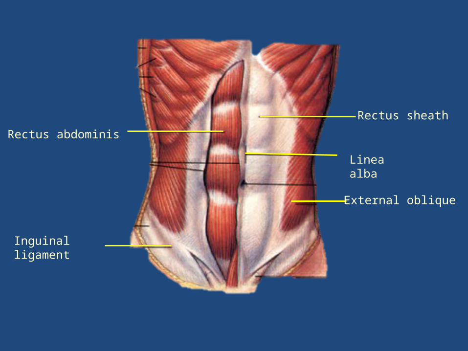
Rectus sheath
Linea alba
External oblique
Rectus abdominis
Inguinal ligament

External oblique
Transversus abdominis
Internal oblique
Rectus abdominis
Rectal sheath