Functional analysis of the Bazooka protein in the ...
156
Functional analysis of the Bazooka protein in the establishment of cell polarity in Drosophila melanogaster Dissertation submitted in partial fulfillment of the requirements for the degree of “doctor rerum naturalium” of the Georg-August-University Göttingen from Michael Peter Rolf Krahn born in Münster, Germany Göttingen, 2009
Transcript of Functional analysis of the Bazooka protein in the ...

Functional analysis of the Bazooka protein in the establishment of
cell polarity in Drosophila melanogaster
Dissertation submitted in partial fulfillment of the requirements for the degree of “doctor rerum naturalium”
of the Georg-August-University Göttingen
from
Michael Peter Rolf Krahn
born in Münster, Germany
Göttingen, 2009

D7 Referent: Prof. Dr. Andreas Wodarz Korreferent: Prof. Dr. Ernst A. Wimmer Tag der mündlichen Prüfung: 18.06.2009
3
Danksagung Vor allem möchte ich mich bei den Mitgliedern der Abteilung Stammzellbiologie für
drei schöne und erfolgreiche Jahre bedanken, insbesondere bei Prof. Dr. Andreas
Wodarz für die Möglichkeit, diese Promotion in seiner Abteilung durchzuführen und
ihm persönlich möchte ich auch für offene Ohren, Ratschläge und Diskussionen
danken.
Meiner Freundin Lisa Langhorst danke ich für die viele mentale und auch physische
Unterstützung während der Auf’s und Ab’s der letzten zwei Jahre.

Table of contents
5
Table of contents
1. ZUSAMMENFASSUNG 5
2. SUMMARY 6
3. INTRODUCTION 7
3.1. Cell polarity 7
3.2. The Drosophila embryonic epidermis as a model for epithelial polarity 9
3.3. The early development of the Drosophila nervous system 11
3.4. The PAR-complex 12
3.5. Bazooka 13
3.6. Research objectives 16
4. RESULTS 17
4.1. Membrane targeting of Bazooka/PAR-3 is mediated by a novel phosphoinositide-binding domain 18
4.2. PP2A antagonizes phosphorylation of Bazooka by PAR-1 to control apical-basal polarity in dividing embryonic neuroblasts 64
4.3. Imapired phosphorylation of Bazooka by aPKC leads to a dominant negative phenotype 110
5. DISCUSSION 136
5.1. Implications of the structural analysis of the Bazooka protein 136
5.2. Phosphorylation of Bazooka: Only two pieces of a great puzzle 139
6. REFERENCES 144
7. APPENDIX 149
7.1. Abbreviations 149
7.2. Bazooka sequence 150
7.3. Western Blot of Baz constructs 154
8. CURRICULUM VITAE 155

1 Zusammenfassung
5
1. Zusammenfassung Für Komponenten des sogenannten PAR/aPKC- (partitioning-defective / atypische
Proteinkinase C) Komplexes wurde nachgewiesen, dass sie eine Schlüsselrolle in der
Entstehung und Erhaltung der Zellpolarität in unterschiedlichen Zelltypen spielen. Die
grundlegenden Mechanismen scheinen hierbei in der Evolution zwischen Wurm und
Mensch stark konserviert zu sein. Forschung an der Fruchtfliege Drosophila
melanogaster hat gezeigt, dass Bazooka als Kernkomponente des PAR/aPKC
Komplexes an der Spitze einer komplexen Hierachie steht, die die Zellpolarität
reguliert. Nicht nur für die Etablierung der Zellpolarität in epithelialen Zellen,
sondern auch für die asymmetrische Zellteilung der neuralen Stammzellen
(Neuroblasten) und für die Determinierung der Schicksale der beiden Tochterzellen
ist die asymmetrische Lokaliserung von Bazooka essentiell. Trotzdem ist immer noch
nicht geklärt, wie genau Bazooka selbst an die Membran lokalisiert wird und wie
diese Rekrutierung während der Etablierung der Zellpolarität reguliert wird.
In der vorliegenden Studie wurde eine systematische Strukturanalyse des Bazooka-
Proteins vorgenommen, indem Fusionsproteine aus Bazooka-Deletionskonstrukten
und dem grünen fluoreszierenden Protein (GFP) in transgenen Fliegen und in der
Zellkultur exprimiert wurden. Dabei wurde festgestellt, dass die C-terminale Region
von Bazooka ein neues Lipid-Bindemotiv enthält und essentiell für die
Membranlokalisierung des Proteins ist.
Des weiteren wurde die Rolle von zwei Phosphorylierungen näher untersucht: Zum
einen die Phosphorylierung und Dephosphorylierung des konservierten Serinrestes
1085 durch die Kinase PAR-1 und die Phosphatase PP2A, wodurch die apikal-basale
Polarität in Neuroblasten kontrolliert wird. Dies geschieht durch die Regulierung
einer Bindestelle für die Adaptorproteine 14-3-3ε und Leonardo. Defekte in dieser
Signalkaskade führen in einem hohen Anteil embryonaler Neuroblasten zu einer
Umkehr der apikal-basalen Polarität.
Zweitens wurde die Interaktion zwischen Bazooka und aPKC, welches Bazooka an
dem konservierten Serinrest 980 phosphoryliert, genauer charakterisiert. Hierbei
konnte gezeigt werden, dass die Überexpression einer nicht phosphorylierbaren
Variante von Bazooka zu einem drastischen dominant-negativen Phänotyp führt, der
mit einem Verlust der Zellpolarität und embryonaler Letalität verbunden ist.

2 Summary
6
2. Summary Components of the PAR/aPKC (partitioning-defective / atypical protein kinase C)
complex have been found to play a key role in the establishment and maintenance of
cell polarity in various cell types. The underlying mechanisms are highly conserved
throughout evolution, from worm to mammals. Research in the fruit fly Drosophila
melanogaster revealed that Bazooka as the core component of the PAR/aPKC
complex acts on top of a hierarchy in the regulation of cell polarity. Not only the
establishment of epithelial cell polarity, but also the asymmetric cell division of the
neural stem cell (neuroblast, NB) and the determination of the distinct cell fates of the
two daughter cells are dependent on asymmetric localization of Bazooka. However, it
is not yet fully elucidated how exactly Bazooka itself is localized to the apical
membrane domain and how its targeting is regulated during the establishment and
maintenance of cell polarity.
In this study, a systematic structural analysis of the Bazooka protein was performed,
using deletion constructs tagged with green fluorescent protein (GFP) in transgenic
flies and in cell culture experiments in order to clarify the role of the distinct domains
of the protein. We found that the C-terminal region of Bazooka, contains a new lipid
binding motif and is crucial for membrane association of the protein.
Furthermore, the role of two different phosphorylation events of Bazooka were
elucidated: First, (de)phosphorylation at the conserved serine residue 1085 by the
kinase PAR-1 and the phosphatase PP2A controls apical-basal polarity in dividing
embryonic NBs by regulating a binding site for the adaptor proteins 14-3-3ε and
Leonardo. Defects in this pathway lead to frequent reversal of apical-basal polarity in
embryonic NBs.
Second, the interaction of Bazooka with aPKC, which phosphorylates Bazooka at the
conserved serine residue 980, was investigated in more detail. Overexpression of a
non-phosphorylatable version of Baz leads to a drastic dominant negative phenotype
with a total loss of cell polarity and embryonic lethality.

3 Introduction
7
3. Introduction
3.1. Cell polarity Cell polarity is one of the key features of multicellular organisms and is the
prerequisite for various complex functions including the establishment of epithelial
barriers, directed growth and movement and the three dimensional development of the
nervous system.
After more than one century of intensive research we are far from understanding the
interactions of genes, proteins and regulatory RNAs involved in the regulation of cell
polarity, and many pieces of this puzzle remain to be identified. Nevertheless, some
common principles and key players of polarity have been revealed and investigated.
Interestingly, most of them are well conserved throughout evolution and have a
general function in different polarized cell types.
The approach of developmental biology and the work on model organisms like
Drosophila melanogaster provides versatile tools not only for the understanding of
fundamental mechanisms of life and diseases but also for the development of specific
drugs and therapies. In contrast to mammalian cell culture systems, the fruit fly
Drosophila offers not only the opportunity of a real “in vivo” approach to test all
mechanisms, mutations, candidates etc. for their implications on the entire organism.
It also allows to investigate them in different cell types, tissues and developmental
stages and thereby to compare directly the underlying mechanisms.
In Drosophila, at least five different polarized cell types are easily accessible for in
vivo research:
1. The oocyte, which is surrounded by the follicle epithelium exhibits an anterior
(facing the nurse cells) – posterior (facing the next egg chamber) - polarity, which is
reflected not only by the specific localization of proteins but also by the directed,
microtubule based transport and localization of mRNAs.
2. The ectodermal epithelium surrounds the developing embryo, secreting a protective
cuticle. It also forms part of the intestinal system, the tracheae and the salivary glands
(see also 2.2).
3. The mesodermal follicle cell epithelium. Similar to the ectodermal cells of the
epidermis, it also forms a polarized single layer of cuboidal cells, but in contrast to
3 Introduction
8
ectodermal epithelia, whose apical membranes face the outside world or a lumen, its
apical membrane forms cell-cell contacts with the germline cells.
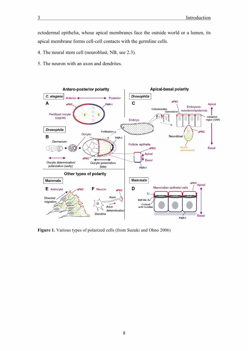
4. The neural stem cell (neuroblast, NB, see 2.3).
5. The neuron with an axon and dendrites.
Figure 1. Various types of polarized cells (from Suzuki and Ohno 2006)

3 Introduction
9
3.2. The Drosophila embryonic epidermis as a model for epithelial polarity
The ectodermal epidermis of the Drosophila embryo is a good model to study
fundamental mechanism of cell polarity. The polarity is first established during
blastoderm stage (ca. 2:10h after egg deposition), concomitantly with the invagination
of the plasma membrane separating the syncytium (Lecuit, 2004). Compared with the
mammalian cell culture system, it has been shown that many of the basic mechanisms
and genes regulating epithelial polarity are highly conserved throughout evolution
(Knust and Bossinger, 2002).
Polarity in epithelial cells is based on the segregation of proteins and lipids between
an apical membrane domain, a lateral cell-cell contact zone and a basal cortex, which
is in close contact to the underlaying tissue. The last two domains are often subsumed
as the basolateral domain. One key step in the establishment and restriction of the
membrane domains is the formation of specialized cell-cell contact zones.
Figure 2. Junctional complexes of epithelial cells in vertebrates and Drosophila (from Knust
and Bossinger 2002)
In vertebrates, adherens junctions between neighbouring cells are formed in the
zonula adherens (ZA), a process which mainly involves the cadherin-catenin complex,
Therefore, the transmembrane protein E-cadherin (or other members of the cadherin
family) forms first cis-cellular and later trans-cellular dimers in a calcium dependent
fashion (Nelson, 2008). By their intracellular domain, cadherins recruit β-catenin,
which in turns bind to α-catenin which finally links the cadherin-catenin complex
directly or viaa vinculin and α-actinin to the actin cytoskeleton (Nelson, 2008; Perez-
Moreno et al., 2003). The correct formation of the ZA is a crucial prerequisite for the
establishment of the tight junctions (TJ), which are located apical of the ZA and

3 Introduction
10
composed of different protein complexes which finally act together to seal the
intercellular space (Matter, 2000; Tsukita et al., 2001). Beside members of the
transmembrane-protein families JAM (junctional adhesion molecule), claudin and
occludin, there are also some cytoplasmic proteins localized to the TJ, namely the
zonula occludens proteins (ZO-1-3), MAGI-proteins and the PAR/aPKC complex
proteins (cp 2.4) (Tsukita et al., 2001). One more TJ complex, which is also
conserved throughout evolution is the Crumbs (Crb) / PALS1 (protein associated with
Lin7) / PATJ (PALS1-associated TJ protein) complex. As an antagonist to the apical
junctional regulators functions the Discs Large (Dlg) / Scribble / Lethal (2) giant
larvae (Lgl) complex at the basolateral domain.
The components of the AJ, its assembly and regulation is mostly conserved in from
fly to man but in contrast to mammalian cells there is no real TJ in the Drosophila
epithelium but a so called sub-apical region (SAR), which is located apical of the AJ.
This junctional belt is predominately established by the transmembrane protein Crb
and its intracellular binding partner Stardust, although components of the PAR/aPKC
complex are also partly localized to the SAR and regulate SAR and AJ assembly
(Bilder et al., 2003; Harris and Peifer, 2005; Knust and Bossinger, 2002). Analogue to
mammalian epithelial cells, the Dlg complex is located at the basolateral membrane.
Figure 3. Localization of protein complexes in the Drosophila epithelium (Beati, personal
communication)

3 Introduction
11
3.3. The early development of the Drosophila nervous system
The development of the nervous system of Drosophila starts with the delamination of
the NBs during stage 9 of embryogenesis (approximately four hours after egg
deposition) from the overlying ectodermal epithelium in the so-called “neurogenic
region”. Prior to the first mitosis, apical-basal polarity is established, partly inherited
from the epithelium (Wodarz, 2005; Wodarz and Huttner, 2003). In metaphase,
members of the PAR/aPKC-complex (see below) are positioned at the apical
membrane domain, together with the Insc/Pins/Gαi complex. In contrast, certain cell
fate determinants like the transcription factor Prospero, the proteins Brain Tumor
(Brat) and Numb and their adaptor proteins Miranda and Partner of Numb are
localized to the basal cortex. Additionally, the spindle, which is first in parallel to the
overlaying epithelium, rotates by 90° and upon unequal cytokinesis the NB divides
asymmetrically into a bigger, apically localized daughter cell and a smaller, basally
localized daughter cell. Proteins localized apically during metaphase are inherited by
the bigger daughter cell, which retains stem cell abilities and undergoes more cycles
of asymmetric cell division. In contrast, proteins targeted to the basal cortex in the
dividing NB segregate exclusively into the smaller daughter cell, the so-called
“ganglion mother cell” (GMC), which divides only once more, giving rise to two
neurons or glial cells. The apical-basal polarity of the NB, which is coordinated with
spindle orientation in metaphase, is crucial for asymmetric cell division and thereby
also for the development of the nervous system: Loss of polarity often results in a
symmetric division, generating two daughter cells with stem cell abilities that both
continue to divide, eventually leading to tumor formation (Bello et al., 2006;
Betschinger et al., 2006; Lee et al., 2006; Wodarz and Näthke, 2007).
Figure 4. Delamination and asymmetric cell division in Drosophila NBs (Wodarz 2003).

3 Introduction
12
3.4. The PAR-complex One of the most important regulators of cell polarity is the PAR-aPKC- (partitioning-
defective – atypical protein kinase C) complex. It is highly conserved throughout
evolution from worm to man (Suzuki and Ohno, 2006) and consists of the scaffolding
proteins PAR-3 (Bazooka, Baz in Drosophila) and PAR-6 and the serine-threonine
kinase aPKC. This complex localizes to the apical cortex in epithelial cells and NBs
and to the anterior cortex in the C.elegans zygote and the oocyte of Drosophila
(Figure 1). It is antagonized by other PAR proteins, namely PAR-1, a serine-threonine
kinase that localizes basolaterally in epithelia and posterior in the oocyte, and the
adaptor protein PAR-5 (14-3-3ε and leonardo in Drosophila).
Figure 5. Interacting domains in the PAR-complex (adapted from Johnson and Wodarz
2003). Baz serves as a scaffold to recruit PAR-6 and aPKC to the cortex: The first PDZ
domain of Baz interacts with the PDZ domain of PAR-6 and the aPKC binding domain with
the kinase domain of aPKC. Additionally, aPKC can directly interact with PAR-6 via their
PB1 (phagocyte oxidase/Bem1) domains.

3 Introduction
13
3.5. Bazooka The bazooka (baz) gene was first identified in a screen for embryonic patterning
defects and obtained its name due to the big holes in the cuticle of baz mutant
embryos (Wieschaus et al., 1984). baz encodes a large protein of 1464 amino acids
that possesses three highly conserved PDZ-(Psd95, Disc large, ZO-1) domains and a
conserved N-terminal oligomerization domain (CR1) (Kuchinke et al., 1998;
Wieschaus et al., 1984) (Benton & St Johnston, 2003). Furthermore, for the
mammalian and worm homologue of Baz, PAR-3, a conserved region of twenty
amino acid residues has been described to interact with the kinase domain of aPKC
(Izumi et al., 1998; Tabuse et al., 1998). In contrast, for Baz, the interaction with
aPKC was mapped to the second and third PDZ domain (Wodarz et al., 2000).
PAR-6 can bind to the first PDZ domain of PAR-3 and additionally directly to aPKC
(Joberty et al., 2000; Lin et al., 2000). In addition to these three “core” components of
the PAR/aPKC complex, the small GTPase Cdc42 is often recruited into this
complex. In fact, it can bind directly to PAR-6, regulating the binding affinity of the
PAR-6-aPKC interaction and thereby aPKC kinase activity in various cell types of
different species (Garrard et al., 2003; Joberty et al., 2000; Lin et al., 2000; Peterson
et al., 2004). The specific contribution of Cdc42 to the function of the PAR-complex
in the regulation of cell polarity still remains unclear, because Cdc42 is involved in
several additional pathways connected with polarity.
Various studies have shown that the PAR complex and particularly Baz/PAR-3 acts at
the top of a genetic hierarchy in the regulation of cell polarity (Johnson and Wodarz,
2003). Loss of Baz leads to a complete loss of cell polarity in most polarized cell
types investigated so far. In fact, in Drosophila, Baz is one of the first apical cues in
the ectodermal epithelium and it is essential for the establishment of the first adherens
junctions during cellularization (Harris and Peifer, 2004). It is necessary for the
correct targeting of Crumbs (Crb), a conserved transmembrane protein and key
regulator of epithelial cell polarity, to the apical membrane (Harris and Peifer, 2004).
In contrast, mutation of crb does not alter the apical localization of Baz in early
embryogenesis (Bilder et al., 2003; Johnson and Wodarz, 2003). Moreover, Baz
mediates assembly of the junctional protein complex of DE-cadherin (Drosophila E-
cadherin) and Armadillo (the Drosophila homologue of β-catenin) (Harris and Peifer,

3 Introduction
14
2004; Harris and Peifer, 2005). Consequently, loss of Baz results in an impaired
assembly of the AJ.
In addition to epithelial polarity, the asymmetric cell division in embryonic and larval
NBs is controlled by Baz (Knoblich, 2008; Wodarz, 2005). Here, Baz recruits
Inscuteable (Insc) and Partner of Inscuteable (Pins) to the apical cortex, which in turn
stabilizes the Baz protein (Schober et al., 1999; Wodarz et al., 1999). Like in
epithelial cells, Baz also targets PAR-6 and aPKC to the apical cortex in dividing NBs
(Petronczki and Knoblich, 2001; Wodarz et al., 2000). The apical accumulation of
Baz is not affected upon loss of PAR-6 or aPKC, in contrast to the asymmetric
localization of cell fate determinants, which ensure that only one daughter cell retains
stem cell abilities (Petronczki and Knoblich, 2001; Rolls et al., 2003). This supports
the hypothesis that Baz serves as a scaffold to ensure the correct localization and
regulation of aPKC kinase activity (Wirtz-Peitz et al., 2008).
1 83 292 400 441 527 665 732 968-996 1464
Baz
Baz CR1 PDZ domain aPKC BR
Figure 6. Structure of the Baz protein
Up to now, three conserved serine residues of Baz have been reported to be
phosphorylated: serine 980 as mentioned above is phosphorylated by aPKC (Kim et
al. submitted, (Nagai-Tamai et al., 2002). In a mammalian cell culture system, this
phosphorylation has been shown to be crucial for the establishment but not for the
maintenance of cell polarity (c.p. 3.3)(Nagai-Tamai et al., 2002).
Serine 151 and serine 1085 are phosphorylated by PAR-1, thus creating a binding site
for 14-3-3 proteins (Benton and St Johnston, 2003). Furthermore, it has been
demonstrated that the phosphorylation of Baz at these two sites cooperates in the
exclusion of Baz from the lateral and basal membrane domain in the follicle
epithelium and from the posterior cortex in the oocyte. Recently, a first genetic

3 Introduction
15
interaction study suggested a role for PP2A as a counterpart of PAR-1 kinase activity
in the development of the polarized photoreceptor cells (Nam et al., 2007).

3 Introduction
16
3.6. Research objectives Although various aspects of the function of Baz/PAR-3 in the control of cell polarity
have been elucidated during the last decade, there are still many unanswered
questions. One of the most intriguing problems is how exactly Baz is recruited to the
membrane and how it is targeted to the apical membrane domain.
Therefore, the first aim of this study was to characterize the Baz protein functionally
by a structural analysis using deletion constructs in transgenic flies and cell culture.
From the subcellular localization of the mutated proteins conclusions can be drawn
regarding the function of the different domains. This analysis was performed in four
different polarized cell types, namely the ectodermal epidermis, the mesodermal
follicle epithelium, the adult female germ line and the embryonic NBs.
Secondly, I analyzed the interaction between Baz and protein phosphatase 2A
(PP2A), a potential interaction partner of Baz found in a yeast-two-hybrid screen. The
focus of this project was to determine whether the potential dephosphorylation of
three conserved serine residues in Baz by PP2A is required for the establishment and
maintenance of cell polarity in NBs.
Finally, the phosphorylation of Baz by aPKC at the conserved serine 980, which has
already been described to play an essential role in the establishment of cell polarity in
mammalian epithelial cells (Nagai-Tamai et al., 2002), was elucidated by generation
of mutations in this site and expression of the mutant constructs in flies and cell
culture. The consequences of such mutations on cell polarity in different cell types
and on the interaction between Baz and aPKC were characterized in detail.

4 Results
17
4. Results
Every chapter within the results starts with a one-page description of:
• the main aim of the particular manuscript in the context of the complete thesis
• the authors and their contributions to the work, and
• the status of the manuscript.
4.1 Membrane targeting of Bazooka Results
18
4.1. Membrane targeting of Bazooka/PAR-3 is mediated by a novel phosphoinositide-binding domain
Within that project, various deletion constructs of Baz were expressed in different
polarized tissues in the Drosophila embryo and adult female germ line using the
UAS-GAL4 system. By indirect immunofluorescence and confocal laser microscopy,
the subcellular localization of the mutated transgenes was investigated and its
functionality was tested by a rescue experiment with two Baz NULL-alleles.
The potential lipid-binding capability of the PDZ domains and the C-terminus of Baz
were tested using membrane lipids-strips.
Michael P. Krahn, Nannette Fischer and Andreas Wodarz
Author contributions to the work: Michael P. Krahn: All experiments, besides*
writing of the manuscript Nannette Fischer: *Sequencing of the Baz alleles Andreas Wodarz: Editing of the manuscript STATUS: SUBMITTED to Current Biology

4.1 Membrane targeting of Bazooka/PAR-3 Results
19
Membrane targeting of Bazooka/PAR-3 is mediated by a
novel phosphoinositide-binding domain
Michael P. Krahn1, Nannette Fischer1,2 and Andreas Wodarz1*
1Abteilung Stammzellbiologie, DFG Research Center for Molecular Physiology of the
Brain (CMPB), Georg-August-Universität Göttingen, Justus-von-Liebig-Weg 11,
37077 Göttingen, Germany
2Institut für Genetik, Heinrich-Heine-Universität Düsseldorf, Universitätsstr. 1,
Germany
*author for correspondence: [email protected]
Running title: Membrane targeting of Bazooka/PAR-3
Keywords: epithelia, neuroblast, polarity, PAR proteins, membrane targeting

4.1 Membrane targeting of Bazooka/PAR-3 Results
20
Summary
Background
Cell polarity in higher animals is controlled by evolutionarily conserved protein
complexes, which localize to the cytocortex in a polarized manner. The
PAR-3/PAR-6/aPKC complex is the first to become asymmetrically localized and it
controls the localization of additional complexes functioning further downstream in
the regulation of cell polarity, including the Crumbs/Stardust/PATJ complex in
epithelia and the Partner of Inscuteable/Gαi complex in neural precursor cells. The
first component of the PAR-3/PAR-6/aPKC complex that is localized to the cortex is
Bazooka/PAR-3 (Baz), a large scaffolding protein. How Baz is recruited to the
membrane is unknown so far.
Results
Here we present a structure-function analysis of Baz focussing on its subcellular
localization and function in four different polarized cell types of Drosophila: the
ectodermal embryonic epidermis, the mesodermal follicle epithelium, embryonic
neuoblasts and the oocyte. We show that the PDZ domains of Baz are dispensable for
its correct localization, whereas a conserved region in the C-terminal part of Baz to
which no function had been assigned so far is required and sufficient for membrane
localization. This domain binds strongly to phosphoinositide membrane lipids and
thus mediates cortical localization of Baz by direct interaction with the plasma
membrane.
Conclusions
We have identified a novel phosphoinositide-binding domain that is necessary and
sufficient for recruitment of Baz to the plasma membrane. Our findings reveal a
mechanism for the coupling of plasma membrane polarity and cortical polarity.

4.1 Membrane targeting of Bazooka/PAR-3 Results
21
Introduction
Baz/PAR-3 is a core component of the PAR-3/PAR-6/aPKC complex, which is
conserved throughout evolution from worm to man [1, 2]. In a broad range of
polarized cell types, the PAR-3/PAR-6/aPKC complex is required to define the axis
of polarity: apical versus basal or anterior versus posterior [2]. During the past decade
it became clear that Baz acts at the top of a hierarchy of molecules which are
responsible for this polarization [3-5]. For example, in C.elegans, PAR-3 can localize
to the anterior cortex in a PAR-6 and aPKC- independent fashion [6, 7]. In
Drosophila neural precursor cells (neuroblasts, NBs), Baz does not only recruit aPKC
and PAR-6 to the apical cortex, but also the Inscuteable/Pins/Gαi complex [8-13].
Similarly, in the Drosophila ectodermal epithelium, Baz serves as the first apical cue
required for localization of Crumbs (Crb) to the apical membrane domain [14]. In all
these cell types, loss of Baz/PAR-3 function leads to loss of cell polarity [10, 13, 15,
16].
These findings raise the question of how Baz itself is recruited to the membrane and
how it obtains its polarized subcellular localization. This could be achieved in several
ways, for instance by binding to an integral transmembrane protein, by binding to a
membrane associated protein, by lipid modification or by direct binding to membrane
lipids. Mammalian PAR-3 is recruited to tight junctions in epithelial cells by binding
to the transmembrane protein Junctional Adhesion Molecule (JAM) via its first PDZ
domain [17, 18]. However, there are no annotated homologs of JAM in Drosophila,
ruling out this mechanism for membrane recruitment of Baz.
It was recently shown that the highly conserved second PDZ domain of rat PAR-3
binds to phosphoinositide lipids and is crucial for membrane association of PAR-3 in
mammalian epithelial cells [19]. Binding to phosphoinositides is not a unique feature

4.1 Membrane targeting of Bazooka/PAR-3 Results
22
of the second PDZ domain of PAR-3 but was also demonstrated for a variety of other
PDZ domains [19-22]. Given the high conservation of Baz/PAR-3 throughout
evolution, it is tempting to speculate that the second PDZ domain of Baz may be
responsible for its membrane localization, but this has not been tested yet.
Additional evidence for the potential involvement of phosphoinositides in the
localization of the PAR-3/PAR-6/aPKC complex has come from studies in cultured
hippocampal neurons, where the phosphatidyl-inositol-3-kinase (PI3-kinase) pathway
is required for the polarized localization of the complex to the tip of the axon [23].
Intriguingly, different phosphoinositides are restricted to different domains of the
plasma membrane. Phosphatidylinositol (4, 5) bisphosphate (PIP2) is restricted to the
apical plasma membrane domain and Phosphatidylinositol (3, 4, 5) trisphosphate
(PIP3) to the basolateral domain in mammalian polarized epithelia [24, 25]. In
Drosophila photoreceptor cells and ectodermal embryonic epithelia the distribution of
PIP2 and PIP3 is reversed, with PIP3 accumulating in the apical and PIP2 in the
basolateral membrane domain [26, 27]. The balance between PIP2 and PIP3 in the
membrane is regulated by PI3-kinase and its antagonist, the lipid phosphatase PTEN
[28]. PTEN directly binds to the third PDZ domain of Baz, revealing another
important link between the PAR-3/PAR-6/aPKC complex and phosphoinositide
signaling [19, 26, 27].
To address the mechanism of how Baz gets recruited to the plasma membrane and
which domains of Baz are required for its function, we performed a structure-function
analysis using a series of GFP-Baz fusion proteins lacking different regions of the
protein. These GFP fusion proteins were expressed from UAS-driven transgenes in
the embryonic epidermis, in embryonic NBs, in the follicle epithelium and in oocytes
and their subcellular localization in these cell types was analyzed by confocal
4.1 Membrane targeting of Bazooka/PAR-3 Results
23
microscopy. Furthermore, the mutant proteins were tested for their ability to rescue
the lethality of strong loss-of-function mutations of baz.
Contrary to our expectations based on the study by Wu et al. [19], we found that
deletion of the second or even of all three PDZ domains did not result in
mislocalization of Baz, whereas a domain in the C-terminal region of Baz was
necessary and sufficient for membrane targeting. We show that this domain binds
strongly to phosphoinositides, in contrast to a protein fragment comprised of the three
PDZ domains of Baz. Thus, our data reveal that Baz is recruited to the membrane by
direct binding to phosphoinositides via a novel phosphoinositide-binding domain.
Results
Molecular analysis of loss-of-function alleles of baz
Baz is a large scaffolding protein of 1464 aa, but with the exception of the N-terminal
conserved region 1 (CR1; Fig. 1), which is required for homo-dimerization [29], and
two conserved phosphorylation sites for the kinase PAR-1 (S151 and S1085), which
serve as binding sites for 14-3-3 proteins [30, 31], little is known about the
importance of individual domains for the function and proper subcellular localization
of Baz. To identify protein domains and individual amino acid residues that are
crucial for the function of Baz, we sequenced four mutant baz alleles that were either
induced by treatment with ethyl-methanesulfonate (EMS) (bazXi106; [32], bazEH747;
[33]) or by exposure to X-rays (baz815-8; [34], bazXR11; [35], R. Stanewsky,
unpublished). Three alleles contained nonsense mutations giving rise to Baz proteins
truncated in the N-terminal quarter of the protein that most likely are completely
nonfunctional (Fig. 1A). In bazXR11 we could not detect any mutation that would lead

4.1 Membrane targeting of Bazooka/PAR-3 Results
24
to changes in the Baz protein sequence, pointing to a mutation in a regulatory element
outside of the coding region in this allele.
Structure-function analysis of Baz using GFP-Baz fusion proteins
Because the molecular analysis of the four mutant alleles of baz did not allow us to
draw any conclusions on the functional requirement for individual domains of Baz,
we generated a series of constructs encoding full-length and deletion mutants of Baz
tagged at the N-terminus with green fluorescent protein (GFP) (Fig. 1B). These
constructs were expressed with the UAS-GAL4 system [36] in four different
polarized cell types: In the embryonic epidermis using the ubiquitous driver
daughterless::GAL4 (da::G4), in the adult follicle epithelium, which is derived from
mesodermal stem cells by Cu::GAL4 (Cu::G4), in embryonic NBs using
worniu::GAL4 (wor::G4) and in the adult female germ line with nanos::GAL4
(nos::G4), in order to test the requirement of the different domains of the Baz protein
for proper subcellular localization in these cell types.
Full length GFP-Baz is fully functional as it rescued the embryonic lethality of two
strong baz alleles (baz815-8 and bazXR11; Table 1), like untagged full length Baz [35].
Furthermore, the GFP-Baz fusion protein showed exactly the same subcellular
localization as the endogenous Baz protein in all cell types that we analyzed (Fig. 2).
In embryonic epidermal cells and in the follicle epithelium, full-length GFP-Baz
localized to the ZA and colocalized there with DE-Cadherin (DE-Cad) but not with
Discs large (Dlg), a marker for the basolateral membrane (Fig. 2A, B). In embryonic
NBs, GFP-Baz formed an apical crescent at pro- and metaphase and colocalized with
aPKC, whereas Miranda (Mira) formed a crescent at the basal cortex (Fig. 2C). In

4.1 Membrane targeting of Bazooka/PAR-3 Results
25
stage 10 oocytes GFP-Baz formed a gradient at the membrane with high
concentrations anterior and low concentrations at the posterior pole (Fig. 2D).
The N-terminal region of Baz is required for polarized localization in NBs and
oocytes
The N-terminal region of Baz (CR1; Fig. 1) is highly conserved and is predicted to
adopt a three-dimensional structure similar to that of the E. coli protein DinI and a
homologous protein in coliphage 186. Baz CR1 is required for homophilic di- or
oligomerization of the Baz protein [29]. Furthermore, Baz CR1 was found to be
necessary but not sufficient for apical membrane localization of a Baz-GFP fusion
protein in the follicle epithelium [29]. We investigated the subcellular localization of
a mutant GFP-Baz fusion protein lacking the first 317 amino acids including CR1
(GFP-BazΔ1-317, Fig. 1B). In the embryonic epidermis GFP-BazΔ1-317 colocalized
with endogenous Baz (data not shown) and with DE-cad in the ZA (Fig. 3A). In the
follicle epithelium, the subcellular localization of GFP-BazΔ1-317 was dependent on
the level of overexpression. At low levels of overexpression, GFP-BazΔ1-317 was
targeted correctly to the ZA and colocalized with DE-Cad (Fig. 3B). In cells
containing higher levels of the protein, GFP-BazΔ1-317 was partly diffusely
distributed in the cytosol (Fig. 3B, arrows) but still a considerable amount of protein
accumulated in the ZA.
In embryonic NBs, GFP-BazΔ1-317 was associated with the membrane but localized
to the entire cortex instead of forming an apical crescent (Fig. 3C). GFP-BazΔ1-317
also localized homogeneously to the entire cortex of the oocyte (Fig. 3D),
demonstrating that the N-terminal region of Baz is required for its polarized
localization in these two non-epithelial cell types.

4.1 Membrane targeting of Bazooka/PAR-3 Results
26
The PDZ domains of Baz are not required for membrane targeting
For rat PAR-3 (also called atypical protein kinase C specific interacting protein,
ASIP) [37], the second PDZ domain was shown to interact with phosphoinositide
membrane lipids [19]. This interaction depends on a PIP head group polar binding
pocket and a positively charged cluster of amino acids in the second PDZ domain of
PAR-3. Deletion of the second PDZ domain or mutation of single conserved amino
acid residues involved in lipid binding was reported to result in a total loss of
membrane association of PAR-3 in MDCK cells [19]. To test whether the second
PDZ domain has a similar function in Drosophila Baz, we expressed deletion mutants
of Baz lacking individual PDZ domains (GFP-BazΔPDZ1/2/3), or all three PDZ
domains together (GFP-BazΔPDZ1-3) (Fig. 1B) in Drosophila embryonic and adult
tissues. Neither deletion of any single PDZ domain (data not shown), nor deletion of
all three PDZ domains resulted in a disturbed localization of the respective mutant
Baz protein compared to wild type Baz (Suppl. Fig. 1).
One explanation could be that correctly localized endogenous Baz protein localizes
the mutant protein via its N-terminal oligomerization domain [29]. However, in
hemizygous baz815-8 mutant embryos the GFP-BazΔPDZ1-3 protein still localized
normally at late embryonic stages when the maternally contributed endogenous wild
type Baz protein had already disappeared (data not shown). Additionally, in S2R+
cells, which express only low levels of endogenous Baz, GFP-BazΔPDZ1-3 was
correctly targeted to the membrane indistinguishable from its wild type counterpart
(Suppl. Fig. 2B).
These results show that the correct subcellular localization of Baz in the four cell
types investigated here is independent of its PDZ domains. Consistent with this

4.1 Membrane targeting of Bazooka/PAR-3 Results
27
conclusion, fragments of Baz containing all three PDZ-domains but lacking portions
of the C-terminal region did neither show any significant membrane localization in
the embryonic epidermis or in follicle cells nor in S2R+ cells (see below). This
further suggests that the membrane binding ability of the PDZ domains of Baz is not
sufficient to link Baz to the membrane in vivo. Nonetheless, the PDZ domains 1 and 2
are essential for the function of Baz, because mutant forms of Baz lacking these
domains fail to rescue the lethality of strong baz loss-of-function alleles (Table 1).
Truncation of the C-terminal region of Baz abolishes membrane association
In contrast to Baz CR1, the three PDZ domains and the phosphorylation sites for
aPKC and PAR-1 (S980 and S151/S1085 respectively), the C-terminal region of Baz
is quite divergent from vertebrate and C. elegans PAR-3. To assess the function of
this part of Baz, we generated a series of constructs encoding proteins with C-terminal
truncations (Fig. 1B). Deletion of the non-conserved potential PDZ binding motif at
the very C-terminus of Baz (SEVL; GFP-BazΔ1461-1464) did not affect the normal
subcellular localization of Baz (data not shown). GFP-BazΔ1325-1464 (data not
shown) and GFP-BazΔ1222-1464 were also localized correctly in all tissues analyzed
(Suppl. Fig. 3). Deletion of 349 or 463 aa from the C-terminus (GFP-BazΔ1097-1464
and GFP-BazΔ1001-1464, respectively) led to increased accumulation of the
truncated protein in the cytoplasm (Fig. 4A – C; Suppl. Fig. 4A). Only a small
fraction of these mutant Baz proteins was still localized correctly to the ZA in
epithelial cells (Fig. 4A) and to the apical cortex in NBs (Fig. 4C). Deletion of 496 aa
from the C-terminus (GFP-BazΔ969-1464) completely abolished membrane
localization of Baz in all tissues analyzed (data not shown).

4.1 Membrane targeting of Bazooka/PAR-3 Results
28
In S2R+ cells, which only express low levels of endogenous Baz protein,
GFP-BazΔ1097-1464 and GFP-BazΔ1001-1464 were homogeneously distributed in
the cytosol without accumulation at the cell cortex (Suppl. Fig. 2E, F). In contrast,
GFP-Baz proteins with smaller truncations were correctly targeted to the membrane
(Suppl. Fig. 2C, D).
We assessed the functionality of the different variants of Baz with C-terminal
truncations by rescue experiments. Deletion of up to 367 aa from the C-terminus
allowed rescue of the lethality of baz815-8 and of bazXR11 to the adult, whereas larger
deletions eliminating the conserved regions surrounding the phosphorylation sites for
PAR-1 and aPKC did not rescue (Table 1).
A domain close to the C-terminus of Baz is sufficient for localization to the plasma
membrane
From our experiments we conclude that the C-terminal region is required for
membrane localization of Baz in different polarized tissues of the fly. In order to test
whether the isolated C-terminal region is sufficient for membrane binding of Baz, we
generated transgenic flies expressing a GFP-tagged C-terminal fragment of Baz (aa
905-1464, GFP-BazΔ1-904). The truncated protein was targeted to the plasma
membrane in the epidermis of stage 12 embryos and showed partial colocalization
with endogenous Baz protein (data not shown) and DE-Cad (Fig. 4D; Suppl. Fig. 7A).
Only a small fraction of GFP-BazΔ1-904 was mislocalized to the cytosol (Fig. 4D;
Suppl. Fig. 7A). In S2R+ cells, GFP-BazΔ1-904 was localized to the cortex (Suppl.
Fig. 2G), consistent with the hypothesis that the C-terminal region of Baz contains a
membrane-targeting domain.

4.1 Membrane targeting of Bazooka/PAR-3 Results
29
In the follicle epithelium, GFP-BazΔ1-904 localized to the entire apical and lateral
membrane and to the cytosol, instead of being restricted to the ZA (Fig. 4E). In
embryonic NBs, GFP-BazΔ1-904 was still localized at the membrane but did not form
an apical crescent (Fig. 4F). This underlines our previous finding that the N-terminal
region of Baz is required for correct apical targeting in embryonic NBs (see above). In
the oocyte, GFP-BazΔ1-904 was cytosolic and did not show any membrane
localization (Suppl. Fig. 4B).
Because a truncation of 243aa (GFP-BazΔ1222-1464) still allowed correct
localization of Baz to the ZA in the embryonic epidermis (Suppl. Fig. 3), we tested
whether an even smaller C-terminal region is able to associate with the membrane.
Therefore we expressed aa 905-1221 of Baz as a GFP fusion protein (Fig. 1B;
GFP-BazΔ1-904Δ1222-1464). GFP-BazΔ1-904Δ1222-1464 localized to the plasma
membrane, similar to GFP-BazΔ1-904 and also localized to the cortex in S2R+ cells
(data not shown; Suppl. Fig. 2H).
Within the region from aa 905-1221, which is sufficient for membrane association of
Baz, three regions are highly conserved between Baz and vertebrate PAR-3 (Suppl.
Fig. 5): 1) the aPKC-binding domain (aa 971-985) including the phosphorylation site
for aPKC (S980) [7, 37, 38], 2) the binding site for 14-3-3/PAR5 proteins (aa 1073-
1093), which contains a phosphorylation site for PAR-1 (S1085) [30, 39, 40] (Krahn
et al. in revision) and 3) a 20 aa stretch (aa 1173-1193) with up to now unknown
function.
Deletion of the aPKC-binding domain (GFP-BazΔ968-996) did not affect correct
localization of Baz in the epidermis (Suppl. Fig. 6A) nor in embryonic NBs (Suppl.
Fig. 6D). In the follicle epithelium at stage 6, the mutated Baz was correctly targeted

4.1 Membrane targeting of Bazooka/PAR-3 Results
30
to the ZA (Suppl. Fig. 6B), whereas at stage 10 it accumulated in dot-like structures in
the cytosol (Suppl. Fig. 6C). In the oocyte, only a faint membrane staining was
detectable, most of the protein accumulated in aggregates in the cytoplasm (Suppl.
Fig. 6E). Variants of Baz deleted for the binding site for 14-3-3/PAR5 proteins
(GFP-BazΔ1073-1093) or the conserved stretch of aa with unknown function
(GFP-BazΔ1173-1193) localized normally in epithelia and NBs (data not shown).
To investigate the function of the three conserved sequence blocks within the region
sufficient for membrane localization of Baz (aa 905-1464), we generated constructs
comprising aa 905-1464 of Baz with the corresponding small internal deletions (Fig.
1B). GFP-BazΔ1-904Δ968-996 and GFP-BazΔ1-904Δ1073-1093 localized to the
membrane similar to GFP-BazΔ1-904 (Suppl. Fig. 7A - C). In contrast,
GFP-BazΔ1-904Δ1173-1193 did not show any membrane localization and was
completely cytosolic (Suppl. Fig. 7D), demonstrating that in the absence of the N-
terminal 904 aa, the conserved sequence block from aa 1173-1193 is essential for
membrane localization.
The C-terminal region of Baz binds to phosphoinositides
Attachment of a cytoplasmic protein to the plasma membrane can be achieved either
by binding to a transmembrane or membrane-associated protein or by direct
anchorage to the lipid bilayer of the membrane. The latter can be mediated by
posttranslational protein modification, e.g. prenylation and palmitoylation [41, 42] or
by lipid binding domains, such as PH, FYVE and PX domains [43]. Sequence
analyses using the BLAST and SMART algorithms did not reveal the existence of any
known lipid-binding domain in the C-terminal region of Baz.

4.1 Membrane targeting of Bazooka/PAR-3 Results
31
To elucidate whether the C-terminal region of Baz, which is necessary and sufficient
for membrane association as shown here, binds to either a transmembrane or
membrane associated protein, we performed a yeast-two-hybrid screen with aa 725-
1464 of Baz as bait. After screening of 225 mio interactions, we did not find any
interaction with a transmembrane or membrane associated protein that might serve as
a linker to the membrane (data not shown). We then performed lipid-binding assays
with two fragments (amino acids 905-1221 and 947-1464) of Baz fused to
glutathione-S-transferase (GST) (GST-Baz905-1221 and GST-Baz947-1464). Both
fusion proteins bound strongly to PI(4, 5)P2 and PI(3, 4, 5)P3, in contrast to a GST
fusion protein containing all three PDZ domains of Baz (GST-BazPDZ1-3) and GST
alone (Fig. 5).
These findings show that the C-terminal region from aa 947-1221 of Baz is sufficient
for membrane association, raising the question of whether replacement of this region
for an unrelated phosphoinositide binding domain can restore proper localization of
Baz in different cell types. To answer that question, we generated transgenic flies
expressing a chimeric protein (GFP-BazΔ1107-1464PHP; Fig. 1B) consisting of the
first 1106 aa of Baz and the pleckstrin homology (PH) domain of human
phospholipase Cδ, which binds specifically to PI(4, 5)P2 [44].
GFP-BazΔ1107-1464PHP was localized correctly to the ZA in the embryonic
epidermis (Fig. 4G). In the follicle epithelium, GFP-BazΔ1107-1464PHP was
localized to the plasma membrane, but instead of being strongly enriched at the ZA,
the protein was found at the free apical membrane and to a lesser extent along the
lateral membrane (Fig. 4H). GFP-BazΔ1107-1464PHP was targeted correctly to the
apical cortex in embryonic NBs (Fig. 4I) but was not excluded form the posterior
oocyte cortex at stage 10 (Suppl. Fig. 4C). Essentially the same subcellular

4.1 Membrane targeting of Bazooka/PAR-3 Results
32
localization was observed for a variant of Baz (GFP-BazΔ1107-1464PHS; Fig. 1B) in
which aa 1108-1464 were replaced by the PH domain of the protein Stepke [45],
which specifically binds to PI(3, 4, 5)P3 (data not shown). These findings suggest that
the binding to phosphoinositides as such is sufficient for localization of Baz to the
membrane, and that the specific localization to the ZA and to the apical membrane
domain is mediated by domains located in the N-terminal half of the protein.
Discussion
In all higher animals, cell polarity in a wide variety of cell types is controlled by the
activity of the PAR-3/PAR-6/aPKC complex. PAR-3/Baz is the first component of
this complex to become asymmetrically localized to the cortex underlying the plasma
membrane, raising the question of how PAR-3/Baz is anchored at the membrane. So
far, no transmembrane protein has been identified as a direct binding partner of Baz.
Baz could be indirectly associated with the transmembrane protein Crb, since both
Baz and Crb can bind to PAR-6 [12, 46-49]. However, indirect binding of Baz to Crb
could only explain the membrane localization of Baz in epithelial cells and not in NBs
or S2R cells, where Crb is not expressed. Furthermore, Baz is already localized to the
membrane before Crb expression starts and Baz is positioned normally at the ZA in
crb mutant embryonic epithelia, indicating that Crb cannot be responsible for
membrane localization of Baz [5, 50]. Baz has also been reported to bind Armadillo
(Arm), the Drosophila homolog of beta-catenin, which binds to the cytoplasmic tail
of cadherins [51]. However, this interaction cannot be responsible for membrane
recruitment of Baz, because Baz localization to the membrane is independent of the
formation of E-cadherin-dependent cell-cell-contacts [50]. Moreover, deletion of the

4.1 Membrane targeting of Bazooka/PAR-3 Results
33
first PDZ domain of Baz, which mediates binding to Arm, does not affect membrane
localization of Baz.
The N-terminal conserved region 1 (CR1) is responsible for the homodimerization of
Baz and PAR-3 [29, 52]. A mutant Baz-GFP fusion protein lacking CR1 localized to
the cytoplasm in follicle cells, instead of being localized to the apical membrane and
the ZA like wild type Baz [29]. In our hands, the localization of GFP-Baz lacking
CR1 (GFP-BazΔ1-317) in follicle cells was dependent on the level of overexpression.
At low levels of overexpression, most of the mutant protein was correctly localized to
the ZA and to the apical membrane, and only upon stronger overexpression the
mutant protein was partly mislocalized to the cytoplasm. In the embryonic epidermis,
the localization of GFP-BazΔ1-317 was indistinguishable from wild type Baz,
demonstrating that CR1 is dispensable for proper localization of Baz in this tissue. In
contrast, GFP-BazΔ1-317 was localized uniformly around the cortex in neuroblasts
and in the oocyte, revealing that aa 1-317 are required for the exclusion of Baz from
the basal neuroblast cortex and the posterior oocyte cortex. At present we do not
know whether these defects are due to compromised oligomerization or due to other,
up to know unknown functions of the N-terminal region of Baz. In addition to CR1,
the region deleted in GFP-BazΔ1-317 contains S151, a phosphorylation target for the
kinase PAR-1, which localizes to the posterior oocyte cortex and the basal neuroblast
cortex and destabilizes Baz at these sites [30, 31]. However, we do not think that
deletion of S151 is responsible for the mislocalization of the GFP-BazΔ1-317 fusion
protein, because a point mutation changing S151 to A does not significantly affect the
localization of GFP-Baz [30] (MPK and AW, unpublished).
The PDZ domains of mammalian PAR-3 have been implicated in membrane targeting
by two different mechanisms. The first PDZ domain of rat PAR-3 binds to the C-

4.1 Membrane targeting of Bazooka/PAR-3 Results
34
terminus of junctional adhesion molecule 1 (JAM-1), a transmembrane protein
localized at the tight junction [17]. The second PDZ domain of rat PAR-3 was shown
to bind phosphoinositides and deletion of this domain led to cytoplasmic localization
of the mutant PAR-3 in MDCK II epithelial cells [19]. However, deletion of all three
PDZ domains of mouse PAR-3 did not affect its localization to the tight junction in
MDCK cells [52], questioning the functional significance of phosphoinositide binding
by the second PDZ domain of PAR-3. In Drosophila, deletion of individual PDZ
domains or of all three PDZ domains together did neither affect the membrane
localization of Baz per se, nor the asymmetric localization of Baz in any of the four
cell types that we analyzed in this study. A GST fusion protein comprising all three
PDZ domains of Baz showed weak if any binding to phosphoinositides in vitro,
suggesting that this functional feature may not be shared between flies and mammals.
Nonetheless, with the exception of PDZ domain 3, which appears to be dispensable
for development of the fly, deletion mutants lacking the first or second PDZ domain
of Baz were not capable of rescuing strong baz loss-of-function mutations,
demonstrating an essential function for these two PDZ domains unrelated to
membrane targeting, presumably by recruiting distinct interaction partners to the
membrane.
Here we have shown that membrane localization of Baz depends on the region
between aa 947-1221 containing the aPKC target site S980, the PAR-1 target site
S1085 and a third conserved stretch of amino acids (aa 1173-1193) to which no
function had been assigned so far. A fusion protein of this region with GST binds
strongly to phosphoinositide membrane lipids in vitro. The same region fused to GFP
is sufficient to target the fusion protein to the membrane in epithelia and neuroblasts
of transgenic animals. However, in contrast to full-length Baz, this fusion protein does

4.1 Membrane targeting of Bazooka/PAR-3 Results
35
not get asymmetrically localized in the four cell types we analyzed. A detailed
deletion analysis of this lipid-binding region revealed that all three conserved
sequence blocks are dispensable for proper localization of Baz when deleted
individually in the context of the full-length protein, but that aa 1173-1193 are
essential for membrane localization of the smaller C-terminal fragment of Baz. Our
findings are consistent with a previous report showing that the region between aa 937-
1024 of mouse PAR-3 (corresponding to aa 1124-1188 in Baz) is required for
localization to the tight junction in MDCK cells [52].
Conclusions
We have shown that the proper asymmetric localization of Baz in four different cell
types of Drosophila generally involves two separate mechanisms. A novel
phosphoinositide-binding domain in the C-terminal region of Baz is responsible for
the recruitment of Baz to the plasma membrane. In addition to this lipid-binding
domain, the N-terminal 317 aa, which mediate homodimerization of Baz/PAR-3 [29,
52], are required for the asymmetric localization of Baz in NBs and the oocyte. This
finding suggests that Baz may have to form higher order complexes in order to
localize asymmetrically at the membrane.
Experimental Procedures
Fly stocks and genetics
The following alleles of baz were used in this study: bazXi106 [32], baz815-8 [34],
bazEH747 [33] and bazXR11 [35], (R. Stanewsky, unpublished). Transgenic flies carrying
UAS::GFP-Baz constructs were generated using standard germ line transformation.
The following GAL4 driver lines were used for expression of the transgenes in

4.1 Membrane targeting of Bazooka/PAR-3 Results
36
different tissues: daughterless::GAL4 (da::G4) [53], Cu::GAL4, worniu::GAL4
(wor::G4), nanos::GAL4 (nos::G4). If not indicated otherwise, fly stocks were
obtained from the Bloomington Drosophila stock center at the University of Indiana.
Immunohistochemistry
Embryos and ovaries were fixed in 4% formaldehyde, phosphate buffer pH 7.4. The
primary antibodies used were rabbit anti Baz (1:1000) [10], rat anti Baz (1:500) [10],
guinea-pig anti Mira (1:1000; Kim et al. submitted), rabbit anti PKCζ C20 (1:1000;
Santa Cruz Biotechnology, Inc.), rat anti DE-Cadherin DCAD2 (1:50; Developmental
Studies Hybridoma Bank, DSHB), mouse anti Dlg 4F3 (1:50; DSHB), rabbit anti
Staufen (1:1000) [54], mouse anti Gurken 1D12 (1:10, DSHB), mouse anti GFP 3E6
(1:1000; Invitrogen). DNA was stained with DAPI (Invitrogen). Secondary antibodies
conjugated to Cy2 and Cy3 were obtained from Jackson Laboratories. Secondary
antibodies conjugated to Alexa 647 were obtained from Invitrogen. Images were
taken on a Zeiss LSM 510 Meta confocal microscope and processed using Adobe
Photoshop.
Lipid binding assays
Fusion proteins of different regions of Baz with GST were expressed in E. coli and
affinity-purified according to the manufacturers instructions (Roche). Lipid strips
containing spots of different membrane lipids (Echelon Inc) were then incubated with
the purified GST-Baz fusion proteins according to the manufacturers instructions,
washed and probed with antibodies against GST (SIGMA G7781) according to
standard Western blot procedures.

4.1 Membrane targeting of Bazooka/PAR-3 Results
37
Acknowledgements
We thank E. Knust, I. Macara, R. Stanewsky and D. St Johnston for sending fly
stocks, DNAs and antibodies. We also thank the Bloomington Drosophila stock
center at the University of Indiana for sending numerous fly stocks and the
Developmental Studies Hybridoma Bank at the University of Iowa for sending
hybridoma cells and supernatants. We also thank T. Hanke for help in the sequencing
of mutant baz alleles. A. Grimm, M. Müller-Borg, K. Fricke and M. Honemann-
Capito provided expert technical assistance. We also thank the members of the
Wodarz lab for discussion. This work was supported by grants from the Deutsche
Forschungsgemeinschaft to A. W. (SPP 1109, Stem Cells, WO584/5-1, WO584/7-1;
DFG Research Center Molecular Physiology of the Brain, CMPB).
References
1. Wodarz, A. (2002). Establishing cell polarity in development. Nat Cell Biol 4, E39-44.
2. Suzuki, A., and Ohno, S. (2006). The PAR-aPKC system: lessons in polarity. J Cell Sci 119, 979-987.
3. Johnson, K., and Wodarz, A. (2003). A genetic hierarchy controlling cell polarity. Nat Cell Biol 5, 12-14.
4. Tanentzapf, G., and Tepass, U. (2003). Interactions between the crumbs, lethal giant larvae and bazooka pathways in epithelial polarization. Nat Cell Biol 5, 46-52.
5. Bilder, D., Schober, M., and Perrimon, N. (2003). Integrated activity of PDZ protein complexes regulates epithelial polarity. Nat Cell Biol 5, 53-58.
6. Hung, T.J., and Kemphues, K.J. (1999). PAR-6 is a conserved PDZ domain-containing protein that colocalizes with PAR-3 in Caenorhabditis elegans embryos. Development 126, 127-135.
7. Tabuse, Y., Izumi, Y., Piano, F., Kemphues, K.J., Miwa, J., and Ohno, S. (1998). Atypical protein kinase C cooperates with PAR-3 to establish embryonic polarity in Caenorhabditis elegans. Development 125, 3607-3614.
8. Wodarz, A. (2005). Molecular control of cell polarity and asymmetric cell division in Drosophila neuroblasts. Curr Opin Cell Biol 17, 475-481.
9. Wodarz, A., Ramrath, A., Grimm, A., and Knust, E. (2000). Drosophila atypical protein kinase C associates with Bazooka and controls polarity of epithelia and neuroblasts. J Cell Biol 150, 1361-1374.

4.1 Membrane targeting of Bazooka/PAR-3 Results
38
10. Wodarz, A., Ramrath, A., Kuchinke, U., and Knust, E. (1999). Bazooka provides an apical cue for Inscuteable localization in Drosophila neuroblasts. Nature 402, 544-547.
11. Knoblich, J.A. (2008). Mechanisms of asymmetric stem cell division. Cell 132, 583-597.
12. Petronczki, M., and Knoblich, J.A. (2001). DmPAR-6 directs epithelial polarity and asymmetric cell division of neuroblasts in Drosophila. Nat Cell Biol 3, 43-49.
13. Schober, M., Schaefer, M., and Knoblich, J.A. (1999). Bazooka recruits Inscuteable to orient asymmetric cell divisions in Drosophila neuroblasts. Nature 402, 548-551.
14. Harris, T.J., and Peifer, M. (2005). The positioning and segregation of apical cues during epithelial polarity establishment in Drosophila. J Cell Biol 170, 813-823.
15. Müller, H.A., and Wieschaus, E. (1996). armadillo, bazooka, and stardust are critical for early stages in formation of the zonula adherens and maintenance of the polarized blastoderm epithelium in Drosophila. J Cell Biol 134, 149-163.
16. Etemad-Moghadam, B., Guo, S., and Kemphues, K.J. (1995). Asymmetrically distributed PAR-3 protein contributes to cell polarity and spindle alignment in early C. elegans embryos. Cell 83, 743-752.
17. Ebnet, K., Suzuki, A., Horikoshi, Y., Hirose, T., Meyer Zu Brickwedde, M.K., Ohno, S., and Vestweber, D. (2001). The cell polarity protein ASIP/PAR-3 directly associates with junctional adhesion molecule (JAM). Embo J 20, 3738-3748.
18. Itoh, M., Sasaki, H., Furuse, M., Ozaki, H., Kita, T., and Tsukita, S. (2001). Junctional adhesion molecule (JAM) binds to PAR-3: a possible mechanism for the recruitment of PAR-3 to tight junctions. J Cell Biol 154, 491-497.
19. Wu, H., Feng, W., Chen, J., Chan, L.N., Huang, S., and Zhang, M. (2007). PDZ domains of Par-3 as potential phosphoinositide signaling integrators. Mol Cell 28, 886-898.
20. Mortier, E., Wuytens, G., Leenaerts, I., Hannes, F., Heung, M.Y., Degeest, G., David, G., and Zimmermann, P. (2005). Nuclear speckles and nucleoli targeting by PIP2-PDZ domain interactions. Embo J 24, 2556-2565.
21. Zimmermann, P., Meerschaert, K., Reekmans, G., Leenaerts, I., Small, J.V., Vandekerckhove, J., David, G., and Gettemans, J. (2002). PIP(2)-PDZ Domain Binding Controls the Association of Syntenin with the Plasma Membrane. Mol Cell 9, 1215-1225.
22. Yan, J., Wen, W., Xu, W., Long, J.F., Adams, M.E., Froehner, S.C., and Zhang, M. (2005). Structure of the split PH domain and distinct lipid-binding properties of the PH-PDZ supramodule of alpha-syntrophin. Embo J 24, 3985-3995.
23. Shi, S.H., Jan, L.Y., and Jan, Y.N. (2003). Hippocampal Neuronal Polarity Specified by Spatially Localized mPar3/mPar6 and PI 3-Kinase Activity. Cell 112, 63-75.
24. Martin-Belmonte, F., Gassama, A., Datta, A., Yu, W., Rescher, U., Gerke, V., and Mostov, K. (2007). PTEN-mediated apical segregation of phosphoinositides controls epithelial morphogenesis through Cdc42. Cell 128, 383-397.

4.1 Membrane targeting of Bazooka/PAR-3 Results
39
25. Gassama-Diagne, A., Yu, W., ter Beest, M., Martin-Belmonte, F., Kierbel, A., Engel, J., and Mostov, K. (2006). Phosphatidylinositol-3,4,5-trisphosphate regulates the formation of the basolateral plasma membrane in epithelial cells. Nat Cell Biol 8, 963-970.
26. Pinal, N., Goberdhan, D.C., Collinson, L., Fujita, Y., Cox, I.M., Wilson, C., and Pichaud, F. (2006). Regulated and polarized PtdIns(3,4,5)P3 accumulation is essential for apical membrane morphogenesis in photoreceptor epithelial cells. Curr Biol 16, 140-149.
27. von Stein, W., Ramrath, A., Grimm, A., Muller-Borg, M., and Wodarz, A. (2005). Direct association of Bazooka/PAR-3 with the lipid phosphatase PTEN reveals a link between the PAR/aPKC complex and phosphoinositide signaling. Development 132, 1675-1686.
28. Leslie, N.R., Batty, I.H., Maccario, H., Davidson, L., and Downes, C.P. (2008). Understanding PTEN regulation: PIP2, polarity and protein stability. Oncogene 27, 5464-5476.
29. Benton, R., and St Johnston, D. (2003). A conserved oligomerization domain in drosophila Bazooka/PAR-3 is important for apical localization and epithelial polarity. Curr Biol 13, 1330-1334.
30. Benton, R., and St Johnston, D. (2003). Drosophila PAR-1 and 14-3-3 inhibit Bazooka/PAR-3 to establish complementary cortical domains in polarized cells. Cell 115, 691-704.
31. Krahn, M.P., Egger-Adam, D., and Wodarz, A. (2009). PP2A antagonizes phosphorylation of Bazooka by PAR-1 to control apical-basal polarity in dividing embryonic neuroblasts. Dev. Cell 16, in press.
32. Wieschaus, E., Nüsslein-Volhard, C., and Jürgens, G. (1984). Mutations affecting the pattern of the larval cuticle in Drosophila melanogaster. III. Zygotic loci on the X chromosome and fourth chromosome. Wilhelm Roux's Arch 193, 296-307.
33. Eberl, D.F., and Hilliker, A.J. (1988). Characterization of X-linked recessive lethal mutations affecting embryonic morphogenesis in Drosophila melanogaster. Genetics 118, 109-120.
34. McKim, K.S., Dahmus, J.B., and Hawley, R.S. (1996). Cloning of the Drosophila melanogaster meiotic recombination gene mei-218: a genetic and molecular analysis of interval 15E. Genetics 144, 215-228.
35. Kuchinke, U., Grawe, F., and Knust, E. (1998). Control of spindle orientation in Drosophila by the Par-3-related PDZ- domain protein Bazooka. Curr Biol 8, 1357-1365.
36. Brand, A.H., and Perrimon, N. (1993). Targeted gene expression as a means of altering cell fates and generating dominant phenotypes. Development 118, 401-415.
37. Izumi, Y., Hirose, T., Tamai, Y., Hirai, S., Nagashima, Y., Fujimoto, T., Tabuse, Y., Kemphues, K.J., and Ohno, S. (1998). An atypical PKC directly associates and colocalizes at the epithelial tight junction with ASIP, a mammalian homologue of caenorhabditis elegans polarity protein PAR-3. J Cell Biol 143, 95-106.
38. Nagai-Tamai, Y., Mizuno, K., Hirose, T., Suzuki, A., and Ohno, S. (2002). Regulated protein-protein interaction between aPKC and PAR-3 plays an essential role in the polarization of epithelial cells. Genes Cells 7, 1161-1171.

4.1 Membrane targeting of Bazooka/PAR-3 Results
40
39. Hurd, T.W., Fan, S., Liu, C.J., Kweon, H.K., Hakansson, K., and Margolis, B. (2003). Phosphorylation-dependent binding of 14-3-3 to the polarity protein Par3 regulates cell polarity in mammalian epithelia. Curr Biol 13, 2082-2090.
40. Traweger, A., Wiggin, G., Taylor, L., Tate, S.A., Metalnikov, P., and Pawson, T. (2008). Protein phosphatase 1 regulates the phosphorylation state of the polarity scaffold Par-3. Proc Natl Acad Sci U S A 105, 10402-10407.
41. Smotrys, J.E., and Linder, M.E. (2004). Palmitoylation of intracellular signaling proteins: regulation and function. Annu Rev Biochem 73, 559-587.
42. Resh, M.D. (2006). Trafficking and signaling by fatty-acylated and prenylated proteins. Nat Chem Biol 2, 584-590.
43. Lemmon, M.A. (2003). Phosphoinositide recognition domains. Traffic 4, 201-213.
44. Varnai, P., and Balla, T. (1998). Visualization of phosphoinositides that bind pleckstrin homology domains: calcium- and agonist-induced dynamic changes and relationship to myo-[3H]inositol-labeled phosphoinositide pools. J Cell Biol 143, 501-510.
45. Britton, J.S., Lockwood, W.K., Li, L., Cohen, S.M., and Edgar, B.A. (2002). Drosophila's insulin/PI3-kinase pathway coordinates cellular metabolism with nutritional conditions. Dev Cell 2, 239-249.
46. Joberty, G., Petersen, C., Gao, L., and Macara, I.G. (2000). The cell-polarity protein Par6 links Par3 and atypical protein kinase C to Cdc42. Nat Cell Biol 2, 531-539.
47. Lin, D., Edwards, A.S., Fawcett, J.P., Mbamalu, G., Scott, J.D., and Pawson, T. (2000). A mammalian Par-3-Par-6 complex implicated in CdC42/Rac1 and aPKC signalling and cell polarity. Nat. Cell Biol. 2, 540-547.
48. Kempkens, O., Medina, E., Fernandez-Ballester, G., Ozuyaman, S., Le Bivic, A., Serrano, L., and Knust, E. (2006). Computer modelling in combination with in vitro studies reveals similar binding affinities of Drosophila Crumbs for the PDZ domains of Stardust and DmPar-6. Eur J Cell Biol 85, 753-767.
49. Lemmers, C., Michel, D., Lane-Guermonprez, L., Delgrossi, M.H., Medina, E., Arsanto, J.P., and Le Bivic, A. (2004). CRB3 binds directly to Par6 and regulates the morphogenesis of the tight junctions in mammalian epithelial cells. Mol Biol Cell 15, 1324-1333.
50. Harris, T.J., and Peifer, M. (2004). Adherens junction-dependent and -independent steps in the establishment of epithelial cell polarity in Drosophila. J Cell Biol 167, 135-147.
51. Wei, S.Y., Escudero, L.M., Yu, F., Chang, L.H., Chen, L.Y., Ho, Y.H., Lin, C.M., Chou, C.S., Chia, W., Modolell, J., et al. (2005). Echinoid is a component of adherens junctions that cooperates with DE-Cadherin to mediate cell adhesion. Dev Cell 8, 493-504.
52. Mizuno, K., Suzuki, A., Hirose, T., Kitamura, K., Kutsuzawa, Y., Futaki, M., Amano, Y., and Ohno, S. (2003). Self-association of PAR-3 mediated by the conserved N-terminal domain contributes to the development of epithelial tight junctionsr. J Biol Chem.
53. Wodarz, A., Hinz, U., Engelbert, M., and Knust, E. (1995). Expression of Crumbs confers apical character on plasma membrane domains of ectodermal epithelia of Drosophila. Cell 82, 67-76.
54. St Johnston, D., Beuchle, D., and Nusslein-Volhard, C. (1991). Staufen, a gene required to localize maternal RNAs in the Drosophila egg. Cell 66, 51-63.

4.1 Membrane targeting of Bazooka/PAR-3 Results
41
Figure Legends
Figure 1. Structure-function analysis of Baz. (A) Structure of the Baz protein. The
positions of identified point mutations in three baz alleles are indicated by
arrowheads. (B) Schematic representation of deletion mutants of Baz. All versions of
Baz were N-terminally tagged with GFP and were expressed under control of the
UAS-GAL4 system in transgenic flies and tissue culture cells. The amino acid
residues still present in the deletion mutants are given in numbers at the borders of the
deletions.
Figure 2. Subcellular localization of wild type GFP-Baz. (A) In the embryonic
epidermis at stage 12, GFP-Baz (GFP) colocalizes with DE-Cadherin (DE-Cad) at the
ZA, but does not overlap with basolateral Discs Large (Dlg). (B) In the follicle
epithelium at stage 10 of oogenesis, GFP-Baz also colocalizes with DE-cadherin and
is excluded from the basolateral membrane. (C) In embryonic metaphase neuroblasts
(arrow), GFP-Baz colocalizes with aPKC in an apical cortical crescent opposite to the
basal crescent of Miranda (Mira). (D) In stage 10 oocytes, GFP-Baz localizes to the
cortex but is excluded from the posterior tip of the oocyte, marked by the presence of
Staufen (Stau). The anterior-dorsal region of the oocyte is marked by the Gurken
(Grk) protein. Genotypes are indicated in the respective panels. oc, oocyte, nc, nurse
cell. DNA was stained with DAPI. Scale bars = 10 µm. In (A – C) apical is up. In (D)
anterior is to the left.
Figure 3. Subcellular localization of GFP-BazΔ1-317. (A) In the embryonic epidermis
at stage 12, the subcellular localization of GFP-BazΔ1-317 is indistinguishable from
full-length wild type GFP-Baz (cf. Fig. 2A). (B) In the follicle epithelium at stage 10
of oogenesis, GFP-BazΔ1-317 colocalizes with DE-cadherin at the ZA and is

4.1 Membrane targeting of Bazooka/PAR-3 Results
42
excluded from the basolateral membrane. Only few cells show increased staining in
the cytoplasm (arrows). (C) In embryonic metaphase neuroblasts (arrow),
GFP-BazΔ1-317 localizes all around the cortex. (D) In stage 10 oocytes,
GFP-BazΔ1-317 localizes all around the cortex and is not excluded from the posterior
tip of the oocyte, marked by the presence of Staufen. Genotypes are indicated in the
respective panels. oc, oocyte, nc, nurse cell. DNA was stained with DAPI. Scale bars
= 10 µm. In (A – C) apical is up. In (D) anterior is to the left.
Figure 4. The C-terminal region of Baz is necessary and sufficient for membrane
localization. (A – C) GFP-BazΔ1097-1464 lacking 367 aa of the C-terminal region of
Baz shows strongly reduced membrane localization and accumulates in the cytoplasm
in the epidermis (A), in the follicle epithelium (B) and in neuroblasts (C, arrow). (D –
F) GFP-BazΔ1-904 lacking CR1 and all PDZ domains localizes to the membrane but
does not accumulate apically in the epidermis (D), in the follicle epithelium (E) and in
mitotic neuroblasts (F, arrow). (G – I) Replacement of the C-terminal 357 aa of Baz
by the pleckstrin homology (PH) domain of phospholipase C δ, which binds to PI(4,
5)P2 leads to normal localization of the GFP-BazΔ1107-1464PH fusion protein in the
epidermis (G), and in mitotic neuroblasts (I, arrow). In the follicle epithelium, the
localization of GFP-BazΔ1107-1464PH is not restricted to the ZA but spreads along
the apical and lateral membrane. Genotypes are indicated to the left and to the top of
the respective image panels. Scale bars = 10 µm. Apical is up in all panels.
Figure 5. The region between aa 947-1221 of Baz binds to phosphoinositides. Lipid
membrane strips were incubated with the GST fusion proteins indicated at the bottom
and bound proteins were detected with anti GST antibody.

4.1 Membrane targeting of Bazooka/PAR-3 Results
43
Construct Rescue baz815-8 Rescue bazXR11
GFP-Baz + +
GFP-BazΔ1-317 - -
GFP-BazΔPDZ1 - -
GFP-BazΔPDZ2 - -
GFP-BazΔPDZ3 + +
GFP-BazΔPDZ1-3 - -
GFP-BazΔ968-996 - -
GFP-BazΔ1073-1093
GFP-BazΔ1173-1193 + +
GFP-BazΔ969-1464 - -
GFP-BazΔ1001-1464 - -
GFP-BazΔ1097-1464 + +
GFP-BazΔ1222-1464 + +
GFP-BazΔ1325-1464 + +
GFP-BazΔ1461-1464 + +
GFP-BazΔ1-904 - -
GFP-BazΔ1107-1464PHP + +
GFP-BazΔ1107-1464PHS
Table 1. Rescue of the lethality of two strong baz alleles by GFP-Baz fusion proteins
expressed with the UAS-GAL4 system under control of the da::GAL4 driver line. (+)
indicates that rescued adult hemizygous baz mutant males were obtained that
expressed the respective GFP-Baz transgene.

4.1 Membrane targeting of Bazooka/PAR-3 Results
44
Figure 1

4.1 Membrane targeting of Bazooka/PAR-3 Results
45
Figure 2

4.1 Membrane targeting of Bazooka/PAR-3 Results
46
Figure 3

4.1 Membrane targeting of Bazooka/PAR-3 Results
47
Figure 4

4.1 Membrane targeting of Bazooka/PAR-3 Results
48
Figure 5

4.1 Membrane targeting of Bazooka/PAR-3 Results
49
Supplemental Material
Supplemental Experimental Procedures
DNA and constructs
N-terminal deletion versions of Baz were generated by PCR from a full-length Baz
cDNA clone (Krahn et al. 2009) as template using the following oligonucleotides (in
5’ – 3’ orientation):
BazΔ1-904-for: CACCATGTCTCCAACACTACCGGCACG
BazΔ1-904-rev: TCACACCTTGGAGGCGTGTG
BazΔ1-317-for: CACCATGGAGAGCAAGCGAAAGGAGCCC
BazΔ1-317-rev: TCACACCTTGGAGGCGTGTG
The PCR products were cloned into the pENTR vector using the pENTR Directional
TOPO Cloning Kit (Invitrogen).
For generation of C-terminal deletion versions of Baz the following oligonucleotides
(in 5’ – 3’ orientation) were used for site directed mutagenesis of wild type Baz-
pEntry cDNA to introduce a premature stop codon:
BazΔ969-1464-for:
GAGACAAACTCGGGCTGAGGATCCGGAGGTCACGCCTCCAAGGTG
BazΔ969-1464-rev:
CACCTTGGAGGCGTGACCTCCGGATCCTCAGCCCGAGTTTGTCTC
BazΔ1001-1464-for: TATCAGCGGAATTAGATCTTACGCGAGGAGCGC
BazΔ1001-1464-rev: GCGCTCCTCGCGTAAGATCTAATTCCGCTGATA
BazΔ1097-1464-for: ATGGTGCAGGAGCTGTAGATGTCGGATGAGCCG
BazΔ1097-1464-rev: CGGCTCATCCGACATCTACAGCTCCTGCACCAT

4.1 Membrane targeting of Bazooka/PAR-3 Results
50
BazΔ1222-1464-for: ACATCGCCGCAGCTGTGAAAGGGTGGGCGC
BazΔ1222-1464-rev: GCGCCCACCCTTTCACAGCTGCGGCGATGT
BazΔ1325-1464-for: ATGCACTCGACGAGCTGAGGATCCCAGCCAGGA
BazΔ1325-1464-rev: TCCTGGCTGGGATCCTCAGCTCGTCGAGTGCAT
BazΔ1461-1464-for:
BazΔ1461-1464-rev:
To introduce small internal deletions, the following mutagenesis primers were used:
BazΔ968-996-for: GAGACAAACTCGGGCTATCAGCGGAATAAG
BazΔ968-996-rev: CTTATTCCGCTGATAGCCCGAGTTTGTCTC
BazΔ1073-1093-for: AGGGATCAGCTGGGCCTGCAGATGTCGGAT
BazΔ1073-1093-rev: ATCCGACATCTGCAGGCCCAGCTGATCCCT
BazΔ1173-1193-for: AAGTCGTCGCGGGCCGGCGTGGTGCCAGTG
BazΔ1173-1193-rev: CACTGGCACCACGCCGGCCCGCGACGACTT
For deletion of PDZ domains, N-terminal and C-terminal fragments of Baz were
amplified separately by PCR using oligonucleotides with newly introduced restriction
sites and cloned into the pUAST vector. From these constructs, the region containing
the respective PDZ deletion was cut out by restriction digest with Psy1 and EcoR1
and ligated into the Baz full-length pEntry construct cut with the same enzymes. The
following oligonucleotides were used for PCR:
BazΔPDZ1-for1:
AAGGAAAAAAGCGGCCGCCATGAAGGTCACCGTCTGCTTCG
BazΔPDZ1-rev1: AAGGAAAAAAGCGGCCGCCTCCCTGGGCAAGGACTTGC
4.1 Membrane targeting of Bazooka/PAR-3 Results
51
BazΔPDZ1-for2:
AAGGAAAAAAGCGGCCGCCCAACAGCGTGACTCTAAGGTGGC
BazΔPDZ1-rev2: GGGGTACCGCAATGTGTTCAGCACTCGGTGG
All other PDZ deletion constructs were also made using the BazΔPDZ1-for1 and
BazΔPDZ1-rev2 oligonucleotides, so in the following only the respective rev1 and
for2 oligonucleotides are listed:
BazΔPDZ2-rev1: CCGCTCGAGCACCTTAGAGTCACGCTGTTGG
BazΔPDZ2-for2: CCGCTCGAGAGTTCCAGTGACATTCTGGACC
BazΔPDZ3-rev1:
AAGGAAAAAAGCGGCCGCCGCCACACCAGCACTTTTCTCC
BazΔPDZ3-for2:
AAGGAAAAAAGCGGCCGCCAGTTCCAGTGACATTCTGGACCAC
BazΔPDZ1-3-rev1: CCGCTCGAGCTCCCTGGGCAAGGACTTGC
BazΔPDZ1-3-for2: CCGCTCGAGAGTTCCAGTGACATTCTGGACC
BazΔ1107-1464PHP and BazΔ1107-1464PHS were generated as follows: first, a
HindIII cutting site was introduced into Baz-pEntry by site directed mutagenesis
using the following oligonucleotides (in 5’ – 3’ orientation, modified nucleotides
underlined) :
BazHindIIIat1107-for: CCGCGTGGTCATCAAGCTTTGCGCGCACCGCGTG
BazHindIIIat1107-for: CACGCGGTGCGCGCAAAGCTTGATGACCACGCGG
Then, the PH domain of phospholipase C δ or Steppke was amplified using the
follwing primers (in 5’ – 3’ orientation) :
PLC-PH-for: AAGCTTTG GATGAGGATCTACAGGCGCT
PLC-PH-for: GGATCC CTAGATCTTGTGCAGCCCCAG

4.1 Membrane targeting of Bazooka/PAR-3 Results
52
Steppke-PH-for: AAGCTTTGCCCGACAAGGAGGGCTG
Steppke-PH-rev: GGATCCTTAACTCTTGCTGAGTGCCTTTTT
Then, PCR fragments were digested with HindIII and BamHI and ligated into
modified Baz-pEntryHindIII.
Using Gateway Technology (Invitrogen) all constructs were recombined into the
pPGW expression vector carrying N-terminal EGFP under control of the UASp
promoter.
Sequencing of mutant alleles of baz
Mutations in the alleles baz815-8, bazEH747, bazXR11 and bazXi106 were identified by
sequencing of PCR fragments encompassing the whole coding region of baz from
genomic DNA of heterozygous mutant female flies balanced over FM7 using the
following oligonucleotides (always in 5’ – 3’ orientation):
Exon 1: TTGCGAGCGAGAGCGAAGAA (forward)
ACCTCCCGTAACTCCAGAAG (reverse)
Exon 2 (5’ part): CTTCCACGGCCCCCAGTCTAAT (forward)
GGGCTCCTTTCGCTTGCTCTC (reverse)
Exon 2 (3’ part): GACATATCAGCAGGGTTCTCATC (forward)
CGGCCTTTTCGGTGTCGTGTA (reverse)
Exons 3 and 4: GCGAGTCGGCGGCTTCAAATGTA (forward)
GGACGTGCCGGTAGTGTTCTG (reverse)
Exon 5: CAACACGGCGCTGCTTATCG (forward)
CCAGGTCTTGCGGGGCTCTA (reverse)
Exon 6: ACCGGATGCGAGCAGTAAGTTTTT (forward)
GTGTGGGTGAGTTTGCGTGTGATG (reverse)
Exon 7 (5’ part): CATGAATTTTGGCTGGCTACTTGT (forward)

4.1 Membrane targeting of Bazooka/PAR-3 Results
53
CCCCTTATGCATGTGGTTTA (reverse)
Exon 7 (3’ part): TCAGCGAGGTCAACGAGGAGGTG (forward)
GGAAGTCAGCGTTGCAGTCAGTCG (reverse)

4.1 Membrane targeting of Bazooka/PAR-3 Results
54
Supplemental Figure Legends
Supplemental Figure 1. The PDZ domains are not required for proper localization of
Baz. (A) In the embryonic epidermis at stage 12, GFP-BazΔPDZ1-3 (GFP)
colocalizes with DE-Cad at the ZA, but does not overlap with basolateral Dlg. (B) In
the follicle epithelium at stage 10 of oogenesis, GFP-BazΔPDZ1-3 also colocalizes
with DE-cad and is excluded from the basolateral membrane. (C) In embryonic
metaphase NBs (arrow), GFP-BazΔPDZ1-3 colocalizes with aPKC in an apical
cortical crescent opposite to the basal crescent of Mira. (D) In stage 10 oocytes,
GFP-BazΔPDZ1-3 localizes to the cortex and is excluded from the posterior tip of the
oocyte, marked by the presence of Stau. The anterior-dorsal region of the oocyte is
marked by the Grk protein. Genotypes are indicated in the respective panels. oc,
oocyte, nc, nurse cell. DNA was stained with DAPI. Scale bars = 10 µm. In (A – C)
apical is up. In (D) anterior is to the left.
Supplemental Figure 2. Subcellular localization of GFP-Baz fusion proteins in S2r+
cells. Cells were cotransfected with the respective UAS::GFP-Baz constructs and
actin 5C::GAL4 and stained for GFP and DAPI. For a schematic representation of the
transfected constructs see Fig. 1B. Scale bars = 10 µm.
Supplemental Figure 3. Truncation of 242 aa from the C-terminus of Baz does not
affect its subcellular localization. (A) In the embryonic epidermis at stage 12,
GFP-BazΔ1222-1464 (GFP) colocalizes with DE-Cad at the ZA, but does not overlap
with basolateral Dlg. (B) In the follicle epithelium at stage 10 of oogenesis,
GFP-BazΔ1222-1464 also colocalizes with DE-cad and is excluded from the
basolateral membrane. (C) In embryonic metaphase NBs (arrow),
GFP-BazΔ1222-1464 colocalizes with aPKC in an apical cortical crescent opposite to

4.1 Membrane targeting of Bazooka/PAR-3 Results
55
the basal crescent of Mira. (D) In stage 10 oocytes, GFP-BazΔ1222-1464 localizes to
the cortex but is excluded from the posterior tip of the oocyte, marked by the presence
of Stau. The anterior-dorsal region of the oocyte is marked by the Grk protein.
Genotypes are indicated in the respective panels. oc, oocyte, nc, nurse cell. DNA was
stained with DAPI. Scale bars = 10 µm. In (A – C) apical is up. In (D) anterior is to
the left.
Supplemental Figure 4. The C-terminal 367 aa of Baz are necessary but not sufficient
for membrane localization in the oocyte. (A) Deletion of aa 1097-1464 at the C-
terminus of Baz leads to cytoplasmic localization of the protein in the oocyte. (B) A
C-terminal fragment of Baz (aa 905-1464) also localizes to the cytoplasm and fails to
associate with the oocyte membrane. (C) A chimeric GFP-Baz fusion protein in which
aa 1107-1464 have been replaced by the pleckstrin homology (PH) domain of
phospholipase C δ localizes uniformly to the oocyte membrane but is not excluded
from the posterior pole of the oocyte, marked by Stau. The anterior-dorsal region of
the oocyte is marked by the Grk protein. Genotypes are indicated to the left and to the
top of the respective panels. oc, oocyte. DNA was stained with DAPI. Scale bars = 10
µm. Anterior is to the left.
Supplemental Figure 5. Sequence conservation in the C-terminal region of Baz
required for membrane binding. The sequences of Baz (Baz Dm), human PAR-3
(PAR-3 Hs), rat PAR-3 (PAR-3 Rn), chick PAR-3 (PAR-3 Gg) and zebrafish PAR-3
(PAR-3 Dr) were aligned with Megalign (DNAStar) using the Clustal V algorithm.
The region between aa 898-1248 of Baz is shown. Blocks of highly conserved
sequences are underlined in red. The phosphorylation sites for aPKC (S980) and
PAR-1 (S1085) are marked by red arrowheads.

4.1 Membrane targeting of Bazooka/PAR-3 Results
56
Supplemental Figure 6. The aPKC binding region is not required for proper
localization of Baz. (A) In the embryonic epidermis at stage 12, GFP-BazΔ968-996
(GFP) colocalizes with DE-Cad at the ZA, but does not overlap with basolateral Dlg.
(B) In the follicle epithelium at stage 10 of oogenesis, GFP-BazΔ968-996 also
colocalizes with DE-cad and is excluded from the basolateral membrane. (C) In
embryonic metaphase NBs (arrow), GFP-BazΔ968-996 colocalizes with aPKC in an
apical cortical crescent opposite to the basal crescent of Mira. (D) In stage 10 oocytes,
GFP-BazΔ968-996 localizes to the cortex but is excluded from the posterior tip of the
oocyte, marked by the presence of Stau. The anterior-dorsal region of the oocyte is
marked by the Grk protein. Genotypes are indicated in the respective panels. oc,
oocyte, nc, nurse cell. DNA was stained with DAPI. Scale bars = 10 µm. In (A – C)
apical is up. In (D) anterior is to the left.
Supplemental Figure 7. The region between aa 1173-1193 is essential for membrane
localization of the C-terminal phosphoinositide binding fragment of Baz. (A -D)
Superficial optical sections of embryos at stage 13 expressing the GFP-Baz fusion
proteins indicated on the top under control of the da::G4 driver. Embryos were stained
for GFP (top row) and DE-Cad (middle row), the merged image is shown at the
bottom. While in (A – C) the respective GFP-Baz fusion proteins colocalize at the
plasma membrane with DE-Cad, the variant of Baz lacking aa 1173-1193 (D) is
diffuse cytoplasmic. Scale bars = 10 µm.
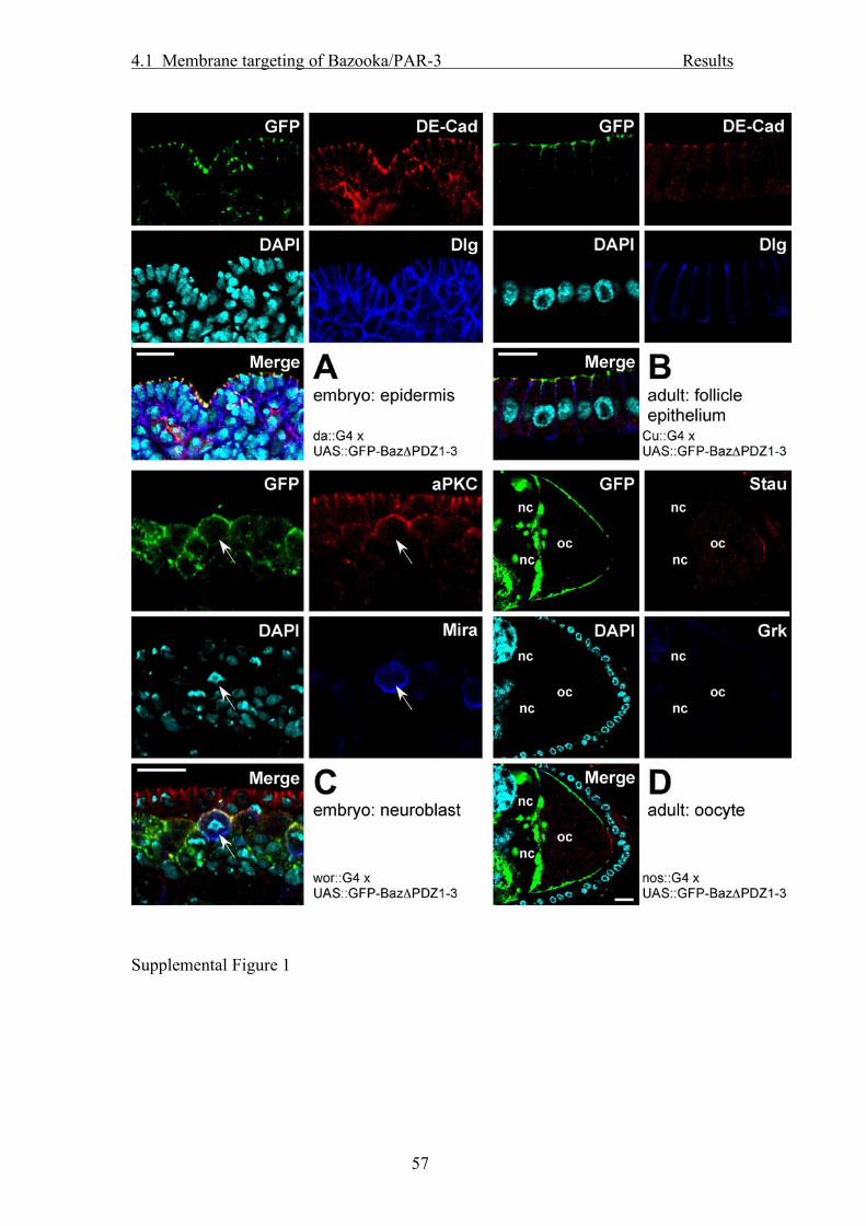
4.1 Membrane targeting of Bazooka/PAR-3 Results
57
Supplemental Figure 1

4.1 Membrane targeting of Bazooka/PAR-3 Results
58
Supplemental Figure 2

4.1 Membrane targeting of Bazooka/PAR-3 Results
59
Supplemental Figure 3
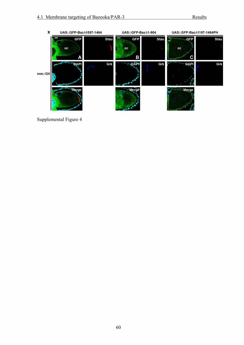
4.1 Membrane targeting of Bazooka/PAR-3 Results
60
Supplemental Figure 4

4.1 Membrane targeting of Bazooka/PAR-3 Results
61
Supplemental Figure 5

4.1 Membrane targeting of Bazooka/PAR-3 Results
62
Supplemental Figure 6

4.1 Membrane targeting of Bazooka/PAR-3 Results
63
Supplemental Figure 7
4.2 PP2A controls neuroblast polarity Results
64
4.2. PP2A antagonizes phosphorylation of Bazooka by PAR-1 to control apical-basal polarity in dividing embryonic neuroblasts
In this part I investigated the interaction between the Bazooka protein and one
particular potential interaction partner, protein phosphatase 2A (PP2A), which was
found in a yeast-two-hybrid screen in the lab before.
After verification of the interaction by co-immunoprecipitation experiments, I showed
that of the three described phosphorylation sites, only one is dephosphorylated by
PP2A in vivo.
Furthermore, the effect of PP2A phosphatase activity knock down or the
overexpression of the complementary kinase activity (PAR-1) on the apical-basal
polarity in metaphase NBs is was investigated. Michael P. Krahn, Diane Egger-Adam and Andreas Wodarz
Author contributions to the work: Michael P. Krahn: All experiments, besides*
writing of the manuscript Diane Egger-Adam: *Initial Yeast-Two-Hybrid-Screen for new interaction partners of
Baz, in which PP2A was found Andreas Wodarz: Editing of the manuscript STATUS: ACCEPTED in Developmental Cell, scheduled for issue June 16th 2009.

4.2 PP2A controls neuroblast polarity Results
65
PP2A antagonizes phosphorylation of Bazooka by PAR-1
to control apical-basal polarity in dividing embryonic
neuroblasts
Michael P. Krahn1, Diane Egger-Adam1,2 and Andreas Wodarz1*
1Abteilung Stammzellbiologie, DFG Research Center for Molecular Physiology
of the Brain (CMPB), Georg-August-Universität Göttingen, Justus-von-Liebig-
Weg 11, 37077 Göttingen, Germany
2Fakultät für Biologie, Universität Konstanz, 78434 Konstanz, Germany
*author for correspondence: [email protected], phone: +49-551-3913711,
fax: +49-551-3913713
Running title: PP2A controls neuroblast polarity
Keywords: neuroblast, polarity, stem cell, PAR proteins, PP2A, asymmetric
cell division

4.2 PP2A controls neuroblast polarity Results
66
Abstract
Bazooka/Par-3 (Baz) is a key regulator of cell polarity in epithelial cells and
neuroblasts (NBs). Phosphorylation of Baz by PAR-1 and aPKC is required for
its function in epithelia, but little is known about the dephosphorylation
mechanisms that antagonize the activities of these kinases or about the
relevance of Baz phosphorylation for NB polarity. We found that protein
phosphatase 2A (PP2A) binds to Baz via its structural A subunit. By using
phospho-specific antibodies, we show that PP2A dephosphorylates Baz at the
conserved serine residue 1085 and thereby antagonizes the kinase activity of
PAR-1. Loss of PP2A function leads to complete reversal of polarity in NBs,
giving rise to an “upside down” polarity phenotype. Overexpression of PAR-1
or Baz, or mutation of 14-3-3 proteins that bind phosphorylated Baz, causes
essentially the same phenotype, indicating that the balance of PAR-1 and
PP2A effects on Baz phosphorylation determines NB polarity.

4.2 PP2A controls neuroblast polarity Results
67
Introduction
The generation of cell fate diversity in developing and adult organisms
depends on the asymmetric division of stem cells. One of the best model
systems to study this process are the embryonic neuroblasts (NBs) in the fruit
fly Drosophila. Upon unequal cytokinesis, the NB gives rise to a ganglion
mother cell (GMC) which divides only once more and produces two neurons
or glia cells, and another NB that continues to divide asymmetrically (Wodarz,
2005; Knoblich, 2008; Zhong and Chia, 2008). A crucial prerequisite for
asymmetric cell division is the polarization of the stem cell. NB polarity is
controlled by a group of genes encoding cortical proteins which function in a
hierarchy, leading to the asymmetric localization of the cell fate determinants
Prospero, Brain Tumor (Brat) and Numb (Wodarz, 2005; Knoblich, 2008;
Zhong and Chia, 2008).
The PDZ domain protein Baz, the Drosophila homolog of C. elegans and
vertebrate PAR-3, is among the first proteins that localize to the apical NB
cortex where it recruits PAR-6 and aPKC to form the Baz/PAR-6/aPKC
complex (Kuchinke et al., 1998; Schober et al., 1999; Wodarz et al., 1999;
Wodarz et al., 2000; Petronczki and Knoblich, 2001). Together with the
proteins Discs Large (Dlg), Lethal giant larvae (Lgl) and Scribble (Scrib), the
Baz/PAR-6/aPKC complex is essential for the localization of cell fate
determinants and their adaptor proteins Miranda (Mira) and Partner of Numb
(Pon) to the basal cortex of mitotic NBs (Rhyu et al., 1994; Hirata et al., 1995;
Knoblich et al., 1995; Spana and Doe, 1995; Ohshiro et al., 2000; Peng et al.,
2000; Albertson and Doe, 2003). The assembly and activity of the Baz/PAR-
6/aPKC complex is controlled by a phosphorylation cascade triggered by

4.2 PP2A controls neuroblast polarity Results
68
activation of the mitotic kinase Aurora A, which leads to phosphorylation of
PAR-6 and activation of aPKC (Wirtz-Peitz et al., 2008). aPKC in turn
phosphorylates and inactivates Lgl at the apical cortex (Betschinger et al.,
2003). Phosphorylation of Lgl leads to dissociation of Lgl from PAR-6 and
aPKC and to its exchange for Baz. The Baz/PAR-6/aPKC complex finally
phosphorylates Numb and prevents its localization to the apical NB cortex
(Wirtz-Peitz et al., 2008). These findings clearly show that modification of
polarity proteins by phosphorylation is an important mechanism to regulate
their function in asymmetric cell division.
Baz itself is also a phosphoprotein and some phosphorylation sites relevant
for its function have been identified. Phosphorylation of S151 and S1085 by
PAR-1 is crucial for Baz function and localization in cells of the follicle
epithelium and in the oocyte (Benton and St Johnston, 2003). In rat PAR-3,
phosphorylation of S827, corresponding to S980 in Baz, by aPKC results in
decreased stability of the aPKC-PAR-3 interaction (Nagai-Tamai et al., 2002).
Overexpression of a mutant version of PAR-3 that cannot be phosphorylated
at this site (PAR-3 S827A) in MDCK cells leads to polarity defects similar to
those observed upon overexpression of a dominant-negative form of aPKC
(Suzuki et al., 2001; Nagai-Tamai et al., 2002).
In contrast, up to now little is known about the mechanisms of Baz or PAR-3
dephosphorylation counteracting the activities of PAR-1 and aPKC kinases.
Protein phosphatase 1 (PP1) has recently been shown to bind mouse PAR-3
and to dephosphorylate its serine residues S144 and S824, corresponding to
S151 and S980 of Baz (Traweger et al., 2008). A function for protein
phosphatase 2A (PP2A) as an antagonist of PAR-1 in photoreceptor cells of

4.2 PP2A controls neuroblast polarity Results
69
the Drosophila pupal retina was recently described (Nam et al., 2007). In this
system, overexpression of PAR-1 or reduction of PP2A activity led to
mislocalization of Baz and disturbed cell polarity.
PP2A is a ubiquitously expressed serine-threonine-specific phosphatase
which has crucial functions in various cellular processes such as cell cycle
control, cytoskeleton reorganization and cell polarity (Janssens and Goris,
2001). PP2A is a heterotrimeric enzyme complex consisting of a catalytic
(PP2A-C) subunit, which is only stable if tightly bound to a structural subunit of
65kD (PP2A-A). This core complex recruits one out of several regulatory
subunits (PP2A-B) which provide substrate specificity (Janssens and Goris,
2001). Whereas in vertebrates two different A subunits and various regulatory
B subunits are expressed, in Drosophila the A subunit is encoded by a single
gene and only four genes for regulatory B subunits are annotated: Twins
(Tws), Widerborst (Wdb), B56-1 and PR72.
In this study we show that a heterotrimeric complex of PP2A-A, PP2A-C(Mts)
and Tws binds to Baz and dephosphorylates Baz at the conserved serine
residue 1085. Furthermore, impaired dephosphorylation of this site caused by
lack of PP2A activity induces a dramatic phenotype in late stage embryonic
NBs characterized by a total reversal of apical-basal cell polarity. This finding
can be phenocopied either by overexpression of PAR-1 or Baz. We
furthermore show that the Drosophila homologs of PAR-5, 14-3-3ε and 14-3-
3ζ (Leonardo), function in the same pathway to control apical-basal NB
polarity.
Results

4.2 PP2A controls neuroblast polarity Results
70
PP2A is found in a complex with Baz but not with aPKC or PAR-6
In a yeast two-hybrid screen for interaction partners of the N-terminal region of
Baz (aa 1-318), we isolated a clone corresponding to aa 399-590 of the
structural A subunit of PP2A (PP2A-A) that specifically interacted with the Baz
bait (Figure S1). In an independent experimental approach, we performed
coimmunoprecipitation experiments from embryonic extracts using an
antibody directed against Baz. A 39 kD band coimmunoprecipitating with Baz
was analyzed by mass spectrometry and three peptides matched Mts, the
only catalytic subunit of PP2A annotated in the Drosophila genome (Figure
S1).
In order to test whether PP2A is able to bind Baz in vivo, we performed
coimmunoprecipitation experiments from embryonic lysates. To that aim, we
generated polyclonal peptide antisera directed against PP2A-A. These
antisera recognize a single band of 65 kD in Western blots, which
corresponds to the predicted size of PP2A-A (Figure 1A). A small proportion
of PP2A-A and Mts coimmunoprecipitated with Baz (Figure 1A). Vice versa,
GFP-Baz was present in immunoprecipitates pulled down with the anti PP2A-
A antibody (Figure 1B). In contrast, neither aPKC nor PAR-6 were found in a
complex with PP2A (Figure 1C, D).
To clarify which of the four regulatory B-subunits encoded in the Drosophila
genome were associated with the PP2A-Baz complex, HA-tagged versions of
all four B-subunits were coexpressed with GFP-Baz in S2 cells and tested for
coimmunoprecipitation with GFP-Baz. Both the Tws (Figure 1E) and the B56-
1 (Figure 1F) regulatory subunits were found in a complex with GFP-Baz,
whereas no interaction was found with Wdb (Figure 1G) or PR72 (Figure 1H).

4.2 PP2A controls neuroblast polarity Results
71
Inhibition of PP2A activity in S2 cells results in increased phosphorylation of
Baz at serine 1085
To test whether the association of Baz with PP2A controls the phosphorylation
of one of the three previously described phosphorylation sites of Baz, we
raised phospho-specific peptide antibodies against each of these
phosphorylated serine residues (pS151, pS980, pS1085). S2 cells
overexpressing GFP-Baz were treated for 4h with either DMSO as negative
control, with okadaic acid (5 nM) or with cantharidin (0.5 µM). At these
inhibitor concentrations, the inhibition of PP2A is more than tenfold higher
than the inhibition of PP1, another major cellular protein phosphatase that can
be inhibited with these substances (Barford, 1996). Subsequently, cells were
lysed, GFP-Baz was immunoprecipitated and the precipitates were subjected
to Western blot with the phospho-specific antibodies (Figure 2B - D). Whereas
GFP-Baz from DMSO treated cells was only weakly phosphorylated at any of
the three investigated sites, treatment with the phosphatase inhibitors resulted
in a strong increase of the band intensity with all three phospho-specific
antibodies (Figure 2B - D). In contrast, the total amount of Baz protein was not
affected (Figure 2A). Treatment of the cells with 100µM cantharidin, a 10-fold
excess over the EC50 of PP1, did not lead to further increase of
phosphorylation at any of the three serines (Figure 2B – D). However, a
distinct shift in the mobility of the Baz band was detectable, indicating that
PP1 or other phosphatases affected by this high concentration of the inhibitor
may dephosphorylate other, yet undescribed phosphorylation sites of Baz.
In contrast to the three phosphorylation sites in Baz, phosphorylation of aPKC
at T422 was not affected by the low concentrations of cantharidin or okadaic

4.2 PP2A controls neuroblast polarity Results
72
acid that predominantly inhibit PP2A (Figure 2G). Increased phosphorylation
of T422 was only observed after inhibition of PP1 by higher concentrations of
cantharidin (Figure 2G).
While these inhibitor experiments pointed to PP2A as the phosphatase that
specifically dephosphorylated the three serine residues of Baz, the specificity
of the inhibitors is not high enough to exclude the involvement of other
phosphatases, such as PP4 or PP6. We therefore knocked down PP2A by
application of double-stranded RNA in S2 cells, which has been shown to
work efficiently for PP2A subunits (Silverstein et al., 2002; Sathyanarayanan
et al., 2004). This approach also allowed us to clarify which one of the
different regulatory B subunits is involved in the dephosphorylation of Baz.
Knock down of PP2A-A, Mts and Tws resulted in strongly elevated
phosphorylation of S1085 (Figure 2K), whereas phosphorylation of S151 and
S980 remained unaffected (Figure 2I, J). In the absence of phosphatase
inhibitors only a smaller form of Baz (110 kD) was detectable with the anti
BazpS151 antibody (Figure 2I, cf. Figure 2B). Knock down of Wdb, B56-1 and
PR72 did not lead to elevated phosphorylation of S1085 (Figure 2K),
indicating that Tws is the only regulatory B subunit of PP2A that functions in a
complex with the core dimer of PP2A-A and Mts to dephosphorylate Baz at
S1085. PP2A-A and Mts were mutually required to stabilize each other and
the B subunits Tws and Wdb, whereas the knock-down of Wdb or Tws did not
affect the stability of PP2A-A or Mts (Figure 2L – O) (Silverstein et al., 2002).
Apical-basal polarity is totally reversed in a fraction of PP2A deficient
metaphase-NBs

4.2 PP2A controls neuroblast polarity Results
73
In order to investigate the consequences of PP2A depletion in embryonic
NBs, we examined embryos homozygous mutant for PP2A-29BGE16781, a
predicted null allele for the PP2A-A subunit which carries a P-element
insertion 38 bp 3’ of the start-codon and is embryonic lethal. The lethality of
PP2A-29BGE16781 was fully rescued by ubiquitous expression of a UAS::PP2A-
29B transgene (data not shown). In NBs at early stages of neurogenesis
(stage 9-11) we did not observe any abnormalities regarding spindle
orientation or localization of polarity proteins (Figure 3B). However, in late
neurogenesis (stage 13-15), a significant percentage of PP2A-29BGE16781
mutant NBs showed either a spindle orientation parallel to the plane of the
neuroectodermal epithelium (27%, n=99; Figure 3E, J) or a total reversal of
apical-basal cell polarity (22%; Figure 3D, J). Very few NBs with oblique
spindle orientation were found in PP2A-29BGE16781 mutant embryos (Figure
3J). In the NBs with reversed polarity, Baz, which localizes to the apical cortex
in wild type (Figure 3A, C) formed basal crescents (Figure 3D), while Mira,
which localizes to the basal cortex in wild type (Figure 3A, C), formed apical
crescents (Figure 3D). Analysis of other apically (Figure S2A – H) and basally
(Figure S2I – N) localized proteins confirmed that cortical polarity was
completely reversed in PP2A mutant embryos. To verify that this phenotype
was due to decreased activity of PP2A, we analyzed embryos homozygous
mutant for mtsXE2258 (Figure 3F) (Wassarman et al., 1996) and tws60 (Figure
3G) (Uemura et al., 1993) and found essentially the same abnormalities. To
analyze the functional requirement for PP2A in NB polarity by an additional
approach, we expressed a dominant negative version of Mts (UAS::dnMts)
(Hannus et al., 2002) in a NB specific pattern using wor::GAL4 or in a

4.2 PP2A controls neuroblast polarity Results
74
ubiquitous pattern using tubulin::GAL4 or mat67::GAL4. In all of these cases
we identified “upside down” NBs at late stages of neurogenesis at a frequency
comparable to embryos homozygous for mutations in PP2A-A, mts or tws
(Figure 3I). No polarity defects were observed in NBs at earlier stages of
neurogenesis (Figure 3H), although the UAS::dnMts was expressed from the
beginning of neurogenesis when the maternal tubulin::GAL4 or mat67::GAL4
driver lines were used. We did not detect any PP2A-29BGE16781 mutant ana- or
telophase NBs showing reversed polarity, which is probably due to an arrest
in mitosis upon loss of PP2A activity (Chen et al., 2007).
The NB polarity phenotype of PP2A mutants can be phenocopied by
overexpression of PAR-1
PAR-1 can phosphorylate Baz at relevant serine residues S151 and S1085
(Benton and St Johnston, 2003) and acts as an antagonist of PP2A with
respect to cell polarity and Baz localization in pupal photoreceptor cells (Nam
et al., 2007). In NBs, GFP-tagged PAR-1 localized predominantly to the basal
cortex (Figure S3A). To test whether PAR-1 antagonizes PP2A also in the
control of NB polarity, we overexpressed PAR-1 in NBs using the UAS-GAL4
system. The consequences on NB polarity at metaphase were essentially the
same as in PP2A mutants and upon overexpression of dominant negative Mts
(Figure 3L; Table 1). Again, polarity defects were only observed in older NBs
from stage 13 onwards. The reversion of NB polarity was dependent on the
kinase activity of PAR-1, as overexpression of a kinase-dead version of PAR-
1 (PAR-1T408A) did not result in NB polarity defects (Figure 3O; Table 1). In
contrast to the situation in PP2A mutants, overexpression of PAR-1 did not
result in mitotic arrest at metaphase. Consequently, we did observe a

4.2 PP2A controls neuroblast polarity Results
75
significant number of ana- and telophase NBs that budded off the GMC to the
apical side (Figure 3M), confirming our hypothesis that these NBs are entirely
upside-down. Besides NBs dividing with an upside down polarity we also
observed an increased number of telophase NBs dividing in an orientation
parallel to the plane of the neuroectodermal epithelium (Figure 3N).
Moderately elevated level of Baz results in reversed apical-basal NB polarity
We next addressed the question of whether changes in Baz phosphorylation
are responsible for the reversal of NB polarity upon loss of PP2A function or
overexpression of PAR-1. Using different driver lines (Wor::GAL4,
Pros::GAL4, asense::GAL4) and different temperatures, we determined that
the effects of Baz overexpression were dosage dependent. Weak
overexpression of wild type Baz, either untagged or N-terminally tagged with
GFP, led to complete reversal of NB polarity (Figure 3P) in a significant
fraction of NBs from stage 13 onwards (23.3%, Table 1). Similar to PP2A
knockdown or PAR-1 overexpression, the number of dividing NBs showing a
spindle orientation rotated by 90° was also increased (20.8%, Table 1).
Furthermore, we frequently observed NBs with reversed polarity in ana- and
telophase, budding off the GMC to the apical side, similar to the phenotype
upon PAR-1 overexpression (Table 1). In order to visualize this event and to
further elucidate if the reversed NB polarity was immediately established or
was caused by relocalization of initially correctly targeted apical and basal
proteins, we performed live imaging of embryos co-overexpressing wild type
Baz and Pon-GFP (Lu et al., 1999) (Movies S2 and S3). In wild type embryos,
Pon-GFP accumulated at the basal cortex and exclusively segregated into the
basally localized GMC (Movie S1). By contrast, upon overexpression of Baz

4.2 PP2A controls neuroblast polarity Results
76
we frequently observed asymmetric divisions in parallel to the plane of the
overlying epithelium (Movie S2) and also asymmetric divisions in which the
GMC budded off to the apical side (Movie S3). In the latter case, Pon-GFP
was directly targeted to the apical cortex without prior accumulation at the
basal cortex (Movie S3).
Our observations bring up the question of whether the reversal of cortical
apical-basal NB polarity was due to a mistargeting of apical and basal protein
complexes without affecting the intrinsic asymmetry of the mitotic spindle, or
whether the whole NB, including the mitotic spindle, was upside down. To
investigate this, we stained centrosomes with γ-tubulin (Figure S4) and Cnn
(data not shown). In wild type, the apical centrosome of metaphase and
anaphase NBs was significantly larger than the basal one (Figure S4A, B)
(Kaltschmidt et al., 2000). In NBs showing reversed apical-basal polarity upon
overexpression of Baz, the basal centrosome was larger than the apical
centrosome (Figure S4C). In general, the centrosome close to the Baz
crescent was larger than the centrosome opposite to the Baz crescent,
irrespective of the orientation of the NB relative to the overlying epithelium
(Figure S4C, D). From these observations we conclude that the affected NBs
do not only exhibit reversed apical-basal polarity of the cytocortex but are
entirely upside down.
Next we investigated if the phosphorylation of serine 1085 is crucial for the
polarity reversal in NBs. Overexpression of Baz in which serine 1085 was
exchanged for glutamate (BazS1085E), mimicking a constitutively
phosphorylated protein, showed similar effects as overexpression of wild type
Baz (20.4% reversed polarity and 25.7% spindle rotated 90°, Table 1) without

4.2 PP2A controls neuroblast polarity Results
77
further enhancement of the phenotype, consistent with our observation that
wild type Baz gets phosphorylated on S1085 upon overexpression (Figure
S5). In contrast, overexpression at the same level (Figure S6) of
nonphosphorylatable Baz in which serine 1085 was exchanged for alanine
(BazS1085A) caused polarity reversal in only 4,1% of NBs and spindle
misorientation in 14,6% of NBs (Table 1). Overexpression of a version of Baz
in which both S151 and S1085 were mutated to alanine (BazS151AS1085A)
did not cause any significant increase of NBs with abnormal polarity compared
to wild type (Table 1), suggesting that S151 contributes to a minor extent to
the polarity defects observed upon overexpression of Baz.
14-3-3 proteins interact with Baz in the control of neuroblast polarity
In Drosophila, two homologues of PAR-5 exist: 14-3-3ε and 14-3-3ζ
(Leonardo, Leo). Both have been shown to interact with Baz posphorylated by
PAR-1 and thereby exclude Baz from the basolateral membrane in the follicle
cell epithelium (Benton and St Johnston, 2003). We have confirmed the
phosphorylation-dependent interaction of 14-3-3ε with S1085 of Baz by
coimmunoprecipitation experiments in transfected S2 cells (Figure S7).
Interestingly, the binding of 14-3-3ε to Baz was inversely correlated with
binding of aPKC to Baz (Figure S7), suggesting that phosphorylation of S1085
regulates the binding of Baz to aPKC.
In embryos mutant for a loss-of-function and a hypomorphic allele of 14-3-3ζ
(14-3-3leo12BL and 14-3-3leoP1188), we detected the same phenotype of
reversed apical-basal NB polarity as in PP2A mutants and upon PAR-1
overexpression (Figure 3Q, Table 1 and data not shown). In zygotic mutants
4.2 PP2A controls neuroblast polarity Results
78
of a null allele of 14-3-3ε, 14-3-3εj2B10, only a few (6.5%, Table 1) embryonic
NBs showed reversed polarity, which could be due to the fact that the
maternal component is stronger and longer lasting than that of 14-3-3ζ.
Indeed, zygotic mutant 14-3-3εj2B10 animals survive until late larval stages.
Based on these findings we speculated that like in the follicle epithelium, 14-3-
3 proteins may be required to exclude Baz from the basal cortex in NBs and
thus to ensure proper NB polarity. To test our hypothesis, we co-
overexpressed wild type Baz and 14-3-3ζ or 14-3-3ε in NBs. Indeed, the
percentage of NBs showing a reversed polarity was strongly decreased
compared to overexpression of Baz alone (Table 1).
Discussion
Apical-basal polarity of NBs is controlled by a relatively small number of
proteins which assemble into protein complexes localized to the NB cortex in
an asymmetric fashion (Wodarz, 2005; Knoblich, 2008). These cortical
proteins interact with each other in a functional hierarchy. At the top of the
hierarchy is Baz, because it can localize to the apical NB cortex in loss-of-
function mutants for any of the other factors, including PAR-6, aPKC, Insc,
Pins and others (Schober et al., 1999; Wodarz et al., 1999; Yu et al., 2000;
Rolls et al., 2003); Kim et al., submitted).
Here we have shown that Baz gets frequently mislocalized to the basal NB
cortex when it is moderately overexpressed or when it is excessively
phosphorylated at S1085, either by overexpression of PAR-1 or by loss-of-
function of PP2A. We expect that similar antagonistic activities of kinases and

4.2 PP2A controls neuroblast polarity Results
79
phosphatases regulate the phosphorylation state of additional sites of
Baz/PAR-3 that are relevant in different cellular contexts. Loss-of-function of
14-3-3ζ and to a lesser extent of 14-3-3ε causes mislocalization of
endogenous Baz in NBs, whereas overexpression of 14-3-3ζ and 14-3-3ε
suppresses the mislocalization of overexpressed Baz. We therefore suggest
that the ratio of Baz phosphorylated at S1085 to the amount of available 14-3-
3 determines whether Baz gets mislocalized to the basal cortex. In this model,
the 14-3-3 proteins function as a buffer to inactivate mislocalized,
phosphorylated Baz. This inactivation could be explained by the inhibition of
aPKC binding to Baz upon association of 14-3-3 with Baz. If the amount of
overexpressed Baz exceeds the buffering capacity of 14-3-3, this would lead
to the formation of active Baz/aPKC complexes at the basal cortex. These
basally localized, active Baz/aPKC complexes may in turn affect the
localization of PAR-1. The mammalian aPKC homolog PKCζ can
phosphorylate PAR-1 at a conserved serine residue and this phosphorylation
causes a strong reduction of PAR-1 kinase activity and the release of PAR-1
from the plasma membrane (Hurov et al., 2004). If the same was true in
Drosophila, it would explain the total reversal of NB polarity, because the now
basally localized aPKC would phosphorylate PAR-1, which would cause its
release from the membrane and the establishment of a new apical cortical
domain at the previously basal cortex.
PAR-1, 14-3-3 proteins and PP2A are strongly expressed during oogenesis
and maternal contributions may account for difficulties identifying
requirements during early embryogenesis. On the other hand, eliminating
maternal expression of these genes results in phenotypes too severe to allow

4.2 PP2A controls neuroblast polarity Results
80
the study of neurogenesis (Wassarman et al., 1996; Shulman et al., 2000;
Benton et al., 2002). However, overexpression of a dominant-negative form of
Mts from early neurogenesis onwards also caused polarity reversal only in
late stage NBs. While this experiment does not exclude the possibility that the
late onset of polarity reversal in NBs is due to the perdurance of the maternal
gene products, it points to a fundamental difference in the mechanism of how
NB polarity is controlled immediately after delamination as opposed to
subsequent asymmetric divisions. The majority of late stage NBs showing
polarity reversal were not in direct contact with the overlying epithelium and
thus may rely exclusively on intrinsic polarity cues, in contrast to NBs that
have just delaminated and maintain contact to the overlying epithelium. Late
stage NBs lacking contact to the overlying epithelium show a higher variability
of spindle orientation as compared to early stage NBs in close contact to the
epithelium (Siegrist and Doe, 2006). Thus, late stage NBs may be particularly
sensitive to changes in the phosphorylation state and general activity level of
Baz, because they rely on Baz as the main cue for orienting their polarity axis.
It is interesting to note that mutations uncoupling spindle orientation from the
localization of cell fate determinants commonly show fully random spindle
orientation, including a variety of oblique orientations (Izumi et al., 2006; Siller
and Doe, 2009). In contrast, hyperphosphorylation of Baz at S1085 resulted
very rarely in oblique orientations and spindles were always aligned with the
asymmetric crescents of cell fate determinants. Although we currently do not
have a good explanation for why there is a strong bias for either total reversal
of polarity or misorientation of the spindle by 90°, our findings point to the

4.2 PP2A controls neuroblast polarity Results
81
existence of a spatial cue functioning upstream of Baz that defines a polarity
axis perpendicular to the plane of the epithelium.

4.2 PP2A controls neuroblast polarity Results
82
Experimental Procedures
Antibodies
Phospho-specific antibodies against Baz phosphorylated at serine 151, 980
and 1085 were raised by injection of the phosphorylated peptides
CLMVRRSpSDPNLL (pS151), CDALGRRpSISEKH (pS980) and
CGMKKSSpSLESLQ (pS1085) into rabbits and subsequent affinity
purification against the phosphorylated peptide.
Peptide antibodies directed against PP2A-A (PP2A-29B) were raised by
injection of the peptides AASDKSVDDSLYPIAC (aa 2-16) and
PYVRDLVSDPNPHVKC (aa 330-344) into rabbits (Eurogentec, Seraing,
Belgium).
Immunoprecipitation and Western blotting
For immunoprecipitations, wild type embryos from an overnight collection
were dechorionated and lysed in lysis buffer (1% Triton X-100, 150mM NaCl,
1mM CaCl2, 1mM MgCl2, 25mM Hepes pH 7.44) supplemented with protease
inhibitors. S2 cells were lysed in the same buffer. After centrifugation, 2 µl of
rat anti Baz (Wodarz et al., 1999), 2 µl of rabbit anti PP2A-A, 2µl of rabbit anti
PKCζ C20 (Santa Cruz Biotechnology, Inc.), 2 µl of guinea-pig anti PAR-6, 2
µl of rabbit anti-GFP (Molecular Probes #A11122), or 2 µl of the
corresponding preimmune serum were added to cell lysate corresponding to
500 µg total protein. Immune complexes were harvested using protein A/G-
conjugated agarose (Roche), washed five times in lysis buffer and boiled in 2x
SDS sample buffer before SDS-PAGE and Western blot. For mass
spectrometry, immune complexes precipitated with rabbit anti Baz were

4.2 PP2A controls neuroblast polarity Results
83
separated by SDS-PAGE, gels were silver stained and selected bands were
cut out. Bands were digested with trypsin and analyzed by MALDI-TOF at the
ZMMK of the University of Cologne.
Western blotting was done according to standard procedures. Primary
antibodies used for Western blotting are listed in the Supplemental Material.
Immunohistochemistry
Embryos were fixed in 4% formaldehyde, phosphate buffer pH 7.4 according
to standard procedures. Primary antibodies used for indirect
immunofluorescence are listed in the Supplemental Material. Images were
taken on a Zeiss LSM 510 Meta confocal microscope and processed using
Adobe Photoshop.
Phosphatase inhibition and RNA interference in S2 cells
Inhibition of phosphatases was achieved by incubation of S2 cells with
okadaic acid (5 nM) or cantharidin (5 µM or 100 µM) for four h, followed by
lysis in lysis buffer supplemented with the same concentration of phosphatase
inhibitors.
Knockdown of the different PP2A subunits by RNA interference in S2 cells
was done as described (Silverstein et al., 2002; Sathyanarayanan et al.,
2004).
References
Albertson, R., and Doe, C.Q. (2003). Dlg, Scrib and Lgl regulate neuroblast cell size and mitotic spindle asymmetry. Nat Cell Biol 5, 166-170.
Barford, D. (1996). Molecular mechanisms of the protein serine/threonine phosphatases. Trends Biochem Sci 21, 407-412.
Benton, R., Palacios, I.M., and St Johnston, D. (2002). Drosophila 14-3-3/PAR-5 is an essential mediator of PAR-1 function in axis formation. Dev Cell 3, 659-671.
4.2 PP2A controls neuroblast polarity Results
84
Benton, R., and St Johnston, D. (2003). Drosophila PAR-1 and 14-3-3 inhibit Bazooka/PAR-3 to establish complementary cortical domains in polarized cells. Cell 115, 691-704.
Betschinger, J., Mechtler, K., and Knoblich, J.A. (2003). The Par complex directs asymmetric cell division by phosphorylating the cytoskeletal protein Lgl. Nature 422, 326-330.
Chen, F., Archambault, V., Kar, A., Lio, P., D'Avino, P.P., Sinka, R., Lilley, K., Laue, E.D., Deak, P., Capalbo, L., et al. (2007). Multiple protein phosphatases are required for mitosis in Drosophila. Curr Biol 17, 293-303.
Hannus, M., Feiguin, F., Heisenberg, C.P., and Eaton, S. (2002). Planar cell polarization requires Widerborst, a B' regulatory subunit of protein phosphatase 2A. Development 129, 3493-3503.
Hirata, J., Nakagoshi, H., Nabeshima, Y., and Matsuzaki, F. (1995). Asymmetric segregation of the homeodomain protein Prospero during Drosophila development. Nature 377, 627-630.
Hurov, J.B., Watkins, J.L., and Piwnica-Worms, H. (2004). Atypical PKC phosphorylates PAR-1 kinases to regulate localization and activity. Curr Biol 14, 736-741.
Izumi, Y., Ohta, N., Hisata, K., Raabe, T., and Matsuzaki, F. (2006). Drosophila Pins-binding protein Mud regulates spindle-polarity coupling and centrosome organization. Nat Cell Biol 8, 586-593.
Janssens, V., and Goris, J. (2001). Protein phosphatase 2A: a highly regulated family of serine/threonine phosphatases implicated in cell growth and signalling. Biochem J 353, 417-439.
Kaltschmidt, J.A., Davidson, C.M., Brown, N.H., and Brand, A.H. (2000). Rotation and asymmetry of the mitotic spindle direct asymmetric cell division in the developing central nervous system. Nat Cell Biol 2, 7-12.
Knoblich, J.A. (2008). Mechanisms of asymmetric stem cell division. Cell 132, 583-597.
Knoblich, J.A., Jan, L.Y., and Jan, Y.N. (1995). Asymmetric segregation of Numb and Prospero during cell division. Nature 377, 624-627.
Kuchinke, U., Grawe, F., and Knust, E. (1998). Control of spindle orientation in Drosophila by the Par-3-related PDZ- domain protein Bazooka. Curr Biol 8, 1357-1365.
Lu, B., Ackerman, L., Jan, L.Y., and Jan, Y.N. (1999). Modes of protein movement that lead to the asymmetric localization of partner of Numb during Drosophila neuroblast division. Mol Cell 4, 883-891.
Nagai-Tamai, Y., Mizuno, K., Hirose, T., Suzuki, A., and Ohno, S. (2002). Regulated protein-protein interaction between aPKC and PAR-3 plays an essential role in the polarization of epithelial cells. Genes Cells 7, 1161-1171.
Nam, S.C., Mukhopadhyay, B., and Choi, K.W. (2007). Antagonistic functions of Par-1 kinase and protein phosphatase 2A are required for localization of Bazooka and photoreceptor morphogenesis in Drosophila. Dev Biol 306, 624-635.
Ohshiro, T., Yagami, T., Zhang, C., and Matsuzaki, F. (2000). Role of cortical tumour-suppressor proteins in asymmetric division of Drosophila neuroblast. Nature 408, 593-596.
Peng, C.Y., Manning, L., Albertson, R., and Doe, C.Q. (2000). The tumour-suppressor genes lgl and dlg regulate basal protein targeting in Drosophila neuroblasts. Nature 408, 596-600.
Petronczki, M., and Knoblich, J.A. (2001). DmPAR-6 directs epithelial polarity and asymmetric cell division of neuroblasts in Drosophila. Nat Cell Biol 3, 43-49.
Rhyu, M.S., Jan, L.Y., and Jan, Y.N. (1994). Asymmetric distribution of numb protein during division of the sensory organ precursor cell confers distinct fates to daughter cells [see comments]. Cell 76, 477-491.
Rolls, M.M., Albertson, R., Shih, H.P., Lee, C.Y., and Doe, C.Q. (2003). Drosophila aPKC regulates cell polarity and cell proliferation in neuroblasts and epithelia. J Cell Biol 163, 1089-1098.

4.2 PP2A controls neuroblast polarity Results
85
Sathyanarayanan, S., Zheng, X., Xiao, R., and Sehgal, A. (2004). Posttranslational regulation of Drosophila PERIOD protein by protein phosphatase 2A. Cell 116, 603-615.
Schober, M., Schaefer, M., and Knoblich, J.A. (1999). Bazooka recruits Inscuteable to orient asymmetric cell divisions in Drosophila neuroblasts. Nature 402, 548-551.
Shulman, J.M., Benton, R., and St Johnston, D. (2000). The Drosophila homolog of C. elegans PAR-1 organizes the oocyte cytoskeleton and directs oskar mRNA localization to the posterior pole. Cell 101, 377-388.
Siegrist, S.E., and Doe, C.Q. (2006). Extrinsic cues orient the cell division axis in Drosophila embryonic neuroblasts. Development 133, 529-536.
Siller, K.H., and Doe, C.Q. (2009). Spindle orientation during asymmetric cell division. Nat Cell Biol 11, 365-374.
Silverstein, A.M., Barrow, C.A., Davis, A.J., and Mumby, M.C. (2002). Actions of PP2A on the MAP kinase pathway and apoptosis are mediated by distinct regulatory subunits. Proc Natl Acad Sci U S A 99, 4221-4226.
Spana, E.P., and Doe, C.Q. (1995). The prospero transcription factor is asymmetrically localized to the cell cortex during neuroblast mitosis in Drosophila. Development 121, 3187-3195.
Suzuki, A., Yamanaka, T., Hirose, T., Manabe, N., Mizuno, K., Shimizu, M., Akimoto, K., Izumi, Y., Ohnishi, T., and Ohno, S. (2001). Atypical protein kinase C is involved in the evolutionarily conserved par protein complex and plays a critical role in establishing epithelia- specific junctional structures. J Cell Biol 152, 1183-1196.
Traweger, A., Wiggin, G., Taylor, L., Tate, S.A., Metalnikov, P., and Pawson, T. (2008). Protein phosphatase 1 regulates the phosphorylation state of the polarity scaffold Par-3. Proc Natl Acad Sci U S A 105, 10402-10407.
Uemura, T., Shiomi, K., Togashi, S., and Takeichi, M. (1993). Mutation of twins encoding a regulator of protein phosphatase 2A leads to pattern duplication in Drosophila imaginal discs. Genes Dev 7, 429-440.
Wassarman, D.A., Solomon, N.M., Chang, H.C., Karim, F.D., Therrien, M., and Rubin, G.M. (1996). Protein phosphatase 2A positively and negatively regulates Ras1-mediated photoreceptor development in Drosophila. Genes Dev 10, 272-278.
Wirtz-Peitz, F., Nishimura, T., and Knoblich, J.A. (2008). Linking cell cycle to asymmetric division: Aurora-A phosphorylates the Par complex to regulate Numb localization. Cell 135, 161-173.
Wodarz, A. (2005). Molecular control of cell polarity and asymmetric cell division in Drosophila neuroblasts. Curr Opin Cell Biol 17, 475-481.
Wodarz, A., Ramrath, A., Grimm, A., and Knust, E. (2000). Drosophila atypical protein kinase C associates with Bazooka and controls polarity of epithelia and neuroblasts. J Cell Biol 150, 1361-1374.
Wodarz, A., Ramrath, A., Kuchinke, U., and Knust, E. (1999). Bazooka provides an apical cue for Inscuteable localization in Drosophila neuroblasts. Nature 402, 544-547.
Yu, F., Morin, X., Cai, Y., Yang, X., and Chia, W. (2000). Analysis of partner of inscuteable, a novel player of Drosophila asymmetric divisions, reveals two distinct steps in inscuteable apical localization. Cell 100, 399-409.
Zhong, W., and Chia, W. (2008). Neurogenesis and asymmetric cell division. Curr Opin Neurobiol 18, 4-11.
Acknowledgements

4.2 PP2A controls neuroblast polarity Results
86
We thank J. Botas, S. Bray, W. Chia, J. Knoblich, B. Lu, W. Odenwald, E.
Schejter, A. Segal, D. St Johnston, T. Uemura and G. Vorbrueggen for
sending fly stocks and antibodies. We also thank the Bloomington Drosophila
stock center at the University of Indiana for sending numerous fly stocks, the
Developmental Studies Hybridoma Bank at the University of Iowa for sending
hybridoma cells and supernatants and the Drosophila Genomics Resource
Center for sending ESTs and vectors. Stefan Mueller (ZMMK, University of
Cologne) performed the MALDI-TOF analyses of Baz interaction partners.
Karen Fricke and Mona Honemann-Capito provided expert technical
assistance. We also thank the members of the Wodarz lab for discussion.
This work was supported by grants from the Deutsche
Forschungsgemeinschaft to A. W. (SFB 590, TP A2; WO 584/4-1, 4-2; DFG
Research Center Molecular Physiology of the Brain, CMPB).
Figure Legends
Figure 1. Baz binds to PP2A in vivo. (A) Wild type embryonic extracts were
immunoprecipitated with anti Baz antiserum (IP Baz) or the corresponding
preimmune serum (IP pre). Blots were probed for Baz, PP2A-A and Mts. (B)
PP2A-A antibody was used for immunoprecipitation from extracts of embryos
expressing GFP-Baz. Blots were probed for PP2A-A and GFP to detect GFP-
Baz. (C, D) Immunoprecipitates of wild type embryonic extracts pulled down
with anti aPKC (C) or anti PAR-6 antibody (D). (E – H) The Tws and B56-1
regulatory B subunits of PP2A coimmunoprecipitate with GFP-Baz. S2 cells
were cotransfected with GFP-Baz and HA-tagged Tws (E), B56-1 (F), Wdb
(G) and PR72 (H). Lysates were precipitated with anti-GFP and probed for

4.2 PP2A controls neuroblast polarity Results
87
Baz and the HA-tag. Bands of interest are indicated by asterisks. Note that
Baz always runs as a series of bands (marked by a bar next to the asterisks)
in SDS-PAGE that are generated by proteolytic processing or degradation of
the protein. In (E – H) S2 cells transfected only with the HA-tagged B subunits
of PP2A were used as negative controls.
Figure 2. The phosphorylation state of three conserved serine residues of Baz
can be monitored by phospho-specific antibodies. (A – G) S2 cells expressing
GFP-Baz were treated either with DMSO as negative control or with the
phosphatase inhibitors okadaic acid (OA) and cantharidin (Canth) at the
indicated concentrations. GFP-Baz was immunoprecipitated with anti GFP
antibody and the precipitates were subsequently probed with anti Baz (A), anti
BazpS151 (B), anti BazpS980 (C) and anti BazpS1085 (D). Lysates were also
probed for actin (E), aPKC (F) and aPKCpT422 (G). (H – P) Serine 1085 of
Baz is specifically dephosphorylated by a heterotrimeric complex of PP2A-A,
Mts and Tws. S2 cells were treated with double stranded RNA corresponding
to different subunits of PP2A indicated on top (X RNAi). Double stranded RNA
corresponding to GFP was used as negative control. Lysates were subjected
to Western blots with the antibodies indicated on the left.
Figure 3. (A – I) Loss of PP2A function leads to complete apical-basal polarity
reversal in a fraction of embryonic NBs. Embryos of the indicated genotypes
and developmental stages were stained for Baz (red), Mira (blue) and DAPI
(turquoise). (J) Quantification of spindle orientation in wild type and PP2A-
29BGE16781 mutant embryos. Spindle orientation was determined by drawing a
line through the center of the NB that dissected the Baz crescent in the

4.2 PP2A controls neuroblast polarity Results
88
middle. The angle of that line to a line perpendicular to the plane of the
overlying epithelium was measured in increments of 10°. (K – Q) Stage 13 NB
polarity is reversed upon overexpression of PAR-1 and Baz and in leo
mutants. Scale bars = 5 µm. Apical is up.

4.2 PP2A controls neuroblast polarity Results
89
Figure 1

4.2 PP2A controls neuroblast polarity Results
90
Figure 2

4.2 PP2A controls neuroblast polarity Results
91
Figure 3

4.2 PP2A controls neuroblast polarity Results
92
Table
Genotype NB polarity [%] n metaphase reversed rotated 90° normal
wild type 0.6 10.6 88.8 170 PP2A-29BGE16781/PP2A-29BGE16781 26.5 24.9 49.6 163 UAS::PAR-1 x Wor::GAL4 19.0 22.1 58.9 168 UAS::PAR-1T408A x Wor::GAL4 2.6 12.9 84.5 162 UAS::Baz x Wor::GAL4 23.3 20.8 55.9 168 UAS::BazS1085E x Wor::GAL4 20.4 25.7 53.9 161 UAS::BazS1085A x Wor::GAL4 4.1 14.6 81.3 167 UAS::BazS151AS1085A x Wor::G4
0.6 8.1 91.3 160
14-3-3leo12BL/ 14-3-3leo12BL 25.8 20.5 53.7 160 14-3-3leoP1188/ 14-3-3leoP1188 18.4 17.9 63.7 163 14-3-3εj2B10 / 14-3-3εj2B10 6.5 11.3 82.2 165 UAS::Baz, UAS::14-3-3leo x Wor::GAL4
7.9 12.7 79.4 161
UAS::Baz, UAS::14-3-3ε x Wor::GAL4
5.4 13.0 71.6 168
telophase wild type 0 4.9 95.1 61 UAS::Baz x Wor::GAL4 21.1 31.6 47.4 57 Table 1. Quantification of NB polarity defects in embryos at stage 13-15. For
measurements of NB polarity at metaphase, the angle between a line
perpendicular to the plane of the ectodermal epithelium and a line from the
center of the NB through the center of the Baz crescent was measured. For
telophase NBs, the angle of a line connecting the centers of the two
separating daughter cells to the line perpendicular to the plane of the
epithelium was measured. Angles between 0° and 30° were scored as normal
polarity, angles between 60° and 120° were scored as polarity rotated 90° and
angles between 150° and 180° were scored as reversed polarity. Oblique
spindle orientations between 30° and 60° or between 120° and 150° were so
rare (see Figure 3J) that they were not counted here. The numbers for wild
type and homozygous mutant PP2A-29BGE16781 NBs shown here differ from

4.2 PP2A controls neuroblast polarity Results
93
those shown in Figure 3J because they were obtained in independent
experiments.
4.2 PP2A controls neuroblast polarity Results
94
Supplemental Material
Supplemental Methods
Fly stocks
The PP2A-29B null allele, the P-element insertion P{GE16781} was obtained
from GenExel (Daejeon, South Korea). tws60 was obtained from Tadashi
Uemura (Uemura et al., 1993), UAS::dnMts from Amita Sehgal (Hannus et al.,
2002; Sathyanarayanan et al., 2004), UAS::PAR-1 and UAS::PAR-1T408A
from Bingwei Lu (Sun et al., 2001), UAS::PAR-1-GFP from D. St Johnston
(Huynh et al., 2001), UAS::14-3-3ε from J. Botas (Chen et al., 2003), UAS::14-
3-3ζ from G. Vorbrueggen (Kockel et al., 1997). UAS::Baz- and UAS::PP2A-
29B transgenic fly lines were generated using standard germ line
transformation. For overexpression studies with UAS::Baz, five independent
P-element insertions were used for each experiment. All other strains used in
this study were provided by the Bloomington Drosophila stock center.
Antibodies for Western Blotting
Primary antibodies were used for Western blotting according to standard
procedures (Wodarz, 2008) as follows: rabbit anti Baz (1:2000) (Wodarz et al.,
1999), rabbit anti PP2A-A (1:2000), rat anti Mts (1:500) (Shiomi et al., 1994),
mouse anti GFP (1:1000; Roche 11814460001), rabbit anti-PKCζ C20
(1:2000; Santa Cruz Biotechnology, Inc.), guinea-pig anti PAR-6 (1:1000; Kim
et al. submitted), mouse anti HA 12CA5, (1:1000; Roche), rabbit anti
BazpS151 (1:100), rabbit anti BazpS980 (1:100), rabbit anti BazpS1085
(1:100), rabbit anti actin A2066, (1:1000; SIGMA), rabbit anti-phospho-PKCζ

4.2 PP2A controls neuroblast polarity Results
95
T410 (1:1000; Santa Cruz Biotechnology, Inc.), guinea pig anti Wdb (1:2000)
(Sathyanarayanan et al., 2004), rat anti Tws (1:500) (Shiomi et al., 1994).

4.2 PP2A controls neuroblast polarity Results
96
Antibodies for immunohistochemistry
The primary antibodies used were rabbit anti Baz (1:1000) (Wodarz et al.,
1999), rat anti Baz (1:500) (Wodarz et al., 1999), guinea-pig anti Mira (1:1000;
Kim et al. submitted), rabbit anti PKCζ C20 (1:1000; Santa Cruz
Biotechnology, Inc.), rabbit anti Gαi (1:200) (Schaefer et al., 2001), rabbit anti
Insc (1:1000) (Kraut and Campos-Ortega, 1996), rabbit anti Pins (1:1000) (Yu
et al., 2000), rabbit anti Numb (1:1000) (Rhyu et al., 1994), rabbit anti Pon
(1:1000) (Lu et al., 1998), mouse anti Pros MR1A (1:50; DSHB), mouse anti γ-
tubulin GTU-88 (1:1000; Sigma), mouse anti GFP 3E6, (1:1000; Invitrogen),
rabbit anti BazpS1085 (1:200). DNA was stained with DAPI (Invitrogen).
Primary antibodies were detected with secondary antibodies conjugated to
Cy2 and Cy3 (Jackson) or Alexa 647 (Invitrogen).
DNA and constructs
The coding regions of PP2A B-subunits (Tws, Wdb, B56-1 and PR72), 14-3-
3ε and 14-3-3ζ were amplified by PCR from full-length EST clones
(Drosophila Genomics Resources Center) as template using the following
oligonucleotides (in 5’ – 3’ orientation):
Tws-for: CACCATGGCCGGTAATGGAGAGG
Tws-rev: GGCTAAAATTTATCCTGAAATATGAAGAGG
Wdb-for: CACCATGTCATCGGGCACGTTTGTG
Wdb-rev: TTAGTTGTCCGCCTTATCCTGTTTG
B56-1-for: CACCATGGTCTTCGGTGCTATGTTG
B56-1-rev: TTAGTAGTTGTTTAGTGTTGTTAATGTGG
PR72-for: CACCATGCAAGCCAATTCGAGCA
PR72-rev: TTAATCGTTTTCCTCGGATATGAGC
4.2 PP2A controls neuroblast polarity Results
97
14-3-3ε for: CACCATGACTGAGCGCGAGAACAAT
14-3-3ε rev: CGACACGTCCTGATCCTCAAC
14-3-3ζ for: CACCATGTCGACAGTCGATAAGGAAGAG
14-3-3ζ rev: GTTGTCGCCGCCCTCC
The PCR products were cloned into the pENTR vector using the pENTR
Directional TOPO Cloning Kit (Invitrogen). Using Gateway Technology
(Invitrogen) these constructs were recombined into the pAHW expression
vector carrying N-terminal HA under control of the actin-5C promoter (PP2A
B-subunits) or into the pAWH vector carrying C-terminal HA under control of
the actin-5C promoter (14-3-3 constructs).
Baz full length was cloned into pENTR and was subsequently recombined into
different expression vectors (AGW, TGW, PW; Murphy lab, Drosophila
Genomics Resources Center) using the following primers:
Baz-for: CACCATGAAGGTCACCGTCTGCTTCGGC
Baz-rev: TCACACCTTGGAGGCGTGTG
The QuickChange Site-Directed Mutagenesis Kit (Stratagene) was used to
generate defined point mutations with full length Baz cDNA in pENTR as
template. The following oligonucleotides were used for mutagenesis (mutation
underlined):
BazS1085A-for: GAAGTCCTCGGCGTTGGAGTCGCTCCAGACCATGG
BazS1085A-rev: CCATGGTCTGGAGCGACTCCAACGCCGAGGACTTC
BazS1085E-for: AAGAAGTCCTCGGAGTTGGAGTCGCTCCAGACCATG
BazS1085E-rev: CATGGTCTGGAGCGACTCCAACTCCGAGGACTTCTT

4.2 PP2A controls neuroblast polarity Results
98
Supplemental Figure and Movie Legends
Figure S1. Identification of PP2A as an interaction partner of Baz. (A) In a
yeast two-hybrid screen with the N-terminal 318 aa of Baz as bait we
identified an interacting clone containing aa 399 – 480 of PP2A-A. (B) In an
independent coimmunoprecipitation experiment, we precipitated Baz from wild
type embryonic extract with a polyclonal antibody raised against the N-
terminal 297 aa of Baz (Wodarz et al., 1999). Coprecipitating proteins were
separated by SDS-PAGE and stained with silver nitrate. A band of 39 kD that
was not present in the preimmune control was cut out and analyzed by mass
spectrometry. The Mascot search algorithm revealed that the masses of three
peptides derived from this band correspond to the masses of three peptides of
Mts (marked in red), the 36 kD catalytic subunit of PP2A. While the
significance of this hit is not high enough to claim unambiguously that the 39
kD band contains Mts, it is consistent with the size of Mts (36 kD) and with the
results of our coimmunoprecipitation analysis using Mts specific antibodies
(Figure 1A).
Figure S2. Reversal of apical-basal NB polarity in PP2A-29BGE16781 mutants.
Polarity reversal in PP2A-29BGE16781 mutants was observed with respect to
the apically localized proteins (red) aPKC (A, B), Gαi (C, D), Insc (E, F) and
Pins (G, H) and the basally localized proteins (blue) Numb (I, J), Pon (K, L)
and Pros (M, N). DNA was stained with DAPI (turquoise). Scale bars = 5 µm.
Apical is to the top in all panels.
Figure S3. (A) PAR-1-GFP localizes predominantly to the basal cortex of
mitotic NBs. PAR-1-GFP (green) was expressed in the embryonic nervous

4.2 PP2A controls neuroblast polarity Results
99
system using the Pros::GAL4 driver line. The stage 13 embryo shown here
was also stained for DAPI (turquoise), Baz (red) and Mira (blue). (B) In NBs
with reversed polarity that overexpress both PAR-1-GFP and Baz under
control of the worniu::GAL4 driver, PAR-1-GFP localizes to the apical cortex
opposite of the basally localized Baz. Scale bar = 5 µm. Apical is to the top.
Figure S4. The asymmetry of centrosome size correlates with the localization
of Baz in NBs with altered polarity. (A, B) Wild type embryos and (C, D)
embryos overexpressing Baz were stained for γ-tubulin (red), Mira (blue),
DAPI (turquoise) and Baz (green, only in (C) and (D)). Note that in the wild
type NB at metaphase (A) the apical centrosome (arrow) is larger than the
basal centrosome (arrowhead). (B) This size asymmetry is more pronounced
in ana- and telophase. (C, D) In the NBs with altered polarity due to
overexpression of Baz, the centrosome close to the Baz crescent (arrow) is
always larger than the centrosome close to the Mira crescent (arrowhead),
irrespective of the orientation of the spindle. Images are maximum projections
of z-stacks. Scale bar = 5 µm. Apical is up.
Figure S5. Overexpressed wild type Baz gets phosphorylated on S1085. (A)
wild type GFP-Baz, (B) GFP-BazS1085A and (C) GFP-BazS1085E were
overexpressed in embryos using the engrailed::GAL4 (en::G4) driver line.
Stripes of GFP tagged Baz were detectable in all lines in the GFP channel
(green) and in stainings using an antibody against Baz that does not
discriminate between phosphorylated and unphosphorylated forms (blue).
Using the phosphospecifc anti BazpS1085 antibody (red), stripes were only
detectable upon overexpression of wild type GFP-Baz (A) and GFP-
4.2 PP2A controls neuroblast polarity Results
100
BazS1085E (C), but not upon overexpression of GFP-BazS1085A (B).
Anterior is to the left and dorsal up. Scale bar = 100 µm.
Figure S6. Expression levels of UAS::Baz transgenes are equivalent. The
expression levels of the different UAS::Baz transgenes used in this study were
compared by Western blot. Equal amounts of embryonic lysate from embryos
expressing the respective UAS::Baz constructs under control of
daughterless::GAL4 (da::G4) were subjected to Western blot analysis with anti
Baz and anti Actin antibodies. Since the Baz proteins encoded by the
transgenes were untagged, the signal in the Baz Western is the combination
of endogenous Baz and the respective form of Baz encoded by the transgene.
Figure S7. Phosphorylation of S1085 of Baz is inversely correlated with
binding of Baz to aPKC and promotes binding of Baz to 14-3-3ε. HA-tagged
14-3-3ε was coexpressed in S2r cells with full length GFP-Baz or with the
phosphorylation site mutants GFP-BazS1085A and GFP-BazS1085E. Empty
vector instead of the GFP-Baz constructs was used as negative control.
Lysates of the transfected cells were immunoprecipitated with an antibody
against GFP, followed by Western blot with the indicated antibodies. The input
blots of the lysates show that equal amounts of endogenous aPKC and of
transfected HA-14-3-3ε were present in all experiments.
Movie S1. Asymmetric NB division in wild type. The movie shows the
asymmetric division of a wild type NB expressing PON-GFP. Note that PON-
GFP localizes as a basal crescent before segregating into the budding GMC.
Apical is up.

4.2 PP2A controls neuroblast polarity Results
101
Movie S2. Asymmetric NB division with abnormal spindle orientation in a NB
overexpressing full length Baz and PON-GFP. Note that PON-GFP localizes
as a lateral crescent before segregating into the laterally budding GMC. Apical
is up.
Movie S3. Asymmetric NB division with reverse spindle orientation in a NB
overexpressing full length Baz and PON-GFP. Note that PON-GFP localizes
as an apical crescent before segregating into the apically budding GMC.
Apical is up.
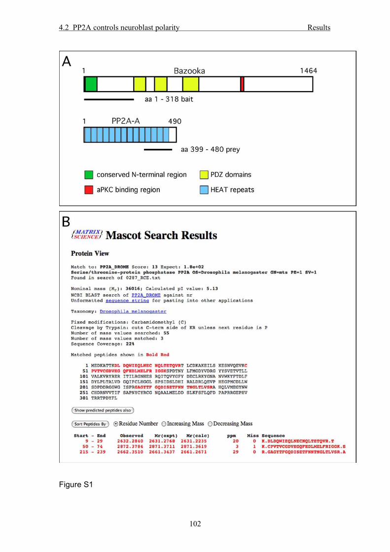
4.2 PP2A controls neuroblast polarity Results
102
Figure S1

4.2 PP2A controls neuroblast polarity Results
103
Figure S2

4.2 PP2A controls neuroblast polarity Results
104
Figure S3

4.2 PP2A controls neuroblast polarity Results
105
Figure S4
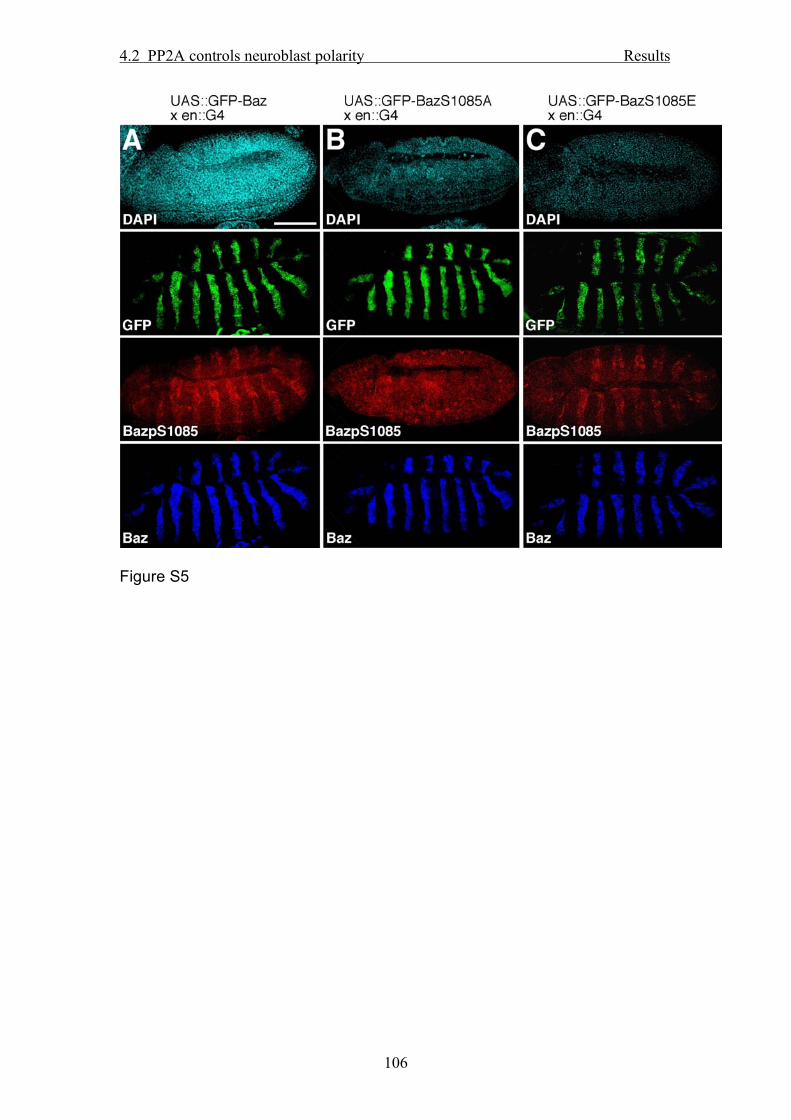
4.2 PP2A controls neuroblast polarity Results
106
Figure S5

4.2 PP2A controls neuroblast polarity Results
107
Figure S6
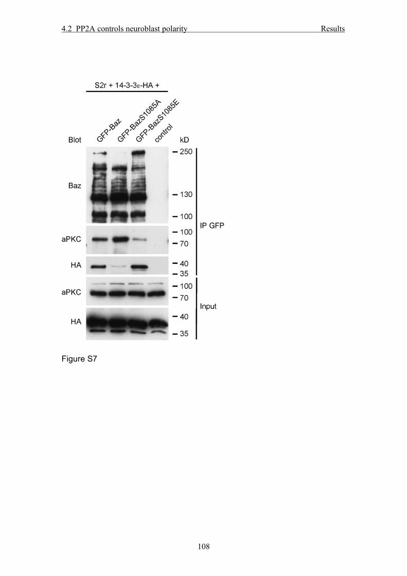
4.2 PP2A controls neuroblast polarity Results
108
Figure S7

4.2 PP2A controls neuroblast polarity Results
109
Supplemental References
Chen, H.K., Fernandez-Funez, P., Acevedo, S.F., Lam, Y.C., Kaytor, M.D., Fernandez, M.H., Aitken, A., Skoulakis, E.M., Orr, H.T., Botas, J., et al. (2003). Interaction of Akt-phosphorylated ataxin-1 with 14-3-3 mediates neurodegeneration in spinocerebellar ataxia type 1. Cell 113, 457-468.
Hannus, M., Feiguin, F., Heisenberg, C.P., and Eaton, S. (2002). Planar cell polarization requires Widerborst, a B' regulatory subunit of protein phosphatase 2A. Development 129, 3493-3503.
Huynh, J.R., Shulman, J.M., Benton, R., and St Johnston, D. (2001). PAR-1 is required for the maintenance of oocyte fate in Drosophila. Development 128, 1201-1209.
Kockel, L., Vorbruggen, G., Jackle, H., Mlodzik, M., and Bohmann, D. (1997). Requirement for Drosophila 14-3-3 zeta in Raf-dependent photoreceptor development. Genes Dev 11, 1140-1147.
Kraut, R., and Campos-Ortega, J.A. (1996). inscuteable, a neural precursor gene of Drosophila, encodes a candidate for a cytoskeleton adaptor protein. Dev Biol 174, 65-81.
Lu, B., Rothenberg, M., Jan, L.Y., and Jan, Y.N. (1998). Partner of Numb colocalizes with Numb during mitosis and directs Numb asymmetric localization in Drosophila neural and muscle progenitors. Cell 95, 225-235.
Rhyu, M.S., Jan, L.Y., and Jan, Y.N. (1994). Asymmetric distribution of numb protein during division of the sensory organ precursor cell confers distinct fates to daughter cells. Cell 76, 477-491.
Sathyanarayanan, S., Zheng, X., Xiao, R., and Sehgal, A. (2004). Posttranslational regulation of Drosophila PERIOD protein by protein phosphatase 2A. Cell 116, 603-615.
Schaefer, M., Petronczki, M., Dorner, D., Forte, M., and Knoblich, J.A. (2001). Heterotrimeric g proteins direct two modes of asymmetric cell division in the drosophila nervous system. Cell 107, 183-194.
Shiomi, K., Takeichi, M., Nishida, Y., Nishi, Y., and Uemura, T. (1994). Alternative cell fate choice induced by low-level expression of a regulator of protein phosphatase 2A in the Drosophila peripheral nervous system. Development 120, 1591-1599.
Sun, T.Q., Lu, B., Feng, J.J., Reinhard, C., Jan, Y.N., Fantl, W.J., and Williams, L.T. (2001). PAR-1 is a Dishevelled-associated kinase and a positive regulator of Wnt signalling. Nat Cell Biol 3, 628-636.
Uemura, T., Shiomi, K., Togashi, S., and Takeichi, M. (1993). Mutation of twins encoding a regulator of protein phosphatase 2A leads to pattern duplication in Drosophila imaginal discs. Genes Dev 7, 429-440.
Wodarz, A. (2008). Extraction and immunoblotting of proteins from embryos. In Drosophila: Methods and Protocols, C. Dahmann, ed. (Totowa, NJ, Humana Press), pp. 335-345.
Wodarz, A., Ramrath, A., Kuchinke, U., and Knust, E. (1999). Bazooka provides an apical cue for Inscuteable localization in Drosophila neuroblasts. Nature 402, 544-547.
Yu, F., Morin, X., Cai, Y., Yang, X., and Chia, W. (2000). Analysis of partner of inscuteable, a novel player of Drosophila asymmetric divisions, reveals two distinct steps in inscuteable apical localization. Cell 100, 399-409.

4.3 Dominant negative version of Bazooka Results
110
4.3. Imapired phosphorylation of Bazooka by aPKC leads to a dominant negative phenotype
Here I investigated the interaction between the Bazooka protein and another core
component of the PAR/aPKC-complex, aPKC.
I show that in addition to the known aPKC binding domain and the PDZ domains, the
poorly conserved linker region between these two domains is essential for binding of
aPKC to Baz and phosphorylation of Baz at serine 980..
Impaired phosphorylation of Baz at serine 980 leads to a complete loss of polarity in
the embryonic epidermis and in the compound eye, but not in NBs and in the female
germ line. Michael P. Krahn and Andreas Wodarz
Author contributions to the work: Michael P. Krahn: All experiments, besides*
writing of the manuscript STATUS: MANUSCRIPT IN PREPARATION

4.3 Dominant negative version of Bazooka Results
111
Impaired phosphorylation of Bazooka by aPKC leads to
a dominant negative phenotype
Michael P. Krahn1 and Andreas Wodarz1*
1Abteilung Stammzellbiologie, DFG Research Center for Molecular Physiology of the
Brain (CMPB), Georg-August-Universität Göttingen, Justus-von-Liebig-Weg 11,
37077 Göttingen, Germany
*author for correspondence: [email protected]
Running title: Dominant negative version of Bazooka
Keywords: epithelia, aPKC, polarity, PAR proteins, phosphorylation

4.3 Dominant negative version of Bazooka Results
112
Abstract
The conserved phosphorylation of Bazooka/PAR-3 by aPKC at serine 980 is
supposed to be a crucial event in the establishment of cell polarity. However, many
details of the interaction between aPKC and Baz as well as the function of their
alliance and the phosphorylation event are not yet fully understood.
Here we show that Baz provides several binding sites for aPKC, with the strongest
affinity in the PDZ domains and that phosphorylation of S980 increases the binding
affinity of Baz to aPKC. A non-phosphorylatable version of Baz exhibits a strong
dominant negative phenotype in the embryonic epidermis which can be partially
rescued by reduction of aPKC activity and resembles the defects observed in crb loss
of function alleles.
Introduction
Cell polarity is one of the most important features which is acquired throughout the
evolution of multicellular organisms. It is not only represented by a variety of cell
types and tissues but also controlled by a complicated network of genes. Most of these
Key regulators are highly conserved from worm to man, underlining their importance
and success in this complex process.
One of the proteins which functions upstream in the hierarchy controlling cell polarity
is Bazooka (Baz), the Drosophila homologue of PAR-3, which is the core component
of the PAR-3/PAR-6/aPKC complex (Wodarz, 2002; Suzuki and Ohno, 2006). In the
Drosophila ectodermal epithelium, Baz serves as the first apical cue required for
correct targeting of the transmembrane protein Crumbs (Crb) to the apical membrane
(Harris and Peifer, 2004; Harris and Peifer, 2005). Loss of Baz does not only result in

4.3 Dominant negative version of Bazooka Results
113
mislocalization of Crb and the components of the PAR-complex but leads to a
complete loss of cell polarity (Müller and Wieschaus, 1996; Tanentzapf and Tepass,
2003).
Apart from the correct targeting of diverse proteins to their distinct destinations, e.g.
the zones of cell-cell contacts or the apical membrane domain, dynamic protein
modification has been turned out to be involved in the regulation of cell polarity.
Recently it has been shown, that a complex signal cascade triggers the segregation of
cell fate determinants into one daughter cell during asymmetric division in
neuroblasts (NBs) (Wirtz-Peitz et al., 2008). In mammalian epithelial cells, PAR-1 is
phosphorylated by aPKC leading to an inactivation and dissociation of PAR-1 from
the apical membrane domain (Hurov et al., 2004). Vice versa, in Drosophila
ectodermal epithelial cells, the highly conserved intracellular domain of Crb is
phosphorylated by aPKC resulting in a stabilization of Crb protein at the apical
membrane domain (Sotillos et al., 2004).
For Baz different phosphorylation sites have been already described to play a crucial
role in the establishment and maintenance of polarity: Two conserved serines (S151
and S1085) are phosphorylated by PAR-1, creating a binding site for 14-3-3 proteins
(the Drosophila homologue of C.elegans PAR-5) and thereby support the exclusion of
Baz from the basolateral membrane in epithelial cells and dividing neural stem cells
(Benton and St Johnston, 2003)(Krahn et al. 2009).
Baz is also phosphorylated by aPKC (Kim et al. submitted), however the relationship
between them seems to be more then a mere kinase-substrate encounter. aPKC binds
to the second and third PDZ domain of Baz (Wodarz et al., 2000) and this interaction
is supposed to recruit aPKC to the membrane with Baz serving as a scaffold for
aPKC. Interestingly, aPKC can also bound to a conserved region at the C-terminal

4.3 Dominant negative version of Bazooka Results
114
third of Baz, depicted “aPKC-binding domain”, although all conclusions regarding
this interaction are derived from the C.elegans and mammalian homologues PAR-3
and ASIP which are studied excessively in vitro and in vivo (Izumi et al., 1998; Lin et
al., 2000; Tabuse et al., 1998).
In mammalian PAR-3 S827 (S980 in Baz) has been shown to be phosphorylated by
aPKC resulting in a decreased binding affinity between aPKC and PAR-3 (Nagai-
Tamai et al., 2002). Replacement of S827 by alanine, mimicking a not-
phosphorylatable version of Baz, results in a loss of cell polarity after calcium switch
in mammalian epithelial cells.
However, little to nothing is known if these correlations are also true for Drosophila
and if they are significant for all kind of polarized cell types. Therefore in this study
we elucidated the association of Baz and aPKC in vitro and in vivo in different
polarized cell types and describe the effects of overexpressing versions of Baz which
cannot be phosphorylated by aPKC or which mimic a constitutive phosphorylation.
Results and Discussion
Baz binds aPKC by its PDZ domains and its aPKC-binding domain
In mammalian cells, the minimal region of PAR-3 which is sufficient for binding of
aPKC has been reported to be a twenty amino acid stretch at the C-terminal third of
the protein (aa 820-840, aa 968-996 in Baz). However, in Drosophila, the first
association between aPKC and Baz was mapped to the second and third PDZ domain
by Yeast-2-Hybrid experiments (Wodarz et al., 2000). In order to clarify which
domains are crucial and sufficient for binding of aPKC in vivo, we performed co-

4.3 Dominant negative version of Bazooka Results
115
immunoprecipitation with GFP-tagged Baz constructs and endogenous aPKC in S2R+
cells.
Remarkably, deletion of the aPKC-binding domain (BazΔ968-996) leads to a
decreased binding of aPKC compared to wt Baz (Figure 1A), but it does not
completely abolish this binding. In contrast, deletion of the second PDZ domain
(BazΔPDZ2) decreases binding of aPKC and deletion of all three PDZ domains
(BazΔPDZ1-3) totally blocks the association between Baz and aPKC. This is in
contrast to our previous findings identifying the second and third PDZ domain to be
responsible for aPKC binding (Wodarz et al., 2000). However, it fits with data from
rescue experiments, indicating that a transgene lacking the third PDZ domain is able
to rescue the embryonic lethality of two strong Baz null alleles (Krahn et al.
submitted). Concomitantly, the isolated C-terminal region of Baz (BazΔ1-905), which
includes the aPKC-binding domain, is not capable to pull down aPKC. Surprisingly,
deletion of aa 737-945, a rather unconserved region, which has not been assigned any
function yet, also blocks aPKC binding, although the mutant protein localizes
normally at the apical junctional region in vivo (data not shown).
These finding were supported in vivo by segmental overexpression of UAS::GFP-Baz
deletion constructs in wild type embryos using an en::GAL4 driver line, which results
in segmental accumulation of aPKC only if Baz but not if BazΔPDZ1-3 are
overexpressed (data not shown).
Phosphorylation of S980 increases binding of aPKC
For mammalian PAR-3 it has been shown that phosphorylation of S827 (S980 in Baz)
decreases the binding affinity of aPKC and Baz (Nagai-Tamai et al., 2002). However,

4.3 Dominant negative version of Bazooka Results
116
we find it vice versa: co-immunoprecipitation of full length Baz either as wild type, a
not phosphorylatable version (S980A) or a constitutive phosphorylated version
(S980E) revealed that binding of aPKC is increased after phosphorylation of S980
and strongly decreased if this serine can not be phosphorylated (Fig. 2A).
Concomitantly, addition of a phosphatase inhibitor, preventing hydrolysis of the
phosphate groups, also results in a drastic increase of affinity between aPKC and Baz
(Fig. 2A). However, this is also true for BazS980E, suggesting that either other
phosphorylation sites of Baz apart from S980 are involved or that the phosphorylation
state of aPKC itself is increased, leading to a conformational change and enhanced
binding affinity to Baz. It has already been shown that for its activation, aPKC must
be phosphorylated by PDK-1 (Gao et al., 2001). Therefore, prevention of
dephosphorylation would result in an increased activation of aPKC, which in turns
could enhance phosporylation of S980 of Baz. Notably, after treatment with a
phosphatase inhibitor, a substantial interaction between BazΔPDZ1-3 and aPKC can
be detected, whereas it was below the detection level of our assay without inhibitor
(Fig.1)
The unconserved region linking the PDZ domains to the aPKC binding region is
crucial for binding of Baz to aPKC and phosphorylation of S980
Interaction between kinases and substrates are supposed to be rather transient and
hard to detect in vivo. Baz has been supposed to function as a scaffold for aPKC,
presumably to recruit the kinase activity to the membrane. Therefore, the binding is
rather strong. As after deletion of the PDZ domains of Baz no binding to aPKC can be
detected in vivo, we investigated if this results in an impaired phosphorylation of
S980. Therefore we either wild type GFP-Baz, GFP-BazΔPDZ1-3 or BazΔ737-945

4.3 Dominant negative version of Bazooka Results
117
was immunoprecipitated from lysates of transfected cells and blotted with an antibody
specific for phosphorylated BazS980. BazS980A was used as a negative control.
Although binding of aPKC can only be demonstrated after addition of phosphatase
inhibitor in vivo, BazΔPDZ1-3 is readily phosphorylated at S980 (Fig. 3), indicating,
that there exist at least two independent binding sites for aPKC in Baz and that
recognition and phosphorylation of the aPKC target motif surrounding S980 is not
impaired after deletion of the PDZ domains.
In contrast, deletion of aa 737-945 strongly reduces or even abolishes phosphorylation
of S980. BazΔ737-945 is also unable to bind aPKC and fails to rescue the embryonic
lethality of Baz alleles. Therefore we conclude that this region is not only involved in
the binding of aPKC (Fig. 1) but is also crucial for the phosphorylation of S980 and
therefore for the functionality of Baz. As this region is only poorly conserved we
speculate that the observed negative effect effect might be due to an altered folding of
the protein, concealing the binding site for aPKC surrounding S980 and the binding
site located in the second PDZ domain.
Not phosphorylatable Baz results in a dominant negative phenotype
Nagai-Tamai et al. already showed for mammalian epithelial cells that overexpression
of a phosphorylation-defective version of Baz (S827A) results in an impaired
formation of cell polarity after calcium switch (Nagai-Tamai et al., 2002). To test if
this is also true for Drosophila and in particular if the polarity of all polarized cell
types is affected, we expressed UAS::GFP-BazS980A with tissue specific driver
lines. In epithelial cells of the epidermis, the effect of BazS980A overexpression is
drastic: embryos die in late embryogenesis with a totally messed up morphology and

4.3 Dominant negative version of Bazooka Results
118
no cuticle secreted (Fig. 4A, Fig. 5, supplemental movie 2). Interestingly, epidermal
structures are nearly totally destructed whereas the intestine is proliferating and
expanding, forming large bulbs (Fig. 4A, arrow, Fig. 4A’ asterisk). In these structures,
BazS980A is mostly correctly localized to the apical (luminal) junctional region (Fig.
4A’, arrow) with some aggregates mislocalized to the basal site (Fig. 3A’,
arrowhead). Similarly, other polarity markers as DE-cad, Dlg (Fig. 4A’), aPKC, Arm
and Dystrophin (data not shown) are correctly localized in this tissue. Notably,
instead of being restricted to the subapical region, BazS980A-GFP and DE-Cad are
frequently found to be spread over the entire free apical membrane. Crb can not be
detected in these structures (Fig. 4A and data not shown), supporting the hypothesis
that the bulb-like structures are originated from the midgut.
Analyzed in more detail, it is obvious that in the epidermis of earlier stages,
BazS980A localizes predominately to the apical junctional region (Fig 4A’’), whereas
in later stages, it forms cytoplasmic and cortical aggregates, which also include Crb,
PAR-6 (Fig. 4A’’’), aPKC and DE-Cad (data not shown). Striking is the disturbance
of Crb in earlier stages when BazS980A is expressed: Although Baz S980A is still
cortical and without bigger aggregates, Crb is to some extent depleted from the
membrane (Fig. 4’’). Taken together these data indicate that the overexpression of
BazS980A severely disturbs the polarity in the epidermal epithelium.
Surprisingly, in the follicle epithelium and in the oocyte, the polarity is not affected
upon BazS980A overexpression and the transgene localizes correctly to the ZA and to
the anterior cortex, respectively (Fig. 4B, C). Concomittantly, female flies
overexpressing BazS980A in the germ line lay normal eggs.
4.3 Dominant negative version of Bazooka Results
119
Similarly, in embryonic NBs, BazS980A localizes to the apical membrane as its wild
type counterpart, without affecting the localization of cell fate determinants, spindle
orientation or the asymmetric cell division (Fig. 4D and data not shown).
Finally, overexpression of BazS980A in the eye using GMR::G4 also results in a
drastic phenotype: Upon overexpression of wild type Baz in the eye, a “rough eye”
phenotype can be observed, indicating that the polarity of the omatidia is disturbed.
This phenomenon is dramatically increased upon overexpression of BazS980A,
leading to a nearly complete destruction of the compound eye (Fig. 4E).
Membrane association is crucial for the dominant negative phentoype of
BazS980A overexpression
In order investigate which of the domain of Baz are involved in the dominant negative
phenotype of BazS980A overexpression, we tested different deletion constructs of
Baz with the S980A mutation: First, deletion of the conserved N-terminal
oligomerization domain (BazΔ1-317S980A) does not diminish the dominant negative
phenotype of S980A. The same is true if all three PDZ domains are deleted together
with the S980A mutation. Interestingly, in contrast to BazΔPDZ1-3, BazΔPDZ1-
3S980A is capable to bind aPKC in S2 cells (Fig. 1) and segmental overexpression of
BazΔPDZ1-3S980A results in a strong accumulation of aPKC (Fig. 5).
aPKC has been reported to act as a constitutive active version, if it is tethered to the
membrane by addition of a prenylation signal (aPKCCAAX)(Sotillos et al., 2004). We
have shown recently, that the C-terminal region of Baz is crucial for the membrane
localization of the protein (Krahn et al. submitted). To test, whether membrane
association of Baz is essential for the S980A phenotype, we overexpressed a C-

4.3 Dominant negative version of Bazooka Results
120
terminal truncated version of Baz carrying the S980A mutation (BazΔ1097-
1464S980A), which shows a strongly decreased membrane association (Krahn et al.
submitted and data not shown). Indeed, overexpression of BazΔ1097-1464S980A
does not affect cell polarity or viability of the embryos.
However, testing the isolated C-terminus including the aPKC-binding region with the
S980A mutation (BazΔ1-904S980A), we were not able to detect any defects in the
embryonic epidermis. This might be due to the fact, that BazΔ1-904 does not entirely
localizes to the ZA as full length Baz but it also accumulates to some extent in the
cytoplasma and at the basolateral membrane (Krahn et al. submitted).
Taken together, our data indicate that the effects of BazS980A overexpression are
facilitated by the membrane-association of Baz but are not dependent on the N-
terminal oligomerization domain or the PDZ domains.
Therefore one explanation of the mechanism underlying the dominant negative
phenotype of BazS980A overexpression could be that physiologically, aPKC is
recruited to the membrane by Baz, phosphorylates S980 resulting in an increased
binding to this domain, which in turn results in a block of aPKC kinase activity.
Thereby Baz would serve not only as a scaffold for aPKC but also as a trigger for
aPKC activity. If S980 is not phosphorylatable anymore, the inhibitory effect of Baz
would be omitted, releasing untriggered aPKC activity.
Cuticle defects of BazS980A overexpression resemble crb mutant phenotypes
An intact cell polarity is the prerequisite for the secretion of a physiological cuticle by
the embryonic epidermis at the end of embryogenesis. Alleles of several genes
involved in the regulation of cell polarity show defects in cuticle secretion (Knust et

4.3 Dominant negative version of Bazooka Results
121
al., 1993; Kuchinke et al., 1998; Petronczki and Knoblich, 2001; Tepass et al., 1996;
Wieschaus et al., 1984; Wodarz et al., 2000). Therefore we investigated the effect of
BazS980A overexpression on the cuticle secretion. As shown in Fig. 6, only minimal
debris of cuticle can be detected, a defect which is strikingly similar to the phenotype
of crb loss of function alleles (Knust et al., 1993), which is very characteristically,
leading to the denotation of the gene “crumbs” (Knust et al., 1993).
This raises the question if the overexpression of BazS980A somehow affects either
the stabilization/degradation of the Crb protein or its functionality. As no differences
in protein levels of Crb upon BazS980A overexpression compared to wild type lysate
or wild type Baz overexpression can be detected (data not shown), we focused on the
second hypothesis.
The dominant negative phenotype of BazS980A-overexpression is partly rescued
by reduction of aPKC activity
Crb localization and functions is dependent on the correct formation of the
PAR/aPKC complex (Bilder et al., 2003), which has recently been described in more
detail: aPKC phosphorylates two threonines in the conserved intracellular domain of
Crb, which is crucial for proper localization and function of the protein (Sotillos et al.,
2004). Replacement of the involved residues by alanine leads to a loss of membrane
association of Crb. Concomitantly, overexpression of a dominant negative, kinase
dead version of aPKC (aPKCCAAX DN) also leads to a downregulation of Crb, whereas
overexpression of an active, membrane bound aPKC form (aPKCCAAX) results in an
expansion of the Crb-positive apical domain to the basolateral membrane (Sotillos et
al., 2004).

4.3 Dominant negative version of Bazooka Results
122
In order to elucidate the interaction between BazS980A overexpression and aPKC
activity, we tested whether reduction of aPKC activity can rescue the phenotype of
BazS980A overexpression. Indeed, loss of one copy of aPKC decreases the
severeness of the BazS980A overexpression phenotype in cuticle preparations (Fig.
6), although the dominant negative phenotype is not fully rescued and the embryonic
lethality is still 100%.
Taken together we have shown that Baz serves as a multiple scaffold for aPKC by
binding to aPKC via its second PDZ domain and the aPKC binding domain. Although
not conserved, the region between these two identified binding region is crucial for
aPKC association and phosphorylation of S980. This phosphorylation serves as a
trigger to increase the binding to aPKC, presumably to inhibit its cortical kinase
acitivity. The key question is, what is the substrate for aPKC? aPKC has already been
reported to phosphorylate several regulators of polarity: PAR-1, which is thereby
inactivated and released from the membrane (Hurov et al., 2004), lgl, which is also
inactivated (Betschinger et al., 2005; Betschinger et al., 2003; Ponting, 1997;
Yamanaka et al., 2003), numb, leading to a restriction to the basal cortex of dividing
NBs (Smith et al., 2007; Wirtz-Peitz et al., 2008) and Crb (Sotillos et al., 2004).
Taking in account first the fact that the most predominant phenotype of BazS980A
overexpression is found in the eye and in the embryonic epidermis; second, the
polarity of the oocyte and embryonic NBs are not affected, third, Crb-negative
luminal structures, presumably the midgut, proliferate in otherwise totally messed up
embryos leads to the conclusion that Crb is the most likely candidate of the known
substrates of aPKC. In fact, the cuticle phenotype of BazS980A overexpression is
identical to that of crb mutant. However, it still remains unclear, how Crb should be
degraded upon BazS980A overexpression: Given the fact that Baz recruits aPKC to

4.3 Dominant negative version of Bazooka Results
123
the membrane and regulates its kinase activity via its aPKC binding domain, the
S980A mutation should lead to an increase in aPKC kinase activity, which in turn
excessively phosphorylates Crb. Following this hypothesis, the resulting phenotype
would be instead of a loss of function of crb a gain of function, reflected by ectopic
cuticle and a spatially multilayered ectodermal epithelium (Wodarz et al., 1995).
Therefore, BazS980A-overexpression must somehow provide an increased feedback
loop to restrict aPKC activity. One possibility would be that the S980A mutation
results in a conformational change of the Baz protein, which now binds differentially
to aPKC, not providing a scaffold for membrane tethering but inhibiting its activity.
Another possibility would be that by this conformational change, Baz exposes a
different binding site for an up to now unidentified protein, which in turn inhibits
aPKC. One candidate for inhibition of aPKC is PAR-6, although we have not yet any
evidence that S980A increases the binding of PAR-6.
Materials and Methods
Fly stocks and genetics
Transgenic flies carrying UAS::GFP-Baz constructs were generated using standard
germ line transformation. The following GAL4 driver lines were used for expression
of the transgenes in different tissues: daughterless::GAL4 (da::G4) (Wodarz et al.,
1995), worniu::GAL4 (wor::G4), nanos::GAL4 (nos::G4). Cuticle preparations werde
performed according to standard protocols.

4.3 Dominant negative version of Bazooka Results
124
DNA and constructs
Cloning of wild type Baz in pEntr and destination vectors has been described
previously (Krahn et al. 2009). For BazS980A, following oligonuceotides were used
(in 5’-3’ orientation, mutation underlined) :
BazS980A-for: GATGCATTGGGACGACGCGCCATCTCTGAGAAG
BazS980A-rev: CTTCTCAGAGATGGCGCGTCGTCCCAATGCATC
Cloning of all other constructs have been described elsewhere (Krahn et al.
submitted).
Immunoprecipitation and Western Blot
For immunoprecipitations, embryos from an overnight collection were dechorionated
and lysed in lysis buffer (1% Triton X-100, 150mM NaCl, 1mM CaCl2, 1mM MgCl2,
25mM Hepes pH 7.44) supplemented with protease inhibitors. After centrifugation, 2
µl of mouse anti GFP (Invitrogen) were added to cell lysate corresponding to 500 µg
total protein. Immune complexes were harvested using protein G-conjugated agarose
(Roche), washed five times in lysis buffer and boiled in 2x SDS sample buffer before
SDS-PAGE and Western blot.
Primary antibodies were used for Western blotting according to standard procedures
(Wodarz, 2008) as follows: mouse anti GFP (1:1000; Roche 11814460001), rabbit
anti-PKCζ C20 (1:2000; Santa Cruz Biotechnology, Inc.).
Immunohistochemistry
Embryos and ovaries were fixed in 4% formaldehyde, phosphate buffer pH 7.4. The
primary antibodies used were guinea-pig anti Mira (1:1000; Kim et al. submitted),
rabbit anti PKCζ C20 (1:1000; Santa Cruz Biotechnology, Inc.), rat anti DE-Cadherin
DCAD2 (1:50; Developmental Studies Hybridoma Bank, DSHB), mouse anti Dlg
4F3 (1:50; DSHB), rabbit anti Staufen (1:1000) (St Johnston et al., 1991), mouse anti

4.3 Dominant negative version of Bazooka Results
125
Gurken 1D12 (1:10, DSHB), mouse anti Crb (1:10, DSHB), mouse anti GFP 3E6
(1:1000; Invitrogen). DNA was stained with DAPI (Invitrogen). Secondary antibodies
conjugated to Cy2 and Cy3 were obtained from Jackson Laboratories. Secondary
antibodies conjugated to Alexa 647 were obtained from Invitrogen. Images were
taken on a Zeiss LSM 510 Meta confocal microscope and processed using Adobe
Photoshop.
References
Benton, R. and St Johnston, D. (2003). Drosophila PAR-1 and 14-3-3 inhibit
Bazooka/PAR-3 to establish complementary cortical domains in polarized cells. Cell
115, 691-704.
Betschinger, J., Eisenhaber, F. and Knoblich, J. A. (2005). Phosphorylation-
induced autoinhibition regulates the cytoskeletal protein Lethal (2) giant larvae. Curr
Biol 15, 276-82.
Betschinger, J., Mechtler, K. and Knoblich, J. A. (2003). The Par complex directs
asymmetric cell division by phosphorylating the cytoskeletal protein Lgl. Nature 422,
326-30.
Bilder, D., Schober, M. and Perrimon, N. (2003). Integrated activity of PDZ protein
complexes regulates epithelial polarity. Nat Cell Biol 5, 53-58.
Gao, T., Toker, A. and Newton, A. C. (2001). The carboxyl terminus of protein
kinase c provides a switch to regulate its interaction with the phosphoinositide-
dependent kinase, PDK-1. J Biol Chem 276, 19588-96.
Harris, T. J. and Peifer, M. (2004). Adherens junction-dependent and -independent
steps in the establishment of epithelial cell polarity in Drosophila. J Cell Biol 167,
135-47.
Harris, T. J. and Peifer, M. (2005). The positioning and segregation of apical cues
during epithelial polarity establishment in Drosophila. J Cell Biol 170, 813-23.
Hurov, J. B., Watkins, J. L. and Piwnica-Worms, H. (2004). Atypical PKC
phosphorylates PAR-1 kinases to regulate localization and activity. Curr Biol 14, 736-
41.
Izumi, Y., Hirose, T., Tamai, Y., Hirai, S., Nagashima, Y., Fujimoto, T., Tabuse,
Y., Kemphues, K. J. and Ohno, S. (1998). An atypical PKC directly associates and

4.3 Dominant negative version of Bazooka Results
126
colocalizes at the epithelial tight junction with ASIP, a mammalian homologue of
caenorhabditis elegans polarity protein PAR-3 [In Process Citation]. J Cell Biol 143,
95-106.
Knust, E., Tepass, U. and Wodarz, A. (1993). crumbs and stardust, two genes of
Drosophila required for the development of epithelial cell polarity. Dev Suppl 1993,
261-8.
Kuchinke, U., Grawe, F. and Knust, E. (1998). Control of spindle orientation in
Drosophila by the Par-3-related PDZ- domain protein Bazooka. Curr Biol 8, 1357-65.
Lin, D., Edwards, A. S., Fawcett, J. P., Mbamalu, G., Scott, J. D. and Pawson, T.
(2000). A mammalian Par-3-Par-6 complex implicated in CdC42/Rac1 and aPKC
signalling and cell polarity. Nat. Cell Biol. 2, 540-547.
Müller, H. A. and Wieschaus, E. (1996). armadillo, bazooka, and stardust are
critical for early stages in formation of the zonula adherens and maintenance of the
polarized blastoderm epithelium in Drosophila. J Cell Biol 134, 149-63.
Nagai-Tamai, Y., Mizuno, K., Hirose, T., Suzuki, A. and Ohno, S. (2002).
Regulated protein-protein interaction between aPKC and PAR-3 plays an essential
role in the polarization of epithelial cells. Genes Cells 7, 1161-71.
Petronczki, M. and Knoblich, J. A. (2001). DmPAR-6 directs epithelial polarity and
asymmetric cell division of neuroblasts in Drosophila. Nat Cell Biol 3, 43-49.
Ponting, C. P. (1997). Evidence for PDZ domains in bacteria, yeast, and plants.
Protein Sci 6, 464-8.
Smith, C. A., Lau, K. M., Rahmani, Z., Dho, S. E., Brothers, G., She, Y. M.,
Berry, D. M., Bonneil, E., Thibault, P., Schweisguth, F. et al. (2007). aPKC-
mediated phosphorylation regulates asymmetric membrane localization of the cell fate
determinant Numb. Embo J 26, 468-80.
Sotillos, S., Diaz-Meco, M. T., Caminero, E., Moscat, J. and Campuzano, S.
(2004). DaPKC-dependent phosphorylation of Crumbs is required for epithelial cell
polarity in Drosophila. J Cell Biol 166, 549-57.
St Johnston, D., Beuchle, D. and Nusslein-Volhard, C. (1991). Staufen, a gene
required to localize maternal RNAs in the Drosophila egg. Cell 66, 51-63.
Tabuse, Y., Izumi, Y., Piano, F., Kemphues, K. J., Miwa, J. and Ohno, S. (1998).
Atypical protein kinase C cooperates with PAR-3 to establish embryonic polarity in
Caenorhabditis elegans. Development 125, 3607-14.

4.3 Dominant negative version of Bazooka Results
127
Tanentzapf, G. and Tepass, U. (2003). Interactions between the crumbs, lethal giant
larvae and bazooka pathways in epithelial polarization. Nat Cell Biol 5, 46-52.
Tepass, U., Gruszynski, D. E., Haag, T. A., Omatyar, L., Török, T. and
Hartenstein, V. (1996). shotgun encodes Drosophila E-cadherin and is preferentially
required during cell rearrangement in the neurectoderm and other morphogenetically
active epithelia. Genes Dev 10, 672-85.
Wieschaus, E., Nüsslein-Volhard, C. and Jürgens, G. (1984). Mutations affecting
the pattern of the larval cuticle in Drosophila melanogaster. III. Zygotic loci on the X
chromosome and fourth chromosome. Wilhelm Roux's Arch 193, 296-307.
Wirtz-Peitz, F., Nishimura, T. and Knoblich, J. A. (2008). Linking cell cycle to
asymmetric division: Aurora-A phosphorylates the Par complex to regulate Numb
localization. Cell 135, 161-73.
Wodarz, A. (2008). Extraction and immunoblotting of proteins from embryos.
Methods Mol Biol 420, 335-45.
Wodarz, A., Hinz, U., Engelbert, M. and Knust, E. (1995). Expression of Crumbs
confers apical character on plasma membrane domains of ectodermal epithelia of
Drosophila. Cell 82, 67-76.
Wodarz, A., Ramrath, A., Grimm, A. and Knust, E. (2000). Drosophila atypical
protein kinase C associates with Bazooka and controls polarity of epithelia and
neuroblasts. J Cell Biol 150, 1361-74.
Yamanaka, T., Horikoshi, Y., Sugiyama, Y., Ishiyama, C., Suzuki, A., Hirose, T.,
Iwamatsu, A., Shinohara, A. and Ohno, S. (2003). Mammalian Lgl Forms a Protein
Complex with PAR-6 and aPKC Independently of PAR-3 to Regulate Epithelial Cell
Polarity. Curr Biol 13, 734-43.
Figure Legends
Figure 1. The PDZ domains of Baz are crucial for binding of aPKC. Endogenous
aPKC protein was co-immunoprecipitated with GFP-Baz variants from
transfected S2R cells.
Figure 2. Phosphorylation of S980A increases binding of aPKC to Baz. S2R cells
transfected with GFP-Baz constructs werde treated with DMSO as negative
4.3 Dominant negative version of Bazooka Results
128
control or 10µM cantharidin for 2h. GFP-Baz proteins were imunoprecipitated
with an anti-GFP antibody and blotted against aPKC and GFP.
Figure 3. Phosphorylation of S980A is impaired after deletion of aa 737-945.
Different GFP-Baz constructs were expressed in S2R cells and
immunoprecipitated. Proteins were dissolved on SDS-PAGE and blotted
against GFP and pS980.
Figure 4. Overexpression of BazS980A results in severe polarity defects of the
embryonic epidermis. GFP-BazS980A was overexpressed in different
polarized cell types using distinct driver lines. (A) Upon ubiquitous
overexpression using da::G4, the morphology of embryos in later stages is
severely disturbed. (A’) Cavernous structure, presumably representing midgut
tissue. (A’’-A’’’) Segmental overexpression of GFP-BazS980A with en::G4.
In early stages, BazS980A still localizes at the cortex, whereas it forms
aggregates in later stages. Crb expression is decreased in early stages and in
later stages included in BazS980A positive aggregates. (B) Expression of
BazS980A in the oocyte shows wild type localization at the anterior cortex,
whereas Staufen (Stau) localizes posterior. Gurken (Grk) marks the dorso-
posterior pol. (C) BazS980A localizes normally in embryonic NBs at the
apical cortex, overlapping with aPKC. Miranda (Mira) accumulates at the
basal pole. (D) In the eye, overexpression of BazS980A results in a
completely dirsturbed morphology, compared to a discrete rough-eye
phenotype of wild-type Baz overexpression.
Figure 5. BazΔPDZ1-3S980A but not BazΔPDZ1-3 is able to recruit aPKC in vivo.
In order to assay in vivo recruitment of aPKC, aPKC was costained.
BazΔPDZ1-3 and BazΔPDZ1-3S980A were segmental overexpressed using
en::G4.
Figure 6. Embryos overexpressing BazS980A fail to secrete a physiological cuticle
Cuticle preparation of embryos overexpressing BazS980A, wild type Baz or
BazS980A with only one copy of aPKC.

4.3 Dominant negative version of Bazooka Results
129
Figure 1

4.3 Dominant negative version of Bazooka Results
130
Figure 2

4.3 Dominant negative version of Bazooka Results
131
Figure 3

4.3 Dominant negative version of Bazooka Results
132
Figure 4
Figure 5
GFP DE-Cad
DAPI
Merge A’ embryo: epidermis stage 14
da::G4 x UAS::BazS980A
Dlg
GFP
DAPI
Merge A embryo: overview stage 14 da::G4 x UAS::BazS980A
Crb
Merge
GFP
Crb
DAPI
Merge A’’’ embryo: epidermis stage 11
en::G4 x UAS::BazS980A
PAR-6
Crb
DAPI
A’’ embryo: epidermis stage 9
en::G4 x UAS::BazS980A
PAR-6 GFP
Merge

4.3 Dominant negative version of Bazooka Results
133
B adult: oocyte
nos::G4 x UAS::BazS980A
GFP Stau
DAPI
Merge
Grk
C embro: neuroblast
wor::G4 x UAS::BazS980A
GFP aPKC
DAPI
Merge
Mira
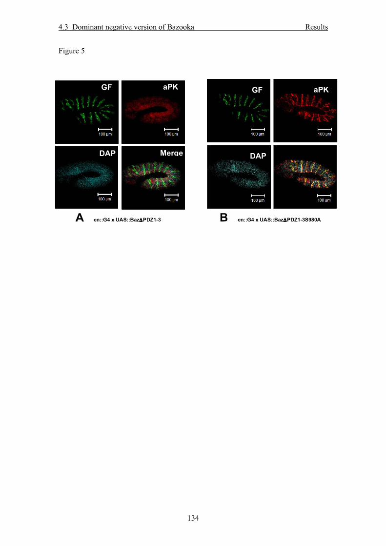
4.3 Dominant negative version of Bazooka Results
134
Figure 5
A en::G4 x UAS::BazΔPDZ1-3
GFP
aPKC
DAPI
Merge
B en::G4 x UAS::BazΔPDZ1-3S980A
GFP
aPKC
DAPI

4.3 Dominant negative version of Bazooka Results
135
Figure 6

5 Discussion
136
5. Discussion
5.1. Implications of the structural analysis of the Bazooka protein
Within this study, a systematic structure-function analysis of the Baz protein in
different polarized cell types in Drosophila was performed, showing that the
membrane association of Baz is facilitated by a new lipid binding motif located at the
C-terminus of the protein, whereas the N-terminal, highly conserved domains are not
crucial for membrane association (see 3.1). Furthermore, this study showed that the
subcellular localization of Baz is differentially regulated in the context of the cell
type. This is important, as although basic mechanisms for the regulation of cell
polarity are conserved between different polarized cell types, they are apparently
modulated in a tissue-specific manner. One example is the difference between the
ectodermal epithelium (e.g. in the embryonic epidermis) and the NBs. Whereas the N-
terminal conserved region 1 (CR1) is dispensable for proper localization of Baz to the
ZA in the epidermis, it is required for localization to the apical cortex in NBs.
In complementation to the study presented in 3.1, it would be very interesting to see
how the mutant proteins described in 3.1 are localized in a baz-negative background.
Furthermore, it would be important to know whether the C-terminal truncation
constructs (BazΔ1096-1464, BazΔ1222-1464, BazΔ1325-1464, BazΔ1461-1464),
BazΔPDZ3 and BazΔ1096-1464PHP, which all are able to rescue the embryonic
lethality of baz zygotic mutants (cp. 3.1), can also rescue maternally and paternally
mutant embryos and whether they can compensate for the loss of baz function in
germ-line clone egg chambers. Particularly for BazΔ1096-1464 we doubt whether it
would rescue baz germ line clone mutants, because it is only to a minor extent
correctly localized at the membrane and shows a significant cytosolic staining.
However, also BazΔ1461-1464 and BazΔ1096-1464PHP are of great interest, as both
localize normally at the apical junctional region, but a potential PDZ-domain
consisting of the last four amino acids of Baz is deleted. In late embryonic, larval and
pupal development this domain appears to be dispensable, but perhaps it plays a
crucial role in oogenesis or in the establishment of the first junctional complexes
during cellularization in the early embryo. In these stages, the maternal component is
still present in zygotic baz mutants. Therefore, experiments are under way in which
insertions of distinct GFP-Baz deletion constructs inserted on the third chromosome

5 Discussion
137
are recombined with a ubiquitous driver line (da::GAL4, cp. 3.1) and crossed to a baz
allele carrying an FRT site (Baz815-8, FRT19A, (McKim et al., 1996)). Using the FLP-
FRT technique (Golic and Lindquist, 1989), female egg chambers can be generated
which lack maternal Baz protein but carry the mutant GFP-tagged protein.
One important open question regarding the structure and function of the Baz protein is
how it is posttranslationally processed: As can be seen by Western blotting with either
GFP antibody using N-terminally GFP-tagged Baz constructs or with an antibody
raised against the N-terminal 317aa (Wodarz et al., 1999), several bands of different
size are detected corresponding to N-terminal fragments of Baz (cp. 3.2 Figure 1 and
Appendix 6.2). In contrast, using an antibody recognizing the last 15 amino acids,
only one band representing full length Baz is detectable (data not shown). As these
bands are stable and reproducible and definitely more predominant than the band
corresponding to full length Baz, they are presumably not only degradation products
but rather the result of specific cleavage steps. This raises several questions: first of
all, what is the function of the distinct cleavage products? Second, which proteases
are involved in the processing of full length Baz? And third, where and in which
context Baz is cleaved: directly after the translation event in the cytosol or after
membrane recruitment, within the PAR/aPKC complex or before association with its
binding partners.
Some ideas can be derived from the structural analysis (cp. 3.1 and Appendix 6.2): As
the GFP fluorochrom which is N-terminally attached to full length Baz is exclusively
detectable at the membrane and never in other subcellular compartments e.g. the
nucleus, the cytosol or vesicular structures, it is clear that either the GFP itself is
cleaved and rapidly degraded or the most N-terminal region is always associated with
the membrane. This is further supported by the observation that the signal detected by
indirect immunofluorescence with the antibody raised against the N-terminal region is
always cortical, too. In contrast, truncation of Baz at amino acid 968 (BazΔ969-1464),
directly N-terminal of the aPKC-binding domain, results in cytosolic localization of
the mutant protein. However, multiple fragments observed in Western Blot with the
N-terminal or GFP antibody are much smaller than the calculated size of BazΔ969-
1464 (Appendix 6.2). This would mean that the proteins corresponding to these bands
should be detectable in the cytoplasm if they are not linked to the membrane by a

5 Discussion
138
different mechanism, which is rather unlikely, as the same cleavage pattern of smaller
bands is also detected in BazΔ969-1464, which localizes to the cytosol. Therefore it
remains elusive, where the forms of Baz corresponding to these smaller bands are
localized in vivo.
Regarding the fate and potential function of the C-terminal cleavage products
corresponding to the N-terminal fragments we know virtually nothing – either they
are degraded or they are targeted to specific compartments, fulfilling distinct
functions. Interestingly, an antibody raised against the peptide Baz973-985
phosphorylated at serine 980 (cp. 3.2), shows besides a weak membrane staining a
strong nuclear staining. This suggests that a fragment containing this peptide localizes
to the nucleus. This is even more exciting because we found several nuclear proteins
as potential interaction partners of Baz in a yeast-two-hybrid screen. For mammalian
PAR-3, nuclear localization and a potential function in DNA-damage repair pathways
has been described previously (Fang et al., 2007). In their experiments, the authors
used an antibody raised against a C-terminal GST-fusion protein of PAR-3 and they
were able to detect PAR-3 in the nucleus. With the peptide-antibody raised against the
last 15aa of Baz, we only detect a cortical staining, indicating that all fragments
containing these aa are not localized to the nucleus.
Taken together there are still many open questions regarding the generation,
regulation and function of different fragments of the Baz protein. In particular, the
potential function of Baz in the nucleus in the control of chromatin structure and
transcription will be in the focus of future research in this field.
5 Discussion
139
5.2. Phosphorylation of Bazooka: Only two pieces of a great puzzle
In the second part of this work, two important phosphorylation events have been
investigated: First, the role of phosphorylation of serine 1085 by PAR-1 and
subsequent dephosphorylation by PP2A in the regulation of apical-basal polarity in
dividing NBs. Second, the phosphorylation of serine 980 by aPKC, which is crucial
for the establishment of cell polarity in ectodermal epithelial cells. Both studies
demonstrate that the posttranslational modification of Baz is necessary and important
for the physiological function of the protein. Therefore, an exactly tuned regulation of
phosphorylation and dephosphorylation events must take place to ensure proper
subcellular localization of Baz in the context of different cell types. One mechanism
to restrict such protein modifications to specific subcellular compartments is the
targeting of the particular kinases or phosphatases to these compartments. This is the
case for PAR-1, which is restricted to the basolateral membrane in epithelial cells and
to the basal cortex in dividing NBs (cp 3.2). The restricition of PAR-1 to these
membrane domains is presumably achieved by an active exclusion pf PAR-1 from the
apical membrane domain by aPKC. In mammalian cells, aPKC phosphorylates PAR-
1, which subsequently dissociates from the membrane (Hurov et al., 2004; Suzuki et
al., 2004).
However, many if not most kinases and phosphatases are not specifically targeted to a
distinct subcellular localization but are rather diffusely distributed in the cytoplasm
and the nucleus, for example Protein phosphatase 1, PP2A and LKB1/PAR-4
(Bonaccorsi et al., 2007; Janssens and Goris, 2001; Jones et al., 1995). Therefore
another possibility to spatially restrict kinase / phosphatase activity is to establish
phosphorylation cascades, leading to the activation or inactivation of proteins and
kinases. Apart from the mentioned aPKC-PAR-1 interaction, the binding of the 14-3-
3 proteins to Baz/PAR-3 is another example: The ubiquitously expressed 14-3-3
proteins are only bound after phosphorylation of their target sequences (Hurd et al.,
2003), in the case of Baz after phosphorylation of serine 151 and 1085 by PAR-1. As
this occurs only at the basal cortex in NBs, where PAR-1 is expressed, the activities
of 14-3-3 proteins are also restricted to that specific localization (see also 3.2).

5 Discussion
140
In this study we described two different phosphorylation events of Baz with dramatic
effects on the regulation of cell polarity. However, we have obtained evidence that
there exist far more phosphorylation sites in the Baz protein: Treatment of cells
transfected with Baz-GFP with high concentrations of phosphatase inhibitors and
subsequent immunoprecipitation of Baz protein and Western blot against Baz
revealed a significant change in the protein mobility in SDS-PAGE by more than
10kDa (cp 3.2, Figure 2). This band-shift may be due to an altered charge of the Baz
protein upon modification by phosphate groups. As the three described
phosphorylation sites/events do not lead to such a dramatic increase in the band size,
it is clear that Baz is phosphorylated at additional residues. Indeed, two proteome-
wide screens for phosphorylated proteins in Drosophila identified several
phosphorylated amino acids in Baz: In the database Phospho Pep, phosphorylated
peptides isolated from Drosophila Kc167 cells identified by mass spectrometry were
assigned to proteins (Bodenmiller et al., 2008). In this study, 14 serine residues and
two threonine residues of Baz were found to be phosphorylated. Interestingly, of the
three previously described phosphorylation sites only S151 was found in this screen,
indicating that by this approach not all phosphorylated peptides were detected. This
implies that Baz may be phosphorylated at more than 20 different amino acid
residues. A second study used sodium orthovanadate as a specific inhibitor for
tyrosine phosphatases to enhance tyrosine phosphorylation of proteins in Drosophila
S2 cells. After precipitation of proteins containing phosphorylated tyrosines with an
anti phospho-tyrosine antibody, proteins were digested and peptides analyzed by mass
spectrometry (Chang et al., 2008). For Baz, four peptides carrying phosphorylated
tyrosines were found. Interestingly, the mammalian homolog PAR-3 is tyrosine
phosphorylated at residue 1127 in an epidermal growth factor dependent manner
(Wang et al., 2006). Although Y1127 is not conserved in Baz, these two publications
demonstrate that Baz might be tyrosine phosphorylated, too. However, we failed to
detect tyrosine phosphorylation of Baz immunoprecipitated from cell lysates in
Western blotting (data not shown). One reason might be that the phosphorylation is
rather transient and not stable enough to be detected in Western blotting. Nevertheless
the discussed findings demonstrate that we are far from understanding the entire
phosphorylation repertoire of the Baz protein and especially how it is regulated in the
context of Baz’s function in the establishment of cell polarity. To obtain more
information about additional phosphorylation sites, the corresponding

5 Discussion
141
kinases/phosphatases and the role of the particular phosphorylation events in the
context of cell polarity will be one of the future goals of our research.
One kinase of particular interest is LKB1, the Drosophila homologue of C.elegans
PAR-4. LKB1 was originally identified in C.elegans in a screen for embryonic
asymmetry (Watts et al., 2000). This function is conserved in the Drosophila oocyte,
where LKB1 functions in the establishment of anterior-posterior axis formation
(Martin and St Johnston, 2003). Various studies in mammalian cells indicate that
LKB1 is also involved in the regulation of epithelial cell polarity and functions as a
tumor suppressor protein (for review see (Baas et al., 2004).
In mammalian cells, LKB1 phosphorylates PAR-1 in the activation loop of the kinase
domain, resulting in activation of PAR-1 (Lizcano et al., 2004). Martin and St.
Johnston obtained contradictory results and showed that in Drosophila LKB1 acts
genetically downstream of PAR-1 and is itself phosphorylated by PAR-1 (Martin and
St Johnston, 2003). It is also possible that LKB1 phosphorylates not only PAR-1 but
also other members of the PAR family, e.g. Baz, PAR-6 or aPKC which would be
another intriguing piece in the network of PAR-proteins regulating cell polarity. In
Drosophila NBs, LKB1 has been shown to be crucial not only for asymmetric cell
division and spindle formation, but also for correct localization of the PAR/aPKC
complex to the apical and Mira to the basal cortex (Bonaccorsi et al., 2007). However,
in this context, no phosphorylation target has been identified yet. Therefore,
components of the PAR/aPKC complex would be ideal targets of LKB1, particularly
because their localization is disturbed whereas Pins and Gαi are localized normally.
These proteins have been shown to act in a parallel pathway which functions
redundantly to the PAR/aPKC complex in asymmetric division and spindle
orientation (Cai et al., 2003; Fuse et al., 2003; Izumi et al., 2004; Yu et al., 2003). The
penetrance of spindle orientation and division defects is increased in double mutant
for LKB1 and Pins compared to the according single mutants, indicating that they act
in parallel pathways, which suggests that LKB1 may be involved in the regulation of
the PAR/aPKC complex.
Another candidate for a Baz phosphorylating kinase is LK6, a serine-threonine kinase,
which was found to interact with the Baz N-terminal region in the same yeast-two-
hybrid screen as PP2A (cp. 3.2, Egger-Adam PhD thesis 2006). This is of great
interest, because LK6 associates with microtubules and localizes to the centrosomes

5 Discussion
142
(Kidd and Raff, 1997), which might provide a direct link between cortical Baz and the
formation and orientation of the mitotic spindle, which has up to now only been
shown by genetic interactions (Wodarz, 2005).
On the other hand, regarding additional phosphatases implicated in Baz
dephosphorylation, protein phosphatase 1 (PP1) is one of the most likely candidates.
Besides PP2A, PP1 is the second major, ubiquitously expressed phosphatase and it
has already been demonstrated to dephosphorylate mouse PAR-3 (Traweger et al.,
2008). Interestingly, in that study the serine residues corresponding to serine 151, 980
and 1085 were described to be dephosphorylated by PP1. Furthermore, PP1 binds to
several fragments of PAR-3, including the PDZ domains, which indicates that there
might be more phosphorylation sites affected by PP1. As from our experiments we
conclude that PP2A dephosphorylates serine 1085 (see 3.2) there is either a redundant
mechanism or in contrast to the phosphorylation event, the dephosphorylation of
serine 1085 is not conserved throughout evolution. Hoewever, it also has to be
considered whether the reported in vito dephosphorylation of all three reported
phosphorylation sites really takes place in vivo and if so, whether this is of
physiological relevance. Nevertheless PP1 might dephosphorylate distinct
serine/threonine residues of Baz and thereby it may take part in the complex
regulation of Baz in the context of cell polarity.
Apart from PP1, several other phosphatases must be taken into account with respect to
Baz dephosphorylation. One example is protein phosphatase 4, whose regulatory
subunit Falafel associates with Mira in larval NBs (Sousa-Nunes et al., 2009). Nuclear
Falafel prevents Pros from entering the nucleus in the NB and knock-down of falafel
results in dissociation of Mira from the cortex, indicating that Falafel plays a crucial
role in the establishment of apical-basal polarity of dividing NBs and thereby in the
asymmetric cell division. However, direct dephosphorylation of Mira or other proteins
by Falafel has not been demonstrated yet.
Apart from phosphorylation, other posttranslational modifications of Baz might
regulate its localization, its affinity to binding partners etc. For example, attachment
of ubiquitin (mono- or polyubiquitination) emerges more and more to be not only
important for the targeting to the proteasome and subsequent degradation of a protein,
but can also function to modify localization or function of a protein (Sun and Chen,

5 Discussion
143
2004). One example in NBs is Mira, which has been found to be ubiquitinylated at its
C-terminal region. Removal of this domain results in mislocalization of Mir to the
cytosol, whereas replacement of this domain by ubiquitin restores the physiological
localization (Slack et al., 2007). Blotting of precipitated Baz protein reveals that
several cleavage products of Baz are ubiquitinylated (data not shown), but it remains
elusive, whether this leads to protein degradation or whether there is any additional
functional relevance in the attachment of ubiquitin molecules to Baz.
A mechanism similar to ubiquitinylation is SUMOylation. Here, a small molecule
called SUMO (small ubiquitin-homologous modifier) is attached to lysine residues of
the protein, preferentially at sites matching a consensus motif (hydrophobic – K – X –
D/E). Similar to ubiquitinylation, SUMOylation has been reported in several contexts
to regulate protein localization and activity, especially in transcriptional regulation
(Perry et al., 2008; Talamillo et al., 2008). Baz contains several potential
SUMOylation sites (predicted by SUMOPlotTM,
http://www.abgent.com/tools/sumoplot). However, according to Western blots with
an anti-SUMO antibody on precipitated Baz protein, Baz does not appear to be
SUMOylated (data not shown).

6 References
144
6. References Baas, A. F., Smit, L. and Clevers, H. (2004). LKB1 tumor suppressor protein: PARtaker in cell polarity. Trends Cell Biol 14, 312-9.
Bello, B., Reichert, H. and Hirth, F. (2006). The brain tumor gene negatively regulates neural progenitor cell proliferation in the larval central brain of Drosophila. Development 133, 2639-48.
Benton, R. and St Johnston, D. (2003). Drosophila PAR-1 and 14-3-3 inhibit Bazooka/PAR-3 to establish complementary cortical domains in polarized cells. Cell 115, 691-704.
Betschinger, J., Mechtler, K. and Knoblich, J. A. (2006). Asymmetric segregation of the tumor suppressor brat regulates self-renewal in Drosophila neural stem cells. Cell 124, 1241-53.
Bilder, D., Schober, M. and Perrimon, N. (2003). Integrated activity of PDZ protein complexes regulates epithelial polarity. Nat Cell Biol 5, 53-58.
Bodenmiller, B., Campbell, D., Gerrits, B., Lam, H., Jovanovic, M., Picotti, P., Schlapbach, R. and Aebersold, R. (2008). PhosphoPep--a database of protein phosphorylation sites in model organisms. Nat Biotechnol 26, 1339-40.
Bonaccorsi, S., Mottier, V., Giansanti, M. G., Bolkan, B. J., Williams, B., Goldberg, M. L. and Gatti, M. (2007). The Drosophila Lkb1 kinase is required for spindle formation and asymmetric neuroblast division. Development 134, 2183-93.
Cai, Y., Yu, F., Lin, S., Chia, W. and Yang, X. (2003). Apical Complex Genes Control Mitotic Spindle Geometry and Relative Size of Daughter Cells in Drosophila Neuroblast and pI Asymmetric Divisions. Cell 112, 51-62.
Chang, Y. C., Lin, S. Y., Liang, S. Y., Pan, K. T., Chou, C. C., Chen, C. H., Liao, C. L., Khoo, K. H. and Meng, T. C. (2008). Tyrosine phosphoproteomics and identification of substrates of protein tyrosine phosphatase dPTP61F in Drosophila S2 cells by mass spectrometry-based substrate trapping strategy. J Proteome Res 7, 1055-66.
Fang, L., Wang, Y., Du, D., Yang, G., Tak Kwok, T., Kai Kong, S., Chen, B., Chen, D. J. and Chen, Z. (2007). Cell polarity protein Par3 complexes with DNA-PK via Ku70 and regulates DNA double-strand break repair. Cell Res 17, 100-16.
Fuse, N., Hisata, K., Katzen, A. L. and Matsuzaki, F. (2003). Heterotrimeric g proteins regulate daughter cell size asymmetry in Drosophila neuroblast divisions. Curr Biol 13, 947-54.
Garrard, S. M., Capaldo, C. T., Gao, L., Rosen, M. K., Macara, I. G. and Tomchick, D. R. (2003). Structure of Cdc42 in a complex with the GTPase-binding domain of the cell polarity protein, Par6. Embo J 22, 1125-33.

6 References
145
Golic, K. G. and Lindquist, S. (1989). The FLP recombinase of yeast catalyzes site-specific recombination in the Drosophila genome. Cell 59, 499-509.
Harris, T. J. and Peifer, M. (2004). Adherens junction-dependent and -independent steps in the establishment of epithelial cell polarity in Drosophila. J Cell Biol 167, 135-47.
Harris, T. J. and Peifer, M. (2005). The positioning and segregation of apical cues during epithelial polarity establishment in Drosophila. J Cell Biol 170, 813-23.
Hurd, T. W., Fan, S., Liu, C. J., Kweon, H. K., Hakansson, K. and Margolis, B. (2003). Phosphorylation-dependent binding of 14-3-3 to the polarity protein Par3 regulates cell polarity in mammalian epithelia. Curr Biol 13, 2082-90.
Hurov, J. B., Watkins, J. L. and Piwnica-Worms, H. (2004). Atypical PKC phosphorylates PAR-1 kinases to regulate localization and activity. Curr Biol 14, 736-41.
Izumi, Y., Hirose, T., Tamai, Y., Hirai, S., Nagashima, Y., Fujimoto, T., Tabuse, Y., Kemphues, K. J. and Ohno, S. (1998). An atypical PKC directly associates and colocalizes at the epithelial tight junction with ASIP, a mammalian homologue of caenorhabditis elegans polarity protein PAR-3 [In Process Citation]. J Cell Biol 143, 95-106.
Izumi, Y., Ohta, N., Itoh-Furuya, A., Fuse, N. and Matsuzaki, F. (2004). Differential functions of G protein and Baz-aPKC signaling pathways in Drosophila neuroblast asymmetric division. J Cell Biol 164, 729-38.
Janssens, V. and Goris, J. (2001). Protein phosphatase 2A: a highly regulated family of serine/threonine phosphatases implicated in cell growth and signalling. Biochem J 353, 417-39.
Joberty, G., Petersen, C., Gao, L. and Macara, I. G. (2000). The cell-polarity protein Par6 links Par3 and atypical protein kinase C to Cdc42. Nat Cell Biol 2, 531-9.
Johnson, K. and Wodarz, A. (2003). A genetic hierarchy controlling cell polarity. Nat Cell Biol 5, 12-4.
Jones, D. H., Martin, H., Madrazo, J., Robinson, K. A., Nielsen, P., Roseboom, P. H., Patel, Y., Howell, S. A. and Aitken, A. (1995). Expression and structural analysis of 14-3-3 proteins. J Mol Biol 245, 375-84.
Kidd, D. and Raff, J. W. (1997). LK6, a short lived protein kinase in Drosophila that can associate with microtubules and centrosomes. J Cell Sci 110, 209-19.
Knoblich, J. A. (2008). Mechanisms of asymmetric stem cell division. Cell 132, 583-97.
Knust, E. and Bossinger, O. (2002). Composition and formation of intercellular junctions in epithelial cells. Science 298, 1955-9.

6 References
146
Kuchinke, U., Grawe, F. and Knust, E. (1998). Control of spindle orientation in Drosophila by the Par-3-related PDZ- domain protein Bazooka. Curr Biol 8, 1357-65.
Lecuit, T. (2004). Junctions and vesicular trafficking during Drosophila cellularization. J Cell Sci 117, 3427-33.
Lee, C. Y., Robinson, K. J. and Doe, C. Q. (2006). Lgl, Pins and aPKC regulate neuroblast self-renewal versus differentiation. Nature 439, 594-8.
Lin, D., Edwards, A. S., Fawcett, J. P., Mbamalu, G., Scott, J. D. and Pawson, T. (2000). A mammalian Par-3-Par-6 complex implicated in CdC42/Rac1 and aPKC signalling and cell polarity. Nat. Cell Biol. 2, 540-547.
Lizcano, J. M., Goransson, O., Toth, R., Deak, M., Morrice, N. A., Boudeau, J., Hawley, S. A., Udd, L., Makela, T. P., Hardie, D. G. et al. (2004). LKB1 is a master kinase that activates 13 kinases of the AMPK subfamily, including MARK/PAR-1. Embo J 23, 833-43.
Martin, S. G. and St Johnston, D. (2003). A role for Drosophila LKB1 in anterior-posterior axis formation and epithelial polarity. Nature 421, 379-84.
Matter, K. (2000). Epithelial polarity: sorting out the sorters. Curr Biol 10, R39-42.
McKim, K. S., Dahmus, J. B. and Hawley, R. S. (1996). Cloning of the Drosophila melanogaster meiotic recombination gene mei-218: a genetic and molecular analysis of interval 15E. Genetics 144, 215-28.
Nagai-Tamai, Y., Mizuno, K., Hirose, T., Suzuki, A. and Ohno, S. (2002). Regulated protein-protein interaction between aPKC and PAR-3 plays an essential role in the polarization of epithelial cells. Genes Cells 7, 1161-71.
Nam, S. C., Mukhopadhyay, B. and Choi, K. W. (2007). Antagonistic functions of Par-1 kinase and protein phosphatase 2A are required for localization of Bazooka and photoreceptor morphogenesis in Drosophila. Dev Biol 306, 624-35.
Nelson, W. J. (2008). Regulation of cell-cell adhesion by the cadherin-catenin complex. Biochem Soc Trans 36, 149-55.
Perez-Moreno, M., Jamora, C. and Fuchs, E. (2003). Sticky business: orchestrating cellular signals at adherens junctions. Cell 112, 535-48.
Perry, J. J., Tainer, J. A. and Boddy, M. N. (2008). A SIM-ultaneous role for SUMO and ubiquitin. Trends Biochem Sci 33, 201-8.
Peterson, F. C., Penkert, R. R., Volkman, B. F. and Prehoda, K. E. (2004). Cdc42 regulates the Par-6 PDZ domain through an allosteric CRIB-PDZ transition. Mol Cell 13, 665-76.
Petronczki, M. and Knoblich, J. A. (2001). DmPAR-6 directs epithelial polarity and asymmetric cell division of neuroblasts in Drosophila. Nat Cell Biol 3, 43-49.

6 References
147
Rolls, M. M., Albertson, R., Shih, H. P., Lee, C. Y. and Doe, C. Q. (2003). Drosophila aPKC regulates cell polarity and cell proliferation in neuroblasts and epithelia. J Cell Biol 163, 1089-98.
Schober, M., Schaefer, M. and Knoblich, J. A. (1999). Bazooka recruits Inscuteable to orient asymmetric cell divisions in Drosophila neuroblasts. Nature 402, 548-51.
Slack, C., Overton, P. M., Tuxworth, R. I. and Chia, W. (2007). Asymmetric localisation of Miranda and its cargo proteins during neuroblast division requires the anaphase-promoting complex/cyclosome. Development 134, 3781-7.
Sousa-Nunes, R., Chia, W. and Somers, W. G. (2009). Protein phosphatase 4 mediates localization of the Miranda complex during Drosophila neuroblast asymmetric divisions. Genes Dev 23, 359-72.
Sun, L. and Chen, Z. J. (2004). The novel functions of ubiquitination in signaling. Curr Opin Cell Biol 16, 119-26.
Suzuki, A., Hirata, M., Kamimura, K., Maniwa, R., Yamanaka, T., Mizuno, K., Kishikawa, M., Hirose, H., Amano, Y., Izumi, N. et al. (2004). aPKC acts upstream of PAR-1b in both the establishment and maintenance of mammalian epithelial polarity. Curr Biol 14, 1425-35.
Suzuki, A. and Ohno, S. (2006). The PAR-aPKC system: lessons in polarity. J Cell Sci 119, 979-87.
Tabuse, Y., Izumi, Y., Piano, F., Kemphues, K. J., Miwa, J. and Ohno, S. (1998). Atypical protein kinase C cooperates with PAR-3 to establish embryonic polarity in Caenorhabditis elegans. Development 125, 3607-14.
Talamillo, A., Sanchez, J. and Barrio, R. (2008). Functional analysis of the SUMOylation pathway in Drosophila. Biochem Soc Trans 36, 868-73.
Traweger, A., Wiggin, G., Taylor, L., Tate, S. A., Metalnikov, P. and Pawson, T. (2008). Protein phosphatase 1 regulates the phosphorylation state of the polarity scaffold Par-3. Proc Natl Acad Sci U S A 105, 10402-7.
Tsukita, S., Furuse, M. and Itoh, M. (2001). Multifunctional strands in tight junctions. Nat Rev Mol Cell Biol 2, 285-93.
Wang, Y., Du, D., Fang, L., Yang, G., Zhang, C., Zeng, R., Ullrich, A., Lottspeich, F. and Chen, Z. (2006). Tyrosine phosphorylated Par3 regulates epithelial tight junction assembly promoted by EGFR signaling. Embo J 25, 5058-70.
Watts, J. L., Morton, D. G., Bestman, J. and Kemphues, K. J. (2000). The C. elegans par-4 gene encodes a putative serine-threonine kinase required for establishing embryonic asymmetry. Development 127, 1467-75.
Wieschaus, E., Nüsslein-Volhard, C. and Jürgens, G. (1984). Mutations affecting the pattern of the larval cuticle in Drosophila melanogaster. III. Zygotic loci on the X chromosome and fourth chromosome. Wilhelm Roux's Arch 193, 296-307.

6 References
148
Wirtz-Peitz, F., Nishimura, T. and Knoblich, J. A. (2008). Linking cell cycle to asymmetric division: Aurora-A phosphorylates the Par complex to regulate Numb localization. Cell 135, 161-73.
Wodarz, A. (2005). Molecular control of cell polarity and asymmetric cell division in Drosophila neuroblasts. Curr Opin Cell Biol 17, 475-81.
Wodarz, A. and Huttner, W. B. (2003). Asymmetric cell division during neurogenesis in Drosophila and vertebrates. Mech Dev 120, 1297-309.
Wodarz, A. and Näthke, I. (2007). Cell polarity in development and cancer. Nat. Cell Biol. 9, in press.
Wodarz, A., Ramrath, A., Grimm, A. and Knust, E. (2000). Drosophila atypical protein kinase C associates with Bazooka and controls polarity of epithelia and neuroblasts. J Cell Biol 150, 1361-74.
Wodarz, A., Ramrath, A., Kuchinke, U. and Knust, E. (1999). Bazooka provides an apical cue for Inscuteable localization in Drosophila neuroblasts. Nature 402, 544-7.
Yu, F., Cai, Y., Kaushik, R., Yang, X. and Chia, W. (2003). Distinct roles of Galphai and Gbeta13F subunits of the heterotrimeric G protein complex in the mediation of Drosophila neuroblast asymmetric divisions. J Cell Biol 162, 623-33.

7 Appendix
149
7. Appendix
7.1. Abbreviations aa amino acids
aPKC atypical protein kinase C
Baz Bazooka
Crb Crumbs
Dlg Discs large
GαI G-protein αi
GFP green fluorescent protein
GMC ganglion mother cell
Insc Inscuteable
kDa kilodalton
Lgl lethal giant larvae
Mira Miranda
NB neuroblast
PAR partitioning-defective
Pins Partner of Inscuteable
PP1 protein phosphatase 1
PP2A protein phosphatase 2A
SAR sub-apical region
TJ tight junction
ZA zonula adherens
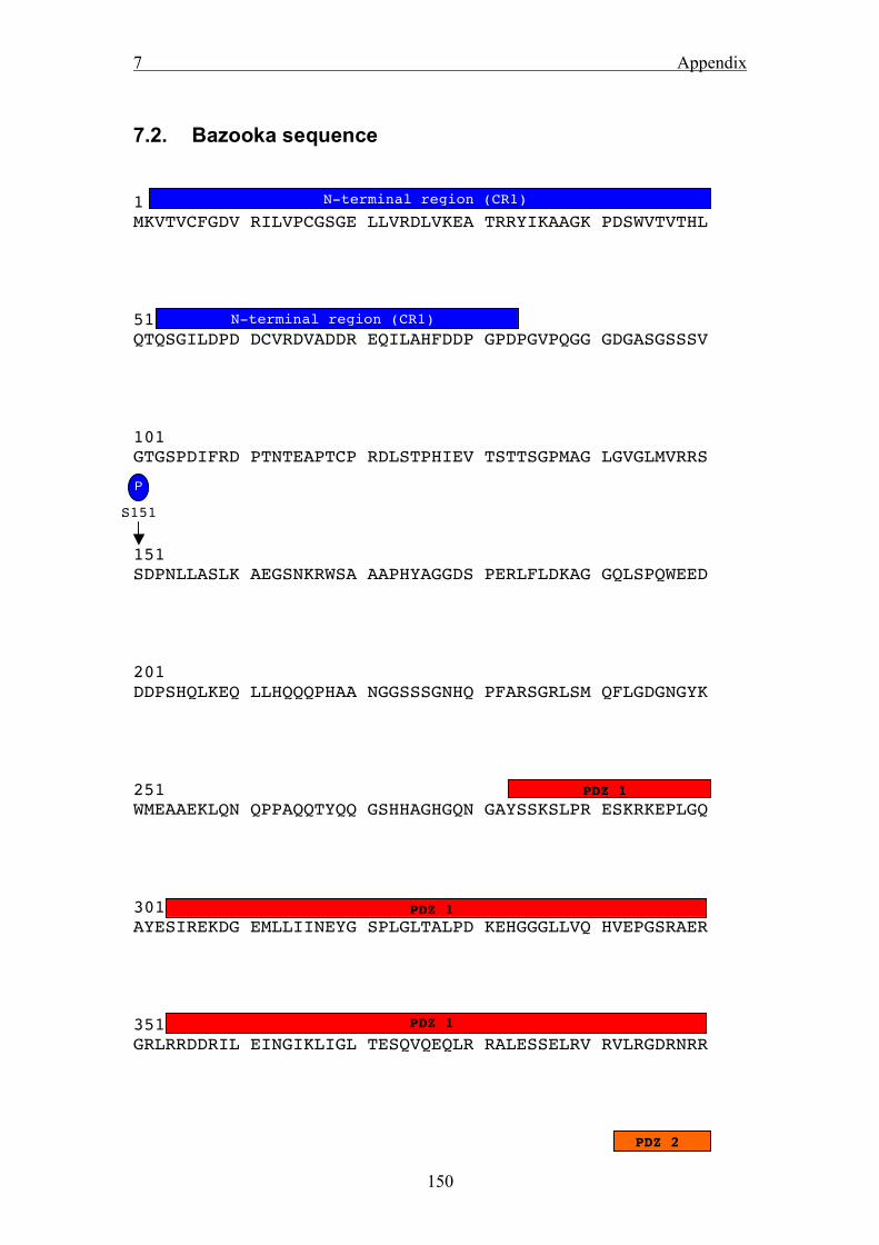
7 Appendix
150
7.2. Bazooka sequence 1 MKVTVCFGDV RILVPCGSGE LLVRDLVKEA TRRYIKAAGK PDSWVTVTHL 51 QTQSGILDPD DCVRDVADDR EQILAHFDDP GPDPGVPQGG GDGASGSSSV 101 GTGSPDIFRD PTNTEAPTCP RDLSTPHIEV TSTTSGPMAG LGVGLMVRRS 151 SDPNLLASLK AEGSNKRWSA AAPHYAGGDS PERLFLDKAG GQLSPQWEED 201 DDPSHQLKEQ LLHQQQPHAA NGGSSSGNHQ PFARSGRLSM QFLGDGNGYK 251 WMEAAEKLQN QPPAQQTYQQ GSHHAGHGQN GAYSSKSLPR ESKRKEPLGQ 301 AYESIREKDG EMLLIINEYG SPLGLTALPD KEHGGGLLVQ HVEPGSRAER 351 GRLRRDDRIL EINGIKLIGL TESQVQEQLR RALESSELRV RVLRGDRNRR
N-terminal region (CR1)
N-terminal region (CR1)
PDZ 1
PDZ 1
PDZ 1
PDZ 2
S151
P

7 Appendix
151
401 QQRDSKVAEM VEVATVSPTR KPHAAPVGTS LQVANTRKLG RKIEIMLKKG 451 PNGLGFSVTT RDNPAGGHCP IYIKNILPRG AAIEDGRLKP GDRLLEVDGT 501 PMTGKTQTDV VAILRGMPAG ATVRIVVSRQ QELAEQADQP AEKSAGVAVA 551 PSVAPPAVPA AAAPAPPIPV QKSSSARSLF THQQQSQLNE SQHFIDAGSE 601 SAASNDSLPP SSNSWHSREE LTLHIPVHDT EKAGLGVSVK GKTCSNLNAS 651 GSSASSGSNG LMKHDGDLGI FVKNVIHGGA ASRDGRLRMN DQLLSVNGVS 701 LRGQNNAEAM ETLRRAMVNT PGKHPGTITL LVGRKILRSA SSSDILDHSN 751 SHSHSHSNSS GGSNSNGSGN NNNSSSNASD NSGATVIYLS PEKREQRCNG 801 GGGGGSAGNE MNRWSNPVLD RLTGGICSSN SAQPSSQQSH QQQPHPSQQQ
PDZ 2
PDZ 2
PDZ 3
PDZ 3

7 Appendix
152
851 QQQRRLPAAP VCSSAALRNE SYYMATNDNW SPAQMHLMTA HGNTALLIED 901 DAEPMSPTLP ARPHDGQHCN TSSANPSQNL AVGNQGPPIN TVPGTPSTSS 951 NFDATYSSQL SLETNSGVEH FSRDALGRRS ISEKHHAALD ARETGTYQRN 1001 KKLREERERE RRIQLTKSAV YGGSIESLTA RIASANAQFS GYKHAKTASS 1051 IEQRETQQQL AAAEAEARDQ LGDLGPSLGM KKSSSLESLQ TMVQELQMSD 1101 EPRGHQALRA PRGRGREDSL RAAVVSEPDA SKPRKTWLLE DGDHEGGFAS 1151 QRNGPFQSSL NDGKHGCKSS RAKKPSILRG IGHMFRFGKN RKDGVVPVDN 1201 YAVNISPPTS VVSTATSPQL QQQQQQQLQQ HQQQQQIPTA ALAALERNGK 1251 PPAYQPPPPL PAPNGVGSNG IHQNDIFNHR YQHYANYEDL HQQHQQHQIS
aPKC-binding
CR2
CR3
S980
P
S1085
P

7 Appendix
153
1301 RRHQHYHSQR SARSQDVSMH STSSGSQPGS LAQPQAQSNG VRPMSSYYEY 1351 ETVQQQRVGS IKHSHSSSAT SSSSSPINVP HWKAAAMNGY SPASLNSSAR 1401 SRGPFVTQVT IREQSSGGIP AHLLQQHQQQ QLQQQQPTYQ TVQKMSGPSQ 1451 YGSAAGSQPH ASKV
Baz protein sequence (isoform A). The conserved domains are marked with bars (the
up to now unannotated conserved regions aa 1073-1093 and 1173-1193 are annotated
as CR2 and CR3). The three reported phosphorylation sites are indicated.
PDZ-B

7 Appendix
154
7.3. Western Blot of Baz constructs
S2R cells were transfected with Baz expression constructs, which were N-terminally
GFP-tagged. GFP-Baz proteins were immunoprecipitated using an anti-GFP antibody
(see 3.2 for details), proteins were resolved on 10% SDS-PAGE and blotted against
GFP.
8 Curriculum vitae
155
8. Curriculum vitae Dr. med. vet. Michael Krahn Department of Stem Cell Biology University of Goettingen Justus-von-Liebig-Weg 11 37 077 Goettingen Germany 08.12.1980 : Born in Muenster (Germany) 1987-1990 : Visit of the primary school in Everswinkel 1990-2000 : Visit of the grammar school in Warendorf, graduation with the “Abitur” 2000-2006 : Study of veterinary medicine at the School of Veterinary Medicine,
Hannover, Graduation in 2006 2003 : Participation in the Cornell Leadership Program for Veterinary Students 2003-2006 : Experimental PhD work in the Institute of Physiological Chemistry :
„Expression and characterization of Protocadherin LKC in mammalian cells – an new potential growth regulator in epithelial cells“, graduation
as “Dr. med vet.” in Mai 2006
2006-2009 : PostDoc position in the Department of Stem Cell Biology, University of
Goettingen Posters and Presentations :
- Poster at the meeting Horizons in Molecular Biology (12.09.-15.09.2007):
1.) Krahn and Wodarz : „Functional interaction of Protein Phosphatase 2A with Bazooka/PAR-3 during Drosophila development”
2.) Trierweiler, Krahn and Wodarz : „Drosophila as a model organism to study the physiological role of Dymeclin in context with the Dyggve-Melchior-Clausen Dysplasia”
3.) Neugebauer, Krahn and Wodarz : „Analysis of the Function of CG31534 during epithelial development in Drosophila”
- Poster at the annual meeting of the German Society for Cell Biology (12.-
15.03.2008): Krahn and Wodarz : „Dephosphorylation of Bazooka by PP2A is required for proper apical-basal polarity in embryonic neural stem cells”
- Presentation at the International Drosophila Research Conference (San Diego, 02.-06.04.2008) Krahn and Wodarz : „Dephosphorylation of Bazooka by

8 Curriculum vitae
156
PP2A is required for proper apical-basal polarity in Drosophila embryonic neuroblasts”
- Poster at the meeting Horizons in Molecular Biology (10.09.-13.09.2008): Hogl, Krahn and Wodarz : „Proper phosphorylation of Bazooka is required for maintenance of epithelial polarity in Drosophila melanogaster”
- Poster at the meeting of the Germany Society for Developmental Biology (25.-28.03.2009):
1.) Beati, Krahn and Wodarz : “The role of the Bazooka-CG31534 interaction for the establishment of cell polarity in Drosophila melanogaster“
2.) Krahn et al. : “Imperfect conservation from fly to man : The C-terminal
region of Bazooka but not of PAR-3 facilitates membrane binding”
- Presentation at the meeting of the Germany Society for Developmental Biology (25.-28.03.2009): Krahn and Wodarz : „PP2A antagonizes phosphorylation of Bazooka by PAR-1 to control apical-basal polarity in dividing embryonic neuroblasts”