Project 3: Functional validation of ncRNAs Norbert Perrimon and Bernard Mathey-Prevot.
Upload
truongminhCategory
view
216download
2
3493
IntroductionMorphogenetic movements such as cell migration are crucialfor the development of multicellular organisms. Cells that areborn at distinct locations in the developing animal oftenundergo precise, spatiotemporally regulated migration todistant sites where they eventually build specialized tissues andorgans. How input from multiple signaling pathways iscoordinated to ensure proper cell movement remains one of themain challenges in the field of cell migration anddevelopmental biology.
The migration of border cells (BCs) in the Drosophila eggchamber provides a unique system with which to geneticallydissect the mechanisms regulating invasive cell migration invivo (Montell, 2003; Rorth, 2002). During oogenesis, a groupof approximately eight cells, called BCs, is specified at theanterior pole of the ovarian follicular epithelium (Montell etal., 1992). At stage 9 of oogenesis, BCs change their shape,exit the epithelium and become migratory (Fig. 1A). BCscomprise two inner cells, called polar cells, surrounded by sixto eight outer BCs (Niewiadomska et al., 1999). The highlydynamic and polarized cytoskeleton of outer BCs enables themto migrate (Fulga and Rorth, 2002) and transport the non-motile polar cells with them. BCs migrate in between nursecells (NCs) until they reach the NC-oocyte boundary at stage10 of oogenesis.
A variety of molecules have been identified to regulate BCmigration. The Drosophila C/EBP transcription factorhomolog Slow border cells (Slbo) is a key regulator of BCmigration (Montell et al., 1992). Slbo is specifically expressedin BCs at the time when BCs become migratory. Slbo promotes
the expression of shotgun (shg), the gene encoding theDrosophila homolog of the cell-cell adhesion molecule E-Cadherin (DE-Cad), which has been shown to be crucial inboth BCs and NCs for BC motility (Niewiadomska et al.,1999). Specifically, loss of shg leads to delayed BC migration,and it has been speculated, although not tested, that an increasein DE-Cad expression might also result in BC migrationdefects. These findings have led to a model proposing that DE-Cad-containing adhesive complexes, together with myosins,provide the traction between BC and NC surfaces that allowsBCs to migrate (Fulga and Rorth, 2002; Geisbrecht andMontell, 2002).
Genetic screens have further identified four signalingpathways that control the stereotypic migration of BCs. Duringearly oogenesis, the cytokine-like molecule Unpaired (Upd) issecreted from the pair of anterior polar cells to activate theJAK/STAT pathway in the surrounding cells. JAK/STATactivation induces the expression of slbo, which specifies BCs(Silver and Montell, 2001). Like JAK/STAT, Notch signaling isalso required for the expression of slbo in anterior follicle cells(Gonzalez-Reyes and St Johnston, 1998), indicating that Notchsignaling might be crucial for BC specification and migration.Once specified, BCs exit the follicular epithelium and becomemigratory, a process that relies on nuclear hormone signaling(Bai et al., 2000). Ecdysone hormone co-receptor taiman (tai)mutants have defects in BC migration even though Slbo isexpressed and DE-Cad-containing adhesive complexes areformed at the BC-NC interface, leading to the proposal that themigration defects of tai mutant BCs are due to problems in theturnover of adhesive complexes (Bai et al., 2000).
Invasive cell migration in both normal development andmetastatic cancer is regulated by various signalingpathways, transcription factors and cell-adhesionmolecules. The coordination between these activities in thecontext of cell migration is poorly understood. DuringDrosophila oogenesis, a small group of cells called bordercells exit the follicular epithelium to perform a stereotypic,invasive migration. We find that the ETS transcriptionfactor Yan is required for border cell migration and thatYan expression is spatiotemporally regulated as bordercells migrate from the anterior pole of the egg chamber
towards the nurse cell-oocyte boundary. Yan expression isdependent on inputs from the JAK/STAT, Notch andReceptor Tyrosine Kinase pathways in border cells.Mechanistically, Yan functions to modulate the turnover ofDE-Cadherin-dependent adhesive complexes to facilitateborder cell migration. Our results suggest that Yan acts asa pivotal link between signal transduction, cell adhesionand invasive cell migration in Drosophila border cells.
Key words: Cell migration, Oogenesis, Drosophila, ETS, Notch,JAK/STAT, RTK, Border cells
Summary
Function of the ETS transcription factor Yan in border cell migrationMarkus Schober1,4,*, Ilaria Rebay3 and Norbert Perrimon1,2,*1Department of Genetics, Harvard Medical School, Boston, MA 02115, USA2Howard Hughes Medical Institute, Harvard Medical School, Boston, MA 02115, USA3University of Chicago, 5801 South Ellis Avenue, Chicago, IL 60637, USA4The Rockefeller University, 1230 York Avenue, New York, NY 10021, USA*Authors for correspondence (e-mail: [email protected] and [email protected])
Accepted 19 May 2005
Development 132, 3493-3504Published by The Company of Biologists 2005doi:10.1242/dev.01911
Research article
Dev
elop
men
t

3494
Two RTK pathways, the Platelet-Derived GrowthFactor/Vascular Endothelial Growth Factor Receptor (PVR)and the Epithelial Growth Factor Receptor (EGFR) pathways,control the migration of BCs. Pvf1, one of three secreted PVRligands (Duchek et al., 2001), is expressed in an increasinggradient along the anteroposterior (AP) axis of the eggchamber. This gradual expression, together with the delayed ormisrouted migration that results when dominant-negativeforms of both EGFR and PVR are co-expressed in BCs, orwhen the ligand Pvf1 is ectopically expressed in ovarian eggchambers, have led to a model whereby PVR and EGFR exertredundant functions in guiding BC migration (Duchek andRorth, 2001; Duchek et al., 2001; McDonald et al., 2003).Although the MAPK pathway is activated in migrating BCs(Duchek and Rorth, 2001), it is not known whether its targetgenes are crucial for BC migration.
The ETS (E-26) transcription factor gene yan, whichencodes the Drosophila homolog of the TEL oncogene (Laudetet al., 1999), is one of the main target genes of the Notch andMAPK signal transduction pathways during photoreceptorspecification in the Drosophila developing eye (O’Neill et al.,1994; Rohrbaugh et al., 2002). While Notch-mediatedSuppressor of Hairless [Su(H)] activity promotes yanexpression (Rohrbaugh et al., 2002), MAPK pathwaystimulation above a critical threshold triggers Yanphosphorylation (Rebay, 2002; Rebay and Rubin, 1995). Thispost-translational modification of Yan stimulates its nuclearexport and subsequent degradation, enabling photoreceptorcells to differentiate. Thus, Yan, which is antagonisticallyregulated by Notch and RTK signaling, probably provides anegative-feedback loop that allows the tight regulation of RTK-mediated cell differentiation.
Although many molecules involved in BC migration havebeen accounted for, it is not yet clear how input from multiplesignaling pathways is integrated to ensure the fidelity andprecise orchestration of cell movements. Here, we describe anovel function for Yan in BC migration. We find that Yan isdynamically expressed and regulated in migrating BCs.Specifically, we find that upregulation of Yan expression in thefollicular epithelium depends on the Notch and JAK/STATsignaling pathways, and precedes BC motility and exit fromthe follicular epithelium, whereas downregulation of Yan inresponse to PVR and EGFR is important for continued invasivemigratory behavior. We further demonstrate that BC migrationcan be delayed by increasing DE-Cad levels and that Yan mayinfluence this process by regulating the turnover of DE-Cad-containing adhesive complexes at the plasma membrane.
Materials and methodsFly strainsyanP(lacZ), yan1, tai61G1, pntP(lacZ), Nts and UAS-Ser were obtained fromthe Bloomington stock center. stat92E85C9, stat92E397 and UAS-DE-Cad were obtained from Denise Montell. UAS-DE-Cad5,9 wasobtained from Benedicte Sanson. UAS-Rab5S43N was obtained fromMarcos Gonzalez-Gaitan. yan443 and yan884 were identified in agenetic screen for Ras modifiers in the eye (Karim et al., 1996). yan443
is a non-sense mutation in Q111*, deleting most of the proteinincluding all of the MAPK phosphorylation sites and the entire ETSDNA-binding domain. yan443 is presumably a null allele. yan884 hasan identical phenotype to yan443 in eye clones, embryos and bordercell clones. Negatively marked follicle cell clones were generated by
crossing ubGFP FRT40A/CyO; T155-Gal4 UAS-Flp/TM6c virginfemales to yan FRT40A /CyO males. Positively marked clones weregenerated by crossing y w UAS-GFP hs-Flp; tub-Gal4; FRT82B tub-Gal80 virgin females to stat85C9 FRT82B /TM3, Sb males. Mosaicmutant females were fed for 5 days at 25°C before dissection.Overexpression experiments were performed using slbo-Gal4; UAS-yanWT, UAS-yanACT, UAS-λpvr, UAS-λtop, UAS-rafF179, UAS-λBtl,UAS-DE-Cad, UAS-DE-Cad5.9 and UAS-RabS43N. Epistasisexperiments between slbo and yan were performed using slbo1310;slbo1310, slbo-Gal4/CyO; slbo1310, UAS-yanWT/CyO and slbo1310, UAS-yanACT/CyO. All experiments were performed at 25°C. Nts flies weregrown and used for control experiments at the permissive temperatureof 18°C. Nts female flies were shifted to the restrictive temperature of29°C for 8, 12 and 16 hours prior to dissection in the Notch mutantanalysis.
Immunohistochemistry and immunofluorescenceOvarioles were dissected from 5-day-old, well-fed females inSchneider S2 medium. For β-galactosidase staining, ovaries weredissected, fixed for 10 minutes in 0.1% glutaraldehyde in PBS-0.1%Triton X-100 and stained as described previously (Bai et al., 2000).For immunofluorescence analysis, ovaries were fixed with 4%formaldehyde in PBS-0.1%Triton X-100 for 30 minutes. Ovaries wereincubated in primary antibody overnight, washed twice for 20 minutesin PBS-0.1% Triton X-100, followed by a 2-hour incubation insecondary antibody containing Alexa-568-Phalloidin (1:100,Molecular Probes) to stain the actin cytoskeleton. After washing thetissue twice for 30 minutes with PBS-0.1% Triton X-100, it wasmounted in Slow Fade (Molecular Probes). The following primaryantibodies were used: mouse anti-FAS III at 1:10 (2B5,Developmental Hybridoma Bank); mouse anti-Yan at 1:200 (I.Rebay); rat anti-Slbo at 1:3000 (P. Rorth); rat anti-DE-Cad at 1:100(H. Oda); and rabbit anti-β-Gal at 1:2000 (Cappel). As secondaryantibodies, Alexa-488 and Alexa-598 were used at 1:500 (MolecularProbes). Images were obtained using a Leica-SP2 confocalmicroscope.
FM1-43 uptake experimentDouble-stranded RNA was generated against the following regionsusing the Ambion MegaScript kit: Yan (NM_078731) nucleotides394-1359 and GFP nucleotides 3-549 of the open reading frame. SL2cells were seeded in serum-free Schneider’s medium (Gibco) into a96-well plate, with 8�105 cells and 1 μg dsRNA per well). Serumwas added after 1 hour of serum starvation and cells were grown for4 days at 25°C. SL2 cells (8�105) were transfected with 0.1 μgMT(metallothionin)-YanACT using effectene transfection reagents.Expression was induced with 1 mM CuSO4 12 hours before theendocytosis assay. Cells were replated on eight-well coverslipchambers coated with concavidin A and washed with HL3 medium(Kuromi and Kidokoro, 1999). HL3 medium was exchanged withHL3 medium containing 10 μM FM1-43 (Molecular Probes). FM1-43 incorporation was observed in real time. Eight confocal sectionsper minute were acquired on a Leica-SP2 inverted confocalmicroscope over a time period of 30 minutes. A maximum projectionwas generated for each frame and the relative fluorescence intensitywas plotted against time to assess the kinetics of FM1-43incorporation. Addition of CuSO4 in mock-transfected cells did notalter the endocytic rate when compared with untreated control cells.
ResultsYan expression is dynamic in migrating border cellsand is required for their migrationIn our search for genes that regulate Drosophila follicle cellmorphogenesis (M.S., unpublished), we identified a lacZ P-element insertion in the yan gene, yanP(lacZ), that is expressed
Development 132 (15) Research article
Dev
elop
men
t
3495The role of Yan in cell migration
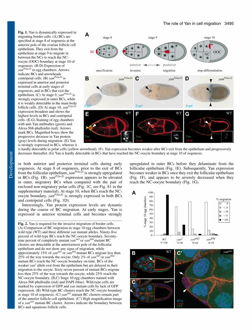
in both anterior and posterior terminal cells during earlyoogenesis. At stage 8 of oogenesis, prior to the exit of BCsfrom the follicular epithelium, yanP(lacZ) is strongly upregulatedin BCs (Fig. 1B). yanP(lacZ) expression appears to be elevatedin outer, migratory BCs when compared with the pair ofenclosed non-migratory polar cells (Fig. 1C, see Fig. S1 in thesupplementary material). At stage 10, when BCs reach the NC-oocyte boundary, yanP(lacZ) is strongly expressed in both BCsand centripetal cells (Fig. 1D).
Interestingly, Yan protein expression levels are dynamicduring the course of BC migration. At early stages, Yan isexpressed in anterior terminal cells and becomes strongly
upregulated in outer BCs before they delaminate from thefollicular epithelium (Fig. 1E). Subsequently, Yan expressionbecomes weaker in BCs once they exit the follicular epithelium(Fig. 1F), and appears to be severely decreased when theyreach the NC-oocyte boundary (Fig. 1G).
Fig. 1. Yan is dynamically expressed inmigrating border cells. (A) BCs arespecified at stage 8 of oogenesis at theanterior pole of the ovarian follicle cellepithelium. They exit from theepithelium at stage 9 to migrate inbetween the NCs to reach the NC-oocyte (OOC) boundary at stage 10 ofoogenesis. (B-D) Expression ofyanP(lacZ) in egg chambers. Arrowsindicate BCs and arrowheadscentripetal cells. (B) yanP(lacZ) isexpressed in anterior and posteriorterminal cells at early stages ofoogenesis, and in BCs that exit theepithelium. (C) At stage 9, yanP(lacZ) isstrongly expressed in outer BCs, whileit is weakly detectable in the main bodyfollicle cells. (D) At stage 10, yanP(lacZ)
expression broadens and shows thehighest levels in BCs and centripetalcells. (E-G) Staining of egg chamberswith anti-Yan antibodies (green) andAlexa-568-phalloidin (red). Arrowsmark BCs. Magnified boxes show theprogressive decrease in Yan protein(gray) levels during migration. (E) Yanis strongly expressed in BCs, whereas itis hardly detectable in polar cells (yellow arrowhead). (F). Yan expression becomes weaker after BCs exit from the epithelium and progressivelydecreases thereafter. (G) Yan is hardly detectable in BCs that have reached the NC-oocyte boundary at stage 10 of oogenesis.
Fig. 2. Yan is required for the invasive migration of border cells.(A) Comparison of BC migration in stage 10 egg chambers betweenwild type (WT) and three different yan mutant alleles. Ninety-fivepercent of wild-type BCs reach the NC-oocyte boundary. Seventy-nine percent of completely mutant yan443 or yan884 mutant BCclusters are detectable at the anteriormost pole of the follicularepithelium and do not show any signs of migration, whileapproximately 15% of yan443 or yan884 mutant BCs migrate less than25% of the way towards the oocyte. Only 2% of yan443 or yan884
mutant BCs reach the NC-oocyte boundary on time. BCs of theweaker yan1 allele exit from the epithelium but are delayed in theirmigration to the oocyte. Sixty-seven percent of mutant BCs migrateless then 25% of the way towards the oocyte, while 25% reach theNC-oocyte boundary. (B,C) Stage 10 egg chambers stained withAlexa-568-phalloidin (red) and DAPI (blue). Wild-type cells aremarked by expression of GFP and yan mutant cells by lack of GFPexpression. (B) Wild-type BC clusters reach the NC-oocyte boundaryat stage 10 of oogenesis. (C) yan443 mutant BC clusters remain partof the anterior follicle cell epithelium. (C′) High magnification imageof a yan443 mutant BC cluster. Arrows indicate the boundary betweenBCs and squamous follicle cells.
Dev
elop
men
t

3496
To address whether Yan is required for BC migration, wegenerated mitotic clones of BCs that lack Yan activity.Migration is significantly delayed in hypomorphic yan1 clones(Fig. 2A). Strikingly, clones of yan443, a protein null allele,exhibit stronger migration defects, where the majority ofmutant BCs fail completely to migrate and remain part of theanterior follicular epithelium (Fig. 2A,C). High-magnificationconfocal microscopy of yan443 mutant egg chambers labeledwith Alexa-568-phalloidin indicated that mutant BCs areconnected laterally to squamous follicle cells, suggesting thatthey remain attached to the follicular epithelium (Fig. 2C′).
Yan modulates DE-Cad-containing adhesivecomplexesTo address whether the yan mutant phenotype is caused by amisspecification of BCs, we examined Slbo expression inyan443 mutant BCs. Removal of Yan function had no effect onSlbo expression (Fig. 3A,A′). In addition, expression of thepolar cell-specific marker Fasciclin 3 (Fas3) was unaffected,indicating that the fate of migratory outer BCs was nottransformed into non-migratory polar cells (Fig. 3B,B′).Together, these data suggested that Yan does not function tospecify BCs or to discriminate between inner versus outer BCidentity.
To test whether Yan regulates cell motility, we analyzed DE-Cad expression in yan mutant BCs. DE-Cad is a key regulatorof BC migration (Niewiadomska et al., 1999) and the dynamicregulation of E-cadherin is crucial for epithelial-to-mesenchymal transitions and morphogenetic cell movementsin many systems (Thiery, 2002). In wild-type BC clusters, DE-Cad localized primarily to the surface between polar cells andouter BCs, as well as to the interface between outer BCs (Fig.3C). It is further detectable at a significantly lower level at themargins between BCs and NCs. In yan443 mutant BC clusters,DE-Cad was strongly expressed in both polar cells and mutantouter BCs (Fig. 3C′). Specifically, DE-Cad was stronglyenriched at the boundary between BCs and squamous follicle
cells (arrow, Fig. 3C′, see also Fig. S2 in the supplementarymaterial). Interestingly, even partially yan443 mutant BCclusters can show severe migration defects, and yan mutantBCs stay connected to squamous follicle cells, showingelevated DE-Cad accumulation at their plasma membraneinterface (arrows, Fig. 3D,D′).
To test whether Yan might suppress DE-Cad expression atthe initiation of BC migration, we overexpressed Yanspecifically in outer BCs using slbo-Gal4. Althoughoverexpression of wild-type Yan had no effects on BCmigration, overexpression of a dominant-active form of Yan(UAS-yanACT) strongly delayed migration (Fig. 4B,D).Interestingly, outer BCs expressing activated Yan showeddecreased cortical DE-Cad staining (Fig. 4B,B′, Fig. S3 in thesupplementary material). To genetically test whether the BCmigration defects associated with slbo-Gal4/UAS-yanACT aredue to reduced cortical DE-Cad expression, we co-expressedUAS-DE-Cad and observed a significant, but incomplete,rescue of the BC migration defects (Fig. 4D). BC migrationdefects observed in yan mutant egg chambers could thereforebe due to elevated and/or mislocalized DE-Cad. Indeed,overexpression of UAS-DE-Cad5,9 in BCs severely delayedtheir migration and in many cases blocked their detachmentfrom the anterior follicular epithelium (Fig. 4C,D). Confocalimmunofluorescence analysis further indicated that, uponoverexpression, DE-Cad was localized in a polarized fashionand was primarily concentrated at the trailing edge where BCsand squamous cells interface (arrowheads, Fig. 4C′). Together,these data suggest that DE-Cad expression is regulated, eitherdirectly or indirectly, by Yan in BCs, and that precise amountsand/or localization of DE-Cad are important for BC migration.
The transcriptional suppression of E-Cadherin is a crucialstep in mediating epithelial-mesenchymal transitions (Thiery,2002). We therefore tested whether Yan might function tosuppress shg transcription to allow BCs to exit the follicularlayer and become migratory. However, shg transcription wasnot affected in yan mutant BCs, nor could we find significant
Development 132 (15) Research article
Fig. 3. Accumulation of DE-Cad in yan mutantborder cells. The expression of Fas3 and DE-Cad was compared between yan/+ (WT) andyan mutant clones (see Materials and methodsfor details). (A-D′) Actin is visualized by Alexa-568-phalloidin (red). (A-D) Expression of GFP(blue) marks non-mutant cells in stage 9 mosaicmutant egg chambers. (A′-D′) yan443 mutantfollicle cell clones are marked by the absence ofGFP (blue). (A,A′) Slbo is expressed both in yanmutant and non-mutant BCs. (B,B′) The polarcell marker Fas3 is restricted to polar cells, evenwhen outer BCs lack Yan expression. (C,C′) DE-Cad accumulates in yan443 mutant BCs at theboundaries between outer BCs and polar cells(asterisk), as well as at the interface betweenBCs and squamous follicle cells (arrow).(D,D′) Mutant BCs accumulate high levels ofDE-Cad on their surface (arrows) and connectnon-mutant BCs and the follicular epithelium.Asterisks mark polar cells.
Dev
elop
men
t

3497The role of Yan in cell migration
alterations in the activity of a shg-luciferase reporterconstruct in S2R+ cells, either when yan expressionwas knocked down by RNA interference using a yan-specific double-stranded RNA (yanRNAi) or whenYanACT was overexpressed (data not shown), indicatingthat Yan does not affect shg transcription.
Changes in E-cadherin expression at the cell surfacecan alter the adhesive strength between cells (Yap etal., 1997). Endocytosis has recently surfaced as acrucial step in modulating E-Cadherin turnover at thecell surface, which might be crucial in remodelingadhesive complexes in morphogenetic movements andcell migration where E-Cadherin expression is nottranscriptionally suppressed (Paterson et al., 2003). Wethus tested whether Yan might modulate endocytosisto potentially affect DE-Cad turnover. UsingDrosophila SL2 cells, we performed an endocytosisassay in which we followed the uptake of themembrane dye FM1-43 over time. Interestingly, expression ofYanACT strongly enhanced endocytic FM1-43 incorporation(Fig. 5B,D), whereas it was reproducibly decreased afteryanRNAi treatment (Fig. 5A,D). These data suggest that Yanmight influence endocytosis and that way impinge on DE-Cadexpression and BC migration. To further test whetheralterations in the endocytic machinery affect DE-Cad turnoverin vivo, we expressed a dominant-negative form of the smallGTPase Rab5 (UAS-Rab5(S43N)) (Entchev and Gonzalez-Gaitan, 2002) in BCs to block endocytosis. In support of ourhypothesis, the migration of BCs expressing UAS-Rab5(S43N)
was severely delayed and elevated DE-Cad levels could beclearly detected at the cell cortex (Fig. 5F-H).
yan functionally interacts with slboSlbo is specifically expressed in BCs and is a crucial regulator
of shg expression and BC migration. Similar to Slbo, Yan isupregulated in BCs just before they become migratory (Fig. 1).To test whether the expression and upregulation of Yan requiresSlbo function, we analyzed Yan expression in slbo1310 mutantegg chambers. Interestingly, Yan was strongly expressed inBCs of stage 9 egg chambers (Fig. 6A), indicating that Yanexpression does not require Slbo activity. We further noticedthat Yan remained strongly expressed in slbo1310 BCs thatfailed to migrate (Fig. 6B), suggesting that migration towardsthe NC-oocyte boundary is required for Yan downregulation inBCs.
Our data indicate that the two transcription factors Yan andSlbo do not regulate the expression of one other (Fig. 3A′, Fig.6A), suggesting that they might not operate in a direct, linearpathway. However, Yan and Slbo could instead function inindependent pathways that converge to regulate BC migration.
Fig. 4. Yan regulates DE-Cad expression levels. (A-C) Eggchambers are stained for actin (with Alexa-568-phalloidin,red), anti-DE-Cad (green) and DAPI (blue). (A′-C′) Highermagnification views of A-C; grayscale images of DE-Cadallow the visualization of DE-Cad expression levels. Yellowarrows point towards polar cells, white arrowheads point atouter BCs, white arrows point at the interface between BCsand squamous follicle cells. (A,A′) In wild type, high levelsof DE-Cad are found at the boundary between polar cellsand outer BCs, as well as between outer BCs. DE-Cad isalso strongly detectable at the edge of outer BCs.(B,B′) DE-Cad surface expression is reduced in outer BCswhere UAS-yanACT is ectopically expressed using slbo-Gal4. (C,C′) Expression of UAS-DE-Cad5,9 in BCs by slbo-Gal4; arrow in C indicates arrested BCs. (D) Functionalinteraction between DE-Cad and Yan to regulate BCmigration. In wild type, 95% of BCs reach the NC-oocyteboundary. Ectopic expression of UAS-yanACT induces strongBC migration defects (60% of BCs migrate less than 25%,26% migrate 25-50%, 12% migrate 50-75% and 2%complete their migration). Weak expression of UAS-DE-Cad partially suppresses UAS-yanACT migration defects(37% migrate less than 25%, 29% migrate 25-50%, 29%migrate 50-75% and 5% reach the nurse cell-oocyteboundary). Forced expression of UAS-DE-Cad5,9 delays BCmigration (20% migrate less than 25%, 30% migrate 25-50%, 32% migrate 50-75% and 18% complete theirmigration by stage 10 of oogenesis). n>100.
Dev
elop
men
t

3498
We therefore tested whether yan and slbo functionally interactin BCs. While overexpression of YanWT had no or little effecton BC migration, expression of constitutively active YanACT
severely delayed their migration (Fig. 6C). Furthermore,overexpression of YanWT in heterozygous slbo1310 mutantsweakly enhanced BC migration defects, whereas ectopicexpression of YanACT in heterozygous slbo1310 mutant BCscaused BC migration defects that were even stronger than thedefects observed in homozygous slbo1310 mutant eggchambers. Strikingly, BC migration was completely blocked inhomozygous slbo1310 mutants overexpressing YanWT (Fig. 6C).Altogether, these data indicate that Yan and Slbo functionally
interact to control BC migration without influencing theexpression of each other.
Early Yan expression is regulated by both theJAK/STAT and Notch pathwaysOur observation, that both gain- and loss-of-Yan functioninfluence DE-Cad expression and BC migration, prompted usto re-examine yan expression during the course of BCmigration. As described above, yan is expressed in anteriorterminal cells, becomes upregulated in BCs before theybecome migratory and then the Yan protein levels decay as BCsapproach the NC-oocyte boundary (Fig. 1E-G). This complexspatiotemporal expression pattern suggests that different signaltransduction pathways are likely to cooperate to control Yanexpression. Previous studies have shown that yan is atranscriptional target of the Notch/[Su(H)] pathway in
Development 132 (15) Research article
Fig. 5. Yan modulates endocytosis. (A-C) Live FM1-43 uptake inDrosophila SL2 cells. FM1-43 membrane incorporation in 20minutes after yanRNAi (A), overexpression of yanACT (B) and gfpRNAi
control (C). (D) Kinetics of FM1-43 uptake is accelerated in cellsoverexpressing yanACT (red) and decreased in yanRNAi cells (blue),when compared with a gfpRNAi control (orange). (E) Twenty-minuteFM1-43 uptake after yanRNAi, gfpRNAi and MT-YanACT in triplicate.(F,G) Stage 10 egg chamber, stained with anti-DE-Cad (green),Alexa-568-phalloidin (red) and DAPI (blue). slbo-Gal4, UAS-Rab5(S43N)-expressing BCs (G) show impaired ability to migrate andelevated levels of cortical DE-Cad expression when compared withcontrol egg chambers (F). (H) Overexpression of dominant-negativeRab5 in BCs delays their migration (46% migrate less than 25%,25% migrate between 25-50%, 15% migrate 50-75% and 14%complete their migration by stage 10 of oogenesis).
Fig. 6. Activated Yan enhances slbo mutant border cell migrationdefects. (A,B) slbo1310 mutant egg chamber stained with Alexa-568-phalloidin (red) and anti-Yan (green). Arrows indicate BCs. (A) Yanis normally expressed in slbo1310 mutant egg chambers at stage 9.(B) Yan is strongly expressed in border cells of slbo1310 mutant eggchambers that fail to migrate towards the oocyte at stage 10.(C) Statistical representation of BC migration defects. Overexpressionof wild-type Yan in BCs has only minor effects on BC migration. Bycontrast, expression of YanACT specifically in BCs delays theirmigration; 38% completely failed to migrate, 22% migrated less than25% and 26% of BC clusters migrate less than 50% towards the NC-oocyte boundary. Interestingly, this phenotype can be enhanced byremoving one copy of slbo1310 resulting in 72% of BCs thatcompletely failed to migrate, 17% that migrated less than 25% and11% that migrated less than 50% towards the oocyte. Expression ofYan and YanACT enhances the BC migration defects of slbo mutantegg chambers. These BC migration defects are significantly strongerthan the defects observed in homozygous slbo1310 mutant eggchambers, where 56% of mutant BCs show no migration and 42%migrate less than 25% of the way towards the oocyte.
Dev
elop
men
t

3499The role of Yan in cell migration
photoreceptor cell development (Rohrbaugh et al., 2002).Furthermore, during oogenesis Notch signaling functions atmultiple times to control axis specification, proliferation andthe determination of various follicle cell fates (Gonzalez-Reyesand St Johnston, 1998; Grammont and Irvine, 2001; Grammontand Irvine, 2002; Keller Larkin et al., 1999; Larkin et al., 1996;Lopez-Schier and St Johnston, 2001; Ruohola et al., 1991)(Fig. 7A). To test whether the Notch pathway is active at thetime of BC migration, we analyzed the expression of a β-galactosidase reporter construct that contains both Su(H)- andGrainy Head-binding sites (Furriols and Bray, 2001).Interestingly, expression of this Notch activity reporter largelyresembled the yanP(lacZ) expression pattern (Fig. 1B-D). Su(H)-lacZ was strongly expressed in the anterior and posteriorterminal cells at early stages of oogenesis (Fig. 7B). At stage9 and 10 it was strongly active in BCs (Fig. 7B,C).
Furthermore, we detected an asymmetric distribution of theNotch ligand Delta. Delta was predominantly expressed inanterior follicle cells that express Yan in stage 8 egg chambers(data not shown).
Because Notch signaling appeared to be active at the timeof BC migration, we analyzed the function of Notch on BCdevelopment using a temperature-sensitive (ts) allele of Notch(Nts) that allows egg chamber development. To prevent defectsin oogenesis prior to stage 8, we shifted Nts females for 8, 12and 16 hours to the restrictive temperature of 29°C. WhereasBC migration was normal in Nts females raised at thepermissive temperature (Fig. 7D), we observed severe BCmigration defects in a significant number of egg chambers inNts females raised at the restrictive temperature (Fig. 7E).Staining of Nts egg chambers with anti-Slbo and anti-Yanantibodies revealed that, in these egg chambers, Slbo
Fig. 7. Yan expression depends on JAK/STAT and Notch pathways. (A) JAK/STAT and Notch signaling around the anterior and posterior polarcells (green) specify terminal cell populations during oogenesis. (B,C) β-galactosidase (β-gal) staining of a Su(H)-lacZ Notch activity reporter.Arrows indicate BCs. (B) β-Gal is dynamically expressed during early oogenesis, strongly in BCs of stage 9 egg chambers. (C) β-Gal isstrongly expressed in BCs of stage 10 egg chambers. (D-K′) Egg chambers are stained with Alexa-568-phalloidin (red) to visualize actin.Arrows indicate BCs and arrowheads mark squamous cells. (D) BCs migrate normally in Nts egg chambers at the permissive temperature.(E) BCs have migration defects in Nts egg chambers at the restrictive temperature. (F-H) Slbo expression is shown in green; (F′-K′) Yan isshown in green. (F,F′) Wild-type control BC cluster expressing Slbo and Yan. (G,G′) Nts BCs have reduced Slbo and Yan expression.(H,H′) Slbo and Yan are normally expressed in BCs where Serrate (Ser) is ectopically expressed. (I,J) stat92E mutant follicle cells arepositively marked by GFP expression (green, arrows), whereas wild-type cells do not express GFP (arrowhead). (I′,J′) stat92E mutant BCs donot express Yan (arrows), whereas Yan expression (green) is normal in wild-type cells (arrowhead). (K,K′) tai61G1 mutant BC clones arenegatively marked by lack of GFP expression. (K) tai mutant BCs do not express GFP (green). (K′) The expression of Yan in tai mutant BCsand squamous follicle cells was similar to that observed in wild-type control egg chambers (F′).
Dev
elop
men
t

3500
expression was strongly reduced (Fig. 7G), whereas Yan wasnot detectable (Fig. 7G′). Ectopic activation of Notch followingexpression of one of its ligands Serrate (Ser), or ectopicexpression of the constitutively active intracellular domain ofNotch (Nintra) in BCs using slbo-Gal4 resulted in robustexpression of Slbo and Yan (Fig. 7H,H′ and data not shown)and normal BC motility. Thus, Notch signaling controls theexpression of Yan and Slbo in anterior terminal cells and BCs,respectively.
In addition to Notch signaling, the JAK/STAT pathway hasrecently been shown to control the specification of variousfollicle cell fates (Keller Larkin et al., 1999; Xi et al., 2003)and BC migration (Beccari et al., 2002; Silver and Montell,2001) (Fig. 7A). To test whether JAK/STAT signalingregulates Yan expression in BCs, we generated positivelymarked stat92E mutant clones. stat mutant follicle cellsshowed strongly reduced Yan expression (arrows, Fig. 7I,I′)when compared with the corresponding wild-type folliclecells (arrowheads, Fig. 7I,I′) in stage 8 egg chambers. statmutant BCs, as well as squamous follicle cells, did notexpress Yan (arrows, Fig. 7J,J′), whereras it was normallyexpressed in wild-type anterior terminal cells(arrowhead, Fig. 7J,J′). Similar results wereobtained in clones that were mutant for hopscotch,the JAK kinase that activates Stat92E (data notshown). Interestingly, BCs of hypomorphicstatF/statP1608 mutant egg chambers showed severeBC migration defects and a significant reduction in
Yan expression (data not shown), suggesting that Yan is acrucial target gene of the JAK/STAT pathway in BCs andanterior terminal cells.
Finally, we found that the ecdysone nuclear hormonepathway, which coordinates BC migration with egg chamberdevelopment (Bai et al., 2000), did not influence Yanexpression. Staining of tai61G1 mutant BC clusters with anti-Yan antibodies revealed that Yan was normally expressed inthe absence of tai gene activity (Fig. 7K,K′). Altogether, weconclude that the JAK/STAT and Notch signaling pathwayscontrol the expression of Slbo and Yan in the developing eggchamber to promote BC migration.
Yan is downregulated in response to increasing PVRand EGFR signalingYan expression decreases in wild-type BCs as they migratefrom the anterior pole of stage 9 egg chamber towards the NC-oocyte boundary (Fig. 1), but remains strongly expressed inslbo mutant BCs, which fail to migrate and remain at theanterior tip of stage 10 egg chambers (Fig. 6). Furthermore,overexpression of yanWT had little or no effects on BC
Development 132 (15) Research article
Fig. 8. RTKs signaling downregulates Yan. (A) Yan(blue) is strongly expressed in BCs that delaminate fromthe follicular epithelium, but expression graduallydecreases as BCs migrate along the increasing gradientof Pvf1 (orange). The inverse expression levels of Yanand Pvf1 suggest that PVR activity counteracts Yanexpression in migrating BCs. (B-E′) Egg chambers arestained with Alexa-568-phalloidin (red) to visualizeactin. Arrows indicate BCs and arrowheads thesquamous follicle cells. (B-E) Anti-Slbo (green) andactin (red) staining; (B′-E′) anti-Yan (green) and actin(red) staining. (B,B′) Slbo and Yan are stronglyexpressed in wild-type BCs that delaminate from thefollicular epithelium. (C) Slbo is normally expressed inBCs that ectopically express activated PVR. (C′) Bycontrast, Yan is undetectable in BCs but is expressednormally in squamous follicle cells where slbo-Gal4 isinactive. Similarly, expression of either activated EGFR(D,D′) or activated Raf (E,E′) in BCs downregulates Yanbut not Slbo. All experiments and stainings were done inparallel, and images were taken with the same settings,together with experiments in Fig. 7F-H. The expressionlevels of Yan in squamous follicle cells serve as aninternal control. (F,G) β-Gal staining indicates yanexpression in yanP(lacZ) (F) and yanP(lacZ)/slbo-Gal4;UAS-λPVR (G) stage 10 egg chambers. Arrows indicateBCs; insets show yan mRNA expression by in situhybridization. (H,I) Stage 10 egg chamber stained withanti-Slbo antibody (red), anti-β-Gal antibody indicatingpnt-lacZ expression (green), DAPI (blue) and Alexa-568-phalloidin (gray). (H) Pnt-lacZ is expressed in posteriorcells and is absent in wild-type BCs. (I) Expression ofactivated PVR in BCs arrests their migration. These BCsexpress Slbo but do not express pnt-lacZ.
Dev
elop
men
t

3501The role of Yan in cell migration
migration, whereas expression of activated yan (yanACT), aform of Yan that cannot be post-translationally regulated byphosphorylation via the MAPK pathway (Rebay and Rubin,1995), showed a severe delay in BC migration. These data,together with the recent finding that Pvf1 is expressed in anincreasing gradient along the AP axis towards the NC-oocyteboundary, and that the PVR and EGFR signaling pathways areactive in BCs and are crucial for their migration (Duchek andRorth, 2001; Duchek et al., 2001; McDonald et al., 2003),suggested that the gradient of PVR and EGFR activity mightcontrol the spatiotemporal expression of Yan during the courseof BC migration (Fig. 8A).
To test whether the PVR and EGFR pathways can trigger Yandownregulation in BCs, we expressed activated forms of PVR,EGFR and the RTK signal transducer RAF in BCs, and assayedSlbo and Yan expression. Whereas Slbo was expressed at normallevels in BCs that expressed activated forms of PVR, EGFR orRAF (Fig. 8B-E), Yan protein levels were strongly reduced (Fig.8B′-E′). As an internal control, normal Yan expression levelswere observed in squamous follicle cells where slbo-Gal4 is notexpressed. Furthermore, expression of activated FGFR hardly
affected BC migration and Yan expression levels werecomparable to wild type (data not shown).
To distinguish whether PVR and EGFR regulate Yanexpression at the transcriptional or post-transcriptional level,we expressed activated PVR using slbo-Gal4 in a yanP(lacZ)
background. BCs that do not express activated PVR (Fig. 8F)are clearly distinguishable from activated PVR-expressing BCs(Fig. 8G) by their migration defects in stage 10 egg chambers.Yan has previously been described to suppress its owntranscription (Rohrbaugh et al., 2002), and loss of Yan proteinmight thus result in activation of the yanP(lacZ) reporter due toan autoregulatory feedback loop. Interestingly, BCs thatexpress activated PVR strongly express β-Gal, suggesting thatPVR activation controls Yan expression post-transcriptionally,which is further supported by RNA in situ hybridization datausing a yan-specific probe (see insets, Fig. 8F,G).
Our data support a model where JAK/STAT and Notchsignaling specify anterior terminal cells including BCs,resulting in a strong expression of Yan in BCs; increasing RTKactivity can decrease Yan expression as BCs approach theirdestination (Fig. 9).
initiation migration stop
A
stage 9 stage 10
JAK/Stat EGFR
JAK/Stat Notch
PC PC p-TC a-TC
stage 8
specification
EcdysonePVR
EGFR PVR
stage 9
Yan expression PVF1 expression
B
JAK/Stat
Notch
yan
mRNA protein protein-degradation
DE-Cad turnover
protein
RTK
MAPK
Cytoskeletal Dynamics
x
Yan Yan
Stereotypic Cell Migration
Yan P
Fig. 9. Integrative model of Yan regulation and function during border cell migration. (A) The JAK/STAT and Notch pathways specify the groupof anterior terminal cells (a-TC) around the pair of polar cells (PC, green). Cells that are directly adjacent to the anterior polar cells are specifiedas BCs expressing Slbo. With the exception of the polar cells, Yan (blue) is expressed in all a-TCs, and becomes upregulated immediately prior totheir transit from a static, epithelial state to a migratory state (dark blue). Posterior terminal cells (red) are specified by Gurken (EGF) signaling.Pvf1 (orange), secreted from the oocyte, guides BCs towards the oocyte. As BCs face increasing Pvf1 levels from anterior to posterior, Yanexpression levels decrease (light blue circles). (B) Regulatory relationships between signal transduction pathways that control BC migration.JAK/STAT and Notch signaling pathways regulate the expression of slbo and yan, whereas PVR and EGFR induction lead to Yanphosphorylation and its inactivation. The transient upregulation of Yan at the initiation of BC migration facilitates DE-Cad turnover at the plasmamembrane to enable BCs to make and break adhesive contacts, and to promote detachment from the epithelium and cell movement. Coordinated,dynamic changes in cell adhesion and cytoskeletal organization enable BCs to migrate in a stereotypic fashion.
Dev
elop
men
t

3502
The Notch and RTK signaling pathways function to controlAP axis specification at early stages of oogenesis, resulting inexpression of the ETS transcription factor pointed (pnt) at theposterior pole. In photoreceptor cells, RTK activation inducesthe downregulation of Yan, which subsequently allows pntexpression and a switch in cell fate. Thus, we tested whetherYan expression at the initiation of BC migration might suppresspnt expression, and Yan downregulation at the NC-oocyteboundary might lead to pnt expression, and therefore,potentially, induce BC differentiation. RNA in situhybridization data and analysis of pntP(lacZ) expression inovaries revealed that pnt is not expressed in BCs at any stageof oogenesis, and ectopic expression of slbo-Gal4::UAS-pntP2does not alter BC motility (data not shown). Furthermore,ectopic activation of PVR in BCs downregulated Yanexpression (Fig. 8C′) and delayed BC migration withoutinduction of pntP(lacZ) in BCs (Fig. 8I). We thus conclude thatalthough the Notch and RTK signaling pathways modulate Yanexpression levels in both photoreceptor cells and BCs, themechanisms used are not identical, and the transcriptionalresponses and downstream mechanisms depend, at least in part,on the developmental context.
DiscussionDirected cell migration is a complex process wherebyextracellular cues stimulate distinct signal transductionpathways to modulate cytoskeletal dynamics, as well as cell-cell and cell-substratum adhesion. Despite considerableadvances in recent years, the relationships between theactivities of signaling pathways, transcription factors and celladhesion molecules in the context of cell migration andmetastatic cancer remain poorly understood. Here, we describea function for the ETS transcription factor Yan in BCmigration. Yan expression levels are spatiotemporallyregulated during the course of BC migration by the Notch,JAK/STAT and RTK signaling pathways (Fig. 9). Either anincrease or a decrease in Yan activity delays BC migration andis associated with an alteration in DE-Cad-dependent adhesivecomplexes, which themselves are crucial for BC motility. Thefinding that Yan functions as a key regulator of BC migrationis an important step towards understanding the molecularmechanisms by which extrinsic cues regulate cell adhesion andcytoskeletal dynamics to control invasive cell migration invivo.
Yan is dynamically expressed in migrating bordercellsOur study reveals that during oogenesis yan mutant BCs aredefective in their invasive migratory behavior. In addition, wefound that Yan is upregulated as BCs exit the epithelium tobecome migratory, and that subsequently Yan protein levelsdecay as BCs approach the NC-oocyte boundary (Fig. 1).Because Yan has previously been shown to function as atranscriptional repressor and an inhibitor of neuronaldifferentiation, we tested whether it regulates BC identity.Although we cannot completely exclude this possibility, BCmarkers are properly expressed in the absence of Yan. Thus,we propose that Yan promotes BC motility, an hypothesiswhich is supported by the observations that: (1) Yan isupregulated prior to the BCs exiting the follicular epithelium
to become migratory; (2) Yan protein levels decreaseprogressively as BCs approach their final destination; and (3)yan mutant BCs exhibit a delay in migration. Interestingly,ectopic expression of constitutively activated Yan in BCs alsodelays their migration, suggesting that the spatiotemporalactivity of Yan protein needs to be precisely controlled duringthe migratory process.
Yan coordinates signal inputs from differentpathwaysThe dynamic expression of Yan is crucial for BC migration, asindicated by the migratory defects associated with both gain-and loss-of-function alleles of yan. Analysis of mutations inthe JAK/STAT (Silver and Montell, 2001; Xi et al., 2003) andNotch (Gonzalez-Reyes and St Johnston, 1998) signalingpathways revealed that they are required for the expression ofat least two transcription factors that are crucial for BCmigration and which themselves influence DE-Cad activity.Slbo is specifically expressed in BCs and enhances shgtranscription. Yan, by contrast, is expressed in anterior terminalcells, but becomes upregulated in BCs at the time they exitfrom the epithelium to become migratory. Yan might enhanceDE-Cad turnover to facilitate the transition from an immobileepithelial state to a migratory one. Enhanced BC migrationdefects of hypomorphic slbo mutant egg chambersoverexpressing Yan further underscore their interaction toregulate DE-Cad expression and BC migration.
Interestingly, we find that Yan expression levels graduallydecrease as BCs move along an increasing PVR/EGFR activitygradient (Duchek and Rorth, 2001; Duchek et al., 2001;McDonald et al., 2003). Yan has been shown to bephosphorylated by the EGFR-MAPK pathway, which triggersits nuclear export and protein degradation (Rebay and Rubin,1995). Consistent with these previous studies, expression ofdominant-active PVR and EGFR in BCs blocks BC migrationand abrogates Yan protein expression, whereas yan transcriptor enhancer trap expression is still detectable. Expression ofactivated Ras and Raf similarly induced Yan downregulation,consistent with an involvement of the canonical Ras/MAPKpathway in mediating PVR/EGFR signaling. We note,however, that although BC migration was significantly delayedupon ectopic expression of activated Ras, activated Raf hardlyaffected their ability to migrate. The basis of this difference,which might be due to complex feedback loops between theimplicated signaling pathways, is unclear at the present timeand will need to be investigated further.
Yan regulates the accumulation of DE-Cadcontaining adhesive complexesIs the function of Yan to facilitate the transition of BCs froman epithelial to a migratory state, or to promote their motility?Although E-Cadherin is often downregulated as cells transitfrom an epithelial to a mesenchymal-like migratory state(Thiery, 2002), this may not be the case in BCs, as DE-Cad isstrongly expressed in BCs and shg mutant BCs fail to migrate(Niewiadomska et al., 1999). However, BCs mutant for yan ortai accumulate ectopic DE-Cad-containing adhesive complexes(Bai et al., 2000). Consistent with these observations, ectopicstimulation of PVR in BCs, which enhances tai mutant BCmigration defects, also results in elevated, cortical DE-Cadstaining (McDonald et al., 2003). Even though the observed
Development 132 (15) Research article
Dev
elop
men
t

3503The role of Yan in cell migration
BC migration defects in these mutants might not be due toaltered surface levels of DE-Cad only, we found thatoverexpression of DE-Cad alone can cause migration impairedBCs. E-cadherin not only mediates homophilic cell-celladhesion but also functions together with its binding partnersas a key regulator of the cortical actin cytoskeleton. It istherefore interesting to note that follicle cells overexpressingDE-Cad show severely enhanced filamentous actin staining(data not shown).
Our experiments revealed that DE-Cad was elevated in yanmutant BCs and suppressed upon expression of UAS-yanACT,suggesting that Yan controls, at least in part, DE-Cadexpression in BCs. These observations find further support inthe partial rescue of slbo-Gal4::UAS-yanACT-induced BCmigration defects upon co-expression of UAS-DE-Cad. Howdoes Yan affect DE-Cad expression in BCs? Although thefunction of Yan as a transcriptional repressor in various tissues(Rebay, 2002) suggests that it may act as a transcriptionalregulator of shg, we could not detect a change in shgtranscription in yan mutant follicle cells. However, increasedFM1-43 incorporation in Drosophila SL2 cells overexpressingYanACT, and a decrease in incorporation after yanRNAi, suggestsa change in endocytic activity. E-Cadherin has previously beenfound in endocytic compartments and endocytosis has beenspeculated to modulate E-Cadherin activity regulation duringmorphogenetic movements (Lanzetti et al., 2004; Paterson etal., 2003). Interestingly, blocking endocytosis by theexpression of dominant-negative Rab5 lead to severe BCmigration defects and increased DE-Cad staining. Consistentwith our observations, expression of shg under a heterologouspromoter has recently been shown to rescue shg mutant BCmigration defects, suggesting that the dynamic expression ofDE-Cad in BCs might depend on both transcriptional and post-transcriptional mechanisms (Pacquelet et al., 2003). Based onthese results, we favor a model whereby Yan might, at least inpart, function to regulate DE-Cad turnover, possibly throughthe transcriptional regulation of as-yet-unidentifiedcomponents of the endocytic machinery.
ETS factors during epithelial-mesenchymaltransition and metastatic cancerETS transcription factors are not only regulators ofmorphogenetic processes but have also been identified asoncogenes. Indeed, several ETS factors are upregulated ininvasive cancers and are currently used as molecular markersto grade their invasiveness (Dittmer and Nordheim, 1998;Oikawa and Yamada, 2003; Sharrocks, 2001). The molecularfunction of ETS factors in tumorigenesis is not clear, as theycan act as both oncogenes and tumor suppressors. Ourobservations that yan is associated with similar gain- and loss-of-function phenotypes support both a positive and negativefunction on invasive migration, dependent on activity levelsand possibly on available cofactors. Furthermore, thecomplexity of invasive tumors makes it difficult to assess whatfunction ETS factors have, as they are upregulated not only inthe cancerous tissue but also, for example, in forming bloodvessels during tumor angiogenesis. Finally, our finding thatYan levels are regulated by JAK/STAT, Notch and RTKsignaling pathways, which have been implicated in metastaticcancer, is another strong connection between Yan-like ETSfactors and tumorigenesis.
We thank Pernille Rorth, Denise Montell, Harioki Oda and theBloomington Stock center for fly strains and antibodies, and MisaoHigashi for helpful suggestions on the FM1-43 assay. We thankBernard Mathey-Prevot, Ramanuj Dasgupta, Craig Micchelli, ColinJamora and Herve Agaisse for helpful discussions and critical readingof the manuscript. This work was supported by the BoehringerIngelheim Fonds (M.S.). N.P. is an investigator of the Howard HughesMedical Institute.
Supplementary materialSupplementary material for this article is available athttp://dev.biologists.org/cgi/content/full/132/15/3493/DC1
ReferencesBai, J., Uehara, Y. and Montell, D. J. (2000). Regulation of invasive cell
behavior by taiman, a Drosophila protein related to AIB1, a steroid receptorcoactivator amplified in breast cancer. Cell 103, 1047-1058.
Beccari, S., Teixeira, L. and Rorth, P. (2002). The JAK/STAT pathway isrequired for border cell migration during Drosophila oogenesis. Mech. Dev.111, 115-123.
Dittmer, J. and Nordheim, A. (1998). Ets transcription factors and humandisease. Biochim. Biophys. Acta 1377, F1-F11.
Duchek, P. and Rorth, P. (2001). Guidance of cell migration by EGF receptorsignaling during Drosophila oogenesis. Science 291, 131-133.
Duchek, P., Somogyi, K., Jekely, G., Beccari, S. and Rorth, P. (2001).Guidance of cell migration by the Drosophila PDGF/VEGF receptor. Cell107, 17-26.
Entchev, E. V. and Gonzalez-Gaitan, M. A. (2002). Morphogen gradientformation and vesicular trafficking. Traffic 3, 98-109.
Fulga, T. A. and Rorth, P. (2002). Invasive cell migration is initiated byguided growth of long cellular extensions. Nat. Cell Biol. 4, 715-719.
Furriols, M. and Bray, S. (2001). A model Notch response element detectsSuppressor of Hairless-dependent molecular switch. Curr. Biol. 11, 60-64.
Geisbrecht, E. R. and Montell, D. J. (2002). Myosin VI is required for E-cadherin-mediated border cell migration. Nat. Cell Biol. 4, 616-620.
Gonzalez-Reyes, A. and St Johnston, D. (1998). Patterning of the follicle cellepithelium along the anterior-posterior axis during Drosophila oogenesis.Development 125, 2837-2846.
Grammont, M. and Irvine, K. D. (2001). fringe and Notch specify polar cellfate during Drosophila oogenesis. Development 128, 2243-2253.
Grammont, M. and Irvine, K. D. (2002). Organizer activity of the polar cellsduring Drosophila oogenesis. Development 129, 5131-5140.
Karim, F. D., Chang, H. C., Therrien, M., Wassarman, D. A., Laverty, T.and Rubin, G. M. (1996). A screen for genes that function downstream ofRas1 during Drosophila eye Development. Genetics 143, 315-329.
Keller Larkin, M., Deng, W. M., Holder, K., Tworoger, M., Clegg, N. andRuohola-Baker, H. (1999). Role of Notch pathway in terminal follicle celldifferentiation during Drosophila oogenesis. Dev. Genes Evol. 209, 301-311.
Kuromi, H. and Kidokoro, Y. (1999). The optically determined size ofexo/endo cycling vesicle pool correlates with the quantal content at theneuromuscular junction of Drosophila larvae. J. Neurosci. 19, 1557-1565.
Lanzetti, L., Palamidessi, A., Areces, L., Scita, G. and Di Fiore, P. P.(2004). Rab5 is a signalling GTPase involved in actin remodelling byreceptor tyrosine kinases. Nature 429, 309-314.
Larkin, M. K., Holder, K., Yost, C., Giniger, E. and Ruohola-Baker, H.(1996). Expression of constitutively active Notch arrests follicle cells at aprecursor stage during Drosophila oogenesis and disrupts the anterior-posterior axis of the oocyte. Development 122, 3639-3650.
Laudet, V., Hanni, C., Stehelin, D. and Duterque-Coquillaud, M. (1999).Molecular phylogeny of the ETS gene family. Oncogene 18, 1351-1359.
Lopez-Schier, H. and St Johnston, D. (2001). Delta signaling from the germline controls the proliferation and differentiation of the somatic follicle cellsduring Drosophila oogenesis. Genes Dev. 15, 1393-1405.
McDonald, J. A., Pinheiro, E. M. and Montell, D. J. (2003). PVF1, aPDGF/VEGF homolog, is sufficient to guide border cells and interactsgenetically with Taiman. Development 130, 3469-3478.
Montell, D. J. (2003). Border-cell migration: the race is on. Nat. Rev. Mol.Cell Biol. 4, 13-24.
Montell, D. J., Rorth, P. and Spradling, A. C. (1992). slow border cells, alocus required for a developmentally regulated cell migration duringoogenesis, encodes Drosophila C/EBP. Cell 71, 51-62.
Dev
elop
men
t

3504
Niewiadomska, P., Godt, D. and Tepass, U. (1999). DE-Cadherin is requiredfor intercellular motility during Drosophila oogenesis. J. Cell Biol. 144, 533-547.
Oikawa, T. and Yamada, T. (2003). Molecular biology of the Ets family oftranscription factors. Gene 303, 11-34.
O’Neill, E. M., Rebay, I., Tjian, R. and Rubin, G. M. (1994). The activitiesof two Ets-related transcription factors required for Drosophila eyedevelopment are modulated by the Ras/MAPK pathway. Cell 78, 137-147.
Pacquelet, A., Lin, L. and Rorth, P. (2003). Binding site for p120/{delta}-catenin is not required for Drosophila E-cadherin function in vivo. J. CellBiol. 160, 313-319.
Paterson, A. D., Parton, R. G., Ferguson, C., Stow, J. L. and Yap, A. S.(2003). Characterization of E-cadherin endocytosis in isolated MCF-7 andchinese hamster ovary cells: the initial fate of unbound E-cadherin. J. Biol.Chem. 278, 21050-21057.
Rebay, I. (2002). Keeping the receptor tyrosine kinase signaling pathway incheck: lessons from Drosophila. Dev. Biol. 251, 1-17.
Rebay, I. and Rubin, G. M. (1995). Yan functions as a general inhibitor ofdifferentiation and is negatively regulated by activation of the Ras1/MAPKpathway. Cell 81, 857-866.
Rohrbaugh, M., Ramos, E., Nguyen, D., Price, M., Wen, Y. and Lai, Z. C.(2002). Notch activation of yan expression is antagonized by RTK/pointedsignaling in the Drosophila eye. Curr. Biol. 12, 576-581.
Rorth, P. (2002). Initiating and guiding migration: lessons from border cells.Trends Cell Biol. 12, 325-331.
Ruohola, H., Bremer, K. A., Baker, D., Swedlow, J. R., Jan, L. Y. and Jan,Y. N. (1991). Role of neurogenic genes in establishment of follicle cell fateand oocyte polarity during oogenesis in Drosophila. Cell 66, 433-449.
Sharrocks, A. D. (2001). The ETS-domain transcription factor family. Nat.Rev. Mol. Cell Biol. 2, 827-837.
Silver, D. L. and Montell, D. J. (2001). Paracrine signaling through theJAK/STAT pathway activates invasive behavior of ovarian epithelial cells inDrosophila. Cell 107, 831-841.
Thiery, J. P. (2002). Epithelial-mesenchymal transitions in tumourprogression. Nat. Rev. Cancer 2, 442-454.
Xi, R., McGregor, J. R. and Harrison, D. A. (2003). A gradient of JAKpathway activity patterns the anterior-posterior axis of the follicularepithelium. Dev. Cell 4, 167-177.
Yap, A. S., Brieher, W. M. and Gumbiner, B. M. (1997). Molecular andfunctional analysis of cadherin-based adherens junctions. Annu. Rev. CellDev. Biol. 13, 119-146.
Development 132 (15) Research article
Dev
elop
men
t