Function in Cell Growth, Differentiation & Development
6
Gene Function in Cell Growth, Differentiation & Development Director Univ.-Prof. Dr. rer. nat. Martin Zenke RWTH Aachen University Hospital Pauwelsstrasse 30, 52074 Aachen Helmholtz Institute for Biomedical Engineering Pauwelsstrasse 20, 52074 Aachen Phone: +49-241-80 80760 (Office) +49-241-80 80759 (Secretary) Fax: +49-241-80 82008 Email: [email protected] Web: http://www.molcell.de http://www.stemcellfactory.de Staff Mierau, Eveline, Administrative Assistant Sous, Renate, Administrative Assistant Aydin, Gülcan, BSc, Technician Delgado Cáceres, Manuel, MSc Student Förster, Malrun, MSc, PhD Student Hapala Jan, PhD, Postdoc Hieronymus, Thomas, PhD, Group Leader Kluge, Frederick, BSc Student Küstermann, Caroline, MSc, PhD Student Kreutzer, Fabian, MSc Student Lennartz, Daniel, MSc Student Mitzka, Saskia, Technician Niessing, Bastian, MSc Student Prithivirat, Sujeethkumar, MSc Student Rösseler, Corinna, MSc, PhD Student Sagi, Zsofia, MSc, PhD student Schalla, Carmen, Technician Sechi, Antonio, PhD, Group Leader Seré, Kristin, PhD, Group Leader Sontag, Stephanie, MSc, PhD Student Szymanski de Toledo, Marcelo, PhD, Postdoc Wanek, Paul, BSc, Technician Stem Cell Biology and Cellular Engineering Univ.-Prof. Dr. med., Dr. rer. nat. Wolfgang Wagner Helmholtz Institute for Biomedical Engineering Pauwelsstrasse 20, 52074 Aachen Phone: +49-241-80 88611 (Office) Fax: +49-241-80 82008 Email: [email protected] Web: http://www.stemcellbiology.ukaachen.de Abagnale, Giulio, MSc, PhD Student Bozic, Tanja, MSc, PhD Student Candido, Danilo, MSc, PhD, Postdoc Eipel, Monika, MSc, PhD Student Fernandez-Rebollo., Eduardo, PhD, Postdoc Franzen, Julia, MSc, PhD Student Frobel, Joana, MSc, PhD Student Götzke, Roman, MSc Student Grezella, Clara, MSc Student Hapala, Jan, PhD, Postdoc Han, Yang, MSc, PhD Student Kalwa, Marie, MD Student Lubberich, Richard, MD Student Ostrowska, Alina, Technician Sieben, Thorsten, Technician Weidner, Carola, MSc, PhD Student Winkelmann, Simone, Technician Yesilyurt, Burcu, MSc Student Computational Biology Research Group Dr. rer. nat. Ivan Gesteira Costa Filho Interdisciplinary Center for Clinical Research (IZKF) Aachen Helmholtz Institute for Biomedical Engineering Centre of Medical Technology (MTZ) Pauwelsstr. 19 52074 Aachen Phone: +49-241-80 80270 Email [email protected] Web: http://www.costalab.org Allhoff, Manuel, PhD Student Gusmao, Eduardo G., MSc, PhD Student Kuo, Chao-Chung, MSc, PhD Student Li, Zhijian, MSc, PhD Student Ticconi, Fabio, MSc, PhD Student Zajzon, Barna, MSc Student
Transcript of Function in Cell Growth, Differentiation & Development

5
2016Helmholtz-Institute for Biomedical EngineeringRWTH Aachen University
Gene Function
in Cell Growth, Differentiation & DevelopmentDirectorUniv.-Prof. Dr. rer. nat. Martin ZenkeRWTH Aachen University HospitalPauwelsstrasse 30, 52074 AachenHelmholtz Institute for Biomedical EngineeringPauwelsstrasse 20, 52074 AachenPhone: +49-241-80 80760 (Office) +49-241-80 80759 (Secretary)Fax: +49-241-80 82008Email: [email protected]: http://www.molcell.de http://www.stemcellfactory.de
StaffMierau, Eveline, Administrative AssistantSous, Renate, Administrative Assistant
Aydin, Gülcan, BSc, TechnicianDelgado Cáceres, Manuel, MSc StudentFörster, Malrun, MSc, PhD StudentHapala Jan, PhD, PostdocHieronymus, Thomas, PhD, Group LeaderKluge, Frederick, BSc StudentKüstermann, Caroline, MSc, PhD StudentKreutzer, Fabian, MSc StudentLennartz, Daniel, MSc StudentMitzka, Saskia, TechnicianNiessing, Bastian, MSc StudentPrithivirat, Sujeethkumar, MSc StudentRösseler, Corinna, MSc, PhD StudentSagi, Zsofia, MSc, PhD studentSchalla, Carmen, TechnicianSechi, Antonio, PhD, Group LeaderSeré, Kristin, PhD, Group LeaderSontag, Stephanie, MSc, PhD StudentSzymanski de Toledo, Marcelo, PhD, PostdocWanek, Paul, BSc, Technician
Stem Cell Biology and Cellular EngineeringUniv.-Prof. Dr. med., Dr. rer. nat. Wolfgang WagnerHelmholtz Institute for Biomedical EngineeringPauwelsstrasse 20, 52074 AachenPhone: +49-241-80 88611 (Office)Fax: +49-241-80 82008Email: [email protected]: http://www.stemcellbiology.ukaachen.de
Abagnale, Giulio, MSc, PhD StudentBozic, Tanja, MSc, PhD StudentCandido, Danilo, MSc, PhD, PostdocEipel, Monika, MSc, PhD StudentFernandez-Rebollo., Eduardo, PhD, PostdocFranzen, Julia, MSc, PhD StudentFrobel, Joana, MSc, PhD StudentGötzke, Roman, MSc StudentGrezella, Clara, MSc StudentHapala, Jan, PhD, PostdocHan, Yang, MSc, PhD StudentKalwa, Marie, MD Student
Lubberich, Richard, MD StudentOstrowska, Alina, TechnicianSieben, Thorsten, TechnicianWeidner, Carola, MSc, PhD StudentWinkelmann, Simone, TechnicianYesilyurt, Burcu, MSc Student
Computational Biology Research GroupDr. rer. nat. Ivan Gesteira Costa FilhoInterdisciplinary Center for Clinical Research (IZKF) AachenHelmholtz Institute for Biomedical Engineering Centre of Medical Technology (MTZ) Pauwelsstr. 19 52074 AachenPhone: +49-241-80 80270Email [email protected]: http://www.costalab.org
Allhoff, Manuel, PhD StudentGusmao, Eduardo G., MSc, PhD Student Kuo, Chao-Chung, MSc, PhD Student Li, Zhijian, MSc, PhD StudentTicconi, Fabio, MSc, PhD StudentZajzon, Barna, MSc Student
6
2016Helmholtz-Institute for Biomedical EngineeringRWTH Aachen University
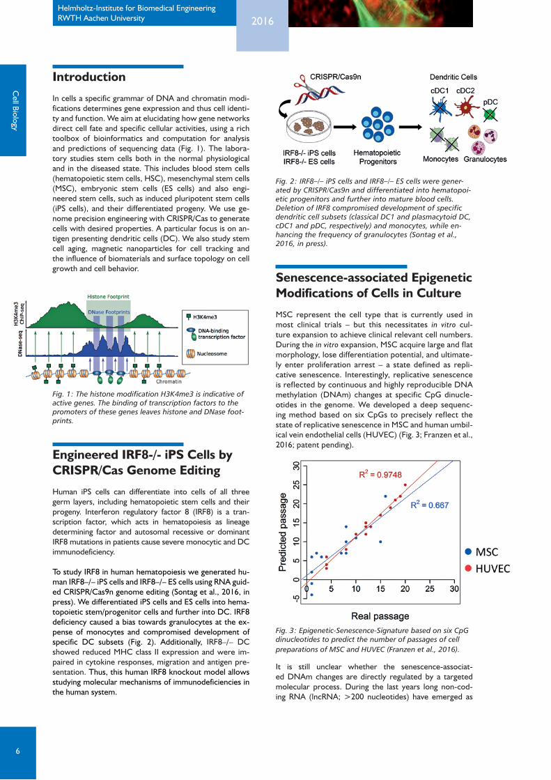
Fig. 2: IRF8–/– iPS cells and IRF8–/– ES cells were gener-ated by CRISPR/Cas9n and differentiated into hematopoi-etic progenitors and further into mature blood cells. Deletion of IRF8 compromised development of specific dendritic cell subsets (classical DC1 and plasmacytoid DC, cDC1 and pDC, respectively) and monocytes, while en-hancing the frequency of granulocytes (Sontag et al., 2016, in press).
Senescence-associated Epigenetic Modifications of Cells in Culture
MSC represent the cell type that is currently used in most clinical trials – but this necessitates in vitro cul-ture expansion to achieve clinical relevant cell numbers. During the in vitro expansion, MSC acquire large and flat morphology, lose differentiation potential, and ultimate-ly enter proliferation arrest – a state defined as repli-cative senescence. Interestingly, replicative senescence is reflected by continuous and highly reproducible DNA methylation (DNAm) changes at specific CpG dinucle-otides in the genome. We developed a deep sequenc-ing method based on six CpGs to precisely reflect the state of replicative senescence in MSC and human umbil-ical vein endothelial cells (HUVEC) (Fig. 3; Franzen et al., 2016; patent pending).
Fig. 3: Epigenetic-Senescence-Signature based on six CpG dinucleotides to predict the number of passages of cell preparations of MSC and HUVEC (Franzen et al., 2016).
It is still unclear whether the senescence-associat-ed DNAm changes are directly regulated by a targeted molecular process. During the last years long non-cod-ing RNA (lncRNA; >200 nucleotides) have emerged as
Introduction
In cells a specific grammar of DNA and chromatin modi-fications determines gene expression and thus cell identi-ty and function. We aim at elucidating how gene networks direct cell fate and specific cellular activities, using a rich toolbox of bioinformatics and computation for analysis and predictions of sequencing data (Fig. 1). The labora-tory studies stem cells both in the normal physiological and in the diseased state. This includes blood stem cells (hematopoietic stem cells, HSC), mesenchymal stem cells (MSC), embryonic stem cells (ES cells) and also engi-neered stem cells, such as induced pluripotent stem cells (iPS cells), and their differentiated progeny. We use ge-nome precision engineering with CRISPR/Cas to generate cells with desired properties. A particular focus is on an-tigen presenting dendritic cells (DC). We also study stem cell aging, magnetic nanoparticles for cell tracking and the influence of biomaterials and surface topology on cell growth and cell behavior.
Fig. 1: The histone modification H3K4me3 is indicative of active genes. The binding of transcription factors to the promoters of these genes leaves histone and DNase foot-prints.
Engineered IRF8-/- iPS Cells by CRISPR/Cas Genome Editing
Human iPS cells can differentiate into cells of all three germ layers, including hematopoietic stem cells and their progeny. Interferon regulatory factor 8 (IRF8) is a tran-scription factor, which acts in hematopoiesis as lineage determining factor and autosomal recessive or dominant IRF8 mutations in patients cause severe monocytic and DC immunodeficiency.
To study IRF8 in human hematopoiesis we generated hu-man IRF8–/– iPS cells and IRF8–/– ES cells using RNA guid-ed CRISPR/Cas9n genome editing (Sontag et al., 2016, in press). We differentiated iPS cells and ES cells into hema-topoietic stem/progenitor cells and further into DC. IRF8 deficiency caused a bias towards granulocytes at the ex-pense of monocytes and compromised development of specific DC subsets (Fig. 2). Additionally, IRF8–/– DC showed reduced MHC class II expression and were im-paired in cytokine responses, migration and antigen pre-sentation. Thus, this human IRF8 knockout model allows studying molecular mechanisms of immunodeficiencies in the human system.
Cell Biology

7
2016Helmholtz-Institute for Biomedical EngineeringRWTH Aachen University
potential epigenetic modifiers. We demonstrated that the HOX transcript antisense RNA (HOTAIR) binds pref-erentially to genomic loci that become hypermethylated during replicative senescence. Gain- and loss-of-function approaches indicated that HOTAIR expression contributes to regulation of cellular senescence by changes in gene ex-pression and DNAm. Notably, in silico and subsequent in vitro analysis indicated that targeting of HOTAIR to specif-ic genomic loci is mediated by triple helix formation (Fig. 4; Kalwa et al., 2016).
Fig. 4: Schematic representation of the triple helix forma-tion. The lncRNA HOTAIR targets specific genomic locations and interacts with epigenetic modifiers, such as PCR2 and LSD1 (Kalwa et al., 2016).
Polyelectrolyte Coating of Magnetic Nanoparticles for Cell Labelling and Tracking by MRI
Translation of cell-based therapies into clinical applications is hampered due to the lack of tools to monitor cell fate and function after transplantation. Labelling of cells with engi-neered magnetic nanoparticles (MNP) before implantation shows great promise in tracking cells in vivo using magnetic resonance imaging (MRI).
We study the tailoring of MNP using layer-by-layer assem-bly of polyelectrolytes (PE) for enhanced labelling of hema-topoietic stem/progenitor cells and DC and their tracking
Fig. 5: Labeling of inflammatory (GM-DC) and steady state (FL-DC) DC with PE-coated and uncoated ferumoxytol particles. (A) Schematic representation of magnetic separation after labelling with MNP. (B) FL-DC after uptake of respective MNP were analyzed by flow cytometry. Representative dot plots show MNP-labeled cDC (black boxes) and pDC (red boxes) subsets.
using MRI (Schwarz et al., Nanomedicine, 2012; Celikkin et al., J. Magn. Magn. Mater, 2015; in collaboration with W. Richtering and J. E. Wong, Institute of Physical Chemistry, RWTH Aachen University, Aachen, Germany and M. Hoehn, In vivo NMR Research Group, MPI for Metabolism Research, Cologne, Germany). Recent studies revealed a differential impact of specific PE coatings on labelling and cellular responses of different DC subsets under steady state and inflammatory conditions (Celikkin et al., manu-script in preparation; Fig. 5).
Regulation of Cell Motility and Adhesion by Surface-grafted Nanogel Arrays
Cellular functions, including cell adhesion and migration, are controlled by the precise spatio-temporal regulation of cytoskeleton dynamics. Several interdisciplinary stud-ies have demonstrated that material chemistry and to-pology can be exploited to modulate cell adhesion and migration.
In this project, we developed highly functional and stim-uli-responsive poly(N-isopropyl acrylamide) nanogel ar-rays grafted onto glass surfaces by a printing process using wrinkled polydimethylsiloxane (PDMS) templates (Sechi et al., 2016). Using low-temperature plasma treatment, nanogels were chemically grafted onto glass supports thus leading to highly stable nanogel layers in cell culture media.
We demonstrate that surface-grafted nanogels can serve as novel substrates for the analysis of cell adhesion and migration (Sechi et al., 2016). Nanogels have a strong im-pact on size, speed and dynamics of focal adhesions and cell motility causing cells to move along straight trajecto-ries (Fig. 6). In addition, modulation of nanogel swelling state or spacing serves as an effective tool for regulation of cell motility.
Our study demonstrates that nanogel arrays deposited on solid surfaces can be used to provide a precise and tun-able system to understand and con-trol cell migration. We anticipate that our surface-grafted nanogel system will contribute to the development of implantable systems aimed at sup-porting and enhancing cell migration and adhesion.
Cel
l Bio
logy

8
2016Helmholtz-Institute for Biomedical EngineeringRWTH Aachen University
site detection from computa-tional footprinting in Gusmao et al., 2016. We showed that several state-of-the-art meth-ods (including our own meth-od) can detect cell specific binding sites with very high ac-curacy. Moreover, we dem-onstrated how to correct for experimental artefacts, such as DNase I enzyme cleavage bias, thereby settling a long-
standing scientific discussion about computational foot-printing (see also Editorial Nat. Methods 13, 185, 2016). In practical examples, we have applied computational footprint to dissected NF-kB regulation during inflam-mation in HUVEC (Kolovos et al., 2016) and to uncov-er regulatory roles of Tbx3 in pancreas development (Perkhofer et al., 2016).
Computational Detection of Cell Specific Binding Sites One of the main molecular mechanisms controlling the temporal and spatial expression of genes is transcription-al regulation. In this process, transcription factors bind to the vi-cinity of a gene to re-cruit (or block) the transcriptional ma-chinery. The identifi-cation of transcription factor binding sites is a first step to under-stand regulatory net-works driving cellular processes, such as cell differentiation and the onset of diseases. Cell specific binding sites can be predict-ed by the analysis of sequencing protocols measuring open chro-matin with compu-tational footprinting methods (Fig. 7).
We have revisited the problem of binding
Fig. 6: Impact of surface-grafted nanogels on cell mor-phology and cytoskeleton organisation (A-C). The micro-filaments (A, green, arrow) and focal adhesions (B, green, arrow) of mouse melanoma B16F1 cells aligned along the major axis of nanogel arrays (long arrows). Cell nuclei were stained with DAPI (blue). Scale bar: 10 µm. Scanning electron microscopy of B16F1 cells seed-ed on nanogel arrays (C). Note the interaction of cell projec-tions (green arrows) with the underlying nanogel arrays (white arrow). Scale bar: 0.25 µm (Sechi et al., 2016).
Fig. 7: Workflow of the computational analysis of the open chromatin protocol DNAse-seq (Gusmao et al., 2016).
Cell Biology

9
2016Helmholtz-Institute for Biomedical EngineeringRWTH Aachen University
(2016). Astrocytic calcium waves signal brain injury to neural stem and progenitor cells. Stem Cell Reports, in press.
[13] Lenz, M., Müller, F.J., Zenke, M., and Schuppert, A. (2016). Principal components analysis and the reported low intrinsic dimensionality of gene expression microarray data. Sci. Rep. 6, 25696.
[14] Lin, Q., Weidner, C.I., Costa, I.G., Marioni, R.E., Ferreira, M.R., Deary, I.J., and Wagner, W. (2016). DNA methylation levels at indi-vidual age-associated CpG sites can be indicative for life expectancy. Aging 8, 394-401.
[15] Nascimento, A.C., Prudencio, R.B., and Costa, I.G. (2016). A multiple kernel learning algorithm for drug-target interaction prediction. BMC Bioinformatics 17, 46.
[16] Paschoalin, R. T., Traldi, B., Aydin, G., Oliveira, J. E., Rütten, S., Mat-toso, L. H. C., Zenke, M. and Sechi, A. (2016). Solution blow spinning fibres: new immunologically inert substrates for the analysis of cell adhesion and motility. Acta Biomaterialia, in press.
[17] Pelusi, N., Kosanke, M., Riedt, T., Rösseler, C., Seré, K., Li, J., Güt-gemann, I., Zenke, M., Janzen, V., and Schorle, H. (2016). The spleen microenvironment influences disease transformation in a mouse model of KIT D816V dependent myeloproliferative neoplasm. Sci. Rep., in press.
[18] Perkhofer, L., Walter, K., Costa, I.G., Carrasco, M.C., Eiseler, T., Hafner, S., Genze, F., Zenke, M., Bergmann, W., Illing, A., Hohwieler, M., Kohntop, R., Lin, Q., Holzmann, K.H., Seufferlein, T., Wagner, M., Liebau, S., Hermann, P.C., Kleger, A., and Müller, M. (2016). Tbx3 fos-ters pancreatic cancer growth by increased angiogenesis and activin/nodal-dependent induction of stemness. Stem Cell Res. 17, 367-378.
[19] Schemionek, M., Herrmann, O., Reher, M.M., Chatain, N., Schubert, C., Costa, I.G., Hänzelmann, S., Gusmao, E.G., Kintsler, S., Braun-schweig, T., Hamilton, A., Helgason, G.V., Copland, M., Schwab, A., Müller-Tidow, C., Li, S., Holyoake, T.L., Brümmendorf, T.H., and Ko-schmieder, S. (2016). Mtss1 is a critical epigenetically regulated tumor suppressor in CML. Leukemia 30, 823-832.
[20] Schulze, I., Rohde, C., Scheller-Wendorff, M., Baumer, N., Krause, A., Herbst, F., Riemke, P., Hebestreit, K., Tschanter, P., Lin, Q., Linhart, H., Godley, L.A., Glimm, H., Dugas, M., Wagner, W., Berdel, W.E., Rosen-bauer, F., and Müller-Tidow, C. (2016). Increased DNA methylation of Dnmt3b targets impairs leukemogenesis. Blood 127, 1575-1586.
[21] Sechi, A., Freitas, J. M. G., Wünnemann, P., Töpel A., Paschoalin, R. T., Ullmann, S., Schröder, R., Aydin, G., Rütten, S., Böker, A., Zenke, M. and Pich, A. (2016). Surface-grafted nanogel arrays direct cell adhe-sion and motility. Adv. Mater. Interfaces 3, 1600455 (Cover story).
[22] Sontag, S., Förster, M., Qin, J., Wanek, P., Mitzka, S., Schüler, H. M., Ko-schmieder, S., Rose-John, S., Seré, K., and Zenke, M. (2016). Modelling IRF8 deficient human hematopoiesis and dendritic cell development with engineered induced pluripotent stem cells. Stem Cells, in press.
[23] Souza, M.B., Araujo, G.S., Costa, I.G., and Oliveira, J.R. for the Alz-heimer’s Disease Neuroimaging Initiative (2016). Combined genome-wide CSF Abeta-42’s associations and simple network properties highlight new risk factors for Alzheimer’s dsease. J. Mol. Neurosci. 58, 120-128.
[24] Vasko, T., Frobel, J., Lubberich, R., Goecke, T.W., and Wagner, W. (2016). iPSC-derived mesenchymal stromal cells are less supportive than primary MSCs for co-culture of hematopoietic progenitor cells. J. Hematol. Oncol. 9, 43.
[25] Wagner, W., Fernandez-Rebollo, E., and Frobel, J. (2016a). DNA-methylation changes in replicative senescence and aging: two sides of the same coin? Epigenomics 8, 1-3.
[26] Wagner, W., Frobel, J., and Goetzke, R. (2016b). Epigenetic quality check - how good are your mesenchymal stromal cells? Epigenomics 8, 889-894.
[27] Weidner, C.I., Lin, Q., Birkhofer, C., Gerstenmaier, U., Kaifie, A., Kirschner, M., Bruns, H., Balabanov, S., Trummer, A., Stockklausner, C., Höchsmann, B., Schrezenmeier, H., Wlodarski, M., Panse, J., Brümmendorf, T.H., Beier, F., and Wagner, W. (2016). DNA methyla-tion in PRDM8 is indicative for dyskeratosis congenita. Oncotarget 7, 10765-10772.
[28] Wilop, S., Chou, W.C., Jost, E., Crysandt, M., Panse, J., Chuang, M.K., Brümmendorf, T.H., Wagner, W., Tien, H.F., and Kharabi Masouleh, B. (2016). A three-gene expression-based risk score can refine the European LeukemiaNet AML classification. J. Hematol. Oncol. 9, 78.
Patent applications [1] Epigenetic classification of human mesenchymal stromal cells; 2016;
EP 16152198.4 (Wagner W, de Almeida DC) [2] Method for analysis of the cellular composition in buccal swabs; 2016;
DE 10 2016 109 291.6 (Wagner W, Eipel M)
AcknowledgementsThis work was supported by:
• GermanResearchFoundation(DFG) • GermanFederalMinistryofEducationandResearch(BMBF) • EuropeanUnion(EU) • InterdisciplinaryCentreforClinicalResearchAachen(IZKFAachen) • Aachen Institute forAdvancedStudy inComputationalEngineering
Science (AICES) • StemCellNetworkNRW,MinistryofInnovation,Science,Research
and Technology of the German Federal State North Rhine-Westphalia (NRW)
• START-ProgramoftheFacultyofMedicine,RWTHAachen • Else-Kröner-FreseniusFoundation • DonationbyU.Lehmann • DonationbyVision4LifeSciences • StemCellFactoryisco-fundedbytheEuropeanUnion(EuropeanRe-
gional Development Fund - Investing in your future) and the German Federal State North Rhine-Westphalia (NRW)
Selected References in 2016 [1] Allhoff, M., Sere, K., Pires, J.F., Zenke, M., and Costa, I.G. (2016). Dif-
ferential peak calling of ChIP-seq signals with replicates with THOR. Nucleic Acids Res. 44, e153.
[2] Berger, A.W., Schwerdel, D., Costa, I.G., Hackert, T., Strobel, O., Lam, S., Barth, T.F., Schröppel, B., Meining, A., Büchler, M.W., Zenke, M., Hermann, P.C., Seufferlein, T., and Kleger, A. (2016). Detection of hot-spot mutations in circulating cell-free DNA from patients with intraductal papillary mucinous neoplasms of the pancreas. Gastroen-terology 151, 267-270.
[3] de Almeida, D.C., Ferreira, M.R., Franzen, J., Weidner, C.I., Frobel, J., Zenke, M., Costa, I.G., and Wagner, W. (2016). Epigenetic clas-sification of human mesenchymal stromal cells. Stem Cell Reports 6, 168-175.
[4] Eipel, M., Mayer, F., Arent, T., Ferreira, M.R., Birkhofer, C., Ger-stenmaier, U., Costa, I.G., Ritz-Timme, S., and Wagner, W. (2016). Epigenetic age predictions based on buccal swabs are more precise in combination with cell type-specific DNA methylation signatures. Aging 8, 1034-1048.
[5] Franzen, J., Zirkel, A., Blake, J., Rath, B., Benes, V., Papantonis, A., and Wagner, W. (2016). Senescence-associated DNA methylation is stochastically acquired in subpopulations of mesenchymal stem cells. Aging Cell, Epub ahead of print.
[6] Gamper, I., Fleck, D., Barlin, M., Spehr, M., El Sayad, S., Kleine, H., Maxeiner, S., Schalla, C., Aydin, G., Hoss, M., Litchfield, D.W., Lüscher, B., Zenke, M., and Sechi, A. (2016). GAR22b regulates cell migration, sperm motility, and axoneme structure. Mol. Biol. Cell 27, 277-294.
[7] Gusmao, E.G., Allhoff, M., Zenke, M., and Costa, I.G. (2016). Analysis of computational footprinting methods for DNase sequencing experi-ments. Nat. Methods 13, 303-309.
[8] Hohwieler, M., Illing, A., Hermann, P.C., Mayer, T., Stockmann, M., Perkhofer, L., Eiseler, T., Antony, J.S., Müller, M., Renz, S., Kuo, C.C., Lin, Q., Sendler, M., Breunig, M., Kleiderman, S.M., Lechel, A., Ze-nker, M., Leichsenring, M., Rosendahl, J., Zenke, M., Sainz, B., Jr., May-erle, J., Costa, I.G., Seufferlein, T., Kormann, M., Wagner, M., Liebau, S., and Kleger, A. (2016a). Human pluripotent stem cell-derived acinar/ductal organoids generate human pancreas upon orthotopic transplantation and allow disease modelling. Gut, Epub ahead of print.
[9] Hohwieler, M., Renz, S., Liebau, S., Lin, Q., Lechel, A., Klaus, J., Perk-hofer, L., Zenke, M., Seufferlein, T., Illing, A., Müller, M., and Kleger, A. (2016b). „Miniguts“ from plucked human hair meet Crohn‘s disease. Z. Gastroenterol .54, 748-759.
[10] Kalwa, M., Hänzelmann, S., Otto, S., Kuo, C.C., Franzen, J., Joussen, S., Fernandez-Rebollo, E., Rath, B., Koch, C., Hofmann, A., Lee, S.H., Teschendorff, A.E., Denecke, B., Lin, Q., Widschwendter, M., Wein-hold, E., Costa, I.G., and Wagner, W. (2016). The lncRNA HOTAIR impacts on mesenchymal stem cells via triple helix formation. Nucleic Acids Res. 44, 10631-10643.
[11] Kolovos, P., Georgomanolis, T., Koeferle, A., Larkin, J.D., Brant, L., Nikolicc, M., Gusmao, E.G., Zirkel, A., Knoch, T.A., van Ijcken, W.F., Cook, P.R., Costa, I.G., Grosveld, F.G., and Papantonis, A. (2016). Binding of nuclear factor kappaB to noncanonical consensus sites reveals its multimodal role during the early inflammatory response. Genome Res. 26, 1478-1489.
[12] Kraft, A., Rosales Jubal, E., von Laer, R., Döring, C., Rocha, A., Greb-bin, M., Zenke, M., Kettenmann, H., Stroh, A., and Momma, S.
Cel
l Bio
logy

10
2016Helmholtz-Institute for Biomedical EngineeringRWTH Aachen University
Cell Biology
Team
At the Top and right: Best Paper Award to Eduardo Gusmao, PhD (right) and Poster Award to Thomas Hieronymus, PhD (top, second from left) at Medi-cal Sciences Day 2016, Medical Faculty RWTH Aachen University.
Below: Lab retreat at Mosel river.