Fulminant Hepatic Failure Monitored by Technetium- 99m-DTPA-Galacto syl-Human Serum
4
trated through water absorption (7). Excess biliary excretion due to improper radiopharmaceutical preparation can be excluded as the cause since there was no visualization of the liver in the serial bone scan at any time and other bone scans performed on the same day showed a normal distribution of tracers. REFERENCES I . vardy PA, Lebenthal E, Schwachman H. Intestinal lymphangiectasia: a reappraisal. Pediatrics I975;55:842— 851. 2. Puri AS, Aggarwal R, Gupta RK, et al. Intestinal lymphangiectasia: evaluation by CT and scintigraphy. Gastrointest Radio/ 1992:17:119—12I. 3. Fakhri A, Fishman EK, Jones B, Kuhajda F, Siegelman 55. Primary intestinal lymphangiectasia: clinical and CT findings. J Comp Assist Tomog l985;9:767—770. 4. Soucy IP, Eybalin MC, Taillefer R, Levasseur A, lobin G. Lymphoscintigraphic demonstration of intestinal lymphangiectasia. C/in Nuc/ Med 1983:8:535—537. 5. Mistilis SP, Skyring AP, Stephan DD. Intestinal lymphangiectasia: mechanism of enteric loss of plasma-protein and fat. Lancet l965;1 :77—79. 6. Divgi CR, Lisann NM, Yeh SDJ, Benua RS. Technetium-99m-albumin scintigraphy in the diagnosis of protein-losing enteropathy. J Nuc/ Med 1986:27: 1710—1712. 7. Conway II, Weiss SC, Khentigan A, Tofe AJ. Thane iT. Gallbladder and bowel localization of bone imaging radiopharmaceuticals [Abstract]. J Nuc/ Med I979;20: 622. hepatic function in patients with diffuse liver diseases but also to assess morphological changes of the liver. We describe a patient with fulminant hepatic failure who was evaluated scintigraphically with 99mTc..GSA. CASE REPORT A 43-yr-old woman consulted a physician because of general fatigue and clouding of consciousness. The results of clinical tests showed severe liver dysfunction. She was referred to our hospital for further examination and therapy. On admission, physical examination showed jaundice and ascites, and there was clouding of consciousness. Her white blood cell count was I0, 100/mm3, red blood cell count was 362 X 104/mm3, total bilirubin was 13.2 mg/dl, aspartate aminotransferase was 109 lU/liter, alkaline phos phatase was 372 lU/liter, serum albumin was 2.8 g/dt, lactate dehydrogenase was 5 12 WU/liter, and the prothrombin time was 25%. Anti-hepatitis A antibody, hepatitis B surface antigen and hepatitis C virus antibody were not detected. Hepatic injury, caused by diclofenac sodium, was diagnosed by results of a lymphocyte stimulation test. On abdominal CT, the liver showed extensive low-density regions and atrophy of both lobes (Fig. 1). The patient responded to intensive therapy including plasma pheresis. Hepatic receptor imaging with @mTc@GSA was per formed four times (on admission and after 1, 2 and 4 mo). One 185-MBq dose of 99mTcGSA was injected intravenously and dynamic imaging was performed with the patient supine under a large field of view gamma camera with a low-energy, alt-purpose parallel-hole collimator. Computer acquisition of the gamma cam era data was started just before injection of @°@Tc-GSA and was stopped 20 mm later. Digital images ( 128 X 128 pixels) were acquired in the byte mode at a rate of 60 sec/frame. Accumulation images of the anterior abdominal view were obtained at 20 mm after the injection. Time-activity curves for the heart and liver were generated from regions of interest (ROIs) for the whole liver and precordium. The receptor index (LHL15) was calculated by divid ing the radioactivity of the liver ROl by the radioactivity of the liver plus heart ROIs at 15 mm after the injection. We describe a 43-yr-old woman with fulminant hepatic failure whose progresswas monitoredscintigrapt'iicaltyusing @Tc-galac tosyl-human serum albumin (@Tc-GSA). On admission, the liver was atrophic and the heart was delineateddistinctly by scintigraphy with @Tc-GSA. The receptor index, calculated by dividing the radioactivity of the liver region of interest by the radioactMty of the liver plus heart regions of interest at 15 mm post-tracer injection, was very low. As the patient's condition improved, the right lobe of the liverenlargedwhile the left lobe became atrophic; after 4 mo, the left lobe almost completely disappeared. Delineationof the heart gradually became less distinct, and the receptor index slowly in creased. Hepatic receptor imaging with @rc-GSA can define both the hepatic functional reserve and morphological changes of the liver,so it is usefulfor the diagnosis and follow-up study of fulminant hepatic failure. Key Words: fulminant hepatic failure; technetium-99m-GSA; he patic receptor imaging J Nuci Med 1996; 37:641-643 Technetium-99m-phytate and sulfur colloid, which have been used as liver imaging agents, are transported to the liver and taken up by Kupffer cells after intravenous injection (1 ). The hepatocyte-oriented radiotracer 99mTc galactosyl-neoglycoalbu mm (99mTc@GSA),developed as a receptor-binding radiophar maceutical for noninvasive assessment of liver function, is a synthetic radioligand to the asialoglycoprotein receptor (hepat ic-binding protein), which resides on the plasma membrane of liver cells. Upon intravenous injection, 99mTc..GSA is directed to hepatocytes because of its chemical recognition and binding by a specific receptor of hepatic-binding protein. After binding, it is transferred to hepatic lysosomes by receptor-mediated endocytosis (2,3). The use of 99mTc@GSA enables us not only to evaluate ReceivedMar.3, 1995;revisionaccepted Jun. 29, 1995. For correspondence or reprints contact: Susumu Shomi, MD, Third Department of Internal Medicine, Osaka Ctty Univers@yMedical School, 1-5-7 Asahimachi, Abeno-ku, Osaka 545, Japan. 641 FULMINANT HEPATITIS MONITORED BY TECHNETIUM-99M-GSA •Shiomi et at. Fulminant Hepatic Failure Monitored by Technetium- 99m-DTPA-Galacto syl-Human Serum Albumin Scintigraphy Susumu Shiomi, Tetsuo Kuroki, Masaru Enomoto, Tadashi Ueda, Kyoko Masaki, Naoko Ikeoka, Tadashi Takeda, Kenzo Kobayashi and Hironobu Ochi Third Department oflnternal Medicine and Division ofNuclear Medicine, Osaka City University Medical School, Osaka, Japan by on April 10, 2019. For personal use only. jnm.snmjournals.org Downloaded from
Transcript of Fulminant Hepatic Failure Monitored by Technetium- 99m-DTPA-Galacto syl-Human Serum
trated through water absorption (7). Excess biliary excretiondue to improper radiopharmaceutical preparation can beexcluded as the cause since there was no visualization of theliver in the serial bone scan at any time and other bone scansperformed on the same day showed a normal distribution oftracers.
REFERENCESI . vardy PA, Lebenthal E, Schwachman H. Intestinal lymphangiectasia: a reappraisal.
PediatricsI975;55:842—851.
2. Puri AS, Aggarwal R, Gupta RK, et al. Intestinal lymphangiectasia: evaluation by CTand scintigraphy. Gastrointest Radio/ 1992:17:119—12I.
3. Fakhri A, Fishman EK, Jones B, Kuhajda F, Siegelman 55. Primary intestinallymphangiectasia: clinical and CT findings. J Comp Assist Tomog l985;9:767—770.
4. Soucy IP, Eybalin MC, Taillefer R, Levasseur A, lobin G. Lymphoscintigraphicdemonstration of intestinal lymphangiectasia. C/in Nuc/ Med 1983:8:535—537.
5. Mistilis SP, Skyring AP, Stephan DD. Intestinal lymphangiectasia: mechanism ofenteric loss of plasma-protein and fat. Lancet l965;1 :77—79.
6. Divgi CR, Lisann NM, Yeh SDJ, Benua RS. Technetium-99m-albumin scintigraphy inthe diagnosis of protein-losing enteropathy. J Nuc/ Med 1986:27: 1710—1712.
7. Conway II, Weiss SC, Khentigan A, Tofe AJ. Thane iT. Gallbladder and bowellocalization of bone imaging radiopharmaceuticals [Abstract]. J Nuc/ Med I979;20:622.
hepatic function in patients with diffuse liver diseases but alsoto assess morphological changes of the liver. We describe apatient with fulminant hepatic failure who was evaluatedscintigraphically with 99mTc..GSA.
CASE REPORTA 43-yr-old woman consulted a physician because of general
fatigue and clouding of consciousness. The results of clinical testsshowed severe liver dysfunction. She was referred to our hospitalfor further examination and therapy. On admission, physicalexamination showed jaundice and ascites, and there was cloudingof consciousness. Her white blood cell count was I0, 100/mm3, redblood cell count was 362 X 104/mm3, total bilirubin was 13.2mg/dl, aspartate aminotransferase was 109 lU/liter, alkaline phosphatase was 372 lU/liter, serum albumin was 2.8 g/dt, lactatedehydrogenase was 5 12 WU/liter, and the prothrombin time was25%. Anti-hepatitis A antibody, hepatitis B surface antigen and
hepatitis C virus antibody were not detected. Hepatic injury, causedby diclofenac sodium, was diagnosed by results of a lymphocytestimulation test. On abdominal CT, the liver showed extensivelow-density regions and atrophy of both lobes (Fig. 1).
The patient responded to intensive therapy including plasmapheresis. Hepatic receptor imaging with @mTc@GSAwas performed four times (on admission and after 1, 2 and 4 mo). One185-MBq dose of 99mTcGSA was injected intravenously anddynamic imaging was performed with the patient supine under alarge field of view gamma camera with a low-energy, alt-purposeparallel-hole collimator. Computer acquisition of the gamma camera data was started just before injection of @°@Tc-GSAand wasstopped 20 mm later. Digital images ( 128 X 128 pixels) wereacquired in the byte mode at a rate of 60 sec/frame. Accumulationimages of the anterior abdominal view were obtained at 20 mmafter the injection. Time-activity curves for the heart and liver weregenerated from regions of interest (ROIs) for the whole liver andprecordium. The receptor index (LHL15) was calculated by dividing the radioactivity of the liver ROl by the radioactivity of theliver plus heart ROIs at 15 mm after the injection.
We describe a 43-yr-old woman with fulminant hepatic failurewhose progresswas monitoredscintigrapt'iicaltyusing @Tc-galactosyl-human serum albumin (@Tc-GSA). On admission, the liverwas atrophic and the heart was delineateddistinctly by scintigraphywith @Tc-GSA.The receptor index, calculated by dividing theradioactivityof the liver region of interest by the radioactMty of theliver plus heart regions of interest at 15 mm post-tracer injection,was very low. As the patient's condition improved, the right lobe ofthe liverenlargedwhile the left lobe becameatrophic;after4 mo, theleft lobe almost completely disappeared. Delineationof the heartgradually became less distinct, and the receptor index slowly increased.Hepatic receptor imagingwith @rc-GSAcan define boththe hepatic functional reserve and morphological changes of theliver,so it is usefulfor the diagnosisand follow-up study of fulminanthepatic failure.Key Words: fulminant hepatic failure; technetium-99m-GSA; hepatic receptor imaging
J Nuci Med 1996;37:641-643
Technetium-99m-phytate and sulfur colloid, which have beenused as liver imaging agents, are transported to the liver andtaken up by Kupffer cells after intravenous injection (1 ). Thehepatocyte-oriented radiotracer 99mTc galactosyl-neoglycoalbumm (99mTc@GSA),developed as a receptor-binding radiopharmaceutical for noninvasive assessment of liver function, is asynthetic radioligand to the asialoglycoprotein receptor (hepatic-binding protein), which resides on the plasma membrane ofliver cells. Upon intravenous injection, 99mTc..GSA is directedto hepatocytes because of its chemical recognition and bindingby a specific receptor of hepatic-binding protein. After binding,it is transferred to hepatic lysosomes by receptor-mediatedendocytosis (2,3).
The use of 99mTc@GSA enables us not only to evaluate
ReceivedMar.3, 1995;revisionaccepted Jun. 29, 1995.For correspondence or reprints contact: Susumu Shomi, MD, Third Department of
Internal Medicine, Osaka Ctty Univers@yMedical School, 1-5-7 Asahimachi, Abeno-ku,Osaka 545, Japan.
641FULMINANT HEPATITIS MONITORED BY TECHNETIUM-99M-GSA •Shiomi et at.
Fulminant Hepatic Failure Monitored byTechnetium- 99m-DTPA-Galacto syl-Human SerumAlbumin ScintigraphySusumu Shiomi, Tetsuo Kuroki, Masaru Enomoto, Tadashi Ueda, Kyoko Masaki, Naoko Ikeoka, Tadashi Takeda,Kenzo Kobayashi and Hironobu OchiThird Department oflnternal Medicine and Division ofNuclear Medicine, Osaka City University Medical School,Osaka, Japan
by on April 10, 2019. For personal use only. jnm.snmjournals.org Downloaded from
D-@L
L@-@-•@H L
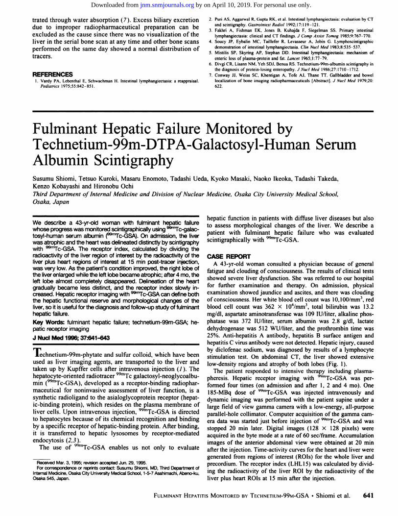
TABLE IChanges in LHL15 Values and Laboratory Data
Onadmission 1 mo 2 mo 3 mo
0.5825
Totalbilirubin(mg/do 13.2
LHL15is calculatedby dividingthe radioactivityof the liverplusheartROlsat 15mmpostinjectionof @‘@‘Tc-GSA.
PT= prothrom@ntime.
pheresis and blood product supplementation. Imaging techniques such as liver scintigraphy (7—9),abdominal CT scan(JO) and abdominal ultrasonography (11 ) have been founduseful in the diagnosis of diffuse hepatic disease such asfulminant hepatic failure. Liver scintigraphy with a radiocolloidagent is especially helpful in establishment of a diagnosis offulminant hepatic failure as it facilitates understanding of thehepatic functional reserve. Waxman (7) noted in a review thepresence of hepatomegaly in 6 (46%) of 13 survivors offulminant hepatic failure and found no hepatomegaly among the9 patients who died. Pulmonary uptake was not seen in any ofthe survivors; on the other hand, it was seen in 3 (33%) of the9 nonsurvivors. Previously, we performed liver scintigraphywith 99mTc@phytate in 44 patients with acute hepatitis and 12patients with fulminant hepatic failure and found that evidenceof liver atrophy and redistribution of the radiocoltoid to thebone marrow is useful in establishing a diagnosis of fulminanthepatic failure (8). Of the 12 patients with fulminant hepaticfailure, pulmonary uptake was not seen in any ofthe 7 survivorsbut was seen in 3 of the 5 nonsurvivors.
CONCLUSIONHepatic receptor imaging with 99mTc@GSAis an accepted
new diagnostic approach for hepatic disease (2,3). The results ofliver scintigraphy with a radiocolloid are affected by the activity ofKupifer cells. Therefore, more reduced uptake into the liver, acondition termed hepatic reticuloendothelial failure, may occasionally be .detected among abusers of alcohol (12, 13). Hepaticreceptor imaging with @‘°@Tc-GSA,however, is affected only bythe function of hepatocytes and not by that of Kupifer cells.Furthermore, hepatic receptor imaging with @mTc@GSApermitsnumerical evaluation of the hepatic functional reserve in terms ofthe LHL15. If analysis of a series of patients shows satisfactoryresults, this method might provide a basis for a more objectivediagnosis than before. The technique also facilitates the diagnosisofmorphological changes in the liver, as seen in our case. In viewofthese advantages, @‘@Tc-GSAscintigraphy ofthe liver may gainacceptance for use in the clinical diagnosis and follow-up offulminant hepatic failure.
LHL15PT(%)
0.70 0.77 0.8840 52 823.6 1.6 1.0
FiGURE1. AbdominalCTshows extensivelow-densityregionsand atrophyof bothlobesinthe liver.
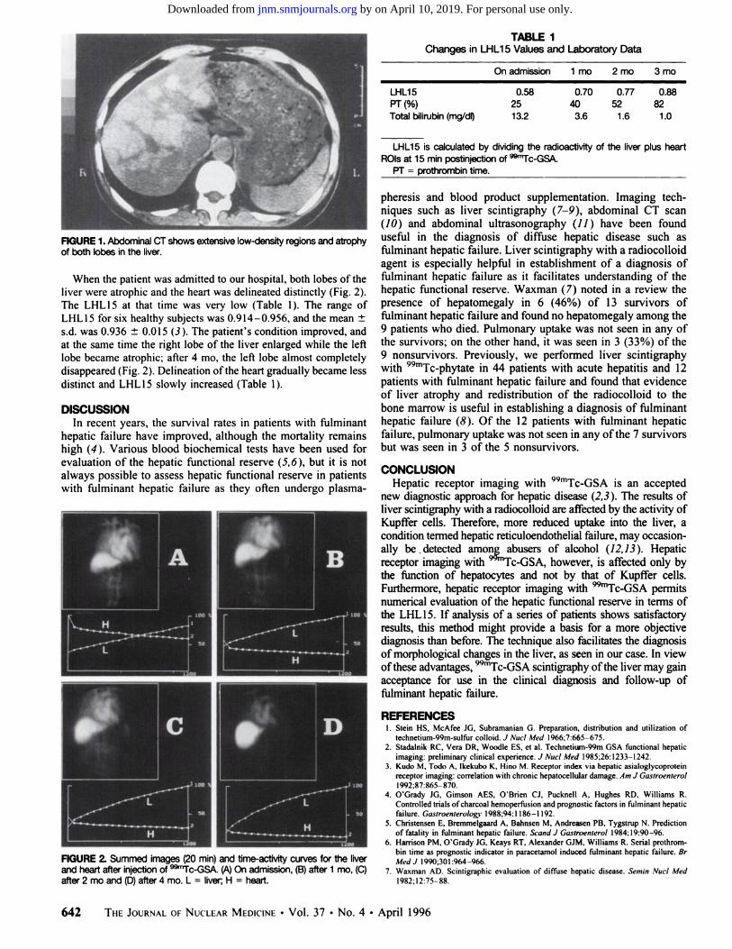
When the patient was admitted to our hospital, both lobes of theliver were atrophic and the heart was delineated distinctly (Fig. 2).The LHL I5 at that time was very low (Table I ). The range ofLHL15 for six healthy subjects was 0.914—0.956,and the mean ±s.d. was 0.936 ±0.015 (3). The patient's condition improved, andat the same time the right lobe of the liver enlarged while the leftlobe became atrophic; after 4 mo, the left lobe almost completelydisappeared (Fig. 2). Delineation ofthe heart gradually became lessdistinct and LHL 15 slowly increased (Table 1).
DISCUSSIONIn recent years, the survival rates in patients with fulminant
hepatic failure have improved, although the mortality remainshigh (4). Various blood biochemical tests have been used forevaluation of the hepatic functional reserve (5,6), but it is notalways possible to assess hepatic functional reserve in patientswith futminant hepatic failure as they often undergo plasma
REFERENCESI. Stein HS, McAfee IG, Subramanian G. Preparation, distribution and utilization of
technetium-99m-sulfur colloid. J Nuc/ Med I966;7:665—675.2. Stadalnik RC, Vera DR. Woodle ES, et al. Technetium-99m GSA functional hepatic
imaging: preliminary clinical experience. J Nuc/ Med 1985:26:1233—1242.3. Kudo M, Todo A, Ikekubo K, Hino M. Receptor index via hepatic asialoglycoprotein
receptor imaging: correlation with chronic hepatocellular damage. Am J Gastroentero/I992;87:865—870.
4. O'Grady IG, Gimson AES, O'Brien CI, Pucknell A, Hughes RD, Williams R.Controlled trials ofcharcoal hemoperfusion and prognostic factors in fulminant hepaticfailure. Gastroenterology 1988;94: I 186—I192.
5. Christensen E, Bremmelgaard A, Bahnsen M, Andreasen PB, Tygstrup N. Predictionof fatality in fulminant hepatic failure. Scand J Gastroentero/ 1984;19:90—96.
6. Harrison PM, O'Grady 1G. Keays RT, Alexander GIM, Williams R. Serial prothrombin time as prognostic indicator in paracetamol induced fulminant hepatic failure. BrMed J 1990;301:964—966.
7. Waxman AD. Scintigraphic evaluation of diffuse hepatic disease. Semin Nuc/ Medl982;I2:75—88.
FIGURE2. Summed images (20 mm)and time-actMtycurves for the liverandheartafterinjectionof @1c-GSA.(A@Onadmission,(B)after1 mo,(C)after2 moand(D)after4 mo.L = liver H = heart.
642 THEJOURNALOFNUCLEARMEDICINE•Vol. 37 â€No. 4 •April 1996
by on April 10, 2019. For personal use only. jnm.snmjournals.org Downloaded from
8. Shiomi 5, Ikeoka N, Minowa T, et al. Diagnostic value of liver scintigraphy infulminant hepatitis and severe acute hepatitis. Acta Hepato/ Jpn I985;26:592—597.
9. Fleischer MR. Sharpstone P. Osbom SB, Williams R. Liver scintiscanning in acutehepatic necrosis. Br J Radio/ I97 1;44:40 I—402.
10. Kumahara 1, Muto Y, Moriwaki H, Yoshida T, Tomita E. Determination of theintegrated CT number of the whole liver in patients with severe hepatitis: as anindicator of the functional reserve of the liver. Gastroenterol Jpn l989;24:290 —297.
I I. Kurtz AB, Rubin CS, Cooper HS. ci al. Ultrasound findings in hepatitis. Radio/ogy1980; 136:717—723.
12. Antar MA, Sziklas II, Spencer RP. Liver imaging during reticuloendothelial failure.C/in Nuci Med 1977;2:293—295.
13. Rao BK, Weir GI Jr. Lieberman LM. Dissociation of reticuloendothelial cell andhepatocyte functions in alcoholic liver disease: a clinical study with a new 99mTclabeled hepatobiliary agent. C/in Nuc/ Med 1981:6:289—294.
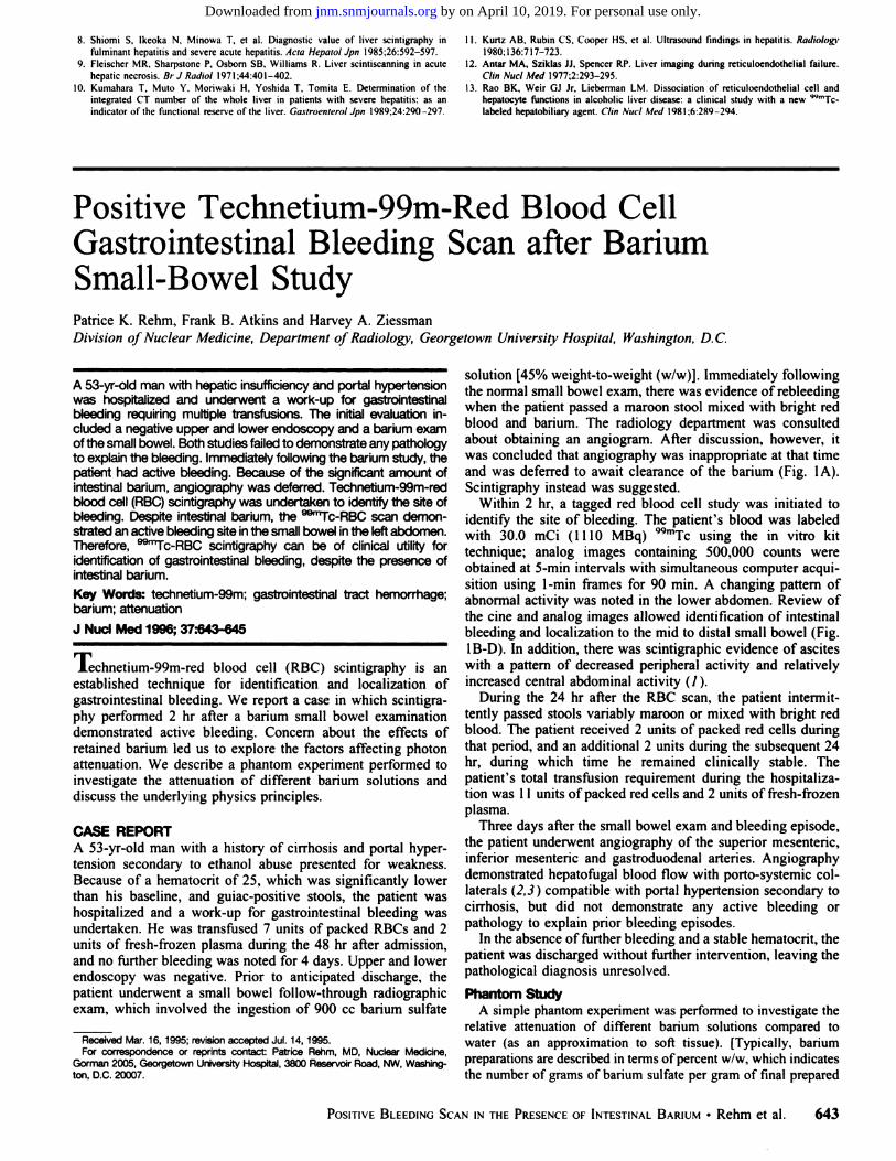
solution [45% weight-to-weight (w/w)}. Immediately followingthe normal small bowel exam, there was evidence of rebteedingwhen the patient passed a maroon stool mixed with bright redblood and barium. The radiology department was consultedabout obtaining an angiogram. After discussion, however, itwas concluded that angiography was inappropriate at that timeand was deferred to await clearance of the barium (Fig. IA).Scintigraphy instead was suggested.
Within 2 hi, a tagged red btood cell study was initiated toidentify the site of bleeding. The patient's blood was labeledwith 30.0 mCi (1 110 MBq) 99mTc using the in vitro kittechnique; analog images containing 500,000 counts wereobtained at 5-mm intervals with simultaneous computer acquisition using 1-mm frames for 90 mm. A changing pattern ofabnormal activity was noted in the tower abdomen. Review ofthe cine and analog images allowed identification of intestinalbleeding and localization to the mid to distal small bowel (Fig.1B-D). In addition, there was scintigraphic evidence of asciteswith a pattern of decreased peripheral activity and relativelyincreased central abdominal activity (1).
During the 24 hr after the RBC scan, the patient intermittently passed stools variably maroon or mixed with bright redblood. The patient received 2 units of packed red cells duringthat period, and an additional 2 units during the subsequent 24hr, during which time he remained clinically stable. Thepatient's total transfusion requirement during the hospitatization was 11 units ofpacked red cells and 2 units of fresh-frozenplasma.
Three days after the small bowel exam and bleeding episode,the patient underwent angiography of the superior mesenteric,inferior mesenteric and gastroduodenal arteries. Angiographydemonstrated hepatofugal blood flow with porto-systemic collaterals (2,3 ) compatible with portal hypertension secondary tocirrhosis, but did not demonstrate any active bleeding orpathology to explain prior bleeding episodes.
In the absence of further bleeding and a stable hematocrit, thepatient was discharged without further intervention, leaving thepathological diagnosis unresolved.
Phantom S@dyA simple phantom experiment was performed to investigate the
relative attenuation of different barium solutions compared towater (as an approximation to soft tissue). [Typically, bariumpreparations are described in terms ofpercent w/w, which indicatesthe number of grams of barium sulfate per gram of final prepared
A 53-yr-old man with hepatic insufficiency and portal hypertensionwas hospitaliZedand underwenta work-upfor gastrointestinalbleeding requiring muttiple transfusions. The initial evaluation included a negativeupper and lower endoscopy and a barium examofthe smallbowel. Both studiesfailedto demonstrateany pathologyto explainthe bleeding. Immediatelyfollowing the barium study, thepatient had active bleeding. Because of the aignificant amount ofintestinal barium, angiography was deferred. Technetium-99m-redblood cell (ABC)scintigraphywas undertaken to identify the site ofbleeding. Despite intestinal barium, the @Tc-RBCscan demonstrated an active bleeding site in the small bowel in the left abdomen.Therefore, @°‘Tc-RBCscintigraphy can be of clinical utility foridentification of gastrointestinal bleeding, despite the presence ofintestinal barium.
Key Words: technetium-99m; gastrointestinal tract hemorrhage;barium; attenuation
J Nuci Med 1996;37.'643-645
Technetium-99m-red blood cell (RBC) scintigraphy is anestablished technique for identification and localization ofgastrointestinal bleeding. We report a case in which scintigraphy performed 2 hr after a barium small bowel examinationdemonstrated active bleeding. Concern about the effects ofretained barium led us to explore the factors affecting photonattenuation. We describe a phantom experiment performed toinvestigate the attenuation of different barium solutions anddiscuss the underlying physics principles.
CASE REPORTA 53-yr-old man with a history of cirrhosis and portal hypertension secondary to ethanol abuse presented for weakness.Because of a hematocrit of 25, which was significantly lowerthan his baseline, and guiac-positive stools, the patient washospitalized and a work-up for gastrointestinat bleeding wasundertaken. He was transfused 7 units of packed RBCs and 2units of fresh-frozen plasma during the 48 hr after admission,and no further bleeding was noted for 4 days. Upper and towerendoscopy was negative. Prior to anticipated discharge, thepatient underwent a small bowel follow-through radiographicexam, which involved the ingestion of 900 cc barium sulfate
ReceivedMar.16,1995;revisionacceptedJul.14,1995.For correspondence or reprints contact: Patnce Rehm, MD, Nuclear Medicine,
Gorman 2005, Georgetown Ur@versityHospital, 3800 Reservoir Road, NW, Washington, D.C. 20007.
643POsITIvE BLEEDING SCAN IN THE PRESENCE OF INTESTINAL BARIUM •Rehm et at.
Positive Technetium-99m-Red Blood CellGastrointestinal Bleeding Scan after BariumSmall-Bowel StudyPatrice K. Rehm, Frank B. Atkins and Harvey A. ZiessmanDivision ofNuclear Medicine, Department of Radiology, Georgetown University Hospital, Washington, D. C.
by on April 10, 2019. For personal use only. jnm.snmjournals.org Downloaded from
1996;37:641-643.J Nucl Med. Kobayashi and Hironobu OchiSusumu Shiomi, Tetsuo Kuroki, Masaru Enomoto, Tadashi Ueda, Kyoko Masaki, Naoko Ikeoka, Tadashi Takeda, Kenzo Serum Albumin ScintigraphyFulminant Hepatic Failure Monitored by Technetium-99m-DTPA-Galactosyl-Human
http://jnm.snmjournals.org/content/37/4/641This article and updated information are available at:
http://jnm.snmjournals.org/site/subscriptions/online.xhtml
Information about subscriptions to JNM can be found at:
http://jnm.snmjournals.org/site/misc/permission.xhtmlInformation about reproducing figures, tables, or other portions of this article can be found online at:
(Print ISSN: 0161-5505, Online ISSN: 2159-662X)1850 Samuel Morse Drive, Reston, VA 20190.SNMMI | Society of Nuclear Medicine and Molecular Imaging
is published monthly.The Journal of Nuclear Medicine
© Copyright 1996 SNMMI; all rights reserved.
by on April 10, 2019. For personal use only. jnm.snmjournals.org Downloaded from