Fronto-insula network activity explains emotional ...
13
Research report Fronto-insula network activity explains emotional dysfunctions in juvenile myoclonic epilepsy: Combined evidence from pupillometry and fMRI Frieder Michel Paulus a,b,*,1 ,S € oren Krach a,*,1 , Marius Blanke c , Christine Roth d , Marcus Belke d , Jens Sommer e , Laura Mu ¨ ller-Pinzler a,b , Katja Menzler d , Andreas Jansen e , Felix Rosenow b,g , Frank Bremmer c , Wolfgang Einh € auser c,f,1 and Susanne Knake d,1 a Department of Psychiatry and Psychotherapy, Social Neuroscience Lab, University of Lu ¨ beck, Lu ¨ beck, Germany b Department of Child- and Adolescent Psychiatry, University of Marburg, Marburg, Germany c Department of Neurophysics, University of Marburg, Marburg, Germany d Department of Neurology, University of Marburg, Marburg, Germany e Section of Brainimaging, Department of Psychiatry and Psychotherapy, University of Marburg, Germany f Center for Interdisciplinary Research (ZiF), Bielefeld, Germany g Epilepsy Center Frankfurt Rhine-Main, Department of Neurology, Center of Neurology and Neurosurgery, Goethe-University, Frankfurt a. M., Germany article info Article history: Received 20 June 2014 Reviewed 1 September 2014 Revised 31 October 2014 Accepted 27 January 2015 Action editor Gus Buchtel Published online 7 February 2015 Keywords: Juvenile myoclonic epilepsy fMRI Pupillometry Anterior cingulate cortex Empathy Emotion abstract Emotional instability, difficulties in social adjustment, and disinhibited behavior are the most common symptoms of the psychiatric comorbidities in juvenile myoclonic epilepsy (JME). This psychopathology has been associated with dysfunctions of mesial-frontal brain circuits. The present work is a first direct test of this link and adapted a paradigm for probing frontal circuits during empathy for pain. Neural and psychophysiological parameters of pain empathy were assessed by combining functional magnetic resonance imaging (fMRI) with simultaneous pupillometry in 15 JME patients and 15 matched healthy controls. In JME patients, we observed reduced neural activation of the anterior cingulate cortex (ACC), the anterior insula (AI), and the ventrolateral prefrontal cortex (VLPFC). This modulation was paralleled by reduced pupil dilation during empathy for pain in patients. At the same time, pupil dilation was positively related to neural activity of the ACC, AI, and VLPFC. In JME patients, the ACC additionally showed reduced functional connectivity with the primary and secondary somatosensory cortex, areas fundamentally implicated in processing the somatic cause of another's pain. Our results provide first evidence that al- terations of mesial-frontal circuits directly affect psychosocial functioning in JME patients and draw a link of pupil dynamics with brain activity during emotional processing. The findings of reduced pain empathy related activation of the ACC and AI and aberrant functional integration of the ACC with somatosensory cortex areas provide further * Corresponding authors. Department of Psychiatry, Social Neuroscience Lab, University of Lu ¨ beck, Ratzeburger Allee 160, 23538 Lu ¨ beck, Germany. E-mail addresses: [email protected] (F.M. Paulus), [email protected] (S. Krach). 1 These authors contributed equally to this work. Available online at www.sciencedirect.com ScienceDirect Journal homepage: www.elsevier.com/locate/cortex cortex 65 (2015) 219 e231 http://dx.doi.org/10.1016/j.cortex.2015.01.018 0010-9452/© 2015 The Authors. Published by Elsevier Ltd. This is an open access article under the CC BY-NC-ND license (http:// creativecommons.org/licenses/by-nc-nd/4.0/). brought to you by CORE View metadata, citation and similar papers at core.ac.uk provided by Elsevier - Publisher Connector
Transcript of Fronto-insula network activity explains emotional ...

www.sciencedirect.com
c o r t e x 6 5 ( 2 0 1 5 ) 2 1 9e2 3 1
brought to you by COREView metadata, citation and similar papers at core.ac.uk
provided by Elsevier - Publisher Connector
Available online at
ScienceDirect
Journal homepage: www.elsevier.com/locate/cortex
Research report
Fronto-insula network activity explains emotionaldysfunctions in juvenile myoclonic epilepsy:Combined evidence from pupillometry and fMRI
Frieder Michel Paulus a,b,*,1, S€oren Krach a,*,1, Marius Blanke c,Christine Roth d, Marcus Belke d, Jens Sommer e, Laura Muller-Pinzler a,b,Katja Menzler d, Andreas Jansen e, Felix Rosenow b,g, Frank Bremmer c,Wolfgang Einh€auser c,f,1 and Susanne Knake d,1
a Department of Psychiatry and Psychotherapy, Social Neuroscience Lab, University of Lubeck, Lubeck, Germanyb Department of Child- and Adolescent Psychiatry, University of Marburg, Marburg, Germanyc Department of Neurophysics, University of Marburg, Marburg, Germanyd Department of Neurology, University of Marburg, Marburg, Germanye Section of Brainimaging, Department of Psychiatry and Psychotherapy, University of Marburg, Germanyf Center for Interdisciplinary Research (ZiF), Bielefeld, Germanyg Epilepsy Center Frankfurt Rhine-Main, Department of Neurology, Center of Neurology and Neurosurgery,
Goethe-University, Frankfurt a. M., Germany
a r t i c l e i n f o
Article history:
Received 20 June 2014
Reviewed 1 September 2014
Revised 31 October 2014
Accepted 27 January 2015
Action editor Gus Buchtel
Published online 7 February 2015
Keywords:
Juvenile myoclonic epilepsy
fMRI
Pupillometry
Anterior cingulate cortex
Empathy
Emotion
* Corresponding authors. Department of PsycGermany.
E-mail addresses: [email protected] These authors contributed equally to thi
http://dx.doi.org/10.1016/j.cortex.2015.01.0180010-9452/© 2015 The Authors. Publishedcreativecommons.org/licenses/by-nc-nd/4.0/
a b s t r a c t
Emotional instability, difficulties in social adjustment, and disinhibited behavior are the
most common symptoms of the psychiatric comorbidities in juvenile myoclonic epilepsy
(JME). This psychopathology has been associated with dysfunctions of mesial-frontal brain
circuits. The present work is a first direct test of this link and adapted a paradigm for
probing frontal circuits during empathy for pain. Neural and psychophysiological
parameters of pain empathy were assessed by combining functional magnetic resonance
imaging (fMRI) with simultaneous pupillometry in 15 JME patients and 15 matched healthy
controls. In JME patients, we observed reduced neural activation of the anterior cingulate
cortex (ACC), the anterior insula (AI), and the ventrolateral prefrontal cortex (VLPFC). This
modulation was paralleled by reduced pupil dilation during empathy for pain in patients.
At the same time, pupil dilation was positively related to neural activity of the ACC, AI, and
VLPFC. In JME patients, the ACC additionally showed reduced functional connectivity with
the primary and secondary somatosensory cortex, areas fundamentally implicated in
processing the somatic cause of another's pain. Our results provide first evidence that al-
terations of mesial-frontal circuits directly affect psychosocial functioning in JME patients
and draw a link of pupil dynamics with brain activity during emotional processing. The
findings of reduced pain empathy related activation of the ACC and AI and aberrant
functional integration of the ACC with somatosensory cortex areas provide further
hiatry, Social Neuroscience Lab, University of Lubeck, Ratzeburger Allee 160, 23538 Lubeck,
k.de (F.M. Paulus), [email protected] (S. Krach).s work.
by Elsevier Ltd. This is an open access article under the CC BY-NC-ND license (http://).

c o r t e x 6 5 ( 2 0 1 5 ) 2 1 9e2 3 1220
evidence for this network's role in social behavior and helps explaining the JME psycho-
pathology and patients' difficulties in social adjustment.
© 2015 The Authors. Published by Elsevier Ltd. This is an open access article under the CC
BY-NC-ND license (http://creativecommons.org/licenses/by-nc-nd/4.0/).
1. Introduction
The ability to share another person's feelings is a prerequisite
for adequate social adjustment and psychosocial behavior
(Davis, 1996). The neurobiology of such empathic processes is
explained by shared representations of one's own and an-
other's affect in similar neural networks. Particularly the
activation of a mesial fronto-insular network including the
anterior cingulate cortex (ACC) and the anterior insula (AI), as
well as somatosensory areas are thought to constitute the
neural pathways for sharing another's feelings (Keysers, Kaas,& Gazzola, 2010). In this line, recent investigations linked the
response of this network tomotivating interpersonal behavior
in healthy participants (Hein, Silani, Preuschoff, Batson, &
Singer, 2010) and to disorders such as autism or psychopa-
thy which are characterized by fundamental impairments in
psychosocial behavior (Meffert, Gazzola, den Boer, Bartels, &
Keysers, 2013; Silani et al., 2008).
Emotional instability, social inadequacy, disinhibition and
difficulties in social adjustment are the most frequent psy-
chiatric comorbidities of juvenile myoclonic epilepsy (JME), a
frequent idiopathic form of generalized epilepsy (Janz, 1985;
Janz & Christian, 1957). On an anatomical level, early au-
topsy studies by Meencke and Janz (1984) as well as Meencke
(1985) have found microscopic malformations including
atypical cells and abnormal cortical architecture (“microdys-
genesis”) post-mortem in about 4% of healthy adults and 18%
of children. In contrast, microdysgenesis was found in 37% of
patients with epilepsies and in 87% of patients with general-
ized epilepsies. They suggested that such microdysgenesis
might be a marker of early developmental damage (for a
critical reflection on the significance of the histological find-
ings reported by Meencke and Janz see Lyon & Gastaut, 1985).
These histological results motivated more recent analyses on
a rather macroscopic scale with non-invasive imaging such
diffusion tensor imaging (DTI), magnetic resonance imaging
(MRI) and magnetic resonance spectroscopy. Voxel-based
morphology (Betting et al., 2006; Kim et al., 2007; Woermann,
Free, Koepp, Sisodiya, & Duncan, 1999) and magnetic reso-
nance spectroscopy studies (Savic, Lekvall, Greitz, & Helms,
2000) have now repeatedly found local structural abnormal-
ities in the mesial-frontal lobe of JME patients that could
explain the peculiarities in the personality structure and
frequent comorbidities of JME with psychiatric conditions
(Gelisse, Thomas, Samuelian, & Gentin, 2007). More recently,
these findings were complemented by connectivity analyses
using DTI, functional MRI (fMRI) and electroencephalography
(EEG). Besides atypical integration of the anterior supple-
mentary motor area (SMA, Vollmar et al., 2011; Vulliemoz
et al., 2011), these studies additionally revealed alterations in
functional and anatomical connections in more ventral as-
pects of the mesial-frontal lobe including the ACC, via fronto-
thalamic (Deppe et al., 2008; Holmes, Quiring, & Tucker, 2010;
O'Muircheartaigh et al., 2012), transcallosal (O'Muircheartaigh
et al., 2011), and pre-SMA (Vollmar et al., 2012) connections.
Despite the evident overlap of these structural disturbances
with networks that regulate interpersonal behavior, no func-
tional neuroimaging study has yet directly examined how
alterations in frontal circuit functioning may contribute to
patients' difficulties in socio-emotional adjustment.
We combined pupillometry and fMRI to investigate frontal
lobe functioning in response to others' bodily pain as an in-
dicator for empathic responding in JME patients and healthy
controls. Since the pupil responds to various neurocognitive
processes (Steinhauer, Siegle, Condray, & Pless, 2004), and
characteristically dilates during negative emotions (Bradley,
Miccoli, Escrig, & Lang, 2008; Janisse, 1974; Partala &
Surakka, 2003) pupillometry provides an outwardly observ-
able measure of brain reactivity to emotional stimuli (Silk
et al., 2007). In this line, a recent study could demonstrate
that the pupil also dilates during the experience of pain.
Importantly, the magnitude of the pupil dilation was thereby
well predictive of the intensity of the pain subjects experi-
enced (Geuter, Gamer, Onat, & Buchel, 2014). Hence, the pu-
pil's response may also help to objectify the experienced
empathy for others' bodily pain beyond the mere self-report
measure. Specifically, we hypothesized that (i) the empathy-
related mesial-frontal circuit activation is diminished in pa-
tients with JME, (ii) pupil dilation in response to others' painaccordingly is affected in JME, (iii) pupil dilation is significantly
coupled with arousal-related neural activation, and (iv) dif-
ferences in neural activation relate to altered connectivity of
the mesial fronto-insular network.
2. Methods
The study was approved by the ethics committee at the Fac-
ulty of Medicine, University Marburg (AZ 46/10). All partici-
pants provided written informed consent.
2.1. Sample characteristics
Fifteen individuals with JME (10 females, mean age ¼ 34.3,
SD ¼ 12.7 years, Table 1) and 15 controls (10 females, mean
age ¼ 32.5, SD ¼ 12.9 years) with no history of psychiatric or
neurologic conditions participated in the combined fMRI-
pupillometry study. Healthy controls (HCs) were matched
with respect to age, sex, handedness, and verbal IQ. The
rationale for matching for verbal IQ was ensuring similar
levels of conceptual understanding of the instructions and
experimental paradigm. Patients were recruited at the local
tertiary epilepsy center. All were taking antiepileptic medi-
cation and were seizure-free for generalized tonic-clonic

Table 1 e Sample characteristics.
JME Controls p
Age (years) 30.3 ± 11 (30 ± 11) 29.9 ± 12.1 (28.8 ± 11.2) .925 (.784)
Sex ratio (f/m) 10/5 (9/3) 10/5 (9/4) 1 (.748)
Handedness 65.6 ± 37.6 (71.8 ± 24.1) 56.3 ± 35.4 (55.1 ± 36.7) .493 (.197)
Verbal IQ 107.8 ± 12.4 (106.7 ± 12.8) 116.3 ± 13.1 (115.1 ± 13.5) .079 (.124)
Education (years) 11.67 ± 1.4 (11.83 ± 1.4) 12.4 ± 1.2 (12.5 ± 1.1) .150 (.178)
Note. Handedness was assessed with the Edinburgh handedness inventory. Verbal IQ was assessed using the German Mehrfachwahl Wort-
schatz Test Version B. Numbers in parentheses indicate frequencies or means and standard deviations for the sub-group of participants for
which valid eye-tracking data was available. p-values result from two-sample t-tests for mean differences or chi-square tests for differences in
frequencies.
c o r t e x 6 5 ( 2 0 1 5 ) 2 1 9e2 3 1 221
seizures for at least 6 months (Table 2). One patient had oc-
casional myoclonic seizures. The mean age at epilepsy onset
was 14.3 years (SD ¼ 3.2) the average disease duration at the
time of fMRI data acquisition was 14.9 years (SD ¼ 11.5). All
participants had normal or corrected-to-normal vision.
Notably, the sample of JME patients for the combined fMRI-
pupillometry study was a subset of a larger sample of JME
patients who participated in an ongoing structural MRI and
DTI study. Within the full sample of N ¼ 19 JME patients and
N ¼ 20 matched HCs we also explored whether JME patients
showed signs for increased levels of alexithymia. Broadly, the
construct of alexithymia conceptualizes the inability to iden-
tify and describe one's own emotions as a stable personality
characteristics (Taylor, 1984; Taylor, Michael Bagby, & Parker,
1991) that is related to various psychiatric conditions (Cox,
Swinson, Shulman, & Bourdeau, 1995). It is not unlikely that
alexithymic traits are also elevated in JME patients since
frontal lobe functioning has strong implications in the un-
derstanding and regulation of one's own emotions (Berthoz
et al., 2002; Damasio et al., 2000; Eippert et al., 2007). To
explore the association of JME with alexithymia, we admin-
istered the Toronto Alexithymia Scale (TAS), a 20-item ques-
tionnaire that assesses the ability to understand, describe and
process one's own emotions. Overall, the internal consis-
tencies of the TAS total score and sub-scales in the present
sample were good and in the range of previous literature
Table 2 e JME patient characteristics and medication.
Patient ID Pupil Sex Age Age at onset IQ
Lam
JME_012 m 33 17 124
JME_013 x m 24 11 93
JME_014 x f 29 11 95
JME_015 m 44 14 112
JME_016 f 18 16 101
JME_018 x f 52 14 130
JME_019 x m 23 19 118
JME_021 x f 24 13 112
JME_024 x f 52 12 112
JME_028 x m 34 18 124
JME_031 x f 25 18 107
JME_037 x f 24 15 92
JME_041 x f 31 13 104
JME_042 x f 22 16 100
JME_046 x f 20 7 93
Note. A cross in the ‘pupil’ column indicates that valid eye-tracking data
(Parker, Taylor, & Bagby, 2003) with all Cronbach's alphas
exceeding .72.
2.2. Functional MRI paradigm
Fifty-six digital color photographs depicting another person'shand or foot from a first-person perspective were selected
from a previously described stimulus set (Jackson, Meltzoff, &
Decety, 2005). Half of the images (28) depicted a painful situ-
ation (‘Pain’), the other half (‘Neutral’) depicted a similar non-
painful situation. Important for the pupillometric analysis,
luminance did not differ between painful and neutral stimuli
[t(27) ¼ .26, p ¼ .79]. Images were presented in pseudo-
randomized order on an LCD screen (12.2 by 9.3� visual
angle) using Presentation 11.0 (Neurobehavioral Systems,
Albany, CA, USA). Participants viewed each image for 4.5 sec,
followed by a blank screen (1.5 sec), rating period (3 sec), and a
jittered baseline (average ¼ 6.1 sec, see Fig. 1A), totaling
15.4 min per experiment. Aided by two example photographs
not used in the actual experiment, participants were
instructed to estimate the intensity of the pain that the person
feels in the depicted situation on a 5-point Likert scale (from
1 ¼ “not at all” to 5 ¼ “very strongly”). During the MRI partic-
ipants gave the rating responses with a button press of the
right hand fingers.
Medication, in mg/day
otrigine Levetiracetame Valproate Carbamazepine
50 2000 1250
1,800
3000
4000 3000 800
200 800
3750 1500
600
200
450
2000
250
400
1500
1000
900
were available for the respective patient.

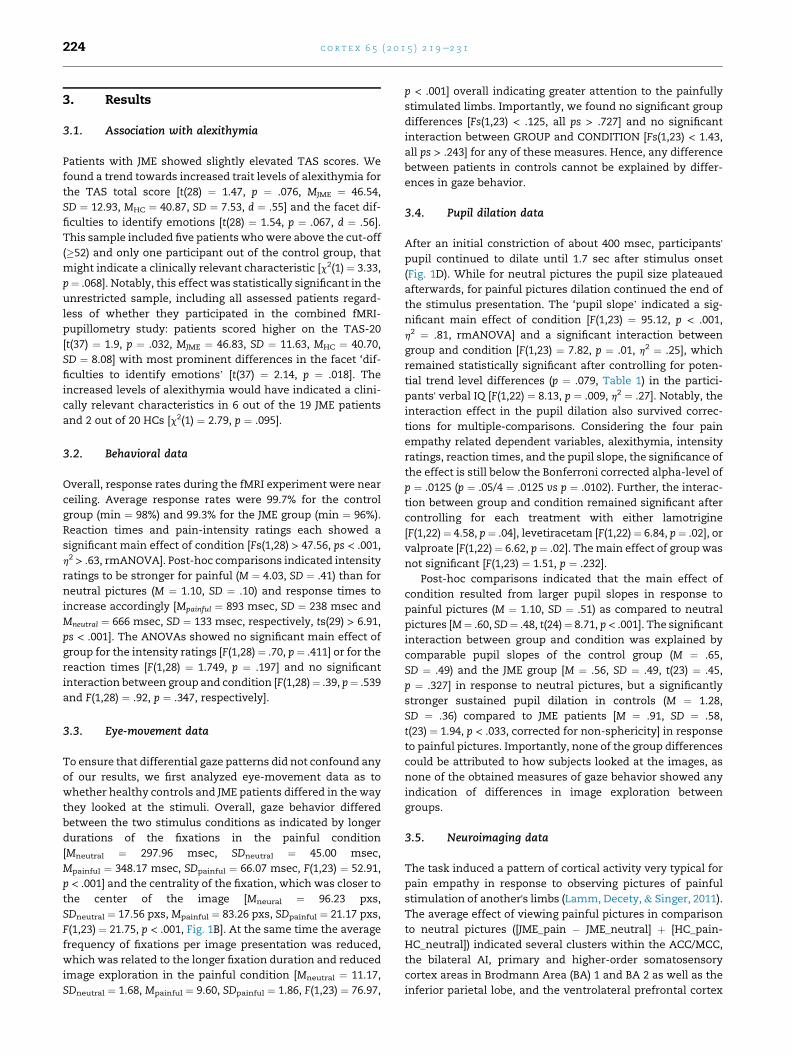
Fig. 1 e Gaze directions, pupil dilation and integration of pupillometry with hemodynamics. A Sequence of the experimental
paradigm and multi-modal-dependent variables. Bottom row: Experimental paradigm to induce empathic pain in the
observers. The trial structure is exemplified with pictures showing painful or neutral, non-painful situations together with
the subsequent rating period and low-level baseline (fixation cross). Middle row, red: z-normalized trace of the pupil
diameter for one subject; for visualization periods of blinks were interpolated using cubic-spline interpolation; blue: a trial-
specific ‘pupil slope’ is computed as optimal linear regression in the least-squares sense on the pupil signal during the time
of stimulus presentation. These trial-specific pupil slopes were entered into the hemodynamic model in order to identify
common variance in the blood oxygen level dependent (BOLD) signal (top row) and the pupil trace. B Heatmaps of the gaze
direction in both groups for each stimulus condition. Gaze patterns of healthy controls (HC) are depicted in the right column
and those of patients with juvenile myoclonic epilepsy (JME) in the left column. The size of the maps corresponds to the full
screen (800 £ 600 pixels, 16.5 £ 12.5� of visual angle) and white rectangles indicate the size of the images that were
presented on the screen (600 £ 450 pixels, 12.5 £ 9.4�). Maps were smoothed with a 20 pixel-wide (.4�) square kernel for
displaying purposes. Colorbars indicate absolute number of samples per bin; colormaps are identical within each row (i.e.,
for the same condition between groups), but differ between rows (i.e., between conditions). C Association of the
hemodynamic response in the right anterior cingulate cortex (ACC) with sustained pupil dilation while watching another'slimb in painful and neutral conditions. The example on the within-subject level illustrates how the observed effects in pupil
dilation relate to neural activation in the group of JME patients and healthy controls. Scatter plots show single trial estimates
of the hemodynamic response in the right ACC at 8, 22, 40 mm and the slope of the pupil dilation for single subjects (left:
patient, right: sex matched HC) in artificial numbers (i.e., b-values). Black dots indicate estimates of the hemodynamic
response and the slope of the pupil dilation in the painful condition, white dots in the neutral condition. Accordingly, black
and white diamonds represent the averaged responses within conditions across trials. Slopes illustrate the positive
association with a least-square fit of a linear regression within the painful condition (dashed, subject 37 Pearson's r ¼ .42
and subject 32 r ¼ .42) and the neutral condition (solid, subject 37 r ¼ .27 and subject 32 r ¼ .51). Consistent with the group-
level analysis, the pupil slope is positively associated with the hemodynamic signal in the ACC, regardless of the condition.
Yet, the mean level of both the hemodynamic response and the pupil slope increased while observing painful stimuli of
another's limbs in the control participant only. D Average dilation of the pupil during the presentation of painful or neutral
stimuli for both groups. For this illustration, blink-interpolated and normalized pupil traces (see panel A, middle row) were
centered on each trial onset and condition averaged for each subject.
c o r t e x 6 5 ( 2 0 1 5 ) 2 1 9e2 3 1222
2.3. Functional MRI data acquisition and preprocessing
Participants were scanned at 3T (Tim TRIO, Siemens Medical
Solutions, Erlangen, Germany) with 36 near-axial slices and a
distance factor of 10% providing whole-brain coverage. An
echo planar imaging (EPI) sequence was used for acquisition
of 395 functional volumes during the experiment (TR: 2.2 sec,
TE: 30 msec, flip angle: 90�, slice thickness: 3 mm, FoV: 192).
fMRI data were analyzed using SPM8 (www.fil.ion.ucl.ac.
uk/spm). After removing the first eight volumes of the time

c o r t e x 6 5 ( 2 0 1 5 ) 2 1 9e2 3 1 223
series the remaining 387 EPI volumes were slice-timed, mo-
tion-corrected, and spatially normalized to the standard EPI-
template of the Montreal Neurological Institute (MNI). The
normalized volumes were re-sliced with a voxel size of
2 � 2 � 2 mm, smoothed with an 8 mm full-width half-
maximum isotropic Gaussian kernel, and high-pass filtered at
1/192 Hz.
2.4. Pupillometry and gaze behavior
Throughout fMRI acquisition, pupil diameter was recorded
monocularly at 500 Hz using a non-invasive MRI-compatible
Eyelink-1000 eye-tracking device (SR Research, Kanata, ON,
Canada). Valid eye-tracking data was obtained for 12 of the
JME patients (9 female, mean age ¼ 30, SD ¼ 11 years) and 13
controls (9 female, mean age ¼ 28.8, SD ¼ 11.2 years). For each
image presentation, the slope of the pupil trace (‘pupil slope’,
Fig. 1A), computed as optimal linear regression in the least-
squares sense on the pupil signal during the time of the
stimulus presentation, provided a trial-specificmeasure of the
pupil dilation (Stoll et al., 2013). The so derived measure
showed excellent reliability for the painful (Cronbach's a¼ .90)
as well as the non-painful condition (a ¼ .86) and was further
used for quantitative analyses: (i) to test the association of
sustained pupil dilation with the hemodynamic signal the
‘pupil slope’ was included as parametric regressor in the fMRI
model (see below) and (ii) to test the condition and group ef-
fects on the ‘pupil slope’ in a 2� 2 repeatedmeasures ANOVA.
To quantify whether gaze behavior differed in terms of
fixated image locations or fixation durations, we determined
periods of fixations using the built-in algorithm of the Eyelink
device with default settings (saccade thresholds: 35 deg/sec
for velocity, 9500 deg/sec2 for acceleration). For illustration
purposes, we in addition use the complete gaze data sampled
at 500 Hz (Fig. 1B).
2.5. fMRI data analyses
2.5.1. Empathy for pain related neural activation andassociation with pupil dilationTwo separate fixed-effects general linear models (GLMs) were
calculated on the within-subject-level. These included three
epoch regressors each modeling the hemodynamic responses
to the painful situations, neutral situations, or the rating
period. Head-movement parameters were included to control
for noise. The first GLMwas implemented to test for activation
differences between the JME and control group. Here, a para-
metric modulator coded the trial-specific intensity rating for
the painful pictures. Weighted b-images contrasting the
painful to the neutral condition were computed and analyzed
at the group level. The second GLM tested the association of
the pupil dilation with the hemodynamic response (Fig. 1A).
Therefore, the trial specific pupil dilation was entered as an
additional parametric weight for the corresponding neutral
and painful picture in the subsample ofN¼ 25 participants for
whom valid eye-tracking data was available. b-images of the
parametric modulators of the pupil dilation during the painful
and neutral condition were analyzed at the group level.
Two separate random-effects GLMs were computed at the
group-level. The first model compared the empathy for pain-
related neural activation in the JME and the healthy control
group. The second model tested the association of trial-by-
trial variability in the pupil slope and the hemodynamic
response with a 2 � 2 repeated measures design including the
parametric weights of the ‘pupil slope’ within the neutral and
painful conditions and the JME and control groups. To control
for potential confounds due to group differences in pupil dy-
namics, this model included the intra-individual standard
deviations of the ‘pupil slope’ as covariates for each group (cf.
Fig. 1C).
To estimate trial-specific responses of the ACC for single
participants, time series were extracted as the first eigen-
variate of a spherical region of interest (ROI) with a radius
of 4 mm. Time series were high-pass filtered with 1/192 Hz,
mean centered, and adjusted for movement-related arti-
facts and hemodynamic responses induced by the rating
period with an effects-of-interest correction. Trial-specific
hemodynamic responses were then estimated with the
above-described stimulus durations of 4.5 sec using
MarsBaR v 0.43 (Brett, Anton, Valabregue, & Jean-Baptiste,
2002).
2.5.2. Functional connectivity analysis of the ACCFor the analysis of functional connectivity, we applied a seed
region approach comparable to procedures described previ-
ously for functional connectivity analyses during task execu-
tion (Paulus et al., 2013; Paulus, Bedenbender, et al., 2014). The
left and right ACC were selected as the seed regions. In order
to determine seed voxels located within the ACC, we con-
strained the search space to the task-activated regions in the
ACC within either the left or right hemisphere. For each
participant the signal was extracted within thesemasks at the
maximum effect for the contrast comparing the hemody-
namic response to the painful and neutral pictures. Neural
signal was extracted as the first eigenvariate in a sphere of
4 mm radius as implemented in SPM8 and task-related vari-
ance was removed by applying an effects-of-interest correc-
tion with an F-contrast set on the six movement parameters.
To account for noise, three additional time-series were
extracted for each subject from the first eigenvariates of all
voxels within masks coveringmedial cerebrospinal fluid (CSF)
or left or right hemispheric white matter (Esslinger et al.,
2009).
The fixed-effects GLM on the subject-level included the
extracted seed time series of the left or right ACC, the left and
right WM noise regressors and the CSF noise regressor.
Additionally, the functional connectivity models included the
three epoch regressorsmodeling the hemodynamic responses
to the painful situations, neutral situations, or the rating
period, the corresponding parametric modulators as well as
head-movement parameters. ß-maps of the left or right ACC
seed time series were analyzed at the group-level using two-
sample t-tests.
All results were corrected for multiple-comparisons using
Gaussian-random field theory as implemented in SPM8 and
overlap between neural activation and cytoarchitectonic
areas was tested with the SPM ANATOMY toolbox v1.8 if
available (Eickhoff et al., 2005). Brain imaging results were
visualized using Caret (Van Essen et al., 2001) and the BrainNet
Viewer (Xia, Wang, & He, 2013).
c o r t e x 6 5 ( 2 0 1 5 ) 2 1 9e2 3 1224
3. Results
3.1. Association with alexithymia
Patients with JME showed slightly elevated TAS scores. We
found a trend towards increased trait levels of alexithymia for
the TAS total score [t(28) ¼ 1.47, p ¼ .076, MJME ¼ 46.54,
SD ¼ 12.93, MHC ¼ 40.87, SD ¼ 7.53, d ¼ .55] and the facet dif-
ficulties to identify emotions [t(28) ¼ 1.54, p ¼ .067, d ¼ .56].
This sample included five patients whowere above the cut-off
(�52) and only one participant out of the control group, that
might indicate a clinically relevant characteristic [c2(1) ¼ 3.33,
p¼ .068]. Notably, this effect was statistically significant in the
unrestricted sample, including all assessed patients regard-
less of whether they participated in the combined fMRI-
pupillometry study: patients scored higher on the TAS-20
[t(37) ¼ 1.9, p ¼ .032, MJME ¼ 46.83, SD ¼ 11.63, MHC ¼ 40.70,
SD ¼ 8.08] with most prominent differences in the facet ‘dif-
ficulties to identify emotions’ [t(37) ¼ 2.14, p ¼ .018]. The
increased levels of alexithymia would have indicated a clini-
cally relevant characteristics in 6 out of the 19 JME patients
and 2 out of 20 HCs [c2(1) ¼ 2.79, p ¼ .095].
3.2. Behavioral data
Overall, response rates during the fMRI experiment were near
ceiling. Average response rates were 99.7% for the control
group (min ¼ 98%) and 99.3% for the JME group (min ¼ 96%).
Reaction times and pain-intensity ratings each showed a
significant main effect of condition [Fs(1,28) > 47.56, ps < .001,
h2 > .63, rmANOVA]. Post-hoc comparisons indicated intensity
ratings to be stronger for painful (M ¼ 4.03, SD ¼ .41) than for
neutral pictures (M ¼ 1.10, SD ¼ .10) and response times to
increase accordingly [Mpainful ¼ 893 msec, SD ¼ 238 msec and
Mneutral ¼ 666 msec, SD ¼ 133 msec, respectively, ts(29) > 6.91,
ps < .001]. The ANOVAs showed no significant main effect of
group for the intensity ratings [F(1,28)¼ .70, p¼ .411] or for the
reaction times [F(1,28) ¼ 1.749, p ¼ .197] and no significant
interaction between group and condition [F(1,28)¼ .39, p¼ .539
and F(1,28) ¼ .92, p ¼ .347, respectively].
3.3. Eye-movement data
To ensure that differential gaze patterns did not confound any
of our results, we first analyzed eye-movement data as to
whether healthy controls and JME patients differed in the way
they looked at the stimuli. Overall, gaze behavior differed
between the two stimulus conditions as indicated by longer
durations of the fixations in the painful condition
[Mneutral ¼ 297.96 msec, SDneutral ¼ 45.00 msec,
Mpainful ¼ 348.17 msec, SDpainful ¼ 66.07 msec, F(1,23) ¼ 52.91,
p < .001] and the centrality of the fixation, which was closer to
the center of the image [Mneural ¼ 96.23 pxs,
SDneutral ¼ 17.56 pxs, Mpainful ¼ 83.26 pxs, SDpainful ¼ 21.17 pxs,
F(1,23) ¼ 21.75, p < .001, Fig. 1B]. At the same time the average
frequency of fixations per image presentation was reduced,
which was related to the longer fixation duration and reduced
image exploration in the painful condition [Mneutral ¼ 11.17,
SDneutral ¼ 1.68, Mpainful ¼ 9.60, SDpainful ¼ 1.86, F(1,23) ¼ 76.97,
p < .001] overall indicating greater attention to the painfully
stimulated limbs. Importantly, we found no significant group
differences [Fs(1,23) < .125, all ps > .727] and no significant
interaction between GROUP and CONDITION [Fs(1,23) < 1.43,
all ps > .243] for any of these measures. Hence, any difference
between patients in controls cannot be explained by differ-
ences in gaze behavior.
3.4. Pupil dilation data
After an initial constriction of about 400 msec, participants'pupil continued to dilate until 1.7 sec after stimulus onset
(Fig. 1D). While for neutral pictures the pupil size plateaued
afterwards, for painful pictures dilation continued the end of
the stimulus presentation. The ‘pupil slope’ indicated a sig-
nificant main effect of condition [F(1,23) ¼ 95.12, p < .001,
h2 ¼ .81, rmANOVA] and a significant interaction between
group and condition [F(1,23) ¼ 7.82, p ¼ .01, h2 ¼ .25], which
remained statistically significant after controlling for poten-
tial trend level differences (p ¼ .079, Table 1) in the partici-
pants' verbal IQ [F(1,22) ¼ 8.13, p ¼ .009, h2 ¼ .27]. Notably, the
interaction effect in the pupil dilation also survived correc-
tions for multiple-comparisons. Considering the four pain
empathy related dependent variables, alexithymia, intensity
ratings, reaction times, and the pupil slope, the significance of
the effect is still below the Bonferroni corrected alpha-level of
p ¼ .0125 (p ¼ .05/4 ¼ .0125 vs p ¼ .0102). Further, the interac-
tion between group and condition remained significant after
controlling for each treatment with either lamotrigine
[F(1,22)¼ 4.58, p¼ .04], levetiracetam [F(1,22)¼ 6.84, p¼ .02], or
valproate [F(1,22)¼ 6.62, p ¼ .02]. Themain effect of group was
not significant [F(1,23) ¼ 1.51, p ¼ .232].
Post-hoc comparisons indicated that the main effect of
condition resulted from larger pupil slopes in response to
painful pictures (M ¼ 1.10, SD ¼ .51) as compared to neutral
pictures [M¼ .60, SD¼ .48, t(24)¼ 8.71, p< .001]. The significant
interaction between group and condition was explained by
comparable pupil slopes of the control group (M ¼ .65,
SD ¼ .49) and the JME group [M ¼ .56, SD ¼ .49, t(23) ¼ .45,
p ¼ .327] in response to neutral pictures, but a significantly
stronger sustained pupil dilation in controls (M ¼ 1.28,
SD ¼ .36) compared to JME patients [M ¼ .91, SD ¼ .58,
t(23) ¼ 1.94, p < .033, corrected for non-sphericity] in response
to painful pictures. Importantly, none of the group differences
could be attributed to how subjects looked at the images, as
none of the obtained measures of gaze behavior showed any
indication of differences in image exploration between
groups.
3.5. Neuroimaging data
The task induced a pattern of cortical activity very typical for
pain empathy in response to observing pictures of painful
stimulation of another's limbs (Lamm, Decety,& Singer, 2011).
The average effect of viewing painful pictures in comparison
to neutral pictures ([JME_pain � JME_neutral] þ [HC_pain-
HC_neutral]) indicated several clusters within the ACC/MCC,
the bilateral AI, primary and higher-order somatosensory
cortex areas in Brodmann Area (BA) 1 and BA 2 as well as the
inferior parietal lobe, and the ventrolateral prefrontal cortex

c o r t e x 6 5 ( 2 0 1 5 ) 2 1 9e2 3 1 225
(VLPFC) to be involved in empathy for pain (p < .05, corrected,
Table 3). Within this network, however, the JME patients
showed significantly less empathy for pain-related
activation compared to the matched controls
([HC_pain � HC_neutral] � [JME_pain � JME_neutral]). ROI
analyses indicated reduced sensitivity to another person'spain in the ACC/MCC cluster [6, 20, 38 mm, t(28) ¼ 3.51,
p ¼ .041, corrected], the right AI [30, 28, 4 mm, t(28) ¼ 3.29,
p¼ .026, corrected], and the VLPFC [�48, 44, 2 mm, t(28)¼ 4.40,
p ¼ .002, corrected, see Table 3 and Fig. 2A,B]. These effects
remained significant after controlling for participants' verbalIQ (ps < .044, corrected). Within the left AI we observed a
similar effect, which however was significant only at an un-
corrected trend level [�40, 18, 6 mm, t(28) ¼ 3.04, p ¼ .136,
corrected, corresponding to p ¼ .003, uncorrected].
To examine common variance of pupil dilation
and hemodynamic responses, we computed the conjunction
of the average effect of the parametric weights of
the pupil dilation across both groups
([JME_pain þ JME_neutral]∩[HC_pain þ HC_neutral]). Signifi-
cant and consistent associations of the pupil slope with the
hemodynamic response were found within the task-
activated network and subcortical regions. The ROI analyses
within the task-activated network revealed significant asso-
ciation of the pupil slope with the hemodynamic signal in the
ACC/MCC cluster [6, 20, 42 mm, t(44) ¼ 4.55, p < .002, cor-
rected], the left AI [�32, 24, �2 mm, t(44) ¼ 3.72, p < .022,
corrected], and the right AI [�38, 24, 4 mm, t(44) ¼ 3.21,
p < .023, corrected, see Table 4, Fig. 2C,D and Fig. 1C for an
illustration of the effects on the within-subject level].
Table 3 e Group differences and empathy for pain-associated ac
Brain region Cyto area Side Cluster si
Pain > No pain
Anterior cingulate R/L 1149
Anterior insula L 1122
Area 44
Anterior insula R 189
Area 44
Anterior insula R 98
Somatosensory IPC(PFop) L 598
IPC(PFt)
Area 1
Somatosensory Area 2 R 95
IPC(PFt)
Ventrolateral prefrontal L 113
Putamen L 31
Putamen R 15
Control > JME £ Pain > No pain
Anterior cingulate R 218
Anterior insula R 39
Ventrolateral prefrontal L 44
Note. All statistics for the average effect of Pain >No Pain are thresholded
k > 10. The interaction effect was examined in the activated regions of in
The cluster extend of the ROI analyses refers to the p < .05 uncorrected
assigned cytoarchitectonic area as indicated by the SPM ANATOMY toolb
Further, the whole-brain analysis indicated a cluster within
the bilateral thalamus to survive the cluster extent threshold
at p ¼ .041 [corresponding to t(44) > 2.69, p < .005, k ¼ 630;
right thalamus at 6, �8, 10 mm, t(44) ¼ 3.84 and left thalamus
�8, �6, 10 mm, t(44) ¼ 3.62].
On average, the pattern of the functional integration of the
ACC showed considerable overlap with the task-activated
network and strongest signal correlations with the bilateral
ACCs were found in the AIs, VLPFCs, and bilateral somato-
sensory cortex areas. In JME patients, thewhole-brain analysis
revealed a significantly decreased coupling of the right ACC
with primary and higher-order somatosensory cortex areas in
the right hemisphere. After controlling for the influences of
task-induced hemodynamics and noise due to head move-
ment or global signal fluctuations, compared to controls, the
right ACC in JME patients showed lower signal correspon-
dence with a cluster covering BA 1 at 64, �12, 28 mm, BA 3b at
40, �26, 44 mm and the parietal operculum at 60, �10, 12 mm
[p ¼ .047, corrected, corresponding to p < .005, uncorrected,
t(28) > 2.76, k ¼ 611, Table 5 and Fig. 3]. A similar and in part
overlapping cluster was observed for the left ACC, showing a
reduced functional connectivity with the right somatosensory
cortex in BA 2 at 44, �28, 56 mm, and BA 3b at 28, �42, 60 mm,
at trend level [p ¼ .064, corrected, corresponding to p < .005,
uncorrected, t(28) > 2.76, k ¼ 492]. Besides these findings,
within the present sample we also found partial support for
previous evidence (Vollmar et al., 2012) indicating reduced
connectivity of the left ACC with SMA regions in the left
hemisphere at 10, �18, 58 mm [p ¼ .124, corrected, corre-
sponding to p < .005, uncorrected, t(28) > 2.76, k ¼ 406].
tivation.
ze MNI coordinates T pFWE
x y z
�4 20 42 10.74 <.001�4 28 40 10.06 <.001�8 30 28 7.43 .002
�40 18 6 9.81 <.001�46 14 �6 8.41 <.001�52 6 14 8.00 .001
48 14 4 8.70 <.00154 12 12 7.12 .003
28 22 6 7.89 .001
38 20 0 6.10 .032
�58 �22 24 8.84 <.001�58 �26 34 8.50 <.001�44 �30 62 7.74 .001
62 �22 44 8.60 <.00148 �28 44 7.21 .003
�48 44 4 8.94 <.001�14 10 2 6.94 .005
14 12 0 6.47 .014
6 20 38 3.51 .041
30 28 4 3.29 .026
�48 44 2 4.40 .002
at p < .05, familywise error (FWE) corrected for a whole-brain analysis,
terests (ROIs) and p values were corrected within the respective ROI.
cluster size within each ROI. The ‘Cyto Area’ column indicates the
ox v1.8 if available.

Fig. 2 e Reduced activation of the JME group within the task-activated network and association of pupil dilation with
hemodynamic responses. A Brain areas showing reduced activation in JME patients (red) compared to healthy controls (HC)
within the empathy for pain-related activation (yellow) rendered on an individual surface image. For display purposes, the
results of the interaction of group and condition ([HC_pain ¡ HC_neutral] ¡ [JME_pain ¡ JME_neutral]) were thresholded at
t(28) > 1.70, p < .05, uncorrected, and were masked by the task-activated network
([JME_pain ¡ JME_neutral] þ [HC_pain ¡ HC_neutral]), which was thresholded at t(28) > 5.89, p < .05, corrected. B Parameter
estimates of clusters that show significant reduction of empathy for pain-related activation in the JME group at corrected
thresholds. Parameter estimates are plotted together with standard errors at the peak voxel and illustrate the contrast of
Pain-Neutral for each group within the left and right anterior cingulate cortex (ACC), the right anterior insula (AI), and the
left ventrolateral prefrontal cortex (VLPFC). C Brain areas where the hemodynamic signal is positively associated with the
'pupil slope' rendered on an individual surface image. Clusters within the empathy for pain-related network (yellow) are
coded in red while areas outside the network are coded in blue. The conjunction analysis of the parametric weights of the
pupil slope in both groups ([JME_pain þ JME_neutral]∩[HC_pain þ HC_neutral]) was thresholded at t(44) > 2.69, p < .005,
uncorrected, and plotted together with the task-activated network ([JME_pain ¡ JME_neutral] þ [HC_pain ¡ HC_neutral]),
which was thresholded at t(28) > 5.89, p < .05, corrected, for displaying purposes. D Parameter estimates of the clusters that
show significant association of sustained pupil dilation with hemodynamic responses consistently across both groups.
Parameter estimates are plotted together with standard errors at the peak voxel and illustrate the average effect of the
conditions (0.5£[pain þ neutral]) for each group within the bilateral AI, the right ACC, and the bilateral thalamus.
c o r t e x 6 5 ( 2 0 1 5 ) 2 1 9e2 3 1226
4. Discussion
In this study we used the specific psychopathology associated
with JME to directly test the role of the mesial fronto-insular
network for social adjustment in patients and healthy con-
trols. Although the peculiarities in the personality structure of
JME patients have been related to structural abnormalities in
the mesial-frontal lobe and dysfunctions of fronto-thalamical

Table 4 e Positive association of hemodynamic responses with variability in the slope of the pupil dilation.
Brain region Cyto area Side Cluster size MNI coordinates T pFWE
x y z
Cluster extend threshold
Thalamus Th-Temporal R 630 6 �8 10 3.84 .041
Th-Temporal L �8 �6 10 3.62
R 10 6 �2 3.60
Th-Prefrontal L �12 �18 12 3.06
ROI analyses
Anterior cingulate R 117 6 20 42 4.55 .002
Anterior insula L 142 �32 24 �2 3.72 .022
Anterior insula R 9 38 24 4 3.21 .023
R 5 28 20 6 3.09 .030
Note. FWE ¼ Familywise error correction for the respective peak voxel in case of the region of interest (ROI) analysis or cluster extent in case of
the whole-brain analysis. Effects represent the average effect of the parametric weights for the Neutral and Pain condition in a conjunction
across the JME and Control group. All cluster extends refer to p < .005, uncorrected. ROIs were similar to those used for examining activation
differences between the JME and Control groups. The ‘Cyto Area’ column indicates the assigned cytoarchitectonic area as indicated by the SPM
ANATOMY toolbox v1.8 if available.
c o r t e x 6 5 ( 2 0 1 5 ) 2 1 9e2 3 1 227
circuits earlier, this hypothesis had not been tested directly
with respect to brain function in context of social paradigms.
By assessing the physiological parameters while observing
others' bodily pain by simultaneous pupillometry and fMRI,
we verified the main hypothesis that JME impacts the
empathy for pain response within mesial-frontal circuits.
Within the task-activated network, patients with JME showed
reduced empathy for pain-related responses in dorsal aspects
of the ACC, the right AI, and the left VLPFC. These data were
supported by less-pronounced pupil dilation to painful stimuli
in the JME group, whichwas linked to the neural activity in the
bilateral ACC, the AI, and the thalamus on the within-subject
level. The neural activation in brain systems that regulate
homeostasis (Craig, 2009) thus directly translated to the
observed pattern of the pupil response providingmulti-modal
evidence for altered pain empathy in patients with JME. The
decreased connectivity of primary and higher-order somato-
sensory cortex areas with the ACC, which are of particular
Table 5 e Reduced functional connectivity of the bilateral ACC in
Brain region Cyto area Side Cluster siz
Functional connectivity of the right anterior cingulate cortex
Somatosensory IPC(PF) R 611
OP4 R
Area 1 R
Area 1 R
Area 3b R
Functional connectivity of the left anterior cingulate cortex
Somatosensory Area 3b R 492
Area 3b R
Area 2 R
Area 4a R
SMA/Somatosensory Area 2 L 406
Area 4p L
Area 6 L
Note. FWE ¼ Familywise error correction for the respective cluster extent
clusters exceeding k > 400. The ‘Cyto Area’ column indicates the assigne
v1.8 if available.
importance for the somatic representations of another per-
son's pain (Keysers et al., 2010), thereby characterizes a neural
pathway that helps explaining the altered response of fronto-
insular circuits and disturbances in psychosocial behavior.
Previous studies have mainly concentrated on structural
abnormalities in JME patients on a micro (Meencke, 1985;
Meencke & Janz, 1984) as well as macro level (Betting et al.,
2006; Deppe et al., 2008; Kim et al., 2007; O'Muircheartaigh
et al., 2012; Woermann et al., 1999), while mesial-frontal
brain functioning was only examined in the context of
cognitive paradigms such as working memory (Vollmar et al.,
2011) or word generation (O'Muircheartaigh et al., 2012).
These studies characterized atypical interactions of higher
motor systems with cortical and subcortical structures,
providing insights into how cognitive effort related to the
specific JME psychopathology. Especially the SMA and pre-
SMA have thus been considered central hub regions in the
pathological architecture of the neural system's functioning
JME patients.
e MNI coordinates T pFWE
x y z
66 �26 34 4.98 .047
60 �10 12 3.78
64 �12 28 3.78
38 �36 62 3.50
40 �26 44 3.34
44 �28 56 4.36 .064
34 �38 58 4.24
28 �42 60 4.23
38 �22 56 3.58
�36 �40 58 3.65 .127
�14 �30 60 3.36
10 �18 58 3.33
. All cluster sizes refer to p < .005, uncorrected, and are reported for
d cytoarchitectonic area as indicated by the SPM ANATOMY toolbox

Fig. 3 e Reduced functional connectivity of the bilateral ACCs with somatosensory cortex areas and the supplementary
motor area in JME patients. A Brain areas with significantly reduced connectivity of the right ACC in JME patients rendered
on the right hemisphere of the ICBM 152 brain surface in MNI space. The depicted cluster survived the family-wise error
correction of the cluster extent threshold in the whole-brain analysis at p < .047. Cytoarchitectonic references indicated
maxima at primary and higher-order somatosensory cortex areas in Area 1, Area 3b, and the parietal operculum. B At trend
level, the left ACC also had reduced connectivity with the somatosensory cortex in Area 2 and Area 3b (p ¼ .064, corrected)
as well as the supplementary motor area in the medial Area 6. All render images were thresholded at t(28) > 2.76, p < .005,
k > 400 and include the location of the left or right hemispheric seed time series for JME patients (green) and healthy
controls (red).
c o r t e x 6 5 ( 2 0 1 5 ) 2 1 9e2 3 1228
(Vollmar et al., 2012), potentially helping to also understand
emotional instabilities and difficulties in socio-emotional
adjustment. Given the frequent comorbidities with psychi-
atric conditions, however, this indirect evidence might not
suffice to understand the complex psychopathology of JME
patients. Instead, the present findings suggest that dysfunc-
tions in the social domain could be explained via altered re-
sponses of the ACC and AI network and reduced functional
integration between primary and higher-order somatosen-
sory cortex areas and the ACC. This is supported by a
considerable body of literature that ascribes the ACC and AI a
crucial role in the understanding and regulation of one's own
emotions (Berthoz et al., 2002; Craig, 2009; Damasio et al.,
2000; Eippert et al., 2007) and altered functioning, specif-
ically of the AI, in alexithymia (Bernhardt et al., 2013; Bird
et al., 2010). Beyond empathy for physical pain, this
network thus also has strong implications in the experience
of other social emotions such as sharing happiness (Jabbi &
Keysers, 2008; Mobbs et al., 2009), disgust (Jabbi,
Bastiaansen, & Keysers, 2008; Wicker et al., 2003) or social
pain (Krach et al., 2011; Paulus, Muller-Pinzler, et al., 2014)
with significant consequences for interpersonal behavior: the
fMRI response pattern observed herein to be altered in JME
patients, had previously been described motivating helping
behavior in healthy observers (Hein et al., 2010; Masten,
Morelli, & Eisenberger, 2011) and related to disorders that
are characterized by impairments in social interactions
(Masten, Colich, et al., 2011; Masten, Morelli, et al., 2011;
Meffert et al., 2013; Silani et al., 2008). In accordance with
the previous evidence for patients with epilepsy in the ACC to
have difficulties in social adjustment (Devinsky, Morrell, &
Vogt, 1995), the present findings corroborate the notion that
dysfunctions within the ACC circuits might mediate distur-
bances in social behavior, also in JME patients. This study
thus is the first to demonstrate that the peculiarities in the
personality structure, including emotional instability, rapid
mood changes or difficulties in social adjustment and the
increased probability of comorbid psychiatric conditions in
JME, may not solely be attributed to the side effects of the
dysexecutive syndrome; instead, our data suggest that they
result from alterations in neural circuits that are directly
involved in regulating affect.
It is unknownwhether our findings result from generalized
alterations in the ACC's functioning during emotion process-
ing or whether the reduced reactivity is specific to impair-
ments in empathizing with another. Even though recent fMRI
studies showed similar involvement of the ACC, AI, and the
VLPFC in pain empathy and the pain experienced on one'sown body, both have to be considered as conceptually and
psychologically distinct phenomena (Keysers & Gazzola,
2007). The psychopathology of JME, specifically the
emotional instability (Gelisse et al., 2007) and the here
described trend for higher levels in alexithymia, might none-
theless suggest the reduced pain-empathy response to be
driven by rather general alterations in the affective response
of the ACC and AI network and reduced integration of infor-
mation from somatosensory cortex areas in mesial-frontal
circuits. The present findings of altered frontal functioning
during empathy for pain thus might have the potential to also
generalize to first-person experiences of pain and explain the
often observed difficulties of JME patients in the experience
and regulation of affect (De Araujo Filho et al., 2013). However,
future studies need to directly address the role of the ACC'sand the AI's functioning in first-person experiences of nega-
tive affect in order to understand their contribution to the JME
psychopathology more comprehensively.
The present findings also provide new insight in the link of
pupil dynamicswith neural systems activity during emotional
processing. While changes in pupil diameter occur in
response to diverse stimulus features and psychological

c o r t e x 6 5 ( 2 0 1 5 ) 2 1 9e2 3 1 229
processes (Preuschoff, 2011; Steinhauer et al., 2004), the cur-
rent pattern of continuous pupil dilation during stimulus
presentation has also been previously related to processing
negative emotions (Bradley et al., 2008) and the experience of
pain on one's own body (Geuter et al., 2014). The physiology of
this characteristic pupil response to emotional stimuli has
been explained by activity of the sympathetic system that
innervates the iris dilator, resulting in the observed increase
of the pupil's size. The involvement of the sympathetic system
in pupil dilation during emotional processing is empirically
supported by close covariation of pupil diameter with skin
conductance responses (Bradley et al., 2008) and the predictive
value of themagnitude of pupil dilation for the intensity of the
experienced affect (Geuter et al., 2014). The now described
association of pupil dilation with ACC and AI activation, brain
systems that have strong implications in the regulation of
homeostasis, is in line with previous work that indicated a
very similar coupling of increase in pupil size with autonomic
activity and stress-induced activation of the ACC (Critchley,
Tang, Glaser, Butterworth, & Dolan, 2005). The link of neural
activity in neural systems that process emotional arousal with
a specific and characteristic pattern observable pupil dy-
namics thus opens up new perspectives for non-invasive in-
vestigations of affective responses in more complex social
scenarios that require ecological plausible environments,
which are difficult to realize in the fMRI setting (Krach,Muller-
Pinzler, Westermann, & Paulus, 2013).
Notably, the reduced pupil dilation in the JME sample could
not be explained by the antiepileptic medication. While a
more recent case report suggested lamotrigine to affect eye
movements in children (Das, Harris, Smyth,& Cross, 2003), an
earlier study with a larger sample of healthy volunteers found
no evidence for this effect (Hamilton et al., 1993) and instead
related carbamazepine to alterations in smooth and saccadic
eye movements. Importantly, this study found no effects on
pupil dynamics and neither did another study for valproate
(DeMet & Sokolski, 1999). Hence there is no prior data sug-
gesting an effect of antiepileptic drugs on pupils' response,which was also supported with the present data; after
including the anticonvulsant medications as nuisance re-
gressors, the significance of the effects remained unchanged.
In conclusion, the present study provides further evidence
for the neural basis of specific aspects of JME patients' psy-chopathology. Consistent with our predictions, the fMRI data
indicated less-pronounced neural responses in the frontal
circuits that mediate the empathic sharing of unpleasant
feelings. This was paralleled by similar effects observed for
pupil dilation, which we link to neural activity in brain sys-
tems with strong implications in homeostatic regulation. The
correlation of the pupils' reactivity with neural activation
within the ACC, thalamic, and insular network represents the
first combined evidence for reduced physiological reactivity of
patients with JME in response to social stimuli which could be
explained by reduced functional interaction among somato-
sensory cortex areas and the ACC. Thismulti-modal approach
thus helps to better understand the complex clinical picture of
JME patients by explaining the peculiarities in psychosocial
behavior with disturbances in mesial-frontal circuits, and
contributes to a better understanding of the neural founda-
tions of social behavior in general.
Acknowledgments
This work was supported by research grants from the ‘Deut-
sche Forschungsgemeinschaft’ (DFG grant no. KR3803/2-1,
KR3803/7-1 and EI852/3-1), the ‘Landes-Offensive zur
Entwicklung Wissenschaftlich-€okonomischer Exzellenz
(LOEWE)’, and the ‘Research Foundation of the University of
Marburg‘. The authors would also like to thank Rita Werner
and Jens Sonntag for their assistance during data collection
and one anonymous reviewer who helped to improve the
quality of the manuscript.
r e f e r e n c e s
Bernhardt, B. C., Valk, S. L., Silani, G., Bird, G., Frith, U., &Singer, T. (2013). Selective disruption of sociocognitivestructural brain networks in autism and alexithymia. CerebralCortex. http://dx.doi.org/10.1093/cercor/bht182.
Berthoz, S., Artiges, E., Van De Moortele, P.-F., Poline, J.-B.,Rouquette, S., Consoli, S. M., et al. (2002). Effect of impairedrecognition and expression of emotions on frontocingulatecortices: an fMRI study of men with alexithymia. The AmericanJournal of Psychiatry, 159(6), 961e967.
Betting, L. E., Mory, S. B., Li, L. M., Lopes-Cendes, I.,Guerreiro, M. M., Guerreiro, C. A. M., et al. (2006). Voxel-basedmorphometry in patients with idiopathic generalizedepilepsies. NeuroImage, 32(2), 498e502. http://dx.doi.org/10.1016/j.neuroimage.2006.04.174.
Bird, G., Silani, G., Brindley, R., White, S., Frith, U., & Singer, T.(2010). Empathic brain responses in insula are modulated bylevels of alexithymiabut not autism. Brain, 133(Pt 5), 1515e1525.http://dx.doi.org/10.1093/brain/awq060. awq060 [pii].
Bradley, M. M., Miccoli, L., Escrig, M. A., & Lang, P. J. (2008). Thepupil as a measure of emotional arousal and autonomicactivation. Psychophysiology, 45(4), 602e607. http://dx.doi.org/10.1111/j.1469-8986.2008.00654.x.
Brett, M., Anton, J.-L., Valabregue, R., & Jean-Baptiste, P. (2002).Region of interest analysis using an SPM toolbox. In 8thInternational Conference on Functional Mapping of the Human Brain.
Cox, B. J., Swinson, R. P., Shulman, I. D., & Bourdeau, D. (1995).Alexithymia in panic disorder and social phobia.Comprehensive Psychiatry, 36(3), 195e198. http://dx.doi.org/10.1016/0010-440X(95)90081-6.
Craig, A. D. (2009). How do you feelenow? The anterior insula andhuman awareness. Nature Reviews Neuroscience, 10(1), 59e70.http://dx.doi.org/10.1038/nrn2555.
Critchley, H. D., Tang, J., Glaser, D., Butterworth, B., & Dolan, R. J.(2005). Anterior cingulate activity during error and autonomicresponse. NeuroImage, 27(4), 885e895. http://dx.doi.org/10.1016/j.neuroimage.2005.05.047.
Damasio, A. R., Grabowski, T. J., Bechara, A., Damasio, H.,Ponto, L. L., Parvizi, J., et al. (2000). Subcortical and corticalbrain activity during the feeling of self-generated emotions.Nature Neuroscience, 3(10), 1049e1056. http://dx.doi.org/10.1038/79871.
Das, K. B., Harris, C., Smyth, D. P. L., & Cross, J. H. (2003). Unusualside effects of lamotrigine therapy. Journal of Child Neurology,18(7), 479e480. http://dx.doi.org/10.1177/08830738030180070301.
Davis, M. H. (1996). Empathy: A social-psychological approach.Boulder: Westview Press.
De Araujo Filho, G. M., de Araujo, T. B., Sato, J. R., Silva, I., Da,Lin, K., Junior, H. C., et al. (2013). Personality traits in juvenilemyoclonic epilepsy: evidence of cortical abnormalities from a

c o r t e x 6 5 ( 2 0 1 5 ) 2 1 9e2 3 1230
surface morphometry study. Epilepsy & Behavior: E&B, 27(2),385e392. http://dx.doi.org/10.1016/j.yebeh.2013.02.004.
DeMet, E. M., & Sokolski, K. N. (1999). Sodium valproate increasespupillary responsiveness to a cholinergic agonist inresponders with mania. Biological Psychiatry, 46(3), 432e436.
Deppe, M., Kellinghaus, C., Duning, T., M€oddel, G.,Mohammadi, S., Deppe, K., et al. (2008). Nerve fiberimpairment of anterior thalamocortical circuitry in juvenilemyclonic epilepsy. Neurology, 71(24), 1981e1985. http://dx.doi.org/10.1212/01.wnl.0000336969.98241.17.
Devinsky, O., Morrell, M. J., & Vogt, B. A. (1995). Contributions ofanterior cingulate cortex to behaviour. Brain, 118, 279e306.
Eickhoff, S. B., Stephan, K. E., Mohlberg, H., Grefkes, C., Fink, G. R.,Amunts, K., et al. (2005). A new SPM toolbox for combiningprobabilistic cytoarchitectonic maps and functional imagingdata. NeuroImage, 25(4), 1325e1335. http://dx.doi.org/10.1016/j.neuroimage.2004.12.034. S1053-8119(04)00792-X [pii].
Eippert, F., Veit, R., Weiskopf, N., Erb, M., Birbaumer, N., &Anders, S. (2007). Regulation of emotional responses elicitedby threat-related stimuli. Human Brain Mapping, 28(5), 409e423.http://dx.doi.org/10.1002/hbm.20291.
Esslinger, C., Walter, H., Kirsch, P., Erk, S., Schnell, K., Arnold, C.,et al. (2009). Neural mechanisms of a genome-wide supportedpsychosis variant. Science, 324(5927), 605. http://dx.doi.org/10.1126/science.1167768, 324/5927/605 [pii].
Gelisse, P., Thomas, P., Samuelian, J. C., & Gentin, P. (2007).Psychiatric disorders in juvenile myoclonic epilepsy. Epilepsia,48(5), 1032e1033. http://dx.doi.org/10.1111/j.1528-1167.2007.01009_4.x. EPI1009_4 [pii].
Geuter, S., Gamer, M., Onat, S., & Buchel, C. (2014). Parametrictrial-by-trial prediction of pain by easily availablephysiological measures. Pain, 155(5), 994e1001. http://dx.doi.org/10.1016/j.pain.2014.02.005.
Hamilton, M. J., Cohen, A. F., Yuen, A. W. C., Harkin, N., Land, G.,Weatherley, B. C., et al. (1993). Carbamazepine andlamotrigine in healthy volunteers: relevance to early toleranceand clinical trial dosage. Epilepsia, 34(1), 166e173. http://dx.doi.org/10.1111/j.1528-1157.1993.tb02393.x.
Hein, G., Silani, G., Preuschoff, K., Batson, C. D., & Singer, T. (2010).Neural responses to ingroup and outgroup members' sufferingpredict individual differences in costly helping. Neuron, 68(1),149e160. http://dx.doi.org/10.1016/j.neuron.2010.09.003.S0896-6273(10)00720-8 [pii].
Holmes, M. D., Quiring, J., & Tucker, D. M. (2010). Evidence thatjuvenile myoclonic epilepsy is a disorder of frontotemporalcorticothalamic networks. NeuroImage, 49(1), 80e93. http://dx.doi.org/10.1016/j.neuroimage.2009.08.004.
Jabbi, M., Bastiaansen, J., & Keysers, C. (2008). A common anteriorinsula representation of disgust observation, experience andimagination shows divergent functional connectivitypathways. PloS One, 3(8), e2939. http://dx.doi.org/10.1371/journal.pone.0002939.
Jabbi, M., & Keysers, C. (2008). Inferior frontal gyrus activitytriggers anterior insula response to emotional facialexpressions. Emotion, 8(6), 775e780. http://dx.doi.org/10.1037/a0014194.
Jackson, P. L., Meltzoff, A. N., & Decety, J. (2005). How dowe perceive the pain of others? A window into theneural processes involved in empathy. NeuroImage, 24(3),771e779. http://dx.doi.org/10.1016/j.neuroimage.2004.09.006.
Janisse, M. P. (1974). Pupil size, affect and exposure frequency.Social Behavior and Personality, 2(2), 125e146. http://dx.doi.org/10.2224/sbp.1974.2.2.125.
Janz, D. (1985). Epilepsy with impulsive petit mal (juvenilemyoclonic epilepsy). Acta Neurologica Scandinavica, 72(5),449e459.
Janz, D., & Christian, W. (1957). Impulsiv-Petit mal. DeutscheZeitschrift Fur Nervenheilkunde, 176(3), 346e386. http://dx.doi.org/10.1007/BF00242439.
Keysers, C., & Gazzola, V. (2007). Integrating simulation andtheory of mind: from self to social cognition. Trends in CognitiveSciences, 11(5), 194e196. http://dx.doi.org/10.1016/j.tics.2007.02.002.
Keysers, C., Kaas, J. H., & Gazzola, V. (2010). Somatosensation insocial perception. Nature Reviews Neuroscience, 11(6), 417e428.http://dx.doi.org/10.1038/nrn2833.
Kim, J. H., Lee, J. K., Koh, S.-B., Lee, S.-A., Lee, J.-M., Kim, S. I., et al.(2007). Regional grey matter abnormalities in juvenilemyoclonic epilepsy: a voxel-based morphometry study.NeuroImage, 37(4), 1132e1137. http://dx.doi.org/10.1016/j.neuroimage.2007.06.025.
Krach, S., Cohrs, J. C., de Echeverria Loebell, N. C., Kircher, T.,Sommer, J., Jansen, A., et al. (2011). Your flaws are my pain:linking empathy to vicarious embarrassment. PloS One, 6(4),e18675. http://dx.doi.org/10.1371/journal.pone.0018675.
Krach, S., Muller-Pinzler, L., Westermann, S., & Paulus, F. M.(2013). Advancing the neuroscience of social emotions withsocial immersion. The Behavioral and Brain Sciences, 36(4),427e428. http://dx.doi.org/10.1017/S0140525X12001951.
Lamm, C., Decety, J., & Singer, T. (2011). Meta-analytic evidencefor common and distinct neural networks associated withdirectly experienced pain and empathy for pain. NeuroImage,54(3), 2492e2502. http://dx.doi.org/10.1016/j.neuroimage.2010.10.014. S1053-8119(10)01306-6 [pii].
Lyon, G., & Gastaut, H. (1985). Considerations on the significanceattributed to unusual cerebral histological findings recentlydescribed in eight patients with primary generalized epilepsy.Epilepsia, 26(4), 365e367. http://dx.doi.org/10.1111/j.1528-1157.1985.tb05664.x.
Masten, C. L., Colich, N. L., Rudie, J. D., Bookheimer, S. Y.,Eisenberger, N. I., & Dapretto, M. (2011). An fMRI investigationof responses to peer rejection in adolescents with autismspectrum disorders. Developmental Cognitive Neuroscience, 1(3),260e270. http://dx.doi.org/10.1016/j.dcn.2011.01.004.
Masten, C. L., Morelli, S. A., & Eisenberger, N. I. (2011). An fMRIinvestigation of empathy for “social pain” and subsequentprosocial behavior. NeuroImage, 55(1), 381e388. http://dx.doi.org/10.1016/j.neuroimage.2010.11.060. S1053-8119(10)01538-7 [pii].
Meencke, H. J. (1985). Neuron density in the molecular layer of thefrontal cortex in primary generalized epilepsy. Epilepsia, 26(5),450e454.
Meencke, H. J., & Janz, D. (1984). Neuropathological findings inprimary generalized epilepsy: a study of eight cases. Epilepsia,25(1), 8e21.
Meffert, H., Gazzola, V., den Boer, J. A., Bartels, A. A. J., &Keysers, C. (2013). Reduced spontaneous but relatively normaldeliberate vicarious representations in psychopathy. Brain,136(8), 2550e2562. http://dx.doi.org/10.1093/brain/awt190.
Mobbs, D., Yu, R., Meyer, M., Passamonti, L., Seymour, B.,Calder, A. J., et al. (2009). A key role for similarity in vicariousreward. Science, 324(5929), 900. http://dx.doi.org/10.1126/science.1170539.
O'Muircheartaigh, J., Vollmar, C., Barker, G. J., Kumari, V.,Symms, M. R., Thompson, P., et al. (2011). Focal structuralchanges and cognitive dysfunction in juvenile myoclonicepilepsy. Neurology, 76(1), 34e40. http://dx.doi.org/10.1212/WNL.0b013e318203e93d.
O'Muircheartaigh, J., Vollmar, C., Barker, G. J., Kumari, V.,Symms, M. R., Thompson, P., et al. (2012). Abnormalthalamocortical structural and functional connectivity injuvenile myoclonic epilepsy. Brain, 135(Pt 12), 3635e3644.http://dx.doi.org/10.1093/brain/aws296.

c o r t e x 6 5 ( 2 0 1 5 ) 2 1 9e2 3 1 231
Parker, J. D., Taylor, G. J., & Bagby, R. M. (2003). The 20-itemToronto alexithymia scale. Journal of Psychosomatic Research,55(3), 269e275. http://dx.doi.org/10.1016/S0022-3999(02)00578-0.
Partala, T., & Surakka, V. (2003). Pupil size variation as anindication of affective processing. International Journal ofHuman-Computer Studies, 59(1e2), 185e198. http://dx.doi.org/10.1016/S1071-5819(03)00017-X.
Paulus, F. M., Bedenbender, J., Krach, S., Pyka, M., Krug, A.,Sommer, J., et al. (2014a). Association of rs1006737 inCACNA1C with alterations in prefrontal activation and fronto-hippocampal connectivity. Human Brain Mapping, 35,1190e1200. http://dx.doi.org/10.1002/hbm.22244.
Paulus, F. M., Krach, S., Bedenbender, J., Pyka, M., Sommer, J.,Krug, A., et al. (2013). Partial support for ZNF804A genotype-dependent alterations in prefrontal connectivity. HumanBrain Mapping, 34(2), 304e313. http://dx.doi.org/10.1002/hbm.21434.
Paulus, F. M., Muller-Pinzler, L., Jansen, A., Gazzola, V., & Krach, S.(2014b). Mentalizing and the role of the posterior superiortemporal sulcus in sharing others' embarrassment. CerebralCortex. http://dx.doi.org/10.1093/cercor/bhu011.
Preuschoff, K. (2011). Pupil dilation signals surprise: evidence fornoradrenaline's role in decision making. Frontiers inNeuroscience, 5(September), 1e12. http://dx.doi.org/10.3389/fnins.2011.00115.
Savic, I., Lekvall, A., Greitz, D., & Helms, G. (2000). MRspectroscopy shows reduced frontal lobe concentrations of N-acetyl aspartate in patients with juvenile myoclonic epilepsy.Epilepsia, 41(3), 290e296.
Silani, G., Bird, G., Brindley, R., Singer, T., Frith, C., & Frith, U.(2008). Levels of emotional awareness and autism: an fMRIstudy. Social Neuroscience, 3(2), 97e112. http://dx.doi.org/10.1080/17470910701577020.
Silk, J. S., Dahl, R. E., Ryan, N. D., Forbes, E. E., Axelson, D. A.,Birmaher, B., et al. (2007). Pupillary reactivity to emotionalinformation in child and adolescent depression: links toclinical and ecological measures. The American Journal ofPsychiatry, 164(12), 1873e1880. http://dx.doi.org/10.1176/appi.ajp.2007.06111816.
Steinhauer, S. R., Siegle, G. J., Condray, R., & Pless, M. (2004).Sympathetic and parasympathetic innervation of pupillarydilation during sustained processing. International Journal ofPsychophysiology: Official Journal of the International Organization
of Psychophysiology, 52(1), 77e86. http://dx.doi.org/10.1016/j.ijpsycho.2003.12.005.
Stoll, J., Chatelle, C., Carter, O., Koch, C., Laureys, S., &Einh€auser, W. (2013). Pupil responses allow communication inlocked-in syndrome patients. Current Biology, 23(15),R647eR648. http://dx.doi.org/10.1016/j.cub.2013.06.011.
Taylor, G. J. (1984). Alexithymia: concept, measurement, andimplications for treatment. The American Journal of Psychiatry,141(6), 725e732.
Taylor, G. J., Michael Bagby, R., & Parker, J. D. A. (1991). Thealexithymia construct: a potential paradigm forpsychosomatic medicine. Psychosomatics, 32(2), 153e164.http://dx.doi.org/10.1016/S0033-3182(91)72086-0.
Van Essen, D. C., Drury, H. A., Dickson, J., Harwell, J., Hanlon, D., &Anderson, C. H. (2001). An integrated software suite forsurface-based analyses of cerebral cortex. Journal of theAmerican Medical Informatics Association, 8(5), 443e459. http://dx.doi.org/10.1136/jamia.2001.0080443.
Vollmar, C., O'Muircheartaigh, J., Barker, G. J., Symms, M. R.,Thompson, P., Kumari, V., et al. (2011). Motor systemhyperconnectivity in juvenile myoclonic epilepsy: a cognitivefunctional magnetic resonance imaging study. Brain, 134(Pt 6),1710e1719. awr098 [pii] 10.1093/brain/awr098.
Vollmar, C., O'Muircheartaigh, J., Symms, M. R., Barker, G. J.,Thompson, P., Kumari, V., et al. (2012). Altered microstructuralconnectivity in juvenile myoclonic epilepsy: the missing link.Neurology, 78(20), 1555e1559. http://dx.doi.org/10.1212/WNL.0b013e3182563b44.
Vulliemoz, S., Vollmar, C., Koepp, M. J., Yogarajah, M.,O'Muircheartaigh, J., Carmichael, D. W., et al. (2011).Connectivity of the supplementary motor area in juvenilemyoclonic epilepsy and frontal lobe epilepsy. Epilepsia, 52(3),507e514. http://dx.doi.org/10.1111/j.1528-1167.2010.02770.x.
Wicker, B., Keysers, C., Plailly, J., Royet, J. P., Gallese, V., &Rizzolatti, G. (2003). Both of us disgusted in my insula: thecommon neural basis of seeing and feeling disgust. Neuron,40(3), 655e664. S0896627303006792 [pii].
Woermann, F. G., Free, S. L., Koepp, M. J., Sisodiya, S. M., &Duncan, J. S. (1999). Abnormal cerebral structure in juvenilemyoclonic epilepsy demonstrated with voxel-based analysisof MRI. Brain, 122, 2101e2108.
Xia, M., Wang, J., & He, Y. (2013). BrainNet Viewer: a networkvisualization tool for human brain connectomics. PloS One,8(7), e68910. http://dx.doi.org/10.1371/journal.pone.0068910.