Frontal Lobe Radionecrosis Following Radiotherapy of an ... · Frontal Lobe Radionecrosis Following...
4
Frontal Lobe Radionecrosis Following Radiotherapy of an Extracranial Tumor Ekstrakraniyal Bir Tiimoriin Radyoterapisi Sonraslnda Olu~an Frontal Lob Radyonekrozu TANJU Ue;AR*, iNANe; GURER**, RECAi TUNCER* * Department of Neurosurgery, University of Akdeniz School of Medicine, Antalya ** Department of Pathology, University of Akdeniz School of Medicine, Antalya Received: 20.11.2001 0 Accepted: 28.05.2003 Abstract: Cerebral radionecrosis is uncommon but important complication of radiation therapy of extra cranial and intracranial tumors.In this report we present a case who operated for squamous cell carcinoma of left eyelid and received radiotherapy a total dose of 5250cGy after operation 18 months before admission. She admitted to our clinic with headache and generalized seizures which started in last month. Cranial MRI showed a dense lesion about 4x4 em. in the left frontal lobe. The patient was operated and mass was removed totally. Histopathological findings were regarded as vascular changes secondary to radiotherapy and coagulative necrosis. Based on a review of the literature a possible mechanism of necrosis, treatment methods and clinical outcome are discussed. Key Words: Radionecrosis, brain tumor, extracranial tumor, radiotherapy INTRODUCTION Cerebral radionecrosis is a rare but well known complication of radiotherapy for intracranial or extracranial tumors (4,11,12,15,17). It may develop as a result of irradiation of nasopharenx, orbita, paranasal sinuses and parotis (1,5,10,15,16). Videly varying frequency of occurrence (0,5%-25%) has been reported (4,11,17). Ozet: Beyinde geli~en radyonekroz intrakranial veya extrakranial radyoterapinin <;okslk kar~lla~llmayan fakat iyi bilinen ve onemli bir komplikasyonudur. Bu yazlda sol goz kapagmda yerle~mi~ squamoz karsinom nedeniyle 18 ay once opere edilmi~ ve takiben toplam 5250cGy radyoterapi alml~ ve klinigimize son bir ay i<;inde ba~layan ba~agnsl ve nobet klinigi ile ba~vuran bir olgu sunulmaktadlr. Kranial MRI da sol frontal bolgede 4x4 em. boyutlannda bir kitle saptanml~hr. Olgu opere edilmi~ ve kitle total olarak <;lkanlml~hr. Histopatolojik tam;radyoterapiye sekonder vaskiiler degi~iklikler ve koagulatif nekroz olarak gelmi~tir. Anahtar Kelimeler: Radyonekroz, beyin tiimorii, ekstrakranial tUmor, radyoterapi Radiation induced brain injury may develop as; an acute reaction to irradiation during the radiotherapy, a temporary post-radiation necrosis occuring several months after receiving radiation or a delayed radiation necrosis occuring 'many months even years after radiation therapy. Most delayed radiation necrosis may appear as intracranial mass lesions.They are life threatening in most cases and need to be surgically removed. 85
Transcript of Frontal Lobe Radionecrosis Following Radiotherapy of an ... · Frontal Lobe Radionecrosis Following...
Frontal Lobe Radionecrosis Following Radiotherapy of anExtracranial Tumor
Ekstrakraniyal Bir Tiimoriin Radyoterapisi Sonraslnda Olu~an Frontal Lob
Radyonekrozu
TANJU Ue;AR*, iNANe; GURER**, RECAi TUNCER*
* Department of Neurosurgery, University of Akdeniz School of Medicine, Antalya
** Department of Pathology, University of Akdeniz School of Medicine, Antalya
Received: 20.11.2001 0 Accepted: 28.05.2003
Abstract: Cerebral radionecrosis is uncommon but
important complication of radiation therapy ofextra cranial and intracranial tumors.In this report wepresent a case who operated for squamous cell carcinomaof left eyelid and received radiotherapy a total dose of5250cGy after operation 18 months before admission. Sheadmitted to our clinic with headache and generalizedseizures which started in last month. Cranial MRIshowed a dense lesion about 4x4 em. in the left frontal
lobe. The patient was operated and mass was removedtotally. Histopathological findings were regarded asvascular changes secondary to radiotherapy andcoagulative necrosis. Based on a review of the literature apossible mechanism of necrosis, treatment methods andclinical outcome are discussed.
Key Words: Radionecrosis, brain tumor, extracranialtumor, radiotherapy
INTRODUCTION
Cerebral radionecrosis is a rare but well
known complication of radiotherapy forintracranial or extracranial tumors (4,11,12,15,17). It
may develop as a result of irradiation ofnasopharenx, orbita, paranasal sinuses and parotis(1,5,10,15,16). Videly varying frequency ofoccurrence (0,5%-25%) has been reported (4,11,17).
Ozet: Beyinde geli~en radyonekroz intrakranial veya
extrakranial radyoterapinin <;okslk kar~lla~llmayan fakat
iyi bilinen ve onemli bir komplikasyonudur. Bu yazlda
sol goz kapagmda yerle~mi~ squamoz karsinom
nedeniyle 18 ay once opere edilmi~ ve takiben toplam
5250cGy radyoterapi alml~ ve klinigimize son bir ay
i<;inde ba~layan ba~agnsl ve nobet klinigi ile ba~vuran
bir olgu sunulmaktadlr. Kranial MRI da sol frontal
bolgede 4x4 em. boyutlannda bir kitle saptanml~hr.
Olgu opere edilmi~ ve kitle total olarak <;lkanlml~hr.
Histopatolojik tam;radyoterapiye sekonder vaskiiler
degi~iklikler ve koagulatif nekroz olarak gelmi~tir.
Anahtar Kelimeler: Radyonekroz, beyin tiimorii,
ekstrakranial tUmor, radyoterapi
Radiation induced brain injury may developas; an acute reaction to irradiation during theradiotherapy, a temporary post-radiation necrosisoccuring several months after receiving radiationor a delayed radiation necrosis occuring 'manymonths even years after radiation therapy. Mostdelayed radiation necrosis may appear asintracranial mass lesions.They are life threateningin most cases and need to be surgically removed.
85
Turkish Neurosurgery 13: 85-88, 2003 U~ar: Frolltal Lobe Radiollecrosis Followillg Radiotherapy of all Extracrnllial TUllioI'
Although there are different VIews on therisky doses, irradiation above 60Gy is generallybelieved to cause necrosis in the brain
(4,8,9,17).The occurrence of radiation therapyrelated central nervous system injuries are dependon both treatment factors such as fraction size, totalradiation dose, treatment volume, and host factors
such as tissue sensitivity, sublethal damage repairtime, concurrent diseases like diabetes mellitus,
hypertension according to Normal TissueComplication Probability Theory. Whenconventional fraction size used, TO 5/5 which
represents the dose of radiation that could cause nomore than a 5% severe complication rate for wholebrain irradiation is 45 Gy, 1/3 brain irradiation is60 Gy and TO 50/5 which represents the dose ofradiation that could cause no more than a 50 %
severe complication rate for whole brainirradiation is 60Gy, 1/3 brain irradiation is 75 Gy.
It is important to note that most frequentcerebral necrosis have been reported in recent yearsthan before. Reasons for this include; relativelyhigher doses with unconventional external beamsfraction size and techniques such as intertitialbrachytherapy, stereotactic radiosurgery,concomittan use of radiosensitizers, to increasesurvival rate.
CASE REPORT
A 56 year old female patient was admitted toour clinic with acute headache started about a
month ago. She also experienced epileptic seizurestwice during that period. The patient had a historyof squamous cell carcinoma located on left eyelidwith invasion to extraoculer muscles.
She had undergone to enucleation with eyelidexcision and a course of postoperativeradiotherapy with daily 250cGy fractions to a totaldose of 5250cGy (250x21 fraction) using anteroposterior and left lateral fields to cover left orbitalregion, Co 60 teletherapy machine in a privatecenter. Two 45 degree wedge filters was applied toachieve homogenous dose distribution. There wasno information regarding selected planning targetvolume, obtained isodose distrubition, the amount
and the region of the brain volume exposuredirradiation. Neurological examination of the
86
patient indicated intact functions except enucleatedleft eye.
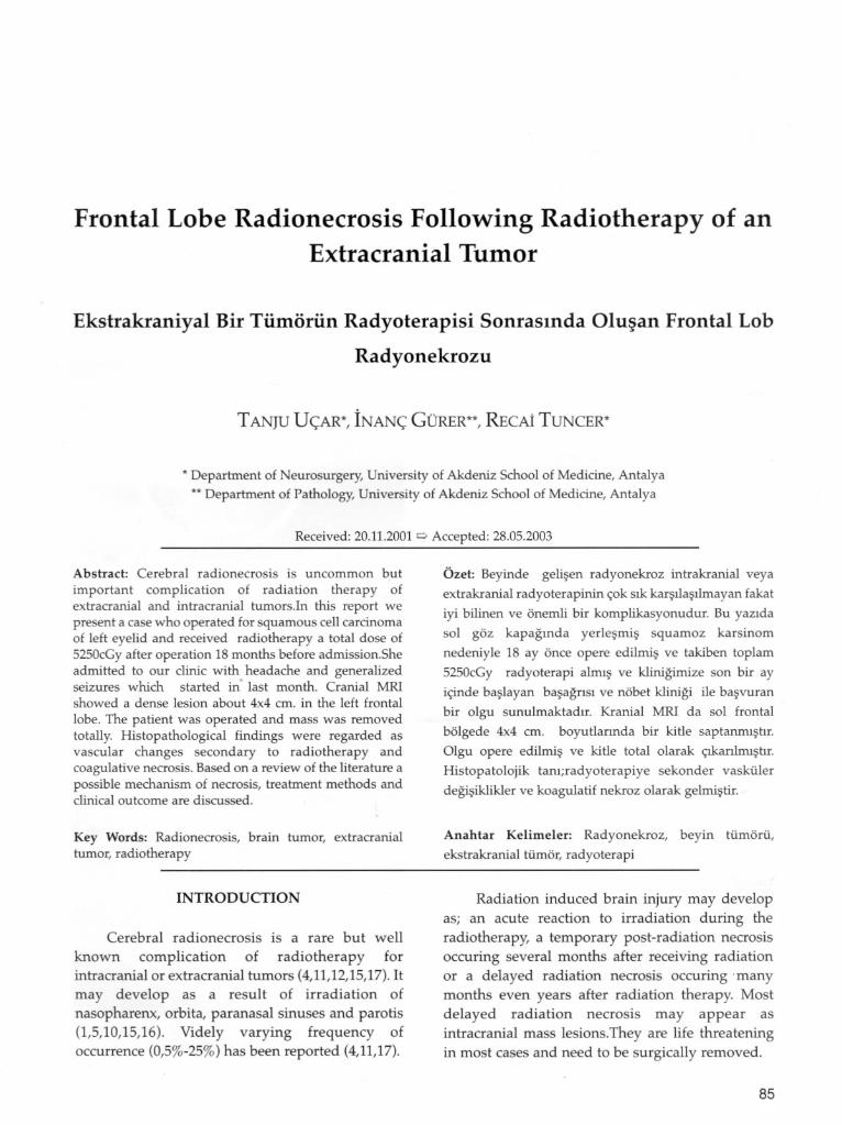
Cranial magnetic resonance imaging (MRI)demonstrated a dense lesion of about 4 cm. m
length in the frontal white matter. MRI revealed adiffuse swelling appearance characterized by T2flair hyperintensity at a lesion site.The massshowed heterogenous peripheral opaquaness aftercontrast injection (Figure 1).
Figure 1: Cranjal MRI demonstrated a dense lesioncompressed the left ventricle
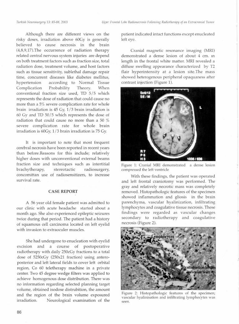
With these findings, the patient was operatedand left frontal craniotomy was performed. Thegray and relatively necrotic mass was completelyremoved. Histopathologic features of the specimenshowed inflammation and gliosis in the brainparenchyma, vascular hyalinization, infiltratinglymphocytes and coagulative tissue necrosis. Thesefindings were regarded as vascular changessecondary to radiotherapy and coagulativenecrosis (Figure 2).
Figure 2: Histopathologic features of the specimen;vascular hyalirusation and infiltrating lymphocytes wasseen.
Turkish Neurosurgery 13: 85-88, 2003 U~ar: Froll/al Lobe Radiollecrosis Followillg Radio/herapy of an Exlracrallial TUllioI'
Postoperative period was complication freeand the patient discharged from hospital in oneweek. She expressed no further complaints duringher last examinations 3 months after operation.
DISCUSSION
Radiation induced necrosis in brain
parenchyma can be caused by either vasculardamage or a direct damage to parenchymal cellsalthough vascular damage is accused to be thecausative agent in most cases (11,12).Thehistopathological evaluation of our case confirmedthe hypothesis of vascular damage as vascularhyalinisation, coagulative necrosis and infiltrativelymphocytes were the dominant changes.
Total radiotherapy dose, fraction dose,volume and radiotherapy techniques, are theimportant factors for development of radiationinduced complications in brain. Complicationprobability can be estimate using fraction size, totalradiation dose, treatment volume, sublethaldamage repair time. Increased fraction size, totaldose and treatment volume increased the
complication rate. Tolerance doses are definedconsidering percent irradiated whole organvolume. Treatment dose and volume description isadministered using the estimated probability rate.Among these, fraction size is considered as themost critical factor followed by others. In thepresent case, fraction dose was 2.5 Gy which is notin conventional fraction dose (1.8- 2 Gy) range andrather higher. To find out equivalent conventionalfractionation schedule of used unconventional
fractionation schedule of this case, LinearQuadratic Formula was used.
Formula: BED: n.d(l+d: a/b)
n: Fraction number, d: Fraction dose, BED :
Biologic equivalent dose
a/b: Suggested values were 1.5, 2 ,3 Gytherefore all three values were used in calculations.
Calculated fraction numbers (n) are 30 in all
three namely 1.5,2,3 Gy a/b values. This meansequivalent conventional fractionation total dose is
approximately 60 Gy with conventional fractionsize 1.8 -2 Gy. Occurrence of severe complicationprobability in this dose level is negligible.Therefore we assume that large fraction size couldresponsible of the radiation damage in the presentcase. The use of larger dose than conventionalfraction size must be approached cautiously.Furthermore the rate of irradiated volume to wholebrain should be considered.
Oppenhaimer and colleagues (9) havereported cases of delayed radiation necrosisfollowing a wide range of 20-69 Gy therapeuticdoses. Rubin and colleagues (12) report anapproximate 5% occurrence of radionecrosis abovetotal doses of 5000cGy. In a report includingfindings from 68 patients, Glass and collabarotors(4) suggested the risky dose as above 4500cGy. Insummary; total doses below 5000cGy are generallyconsidered less risky in terms of the formation ofradionecrosis. However daily doses and thefractions are considered as critical in reducing therisk for necrosis, as the total dose applied (2,7,17).Adaily dose of 200cGy is considered safe . In thisregard , the daily dose of 250cGy given to ourpatient during radiotherapy was most likely amajor factor in the formation of cerebral necrosis.
Although the radiation induced necrosis is awell documented complication, serious challengesexist in diagnosis using radiological techniques orMRI. It is often diffucult to differentiate a necrosis
from a tumor by computerized tomoghraphy (CT)or MRI(3). Positron emission tomography (PET) issuggested as one of the most powerful techniquesin diagnosis (11,14). In addition 18f-PET anddynamic CT and magnetic resonance spectroscopyare also valuable tools in diagnosis ofradionecrosis(6,13,14). During the diagnosticprocess of our case, the histopathology observed inthe left frontal lobe was thought as a tumor, at first,since a tumor in the left eyelid had already beenremoved. However, it was identified as
radionecrosis based on postoperative examinationof the tissue (which would otherwise be diffucult
using CT or MRI).
There are no different opmlOns about thetreatment of radionecrosis. If there is notable
87
Turkish Neurosurgery 13: 85-88, 2003 U~ar: Frolltal Lobe Radiollecrosis Followillg Radiotherapy of all Extracranial Tumor
increase in intracranial pressure, surgical removeof the necrotic tissue seems to be the most
appropriate treatment.
As a conclusion, it is demonstrated in our
case that presence of an intracranial mass in
patients treated for extra / intracranial pathologies
using radiotherapy could be an indication of
radionecrosis. Thus the possibility of radionecrosis,
in addition to the possibilities of reccurence or
metastasis need to be seriously considered in
diagnosis.
Acknowledgement:The authors wish to thank
Melahat Garipagaoglu Assoc. Prof., M.D. for herconsultation
Corresponding: Tanju U;ar, MD.University of Akdeniz School ofMedicine NeurosurgeryDepartment 07070 Antalya-TurkeyPhone: 0242 2274343/66285E-mail: [email protected]
REFERENCES:
1) Coghlan KM, Magenis P. Cerebral radionecrosisfollowing treatment of parotid tumors: a case reportand review of the literature. Int J Oral MaxillofacSurg.; 28(1) :50-52, 1999
2) Curran WJ, Scott CB, Nelson JS, Weinstein AS,Philips TL, Murray K, Fiscbach AI, Yakar D, SchwadeJG, Pow lis WD, Nelson DE "A RandorrUzed trial ofaccelerated hyperfractionated radiation therapy andbis-chlorethyl nitrosurea for malignant glioma.Cancer; 70:2909-2917, 1992
3) Dooms GC, Hecht S, Brant-Zawadski M, BarthiaumeY, Norman D, Newton TH. Brain radiation lesions.Radiology :158 :1149-155, 1986
4) Glass JP, Hwang TL, Leavens ME, Libshitz HI.Cerebral radiation necrosis following treatment ofextracranial malignancies. Cancer; 54:1966-1972,1984
5) Jiang GL, Morrison WH, Garden AS, Gear F,Callender D, Goepfret H, Ang KK. Ethmoid sinuscarcinomas: natural history and treatmentresults.Radiotherapy and Oncology; 49:21-27,1998
88
6) Kamada K, Hovkin K, Abe H, Sawamura Y,Kashibawa T. Differentiation of cerebral radiation
necrosis from tumor reccurence by proton magneticresonance spectroscopy. Neurol Med Chir(Tokyo);37:250-256, 1997
7) Laperriere NI, Wong CS, Milosevic MF, Whitton AC,Wells WA, Patterson B. Accelerated radiationtherapy for primary lymhoma of the brain.Radiotherapy and Oncology:47:21-27,1998
8) Mornex F, Nayel H, Taillander L. Radiation therapyfor malignant astrocytomas in adults. Radiotherapyand Oncology; 27:181-191, 1993
9) Oppenheimer JH, Levy ML, Sinha U, EI-Kadi H,Apuzzu MLI, Luxton G, Petrovich Z, Zee CS, MillerAC. Radionecrosis secondary to intersitialbrachytherapy: Correlation of magnetic resonanceimaging and histopathology. Neurosurgery; 31:336343, 1992
10) Ostwald PM, Cooper SG, Denham Jv, Hamilton CS.Dosimetry of high energy electron therapy to theparotid region. Radiotherapy and Oncology;33:148156,1994
11) Rizzoli HV, Pagnanelli DM. Treatment of delayedradiation necrosis of the brain.A clinical observation.
J Neurosurg: 60(3):589-594, 198412) Rubin P, Gash DM, Hansen JT, Nelson DF, Wilson JP.
Disruption of the blood brain barrier as the primaryeffect of CNS irradiation. Radiotherapy andOncology 31:51-60, 1994
13) Stokkel M, Stevens H, taphoorn M, Van Rijk P.Differentiation between reccurent brain tumor and
postradiation necrosis ;the value of 201TI SPBTversus 18F-FDG PET using a dual headedcoincidence camera. A pilot study. Nucl MedCommun; 20:5:411-417,1999
14) Valk PE, Budinger TF, Silver P, Levin VA, Gutin PH,Doyle WK. PET of malignant cerebral tumors afterinterstitial brachtherapy:demonstration of clinicalactivity and correlation of clinical outcome. JNeurosurg; 69:830-838, 1988
15) Vries BD, Taphoorn M, Van isset JW, Terhaard CHJ,Jansen GH, Elsenburg PHJM. Bilateral temporal lobenecrosis after rdaiotherapy:confounding spectresults. Neurology; 51:1183-1184,1998
16) Wong ET. Reccurent cystic radiation necrosis of thebrain. Oncol Rep;3:685-687, 1998
17) Woo E, Lam K, Yu YL, Huang CY. Cerebralradionecrosis ;is surgery necessary? Journal ofNeurology Neurosurgery Psychiatry; 50:1407-1414,1987