Frontal Lobe Function and Dysfunction - Adele … · Frontal Lobe Function and Dysfunction Edited...
41
Frontal Lobe Function and Dysfunction Edited by HARVEY S. LEVIN HOWARD M. EISENBERG ARTHUR L. BENTON New York Oxford OXFORD UNIVERSITY PRESS 199 1
Transcript of Frontal Lobe Function and Dysfunction - Adele … · Frontal Lobe Function and Dysfunction Edited...

Frontal Lobe Function and Dysfunction
Edited by
HARVEY S. LEVIN HOWARD M. EISENBERG ARTHUR L. BENTON
New York Oxford OXFORD UNIVERSITY PRESS 1991

18 Guidelines for the Study of Brain-Behavior Relationships During Development
ADELE DIAMOND
One way to study the relationship of brain maturation to the elaboration of cognitive abilities during development is to use a two-pronged approach: (I) Study the developmental progression of children's performance on behavioral tasks, and (2) link successful performance on those tasks uniquely to specific neural systems. I would like to suggest a set of guidelines for the conduct and evaluation of such research. I will use the work of myself and others on the development of cognitive abilities linked to dorsolateral prefrontal cortex to illustrate these guiding principles.
A Brief Overview of the Anatomical Connections and Functions of Dorsolateral Prefrontal Cortex
Dorsolateral prefrontal cortex is located between the frontal pole and the arcuate sulcus in the monkey brain (see Fig. 18-4). It is centered around the principal sulcus (Walker's area 46) and is immediately anterior to the supplementary motor area (SMA) and premotor cortex. SMA and premotor cortex, like dorsolateral prefrontal cortex, are among the subregions of frontal cortex. Dorsolateral prefrontal cortex is defined anatomically, in part, by its reciprocal connections with the parvocellular portion of the mediodorsal nucleus of the thalamus (Rose and Woolsey, 1948; Johnson et at., 1968; Leonard, 1969). It also has strong reciprocal connections with parietal cortex, and the dorsolateral prefrontal and parietal cortices appear to be coupled in their projections throughout the brain (Pandya and Kuypers, 1969; Goldman and Nauta, 1977; GoldmanRakic and Schwartz, 1982; Schwartz and Goldman-Rakic, 1984; Selemon and Goldman-Rakic, 1985a,b; 1988; Johnson et al., 1989). The same may be true of the dorsolateral prefrontal and premotor cortices to a lesser extent (Pandya and Vignolo, 1969; 1971; Haaxma and Kuypers, 1975; Pandya and Kuypers, 1969; Goldman and Nauta, 1977; Kunzle, 1978). One of the major output structures

340 PSYCHIATRIC AND DEVELOPMENTAL EFFECTS OF FRONTAL LOBE DAMAGE
of dorsolateral prefrontal cortex is the caudate nucleus (Nauta, 1964; Johnson et al., 1968; Kemp and Powell, 1970; Goldman and Nauta, 1977). Other output sites include the superior colliculus (Goldman and Nauta, 1976; Kunzle, 1978) and the cingulate gyrus (Johnson et al., 1968; Pandya and Vignola, 1969; Goldman and Nauta, 1977; Kunzle, 1978).
Frontal cortex is the largest area of cortex in the human brain; it has increased the most in size (and in the proportion of brain mass devoted to it) over the course of evolution; and it has an unusually protracted period of maturation (probably only reaching full maturity during puberty). There is general agreement that the most anterior regions of frontal cortex (i.e., prefrontal cortex) subserve our highest cognitive abilities. Dorsolateral prefrontal cortex has been most closely associated with functions of memory and inhibitory control (see discussion of the critical abilities thought to be dependent on dorsolateral prefrontal cortex toward the close of this paper). The role of dorsolateral prefrontal cortex in helping us relate information separated in time or space, and in helping us gain control over our actions so we can choose what we want to do and not simply react, makes this area of the brain of great importance for complex cognitive operations.
The classic test for dorsolateral prefrontal cortex function in nonhuman primates is the delayed response task (Jacobsen, 1935; 1936; for reviews: Nauta, 1971; Warren and Akert, 1964; Rosvold, 1972; Markowitsch and Pritzel, 1977; Diamond, 1991a). This hiding task requires both memory and the ability to inhibit merely repeating the last rewarded response. The classic test for dorsolateral prefrontal cortex function in human adults is the Wisconsin Card Sorting Test (Milner, 1963; 1964). Here, as in delayed response, the subject must flexibly switch to a new response after having been rewarded for a particular response. The subject must remember which sorting criteria were most recently tried and found incorrect, and which sorting criterion is now correct.
The guiding principles I would like to suggest for research on brain-behavior relations in development are as follows:
1. Convergent Validity. Use more than one task linked to a given neural circuit and on which performance improves during a given period of development
It is important to look for converging evidence from diverse tests all linked to the same neural system. An impairment, or an improvement, on one test might be due to diverse causes; converging evidence from diverse tests is more convincing. These converging results are more powerful the more dissimilar the tasks.
Thus, in our work on the developmental progression during infancy of abilities dependent on dorsolateral prefrontal cortex, for example, we have used two hiding tasks (AB and delayed response), which require subjects to keep track of where the reward has been hidden in the absence of visible cues, and a transparent barrier detour task (object retrieval), where nothing is hidden and the reward is always visible, but a circuitous route to the goal is required (see e.g., Diamond,

BRAIN-BEHAVIOR RELATIONSHIPS DURING DEVELOPMENT 341
1988; 1991a). AiJ and delayed response are almost identical tasks. They were chosen because delayed response has been so firmly and convincingly linked specifically to dorsolateral prefrontal cortex, and because AB has been repeatedly shown to be a clear marker of developmental change during infancy (e.g., Gratch, 1975; Wellman et al., 1987). The object retrieval task was chosen because it is very different from AB and delayed response, and yet work by Moll and Kuypers ( 1977) has linked it, too, to frontal cortex.
In the AB and delayed response tasks, the subject is centered between two identical hiding wells, one to the left and one to the right. The experimenter holds up an object of keen interest to the subject, and as the subject looks on the experimenter places this object in one of the two hiding wells. The experimenter then covers both hiding wells simultaneously with identical covers and a brief delay of 0-10 seconds is imposed during which the subject is prevented from looking at, or moving or straining toward, the correct well. After the delay, the subject is allowed to reach. In these details the AB and delayed response tasks are identical. The tasks differ solely in the rule for deciding where the reward is to be hidden. In AiJ, the reward is hidden in the same well until the subject is correct to a specified criterion (typically, two consecutively correct responses), then the reward is hidden in the other well and the procedure repeated} In delayed response, the hiding location of the reward is varied randomly by a predetermined schedule.
For the object retrieval task, a plexiglass box open on one side is used. A reward is placed in the box; the subject's task is to retrieve the reward. There is no delay nor time limit; a trial ends when the subject retrieves the reward or stops trying. Experimental variables include (I) which side of the box is open (front, top, left, or right), (2) distance of the reward from the opening (ranging from partially outside the box to deep inside the box), and (3) position of the box on the testing surface (near the front edge of the table or far; far to the left, at the midline, or far to the right). The reward is always visible when the box is transparent, but the experimental variables jointly determine whether the reward is seen through a closed side of the box or through the opening. (For greater detail see Diamond, submitted.) Object retrieval requires inhibition of the strong pull to reach straight to the visible reward (rather than detouring around the barrier), and like delayed response, AB, and the Wisconsin Card Sorting Test, it requires the subject to remember which responses were most recently tried and found incorrect (in this case, which sides of the box were tried and found closed) and to flexibly switch to a new response.
The converging evidence one would like from the different tasks is that (I) they are all linked to the same neural circuit, and (2) developmental improvements in performance on the tasks occur during the same age period. It is important to be as precise as possible here. For example, different regions of frontal cortex (even different regions within prefrontal cortex) participate in different neural circuits (e.g., Goldman and Rosvold, 1970; Bachevalier and Mishkin, 1986). It is not sufficient, then, to use tasks linked simply to frontal cortex, nor even all linked to prefrontal cortex; they must be linked to the same functional region.
The work by Moll and Kuypers ( 1977), upon which our choice of the object

.. 342 PSYCHIATRIC AND DEVELOPMENTAL EFFECTS OF FRONTAL LOBE DAMAGE
too .. . •• •• ... .. •• •• •• t O ·
t OO
10 .. "' 10
10
•• • .. ••
HUMAN INFANTS
I . .
PVt·t 11MONtNI MOfrlltH•
ADULT RHESUS MONKEYS
INFANT RHESUS MONKEYS
too
00
Ia
•• 10
sa
• • sa
•• to
, ...... ~ •IIIONTI'fl WGNfMI
ADULT CYNOMOLGUS MONKEYS
Figure 18- 1 Percentage of trials during object retrieval testing where subjects reached to the box opening without ever having looked into the opening on that trial. Human infants of7~-9 months, adult monkeys with lesions of dorsolateral prefrontal cortex, and infant monkeys of l li-2~ months almost never reach to the opening unless they have looked into the opening on that trial. On the other hand, 12-month-old human infants, 4-month-old infant monkeys, unoperated adult monkeys, and adult monkeys with lesions of parietal cortex or the hippocampus often reach to the opening without ever having looked into the opening on that trial. (This figure summarizes work from Diamond 1990b; submitted; Diamond and Goldman-Rakic, 1985; 1986; and Diamond et al., 1989b.)
retrieval task was based, had relied on very large lesions, spanning the supplementary motor, premotor, and dorsolateral prefrontal regions of frontal cortelt. Our work, however, has shown that lesions restricted specifically to dorsolateral prefrontal corteJt disrupt performance on the object retrieval task (Diamond and Goldman-Rakic, 1985) (Fig. 18-1).2 Previous work had linked delayed response to dorsolateral prefrontal cortex; our work confirmed this (Diamond and Goldman-Rakic, 1989). Given the marked similarity between AB and delayed response it seemed likely that success on AB, too, would depend on involvement of dorsolateral prefrontal cortex. Our work confirmed this suspicion; lesions restricted to dorsolateral prefrontal cortex disrupt performance on the AB task (Diamond and Goldman-Rakic, 1989) (Table 18-1 ).
Moreover, our work has shown that performance on all three tasks improves during the same period of development in both human infants and

BRAIN-BEHAVIOR RELATIONSHIPS DURING DEVELOPMENT
Table 18- 1 Percenlage Correcl on lhe AB Task by Delay and by Expe rimenlal Group
Delay (in seconds)
Experimental groups 2 5
Adult rhesus monkeys with lesions of dorsolateral prefrontal conex Fl 45
10
f2 71 63 58 F3 67 67 64 F4 67 63 S9 Mean 63 64 60
Adult rhesus monkeys with lesions of parietal conex PI 97 94 92 P2 100 100 99 P3 98 99 98 Mean 98 98 96
Unopcmted adult rhesus monkeys Ul 99 98 98 U2 96 911 96 U3 99 96 97 Mean 98 97 97
Adult cynomolgus monkeys with lesions of the hippocampal formation HI 98 93 87 H2 100 88 86 H3 95 95 80 Mean 98 92 84
Unopcr.Jted adult cynomolgus monkeys Cl 92 96 95 C2 99 91 91 C3 87 95 85 Mean 92 92 90
5-month-old infant rhesus monkeys with lesions of dorsolateml prefrontal conex at4 months II 81 75 65 12 75 73 71 Mc-o~n 78 74 68
Unopcr.lled 5-month-old infant rhesus monkey 13 97 97 97
343
infant monkeys. In human infants, performance improves on AB, delayed response, and object retrieval between 7~ and 12 months of age (Diamond, 1985; Diamond and Doar, 1989; Diamond, submitted) (Figs. 18-2 and 18-3). In infant monkeys, performance improves on all three of these tasks between 1 ~ and 4 months of age (Diamond and Goldman-Rakic, 1986) (see Fig. 18-1 and Table 18-1 ).
2. Divergent Validity, 1: Study the role of other neural regions in performance of these tasks
To determine that the tasks of interest are linked specifically to one neural circuit it is important to demonstrate that the functioning of other neural regions is not also related to performance of these tasks.3 For example, is improved perfor-

344 PSYCHIATRIC AND DEVELOPMENTAL EFFECTS OF FRONTAL LOBE DAMAGE
DELAY IN SECONDS
AB - - - Delayed Response
a 9 9t tor 12
AGE IN MONTHS
Figure 18-2 Developmental progression in the delay human inrants can tolerate on the AB and delayed response tasks. AB results are usually reported in tenn of the age at which the AB error occurs. In an attempt to use a comparable measure for the delayed response task, results arc plotted here in tenns or the delay at which errors occurred (i.e., the delay at which performance was below the criterion of 88% correct). The AB results are shown by the solid line and are based on the infants studied longitudinally in Cambridge, MA by Diamond ( 1985). The delayed response results are shown by the dashed line and are based on the infants studied longitudinally in St. Louis, MO by Diamond and Doar (1989). (From Diamond and Doar, 1989, with permission.© John Wiley & Sons, Inc., 1989.)
mance simply due to general brain development? Is impaired performance a sequela that follows damage anywhere in the brain?
To address these kinds of questions, Diamond and Goldman~Rakic investigated the role of parietal cortex in performance of the AB, delayed response, and object retrieval tasks. Parietal cortex is involved in the processing of spatial information and the programming of movements in space (see, e.g., LaMotte and Acuna, 1978; Van Essen, 1979; Andersen, 1988). These abilities would appear to be relevant to all three tasks: In AB and delayed response the hiding wells differ only in spatial location, and in object retrieval the subject must reach around a spatial barrier. Removing all of inferior parietal cortex (Brodmann's

PHASES
4
3
2
PEIICEIITAGE OF
trrFAIITS AT
EACH LEVEL
100\
751
SO\
251
1001
751
SO\
251
1001
l st
~01
151
1001
1B ·· ·j·· ·~~9~~,~· ·J ·· ·· ··· · j ········ i ··· · ···· i · · ::: 1
1001
751
501
251
llt£r.S Of AGE 26 27 28 29 JO Jl 32 JJ 34 35 36 J7 38 39 40 41 4 2 43' 44 45 46 47 . 48 49 50 51 52 53
6 MO . 7 MO, 8 110. 9 MO. 1 0 110 . 11 110 . 12 MO.
Figure 18-3 Developmental progression of the performance of human infants on the object retrieval task with transparent barrier, showing histograms for the age distribution of each phase. (From Diamond, submitted.)

346 PSYCHIATRIC AND DEVELOPMENTAL EFFECTS OF FRONTAL LOBE DAMAGE
area 7), however, had no observable effect on performance of AB and delayed response, and while the monkeys with lesions of parietal cortex showed some initial misreaching errors in aiming their hand to clear the box opening on object retrieval, they showed none of the errors on the task characteristic of monkeys with lesions of dorsolateral prefrontal cortex, infant monkeys, or human infants (Diamond and Goldman-Rakic, 1985; 1986; 1989) (see Table 18-1 and Fig. 18- 1 ).
The abilities most generally considered essential for AB and delayed response are memory and inhibition; the latter is also thought essential for object
...

BRAIN-BEHAVIOR RELATIONSHIPS DURING DEVELOPMENT 347
retrieval (see, e.g., Diamond, 1985; 1988; 1991 a). These abilities are frequently associated with the hippocampus (see, e.g., Squire and Zola-Morgan, 1983; Douglas, 1967). Therefore, Diamond and colleagues (1989b) went on to investigate the role of the hippocampus in performance of these tasks. As was found with parietal cortex, however, the presence or absence of the hippocampus appeared to have no effect on performance of AB or delayed response at the brief delays at which monkeys with lesions of dorsolateral prefrontal cortex, infant monkeys, or human infants fail, and no effect on object retrieval performance (see Table 18-1 and Fig. 18-1 ). This was true even though (I) the lesions of the hippocampus included not only the hippocampus proper (Ammon's hom) but also the dentate gyrus, subiculum, 90% of the parahippocampal gyrus (area TFTH of von Bonin and Bailey, 1947), and the posterior half of the entorhinal cortex (Fig. 18-4); and (2) the monkeys with these hippocampal lesions were profoundly impaired on delayed nonmatching to sample, a classic memory test closely linked to hippocampal function.
Delayed alternation bears some resemblance to delayed response, and performance on it, too, is impaired following lesions of dorsolateral prefrontal cortex. It was not chosen for study, however, because of questions of divergent validity-for delayed alternation is sensitive to damage to diverse regions of the brain (e.g., performance is impaired by lesions of the hippocampus or by lesions of dorsolateral prefrontal cortex). On this task, the reward is hidden (out of sight,
Figure 18-4 Sites of the brain lesions. From top to bottom: dorsolateral prefrontal cortex lesion, inferior parietal cortex lesion, and hippocampal formation lesion (including the subiculum and posterior portion of the en to rhinal cortex). The prefrontal and parietal lesions arc shown on lateral views of the brain. The hippocampus is a deep structure that cannot be seen from the brain's surface. In the ventral view of the brain, the shaded area indicates the cortex that was removed in the hippocampal formation lesion. The hippocampus itself is buried deep beneath this region of cortex.
The dorsolateral prefrontal cortex lesions included cortex in both banks of the principal sulcus, the anterior bank of the arcuate sulcus, and all tissue on the dorsolateral surface rostral to the arcuate sulcus. This area corresponds most closely to area 46 of Walker, including Walker's areas 8 and 9 as well. In the terminology of Brodmann's map of the macaque brain, it corresponds to most of Brodmann 's area 9, area 8, and some of area 10.
The parietal cortex lesions included the posterior bank of the intraparietal sulcus, the posterior bank of the superior temporal sulcus for about 10 mm, and all cortex between the two sulci including roughly 4 mm of the Sylvian fissure (i.e .• most ofBrodmann's area 7).
The hippocampal formation lesions included fields CA 1-4 of the hippocampus proper, the dentate gyrus. the subiculum, 90% of the parahippocampal gyrus (area TFTH of von Bonin and Bailey), and the posterior half of the entorhinal cortex. Inadvertent damage to area TE of von Bonin and Bailey also occurred. The caudate nuclei, lateral geniculate nuclei, amygdaloid nuclei, and the temporal stem were undamaged in all animals. There was some shrinkage and gt:osis in the mammillary bodies. and extensive gliosis bilaterally throughout the fornix.
All lesions were bilateral, symmetric, and performed in one stage.

348 PSYCHIATRIC AND DEVELOPMENTAL EFFECTS OF FRONTAL LOBE DAMAGE
during a brief delay of 5 seconds or so) in the well to which the subject did not reach last time. Note that if a subject reaches perseveratively to one well, the reward will continue to be hidden in the other well, and the subject will continue to be wrong on one trial after another. Perseveration is a frequent consequence of damage to diverse areas of the brain. Any subject who tends to perseverate will tend to do poorly on delayed alternation. However, subjects can perseverate because of diverse reasons and the delayed alternation task tends not to discriminate between these different etiologies.
3. Demonstrate that success on the tasks in question depends specifically on a given neural circuit in the infant, as it does in the adult
3A. Damage to a mature system may produce different effects than damage to a developing, or immature, system There are three issues here. One, it is possible that different neural systems may subserve similar functions at different ages. It has been suggested that lower areas of the brain might mediate infants' performance on a task, even though performance of that task by adults is mediated by a later maturing area of the brain. Thus, if a neural region is late maturing, lesions of that region may produce deficits in the adult, but not in the infant (for examples see Divac et al., 1967; Goldman, 1971; 1974).
Although successful performance on AB, delayed response, and object retrieval depends on dorsolateral prefrontal cortex in the adult, this would not necessarily have to be true in the infant. For this reason it is important to investigate directly the effects of lesions in infant monkeys, and not only in mature animals. Diamond and Goldman-Rakic ( 1986) investigated this for the AB and delayed-response tasks, and found the same impairment in infant monkeys operated at 4 months and tested at 5 months as is found in adult monkeys following lesions of dorsolateral prefrontal cortex (see Table 18-1 ). This was true despite extensive preoperative training on the tasks given to the infant monkeys, and despite their excellent performance even at long delays prior to surgery. The effect of lesions of dorsolateral prefrontal cortex on object retrieval performance in infant monkeys has yet to be investigated, but it is important that this be tested as well.
Table 18-2 summarizes this body of work looking at the performance of human infants, infant monkeys, and monkeys with selective brain lesions on three behavioral tasks (AB, delayed response, and object retrieval):
38. The effects of destruction, or the characteristics of breakdown, are not necessarily a mirror-image of the characteristics of maturation The second issue is that deficits following destruction or breakdown of a system are by no means an infallible guide to what happens during maturation of that system. Lesion studies, even in the infant, are not sufficient. First, impairments that result from the improper functioning, or lack of functioning, of a part do not necessarily tell you the functions of that part. In addition, of course, deficits may not even be due to damage to that part but might be due to damage to fibers

BRAIN-BEHAVIOR RELATIONSHIPS DURING DEVELOPMENT 349
Table 18- 2 Studies of the Developmental Progression of Human Infants, the Developmental Progression of Infant Monkeys, and the Effects of lesions in Infant and Adult Monkeys on the Same Three Behavioral Tasks.
AB Delayed response Object retrieval
Human infants Diamond. 1985 Diamond & Doar. Diamond, submitted show a clear 1989 developmental progression from 7''• to 12 months.
Adult monkeys with lesions or frontal corte;( fail.
Adult monkeys with lesions of parietal corte;( succeed.
Adult monkeys with lesions of the hippocampus succeed.
Infant monkeys show a clear development:ll progression from I 'I to 4 months.
Diamond & Goldman· Diamond & Goldman-Rakic, 1989 Rakic, 1989
Diamond & Goldman· Diamond & Goldman-Rakic, 1989 Rakic, 1989
Diamond, Zola- Squire & Zola-Morgan, & Squire. Morgan. 1983 1989
Diamond & Goldman- Diamond & Goldman-Rakic, 1986 Rakic. 1986
5-month-old infant Diamond & Goldman- Diamond & Goldman-monkeys, who Rakic, 1986 Rakic, 1986 received lesions of frontal corte" at4 mo. fail.
Diamond & Goldman-Rakic. 1985
Diamond & Goldman-Rakic. 1985
Diamond, Zola· Morgan, & Squire. 1989
Diamond & Goldman-Rakic, 1986
of passage or to overlying cortex (in the case of a deep structure such as the hippocampus). Second, the way a system deteriorates from damage or old age is by no means necessarily the reverse of how that system is built up during development, although there are often parallels. For example, the last abilities to develop are often the first to deteriorate, but the order of development is not always exactly the opposite of the order of deterioration. Research must be done on functioning in the intact brain of infants (both monkey and human), and linking maturational changes in the neural circuit in question to improved performance on the behavioral measures.
For this reason, the kind of work being done in the laboratories of investigators such as Fox and Chugani is of great importance. In a longitudinal study of AB and object retrieval performance, Fox and Bell (in press) found increased frontal electroencephalogram (EEG) activity in individual infants at the time that each infant was improving on the tasks. The relation between increased frontal cortex activity and improved performance was significant for each task. Using 2-deoxy-2[18F)fluoro-D-glucose and positron emission tomography (PET), Chugani and associates ( 1987) have been able to measure metabolic rates for glucose uptake in localized regions of the brain in healthy, awake infants at rest, as young as 5 days of age. The more active a neural region, the more glucose it will need to use. Chugani and coworkers ( 1987) found that beginning around

350 PSYCHIATRIC AND DEVELOPMENTAL EFFECTS OF FRONTAL LOBE DAMAGE
8 months, glucose utilization increases specifically in frontal cortex (i.e., activity in frontal cortex appears to increase just before and during the period when infants are improving on the AB, delayed respon_se, and object retrieval tasks).
Much work remains to be done, however, especially on the role that specific maturational changes in dorsolateral prefrontal cortex play in the age-related changes in performance of AB, delayed response, and object retrieval. What exactly is changing in the prefrontal neural system over these months that permits the system to subserve functions it had earlier been incapable of supporting?
One likely maturational change is increasing levels of dopamine in the prefrontal neural system. Dopamine is a particularly important neurotransmitter in prefrontal cortex, where its concentrations are higher than in any other cortical region (Brown et al., 1979). There are two main dopaminergic systems in the brain. One originates in the ventral tegmental area and projects heavily to prefrontal cortex (Divac et al., 1978; Parrino and Goldman-Rakic, 1982). The other originates in the substantia nigra and projects heavily to the caudate nucleus (Anden et al., 1964). The caudate is a major output structure of dorsolateral prefrontal cortex (Selemon and Goldman-Rakic, 1985a). Thus, both cortical and subcortical components of the prefrontal system receive strong dopaminergic input.
High levels of dopamine in dorsolateral prefrontal cortex are essential for proper functioning: (I) If dorsolateral prefrontal cortex is selectively depleted of dopamine by local injection of 6-0HDA, monkeys show impairments on delayed response as large as those found after dorsolateral prefrontal removal (Brozoski et al., 1979). (2) Monkeys treated with MPTP (1-methyl-4-phenyl-1 ,2,3,6-tetra-hydropyridine) show deficits on object retrieval similar to those seen following dorsolateral prefrontal cortex lesions and similar to those seen in young infants (Taylor et al., 1990; Saint-Cyr et al., 1988). MPTP injection results in reduced levels of dopamine in the substantia nigra and in the frontal-striatal system (Elsworth et al., 1987; Mitchel et al., 1986) and is thought to produce behavioral deficits similar to those seen in patients with Parkinson's disease (Bums et al., 1983; Stem and Langston, 1985). Here, depletion of dopamine in the neural circuit appears to produce the same deficits on object retrieval as do lesions to the circuit.
The level of dopamine in the brain increases markedly during the period when performance on AB, delayed response, and object retrieval is improving in infant monkeys (Diamond and Goldman-Rakic, 1986; Goldman-Rakic and Brown, 1982).4 Given the importance of dopamine for dorsolateral prefrontal cortex function and given the increases in brain dopamine levels during the period when tasks dependent on dorsolateral prefrontal cortex are first mastered, it is reasonable that increases in dopamine levels over age may play a causal role in the developmental improvements on these tasks. This remains to be directly demonstrated, however.
3C. Although work with animals or adult patients is important, evidence must, at some point, be obtained directly in children Work with nonhuman primates is useful because the most precise information on brain function comes from invasive procedures that cannot be used with humans. Adult patients enable the investigator to study the effects of brain dam-

BRAIN-BEHAVIOR RELATIONSHIPS DURING DEVELOPMENT 351
age that one would never want, nor would one be allowed, to impose solely for the purposes of research. However, work with animals or adults needs to be complemented by work with children. There is an inescapable inferential leap when drawing conclusions about development in children from studies of animals or adults. At some point, children themselves must be studied.
One way to do this is to study the performance of children with well-localized, verifiable brain damage, much as adult patients are studied. Another valuable approach is to use noninvasive measures of brain function, such as EEG, event related potential (ERP), and magnetoencephalography, to study functioning in intact, healthy children. Yet another approach is to study children with lower levels of dopamine but who appear to have no structural damage to the brain. Children with early-treated phenylketonuria (PKU) may fall in this latter category, as they are thought to have a functional deficit in frontal cortex function due to dopamine depletion with no structural damage. We are now investigating whether these children are impaired specifically on tasks linked to dorsolateral prefrontal cortex function in the face of otherwise preserved cognitive functioning, and whether there is a relationship between their performance and their levels of dopamine.
lfPKU is untreated, it results in severe mental retardation (Primrose, 1983; Tourian and Sidbury, 1978). Dietary regulation, begun early and consistently maintained, results in IQs within the normal range and no gross cognitive impairments (Dobson et al., 1977; Holtzman et at., 1986; Hudson et at., 1970; Koch et al., 1984; Williamson et al., 1977). There are reports, however, that even when a special diet is followed, children with PKU may have residual frontal lobe signs (Cowie, 1971; Pennington et at., 1985; Welsh et at., 1990). It would make sense that these children might have such a selective imP.airment given the tendency of PKU patients, even when they have been maintained on diet from shortly after birth, to have lower dopamine levels (Krause et al., 1985), and given the importance of dopamine for frontal cortex function.
At least for the present, however, studies of children need to be complemented by more precise, invasive procedures with animals. Too often, studies of brain-behavior relationships in children are able to be correlational only. For example, the research with children with early-treated PKU needs to be complemented by work with infant monkeys where the relation between maturational changes in dopamine levels within dorsolateral prefrontal cortex and the emergence of cognitive functions subserved by dorsolateral prefrontal cortex can be studied directly, and where the nature and underlying mechanisms of the causal relation can be explored. Thus, while studies of animals must be complemented by studies of brain-behavior relations in children, so too must studies of children be complemented by studies of brain-behavior relations in animals.
4. Use the same tasks when studying the developmental progression in children and when studying the neural basis in other populations, rather than tasks that are simply similar.
Tasks that appear to be similar, or appear to require similar abilities, often tum out to depend on quite different neural systems. Small changes in a task, which one might have thought would be inconsequential, often tum out to be critical.

352 PSYCHIATRIC AND DEVELOPMENTAL EFFECTS OF FRONTAL LOBE DAMAGE
For example, dorsolateral prefrontal cortex is not required if the rule is to reach to a different object than the one you just saw, but it apparently is required if the rule is to reach to the same object you just saw (Mishkin et al., 1962). As the delay increases on AB and delayed response, these tasks change from being sensitive selectively to dorsolateral prefrontal cortex function at delays of 2-5 seconds to being sensitive to hippocampal function as well at delays of 15 seconds or more (Diamond et al., 1989b). Patients with frontal lobe damage are selectively impaired in producing as many words as they can think of beginning with a given letter (F, A, or S; e.g., Benton, 1968), but they are not selectively impaired in producing as many words as they can think of belonging to a given category (e.g., animals or clothing). The neuropsychological literature is replete with such fascinating dissociations. Similarly, any ability such as memory, attention, or inhibition can be shown to be present in very young children on one test but absent in much older children on a different test, even though both tests are considered measures of"memory" or "inhibition." The exact manner in which something is assessed is terribly important.
For this reason, in our work with human infants, infant monkeys, and adult monkeys, we have used the same tasks, not simply analogous ones. AB, delayed response, and object retrieval were administered to all subjects. Accommodations were made for species differences: e.g., toys served as the reward for human infants; peanuts and raisins served as the reward for monkeys. Aside from these small adjustments, however, children and monkeys were tested in the same way on all three tasks.
This guideline raises a problem, however, in studying brain-behavior relations in development during the preschool and early elementary school years. After 1-2 years of age, children need more sophisticated tasks than can be used with monkeys, but not until 8-12 years of age can they succeed on tests of frontal cortex function used with adult patients. With children 3-8 years of age, we have used modifications of the tasks used with adults (e.g., the Wisconsin Card Sorting Test [Diamond and Boyer, 1989] and the Stroop Test [Diamond et al., in prep.]). It is possible, however, that we have modified the tests in ways that we did not realize were crucial, and this is of great concern to us. Evidence directly linking the versions of the tests we have used to frontal cortex function is critically needed.
Two caveats are in order concerning guideline #4. First, sometimes a task must be modified in order for it to measure the same ability in a different population. An obvious example would be that a test of memory given in English to English-speaking children should be administered in French to French-speaking children if one is interested in testing memory. Similar modifications are sometimes needed to accommodate age or species differences in subjects. Some of the early work in child development foundered because experimenters tried to use the same tasks with children that had been used with animals without modifying the tasks to make them appropriate for children (for some examples, see Donaldson, 1978). Sometimes a task must be modified in order to keep it equivalent for two different populations.
The second caveat is that sometimes using the same task, even when it appears to be appropriate for each population, is not a guarantee that one is

BRAIN-BEHAVIOR RELATIONSHIPS DURING DEVELOPMENT
Table 18- 3 Performance on Delayed Nonmatching to Sample by Age and Delay
5-scc delay 30-scc delay
Mean number of trials Percent passing Percent Percent Ages to criterion• criterion correct correct
12 monthsb 17 50 66 65 (N = 6)' 15 months 16 67 67 65 (N = 8) 18 months 16 67 71 81 (N = 8) 21 months 11 92 80 80 (N = II) 24 months 9 92 87 86(N = II) 27 months 7 100 85 90 30 months 8 100 84 86 3 years 6 100 93 96 4 years 5 100 98 99 5 years 5 100 94 95
"Criterion • S correct responses in 3 row. For subjects who never reached criterion (6 subjects 3t 12 months. 4 subjects c:ach at IS 01nd I 8 months. and I subject each at 21 and 24 months) the total number of trials they were tested (25) is entered here. bN • I 2 for all ages. 'Only subjects who p:ISSCd criterion at 5 sc:c were tested at 30 sc:c.
353
tapping the same ability in two different populations. The delayed non matching to sample task5 is a test of memory sensitive to hippocampal damage in adult monkeys (e.g., Zola-Morgan and Squire, 1986) and sensitive to amnesia in human adults (Squire et al., 1988). It is also a task appropriate for testing young children, and on which they show a developmental progression between 12 and 30 months of age (Diamond, 1990a; Ovennan, 1990).6 However, while this task is a measure of memory ability in adult monkeys and human adults, sensitively reflecting damage to neural structures important for memory, the developmental progression of improved performance on delayed nonmatching to sample in infants does not chart the maturation of this memory function or of the hippocampal neural circuit, even though the same task is used with infants, adults, and monkeys.
Until about 21 months of age, children fail this task even at delays of only 5 seconds (Table 18-3), although it is well established that infants can remember for far longer than 5 seconds well before 21 months (there is evidence of such memory by at least 2-3 months of age: e.g., Werner and Perlmutter, 1979; Rovee-Collier, 1984). Indeed, emerging anatomical and biochemical evidence indicates that the hippocampus develops quite early in humans and monkeys (e.g., Rakic and Nowakowski, 1981; Eckenhoff and Rakic, 1988; Bachevalier et al., 1986; Kretschmann et al., 1986; see Diamond, 1990a). Moreover, in the same session in which children first succeed on delayed nonmatching to sample with a 5-second delay they typically succeed with 30-second delays as well (see Table 18-3). One might expect success at a delay of 30 seconds to come in later if the task were assessing the development of memory. In adult monkeys or humans, in whom other abilities are fully mature, disturbance of the memory

354 PSYCHIATRIC AND DEVELOPMENTAL EFFECTS Of FRONTAL LOBE DAMAGE
system produces errors on the task-for memory is, indeed, one of the abilities required for success, but it is only one of the required abilities. The slow developmental progression in performance of the delayed nonmatching to sample task is due to the late maturation of some other ability. We tested whether this late-developing ability might be the ability to deduce an abstract rule, the ability to quickly encode visual stimuli (speed of processing), or the ability to tolerate retroactive interference (Diamond, 1990a; Towle and Diamond, 1991 ). Telling the children the rule or giving them a long time to encode the stimulus only marginally improved performance. However, reducing retroactive interference (by not introducing a reward after subjects displaced the sample when the sample was presented alone during familiarization) had a dramatic effect: It enabled children to succeed almost 12 months earlier (Towle and Diamond, 199 I).
Here is a case where a task has been linked specifically to a discrete neural circuit, and where the same task has been used with children and the developmental progression documented, yet it is incorrect to conclude that the developmental progression in children's performance of the task reflects maturation of that neural circuit. Thus, while it is true that success on delayed nonmatching to sample requires hippocampal involvement, and it is true that success on delayed nonmatching to sample appears relatively late in development, it is not true that the late emergence of success on this task is due to late maturation of the hippocampus, or of the memory ability it subserves. This illustrates why caution must be used in drawing conclusions about brain-behavior relations in development. In this case important clues came from two sources: (I) Infant monkeys also show a protracted developmental progression in performance of the task despite evidence of early hippocampal maturation in the monkey (Bachevalier and Mishkin, 1984; Brickson and Bachevalier, 1984; Bachevalier, 1990 and (2) the qualitative aspects of the performance of infant monkeys and human infants on the task are different from those of adult monkeys and human patients (e.g., adult monkeys and humans succeed at the shortest delays and perform progressively more poorly as the delay increases; human infants and infant monkeys perform at chance even at the very shortest delay; their performance does not worsen as delay increases). This leads directly to guideline #5.
S. Compare the qualitative, as well as quantitative, aspects of children's performance on the tasks to that of brain-damaged populations
Because someone may fail a task for a variety of reasons, it is important to determine the reasons for failure. The qualitative aspects of performance should be investigated, and not simply rate of success or failure. If one is comparing the performance of children with the performance of animals with selective lesions, then the younger children should fail under the same conditions and in the same ways as do the animals with lesions to the relevant neural system. Changes in task parameters should affect both the young children and the lesioned animals in the same ways. It becomes more likely that the performance of the children and lesioned animals reflects the presence or absence of the same abilities and depends upon the same neural system, the more parameters on which their per-

BRAIN·BEHAVIOR RELATIONSHIPS DURING DEVELOPMENT 355
formance matches and the more identical the circumstances under which these behaviors are elicited.
For example, in comparing human infants and monkeys with lesions of dor· solateral prefrontal cortex we have looked not only at their overall success rates but also at the conditions under which they err and what their errors look like. Figures 18-5 and 18-6 illustrate this. On the AB and delayed response tasks, human infants of 7~-9 months fail at delays of only 2-5 seconds. The only group of adult monkeys to fail at such brief delays are those with lesions of dor· solateral prefrontal cortex. Monkeys with lesions of parietal cortex or the hip· pocampus perform as well as unoperated controls here.
Moreover, at these delays infants show a characteristic pattern of performance: Their errors are confined to trials on which side of hiding is reversed and to the next few trials at that new location. Their performance is excellent when the reward is hidden in the same well as on the previous trial and they were correct on the previous trial (Repeat-following.correct trials; see Fig. 18-5). The only group of adult monkeys to show this pattern of performance are monkeys with lesions of dorsolateral prefrontal cortex (see Fig. 18-5), and they show this pattern at the same delays as do 7Jf-9-month-old human infants. At long delays monkeys with lesions of the hippocampal formation also fail AB, but they never show this differential pattern of performance by type oftrial. At delays of 15-30 seconds, they have difficulty remembering where the reward has been hidden and their performance declines, but it does not decline selectively for reversal trials. When monkeys with hippocampal lesions reach incorrectly, they show some tendency to repeat that error on the next trial, but where these strings of errors begin is randomly distributed over a testing session (Diamond et at., 1989b).
On the object retrieval task, human infants of 8Jf-9 months lean all the way over to look in the opening when the left or right side of the box is open. When they do this they reach with the hand contralateral to the opening, which though easier from this position than reaching with the ipsilateral hand, looks very contorted and is therefore termed an "awkward reach." The awkward reach is not the result of a hand preference, as it is seen on both sides of the box (Diamond, submitted; Bruner et at., 1969; Gaiter, 1973; Schonen and Bresson, 1984). One group of adult monkeys also shows this strange awkward reach-monkeys with lesions of dorsolateral prefrontal cortex (Fig. 18-6 ).
6. Divergent Validity, 2: Study performance on other tasks linked to other neural circuits
It is also important to establish a second kind of divergent validity by determining that performance on tasks linked to other neural circuits and requiring other abilities (I) does not improve over the same age period as does performance on tasks linked to the neural system of interest, and (2) is not affected by disruption of functioning in the neural system of interest. (The first kind of divergent valid· ity discussed previously was that disruption of functioning in other neural systems should not affect performance on tasks linked to the neural system of inter-

356 PSYCHIATRIC AND DEVELOPMENTAL EFFECTS OF FRONTAL LOBE DAMAGE
100
00
AB PERFORMANCE WITH DELAY OF 2·5 SEC
1 HUMAN INFANTS
7 112·9 MONTHS OLD 12 MONTHS OLD
1 1h·2V• MONTHS OLD
2 INFANT RHESUS MONKEYS
4 MONTHS OLD PREFRONTAL
INFANTS
Figure 18-5 Percentage correct by type of trial at delays of 2-5 sec for human infants, infant monkeys, and monkeys with lesions to prefrontal cortex, parietal cortex, and the hippocampal formation. Row I: Human infants of 12 months perform perfectly. Human infants of 7h-9 months perform well on repeat-following-correct trials, but perform significantly worse on reversal trials and on repeat-following-error trials in both the AB task (Diamond, 1985) and the delayed-response task (Diamond and Doar, 1989). Row 2: Infant rhesus monkeys of 4 months perform perfectly. Infant monkeys of 1'~2~, months and infant monkeys who have received bilateral lesions of dorsolateral prefrontal cortex at 4 months and were retested at· 5 months show a similar pattern of differential performance over trials as do 7't.e-9-month-old human infants (Diamond and Goldman-Rakic, 1986).
est.) This is important for determining whether improvements, or impairments, are general or specific. For example, are all abilities improving at the same time, at the same rate, or are developmental changes more pronounced in different abilities during different age periods?
For instance, white human infants improve on AB, delayed response, and object retrieval (tasks all linked to dorsolateral prefrontal cortex) between 7~ and

BRAIN-BEHAVIOR RELATIONSHIPS DURING DEVELOPMENT
PREFRONTAL 100
3 ADULT RHESUS MONKEYS
UNOPERATEO
4 ADULT CYNOMOLGUS MONKEYS
z
= .. .. u w .. .. 0 u I
HIPPOCAMPAL
357
PARIETAL
UNOPERATED
Figure 18-5 (continued) Row 3: Unopcrated adult rhesus monkeys and those with bilateral lesions of inferior parietal conex perform perfectly. Adult rhesus monkeys with bilateral lesions of dorsolateral prefrontal cortex, however, show the same pattern of differential performance over trials as do 7!1.!-9-month-old human infants (Diamond and Goldman-Rakic, 1989). Row 4: Unoperated adult cynomolgus monkeys and those with bilateral lesions of the hippocampal formation perform perfectly at delays of2- 5 sec (Diamond et al., 1989b). (At delays of 15-30 sec hippocampal monkeys no longer perform well on the task, but at these delays they still do not show the pattern of differential performance by type of trial seen in 7Y,-9-month-old human infants, l!f-2h-month-old rhesus monkeys, and in infant and adult rhesus monkeys following lesions of dorsolateral prefrontal cortex).
12 months of age, they improve on visual paired comparison (a task linked to the hippocampal-amygdala system) (Brickson and Bachevalier, 1984; Saunders, 1989) between 2 and 9 months of age (Diamond, 1990a; Fagan, 1990). In the visual paired comparison task, subjects look at a stimulus for a fixed familiarization period or until habituated, a delay is imposed, and then memory of the sample is tested by pairing the sample with another stimulus. Preferential looking at the new stimulus is taken as evidence of memory of the sample, since infants have a natural preference for novelty (Fagan, 1970; Diamond, 1990a).

358 PSYCHIATRIC AND DEVELOPMENTAL EFFECTS OF FRONTAL LOBE DAMAGE
.i H ANT
M .) NKEY
t MO NTNS
tN,ANT
0 MOHTHD
IIOHKEY
Figure 18-6 The "awkward reach" in an infant monkey of 2 months, a human infant of 9 months, and an adult monkey with bilateral lesion of dorsolateral prefrontal cortex. Frame I: Subject leans and looks at bait through opening of box. Frame 2: Subject reaches in awkwardly with the far hand. Frame 3: Opening is on the other side of the box. Performance is the same. Subject leans and looks into the opening. Frame 4: Subject reaches in awkwardly with the far hand.
The visual paired comparison task presents some of the same task requirements as do AB and delayed response: All require that memory be updated on each trial (to remember where the reward was just hidden or which stimulus was just presented). All present the to-be-remembered information visually and only once. All impose the delay within a trial, as opposed to between trials or between testing sessions. However, the visual paired comparison task also differs considerably from AB and delayed response. For example, AB and delayed response require a reaching response; visual paired comparison does not. The to-beremembered information is presented much longer in visual paired comparison. In AB and delayed response the same two hiding wells are used throughout, presenting possible problems of proactive interference; in visual paired comparison, on the other hand, new stimuli are used on each trial (rather than using the same two stimuli repeatedly, varying only which is the sample on a given trial). Subjects must remember spatial location information for AB and delayed response, but not for visual paired comparison.
As more tasks are studied, a better understanding emerges of the abilities developing during each age period and depending on the different neural circuits.
REMAINING QUESTIONS
What Happens After 12 Months of Age in the Neural Circuit of lnteresH
The work on AB, delayed response, and object retrieval appears to provide a window into dorsolateral prefrontal cortex development between 7M and 12

BRAIN-BEHAVIOR RELA liONS HIPS DURING DEVELOPMENT 359
months of age. Neither dorsolateral prefrontal cortex, nor the abilities it subserves, are fully mature by 12 months of age, however. Indeed, frontal cortex is not thought to be fully mature until puberty. For example, children do not perform at adult levels on the Wisconsin Card Sorting Test (a criteria! test of frontal cortex function in adults [Milner, 1963]) until about 9-10 years of age (Chelune and Baer, 1986). What changes occur in the dorsolateral prefrontal neural circuit, and when, after 12 months? In what ways do the abilities dependent on frontal cortex change and improve over these years? Is the improvement simply quantitative, or is it qualitative as well? Is the improvement gradual, or are there growth spurts and plateaus? When are critical points in the development offrontal cortex reached?
We have begun to investigate these questions. In a study of72 children (12 each at 3, 4, 5, 6, 7, and 8 years of age) we have found a significant improvement from 3-6 years on seven different measures of abilities associated with frontal cortex, with a leveling off from 6-8 years (Diamond et al., in prep.) (Fig. 18-7). On all tests we found this same pattern; there were no exceptions. This suggests that the age of 6 may be something of a watershed; on a host of tests linked to frontal cortex, children either reach ceiling performance by 6 years of age or reach a plateau where they remain for at least the next 2 years. Five of our tests assessed inhibitory control of action, one assessed the ability to execute a sequence of actions, and one assessed the ability to remember sequential information. The tests of inhibition were: tapping (Luria, 1973), Stroop (Stroop, 1935), three pegs (Wozniack et al., 1987), Simon Says, and simultaneous switch (Luria, 1973). The test of sequential action was Oat-fist-edge (Luria, 1973). The memory task was multiple boxes (Petrides & Milner, 1982; Passingham, 1985; Petrides, 1988).
On the tapping task, a subject must tap twice when the experimenter taps once, and then tap only once when the experimenter taps twice. This requires inhibiting the pull to do what the experimenter does. Frontal patients fail because, while they may start out correctly, they shortly begin to mirror the experimenter's behavior, rather than following the rule. Similarly, children under 6 years tend to match what the experimenter does rather than follow the rule. Only by 6 years could children consistently succeed on the task (Fig. 18-7A).
In the adult version of the Stroop test, the names of colors are printed in the ink of another color (e.g., the word "blue" is printed in red ink). Subjects are instructed to report the color of the ink. This requires inhibiting the customary response when reading, which is to ignore the ink and attend instead to the meaning of the word. Frontal patients fail the test, as they tend to recite the words and not the color of the ink (Perret, 1974). In our version of the Stroop test for children we use a deck of cards. The front of half the cards is black with a yellow moon and several stars; the front of the other cards is white with a bright sun. Subjects are to say "day" when the experimenter turns over a black card and "night" when a white card appears. This is similar to a modified Stroop test used by Passier and associates ( 1985) where plain black and white cards were used. Passier and colleagues found on their version that children were at ceiling by the youngest age they tested (6 years). We found that most children 2::: 6 years

A Tapping Task
c
"'
... .. ~-------//====~
··r----r/ ____ _ /
.. / " ~""""/
Three Pegs Task ... .S e •1----------------1:~ ~5to /
'!! ~ re '---------7"'--------0~ I / ~~ .,~-----~~./'· ____ , ______ _ ~;; ./ c;S " ~,-/·-------------ti~ / ~ c ·• ~-~~------------C>.
E Simultaneous Switch Task
AgaIn Y~nrts
8 Stroop Task ...
Ag11 In Years
D Simon Says Task .. ... :§ 0: ;; _.............. ~ It 1--------------~·~
... / 5 : • f----//----
3 / u ~ 1-----~~------------------1 ./
Aq'J in Years
F Flat, Fist, Edge Task
ll' ~ !
8 • !-.-----~ -
l· ·~ ·------.,
B ~t------·-------------e ~ :.-:
G Six Boxes Task
.. .. ~ l
w ~ ,, .1l !l . :::
~. -·
~
Figure 18-7 Developmental progression in children aged 3- 8 years on eight tests of abilities associated with frontal cortex.

BRAIN-BEHAVIOR RELATIONSHIPS DURING DEVELOPMENT 361
succeeded, but not most children at younger ages. Indeed, there was little change in the number of children succeeding from 3 to 5 or from 6 to 8 years, but there was a dramatic improvement from 5 to 6 years.
On the three pegs test, the subject is to hammer in a row of pegs (red, yellow, and green) in the order: red, green, and then yellow. The subject must inhibit the tendency to hammer the pegs in spatial left-right order, and instead follow the rule. Performance improved steadily from 3-6 years, and remained at ceiling thereafter (Fig. 18-7C).
Simon Says is a common children's game. The trials of interest here are where the experimenter fails to say "Simon Says." On these trials, the child is not supposed to do anything. This requires inhibiting the tendency to carry out the command anyway. On other trials, the child is to touch his or her nose or eyes, for example, as instructed by "Simon." Before 6 years, children are much more likely to act when they should refrain from acting than are children 2: 6 years.
In the simultaneous switch: fist-flat task, the subject starts with one hand in a fist on the table, and the palm of the other hand flat on the table. The subject is then to switch these hand positions simultaneously back and forth six times. This requires inhibiting one hand from doing what the other is doing. Younger subjects, like frontal patients, tend to err by doing the same thing with both hands or by switching the position of one hand and then the other. We found a marked increase between 4 and 6 years in the degree of simultaneity children showed in switching one hand to one position while simultaneously switching the other hand to the other position. At the 2 extremes of the age range there was little change: Performance did not significantly improve between ages 3 and 4, nor between ages 6 and 8 (Fig. 18-7E).
In the flat-fist-edge task, the subject must change the position of one hand in the following sequence: palm flat on the table, closed fist on the table, and palm at right angles to the table (edge). This sequence is repeated 4 times. Here, the children appeared to reach ceiling at 6 years and thereafter their performance declined slightly.
In the Multiple Boxes task, the subject watches as the experimenter hides a sticker in each of six boxes. All boxes are then closed. The subject is permitted to open one box at a time, in any order he or she chooses. A tO-second delay is imposed between reaches. The goal is to open all the boxes without repeating a choice. The number of reaches needed to open all the boxes progressively decreased until age 6 where it plateaued.This task is selective for dorsolateral prefrontal cortex damage in both monkeys (Passingham, 1985; Petrides, 1988) and human adults (Petrides and Milner, 1982). In short, on all seven of the tasks used, a significant improvement was found up to 6 years. There was little further change from 6- 8 years, either because the tests were not sensitive enough to pick up such changes, or because the abilities tapped by these tests remain relatively constant during the 6-8 year age period.
It should be noted, however, that the Stroop test had to be modified for use with children, as those at the younger end of the age range cannot read, and the three pegs test and Simon Says have never been d irectly linked to frontal cortex function in children or adults. Such evidence, directly linking the modified

362 PSYCHIATRIC AND DEVELOPMENTAL EFFECTS OF FRONTAL LOBE DAMAGE
Stroop test, the three pegs test, and Simon Says to frontal cortex function, is needed. Moreover, even though there is evidence linking performance on the tapping test, the adult version of the Stroop test, and the simultaneous switch tests to the integrity of frontal cortex, information on the role oflocalized regions within frontal cortex in supporting performance on these tasks is lacking. Such information is needed.
What Happens Before 7Yz-8 Months of Age in the Neural Circuit of Interest?
Development does not begin at 7li-8 months. Indeed, it is only because of the developmental accomplishments present by 7li-8 months that testing on AB, delayed response, and object retrieval can begin at roughly those ages. For example, hiding tasks are not possible with younger infants because they will not search for a hidden object. What abilities are developing before 7li-8 months, and what are the neural bases for these accomplishments? What is happening in frontal cortex up until this age? I have suggested (Diamond, 1990b; I99Ib) that some of the accomplishments between 5 and 8 months of age may be made possible by maturational changes in the supplementary motor area (an area of frontal cortex immediately posterior, and medial, to dorsolateral prefrontal cortex). The inhibition of the reflexes of the hand (the grasp and avoidance reactions) and the ability to link two action sequences together into a larger means-end sequence (e.g., removing a cloth in order to then retrieve the reward underneath it) may require involvement of the supplementary motor area. More work is needed in testing this hypothesis and in exploring when and where other neural changes are occurring.
What Abilities Are Dependent on the Neural Circuit of Interest and in What Ways Do These Abilities Change Over Age?
For example, if success on AB, delayed response, and object retrieval depends on involvement of dorsolateral prefrontal cortex, as it now appears, what ability(s) are required by these tasks that accounts for this dependence?
Many of the tasks dependent on dorsolateral prefrontal cortex and on which infants show a developmental progression between 7~ and 12 months (e.g., AB and delayed response) appear to require memory. For instance, monkeys with dorsolateral prefrontal cortex lesions and human infants perform well if there is no delay but fail with a delay, all other conditions being equal. What are the characteristics of this memory ability?
Spatial Memory One prominent theory is that the memory ability subserved by dorsolateral prefrontal cortex is memory of spatial information in particular (e.g., GoldmanRakic, 1987). Evidence for this view includes: Monkeys with lesions of dorsolateral prefrontal cortex are less impaired on some nonspatial memory tasks (e.g., delayed object alternation) than they are on comparable spatial memory

BRAIN-BEHAVIOR RELATIONSHIPS DURING DEVELOPMENT 363
tasks (e.g., delayed spatial alternation) (Mishkin et at., 1969). Monkeys with lesions of the principal sulcus (the ''heart" of dorsolateral prefrontal cortex in the monkey) perform well on spatial tasks that do not require memory but fail spatial tasks that require memory (Goldman and Rosvold, 1970). There are cells in dorsolateral prefrontal cortex that increase firing after a cue is presented and maintain that level of activity throughout the delay (i.e., they appear to serve a memory function); moreover, a subset of these cells is direction-selective, i.e., they fire more if the cue was on the right or left (Fuster and Alexander, 1971; Niki, 1974; Funahashi et at., 1989).
In addition, the anatomical connections between dorsolateral prefrontal cortex and inferior parietal cortex (Brodmann's area 7) are particularly strong (e.g., Schwartz and Goldman-Rakic, 1984). Indeed, not only are there heavy reciprocal connections between these two areas, but throughout diverse areas of the brain, wherever dorsolateral prefrontal cortex projects so does inferior parietal cortex, and in each case their projections interdigitate (i.e., columns of cells receiving projections from prefrontal cortex alternate with columns of cells receiving projections from parietal cortex) (e.g., Goldman-Rakic and Schwartz, 1982; Selemon and Goldman-Rakic, 1985; 1988). This is relevant because parietal cortex participates in that portion of the visual system specialized for the perception of motion rather than the perception of form or texture. It is conceivable that through its connections with parietal cortex, dorsolateral prefrontal cortex might specialize in the memory of spatial information rather than memory of object features.
However, lesions of inferior parietal cortex leave AB and delayed response performance undisturbed. Subjects are evidently able to succeed at these tasks without the perceptual information processed in parietal cortex. Problematic for the spatial memory view is that infants and prefrontally operated monkeys generally perform well at the first location (well A), even though spatial memory is required here as elsewhere. Errors generally first appear only when the location of the reward changes. Some of the cells in dorsolateral prefrontal cortex (and within the principal sulcus itselO that increase firing after a cue is presented and maintain that level of activity throughout the delay fire selectively depending on the color of the cue, just as other cells there fire selectively depending on the location of the cue (Quintana et at., 1988; Wilson and Goldman-Rakic, 1989).
Moreover, most tasks diagnostic of frontal cortex damage in human adults do not appear to have a spatial component. For example, in the Wisconsin Card Sorting Test the sorting criterion (color, shape, or number) changes during testing, and the subject must stay attentive to which criterion is currently correct, but spatial position is irrelevant to the task. Similarly, spatial position is irrelevant on the self-ordered pointing task, which requires subjects to keep track of what stimuli they have already pointed to (Petrides and Milner, 1982). Here, the spatial locations of the stimuli are scrambled after each reach. Indeed, when the stimuli are left stationary, so that the task can be solved by spatial memory, patients with frontal cortex damage perform well. A version of the self-ordered task has recently been used with monkeys, where lesions confined only to the principal sulcus produced severe deficits in performance, even though spatial memory is irrelevant to the task (Petrides, 1988).

364 PSYCHIATRIC AND DEVELOPMENTAl EFFECTS OF FRONTAL LOBE DAMAGE
Memory of Temporal Order Another prominent theory of frontal cortex function is that it is specialized for the memory of temporal order information (e.g., Milner et al., 1985). For exampi~ there is a potential for proactive interference from previous trials during AB and delayed response testing as the same two hiding places are used throughout. Once the reward has been hidden at well A on at least one trial and at well B on at least one trial, one might consider the task to be one of temporal order memory: .. Where was the reward hidden most recently?"
Evidence for this viewpoint includes: When adult patients are shown a series of pictures, patients with frontal cortex damage can tell you which of two pictures they saw before, but not which of the two pictures they saw most recently (Corsi, cited in Milner, 1971 ). When asked about well-known events from the last several decades, patients with frontal cortex damage are impaired in recalling the order in which the events occurred, yet are unimpaired in their recognition and recall of the events (Shimamura et al., 1990). Indeed, all tasks sensitive to frontal cortex damage in the monkey require temporal discrimination (e.g., .. 1 have seen the reward hidden at A and at B. Where was it hidden most recently?"), although the spatial component has been emphasized in theoretical discussion of these tasks. Monkeys with frontal cortex damage, human infants, and infant monkeys generally perform welt at the first location (even though spatial memory is required here as elsewhere); errors appear when the location of the reward changes, i.e., when memory of temporal order is first required.
Most tasks diagnostic of frontal cortex damage in humans appear to require resistance to proactive interference (e.g., the Wisconsin Card Sort: .. Which sorting criterion is the correct one now?", and the self-ordered pointing task: ''Which stimuli have I already pointed to?").
If dorsolateral prefrontal cortex is specialized solely for the memory of temporal relationships, however, why then are there cells in dorsolateral prefrontal cortex that code the left-right location of stimuli or responses? Why, too, does damage to dorsolateral prefrontal cortex produce more profound deficits on spatial tasks (e.g., delayed spatial alternation) than on nonspatial tasks (e.g., delayed object alternation)?
Tulving and Schacter (e.g., Schacter, 1987; Tulving, 1989) have suggested that frontal cortex is critical for the memory of both space and time, specifically memory of the spatial or temporal context in which information is acquired.
Relational Memory Spatial and temporal information (e.g., left, right; earlier, later) are inherently relational. Perhaps memory for relational information in general is dependent on dorsolateral prefrontal cortex and develops between 7~ and 12 months of age in human infants. Evidence consistent with this includes: Patients with frontal cortex damage often do well on typical delayed recall tests but fail delayed comparison tests where they must judge whether a color they saw earlier is the same shade as the color they see now, or whether a tone they just heard is the same pitch as the tone they hear now (Prisko, cited in Milner, 1964). Patients with frontal cortex damage are notoriously poor at relating two pieces of information together (e.g., Barbizet, 1970; Heilman and Valenstein, 1972). Grossman ( 1982)

BRAIN-BEHAVIOR RELATIONSHIPS DURING DEVELOPMENT 365
administered eight visual and auditory reversal tasks (i.e., tasks that required that subjects appreciate the relation between original and transformed states) mediated by linguistic and nonlinguistic symbol systems to adults with localized brain damage. He found no domain-specific deficits; rather, patients with frontal cortex damage were impaired across the board on the reversal tasks regardless of modality or content. Similarly, memory for space and/or time might not be a unique ability but might be a subset of relational memory in general.
It would make sense if relational memory were more difficult than single item memory, as it requires remembering a relation between two things. Perhaps memory of information that is inherently relative, i.e., that requires relating one thing to another (e.g., left, right; smaller, bigger; softer, louder) matures later and more slowly than memory of individual items (e.g., red, girl, circle). Perhaps, too, memory of any relative information, spatial or not, requires involvement of dorsolateral prefrontal cortex.
The idea that memory of space, time, or relational information in general might have a unique developmental trajectory, appear later (i.e., between 7lll and 12 months), and be sensitive to different experimental parameters7 than memory for other, nonrelational information (e.g., color or shape) has never been considered by developmental psychologists, although it deserves investigation.8
Memory Plus Inhibition I have argued that dorsolateral prefrontal cortex is required whenever any information at all must be remembered within each trial as long as the task also demands inhibition of a prepotent response (Diamond, 1985; 1988). That is, it may not matter whether one must remember temporal, spatial, relational, color, or object information. The critical factor may be whether the task demands both memory and inhibition of a dominant response.
Evidence for the role of inhibition is extremely strong: All memory tasks linked to dorsolateral prefrontal cortex in the monkey also impose an inhibitory demand. The pal/ern of error on the AB and delayed response tasks (i.e., errors confined to reversal trials and the trials immediately thereafter) shown by infants or by prefrontally operated monkeys cannot be accounted for by forgetting alone, for the delay is equal on all trials but errors are not equally distributed over trials (Diamond, 1985; Diamond and Goldman-Rakic, 1989). Indeed, monkeys with lesions of the hippocampal formation, who have impaired memory, never show this pattern of error on AB (Diamond et al., 1989b). Moreover, when a task requires memory, but not inhibitory control, human infants perform well months before they first succeed on AB or other tasks dependent on dorsolateral prefrontal cortex (e.g., Baillargeon et al., 1985; Fagan, 1970), and monkeys with lesions of dorsolateral prefrontal cortex also perform well (as on the delayed nonmatching to sample task [e.g., Mishkin et al., 1962; Bachevalier and Mishkin, 1986]).
Note, if dorsolateral prefrontal cortex is necessary whenever a task requires both memory and inhibition, then success on delayed matching to sample should depend on dorsolateral prefrontal cortex involvement, and this appears !o be the case. Monkeys with lesions of lateral frontal cortex fail delayed matchmg to sample, although they succeed at delayed nonmatching to sample (Mish-

366 PSYCHIATRIC AND DEVELOPMENTAl EFFECTS Of FRONTAl LOBE DAMAGE
kin et al., 1962). In delayed matching to sample, a sample object is presented, a brief delay is imposed, and then the subject is given the choice of reaching to the object that matches the sample or to a novel object. A reach to the matching object is rewarded. Infants (e.g., Fantz, 1964; Fagan, 1970; Diamond, 1990a) and monkeys (e.g., Brush et al., 1961 ; Harlow, 1950) have a natural preference for novel stimuli. Therefore, to succeed at delayed matching to sample an infant or monkey must not only remember what he or she has seen but must inhibit the tendency to reach to the new object. (Hence, the importance of using different objects on each trial-for if the objects have been seen on previous trials then neither object will be novel and there will be no response bias to inhibitand the importance of giving subjects sufficient time with the sample so they begin to get bored with it.)
Delayed nonmatching to sample is formally similar to delayed matching to sample (only the rule is different: "Reach to the familiar object" for delayed matching to sample, "reach to the new object" for delayed non matching to sample). However, inhibitory control is not required for delayed nonmatching to sample, as the natural preference is to reach to the new object. Hence, delayed nonmatching to sample requires only memory, not inhibitory control. It is dependent on the hippocampal neural circuit, not dorsolateral prefrontal cortex (Mishkin et at., 1962; Zola-Morgan and Squire, 1986; Zola-Morgan et at., 1989). On the other hand, delayed matching to sample requires both memory and inhibitory control; performance here appears to be impaired by lesions to either the hippocampal system or to dorsolateral prefrontal cortex (Mishkin et at., 1962).
Inhibition Perhaps memory is not one of the abilities dependent on dorsolateral prefrontal cortex at all, and not one of the abilities developing between 7l~ and 12 months. Since the delays at which dorsolateral prefrontal cortex is typically required are so brief(e.g., 2-5 seconds on AB and delayed response), perhaps this might better be described as "maintaining attention" than "memory." If one conceives of the ability to span a few seconds' delay as an ability to resist distraction and maintain focused attention, then one might conceive of AB and delayed response as requiring, not memory plus inhibition, but two types of inhibition (the ability to resist distraction and to resist repeating a rewarded response). Memory may not be required for the object retrieval task, as the goal object is always visible (although memory of which sides of the box have been tried and found closed is probably still important). It is clear, though, that the object retrieval task imposes a strong demand on inhibitory control: The pull to reach straight to the visible goal must be inhibited if the subject is to detour around the transparent box to the opening. For example, subjects perform significantly better on object retrieval when an opaque box is used; i.e., when they cannot see the goal object through a closed side of the box and so do not have to fight the tendency to reach straight to the object (Diamond, submitted; Taylor et al., 1990b). Inhibition is certainly an ability required by all three tasks on which infants improve between 7~ and 12 months: AB, delayed response, and object retrieval.

BRAIN-BEHAVIOR RELA liONS HIPS DURING DEVELOPMENT 367
However, just as a memory demand alone does not appear to be sufficient to require dorsolateral prefrontal cortex involvement, neither is a simple inhibitory demand sufficient. For example, even before 7~- 12 months, infants are able to inhibit their tendency to reach impulsively for any new object (Fox and Belt, 1990}. Infants and prefrontally operated monkeys succeed on AS and delayed response if there is no delay (no requirement to hold something in mind), even on reversal trials where the pull to reach back to where they were previously reinforced must be inhibited (e.g., infants: Gratch and Landers, 1971; Harris, 1973; Gratch et al., 1974; Fox et al., 1979; monkeys: Harlow et al., 1952; Battig et al., 1960; Fuster and Alexander, 1971 ). Still, it is possible that "memory plus inhibition" is not the best way to characterize the abilities maturing between 7~ and 12 months or the abilities dependent on dorsolateral prefrontal cortex. Fox and Bell ( 1990), for example, characterize the ability as "inhibition of a response in the presence of distraction." Further work is needed to help us better understand the underlying abilities and choose among these competing hypotheses.
To better understand the abilities subserved by any neural circuit, or developing during any particular period of life, control tasks must be used. Ideally, they should differ in one way only from the tasks of interest. Examples of such pairs of tasks are delayed nonmatching to sample and delayed matching to sample, or delayed response for spatial location and delayed response for color or objects. In this way, hypotheses can be rigorously tested and eliminated.
What Roles Do Different Regions and Pathways Within the Same Neural Circuit Play in Subserving the Functions of the Circuit?
For example, how do the different regions of frontal cortex, the caudate nucleus, and the cingulate contribute to performance of various tasks? How distinct are the abilities they subserve? How does maturation of these and other related areas affect performance of tasks linked to dorsolateral prefrontal cortex?
The caudate nucleus is a major output structure of dorsolateral prefrontal cortex. Damage to the caudate, as in parkinsonism, appears to result in many frontal cortexlike deficits (Taylor and Saint-Cyr, 1986; see also Lees and Smith, 1983; Stern et at., 1983; Benecke et al., 1987). There has been much suggestion that many of the changes early in life in functions attributed to frontal cortex are due to maturation of the caudate, rather than to maturation of frontal cortex itself (Goldman and Rosvold, 1972; Villablanca et al., 1979; Vicedomini et al., 1982; 1984 ). It is critical to determine, especially during infancy and early childhood, the extent to which the caudate is important for performance of tasks linked to frontal cortex.
A pathway that may be undergoing important changes even as early as 7M-12 months is the callosal connection between the frontal cortices in each hemisphere. Over this age period infants become able, for the first time, to do one action with one hand while simultaneously doing something else with the other hand, such as raising the object retrieval box and, at the same time, reaching for the toy inside (Diamond, 1990b). This complementary use of the two hands may well be dependent on maturation of the interhemispheric connections via the

368 PSYCHIATRIC AND DEVELOPMENTAL EFFECTS OF FRONTAL LOBE DAMAGE
corpus callosum between the right and left frontal cortex, especially between the supplementary motor areas.
Human adults (Luria, 1973; Laplane et aL, 1977) and monkeys (Brinkman, 1984) with lesions of the supplementary motor area have difficulty with the complementary use of the two hands. Their hands tend to do the same thing, making bimanual coordination difficult. For example, patients with damage to the supplementary motor area have great difficulty making a fist with one hand while simultaneously turning their other hand palm up. They either do the same thing with both hands or execute the movements sequentially. This is very similar to the behavior seen in 7~-9-month-old infants. When they raise the object retrieval box with both hands, for instance, these infants have great difficulty not lowering the second hand when one hand goes down to reach in the box. By 8~-9 months, infants can solve this sequentially by first raising the box and then reaching in, but not until 9~-1 0 months can they simultaneously raise the box and reach inside. Simultaneous integration of the movements of the two hands probably requires inhibitory projections via the corpus callosum so that the tendency of one hand to do the same thing as the other hand can be suppressed. Adults who were born without a corpus callosum (congenital acallosals) have difficulty suppressing .. associated movements"; i.e., they have difficulty inhibiting one hand from doing what the other is doing (Dennis, 1976), and the inhibitory control of callosal fibers on movement has been well documented (e.g., Asanuma and Okamoto, 1959).
I hope the guidelines presented here may be of some help to those doing research on brain-behavior relations in development. The importance of converging evidence from diverse experimental approaches, of divergent validity, and of looking at the qualitative, as well as quantitative, aspects of performance cannot be overemphasized. It is important not to stop simply with linking a task to a neural system, but to understand what abilities are required by that task and how that neural system functions to subserve one or more of those abilities. Work ori humans should be supplemented by work with nonhuman primates, work on one neural region should be supplemented by work on other related and unrelated neural regions, work on one age range by work on older and younger ages, and work with one behavioral task by work with other tasks. Many questions remain to be answered, and there is room for much fruitful collaboration. I am convinced that work in neuroscience will continue to help us better understand how the mind changes as a child grows up, and that work in developmental psychology will continue to help us better understand how the brain makes complex cognitive operations possible.
NOTES
I. Infants make a characteristic error on the AB task, from which the task derives its name. They typically reach correctly at the first place the reward is hidden, well A, but when the reward is then hidden at well B, infants still search at well A, although they just saw the reward hidden at B moments earlier. That is, they are correct when the reward is

BRAIN-BEHAVIOR RELATIONSHIPS DURING DEVELOPMENT 369
hidden at A but not when it is hidden at B; they reach to A but not to B. Hence, the name of the task "A-not-B."
2. It will be important in future work to determine the contributions of the supplementary motor area and premotor cortex to performance of the object retrieval task, as well as to determine the relative contributions of the subregions of dorsolateral prefrontal cortex (the sulcus principalis and the periarcuate region). Delayed response appears to depend most crucially on sulcus principal is; it will be important to determine whether the same is true for object retrieval.
3. More precisely stated, the functioning of other neural circuits should not be related in the same ways to the tasks. Sec guideline# 5. Almost any task is going to require multiple abilities, such as visual processing, memory, and motor programming. More than one neural circuit will then be required for successful performance of the task. but the functioning of each neural circuit will affect task performance in different ways and be sensitive to different task parameters.
4. More precise information about changes in dopamine levels specifically in prefrontal cortex during development, or in human infants as opposed to infant monkeys, is not yet known.
5. The delayed nonmatching to sample task assesses recognition memory for objects: First a sample object is presented at the center of the testing area. In order to insure that the subject has seen the sample, the subject must displace it to retrieve a reward underneath. Then an opaque screen is lowered and a delay typically is imposed. The screen is then raised revealing the familiar sample object and a new object the subject has never seen before (one to the left of midline and the other to the right). If the subject displaces the new object (i.e., the one that does not match the object presented before the delay) a hidden reward is revealed for the subject to retrieve. The left-right position of the new and familiar objects is varied randomly over trials.
The testing procedure currently used, with different junk objects on every trial ("trialunique stimuli"), was independently devised by Gaffan (I 974) and by Mishkin and Dclacour ( 1975). Delayed non matching to sample is used because monkeys find it difficult to learn delayed matching to sample, given their natural preference to reach to the new object (Harlow, 1950; Brush et al., 1961; Mishkin et al., 1962; Gaffan et al., 1984). Young children show this same preference (Diamond, in press, b).
6. Children cannot be tested on delayed nonmatching to sample, using the same procedure as used with monkeys or adult patients, until about 12 months of age.
7. For example, might memory for space and/or time be disrupted by delays of only 2-5 seconds when it first appears, while memory for other information is only disrupted by longer delays of~ 15-30 seconds from the very outset? When monkeys with lesions of dorsolateral prefrontal cortex fail memory tasks they do so at delays of only 2-5 seconds; if the task is not sensitive to dorsolateral prefrontal cortex damage at those delays, it is unlikely to be sensitive to it at longer delays (e.g., Diamond and Goldman-Rakic, 1989). Monkeys with hippocampal lesions, on the other hand, usually succeed at delays under 15-30 seconds, even on tasks specifically sensitive to hippocampal damage (e.g., Mishkin, 1978; Zola-Morgan et al., 1989).
8. Somewhat distant, though related questions have been investigated using the AB task, where psychologists have shown that 9-month-old infants tend to encode relative position, rather than absolute location or particular body movement (Bremner and Bryant, 1977; Butterworth, 1975); and infants can learn to associate the color of the cover over a hiding place with the toy and so they will reach to that color regardless of where the toy is hidden. (Bremner, 1978; Butterworth et al., 1982, expt. 2).
Piaget ( 1952 [ 1936]) characterized much of the change occurring in the latter part of the first year as the development of the ability to "put into relation." Certainly, infants

370 PSYCHIATRIC AND DEVELOPMENTAL EFFECTS OF FRONT ALLOBE DAMAGE
begin to be able to relate two objects in play (Fenson et al., 1976) and to relate reaching one place and looking another during this period (Millar and Schaffer, 1972: Diamond, submitted). Holding relational information in memory might be part of this general development.
ACKNOWLEDGMENTS
Work discussed here was supported by NIMH ROI (MH-41842), NIMH F32 (MH-09007). BRSG (RR07054 & RR07083). NSF (BNS-8013-447). and by grants from the Sloan Foundation. and the McDonnell Center for Studies of Higher Brain Function at Washington University School of Medicine to the author. by NICHD (HD-10094) to Jerome Kagan. and by NIMH (MH-00298 & MH-38456) to Patricia Goldman-Rakic. Grateful thanks arc offered to David Sparks for comments on an earlier version of the paper.
REFERENCES
Akert K. Comparative anatomy of the frontal cortex and the thalamocortical connection. In Warren JM, Akert K (eds), The Frontal Granular Cortex and Behavior, New York: McGraw-Hill, 1964, pp. 372-396.
Anden NE, Carlsson A, Dahlstrom A, Fuxe K, Hillarp NA, Larsson K. Demonstration and mapping out of nigro-neostriatal dopamine neurons. Life Sciences 3:523-530, 1964.
Andersen RA. Visual and visual-motor functions of the posterior parietal cortex. In Rakic P, Singer W (eds), Neurobiology of Neocortex, New York: John Wiley & Sons, 1988, pp. 285-295.
Asanuma H, Okamoto K. Unitary study on evoked activity of callosal neurons and its effect on pyramidal tract cell activity on cat. Japanese Journal of Neurophysiology 9:437-483, 1959.
Bachevalier J. Ontogenetic development of habit and memory formation in primates. In Diamond A (ed), The Development and Neural Bases of Higher Cognitive Functions. New York: Annals of the New York Academy of Sciences 608:457-484, 1990.
Bachevalier J, Mishkin M. An early and a late developing system for learning and retention in infant monkeys. Behavioral Neuroscience 98:770- 778, 1984.
Bachevalier J, Mishkin M. Visual recognition impairment follows ventromedial but not dorsolateral prefrontal lesions in monkeys. Behavioural Brain Research 20:249- 261, 1986.
Bachevalier J, Ungerleider LG, O'Neill B, Friedman DP. Regional distribution of [3H) naloxone binding in the brain of a newborn rhesus monkey. Developmental Brain Research 25:302- 308, 1986.
Baillargeon R, Spelke ES, Wasserman S. Object permanence in five month old infants. Cognition 20:191 - 208, 1985.
Barbizet J. Prolonged organic amnesias. In Barbizet J (ed), Human Memory and its Pathology. San Francisco: W. H. Freeman & Co, 1970, pp. 25- 93.
Battig K, Rosvold HE, Mishkin M. Comparison of the effects of frontal and caudate lesions on delayed response and alternation in monkeys. Journal of Comparative and Physiological Psychology 53:400-404, 1960.
Benecke JC, Rothwell JC, Dick JPR, Day BL, Marsden CD.Disturbance of sequential movements in patients with Parkinson's disease. Brain 110:361- 379, 1987.
Benton AL. Differential behavioral effects of frontal lobe disease. Neuropsychologia 6:53-60. 1968.
Bremner JG, Bryant PE. Place versus response as the basis of spatial errors made by young infants. Journal of Experimental Child Psychology 23:162- 171, 1977.

BRAIN-BEHAVIOR RELATIONSHIPS DURING DEVELOPMENT 371
Brickson M, Bachevalier J. Visual recognition in infant rhesus monkeys: Evidence for a primitive memory system. Society for Neuroscience Abstracts 10:137. 1984.
Brinkman C. Supplementary motor area of the monkey's cerebral cortex: Short- and long-term deficits after unilateral ablation and the effects of subsequent collosal section. Journal of Neuroscience 4:918-929, 1984.
Brown RM, Crane AM, Goldman PS. Regional distribution of monoamines in the cerebral cortex and subcortical structures of the rhesus monkey: Concentrations and in vivo synthesis rates. Brain Research 168: 133-150, 1979.
Brozoski T. Brown RM, Rosvold HE, Goldman PS. Cognitive deficit caused by depletion of dopamine in prefrontal cortex of rhesus monkey. Science 205:929-931, 1979.
Bruner JS, Kaye, Lyons K. The Growth of Human Manual Intelligence: Ill. The Development of Detour Reaching. Center for Cognitive Studies. Harvard University. Unpublished manuscript, 1969.
Brush ES, Mishkin M. Rosvold HE. Effects of object preferences and aversions on discrimination learning in monkeys with frontal lesions. Journal of Comparative and Physiological Psychology 54:319-325, 1961.
Burns RS, Chiueh CC, Markey SP, Ebert MH, Jacobowitz DM. Kopin IJ. A primate model of parkinsonism: Selective destruction of dopaminergic neurons in the pars compacta of the substantia nigra by N-methyl-4-phenyl-1,2,3,6-tetrahydropyridine. (Proceedings of the National Academy of Science), Neurobiology 80:4546-4550, 1983.
Butterworth G. Spatial location codes in determining search errors. Child Development 46:866-870, 1975.
Chelune GJ, Baer RA. Developmental norms for the Wisconsin Card Sorting Test. Journal of Clinical and Experimental Neuropsychology 8:219-228. 1986.
Chugani HT, Phelps ME. Mazziotta JC. Positron emission tomography study of human brain functional development. Annals of Neurology 22:487-497. 1987.
Cowie VA. Neurological and psychiatric aspects of phenylketonuria. In Bickel H. Hudson F, Woolf L (eds), Phenylketonuria and Some Other Inborn Errors of Amino Acid Metabolism. Stuttgart: Verlag. 1971.
Dennis M. Impaired sensory and motor differentiation with corpus callosum agenesis: A lack of callosal inhibition during ontogeny? Neuropsychologia 14:455-469, 1976.
Diamond A. Rate of maturation of the hippocampus and the developmental progression of children's performance on the delayed non-matching to sample and visual paired comparison tasks. In Diamond A (ed.), The Development and Neural Bases of Higher Cognitive Functions. New York: Annals of the New York Academy of Sciences 608:394-426, 1990.
Diamond A. Developmental time course in human infants and infant monkeys. and the neural bases of inhibitory control in reaching. /It Diamond A (cd), The Development and Neural Bases of Higher Cognitive Functions. New York: Annals of the New York Academy of Sciences 608:637-676, J990b.
Diamond A. Frontal lobe involvement in cognitive changes during the first year of life. In Gibson KR, Petersen AC (eds). Brain Maturation and Cognitive Development: Comparative and Cross-Cultural Perspectives. New York: Aldine de Gruyter. 1991 a. pp. 127-180.
Diamond A. Neuropsychological insights into the meaning of object concept development. In Carey S. Gelman R (eds). The Epigenesis of Mind: Essays on Biology and Knowledge. Hillsdale, NJ: Lawrence Erlbaum Associates. 199Jb, pp. 67-110.
Diamond A. Retrieval of an object from an open box: The development of visual-tactile control of reaching in the first year of life. Monographs ofthe Society for Research in Child Development. (submitted).

372 PSYCHIATRIC AND DEVELOPMENTAL EFFECTS OF FRONTAL LOBE DAMAGE
Diamond A. Development ~ the ability to use recall to guide action, as indicated by infants' performance on AB. Child Development 56:868-883, 1985.
Diamond A. Differences between adult and infant cognition: Is the crucial variable pres. ence or absence of language? In Weiskrantz L (ed), Thought Without Language. Oxford: Oxford University Press, 1988, pp. 337-370.
Diamond A, Boyer K. A version of the Wisconsin Card Sort Test for usc with preschool children, and an exploration of their sources of error. Journal of Clinical and Experimental Neuropsychology II :83, 1989.
Diamond A, Cruttenden L, Neiderman D. Why have studies found better performance with multiple wells than with only two wells on AB? Society for Research in Child Development Abstracts 6:227. 1989a.
Diamond A, Doar B. The performance of human infants on a measure of frontal cortex function, the delayed response task. Developmental Psychobiology 22:272-294, 1989.
Diamond A, Goldman-Rakic, PS. Evidence for involvement of prefrontal cortex in cognitive changes during the first year of life: Comparison of performance of human infants and rhesus monkeys on a detour task with transparent barrier. Neuroscience Abstracts (Part II) II :832, 1985.
Diamond A, Goldman-Rakic PS. Comparative development in human infants and infant rhesus monkeys of cognitive functions that depend on prefrontal cortex. Neuroscience Abstracts 12:742, 1986.
Diamond A, Goldman-Rakic PS. Comparison of human infants and rhesus monkeys on Piaget's AB task: Evidence for dependence on dorsolateral prefrontal cortex. Experimental Brain Research 74:24-40, 1989.
Diamond A, Towle C, Boyer K. Developmental progression on a test of hippocampal memory function in adult monkeys and human amnesic patients (the delayed nonmatching to sample task) in children aged 1-5 years. (in prep).
Diamond A, Zola-Morgan S, Squire L. Successful performance by monkeys with lesions of the hippocampal formation on AB and object retrieval , two tasks that mark developmental changes in human infants. Behavioral Neuroscience 103:526-537, 1989b.
Divac I, Bjorklund A, Lindvall 0, Passingham R. Converging projections from the mediodorsal thalamic nucleus and mesencephalic dopaminergic neurons in three species. Journal of Comparative Neurology 180:59-71, 1978.
Divac I, Rosvold HE, Szwarcbart MK. Behavioral effects of selective ablation of the caudate nucleus. Journal of Comparative and Physiological Psychology 63: 184-190, 196 7.
Dobson JC, Williamson ML, Azen C, Koch R. Intellectual assessment of Ill four-year-old children with phenylketonuria. Pediatrics 60:822-827, 1977.
Donaldson M. Children's Minds. New York: Norton, 1978. Douglas RJ. The hippocampus and behavior. Psychological Bulletin 67:416-442, 1967. EckenhoffMF, Rakic P. Nature and fate of proliferative cells in the hippocampal dentate
gyrus during the life span of the rhesus monkey. Journal of Neuroscience 8:2729-2747, 1988.
Elsworth JD, Deutsch A Y, Redmond DE Jr, Sladek JR, Roth RH. Differential responsiveness to 1-methyl-4-phenyl-1,2,3,6-tetrahydropyridine toxicity in sub-regions of the primate substantia nigra and striatum. Life Sciences 40:193-202, 1987.
Fagan JF Ill. Memory in the infant. Journal of Experimental Child Psychology 9:217-226, 1970.
Fagan JF III. The paired-comparison paradigm and infant intelligence. In Diamond A (ed), The Development and Neural Bases of Higher Cognitive Functions. New York: Annals of the New York Academy of Sciences, 608:337-364, 1990.
Fantz RL. Visual experience in infants: Decreased attention to familiar patterns relative to novel ones. Science 146:668-670, 1964.

BRAIN-BEHAVIOR RELATIONSHIPS DURING DEVELOPMENT 373
Fox NA. Bell MA. Electrophysiological indices of frontal lobe development: Relations to cognitive and affective behavior in human infants over the first year of life. In Diamond A (ed), The Development and Neural Bases of Higher Cognitive Functions. New York: Annals of the New York Academy of Sciences, 608:677- 704, 1990.
Fox N. Kagan J , & Weiskopf S. The growth of memory during infancy. Genetic Psychology Monographs 99:91-130. 1979.
Funahashi S. Bruce CJ. Goldman-Rakic PS. Mnemonic coding of visual space in the monkey's dorsolateral prefrontal cortex. Journal of Neurophysiology 61:1 - 19, 1989.
Fuster JM, Alexander GE. Neuron activity related to short-term memory. Science 173:652-654. 1971 .
Gaffan D. Recognition impaired and association intact in the memory of monkeys after transection of the fornix. Journal of Comparative and Physiological Psychology 86:1100-1109. 1974.
Gaffan D. Gaffan EA. Harrison S. Effects of fornix transection on spontaneous and trained non-matching by monkeys. Quarterly Journal of Experimental Psychology 36b:285-303. 1984.
Gaiter JL. The Development of Detour Reaching in Infants. Ph.D. dissertation. Brown University, Providence, Rl, I 973.
Goldman PS. Functional development of the prefrontal cortex in early life and the problem of neuronal plasticity. Experimental Neurology 32: 366-387, I 971.
Goldman PS. Recovery of function after CNS lesions in infant monkeys. Neuroscience Research Progress Bulletin I 2:2 I 7-222, I 974.
Goldman-Rakic PS. Circuitry of primate prefrontal cortex and regulation of behavior by representational knowledge. In Plum F, Mountcastle V (eds). Handbook of Physiology 5:373-417. 1987.
Goldman-Rakic PS. Brown RM. Postnatal development of monoamine content and synthesis in the cerebral cortex of rhesus monkeys. Developmental Brain Research 4:339-349, 1982.
Goldman PS. Nauta WJH. Autoradiographic demonstration of a projection from prefrontal association cortex to the superior colliculus in the rhesus monkey. Brain Research 116:145-149, 1976.
Goldman PS, Nauta WJH. An intricately patterned prefronto-caudate projection in the rhesus monkey. Journal of Comparative Neurology 171:369-386, 1977.
Goldman PS. Rosvold HE. Localization of function within the dorsolateral prefrontal cortex of the rhesus monkey. Experimental Neurology 27:291-304, 1970.
Goldman PS, Rosvold HE. The effects of selective caudate lesions in infant and juvenile rhesus monkeys. Brain Research 43:53-66, 1972.
Goldman-Rakic PS, Schwartz ML. Interdigitation of contralateral and ipsilateral columnar projections to frontal association cortex in primates. Science 216:755-757, I 982.
Gratch G. Recent studies based on Piaget's view of object concept development. In Cohen LB. Salapatek P (eds), Infant Perception: From Sensation to Cognition Vol. 2. New York: A cad em ic Press, 197 5.
Gratch G. Appel KJ, Evans WF. LeCompte GK, Wright NA. Piaget's stage IV object concept error. Evidence of forgetting or object conception? Child Development 45:71-77. 1974.
Gratch G. Landers WF. Stage IV of Piaget's theory of infant's object concepts: A longitudinal study. Child Development 42:359-372, 1971.
Grossman M. Reversal operations after brain damage. Brain and Cognition I :331-359, 1982.
Haaxma R & Kuypcrs HGJM. Intrahemispheric cortical connexions and visual guidance of hand and finger movements in the rhesus monkey. Brain 98:239-260. I 975.

374 PSYCHIATRIC AND DEVELOPMENTAL EFFECTS OF FRONTAL LOBE DAMAGE
Harlow HF. Analysis of discrimination learning by monkeys. Journal of Experimental Psychology 40:26-39, 1950.
Harlow HF, Davis RT, Settlage PH, Meyer DR. Analysis of frontal and posterior association syndromes in brain damaged monkeys. Journal of Comparative and Physiological Psychology 54:419-429, 1952.
Harris PL. Perseverative errors in search by young infants. Child Development 44:28-33, 1973.
Heilman KM, Valenstein E. Frontal lobe neglect in man. Neurology 22:660-664, 1972. Holtzman NA, Kronmal RA, van Doornink W, Azen C, Koch R. Effect of age at loss of
dietary control on intellectual performance and behavior of children with phenylketonuria. New England Journal of Medicine 34:593-598, 1986.
Hudson FP, Mordaunt VL, Leahy I. Evaluation of treatment begun in first three months of life in 184 cases of phenylketonuna. Archives of Disease in Childhood 45:5-12, 1970.
Jacobsen CF. Functions of the frontal association areas in primates. Archives of Neurology and Psychiatry 33:558-560, 1935.
Jacobsen CF. Studies of cerebral function in primates: I. The functions of the frontal association areas in monkeys. Comparative Psychology Monographs 13:3-60, 1936.
Johnson PB, Angelucci A, Ziparo RM, Minciacchi D, Bentivoglio M. Caminiti R. Segregation and overlap of callosal and association neurons in frontal and parietal cortices of primates: A spectral and coherency analysis. Journal of Neuroscience 9:23 I 3-2326, 1989.
Johnson TN, Rosvold HE, Mishkin M. Projections of behaviorally defined sectors of the prefrontal cortex to the basal ganglia, septum and diencephalon of the monkey. Experimental Neurology 21 :20-34, 1968.
Kemp JM, Powell TPS. The connexions of the striatum and globus pallidus: Synthesis and speculation. Philosophical Transactions of the Royal Society, London 262:411-457, 1970.
Koch R, Azen C, Friedman EG, Williamson ML. Paired comparisons between early treated PKU children and their matched sibling controls on intelligence and school achievement test results at eight years of age. Journal of Inherited Metabolic Disease 7:86-90, 1984.
Krause WL, Helminski M, McDonald L, Dembure P, SalvoR, Freides D, Elsas U . Biochemical and neuropsychological effects of elevated plasma phenylalanine in patients with treated phenylketonuria, a model for the study of phenylalanine in brain function in man. Journal of Clinical Investigation 75:40-48, 1985.
Kretschmann HJ. Kammradt G, Krauthausen I, Sauer B, Wingert F. Growth of the hippocampal formation in man. Bibliotheca Anatomica 28:27-52. 1986.
Kunzle H. An autoradiographic analysis of the efferent connections from premotor and adjacent prefrontal regions (Areas 6 and 9) in Macaca fascicularis. Brain, Behavior, and Evolution 15:185-234, 1978.
LaMotte RH, Acuna C. Defects in accuracy of reaching after removal of posterior parietal cortex in monkeys. Brain Research 139:309-326, 1978.
Laplane D, Talairach J, Meininger V, Bancaud J, Orgogozo JM. Oinical consequences of corticectomies involving the supplementary motor area in man. Journal of Neurological Science 34:310-314, 1977.
Lees AJ, Smith E. Cognitive deficits in the early stages of Parkinson's disease. Brain 106:257-270, 1983.
Leonard CM. The prefrontal cortex of the rat. I. Cortical projection of the mediodorsal nucleus. Efferent connections. Brain Research 12:321-343, 1963.
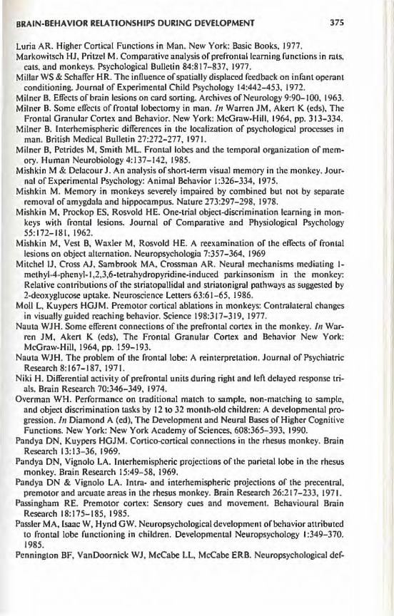
BRAIN-BEHAVIOR RELATIONSHIPS DURING DEVELOPMENT 375
Luria AR. Higher Cortical Functions in Man. New York: Basic Books, 1977. Markowitsch HJ, Pritzel M. Comparative analysis of prefrontal learning functions in rats,
cats, and monkeys. Psychological Bulletin 84:817-837, 1977. Millar WS & Schaffer HR. The influence of spatially displaced feedback on infant operant
conditioning. Journal of Experimental Child Psychology 14:442-453. 1972. Milner B. Effects of brain lesions on card sorting. Archives of Neurology 9 :90-100, 1963. Milner B. Some effects of frontal lobectomy in man. In Warren JM, Akert K (eds), The
Frontal Granular Cortex and Behavior. New York: McGraw-Hill, 1964, pp. 313-334. Milner B. Interhemispheric differences in the localization of psychological processes in
man. British Medical Bulletin 27:272-277, 1971. Milner B, Petrides M, Smith ML. Frontal lobes and the temporal organization of mem
ory. Human Neurobiology 4:137-142, 1985. Mishkin M & Delacour J. An analysis of short-term visual memory in the monkey. Jour
nal of Experimental Psychology: Animal Behavior I :326-334, 1975. Mishkin M. Memory in monkeys severely impaired by combined but not by separate
removal of amygdala and hippocampus. Nature 273:297-298, 1978. Mishkin M, Prockop ES, Rosvold HE. One-trial object-discrimination learning in mon
keys with frontal lesions. Journal of Comparative and Physiological Psychology 55:172-181, 1962.
Mishkin M. Vest B, Waxler M, Rosvold HE. A reexamination of the effects of frontal lesions on object alternation. Neuropsychologia 7:357-364, 1969
Mitchel JJ, Cross AJ, Sambrook MA, Crossman AR. Neural mechanisms mediating 1-methyl-4-phenyl-1 ,2,3,6-tetrahydropyridine-induced parkinsonism in the monkey: Relative contributions of the striatopallidal and striatonigral pathways as suggested by 2-deoxyglucose uptake. Neuroscience Letters 63:61-65, 1986.
Moll L, Kuypers HGJM. Premotor cortical ablations in monkeys: Contralateral changes in visually guided reaching behavior. Science 198:317-319, 1977.
Nauta WJH. Some efferent connections of the prefrontal cortex in the monkey. In Warren JM, Akert K (eds), The Frontal Granular Cortex and Behavior New York: McGraw-Hill, 1964, pp. 159-193.
Nauta WJH. The problem of the frontal lobe: A reinterpretation. Journal of Psychiatric Research 8:167-187, 1971.
Niki H. Differential activity of prefrontal units during right and left delayed response trials. Brain Research 70:346-349, 1974.
Overman WH. Performance on traditional match to sample, non-matching to sample, and object discrimination tasks by 12 to 32 month-old children: A developmental progression. In Diamond A (ed), The Development and Neural Bases of Higher Cognitive Functions. New York: New York Academy of Sciences. 608:365- 393, 1990.
Pandya DN, Kuypers HGJM. Cortico-cortical connections in the rhesus monkey. Brain Research 13: 13- 36, 1969.
Pandya DN, Vignola LA. Interhemispheric projections of the parietal lobe in the rhesus monkey. Brain Research 15:49- 58, 1969.
Pandya DN & Vignola LA. Intra- and interhemispheric projections of the precentral, premotor and arcuate areas in the rhesus monkey. Brain Research 26:217-233, 1971.
Passingham RE. Premotor cortex: Sensory cues and movement. Behavioural Brain Research 18:175-185, 1985.
Passier MA, Isaac W, Hynd GW. Neuropsychological development of behavior attributed to frontal lobe functioning in children. Developmental Neuropsychology 1:349-370. 1985.
Pennington BF, VanDoornick WJ, McCabe LL, McCabe ERB. Neuropsychological def-

376 PSYCHIATRIC AND DEVELOPMENTAL EFFECTS OF FRONTAL LOBE DAMAGE
icits in early treated phenylketonuric children. American Journal of Mental Deficiency 89:467-474, 1985.
Perret E. The left frontal lobe of man and the suppression of habitual responses in verbal categorical behaviour. Neuropsychologia 16:527-537, 1974.
Petrides M. Deficits on self-ordered temporal memory task after dorsolateral prefrontal lesions in the monkey. Society for Neuroscience Abstracts 14, 1988.
Petrides M, Milner B. Deficits in subject-ordered tasks after frontal- and temporal-lobe lesions in man. Neuropsychologia 20:249-262, 1982.
Piaget J. The Origins of Intelligence in Children. (Cook M, Trans.) New York: Basic Books, 1952. (Original work published, 1936.)
Porrino L, Goldman-Rakic PS. Brain stem innervation of prefrontal and anterior cingulate cortex in the rhesus monkey revealed by retrograde transport of HRP. Journal of Comparative Neurology 205:63-76, 1982.
Primrose DA. Phenylketonuria with normal intelligence. Journal of Mental Deficiency Research 27:239-246, 1983.
Quintana J, Yajeya J, Fuster JM. Prefrontal representation of stimulus attributes during delay tasks. I. Unit activity in cross-temporal integration of sensory and sensory-motor integration. Brain Research 474:211-221, 1988.
Rakic P, Nowakowski RS. The time of origin of neurons in the hippocampal region of the rhesus monkey. Journal ofComparative Neurology 196:99-128, 1981.
Rose JE, Woolsey CN. The orbitofrontal cortex and its connections with the mediodorsal nucleus in rabbit, sheep and cat. Research Publication of the Association for Research on Nervous and Mental Disease 27:210-232, 1948.
Rosvold HE. The frontal lobe system: Cortical-subcortical interrelationships. In Konarski J, Teuber HL, Zernicki B (eds), Functions of the Septa-Hippocampal System. Warsaw: Polish Sci. Pub, 1984, pp. 439-460.
Rovee-Collier C. The ontogeny of learning and memory in human infancy. In Kail R, Spear NE (eds), Comparative Perspectives on the Development of Memory. Hillsdale, NJ: Lawrence Erlbaum, 1984, pp. 103-134.
Saint-Cyr JA, Wan RO, Doudet D. Aigner TG. Impaired detour reaching in rhesus monkeys after MPTP lesions. Neuroscience Abstracts (Part I) 14:389, 1988.
Saunders RC. Monkeys demonstrate high level of recognition memory in delayed nonmatching to sample with retention intervals of 6 weeks. Society for Neuroscience Abstracts 15:342, 1989.
Schacter DL. Implicit memory: History and current status. Journal of Experimental Psychology: Learning, Memory & Cognition 13:501-518, 1987.
Schonen S de, Bresson F. Developpement de J'atteinte manuelle d'un objet chez I' enfant. Omportements 1:99-114, 1984.
Schwartz ML, Goldman-Rakic PS. Callosal and intrahemispheric connectivity of the prefrontal association cortex in rhesus monkey: Relation between intraparietal and principal sulcal cortex. Journal of Comparative Neurology 226:403-420, 1984.
Selcmon LD, Goldman-Rakic PS. Longitudinal topography and interdigitation of corticostriatal projections in the rhesus monkey. Journal of Neuroscience 5:776-794, 1985.
Selemon LD, Goldman-Rakic PS. Common cortical and subcortical targets of the dorsolateral prefrontal and posterior parietal cortices in the rhesus monkey: Evidence for a distributed neural network subserving spatially guided behavior. Journal of Neuroscience 8:4049-4068, 1988.
Shimamura AP, Janowsky JS, Squire LR. Memory for the temporal order of events in patients with frontal lobe lesions and amnesic patients. Neuropsychologia 28:803-814, 1990.

BRAIN-BEHAVIOR RELATIONSHIPS DURING DEVELOPMENT 377
Squire LR, Zola-Morgan S. The neurobiology of memory: The case for correspondence between the findings for human and non-human primates. In Deutsch JA (ed), The Physiological Basis of Memory. New York: Academic Press. 1983.
Squire LR, Zola-Morgan S. Chen KS. Human amnesia and animal models of amnesia: Performance of amnesic patients on tests designed for the monkey. Behavioral Neuroscience I 02:210-221 , 1988.
Stern Y, Langston JW. Intellectual changes in patients with MPTP-induced parkinsonism. Neurology 35:1506-1509, 1985.
Stern Y, Mayeux R, Rosen J, llson J. Perceptual motor dysfunction in Parkinson's disease: A deficit in sequential and predictive voluntary movement. Journal of Neurology, Neurosurgery, and Psychiatry 46: 145-151, 1983.
Stroop JR. Studies of interference in serial verbal reactions. Journal of Experimental Psychology 18:643-662, 1935.
Taylor AE, Saint-Cyr JA. Frontal lobe dysfunction in Parkinson'sdisease. Brain 109:845-883, 1986.
Taylor AE, Saint-Cyr JA, Lang AE. Memory and learning in early Parkinson's d isease: Evidence for a "frontal lobe syndrome." Brain and Cognition 13:211-232, 1990a.
Taylor JR. Roth RH, Sladek JR, Jr, Redmond DE, Jr. Cognitive and motor deficits in the performance of the object retrieval detour task in monkeys (Cercopithecus aethiops sabaeus) treated with M PTP: Long-term performance and effect of transparency of the barrier. Behavioral Neuroscience 104:564-576, I 990b.
Tourian AY, Sidbury JB. Phenylketonuria. In Stanbury JD, Wyngaarden JB, Fredrickson D (eds), The Metabolic Basis of Inherited Disease. New York: McGraw-Hill, 1978, pp. 240-255.
Towle C. Diamond A. Developmental progression in children aged 1-5 years on the delayed non-matching to sample task, a test of hippocampal memory function in adult monkeys and human amnesic patients. Paper presented at the Society for Research in Child Development, Biennial Meeting. April 18-21 , Seattle, WA, 1991.
Tulving E. Remembering and knowing the past. The crucial difference between remembering personal experiences and knowing impersonal facts. American Scientist 77:361 -368, 1989.
Van Essen DC. Visual areas of the mammalian cerebral cortex. Annual Review of Neuroscience 2:227-263, 1979.
Vicedomini JP, Corwin JV, Nonneman AJ. Behavioral elfects of lesions to the caudate nucleus or mediodorsal thalamus in neonatal, juvenile, and adult rats. Physiological Psychology 10:246-250, 1982.
Vicedomini JP. Isaac WL, Nonneman AJ. Role of the caudate nucleus in recovery from neonatal mediofrontal cortex lesions in the rat. Developmental Psychobiology 17: 51 -65, 1984.
Villablanca JR. Olmstead CE, Levine MS. Morcus RJ. Elfects of caudate nuclei or frontal cortical ablations in kittens. Experimental Neuropsychology 61 :615-634, 1979.
von Bonin G, Bailey P. Neocortex of Macaca mulatta. Urbana: University of Illinois Press, 1947.
Warren JM. Akert K. The Frontal Granular Cortex and Behavior. New York: McGrawHill, 1964.
Wellman HM. Cross D. Bartsch K. A meta-analysis of research on stage 4 object permanence: The A-not-B error. Monographs of the Society for Research in Child Development 5(3). 1987.
Welsh MC. Pennington BF. Ozonolf S. Rouse 8, McCabe ERB. Neuropsychology of early-treated phenylketonuria: Specific executive function deficits. Child Development 61:1697-1713, \990.

378 PSYCHIATRIC AND DEVELOPMENTAL EFFECTS OF FRONTAL LOBE DAMAGE
Werner JS, Perlmutter M. Development of visual memory in infants. Advances in Child Development and Behavior 14:2-56, 1979.
Williamson M, Dobson JC, Koch R. Collaborative study of children treated for phenyl. ketonuria: Study design. Pediatrics 60:815-821. 1977.
Wilson FA W, Goldman-Rakic PS. Effect of spatial and color cues on delayed-related neuronal responses in prefrontal cortex. Abstracts of the Society for Neuroscience 15:71, 1989.
Wozniak RH & Balamore U. Speech-action coordination in young children. Develop.. mental Psychology 20:850-858, 1984.
Zola-Morgan S, Squire LR. Memory impairment in monkeys following lesions limited to the hippocampus. Behavioral Neuroscience I 00: 15 5-160, 1986.
Zola-Morgan S, Squire LR, Amaral DG. Lesions of the hippocampal formation but not lesions of the fornix or mammillary nuclei produce long-lasting memory impairment in monkeys. Journal of Neuroscience 9:897-912, 1989.