(From the Department of Microbiology and the Department … · (From the Department of Microbiology...
17
RUNT DISEASE INDUCED IN NEONATAL MICE BY STERILE BACTERIAL VACCINES* BY RICHARD D. EKSTEDT, PH.D., AND EDWIN T. NISHIMURA,$ M.D. (From the Department of Microbiology and the Department of Pathology, Northwestern University Medical School, Chicago) PLATES 76 TO 79 (Received for publication, April 27, 1964) In a study concerned with immunity of mice to staphylococcal infection (Ekstedt, references 1 a-1 c) it was observed that if newborn mice were in- jected repeatedly with large amounts of a washed, autoclaved suspension of staphylococci or streptococci, a number of the animals failed to develop nor- really. The phenomenon was strongly reminiscent of the classical "runt disease" described by Billingham and Brent (2), who produced this syndrome by in- jection of homologous spleen cells into mice immediately after birth. The animals most severely affected by the vaccines also resembled those described as developing "wasting disease" following surgical thymectomy at birth (Miller, reference 3). In view of the importance of these phenomena to the concept of immuno- logical tolerance and the current theories of antibody production, it was felt that a description and documentation of our observations should be made. Materials and Methods Animals.--The mice used in these studies were the CF-1 strain obtained originally from Carworth Farms, New York. They were maintained in our laboratory under ideal conditions of temperature, humidity, and nutrition. Adult animals were mated, and the resulting litters were cared for by the mother. In some experiments, ICR-Swlss strain mice, bred from stock obtained from Charles River Laboratories, North Wilmington, Massachusetts, were used. Antigens.--The diffuse variant of the Smith strain of Staphylococcus aureus described by Hunt and Moses (4) was grown in brain-heart infusion broth (Difco Laboratories, Inc., Detroit) at 37°C for 18 to 20 hours. The cells were harvested by centrifugation, washed with saline, adjusted to contain approximately 10 l° organisms per ml of saline suspension, and autoclaved at 121°C for 15 to 20 minutes. A strain (D24) of Group A, Type 30 streptococcus was also grown and prepared for injection in a similar manner. Sterile brain-heart infusion broth, prepared according to the directions of the manufacturer, was injected into some animals as control. All vaccine preparations were checked for sterility prior to use. * This study was supported by grants from the National Institutes of Health (A&I E-2844) and the Streptococcal and Staphylococcal Commission of the Armed Forces Epidemiological Board, Surgeon General (DA-49-193-MD-2186). :~Career Development Awardee, GM-K3-244C3, United States Public Health Service. 795
Transcript of (From the Department of Microbiology and the Department … · (From the Department of Microbiology...

R U N T D I S E A S E I N D U C E D I N N E O N A T A L M I C E BY S T E R I L E B A C T E R I A L V A C C I N E S *
BY RICHARD D. EKSTEDT, PH.D., AND EDWIN T. NISHIMURA,$ M.D.
(From the Department of Microbiology and the Department of Pathology, Northwestern University Medical School, Chicago)
PLATES 76 TO 79
(Received for publication, April 27, 1964)
In a s tudy concerned with immuni ty of mice to s taphylococcal infection (Eks tedt , references 1 a-1 c) i t was observed tha t if newborn mice were in- jected repea ted ly with large amounts of a washed, autoclaved suspension of s taphylococci or streptococci, a number of the animals failed to develop nor- really. The phenomenon was s t rongly reminiscent of the classical " run t disease" described by Bil l ingham and Brent (2), who produced this syndrome by in- ject ion of homologous spleen cells into mice immedia te ly af ter bir th. The animals most severely affected by the vaccines also resembled those described as developing "wast ing disease" following surgical t hymec tomy a t bir th (Miller, reference 3).
I n view of the impor tance of these phenomena to the concept of immuno- logical tolerance and the current theories of an t ibody production, i t was felt tha t a descript ion and documenta t ion of our observat ions should be made.
Materials and Methods
Animals.--The mice used in these studies were the CF-1 strain obtained originally from Carworth Farms, New York. They were maintained in our laboratory under ideal conditions of temperature, humidity, and nutrition. Adult animals were mated, and the resulting litters were cared for by the mother. In some experiments, ICR-Swlss strain mice, bred from stock obtained from Charles River Laboratories, North Wilmington, Massachusetts, were used.
Antigens.--The diffuse variant of the Smith strain of Staphylococcus aureus described by Hunt and Moses (4) was grown in brain-heart infusion broth (Difco Laboratories, Inc., Detroit) at 37°C for 18 to 20 hours. The cells were harvested by centrifugation, washed with saline, adjusted to contain approximately 10 l° organisms per ml of saline suspension, and autoclaved at 121°C for 15 to 20 minutes. A strain (D24) of Group A, Type 30 streptococcus was also grown and prepared for injection in a similar manner. Sterile brain-heart infusion broth, prepared according to the directions of the manufacturer, was injected into some animals as control. All vaccine preparations were checked for sterility prior to use.
* This study was supported by grants from the National Institutes of Health (A&I E-2844) and the Streptococcal and Staphylococcal Commission of the Armed Forces Epidemiological Board, Surgeon General (DA-49-193-MD-2186).
:~ Career Development Awardee, GM-K3-244C3, United States Public Health Service.
795

796 RUN3? DISEASE INDUCED BY STERILE BACTERIAL VACCINES
Treatment.--The animals were given intraperitoneal injections repeatedly every other day of the week for a period of 3 to 5 weeks with 0.1 ml of the killed staphylococcal or streptococcal vaccine or brain-heart infusion broth. The treatment was begun as soon after birth as possible, usually within 1 hour. In some instances, when the animals were born at night or during the weekend, the initial injection was not administered until from 8 to 24 hours after birth. The effect of single injections of vaccine was not investigated.
The animals were weighed at intervals throughout their life and the weights were compared with untreated control animals of the same age. Representative members of the treated groups showing varying degrees of "runting" were sacrificed; the spleen, liver, kidneys, lungs, and thymus were removed and weighed. The tissues were fixed in 10 per cent buffered formalin embedded in paraffin, and sectioned at 5/z. Hematoxylin and eosin were employed for staining
]~.ESULTS
Fig. 1 shows three of the more severely "runted" animals from a litter which was injected with the killed staphylococcal vaccine beside a normal animal of the same age. The smaller animals had a mean weight of 3.5 ~ n at 4 weeks as compared with normal uninjected animals which weigh 12 to 15 gm at this age. The runted animals tended to be hyperirritable, and assumed a characteristic huddled posture at rest which remained the same as they moved about. They appeared to consume adequate amounts of food and water in a normal manner, and their size could not be attributed to avitaminosis or undernourishment. The diet consisted of an unsterilized vitamin- supplemented ration prepared for germ-free mice.
The development of hair in the runted animals was not particularly abnormal in most instances, although an occasional animal showed a lack of hair growth, and in one case a runted animal showed a conspicuous lack of hair growth and an eczematous skin reaction. This animal was 2 weeks old and died shortly after the picture shown in Fig. 2 was taken.
Not all of the animals receiving the vaccines were as severely runted as those shown, and in two separate litters treated with the staphylococcal vaccine, the weights ranged from 3.4 to 17.4 gm at 4 weeks. The susceptibility of litter mates, as well as different litters, varied considerably. Under the same regimens of treatment the number of runts produced varied from none to as much as 80 per cent of the litter. Variation in the severity of the disease in affected animals was also marked. In one litter of 10 animals injected from birth with the killed staphylococcal vaccine, for example, the body weights of the animals at 4 weeks of age were: 3.4, 3.5, 3.5, 7.5, 9.8, 10.0, 12.0, 12.5, 13.2, and 13.5 gin. This marked variation in susceptibility remains to be ex- plained. Both male and female animals were observed to runt under appropriate con- ditions, and the variation observed did not seem to be due to sex differences.
I t was observed that the ul t imate development of the animals receiving the vaccines appeared to depend markedly upon when the initial injections were started. I n general, the most severely affected animals came from litters tha t were injected initially within 1 hour after birth. Those litters tha t were 12 to 24 hours old before receiving the initial antigenic stimulus tended to be less severely affected, and animals 24 to 48 hours old at the time of the initial injection usually developed normally.

RICHARD D. EKSTEDT AND E D W I N T. NISHIMURA 797
In one experiment with a litter injected within 1 hour after birth, 8 surviving mice at 5 weeks of age showed a mean weight of 9.6 4- 1.0 gin. A separate litter born at the same time but not started on its course of injections until 48 hours after birth showed a mean weight of 17.2 4- 1.2 gm for 6 mice. The difference between the means was highly significant (test ffi 4.96; P < 0.01).
Text-fig. 1 shows a graph of the mean weights for a group of animals which were injected with the killed streptococcal vaccine compared with a group of uninjected control animals. In this experiment, the injections were terminated at 3 weeks and after an additional 2 weeks the treated animals showed a considerable gain in weight.
18
16
14
t io°t.°'° . Run%ed ammal~
o I 2 3 / . 5 AGE IN WEEKS
T]~xT-Fm. 1. Comparison of the weights of a group of normal mice and a group of strepto- coccal-runted mice during and following termination of treatment at 3 weeks of age.
The phenomenon of growth retardation did not appear to result in any permanent damage to the animals. Following termination of the treatment, the ruuted animals in most instances would gain weight and remain healthy and essentially indistinguish- able from normal animals.
Litters which were injected with brain-heart infusion broth from birth with the same regiment of injections as the vaccine-treated an{reals never showed any evidence of runting and at 5 weeks of age showed a mean weight of 16.6 gm with a range of 15.1 to 17.2 gm.
As a result of the wide var ia t ion in the response of t rea ted animals to the bacter ia l vaccines, rat ios of organ we igh t /body weight showed no significant differences when compared with normal animals of the same age. This was par t icu la r ly t rue with the liver, kidneys, and spleen. I n the more severely
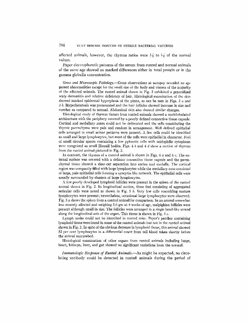
798 RUNT DISEASE INDUCED BY STERILE BACTERIAL VACCINES
affected animals, however, the thymus rat ios were 1/~ to 1/~ of the normal values.
Paper electrophoretic pa t te rns of the serum from runted and normal animals of the same age showed no marked differences either in to ta l protein or in the gamma globulin concentration.
Gross and Microscopic Pathology.--Gross observations at autopsy revealed no ap- parent abnormalities except for the small size of the body and viscera of the majority of the affected animals. The runted animal shown in Fig. 2 exhibited a generalized scaly dermatitis and relative deficiency of hair. Histological examination of the skin showed marked epidermal hyperplasia of the pinna, as can be seen in Figs. 3 a and 3 b. Hyperkeratosis was pronounced and the hair follicles showed increase in size and number as compared to normal. Abdominal skin also showed similar changes.
Histological study of thymus tissues from runted animals showed a multilobulated architecture with the periphery covered by a poorly defined connective tissue capsule. Cortical and medullary zones could not be delineated and the cells constituting the thymic parenchyma were pale and random in arrangement. Well defined epithelial cells arranged in small acinar patterns were present. A few cells could be identified as small and large lymphocytes, but most of the cells were epithelial in character. Foci of small circular spaces containing a few pyknotic cells with acidophilic cytoplasm were recognized as small Hassall bodies. Figs. 4 b and 4 d show a section of thymus from the runted animal pictured in Fig. 2.
In contrast, the thymus of a control animal is shown in Figs. 4 a and 4 c. The ex- ternal surface was covered with a delicate connective tissue capsule and the paren- chymal tissue showed a dear-cut separation into cortex and medulla. The cortical region was compactly filled with large lymphocytes while the medullary zone consisted of large, pale epithelial cells forming a syncytia-like network. The epithelial cells were usually surrounded by clusters of large lymphocytes.
A few poorly developed lymphoid follicles were present in the spleen of the runted animal shown in Fig. 2. In longitudinal section, three foci consisting of aggregated reticular cells were noted as shown in Fig. 5 b. Very few cells resembling mature lymphocytes were present; nevertheless, occasional large lymphocytes were observed. Fig. 5 a shows the spleen from a control animal for comparison. In an animal somewhat less severely affected and weighing 5.6 gm at 4 weeks of age, malpighian follicles were present although small in size. The follicles were arranged in a single bead-like strand along the longitudinal axis of the organ. This tissue is shown in Fig. 5 c.
Lymph nodes could not be identified in runted mice. Peyer's patches containing lymphoid tissue were found in some of the runted animals but not in the runted animal shown in Fig. 2. In spite of the obvious decrease in lymphoid tissue, this animal showed 52 per cent lymphocytes in a differential count from tail blood taken shortly before the animal succumbed.
Histological examination of other organs from runted animals including lungs, heart, kidneys, liver, and gut showed no significant variations from the normal.
Immunologic Response of Runted Animals.--As might be expected, no circu- lat ing an t ibody could be detected in runted animals during the period of

RICHARD D. EKSTEDT AND EDWIN T. NISHIMURA 799
treatment with the bacterial vaccines. These determinations were carried out, using either the capillary precipitin reaction with an ammonium sulfate frac- tion derived from homologous organisms (Ekstedt, reference 1 c) as antigen, or by a sensitive growth-agglutination test described by Boger, Frankel, and Gavin (5). In the latter test the compact variant of the Smith strain of S aureus was used as the antigen. Precipitating antibody could be detected in surviving animals 2 weeks after termination of the injections, and at 1 month 1.0 ml of pooled mouse serum from surviving animals treated with the staph- ylococcal vaccine gave 0.065 mg total protein precipitate with the ammonium sulfate fraction from homologous organisms as antigen.
Further studies are in progress to assess the immunologic competence of runted animals in rejecting heterologous skin grafts or in responding to other antigenic stimuli.
Strain Specificity of the Runting Phenomenon.--In order to determine whether or not the runting phenomenon elicited by injection of bacterial vaccines into neonatal CF-1 mice was related to the strain of animal used, several experiments were carried out, using the ICR-Swiss strain of mice. These animals also proved susceptible to runting with the bacterial vaccines with about the same fre- quency and variation in susceptibility as the CF-1 strain. I t was observed, however, that germ-free ICR mice bred and maintained in our laboratory under carefully controlled conditions of gnotobiosis were much more resistant to runting than conventional animals. In 7 litters of germ-free ICR mice started on their course of injections within 1 hour after birth with the killed staphylococcal vaccine, only one group showed slight evidence of runting. This litter consisted of 8 animals surviving at 4 weeks. Of these, 2 weighed 8.1 and 10.4 gm, respectively, and the rest ranged in weight from 12.0 to 15.8 gm.
These results suggested the possibility that in conventionally raised mice natural antibody to staphylococcal antigens passively transferred through the placenta to the newborn animals might play some role in the runting phenomenon observed. Germ-free animals, although shown recently by Cohen, Newton, Cherry, and Updyke (6) to possess some naturally occurring staph- ylococcal antibodies, might be deficient in this respect. Accordingly, groups of neonatal germ-free mice were treated with the killed staphylococcal vaccine suspended in a 1:100 saline dilution of staphylococcal antiserum prepared in rabbits against the whole cells. In the first experiment of this kind a litter of 10 animals at 3 weeks of age showed a mean weight of 6.5 4- 0.3 gin. These results suggest that antibody does indeed have some effect in inducing the runting syndrome. Further study of the ability of other antigen-antibody complexes to induce a similar runting response in neonatal animals is in prog- ress.
The thought occurred to us that if there were antigens present in the mi-

800 RUNT DISEASE INDUCED BY STERILE BACTERIAL VACCINES
croorganisms which were similar to mouse tissue antigens, the possibility of the animal developing tolerance to these antigens would be greater, and the relation between tolerance and runting might be further clarified. High titered antibody prepared in rabbits against mouse serum was reacted with 3 anti- genic fractions from the Smith, diffuse strain of S . aureus . The fractions tested included a teichoic acid fraction, a protein antigen (Jensen) (7) and a crude ammonium sulfate fraction (lc). In this preliminary experiment no cross-reactivity could be detected using a semi-micro precipitin test. These studies are being continued, using more sensitive methods.
DISCUSSION
A severe, usually fatal disease in mice originally described by Billingham and Brent (2) and termed "runt disease" by them, results from the injection of immunologically competent cells into newborn animals. It has been postu- lated that these transferred cells survive in the immunologically immature host and presumably as a result of antibody production directed against host antigens, the so called "graft versus host" reaction develops. A similar syn- drome has been produced by Miller (3) by neonatal thymectomy of mice. This phenomenon, called "wasting disease", is similar in most respects to runt disease. The most striking abnormality exhibited by these animals is the marked atrophy of lymphoid tissues. This has been suggested by Barnes, Luotit, and Micklem (8) to result in profound metabolic disturbances leading to malformation of the animal.
Although a considerable amount of work has been reported dealing with the graft versus host reaction (9-11), no direct experimental evidence is avail- able to explain the mechanism whereby the antibody response of transferred cells to host tissue antigens results in lymphoid depletion and runting. Circu- lating antibody directed against host tissue has not been demonstrated in runt disease (12).
The runting syndrome reported here, resulting from the strenuous anti- genization of neonatal mice with killed bacterial antigens, requires further speculation as to the mechanism involved in classical runt disease. It would seem conceivable that such treatment might have the same effect, although greatly magnified, as the postulated deletion of "forbidden clones" to self antigens in the normal animal (13). Strenuous treatment with a mixture of foreign antigens such as a bacterial cell, may result in the preemption of primi- tive lymphoid tissues and in their ultimate destruction by some similar mech- anism.
The possibility of an intermediary mechanism being involved in runting must not be overlooked, however. Recently Schlesinger and Mark (14) re- ported the induction of wasting disease in mice by the administration of cortisol acetate. These investigators point out that the destruction of the thymus

RICHARD D. EKSTEDT AND EDWIN T. NISHIMURA 801
in classical runt disease may be due either to a graft v e r s u s host reaction, or to an increased adrenal cortical hormone level resulting from a graft-host interaction. Some similar mechanism mediated by hormonal imbalance may result from bacteria-host interaction in the neonatal mouse.
The report of Schlesinger and Mark (14) appeared during the preparation of this manuscript. If our treatment resulted in a significant increase in secre- tion of adrenal hormones this activity might be reflected in some change in the organ. In reviewing the tissues from our runted animals, no evidence of hyperplasia of the adrenals could be seen either grossly or microscopically.
Another method which has been reported to result in a form of runting in young mice is that reported by Hotchin and Weigand (15). These investigators showed that young mice injected intracerebrally with mouse brain suspensions containing lymphocytic choriomeningitis virus developed a form of runt disease with the usual accompanying abnormalities. In this situation again, the ani- mals were being severely stressed by antigenization with foreign material. The presence of a replicating virus in their material contrasts with the use of killed bacteria in our system. This difference is more apparent than real, however, since repeated injections of the killed bacterial vaccines were required to elicit our runting response, while multiplication of the virus preparation would ultimately result in a similar stress resulting from large amounts of foreign antigen.
Rowe and Capps (16) have isolated a viral agent causing necrosis of the thy- mus in new born mice. The possibility that the treatment of neonatal mice described in this report has resulted in the activation of a latent viral agent of this kind seems unlikely, however. In no case did thymus tissue from severely runted animals show any evidence of necrosis or scarring; however, no effort was made to isolate a viral agent from runted animals.
Another viral agent isolated by Stanley (17) has been reported to result in retardation in the growth of suckling mice. This agent, called a hepatoenceph- alomyelitis virus (HEV) and shown to be identical with reovirus, produced jaundice, peritoneal exudate, ataxia, tremors, and paralysis. Histopathological lesions were observed in the brain, cord, liver, heart, spleen, and lungs. None of these symptoms or lesions was present in the runts induced by bacterial vaccines reported here.
Recently Blaese, Martinez, and Good (18) reported on the immunologic incompetence of immunologically runted animals and showed that mice under- going the graft v e r s u s host reaction failed to form antibodies to T2 bacteriophage. They offer as one explanation of their results the possibility "that the rela- tively limited number of injected parental cells might be totally committed to the tissue antigens of the host, and by virtue of this commitment, incapable of responding to third party antigens following establishment of this commit- ment." Whether a similar situation would obtain in animals runted with bac-

8 0 2 RUNT DISEASE INDUCED BY STERILE BACTERIAL VACCINES
terial vaccines as described here, remains to be determined. I t is clear that following termination of the strenuous treatment with the vaccine antigens surviving animals are capable of recovery and do respond with significant amounts of antibody directed against the homologous vaccine.
Aside from the possibility of a hormonal imbalance being responsible for the results obtained here, the possibility still remains that in newborn, immunolog- ically immature animals presented with large amounts of a mosaic of foreign antigens, large numbers of lymphoid cells either destined to respond to these antigens in later life, or as yet uncommitted may be deleted and removed from the affected animals. Whether severe depletion of lymphoid tissues is the cause or effect of runting cannot as yet be answered.
The observation that germ-free animals are much more resistant to runting with bacterial vaccines, and the possibility of increasing their susceptibility by injecting the organisms along with specific antibody has interesting implica- tions. These results suggest that small amounts of antibody may be required to complex with injected antigen and possibly trigger the destructive response of susceptible lymphoid tissues.
A unifying concept must be sought to better understand the mechanisms by which the diverse methods described above result in the runting of young ani- mals. The graft v e r s u s host phenomenon, wasting resulting from surgical abla- tion of the thymus, hormonal induction of wasting, and strenuous treatment of neonatal animals with bacterial or viral vaccines all lead to a similar end result. The observation in common with all of these phenomena is the profound atrophy of lymphoid tissues and the marked decrease in normal growth of affected ani- mals. Whether or not one is causally related to the other, and whether the underlying mechanism in all cases is the same, remains to be determined.
SUMMARY
A form of runt disease has been produced in neonatal CF-1 and ICR mice by the repeated injection of 109 washed, autoclaved, saline-suspended staphylo- cocci or streptococci. The most severely affected animals showed a marked decrease in lymphoid tissues and resembled grossly and microscopically animals suffering from the classical runt or wasting disease described by others. The timing of the initial antigenic stimulation was of importance, and animals started on their course of injections at an age of 48 hours or older showed no effect. There was a considerable variation in the severity of the disease within litters and from one litter to another. This variation could not be ascribed to a difference in susceptibility between sexes, since both male and female mice were observed to runt under appropriate conditions. Germ-free ICR mice were much more resistant to the runting phenomenon than conventional animals of the same strain, but could be induced to runt by injection of the staphylococcal vaccine suspended in homologous antiserum.

RICHARD D. EKSTEDT AND EDWIN T. NISHIMIIRA 803
The relationship of the runting phenomenon described here and classical runt disease or runting by adrenal hormones is discussed.
We wish to express our appreciation to Mrs. Katheryn Slager for her excellent and devoted technical assistance.
BIBLIOGRAPHY
1 a. Ekstedt, R. I)., Studies on immunity to staphylococcal infection in mice. I. Effect of dosage, viability, and interval between immunization and challenge on resistance to infection following injection of whole cell vaccines, J. Infect. Dis., 1963, 119., 143.
1 b. Ekstedt, R. D., Studies on immunity to staphylococcal infection in mice. II. Effect of immunization with fractions of Staphylococcus aureus prepared by physical and chemical methods, J. Infect. Dis., 1963, 119., 152.
1 c. Ekstedt, R. I)., Studies on immunity to staphylococcal infection in mice. I I I . A protein antigen that induces protection against challenge with saline-sus- pended organisms, J. Infect. Dis., 1963, 113, 105.
2. Billingham, R. E., and Brent, L., A simple method for inducing tolerance of skin homograffs in mice, Transplant. Bull., 1957, 4, 67.
3. Miller, J. F. A. P., Effect of neonatal thymectomy on the immunological respon- siveness of the mouse, Proc. Roy. Soc. London, Series B, 1962, 156, 415.
4. Hunt, G. A., and Moses, A. J., Acute infection of mice with Smith strain of Staphylococcus aureus, Science, 1958, 128, 1574.
5. Boger, W. P., Frankel, J. W., and Gavin, J. J., Detection and titration of Staphy- lococcus aureus agglutinin in serum, Proc. Soc. Exp. Biol. and Med., 1960, 104, 639.
6. Cohen, J. O., Newton, W. C., Cherry, W. B., and Updyke, E. L., Normally oc- curring staphylococcal antibodies in germ free mice, J. Immunol., 1963, 90, 358.
7. LSfkvist, T., and Sj6quist, J., Chemical and serological analysis of antigen prepa- rations from Staphylococcus aureus. A comparison between the products ob- tained by Verwey's and Jensen's techniques, Acta Path. et Microbiol. Scand., 1962, 56, 295.
8. Barnes, D. W. H., Loutit, J. E., and Micklem, H. S., Secondary disease of radia- tion chimeras: A syndrome due to lymphoid aplasia, Ann. New York Acad. Sc., 1962, 99, 374.
9. Billingham, R. E., and Brent, L., Quantitative studies on tissue transplantation immunity. IV. Induction of tolerance in newborn mice and studies on the phenomenon of runt disease, Phil. Tr. Roy. Soc. London, Series B, 1959, 9.42, 439.
10. Billingham, R. E., Brown, V. B., Defendi, V., Silvers, W. K., and Steinmuller, I)., Quantitative studies on the induction of tolerance of homologous tissue and on runt disease in the rat, Ann. New York Acad. Sc., 1960, 87, 457.
11. Cooper, G. N., and Howard, J. G., An effect of the graft-versus-host reaction on resistance to experimental bacteriemia, Brit. J. Exp. Path., 1961, 45, 558.

804 RUNT DISEASE INDUCED BY STERILE BACTERIAL VACCINES
12. Cochrane, C. G., and Dixon, F. J., Antibody production by transferred ceils, Advances Immunol., 1962, 2, 205.
13. Burnet, F. M., The Clonal Selection Theory of Acquired Immunity, London, Cambridge University Press, 1958.
14. Schlesinger, M., and Mark, R., Wasting disease induced in young mice by ad- ministration of cortisal acetate, Science, 1954, 143, 965.
15. Hotchin, J., and Weigand, H., Studies of lymphocytic choriomeningitis in mice. I. The relationship between age at inoculation and outcome of infection, J. Im- munol., 1961, 86, 392.
16. Rowe, W. P., and Capps, W. I., A new mouse virus causing necrosis of the thymus in newborn mice, J. Exp. Med., 1961, 113, 831.
17. Stanley, N. F., Reovirus--a ubiquitous orphan, Med. J. Australia, 1961, 2, 815. 18. Blaese, R. M., Martinez, C., and Good, R. A., Immunologic incompetence of im-
munologically runted animals, J. Exp. Med., 1954, 119, 211.
EXPLANATION OF PLATES
PLATE 76
FIG. 1. Comparison of three staphylococcal vaccine-runted mice with a normal control mouse at 4 weeks of age. X 1.
FIG. 2. A severely affected staphylococcal vaccine-runted mouse at 2 weeks of age showing the eczema-like skin reaction. × 3.5.

THE JOURNAL OF EXPERIMENTAL MEDICINE VOL. 120 PLATE 76
(Ekstedt and Nishimura: Runt disease induced by sterile bacterial vaccines)

PLATE 77
FIG. 3 a. A section from a small portion of the pinna of the runted mouse shown in Fig. 2. The entire external ear was thickened. The epidermis shows moderate hyper- plasia and hyperkeratosis while the hair follicles are numerous, hyperplastic, and un- usually well developed. The cartilaginous plate, not seen here, is thick, irregular and consists of loosely arranged cells with little or no matrix formation. X 200.
FIG. 3 b. A section through the pinna of a control mouse at approximately the same age. X 200.

THE JOURNAL OF EXPERIMENTAL MEDICINE VOL. 120 PLATE 77
(Ekstedt and Nishimura: Runt disease induced by sterile bacterial vaccines)

PLATE 78
FIG. 4 a. Portion of the thymus of a normal mouse. Note the distinct arrangement of cortex which consists of numerous dark staining lymphocytes. The medulla is pale and shows loosely arranged reticuloendothelial cells, large lymphocytes, and blood vessels. X 80.
FIG. 4 b. Almost the entire thymus from a runted mouse. The cortico-medullary pattern is not present. The cells are irregular in shape and arranged in a pleomorphic pattern. X 80.
FIG. 4 c. High-power magnification of normal thymus tissue. X 200. FIG. 4 d. The dark, angular cells are difficult to classify, but a few distinct lympho-
cytes are seen. Several cyst-like structures containing large acidophilic cells with pyknotic nuclei are recognized as Hassall bodies. The pale staining epithelial cells are arranged in an acinar pattern. X 200.

THE JOURNAL OF EXPERIMENTAL MEDICINE VOL. 120 PLATE 78
(Ekstedt and Nishimura: Runt disease induced by sterile bacterial vaccines)

PLATE 79
FIG. 5 a. Normal spleen. Malpighian follicles are prominent and numerous. The clusters of dark staining cells in the red pulp represent myeloid and erythrocytic cells undergoing maturation. × 80.
FIG. 5 b. A section of spleen from the runted mouse shown in Fig. 2. Three small malpighian follicles are seen. A careful survey of the spleen sections revealed very few malpighian follicles. Note the dark staining hemopoietic cells of myeloid and erythro- cytic series. X 100.
FIG. 5 c. A longitudinal section of spleen from a moderately runted mouse. Note single malpighian follicles arranged in a bead-like pattern. Megakaryocytes are seen surrounding the follicles. X 200.
Fro. 5 d. Section of normal spleen. Malpighian follicles are prominent and numer- ous. X 200.

THE JOURNAL OF EXPERIMENTAL MEDICINE VOL. 120 PLATE 79
(Ekstedt and Nishimura: Runt disease induced by sterile bacterial vaccines)