
From Reactive to Endogenously Active Dynamical Conceptions...
48
From Reactive to Endogenously Active Dynamical Conceptions of the Brain Adele Abrahamsen Center for Research in Language, University of California, San Diego William Bechtel Department of Philosophy, Center for Chronobiology, and Interdisciplinary Program in Cognitive Science, University of California, San Diego Abstract We contrast reactive and endogenously active perspectives on brain activity. Both have been pursued continuously in neurophysiology laboratories since the early 20th- century, but the endogenous perspective has received relatively little attention until recently. One of the many successes of the reactive perspective was the identification, in the second half of the 20 th century, of the distinctive contributions of different brain regions involved in visual processing. The recent prominence of the endogenous perspective is due to new findings of ongoing oscillatory activity in the brain at a wide range of time scales, exploiting such techniques as single-cell recording, EEG, and fMRI. We recount some of the evidence pointing to ways in which this endogenous activity is relevant to cognition and behavior. Our major objective is to consider certain implications of the contrast between the reactive and endogenous perspectives. In particular, we relate these perspectives to two different characterizations of explanation in the new mechanistic philosophy of science. In a basic mechanistic explanation, the operations of a mechanism are characterized qualitatively and as functioning sequentially until a terminating condition is realized. In contrast, a dynamic mechanistic explanation allows for non-sequential organization and emphasizes quantitative modeling of the mechanisms's behavior. For example, with appropriate parameter values a set of differential equations can be used to demonstrate ongoing oscillations in a system organized with feedback loops. We conclude that the basic conception of mechanistic explanation is adequate for reactive accounts of brain activity, but dynamical accounts are required to explain sustained, endogenous activity.
Transcript of From Reactive to Endogenously Active Dynamical Conceptions...
From Reactive to Endogenously Active Dynamical Conceptions of the Brain
Adele Abrahamsen
Center for Research in Language, University of California, San Diego
William Bechtel
Department of Philosophy, Center for Chronobiology, and Interdisciplinary Program
in Cognitive Science, University of California, San Diego
Abstract
We contrast reactive and endogenously active perspectives on brain activity. Both
have been pursued continuously in neurophysiology laboratories since the early 20th-
century, but the endogenous perspective has received relatively little attention until
recently. One of the many successes of the reactive perspective was the identification,
in the second half of the 20th century, of the distinctive contributions of different brain
regions involved in visual processing. The recent prominence of the endogenous
perspective is due to new findings of ongoing oscillatory activity in the brain at a
wide range of time scales, exploiting such techniques as single-cell recording, EEG,
and fMRI. We recount some of the evidence pointing to ways in which this
endogenous activity is relevant to cognition and behavior. Our major objective is to
consider certain implications of the contrast between the reactive and endogenous
perspectives. In particular, we relate these perspectives to two different
characterizations of explanation in the new mechanistic philosophy of science. In
a basic mechanistic explanation, the operations of a mechanism are characterized
qualitatively and as functioning sequentially until a terminating condition is realized.
In contrast, a dynamic mechanistic explanation allows for non-sequential
organization and emphasizes quantitative modeling of the mechanisms's behavior.
For example, with appropriate parameter values a set of differential equations can be
used to demonstrate ongoing oscillations in a system organized with feedback loops.
We conclude that the basic conception of mechanistic explanation is adequate for
reactive accounts of brain activity, but dynamical accounts are required to explain
sustained, endogenous activity.

From reactive to endogenously active dynamical conceptions of the brain p. 2
Observe a living organism—from a bacterium to a fellow human being—and you see an
endogenously active system. Introspect and you will observe, as did William James, a continual
flow of thoughts. If pressed, most neuroscientists and psychologists will acknowledge that neural
systems are endogenously active, generating activity even in the absence of any stimulus. But for
decades they have tended to disregard this key characteristic, pursuing programs of research in
which they present discrete stimuli in structured tasks designed to focus on the neural and
behavioral effects of experimental manipulations. In this paper we contrast this perspective
(which we call reactive) with a dynamic perspective emphasizing endogenous activity. In
neuroscience these have a long history of coexistence, but only recently has the endogenous
perspective become less isolated as powerful new strategies for pursuing it have begun changing
the overall research landscape. We provide a selective tour of this history from the vantage point
of the new mechanistic philosophy of science, in which we highlight the interplay between basic
and dynamic mechanistic explanation.
The reactive perspective has deep historical roots and is widely pursued in both neuroscience and
psychology. Neuroscientists, following a tradition initiated by the British neurophysiologist
Charles Scott Sherrington (1923), commonly treat the brain as a reactive mechanism in which
sensory input initiates processing along a neural pathway, terminating in a motor response. One
of their core techniques is to present stimuli and record neural responses in the brain area of
interest; another is to manipulate neural activity and record motor responses. Psychologists more
often target the whole organism, presenting stimuli and recording behavioral responses without
determining the intervening neural activity. Most North American psychologists treated the gap
between stimulus and response as a black box during the behaviorist era and as information
processing thereafter, but for a minority (yesterday’s psychobiologists and today’s cognitive
neuroscientists) the gap is filled by neural activity that should be investigated.
There is no doubt that this reactive framework has been enormously productive for both
neuroscience and psychology. It has served to identify many of the parts and operations within
the mechanisms responsible for cognitive phenomena, as we will show in the case of vision in
section 2. But there are also indications of its limitations. One is the considerable variability
From reactive to endogenously active dynamical conceptions of the brain p. 3
researchers commonly observe in both behavioral and neural responses. While this variability
tends to be construed as noise to be eliminated from experimental data by averaging across time
and subjects, if examined rather than concealed it can reveal compelling signatures of
endogenous activity.
Laboratory research on endogenous activity, while relatively sparse, has historical roots nearly as
deep as those of the reactive approach. Most notably, Thomas Graham Brown (1914) studied
neural mechanisms for motor behavior in decerebrate cats alongside Sherrington in his
laboratory at Liverpool from 1910 to 1913—but arrived at quite different conclusions.
Sherrington was commited to a sequential reflex mechanism, by which peripheral input (e.g., to
the cat’s feet when placed on a moving treadmill) produced a sequence of neural signals (to the
spine, within the spine, and out to flexor and then extensor muscles). Each cycle of stepping
resulted in renewed input (sensory feedback) and hence ongoing, rhythmic stepping movements.
Brown discovered that he could obtain similar rhythmic stepping even after isolating the spinal
cord from afferent (peripheral) input by cutting the dorsal root nerves. This impressive
demonstration of endogenous control led him to propose a neural mechanism that later would be
recognized as the first description of a central pattern generator—central because the key
components were in the spine (sensitive to but not dependent on peripheral input); pattern
because it produced an ongoing oscillatory pattern (observed as rhythmic stepping, in which
flexion alternates with extension); and generator because this mechanism could initiate
production of the pattern. More specifically, Brown proposed what would now be described as
two coupled networks of spinal neurons—one for flexion and one for extension—which
oscillated in inhibiting each other’s activity. However, Sherrington resisted distraction from his
own pursuit of a sequential reflex account of motor behavior;1 as his reactive approach became
more entrenched, Brown was increasingly marginalized (for discussion, see Stuart & Hultborn,
2008).
It was a half-century before Brown’s emphasis on endogenous activity was revived by
researchers who converged on central pattern generators as the explanatory mechanism of choice
1 Early on Sherrington (1913, p. 207) acknowledged that Brown’s view “demands careful attention” but demurred on grounds that his own line of explanation “would be led too far afield by its consideration now.”

From reactive to endogenously active dynamical conceptions of the brain p. 4
for a variety of rhythmic motor behaviors. Wilson and Wyman’s (1965) landmark account of
flight in locusts was followed by identification of central pattern generators in the brain stem and
spinal cord for such activities as walking, swimming, respiration, and circulation (Grillner,
2003). Almost another half-century passed before neuroscientists investigating sensory
processing and central cognition turned their attention to endogenous activity in cerebral cortex
and were rewarded with multiple streams of evidence from single cell recording, EEG, fMRI,
and other techniques. We introduce some of this evidence in section 3, and emphasize that the
resulting conception of the brain as endogenously active poses a profound challenge to the purely
reactive perspective that has dominated much of psychology as well as neuroscience.
The slow spread of the endogenous perspective is unsurprising considering the history of other
sciences. Max Planck (1949, pp. 33-34) famously suggested that “A new scientific truth does not
triumph by convincing its opponents . . . but rather because its opponents eventually die.” He
exaggerated for effect, presumably, but it is not uncommon for scientists to bemoan delays in the
uptake of new approaches. Less remarked upon is the delayed impact of changes in the sciences
on philosophy of science. This is a young field (its first journal, Philosophy of Science, began
publication in 1934), and it has been slow to move beyond its initial roots in twentieth-century
physics to incorporate quite different influences from the biological and cognitive sciences. We
suggest that this delay has been excessive and detrimental to its own development as a field of
inquiry. Philosophers of science did not even recognize the dominant mode of explanation in
these sciences—mechanistic explanation—until pioneering work by William Wimsatt, who
pointed out that “At least in biology, most scientists see their work as explaining types of
phenomena by discovering mechanisms . . .” (Wimsatt, 1976, p. 671). His influence on a cohort
of students gave rise in the 1990s and especially after 2000 to the new mechanists, who have
drawn on biology and cognitive science rather than physics in constructing a new mechanistic
philosophy of science (Bechtel & Richardson, 1993/2010; Bechtel & Abrahamsen, 2005;
Glennan, 1996, 2002; Machamer et al., 2000; Thagard, 2003; Wimsatt, 2007).
Recently we have argued that further developments in these sciences—especially computational
modeling of the dynamics of cognitive and neural mechanisms—require extending the
mechanistic framework to incorporate dynamic mechanistic explanation (Bechtel &
From reactive to endogenously active dynamical conceptions of the brain p. 5
Abrahamsen, 2010, 2011). Thus, in what follows we begin by distinguishing between basic
mechanistic explanation, in which target systems are treated as reactive mechanisms, and
dynamic mechanistic explanation, which has the resources to characterize endogenous as well as
reactive activity and to do so with greater precision (section 1). We then discuss investigations of
brain mechanisms in particular, contrasting those that exemplify the reactive perspective (section
2) with those targeting endogenous activity (section 3). We consider certain implications of the
endogenous perspective for how we understand cognitive activity (section 4). Finally, we return
to the philosophical understanding of dynamic mechanistic explanation and how it can illuminate
research that takes an endogenous perspective on the brain (section 5).
1. Two Conceptions of Mechanism
The new mechanists have primarily focused on basic mechanistic explanation, in which
investigators decompose a system into a set of component parts, each of which performs one or
more operations, and recompose it by figuring out the spatial organization of the parts and
temporal/causal organization of the operations (Bechtel & Abrahamsen, 2005, 2009). The idea is
that going down to a lower level provides the most useful explanation of how the system’s
activity generates a phenomenon of interest.
What makes these explanations “basic” is that the accounts of organization are mostly qualitative
rather than quantitative. Thus, a typical structural decomposition into parts would be recomposed
into a spatial ordering (e.g., the spine’s lumbar vertebrae are designated as L1, L2, L3, L4, L5) or
a schematic layout (e.g., a eukaryotic cell is depicted as a membrane enclosing one nucleus and
numerous organelles in cytoplasm). A typical functional decomposition into operations would be
recomposed most simply into a temporal ordering in which the product of one operation is
operated upon by the next (e.g., the chain of biochemical reactions comprising intermediary
metabolism). The act of constructing a basic mechanistic explanation of a phenomenon is
complete2 when the investigator can specify which parts perform which operations. This task of
2 “Complete” does not imply “final.” An important role for such an account is to provide a framework for further research that elaborates and corrects it and eventually may replace it. Darden and Craver (2002) referred to incomplete accounts as mechanism sketches and traced how two different sketches for protein synthesis in the 1950s
From reactive to endogenously active dynamical conceptions of the brain p. 6
localization sometimes is integral to the discovery process, but may instead be deferred (pending
development of necessary tools, for example, the electron microscope). Once achieved, a well-
supported basic mechanistic account is an important research milestone.
One example, discussed at greater length in section 2, is a pathway through the visual system that
is responsible for the phenomenon of object recognition. Parts of the pathway have been
identified in the retina, lateral geniculate nucleus (LGN), occipital lobe (visual areas V1, V2, and
V4) and temporal lobe. A very simplified version of the basic mechanistic account has each of
these parts in turn performing one or more operations on the output of the preceding operation:3
the retina represents a stimulus object topographically, the LGN modulates or gates the
representation, V1 extracts several types of features, V2 analyzes contours, V4 analyzes form
and color, and inferior temporal cortex performs higher-level, integrative operations that yield a
percept recognized as a particular type of object.
It is possible to find ordered components with no beginning or end: the beads in a bracelet, the
bases in loops of mitochondrial DNA, people circle-dancing, and so forth. But something in us
likes an ordering to be not only invariant but also bounded and unidirectional. Scientists are no
exception, showing a preference for basic mechanistic accounts in which operations are ordered
with a beginning and an end. We will reserve the term sequence for this type of organization in
time or space.
Sequential organization is especially prominent in the definition of mechanism offered by
Machamer, Darden, and Craver (2000):
Mechanisms are entities and activities organized such that they are productive of regular
changes from start or set-up to finish or termination conditions.
Note that their terms “entities” and “activities” are equivalent to parts and operations
respectively. (We use the term “activity” to refer to the overall behavior of a mechanism as
distinguished from the component operations.) were gradually modified and brought together in a basic mechanistic account that was completed (but not final) in the 1960s. 3 The outputs of operations arguably are (a special class of) parts. This is clearer in the case of biochemical reaction pathways, in which the outputs are molecules, than in the case of neural pathways, in which the most useful characterization of the output often is an abstract representation.

From reactive to endogenously active dynamical conceptions of the brain p. 7
What we wish to highlight here is their explicit stipulation of a beginning and end. In the case of
protein synthesis, as discussed by Darden and Craver (2002), this seems appropriate. The start or
set-up conditions are not itemized, but they would seem to include the availability of ribosomes
and amino acids in the cytoplasm, the availability of several kinds of RNA where needed, and
(crucial to initiation of the process) the appropriate RNA polymerase coming into proximity with
the DNA segment that codes for the protein. Highlights of the “regular changes” (sequence of
operations) enabled by those conditions include the RNA polymerase unzipping and transcribing
the DNA into a complementary mRNA base sequence, the transport of the mRNA into the
cytoplasm, each codon (sequence of three bases) on the mRNA forming a weak hydrogen bond
with an appropriate tRNA, guiding its attached amino acid to form a peptide bond with the
previous tRNA’s amino acid. These last two operations are repeated for each codon in turn,
hence synthesizing the protein one amino acid at a time. When the last peptide bond has been
formed, the key termination condition of the protein synthesis mechanism has been satisfied and
it stops.
This case and the definition itself exemplify the reactive perspective, insofar as a sequence of
activity is initiated by satisfaction of set-up conditions and ends with satisfaction of termination
conditions. Many cases in biology are less good exemplars. Machamer, Darden and Craver
(2000, p. 11) acknowledged that set-up conditions “may be the result of prior processes” but
justified requiring them on grounds that “scientists typically idealize them into static time slices
taken as the beginning of the mechanism.” They further noted that “the bulk of the features in the
set-up . . . are not inputs into the mechanism but are parts of the mechanism.” A focus on internal
components is indeed a strength of any mechanistic account, as contrasted with purely functional
accounts of input-output relations. Nonetheless, set-up and termination conditions misleadingly
suggest that the system targeted for explanation is passively awaiting initiation of activity that,
once underway, reaches a stopping point. Since biological mechanisms typically function
continually, what investigators have treated as start-up conditions are better viewed as
perturbations to ongoing endogenous activity.

From reactive to endogenously active dynamical conceptions of the brain p. 8
To build a mechanism capable of sustained, endogenous activity a minimal first step is to allow
at least one operation posited as later in the sequence to feed back on operations posited as
earlier. Adding even a single negative feedback loop to an otherwise feedforward mechanism can
produce ongoing dynamic activity, most notably oscillations. In a mechanism with appropriately
weighted feedback and openness to energy, these oscillations can be regular (exhibiting, for
example, a stable frequency of 10 Hz: 10 cycles of rise and fall per second) and self-sustained
(i.e., not dampen to a steady state over time; see Goodwin, 1965). Many actual biological
systems are well-characterized by a mechanistic account in which positive as well as negative
feedback loops are added to a sequential backbone of operations. Carbohydrate metabolism, for
example, is achieved by a chain of reactions that begins with glycogen and ends with pyruvate.
At least in vitro, sideloops regulate the system such that the amount of pyruvate produced
oscillates with a frequency of about one cycle per minute. (Examples from metabolism are
further discussed in Bechtel & Abrahamsen, 2011). It should be note that this glycolytic
oscillator is harmonic—the amount of pyruvate changes at a constant rate. Neural oscillators, in
contrast, are relaxation oscillators—also regular, but with pulselike activity (spikes) against a
low-activity background.
The addition of feedback loops is not a trivial adjustment: it is a key means of moving beyond a
purely sequential conception of mechanism to a more dynamic conception. Our own earlier
characterization of mechanism gestured in the direction of dynamics in referring to “orchestrated
functioning of the mechanism,” but we recently augmented it as follows:
A mechanism is a structure performing a function in virtue of its component parts,
component operations, and their organization. The orchestrated functioning of the
mechanism, manifested in patterns of change over time in properties of its parts and
operations, is responsible for one or more phenomena. (Bechtel & Abrahamsen, 2010).
The phrase in boldface was added to explicitly cover a broader range of mechanistic accounts
offered by scientists: not only sequential accounts but also those in which the parts and
operations are organized so as to generate endogenous oscillations or other interesting dynamics.
This led us directly to consideration of how the dynamics might be characterized. Those
scientists who emphasize laboratory research tend to look first to their data for this, as in the
example above of pyruvate concentrations oscillating at about one cycle per minute. They then
From reactive to endogenously active dynamical conceptions of the brain p. 9
attend to what operations and organization might be responsible for the dynamics observed. (In
this example, they were able to show that the feedback loops involving one particular enzyme
early in the reaction pathway were crucial.). Another approach is important as well. A
computational biologist can use mathematical tools to construct a computational model that is
explicitly grounded in a mechanistic account. The model offers a precise (and potentially
falsifiable) characterization of the mechanism’s dynamics. The variables in the model are more
or less directly aligned with properties of parts and operations in the mechanistic account.4 A
computational modeler can capture various oscillatory patterns produced by biological
mechanisms and then determine whether there are realistic values of the model’s parameters for
which the oscillations are self-sustaining. This is of particular interest when endogenous
oscillations are claimed. In brief, dynamic mechanistic explanation encompasses both laboratory-
based and computational research. Ideally (but not usually) these are carried out collaboratively.
Endogenously active mechanisms typically can be affected by exogenous inputs, but how they
respond to these depends upon their current endogenous state, which may vary systematically or
irregularly over time. It is important to understand the underlying endogenous behavior of the
mechanism in order to understand how it responds to perturbations. The situation becomes even
more important when the endogenous behavior of one mechanism is affected by endogenous
activity in other mechanisms with which it is dynamically linked (e.g., within an organism’s
body, or within an ecological network in which the organism is behaving). We return to the
discussion of how understanding endogenous activity is relevant to understanding the responses
of mechanisms to exogenous inputs in section 4.
2. Traditional Experimental Approach to the Brain
Although we will focus on shortcomings of the reactive conception of mechanism, research
programs grounded in that conception have been enormously productive. Indeed, researchers
inclined to dynamic mechanistic accounts typically are not in a position to advance serious
4 Most simply there is direct correspondence between variables and properties; for example, c may denote the concentration of the product of a reaction and r the rate of the reaction. Sometimes, though, it is a more complex expression in the model (e.g., a variable multiplied by a scaling parameter) that corresponds to a property in the mechanistic account (e.g., the rate of a reaction).

From reactive to endogenously active dynamical conceptions of the brain p. 10
proposals about the integrated, dynamical behavior of a mechanism until researchers pursuing
the reactive approach have provided a rich understanding of the parts and operations within it.
Further, a premature emphasis on the whole integrated system can be counterproductive. Brain
research in the 18th-20th centuries was marked by ongoing tension between mechanists who
sought to localize specific mental functions in specific areas of cerebral cortex and holists who
argued that the activity required for particular functions was broadly distributed. For example,
Ferrier’s (1876) localization of a number of sensory and motor functions based on ablation and
stimulation experiments in monkeys were countered in the 1880s by Goltz’s claim that such
functions were preserved in dogs with extensive ablations. Post-mortem examinations supported
Ferrier, and some degree of localization of sensory and motor functions became widely accepted
(see Finger, 1994, pp. 54-56). The debate continued with respect to localization of intellectional
functions, such as memory and reasoning, in remaining areas of cerebral cortex. The claim that
what matters is the amount of tissue destroyed in ablations, not which tissue, received its best-
known expression in Lashley’s (1929) “law of mass action” and goes back at least to Flourens
(1824). But whatever its merits, this holistic view that large parts of cortex act as a distributed,
integrated system did not generate a positive program of inquiry. History has adjudicated that it
was the researchers pursuing localization of functions in the brain who achieved results that
could be built upon, leading to our current mechanistic accounts of how the brain performs
cognitive tasks.
The reactive perspective on the brain is well exemplified in research on visual processing. In the
late 19th century it was established that a key area for visual processing was a region of the
occipital lobe distinguished by its pattern of striation (hence, striate cortex; now called V1).
Neural pathways were traced from the eyes to this region, and lesions to it produced visual
deficits in both humans and animals. Salomen Henschen (1893) determined that damage to
particular regions within striate cortex resulted in blindness to specific parts of the visual field,
leading him to propose that striate cortex was organized in terms of a topographic map of the
visual field. While the idea of topographic maps has endured, the particular map Henschen
proposed turned out to be inverted from the ones subsequently developed by Tatsuji Inouye
(1909) and Gordon Holmes (1918) based on correlations between visual deficits and brain
damage in soldiers (Figure 1).

From reactive to endogenously active dynamical conceptions of the brain p. 11
Figure 1. Goldon Holmes’ (1918) map indicating how different areas of the right visual field (the
right side of the image on the right) project onto particular regions of the left occipital lobe (shown
on the left).
Later in the 20th century, electrophysiologists developed techniques for recording activity from
individual neurons in response to carefully selected visual stimuli. Single-cell recording enabled
researchers to determine not only where each neuron was responsive (yielding much finer-
grained topographic maps) but also how it responded. It turned out that the topographic mapping
strategy was relied upon in multiple regions—retina, LGN, striate cortex, and beyond—but that
none produced a simple pixel-like representation. Each region had its own distinctive
computations awaiting discovery. In pioneering work, Kuffler (1953) found that retinal ganglion
cells in cats respond best to light spots on dark backgrounds or dark spots on light backgrounds.
He proposed that these center-surround cells processed stimulation at the center of their
receptive fields and stimulation at the immediately surrounding area in an antagonistic manner
(responding maximally if one was dark and the other light).
Hubel and Wiesel demonstrated that the center-surround design is replicated in cats’ LGN (the
lateral geniculate nucleus of the thalamus), but failed to find it in striate cortex. The edge of a
misoriented slide sparked their realization that striate neurons respond not to spots but to linear
stimuli (light or dark bars or edges). They proceeded to differentiate simple cells (those
responsive to a bar at a specific locus and orientation) from complex cells (which respond to bars
anywhere within a broader area of the visual field but especially to those moving in a preferred
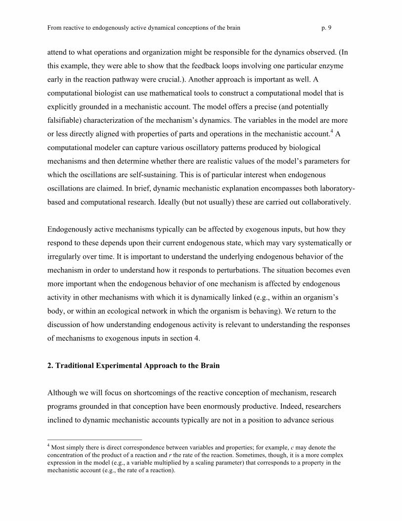
direction). As illustrated in Figure 2, Hubel and Wiesel (1962) proposed that a bar in the visual
From reactive to endogenously active dynamical conceptions of the brain p. 12
field spanned the receptive fields of several center-surround cells in the LGN. These were
connected to at least one simple cell that detected their joint activation, and multiple simple cells
(ideally with closely adjacent receptive fields) in turn were connected to each complex cell. A
complex cell was especially responsive if the simple cells connected to it were triggered in
sequence by a bar moving in the appropriate direction. The simple and complex cells can be
thought of as engaging in feature detection at two levels. In a subsequent study Hubel and Wiesel
replicated these findings of simple and complex cells in monkeys but also reached the conclusion
that their activity “represented a very elementary stage in the handling of complex forms” and
must be followed by further processing “at later stages in the visual path” (Hubel & Wiesel,
1968, p. 242). They began referring to striate cortex as V1 (visual area 1) and the areas involved
in later stages of processing as V2, V3, V4 and MT (medial temporal area)—clearly embracing
the conception of the visual system as a sequential processor, with its sequence of operations
initiated by presentation of a visual stimulus and culminating in a percept.
Figure 2. Hubel and Wiesel’s (1962) proposed simple and complex cells in striate cortex. (a)
Center-surround cells in LGN that detect spots are connected to simple cells that detect location-
and orientation-specific bars. (b) Simple cells are connected to complex cells that detect
orientation-specific bars, especially those moving in a particular direction.
Hubel and Wiesel’s strategy found numerous applications in subsequent years as researchers
inferred function from the classes of visual stimuli that drove responses in specific regions of
occipital, temporal, and parietal cortex. For example, neurons in area V4 were found to achieve
color constancy: in addition to responding to variations in the incoming wavelength due to
From reactive to endogenously active dynamical conceptions of the brain p. 13
changes in the color of an object (like V1) they compensate for variations due to changes in its
illumination. Similarly, neurons in area MT were discovered to respond to the perceived
direction of movement of complex stimuli, whereas those in V1 presented with the same stimuli
respond only to the direction in which components of the stimuli move across the visual field.
Subsequent research revealed regions in the temporal lobe that respond to specific classes of
objects and regions in the parietal lobe that respond to their spatial location (each with distinct
pathways from subareas of LGN, V1 and so forth). By the 1990s over thirty different brain areas
in the macaque had been identified as engaged in processing visual stimuli, and for many of
these areas research pursuing the approach just described succeeded in determining the specific
features of stimuli that evoked a response (van Essen & Gallant, 1994). Each successive brain
area was regarded as operating on the products generated in previous areas to extract new
information about the visual stimulus.
By any measure, this research endeavor that treated the visual system as reacting to visual stimuli
was extremely successful (for a detailed account of this century of research, see Bechtel, 2008).
There are reasons to suspect, however, that the resulting explanatory accounts may be
incomplete and require non-trivial revision. First, the approach assumes sequential processing of
inputs by a succession of processing centers. But researchers have long known that in addition to
forward axonal projections there are extensive backwards and collateral projections in this
system (Lorente de Nó, 1938). Hubel and Wiesel had found that the neurons they recorded were
organized into columns traversing the six layers of cortex and that neurons within a column
responded to stimuli in the same part of the visual field. Forward, backwards, and collateral
projections could be differentiated by the layers from which and to which they projected, which
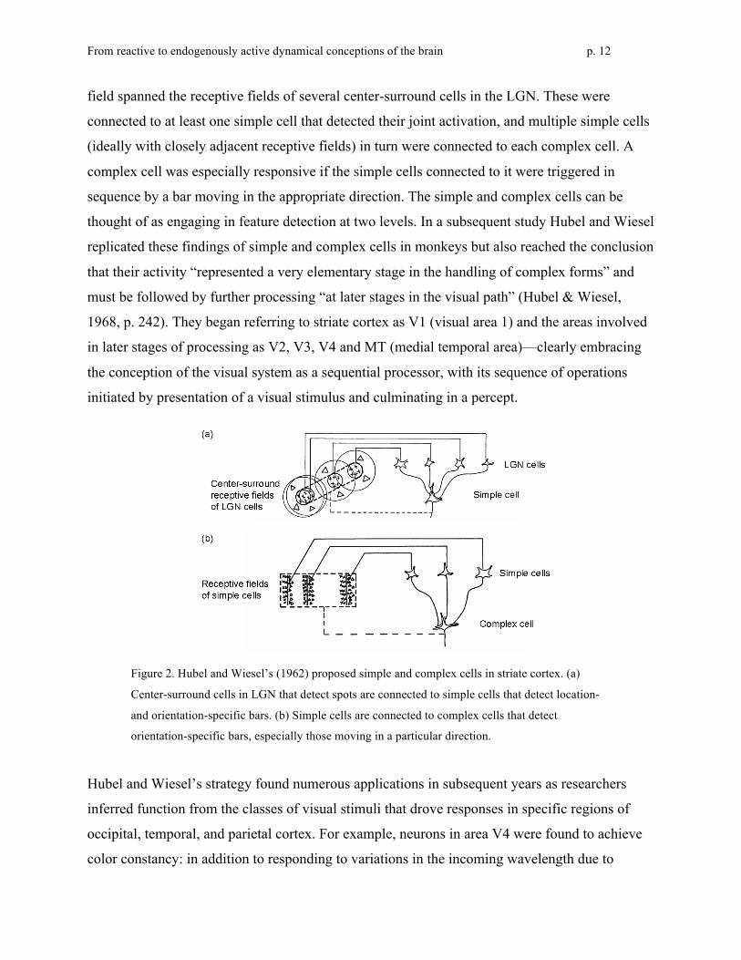
helped researchers uncover the complex pattern of connections between visual areas in the
macaque. The findings were displayed visually in the well-known “subway map” diagram by
Felleman and van Essen (1991), as shown in Figure 3. The other indication that sequentially-
based accounts require revision is that researchers have constantly confronted the problem that
neural responses are highly variable. This variability is generally regarded as noise which needs
to be removed so as to reveal the signal – but a very different research program emerges if
instead it is taken to indicate that much more may be going on within the mechanism than is
revealed by what is regarded as signal.
From reactive to endogenously active dynamical conceptions of the brain p. 14
Figure 3. Felleman and van Essen’s (1991) representation of 32 cortical visual areas identified in
the macaque and the known connections between them. In most cases the connections are
bidirectional, with separate bundles of axons running between different layers for feedforward vs.
feedback signals (not shown).
3. Reconceptualizing the Brain as Endogenously Active
The alternative conception of the brain as an endogenously active mechanism is being pursued
by a growing vanguard of neuroscientists. They are rethinking brain dynamics, are redirecting
the tools of their trade towards detection of endogenous activity, and are devising analyses that
can describe that activity and tease out interactions with activity evoked by stimuli. We will
discuss three key technologies in the order in which they began to be directed to uncovering
endogenous activity in the brain. Since they differ in their temporal and spatial range and
resolution, we conclude this section by asking how activities at multiple timescales might
interrelate.

From reactive to endogenously active dynamical conceptions of the brain p. 15
a. Electroencephalography (EEG)
The vision researchers discussed above implanted electrodes so as to record the activity of
individual neurons, but there is an even longer tradition of inferring aggregate activity from
electrodes placed on or into the scalp or (in animals or surgical patients) on the cortical surface.
The difference in electrical potential between two electrodes fluctuates over time, providing a
measure of electric currents in the brain with high temporal but low spatial resolution. In
pioneering research with rabbits and monkeys, Richard Caton (1875) experimented with various
placements of pairs of electrodes connected to a mirror galvanometer that represented the
currents visually. Despite primitive tools, he made the first observations both of continuous
spontaneous activity (“feeble currents”) and of localized “negative variation” evoked by a
stimulus.5
Caton’s technique was reinvented more than once, but did not give rise to an ongoing program of
research until psychiatrist Hans Berger adapted it to humans in the 1920s. Berger initially
inserted needle electrodes into subcutaneous tissue, often one at the front and one at the back of
the head, but found that he could obtain similar results with the less intrusive procedure of
affixing lead foil electrodes to the scalp. With electrodes connected to a string or double-coil
galvanometer that was attached to a recording apparatus, he could permanently capture
oscillations in the current as lines on long strips of paper (with some delay since the recording
involved a photographic process). In his first publication (Berger, 1929), he called this an
Elektrenkephalogramm or, in English, an electroencephalogram (EEG), in recognition of
existing electrocardiogram instrumentation which he had adapted. In patients and healthy
individuals at rest with eyes closed, he repeatedly observed two distinct waveforms. In his next
report (Berger, 1930), he coined the term alpha waves for the approximately 10 Hz oscillations
that most intrigued him and the term beta waves for smaller, faster 20-30 Hz oscillations.
Moreover, Berger discovered what was later called alpha blocking: when the eyes were opened
5 One of Caton’s objectives was to evaluate Ferrier’s claims regarding localization of motor commands, and he reports (p. 278): “on the areas shown by Dr. Ferrier to be related to rotation of the head and to mastication, negative variation of the current was observed to occur whenever those two acts respectively were performed. Impressions through the senses were found to influence the currents of certain areas; e. g., the currents of that part of the rabbit's brain which Dr. Ferrier has shown to be related to movements of the eyelids, were found to be markedly influenced by stimulation of the opposite retina by light.”
From reactive to endogenously active dynamical conceptions of the brain p. 16
alpha waves declined precipitously, leaving beta waves to predominate. Even with eyes closed,
events in other sensory modalities or attention-demanding tasks such as mental arithmetic could
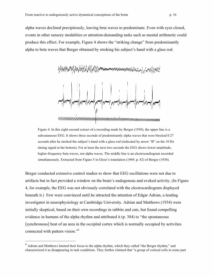
produce this effect. For example, Figure 4 shows the “striking change” from predominantly
alpha to beta waves that Berger obtained by stroking his subject’s hand with a glass rod.
Figure 4. In this eight-second extract of a recording made by Berger (1930), the upper line is a
subcutaneous EEG. It shows three seconds of predominantly alpha waves that were blocked 0.27
seconds after he stroked the subject’s hand with a glass rod (indicated by arrow “B” on the 10 Hz
timing signal at the bottom). For at least the next two seconds the EEG shows lower-amplitude,
higher-frequency beta waves, not alpha waves. The middle line is an electrocardiogram recorded
simultaneously. Extracted from Figure 5 in Gloor’s translation (1969, p. 82) of Berger (1930).
Berger conducted extensive control studies to show that EEG oscillations were not due to
artifacts but in fact provided a window on the brain’s endogenous and evoked activity. (In Figure
4, for example, the EEG was not obviously correlated with the electrocardiogram displayed
beneath it.) Few were convinced until he attracted the attention of Edgar Adrian, a leading
investigator in neurophysiology at Cambridge University. Adrian and Matthews (1934) were
initially skeptical, based on their own recordings in rabbits and cats, but found compelling
evidence in humans of the alpha rhythm and attributed it (p. 384) to “the spontaneous
[synchronous] beat of an area in the occipital cortex which is normally occupied by activities
connected with pattern vision.”6
6 Adrian and Matthews limited their focus to the alpha rhythm, which they called “the Berger rhythm,” and characterized it as disappearing in task conditions. They further claimed that “a group of cortical cells in some part

From reactive to endogenously active dynamical conceptions of the brain p. 17
With Adrian’s imprimatur, human EEG research attracted other pioneering investigators; within
a decade, three additional rhythms had been investigated and named. The term gamma rhythm
was proposed by Jasper and Andrews (1938) to designate frequencies above 30 or 35 Hz, but
high-quality evidence for functionally distinguishing gamma from beta rhythms is much more
recent and often involves evoked rather than endogenous activity. (Proposals regarding functions
of evoked gamma activity have included object perception, cross-modal perception, feature
binding, aspects of short-term memory, and even—focusing more on endogenous than evoked
activity—consciousness and ongoing information processing.) The other two rhythms involved
slower waveforms that Berger had already associated with brain lesions and sleep. W. Grey
Walter (1936), in reporting EEG studies of awake humans with brain tumors, proposed the term
delta rhythm for waves lower in frequency (and typically higher in amplitude) than the alpha
rhythm. Later he designed an automated frequency analyzer and, deploying it on EEGs from a
variety of patients, differentiated a primarily subcortical theta rhythm (4-7 Hz) from the slower
(<4 Hz), primarily cortical delta rhythm (Walter & Dovey, 1944).
The subsequent years brought improvements in recording technologies (e.g., digital EEG in the
1960s) and in analysis of complex EEG waveforms. Methods generally proceed from the
assumption that these waveforms can be decomposed into sinusoidal components of different
frequencies. Even Berger had noted that irregularities in the alpha waves (“notches” on their
descending limbs too small to see in Figure 4) indicated that they were always mixed with the
smaller beta waves. However, automated analysis as pioneered by Walter is far more revealing
than visual inspection of EEG recordings. Since the 1960s computers have allowed efficient
calculation of power density (a measure of amplitude) for each frequency or frequency band
within a time window. Current variations on this method use a fast Fourier or wavelet transform
of the EEG waveform. Herrmann, Grigutsch, & Busch (2005) provide an introduction to wavelet
of the occipital lobe . . . tend to beat synchronously when they are undisturbed, but visual activity or widespread non-visual activity in the brain breaks up the rhythm by exposing the cells to a mosaic of excitations which makes synchronous action impossible. Berger, if we have interpreted him correctly, regards the waves as having a much wider and less specific origin, but the evidence as to localization is the only important point on which our results seem to differ from his” (p. 356). Later researchers confirmed a primary localization in the occipital lobe, but also found other origins and/or broader activity under some conditions. Others confirmed that, although alpha rhythms are not prominent in most animal species, Berger was correct in reporting that they were prominent in dogs as well as humans (see especially pp. 239 and 256 in the review by Shaw, 2003.)
From reactive to endogenously active dynamical conceptions of the brain p. 18
analysis and suggest the following as well-established frequency bands: delta (0-4 Hz), theta (4-8
Hz), alpha (8-12 Hz), beta (12-30 Hz), gamma (30-80 Hz). Additional, less standardized bands
are sometimes specified by those taking advantage of recent advances in technology; examples
include infraslow (0.01 - 0.1 Hz), very slow (0.25 - 0.5 Hz), and very fast (100-500 Hz).
One early application of EEG was in differentiating stages of sleep, with the first comprehensive
proposal advanced by Loomis, Harvey, and Hobart (1937). Most recently (Silber et al., 2007),
the American Academy of Sleep Medicine has identified five stages: wakefulness, rapid eye
movement (REM) sleep, and three stages of lower-frequency, non-REM sleep (NREM 1, 2, and
3).
When people wind down at night, the beta and gamma waves of the cognitively active brain
typically yield to more relaxed alpha waves, which in turn become mixed with yet slower theta
waves as sleep approaches. By definition, an individual is awake (stage W) as long as alpha
exceeds theta activity, and makes the transition to NREM 1 sleep (stage N1) when theta exceeds
alpha activity. For those with little alpha activity, typical accompaniments such as slow rolling
eye movements are counted instead. NREM 2 sleep (stage N2) is characterized by one or more
half-second (or longer) episodes of high-amplitude patterned activity superimposed on a low-
amplitude, mixed-frequency background. One pattern, the sleep spindle, is a train of rhythmic
11-16 Hz waves that increase and then decrease in amplitude, producing a spindle shape in the
EGG. The other pattern, the K-complex, is a sharp negative wave immediately followed by a
slower positive component. NREM 3 sleep (stage N3) begins when at least 20% of activity is
slow wave sleep, restricted by definition to the lower end of the delta range (0.5-2 Hz, with peak-
to-peak amplitude above 75 µV). Typically these slow waves rise to over 50% of EEG activity,
treated as the threshold to a separate stage in older systems. Finally, REM sleep (stage R) has a
complex definition emphasizing three characteristics that tend to co-occur: its namesake rapid
eye movements (during which the most memorable dreaming can occur), low muscle tone, and
low-amplitude, mixed-frequency EEG activity (usually predominantly theta as in stage N1, but
alpha or sawtooth waves may be prominent). After its discovery in 1953, REM sleep was called
paradoxical sleep because the sleeper could not move even though the brain and other systems
were active.
From reactive to endogenously active dynamical conceptions of the brain p. 19
The sequence of stages in a prototypical night begins with W, then N1, then four or five
repetitions of N2-N3-N2-R, but variations are common. It should be kept in mind that the stages
are rigorously defined in part to assure comparability across research laboratories; in an actual
night’s sleep the passage from one stage to another often is gradual or ambiguous. The dynamic
character of sleep, and its relative isolation from environmental influences, make it a highly
relevant context for investigating edogenous activity in the brain.
In summary, EEG research first showed its worth as a means of differentiating overall brain
states. In the early decades it was most usefully applied to the discovery and characterization of
stages of sleep in terms of endogenous alpha, theta, and delta rhythms. From Berger forward,
researchers also recognized that a variety of cognitively active states were marked by faster
rhythms in the beta and gamma ranges. They lacked tools for distinguishing between these states,
however, as would be needed to move towards a brain-based account of cognitive operations and
their orchestration in complex tasks.
This changed beginning in the 1960s (for sensory processing) and 1970s (for more complex
cognitive processing). Instead of recording ongoing brain activity—primarily endogenous in
origin—investigators presented carefully chosen exogenous stimuli and looked for systematic
changes in the EEG pattern, especially within the first half-second or so following stimulus
onset. The time-locked waveform is referred to as an evoked potential (EP), evoked response
potential (ERP), or in cognitive investigations, event-related potential (also ERP).7 This
dramatically repositioned the EEG technology: now it could serve those who found a reactive
conception of the brain most promising for rapid gains in knowledge. Since response to a
stimulus was only one among many influences on the highly variable EEG waveform, however,
it was essential to average the waveforms obtained over multiple trials in which similar stimuli
were presented. Computer processing of digital EEG made this practical. In a classic experiment,
Kutas and Hillyard (1980) presented subjects with 160 seven-word sentences in which the final
word was either anomalous or semantically appropriate. Comparing the average ERPs, they
7 There was interest in such waveforms as far back as Berger (1920s) and Davis (1930s); what was new in the 1960s was powerful new tools for identifying and interpreting components.
From reactive to endogenously active dynamical conceptions of the brain p. 20
discovered a robust negative deflection peaking approximately 400 milliseconds after onset of an
anomalous word but not after an appropriate word. They interpreted this N400 component as
signaling reprocessing of anomalous semantic information and were able to distinguish it from
positive deflections signaling disconfirmation of an expectation (the already well-known P300)
or following a change in font size.
For our purposes, what is most noteworthy here is the considerable variability across trials in the
endogenous components of the EEG waveform that makes it challenging to extract the response
specifically evoked by an exogenous stimulus. Although this variability is viewed as noise from
a reactive perspective, as mentioned above, it can instead be viewed as reflecting the varied
endogenous origins of the brain’s ongoing activity. The challenge is to detect and analyze
patterns in this activity and uncover their origins and functions. For example, thalamocortical
oscillations seem to play a pivotal role in regulating communication between cortical areas
(Buzsáki, 2006). We discuss other proposals regarding the functional importance of endogenous
activity in section 4.
b. Recording from Individual Neurons
EEG rhythms were assumed to reflect neural activity, but what kind of neural activity? Two
opposing explanations were pursued in the 1940s and 1950s (see Kaada, 1953). One explanation
credited individual cortical neurons with the capacity for endogenous generation of rhythmic
firing. If such neurons synchronized their activity, this would be sufficient to produce the overall
rhythms observed in an EEG. The other explanation relied on a reactive conception of the neuron
now known as integrate-and-fire. It was assumed that each neuron continuously performed an
essentially linear integration of inputs received from other neurons at synapses on its dendritic
tree. When a threshold was exceeded it “fired”— that is, it sent an electric pulse (called an action
potential or spike) along its axon towards yet other neurons. This model was an important part of
the conceptual framework for work on the mammalian nervous system, which relied heavily on
single-cell recording studies of motor neurons in the spinal cord. The rate of spiking indicated
responsivity to a stimulus, and it was relatively straightforward to study propagation through
circuits of such neurons. Cortical neurons had much more complex connectivity patterns, but the
From reactive to endogenously active dynamical conceptions of the brain p. 21
same overall framework was assumed to apply to them. To account for alpha rhythms, for
example, it was suggested that a closed, self-re-exciting chain of integrate-and-fire neurons in
cortex, driven by thalamic input, could keep neural impulses circulating rhythmically. In the
version proposed by Eccles (1951, p. 462)8 a 10 Hz firing pattern—the alpha rhythm—was
assured by the fact that each neuron in the chain required approximately 100 msec. of recovery
time before it could respond to above-threshold input with an action potential. On this account,
endogenous rhythmicity was not required to explain observed rhythmicity.
Support for the alternative conception of the neuron as endogenously active came eventually,
from research on invertebrates that was rooted in the dominant reactive perspective but led in
new directions. The initial goal was to achieve a finegrained understanding of the action
potential itself as an event across time, and the giant axon of the squid provided easy access for
intracellular recording under controlled conditions. A key set of researchers were less interested
in how fast the electrical pulse traveled down the axon than in the timecourse and mechanism of
voltage changes at any point along the axon as the pulse passed. The primary mechanism turned
out to be ion movements across the axonal membrane, as captured by Hodgkin and Huxley
(1952) in an elegant set of equations. The membrane’s resting potential is approximately -65
millivolts, reflecting a normal predominance of negative ions inside and positive ions outside. As
a pulse arrives the voltage becomes less negative (depolarizes), triggering an influx of sodium
ions (Na+) from the extracellular space that drives the membrane potential into the positive range
(very quickly, due to positive feedback). At a short delay a less rapid efflux of potassium ions
(K+) brings voltage back into the negative range, first overshooting (hyperpolarizing) and then
returning to the resting potential.
Thus, the shape of the action potential is derived as the net effect of incoming sodium and
outgoing potassium currents. It was not until the 1970s to 1980s that researchers discovered the
molecular mechanism behind these dynamics: proteins in the membrane act as specialized ion
8 Eccles bemoaned the confusing accounts of cortical potentials and advocated the study of motor neurons in the spinal cord as affording a clearer perspective on neural activity: “It is the thesis of this paper that basically the responses of neurones are similar throughout the central nervous system, and that the more easily analysed responses of motoneurones provide the data for a satisfactory explanation of the electrical responses evoked in the cerebral cortex by all conditions of stimulation: by direct electrical stimulation; by afferent volleys; and by antidromic volleys” (Eccles, 1951, p. 449).

From reactive to endogenously active dynamical conceptions of the brain p. 22
channels, opening (or closing) as a function of voltage in the immediate vicinity and thereby
collectively offering high (or low) conductance to their particular type of ion (see Hille, 2001).
Voltage changes at one location on the membrane trigger channel-opening nearby, resulting in
propagation of the action potential along the axon.
Although the Hodgkin-Huxley equations were nonlinear, they fit into the dominant reactive
framework of the era in that the dynamics they described were those of a neuron firing in
response to a depolarizing input. Invertebrate researchers began moving towards an appreciation
for endogenous activity in the 1960s, however, as new findings emerged from intracellular
recording and related experiments. Notably, investigators found specialized pacemaker neurons
that generated their own rhythmic action potentials (Alving, 1968), as well as an unexpected
variety of voltage-gated and other currents producing complex dynamics not only in axons but
also in neurons’ dendrites and cell bodies (reviewed by Kandel, 1976).
Mammalian researchers initially doubted the generality of such findings, but took notice when
Rodolfo Llinás and colleagues found a variety of functionally important ion currents in neurons
of the inferior olive and cerebellum in mammals (and birds) in the 1970s and 1980s. Most were
spatially distributed and gated by voltage differently than the sodium and potassium channels in
the axon, equipping them for functions other than the direct generation of action potentials.
Notably, the dentrites were endowed with channels providing high-threshold conductance to
calcium (Ca2+) ions, enabling dynamically complex dendritic excitation in contrast to earlier
assumptions of passive transmission of signals from synapses.9 Moreover, the cell bodies of
some neurons in the inferior olive had a different kind of calcium channel with a seemingly
paradoxical low-threshold conductance that, in interaction with sodium and high-threshold
calcium conductances, enabled these neurons to function as single-cell oscillators “capable of
self-sustained rhythmic firing independent of synaptic input” (Llinás, 1988, p. 1659).10 They sent
9 This linked nerve excitability with the Ca2+-dependent second messenger system that is important for regulating general cellular functions. It also provided a mechanistic explanation of the suggestion (Bremer, 1958) that EEG primarily reflects synchronized post-synaptic potentials in dendrites—not, as originally thought, action potentials in axons. 10 For further exposition, see Buzsáki (2006, pp. 181-183), who comments: “These findings . . . illustrate that nature went to a lot of trouble bringing together these channels at the right densities and location just to serve one purpose: oscillation.” For evidence extending the findings to sensory neurons in various mammalian species, see Huguenard (1996).

From reactive to endogenously active dynamical conceptions of the brain p. 23
these rhythmic action potentials to target neurons in the cerebellum that were able to respond at
the same frequency, qualifying them as resonators in the dynamical lexicon championed by
Llinás—reacting, but in ways shaped by their internal properties. Llinás also investigated
spontaneous oscillations in electrical potentials elsewhere in the brain. Research on the thalamus
and thalamocortical relay neurons (e.g., Jahnsen & Llinás, 1984) proved particularly useful for
linking dynamic behavior of individual neurons to the large-scale dynamics seen in EEG.
Llinás’ research offers a radically different picture of neural activity than that featured in the
reactive framework. Neurons, on his account, are complex dynamic systems that are constantly
changing their states and spontaneously generating action potentials. To generate oscillations at
the different frequencies found in EEG requires the synchronization of many individual neurons,
but before neurons can synchronize they must first oscillate. By showing how they do so, Llinás
identified the needed foundation for the overall brain to exhibit complex dynamics. A number of
researchers have subsequently built upon this foundation, some examining how multiple ion
channels contribute to the intrinsic oscillatory activity of neurons and others determining how
different neurotransmitters and receptors affect the temporal dynamics of synaptic activity (see
Destexhe & Sejnowski, 2003, for a review and theoretical framework for modeling how these
endogenous and reactive processes interact in producing synchronous thalamocortical
oscillations). We return to this in section 5.
c. Functional magnetic resonance imaging (fMRI)
Single cell recording has been the workhorse technique of neuroscientists using animal models to
understand information processing in the brain, as seen in section 2, but with rare exceptions it is
too invasive for studying the human brain itself. It was the advent of functional neuroimaging
techniques that catalyzed study of the involvement of different brain areas in a variety of human
cognitive performances and gave rise to the distinct field of cognitive neuroscience. An existing
technology, positron emission tomography (PET), was adapted to this end in the 1980s by
Marcus Raichle and colleagues (as reviewed by Posner & Raichle, 1994), followed in the 1990s
by functional magnetic resonance imaging (fMRI). Each of these techniques detects changes in
blood flow in the brain that serve as a proxy for neuronal processing; most commonly used today

From reactive to endogenously active dynamical conceptions of the brain p. 24
is the BOLD (blood oxygen level dependent) signal from fMRI. Until recently the primary
strategy in both PET and fMRI studies has been to identify areas exhibiting higher signal
intensity (greater blood flow) when a subject is performing a target task than when performing a
control task. Those areas are said to be activated by whatever type of processing distinguishes
the two tasks.
If the stimulus is a word, for example, providing a semantically associated word versus merely
reading the stimulus word aloud would call upon semantic processing and thereby activate brain
areas responsible for the relevant semantic operations (Petersen et al., 1989). Often passive
viewing of the same stimulus is included as a control; in the same experiment, reading a word
aloud versus passively viewing it identified areas responsible for certain phonological operations.
Subtracting brain images to find activated areas has been a powerful strategy for localizing
cognitive operations in the brain, currently reaching a spatial resolution as small as 2 mm. It is a
clear success story for the reactive paradigm.
There is a twist, though, that has brought the endogenous perspective back into the story. In the
decades before neuroimaging was deployed on humans responding to stimuli in cognitive tasks,
a few researchers measured blood flow and brain activity in what has been called the resting
state. For Berger (and his successors even today) this was the most appropriate state for detecting
endogenous activity, especially alpha waves. In contemporary experimental designs, the term
references a control condition in which a subject is still with eyes closed and is presented with no
stimuli or task requirements (in variations, the eyes are open with or without a fixation point). In
a pioneering study using the xenon 133 inhalation technique to measure regional cerebral blood
flow, Ingvar (1975) discovered that subjects at rest exhibited high levels of frontal activity. He
surmised that this activity reflected “undirected, spontaneous, conscious mentation, the ‘brain
work,’ which we carry out when left alone undisturbed” (quoted by Buckner et al., 2008, p. 2). In
an even earlier study using nitrous oxide to measure cerebral metabolism, Sokoloff, Mangold,
Wechsler, Kennedy and Kety (1955) found that performing mental arithmetic did not increase
metabolism, indicating that the background activity was as energy demanding as any operations
involved in performing a cognitive task. Recently, Raichle and Mintun (2006) drew upon these
and other findings to argue that while the brain consumes 20% of the energy utilized in the body,
From reactive to endogenously active dynamical conceptions of the brain p. 25
it increases its consumption very little when performing tasks rather than resting. Humans
exhibit, it is then inferred, a great deal of endogenous activity even in the resting state.
The early results indicating substantial endogenous activity received little further attention once
PET and fMRI burst on the scene and researchers focused on identifying brain areas activated in
experimenter-defined tasks. Often, though, they included the resting state as a control condition,
presuming that it would elicit less activation than any of the tasks. A few of these researchers
were intrigued to find that certain brain areas around the midline instead exhibited greater
activation (stronger BOLD signal) in the resting state condition than in task conditions (e.g.,
Ghatan et al., 1995; Baker et al., 1996). They characterized these areas as deactivated during
tasks, but it should be kept in mind that this referred to low relative activity (not negative
activity). The choice of term reflects surprise that some of the subtractions (task condition minus
rest condition) would yield negative numbers.
To determine whether a common set of brain areas manifested task-induced deactivation,
Shulman et al. (1997; see also Mazoyer et al., 2001) conducted a meta-analysis of PET studies in
which a task condition was compared to a non-task condition in which the same stimulus was
present (which turned out to be similar in result to a resting condition with no stimulus). They
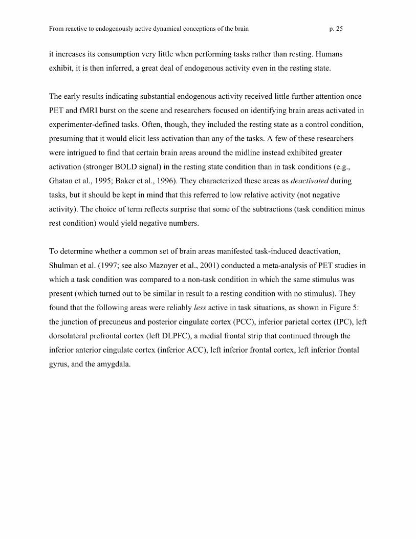
found that the following areas were reliably less active in task situations, as shown in Figure 5:
the junction of precuneus and posterior cingulate cortex (PCC), inferior parietal cortex (IPC), left
dorsolateral prefrontal cortex (left DLPFC), a medial frontal strip that continued through the
inferior anterior cingulate cortex (inferior ACC), left inferior frontal cortex, left inferior frontal
gyrus, and the amygdala.
From reactive to endogenously active dynamical conceptions of the brain p. 26
Figure 5. Metaimage from Shulman et al. (1997) in which areas showing decreases in blood flow
during task performance versus resting state are indicated in yellow and red. Components of this
default network are labeled by numerals as follows: Junction of posterior cingulate and precuneus
(1); Inferior parietal cortex (2, 3, 4); Left dorsolateral prefrontal cortex (5); Medial frontal strip
that continues through the inferior anterior cingulate cortex (6, 7, 8, 9, 10, 12); Left inferior frontal
cortex (11); Left inferior temporal gyrus (13); Right amygdala (14).
Shifting the focus from the fact that these areas are less active during tasks to the fact that they
are more active in the absence of task requirements, Raichle and his collaborators (Raichle et al.,
2001; Gusnard et al., 2001) proposed that these areas constitute a default network – one which
performs actual functions best carried out when there are no external task demands. There are
clues to those functions in Ingvar’s 1975 study, discussed above, and more directly in a
neuroimaging study of autobiographical memory. Andreasen et al. (1995) found that the areas
exhibiting heightened BOLD responses in a resting state condition were also relatively active in
an episodic memory task. In contrast, a different set of areas exhibited heightened BOLD
responses in a more typical semantic memory task. In an attempt to figure out what functions
might elicit increased activity during rest, the researchers queried the subjects. Their reports
pointed towards “a mixture of freely wandering past recollection, future plans, and other
personal thoughts and experiences”—activities that plausibly draw upon episodic memory.
Subsequent research has confirmed that thinking about one’s own experiences is among the
characteristic functions of the default network.
From reactive to endogenously active dynamical conceptions of the brain p. 27
The studies discussed so far focused on relative amount of activity in the default network under
various conditions, but not on the micro-temporal dynamics of this activity. Synchronized
oscillations would be a salient criterion for network status, but finding them with fMRI initially
seemed challenging due to the sluggish nature of the hemodynamic response. The feasibility of
such a temporal analysis of fMRI data was demonstrated first for networks activated by tasks.
Biswal, Yetkin, Haughton, and Hyde (1995) obtained BOLD signal values every 250 msec. for
two minutes following a simple motor task (moving a hand). They identified spontaneous very
low-frequency oscillations bilaterally in sensorimotor cortex, i.e., less than one cycle every 10
seconds (< 0.1 Hz). These oscillations were synchronized across the left and right hemispheres
and also with oscillations in other motor areas. The researchers interpreted their results as
indicting functional connectivity among the regions studied. Cordes et al. (2000) found similar
oscillations in resting state BOLD signals in networks of areas previously identified by their
synchronized activity in sensorimotor, visual, receptive language, or expressive language tasks.
Moreover, their functional connectivity MRI (fcMRI) analysis – applying correlational statistics
to resting state BOLD time series data to determine patterns of synchronization – yielded
functional networks very similar to those identified from activity during tasks. That is, areas
within the same network had correlated patterns of activity across time (rising and falling in
synchrony) regardless of whether overall level of activity was relatively high (e.g., the
sensorimotor network while moving a hand) or relatively low (e.g., the same network in a resting
state condition).
To begin assessing whether the regions proposed to constitute a default network likewise met the
criterion of synchronized oscillation, Greicius, Krasnow, Reiss, and Menon (2003) employed
fcMRI with two seed areas, the PCC and inferior ACC. They regarded their results as providing
“the most compelling evidence to date for the existence of a cohesive, tonically active, default
mode network” (p. 256) and argued that the PCC was a critical node in this network. When it
was used as the seed area for statistical analysis, its resting state oscillations were correlated with
those in much of medial prefrontal cortex (including inferior ACC and orbitofrontal cortex), left
DLPFC, IPC bilaterally, left inferolateral temporal cortex, and left parahippocampal gyrus. (One
of these synchronies is illustrated, using data from another study, in Figure 6.) This is almost the
same set of areas as those deactivated in task conditions according to Shulman’s et al.’s meta-
From reactive to endogenously active dynamical conceptions of the brain p. 28
analysis. Greicius et al. argued that this default network performs higher cognitive functions,
especially various forms of endogenously directed memory retrieval. Turning to their findings
for ventral ACC as the seed area, the correlated areas included the PCC, medial prefrontal
cortex/orbitofrontal cortex, the nucleus accumbens, and the hypothalamus/midbrain. They argued
that these primarily paralimbic and subcortical areas comprised a network important for
calibrating affective and autonomic operations, and further suggested that the strong connection
between inferior ACC and PCC provided a crucial link between the two networks.
Finally, the investigators confirmed that both networks became deactivated during a working
memory task but not in an eyes-closed or eyes-open resting state or even when passively viewing
a blinking checkerboard. Besides indicating a rather high threshold of cognitive demand for
deactivation of the default network, these findings clearly distinguished the default network from
the neural system responsible for alpha rhythms in EEG (which diminish when subjects open
their eyes). Subsequently Greicius and Menon (2004) found that the default network included the
hippocampus and Vincent et al. (2006) determined that by seeding an analysis with a
hippocampal region they could find correlated activity in the rest of the default network.11
Buckner, Andrews-Hanna, and Schacter (2008, pp. 4-5) summed up the perspective provided by
this research: “The default network is a brain system much like the motor system or the visual
system. It contains a set of interacting brain areas that are tightly functionally connected and
distinct from other systems within the brain.”
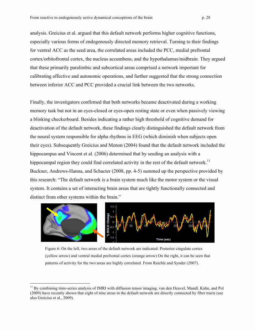
Figure 6: On the left, two areas of the default network are indicated: Posterior cingulate cortex
(yellow arrow) and ventral medial prefrontal cortex (orange arrow) On the right, it can be seen that
patterns of activity for the two areas are highly correlated. From Raichle and Synder (2007).
11 By combining time-series analysis of fMRI with diffusion tensor imaging, van den Heuvel, Mandl, Kahn, and Pol (2009) have recently shown that eight of nine areas in the default network are directly connected by fiber tracts (see also Greicius et al., 2009).

From reactive to endogenously active dynamical conceptions of the brain p. 29
Most of the known networks in the brain, in contrast to the default network, show more BOLD
activation during tasks than at rest. But even at rest there is enough activity to assess whether the
constituent areas of any such task-activated network fluctuate in synchrony with each other (but
not with the default network). Fox et al. (2005) selected a network that was especially active
during attention-demanding tasks (intraparietal sulcus, frontal eye field, middle temporal region,
supplementary motor areas, and the insula). Examining those areas in the resting state, they
found that fluctuations in their BOLD signals indeed were correlated. Moreover, fluctuations in
that network and in the default network were anticorrelated. That is, the areas that were
positively correlated within each network were negatively correlated with areas in the other
network—an outcome more interesting than a zero correlation. Fox et al. emphasized that:
anticorrelations may be as important as correlations in brain organization. Little has been
said previously in the neuronal synchrony literature regarding the role of anticorrelations.
While correlations may serve an integrative role in combining neuronal activity
subserving similar goals or representations, anticorrelations may serve a differentiating
role segregating neuronal processes subserving opposite goals or competing
representations (p. 9677).
This pattern of results in resting state data is a strong indicator that within both the default
network and the network involved in attention-demanding tasks, coordinated activity of some
kind goes on in the absence of external stimulation—activity that is is different for the two
networks.
Subsequently, researchers have used the strategy of finding correlations in resting-state
fluctuations to identify yet other networks. For example, temporally correlated activity was
found by Vincent et al. (2006) in the hippocampus and parietal memory systems and by Fox,
Corbetta, Synder, Vincent, and Raichle (2006) in the dorsal and ventral attention systems.
Deploying an alternative method, independent component analysis, on resting state fMRI data,
Mantini, Perrucci, Del Gratta, Romani and Corbetta (2007) differentiated six different
networks.12 Fox and Raichle (2007, p. 701) concluded: “A consistent finding is that regions with
similar functionality—that is, regions that are similarly modulated by various task paradigms—
12 An interesting finding in their study was the differentiation of the default network from what they characterize as a “self-referential network” that contains areas often associated with the default network: medial-ventral prefrontal cortex, the pregenual anterior cingulate, the hypothalamus, and the cerebellum.
From reactive to endogenously active dynamical conceptions of the brain p. 30
tend to be correlated in their spontaneous BOLD activity.”13 An important unanswered question
is how the anticorrelation is achieved—via a separate control system or as a direct result of
competition between networks.
d. Relating endogenous oscillations at different timescales
Spontaneous oscillations in brain activity for awake humans are found across a wide range of
timescales, depending on measurement technology and practices. Characteristic oscillations are
slowest in fMRI data (< 0.1 Hz), midrange in EEG (1-80 Hz), and fastest in single-cell recording
of action potentials (>100 Hz). This raises the question of whether neural activities as captured
by different methods at different timescales are independent or related.
Intriguing results have been reported by researchers who asked whether the networks inferred
from correlated BOLD signals in fMRI exhibit distinctive EEG signatures. Laufs et al. (2003)
first found that activation of the default network is associated with strong activity in the mid-beta
range, and activation of attention-related frontal and parietal areas is associated with weak
activity in the alpha range, i.e., alpha blocking. Mantini, et al. (2007) expanded this kind of
analysis to the six networks they had identified from resting state fMRI data, seeking
associations across as a broader range of EEG frequency bands. They found that “Each brain
network was associated with a specific combination of EEG rhythms, a neurophysiological
signature that constitutes a baseline for evaluating changes in oscillatory signals during active
behavior” (p. 13170). Their methods were sensitive enough to find alpha and beta rhythms
together providing a broader positive signature for the default network and negative signature for
the attention network. Beta recombined with delta and theta rhythms as the positive signature for
the auditory network and was the sole positive signature for the somatomotor network. Gamma
rhythms were the only positive signature for the self-referential network and the only frequency
band excluded for the vision network. It was assumed that different sets of neurons within an
13 Fox and Raichle also note that finding correlations in the resting state (“spontaneous BOLD activity”) is a good way of identifying the full range of components of a network, since many of them may not be activated by any particular task. “Interestingly, most memory tasks implicate only a subset of regions, whereas the hippocampal formation resting-state correlation map reveals the full distribution of memory-related regions assessed across multiple experiments. Patterns of spontaneous activity could thus serve as a functional localizer, providing a priori hypotheses about the way in which the brain will respond across a wide variety of task conditions” (p. 702).
From reactive to endogenously active dynamical conceptions of the brain p. 31
area were responsible for different rhythms, but also that the resting state BOLD signal indirectly
reflected the endogenous activity of those same neurons.
In another line of research, direct-current-coupled full-band electroencephalography has been
used to detect infraslow oscillations (.01 - .1 Hz, meaning a single cycle lasts 10 to 100 seconds).
The rising phase of a single ultraslow cycle is associated with (a) higher-amplitude activity in
each of the traditional EEG frequency bands and (b) improved accuracy at detecting a
somatosensory signal (Monto et al., 2008). Infraslow EEG oscillations are similar in timescale to
BOLD oscillations in fMRI, and there are indications that these are sensitive to some of the same
activity. For example, He, Snyder, Zempel, Smythe, and Raichle (2008) found a positive
correlation between slow cortical potentials (infraslow and delta activity combined) and
spontaneous fluctuations in the BOLD signal.
A final example of interrelatedness can be found in analyses of local field potentials, which are
recorded in animals by means of electrodes implanted in the extracellular space between
neurons. A variety of neural activities contribute to this signal. If dendritic and other lower-
frequency activities are of primary interest, high frequency oscillations reflecting action
potentials (>150 Hz) can be removed. The highest-amplitude oscillations contributing to the
remaining signal in monkeys turn out to be in the same frequency range (<0.1 Hz) as the
oscillations in the human BOLD signal (Leopold et al., 2003).
4. The Significance of Endogenous Oscillatory Brain Activity
In the previous section we reviewed evidence from EEG, single-cell recording, and fMRI studies
pointing to endogenous oscillatory activity in the brain. An advocate of the reactive framework
might acknowledge these findings but downplay their significance, maintaining that these
oscillations are appropriately regarded as noise with respect to any functional analysis of neural
performance. For example, one would expect basic metabolic activities to be maintained even in
the absence of task demands, so perhaps the endogenous electrical activity is merely an
epiphenomenon of ongoing metabolism. However, the evidence we reviewed indicated that the
oscillations within individual brain areas are periodic, not random, and that those areas are

From reactive to endogenously active dynamical conceptions of the brain p. 32
organized into networks within which oscillations are correlated and between which they are
anticorrelated. As well, these networks exhibit distinctive EEG signatures. This intricate
organization is highly persuasive that the endogenous activity in the brain is functional, and
invites the thought that this functional activity is so important that it cannot be ignored in seeking
to understand how the brain performs its functions. Here we briefly explore four different ways
in which endogenous activity may be crucial for understanding the brain mechanisms involved in
cognition.
First, it seems obvious that for any mechanism that responds to stimuli with an increase (or
decrease) in activity, but also exhibits ongoing endogenous oscillations, the magnitude of any
particular response will depend in part on the phase of that oscillation at the moment the stimulus
arrives. Fox, Snyder, Zacks, and Raichle (2006) devised an innovative strategy for demonstrating
that this is true of neural activity in somatomotor cortex (SMC). Specifically, they showed that
trial-to-trial variability in fMRI BOLD response to an exogenous stimulus could be attributed
largely to spontaneous (endogenous) fluctuations. Data were available from subjects who had
been instructed to press a button with the right hand each time an event was detected. This
evoked a BOLD response in left SMC, peaking on average at eight seconds following the button
press and then returning to baseline. However, there was considerable variability across trials.
Typically such variability is treated as random noise, but Fox et al. suspected that it reflected
endogenous oscillations on which the event-related responses were superimposed. That is, the
endogenous BOLD signal might happen to be relatively high or low or inbetween at the moment
a particular button press was required. The researchers surmised that simultaneous spontaneous
fluctuations in right SMC should serve as a good proxy for the endogenous contribution to the
activity in left SMC, since these were correlated in resting state and only left SMC was involved
in production of a right-hand button press. Indeed, they found that the task-related increase in the
left SMC BOLD signal could be analyzed as a linear addition to the amplitude of the
spontaneous fluctuation. A subsequent study (Fox et al., 2007) was designed to permit
calculations of trial-to-trial variability in the BOLD amplitude at each of eight timepoints
following a button press. As illustrated in Figure 7, variability in left SMC was significantly
reduced by subtracting out the corresponding activity in right SMC. This indicated that much of
the variability reflected ongoing spontaneous fluctuations – not noise – and that event-related
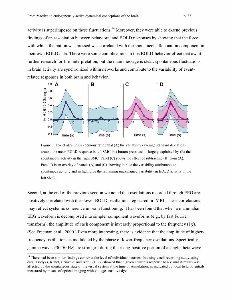
From reactive to endogenously active dynamical conceptions of the brain p. 33
activity is superimposed on these fluctuations.14 Moreover, they were able to extend previous
findings of an association between behavioral and BOLD responses by showing that the force
with which the button was pressed was correlated with the spontaneous fluctuation component in
their own BOLD data. There were some complications in this BOLD-behavior effect that await
further research for firm interpretation, but the main message is clear: spontaneous fluctuations
in brain activity are synchronized within networks and contribute to the variability of event-
related responses in both brain and behavior.
Figure 7. Fox et al.’s (2007) demonstration that (A) the variability (average standard deviation)
around the mean BOLD response in left SMC in a button press task is largely explained by (B) the
spontaneous activity in the right SMC. Panel (C) shows the effect of subtracting (B) from (A).
Panel D is an overlay of panels (A) and (C) showing in blue the variability attributable to
spontanous activity and in light blue the remaining unexplained variability in BOLD activity in the
left SMC.
Second, at the end of the previous section we noted that oscillations recorded through EEG are
positively correlated with the slower BOLD oscillations registered in fMRI. These correlations
may reflect systemic coherence in brain functioning. It has been found that when a mammalian
EEG waveform is decomposed into simpler component waveforms (e.g., by fast Fourier
transform), the amplitude of each component is inversely proportional to the frequency (1/f).
(See Freeman et al., 2000.) Even more interesting, there is evidence that the amplitude of higher-
frequency oscillations is modulated by the phase of lower-frequency oscillations. Specifically,
gamma waves (30-50 Hz) are strongest during the rising-positive portion of a single theta wave 14 There had been similar findings earlier at the level of individual neurons. In a single cell recording study using cats, Tsodyks, Kenet, Grinvald, and Arieli (1999) showed that a given neuron’s response to a visual stimulus was affected by the spontaneous state of the visual system at the time of stimulation, as indicated by local field potentials measured by means of optical imaging with voltage sensitive dye.
From reactive to endogenously active dynamical conceptions of the brain p. 34
(4-10 Hz) within which they are embedded, and in turn the small number of theta waves
concomitant with a single delta wave (1-4 Hz) are strongest during its rising-positive phase.
(These data and the "oscillatory hierarchy hypothesis" proposed to explain them are from
Lakatos et al., 2005; see also Canolty et al., 2006.) Such coupling can be particularly important
when the brain is perturbed by a stimulus, since a modulation in low-frequency oscillations can,
through phase-locking with higher-frequency oscillations, yield rapid changes at those
frequencies. In addition, oscillations at lower frequencies tend to synchronize over more widely
distributed areas of the brain than those at higher frequencies:
[The] 1/f power relationship implies that perturbations occurring at slow frequencies can
cause a cascade of energy dissipation at higher frequencies and that widespread slow
oscillations modulate faster local events. These properties of neuronal oscillators are the
result of the physical architecture of neuronal networks and the limited speed of neuronal
communication due to axon conduction and synaptic delays. Because most neuronal
connections are local, the period of oscillation is constrained by the size of the neuronal
pool engaged in a given cycle. Higher frequency oscillations are confined to a small
neuronal space, whereas very large networks are recruited during slow oscillations”
(Buzsáki & Draguhn, 2004, p. 1926)
In sum, as a result of endogenous activity maintaining synchronized oscillations at different
frequencies, the brain is able to generate coordinated responses to perturbations.
Third, endogenous activity in the brain’s default network is the most obvious candidate for the
neural underpinnings of mindwandering (Antrobus et al., 1970). Ingvar’s (1975) interpretation of
his early blood flow studies noted above suggested such a connection. Further support was
provided by Andreasen et al.’s (1995) subjects, whose informal reports after they had been
imaged in the resting state emphasized recollection and planning. These activities involve
episodic memory, and episodic memory tasks are among the few highly cognitive tasks for
which the default network’s BOLD signal remains as strong as in the resting state. Buckner and
Carroll (2007) in fact found that remembering the past, envisioning future events, and
considering the thoughts and perspectives of other people produced selective activation within
the default network. They construed these results as supporting the view that activity recorded
during the resting state reflects thinking—but thinking that is “engaged when individuals are left
From reactive to endogenously active dynamical conceptions of the brain p. 35
to think to themselves undisturbed” rather than elicited by specific stimuli or tasks (Buckner et
al., 2008, p. 1). They then hypothesized “that the fundamental function of the default network is
to facilitate flexible self-relevant mental explorations—simulations—that provide a means to
anticipate and evaluate upcoming events before they happen” (p. 2).15 In defending this view
they cite not only Andreasen at al.’s results but also correlations found by Mason et al. (2007)
between stimulus independent thoughts (as initially characterized by Antrobus et al., 1970) and
activity in the default network. Intriguingly, Li, Yan, Bergquist, and Sinha (2007) correlated
trials on which subjects failed to detect stop signals in behavioral tasks with increased activity in
the default network, as one would expect if that network were involved in a person thinking
distracting thoughts about past and future experiences.
One factor that renders problematic such a characterization of the activity of the default network
is that its oscillatory behavior is well maintained in sleep (Fukunaga et al., 2006; Larson-Prior et
al., 2009) and under anesthesia (Vincent et al., 2007)—circumstances in which spontaneous
conscious thoughts (such as Andreasen et al.’s subjects report) presumably are not occurring.
This suggests that the default network’s spontaneous activity is more foundational than originally
supposed. Fox and Raichle (2007) considered three possible interpretations of synchronization in
such activity between different areas in this network, as evidenced in correlated BOLD signals;
these interpretations have in common that they reflect cognitive processing but need not be
characterized in terms of conscious thought:
One possibility is that spontaneous activity serves as a record or memory of previous use,
showing correlations between regions that have been modulated together in a task-
dependent manner. Another possibility is that spontaneous activity serves to organize and
coordinate neuronal activity and that this coordination is more prominent between
regions that commonly work in concert. This is similar to the temporal binding, although
spontaneous BOLD occurs at a much slower, broader and more permanent scale. Finally,
spontaneous activity may represent a dynamic prediction about expected use, with
15 They also presented, but did not pursue, an alternative view that activity in the default network generates low-level generalized awareness or watchfulness (Gilbert et al., 2007). This view gains support from Hahn, Ross, and Stein’s (2007) findings of increased activity in the default network in a target-detecting task when the target could appear anywhere, but not when it was expected in a specific location.
From reactive to endogenously active dynamical conceptions of the brain p. 36
correlations occurring between regions that are likely to be used together in the future (p.
709).
Fourth, endogenous brain activity might be crucial for building and maintaining certain types of
organization in the nervous system required for cognitive activity. There is growing evidence
that the brain exhibits small-world organization (Watts & Strogratz, 1998) in which most
connections are between neighboring neurons, creating clusters that can collaborate in processing
specific information, but a few long range connections enable overall coordination. There also is
evidence that while most brain areas have connections to only a few other areas, some have a
large number of connections, thereby constituting hubs. Both neuroanotomical and
neurophysiological studies provide compelling evidence of such an architecture at different
scales in the brains of different organisms. Watts and Strogatz, for example, identified a small-
world architecture in the neural network of the nematode worm Caenorhabditis elegans, whose
structure had been identified by White, Southgate, Thomson, and Brenner (1986) using serial
reconstruction of electron microscopy sections. Also, as shown in Figure 8, Sporns and Zwi
(2004) developed a connection matrix for the 30 cortical areas and 311 connections Felleman
and van Essen (1991) identified in the macaque visual cortex and demonstrated that it exhibited
the features of a small-world network: short characteristic path lengths and high clustering. They
determined that some areas, such as V4, exhibit an atypically large number of connections to
other areas, qualifying them as hubs.

From reactive to endogenously active dynamical conceptions of the brain p. 37
Figure 8. Sporns and Zwi’s (2004) connection matrix based on Felleman and van Essen’s (1991)
depiction of connectivity in the macaque visual cortex (shown in Figure 3). Black squares indicate
a known connection and white squares indicate that no connection has been identified. The
connections are neither random nor purely local, but clustered so as to indicate a small-world
network architecture.
Such an architecture provides a highly efficient organization for information processing. It is
notable that the default network itself exhibits a small-world architecture, as indicated by
neuroanatomical studies on macaque homologues to the areas composing the human default
network. Some of these areas (PCC/Rsp, vMPFC, and IPL) are hubs that link the other areas into
a network. An important question is how such organization might arise. Rubinov, Sporns, van
Leeuwen, and Breakspear (2009) advanced the intriguing suggestion that oscillatory neurons, by
developing connections when synchronized, might self-organize into a small world network with
hubs. In support of this proposal they described a model by Gong and van Leeuwen (2004) that
employed a logistic map activation function for individual units that can individually exhibit
chaotic dynamics. Including a coupling factor enables the emergence of temporary patterns of
synchronized oscillations. A Hebbian learning procedure establishes new connections between
pairs of units whose activity is synchronized and prunes those between unsynchronized units.

From reactive to endogenously active dynamical conceptions of the brain p. 38
Even when these networks begin with random connectivity, they develop clusters linked to each
other through hubs.
Once this structure appears, it in turn generates more coherent patterns of synchronized activity
in subsequent processing, which leads via further Hebbian learning to a structural organization
that is likely to exhibit yet more coherent synchronized patterns. At the larger scale of human
brains, symbiotic interaction between the generation of functional synchrony and the building of
structural connectivity could well result in a highly coordinated brain capable of maintaining
multiple anticorrelated networks. The initial state in real brains presumably is already biased
towards a small-world pattern of connectivity, rather than random connections as in the
simulations, and experience further shapes the emerging organization by Hebbian or other kinds
of learning. Even so, the endogenous activity may be an important molding force so that the very
architecture of the information processing system is in an important way a consequence of both
endogenous and evoked activity.
In this section we have considered four suggestions as to how endogenous activity in the brain
may contribute to its functioning as a cognitive system. Although it is too early to judge which
construals will prove most fruitful, clearly the time for dismissing endogenous activity as mere
noise has passed.
5. Conclusion: Endogenous Brain Activity and Dynamic Mechanistic Explanation
In the last two sections we provided an overview of the now substantial body of evidence that the
brain is an endogenously active mechanism (or assemblage of mechanisms), one that is perturbed
by stimuli or task impositions but changes its activity in ways that depend not just on these
“inputs” but also on internal dynamics. This contrasts with the reactive framework of the vision
research discussed in section 2, in which brain activity is treated as a response evoked by a
stimulus. Llinás suggested viewing the interaction between stimuli and the brain’s endogenous
activity as a conversation:
Although sensory nerve pathways deliver messages to the CNS that are quite invariant
with respect to given sensory stimuli, the manner in which the CNS treats these messages

From reactive to endogenously active dynamical conceptions of the brain p. 39
depends on the functional state of each relay station. Thus, rather than a simple mirror of
the external world, the CNS embodies a dialogue between the internal states generated by
the intrinsic electrical activity of the nerve cells and their connectivity, which represents
the internal context, and the information that reaches the brain from the senses (Llinás,
1988, p. 1633).
As with spoken dialogue, we are unlikely to understand how the brain subserves cognitive
activity if we listen to only one side of the conversation: the message conveyed by a stimulus. It
is equally important to identify endogenous activity and its contribution to what the system does
next.
In this concluding section we return to the question of what conception of mechanism is best
suited to understanding the brain’s endogenous activity. Neuroscience has a long history of
offering explanations in terms of basic mechanisms: parts and operations organized to produce a
phenomenon, with that organization described qualitatively rather than quantitatively. The
definition offered by Machamer, Darden, and Craver (2000) adds the stipulation that the
mechanism’s activity leads from set-up to termination conditions, which suggests sequential
organization and situates basic mechanistic explanation within the reactive perspective that has
long dominated research. This approach has been highly productive, but it offers insufficient
resources for understanding the dynamics of endogenous activity. Oscillations and other complex
dynamics arise only in mechanisms with nonlinear component operations exhibiting some sort of
nonsequential or cyclic organization (particularly feedback loops). Thus, a conception of
mechanism adequate for endogenously active systems must reveal how nonsequentially
organized, nonlinear operations interact quantitatively to generate the overall behavior of the
mechanism. But when we as humans try to understand a particular system’s behavior, we find it
difficult to go beyond mental rehearsal of a sequence of operations; that is, our cognitive limits
predispose us to basic mechanistic explanation. To understand endogenously active mechanisms
we must turn to strategies that extend our capabilities.
The main scientific strategy for understanding how mechanisms generate complex behavior is
computational modeling, in which researchers mathematically describe the dependency of
changes in certain variables on changes in other variables, often by means of differential
From reactive to endogenously active dynamical conceptions of the brain p. 40
equations. As discussed in section 1, some computational models are explicitly grounded in
mechanistic accounts; that is, particular variables or other terms in their equations correspond to
particular properties of the mechanism’s parts and operations. Given such an alignment of
variables with properties, the modeler can simulate the functioning of the mechanism by
assigning initial values to variables and performing numerical integration with an appropriate
time step. The time evolution of each variable (the timecourse of its values) will depend in part
on these initial values, but also on the constants chosen for the model’s parameters. Sometimes
biologically plausible values that have worked well in other simulations are known; if not, the
modeler can determine best-fit values or, more interesting, determine how different values affect
the model’s behavior. Not infrequently, computational models can be best understood by
employing the concepts and representational tools of complexity or dynamical systems theory,
with possible implications for the mechanistic account. For example, when the time evolution of
two of the model’s variables meet criteria for a limit cycle in phase space, this indicates that the
mechanism is capable of producing sustained oscillations.
Thus, the type of explanation required is one that integrates these strategies: dynamic
mechanistic explanation. Mechanistic explanation identifies the parts and operations of a
mechanism and how they are organized, and computational modeling and tools of dynamical
systems theory reveal how such a mechanism will behave. This distinctive type of explanation is
playing an increasingly important role in the sciences and warrants attention in the new
mechanistic philosophy of science as well (Bechtel & Abrahamsen, 2010, 2011).
An instructive example of dynamic mechanistic explanation is provided by accounts of the
endogenous oscillatory capabilities of thalamocortical (TC) relay neurons. These neurons
produce sustained pacemaker oscillations – slow but regular firing at a frequency of 1-3 Hz –
during human sleep. The mechanism has been uncovered by in vitro investigations using cat and
rat TC neurons (Leresche et al., 1991). These oscillations rely upon the coordination of two
voltage-gated currents: (1) Ih is an influx of sodium and potassium ions through channels opened
when the membrane gets hyperpolarized following a spike. (2) IT is an influx of calcium ions
brought about when the depolarization due to Ih opens low-threshold calcium channels. The

From reactive to endogenously active dynamical conceptions of the brain p. 41
ensuing calcium spike16 further depolarizes the membrane, causing the various ion channels to
close. This results in temporary hyperpolarization of the membrane, and the cycle repeats.
The basic mechanistic account provides a spatial layout of the relevant parts (membrane, ion
channels, and ions) and specifies the sequence of their operations. The overall activity of the TC
neuron—its firing pattern—will depend on quantitative properties of these parts and operations.
Several different computational models aligned with the basic mechanistic account have
succeeded in simulating the pacemaker firing pattern. Dextexhe, Bal, McCormick and Sejnowski
(1996), for example, used Hodgkin-Huxley style equations that included variables for the
conductance and activation of each type of channel, membrane voltage, and other properties of
the TC neuron’s component parts and operations.17
One of the parameters specified the maximum conductance of the Ih current (essentially, the
membrane’s capacity to move sodium and potassium ions if the maximum number of relevant
channels were open). There were values of this parameter at which they succeeded in simulating
the pacemaker oscillation pattern. But even more interesting was the fact that values in a higher
range changed the dynamics of the conductance of the Ih current relative to that of the IT current
in such a way that the system now exhibited spindle-like oscillations. A series of spikes at about
1-3 Hz with a more or less spindle-shaped amplitude envelope would be observed for a few
seconds, followed by a quiet period of five seconds or longer, followed by another series of
spikes, then a quiet period, and so forth. This is the pattern that surprised Llinás in the 1980s and
drew him and others to a more dynamic conception of the neuron. Finally, there was yet a higher
range of values for the parameter that produced a quiet resting state.
16 This refers to a rapid depolarization of the membrane due to an influx of calcium, in contrast to the even more rapid depolarization that characterizes action potentials and involves channels specialized to sodium ions. In both cases the membrane then repolarizes less rapidly as the channels close. Hence, a plot of membrane voltage shows a rapid rise followed by a somewhat less rapid decline, typically overshooting—hyperpolarization—before returning to baseline. 17 Neither this basic mechanistic account nor the computational model enumerates individual ions and ion channels—there are simply too many. Thus, the basic mechanistic account states that there is large number of low-threshold calcium channels that can open or close and can vary in permeability. The computational account includes a conductivity variable that is specific to the type of channel and is presumed to represent the collective effect of the number of channels and their properties. Accounts at the level of individual channels are also available, but they focus on how a single channel works.

From reactive to endogenously active dynamical conceptions of the brain p. 42
Destexhe and Sejnowski (2003) provide a comprehensive guide to the many innovative models
and research investigations of thalamocortical neurons including subsequent empirical evidence
that their different patterns of activity are regulated in the manner proposed in their 1996 model.
Buzsáki (2006) makes the case that complex dynamics in various brain regions—not only
endogenous oscillations but also synchronized activity—are crucial to understanding how the
brain works.
One point to emphasize in closing is that adding a focus on dynamics is not intended to replace
the importance of traditional mechanistic research directed at identifying the parts, operations,
and overall organization of a mechanism. There are some who disagree, arguing for dynamical
explanation as a self-contained, successful competitor to mechanistic explanation (e.g., Chemero
& Silberstein, 2008). Dynamical accounts that are not grounded in accounts obtained by
decomposing mechanisms into their parts and operations do describe the activity of possible
systems, but those systems may not be be like those that are actually functioning in the world.
For a dynamical account to offer explanation, it must characterize activity of the actual
mechanism producing the phenomenon of interest. When the variables and terms in a
computational model are grounded in properties of well-established parts and operations, we
have a better basis for trusting the explanation. On the other hand, the model may reveal that the
mechanism, as delineated so far, accounts for some but not all aspects of the actual behavior.
Tinkering with the model to determine what sort of mechanism would account for these
additional aspects can then serve as a discovery heuristic by predicting the occurrence of new
parts, operations, or organizational relationships. Some of these predictions may be borne out,
some not, generating a dialectical engagement between mechanistic research and computational
model building. Such dialectical engagement may provide the only way neuroscience can
account for the endogenous dynamics exhibited in brains and ultimately the cognitive behavior
the brain supports.
From reactive to endogenously active dynamical conceptions of the brain p. 43
References
Adrian, E. D., & Matthews, B. H. C. (1934). The Berger rhythm: Potential changes from the occipita lobes in man. Brain, 57, 355-385.
Alving, B. O. (1968). Spontaneous activity in isolated somata of Aplysia pacemaker neurons. Journal of General Physiology, 51, 29-45.
Andreasen, N. C., O'Leary, D. S., Cizadlo, T., Arndt, S., Rezai, K., Watkins, G. L., et al. (1995). Remembering the past: two facets of episodic memory explored with positron emission tomography. American Journal of Psychiatry, 152, 1576-1585.
Antrobus, J. S., Singer, J. L., Goldstein, S., & Fortgang, M. (1970). Mindwandering and cognitive structure. Transactions of the New York Academy of Sciences, 32, 242-252.
Baker, S. C., Rogers, R. D., Owen, A. M., Frith, C. D., Dolan, R. J., Frackowiak, R. S. J., et al. (1996). Neural systems engaged by planning: a PET study of the Tower of London task. Neuropsychologia, 34, 515-526.
Bechtel, W. (2008). Mental mechanisms. London: Routledge. Bechtel, W., & Abrahamsen, A. (2005). Explanation: A mechanist alternative. Studies in History
and Philosophy of Biological and Biomedical Sciences, 36, 421-441. Bechtel, W., & Abrahamsen, A. (2009). Decomposing, recomposing, and situating circadian
mechanisms: Three tasks in developing mechanistic explanations. In H. Leitgeb & A. Hieke (Eds.), Reduction and elimination in philosophy of mind and philosophy of neuroscience (pp. 173-186). Frankfurt: Ontos Verlag.
Bechtel, W., & Abrahamsen, A. (2010). Dynamic mechanistic explanation: Computational modeling of circadian rhythms as an exemplar for cognitive science. Studies in History and Philosophy of Science Part A, 41, 321-333.
Bechtel, W., & Abrahamsen, A. (2011). Complex biological mechanisms: Cyclic, oscillatory, and autonomous. In C. A. Hooker (Ed.), Philosophy of complex systems. Handbook of the philosophy of science, Volume 10. New York: Elsevier.
Bechtel, W., & Richardson, R. C. (1993/2010). Discovering complexity: Decomposition and localization as strategies in scientific research. Cambridge, MA: MIT Press. 1993 edition published by Princeton University Press.
Berger, H. (1929). Über daas Elektroenkephalogramm des Menschen. Archiv für Psychiatrie und Nervenkrankheiten, 87.
Berger, H. (1930). Über daas Elektroenkephalogramm des Menschen. Zweite Mitteilung. Journal für Psychologie und Neurologie, 40.
Biswal, B., Yetkin, F. Z., Haughton, V. M., & Hyde, J. S. (1995). Functional connectivity in the motor cortex of resting human brain using echo-planar MRI. Magnetic Resonance in Medicine, 34, 537-541.
Bremer, F. (1958). Cerebral and cerebellar potentials. Physiological Reviews, 38, 357-388. Brown, T. G. (1914). On the nature of the fundamental activity of the nervous centres; together
with an analysis of the conditioning of rhythmic activity in progression, and a theory of the evolution of function in the nervous system. The Journal of Physiology, 48, 18-46.
Buckner, R. L., Andrews-Hanna, J. R., & Schacter, D. L. (2008). The Brain's default network. Annals of the New York Academy of Sciences, 1124, 1-38.
Buckner, R. L., & Carroll, D. C. (2007). Self-projection and the brain. Trends in Cognitive Sciences, 11, 49-57.
From reactive to endogenously active dynamical conceptions of the brain p. 44
Buzsáki, G. (2006). Rhythms of the brain. Oxford: Oxford University Press. Buzsáki, G., & Draguhn, A. (2004). Neuronal oscillations in cortical networks. Science, 304,
1926-1929. Canolty, R. T., Edwards, E., Dalal, S. S., Soltani, M., Nagarajan, S. S., Kirsch, H. E., et al.
(2006). High gamma power is phase-locked to theta oscillations in human neocortex. Science, 313, 1626-1628.
Caton, R. (1875). The electric currents of the brain. British Medical Journal, 2. Chemero, A., & Silberstein, M. (2008). After the philosophy of mind: Replacing scholasticism
with science. Philosophy of Science, 75, 1-27. Cordes, D., Haughton, V. M., Arfanakis, K., Wendt, G. J., Turski, P. A., Moritz, C. H., et al.
(2000). Mapping functionally related regions of brain with functional connectivity MR imaging. American Journal of Neuroradiology, 21, 1636-1644.
Darden, L., & Craver, C. (2002). Strategies in the interfield discovery of the mechanism of protein synthesis. Studies in History and Philosophy of Biological and Biomedical Sciences, 33, 1-28.
Destexhe, A. D., Bal, T., McCormick, D. A., & Sejnowski, T. J. (1996). Ionic mechanisms underlying synchronized oscillations and propagating waves in a model of ferret thalamic slices. Journal of Neurophysiology, 76, 2049-2070.
Destexhe, A. D., & Sejnowski, T. J. (2003). Interactions between membrane conductances underlying thalamocortical slow-wave oscillations. Physiological Reviews, 83, 1401-1453.
Eccles, J. C. (1951). Interpretation of action potentials evoked in the cerebral cortex. Electroencephalography and Clinical Neurophysiology, 3, 449-464.
Felleman, D. J., & van Essen, D. C. (1991). Distributed hierarchical processing in the primate cerebral cortex. Cerebral Cortex, 1, 1-47.
Ferrier, D. (1876). The functions of the brain. London: Smith, Elder, and Company. Finger, S. (1994). Origins of neuroscience. Oxford: Oxford University Press. Flourens, J. P. M. (1824). Researches Expérimentales sur les Propriétés et les Fonctions du
Système Nerveux dans les Animaux Vertébris. Paris: Crevot. Fox, M. D., Corbetta, M., Snyder, A. Z., Vincent, J. L., & Raichle, M. E. (2006). Spontaneous
neuronal activity distinguishes human dorsal and ventral attention systems. Proceedings of the National Academy of Sciences, 103, 10046-10051.
Fox, M. D., & Raichle, M. E. (2007). Spontaneous fluctuations in brain activity observed with functional magnetic resonance imaging. Nature Reviews Neuroscience, 8, 700-711.
Fox, M. D., Snyder, A. Z., Vincent, J. L., Corbetta, M., Van Essen, D. C., & Raichle, M. E. (2005). The human brain is intrinsically organized into dynamic, anticorrelated functional networks. Proceedings of the National Academy of Sciences of the United States of America, 102, 9673-9678.
Fox, M. D., Snyder, A. Z., Vincent, J. L., & Raichle, M. E. (2007). Intrinsic fluctuations within cortical systems account for intertrial variability in human behavior. Neuron, 56, 171-184.
Fox, M. D., Snyder, A. Z., Zacks, J. M., & Raichle, M. E. (2006). Coherent spontaneous activity accounts for trial-to-trial variability in human evoked brain responses. Nature Neuroscience, 9, 23-25.
From reactive to endogenously active dynamical conceptions of the brain p. 45
Freeman, W. J., Rogers, L. J., Holmes, M. D., & Silbergeld, D. L. (2000). Spatial spectral analysis of human electrocorticograms including the alpha and gamma bands. Journal of Neuroscience Methods, 95, 111-121.
Fukunaga, M., Horovitz, S. G., van Gelderen, P., de Zwart, J. A., Jansma, J. M., Ikonomidou, V. N., et al. (2006). Large-amplitude, spatially correlated fluctuations in BOLD fMRI signals during extended rest and early sleep stages. Magnetic Resonance Imaging, 24, 979-992.
Ghatan, P. H., Hsieh, J. C., Wirsén-Meurling, A., Wredling, R., Eriksson, L., Stone-Elander, S., et al. (1995). Brain activation induced by the perceptual maze test: A PET study of cognitive performance. Neuroimage, 2, 112-124.
Gilbert, S. J., Dumontheil, I., Simons, J. S., Frith, C. D., & Burgess, P. W. (2007). Comment on "Wandering minds: The default network and stimulus-independent thought". Science, 317, 43b-.
Glennan, S. (1996). Mechanisms and the nature of causation. Erkenntnis, 44, 50-71. Glennan, S. (2002). Rethinking mechanistic explanation. Philosophy of Science, 69, S342-S353. Gloor, P. (1969). Hans Berger on the electroencephalogram of man. Amsterdam: Elsevier. Gong, P., & van Leeuwen, C. (2004). Evolution to a small-world network with chaotic units.
Europhysics Letters, 67, 328-333. Goodwin, B. C. (1965). Oscillatory behavior in enzymatic control processes. Advances in
Enzyme Regulation, 3, 425-428. Greicius, M. D., Krasnow, B., Reiss, A. L., & Menon, V. (2003). Functional connectivity in the
resting brain: A network analysis of the default mode hypothesis. Proceedings of the National Academy of Sciences of the United States of America, 100, 253-258.
Greicius, M. D., & Menon, V. (2004). Default-mode activity during a passive sensory task: Uncoupled from deactivation but impacting activation. Journal of Cognitive Neuroscience, 16, 1484-1492.
Greicius, M. D., Supekar, K., Menon, V., & Dougherty, R. F. (2009). Resting-state functional connectivity reflects structural connectivity in the default mode network. Cerebral Cortex, 19, 72-78.
Grillner, S. (2003). The motor infrastructure: from ion channels to neuronal networks. Nature Reviews Neuroscience, 4, 573-586.
Gusnard, D. A., Akbudak, E., Shulman, G. L., & Raichle, M. E. (2001). Medial prefrontal cortex and self-referential mental activity: Relation to a default mode of brain function. Proceedings of the National Academy of Sciences of the United States of America, 98, 4259-4264.
Hahn, B., Ross, T. J., & Stein, E. A. (2007). Cingulate activation increases dynamically with response speed under stimulus unpredictability. Cerebral Cortex, 17, 1664-1671.
He, B. J., Snyder, A. Z., Zempel, J. M., Smyth, M. D., & Raichle, M. E. (2008). Electrophysiological correlates of the brain's intrinsic large-scale functional architecture. Proceedings of the National Academy of Sciences, 105, 16039-16044.
Henschen, S. E. (1893). On the visual path and centre. Brain, 16, 170-180. Herrmann, C. S., Grigutsch, M., & Busch, N. A. (2005). EEG oscillations and wavelet analysis.
In T. Handy (Ed.), Event-related potentials: a methods handbook (pp. 229-259). Cambridge, MA: MIT.
Hille, B. (2001). Ion channels of excitable membranes. Sunderland, MA: Sinauer.

From reactive to endogenously active dynamical conceptions of the brain p. 46
Hodgkin, A. L., & Huxley, A. F. (1952). A quantitative description of membrane current and its application to the conduction and excitation of nerve. Journal of Physiology, 117, 500-544.
Holmes, G. M. (1918). Disturbances of visual orientation. The British Journal of Ophthalmology, 2, 449-468.
Hubel, D. H., & Wiesel, T. N. (1962). Receptive fields, binocular interaction and functional architecture in the cat's visual cortex. Journal of Physiology, 160, 106-154.
Hubel, D. H., & Wiesel, T. N. (1968). Receptive fields and functional architecture of monkey striate cortex. Journal of Physiology, 195, 215-243.
Huguenard, J. R. (1996). Low-threshold calcium currents in central nervous system neurons. Annual Review of Physiology, 58, 329-348.
Ingvar, D. H. (1975). Patterns of brain activity revealed by measurements of regional cerebral blood flow. In D. H. Ingvar & N. A. Lassen (Eds.), Brain work: The coupling of function, metabolism, and blood flow in the brain: Proceedings of the Alfred Benzon Symposium VIII, Copenhagen, 26-30 May 1974, held at the premises of the Royal Danish Academy of Sciences and Letters, Copenhagen (pp. 397-413). New York: Academic.
Inouye, T. (1909). Die Sehstörungen bei Schussverletzungen der kortikalen Sehsphäre nach Beobachtungen an Verwundeten der letzten japanischen Kriege. Leipzig: Engelmann.
Jahnsen, H., & Llinás, R. R. (1984). Electrophysiological properties of guinea-pig thalamic neurones: an in vitro study. The Journal of Physiology, 349, 205-226.
Jasper, H. H., & Andrews, H. L. (1938). Brain potentials and voluntary muslce activity in man. Journal of Neurophysiology, 1, 87-100.
Kaada, B. R. (1953). Electrical activity of the brain. Annual Review of Physiology, 15, 39-62. Kandel, E. R. (1976). Cellular basis of behavior: An introduction to behavioral neurobiology.
San Francisco: W. H. Freeman. Kuffler, S. W. (1953). Discharge patterns and functional organization of mammalian retina.
Journal of Neurophysiology, 16, 37-68. Kutas, M., & Hillyard, S. A. (1980). Reading senseless sentences: brain potentials reflect
semantic incongruity. Science, 207, 203-205. Lakatos, P., Shah, A. S., Knuth, K. H., Ulbert, I., Karmos, G., & Schroeder, C. E. (2005). An
oscillatory hierarchy controlling neuronal excitability and stimulus processing in the auditory cortex. Journal of Neurophysiology, 94, 1904-1911.
Larson-Prior, L. J., Zempel, J. M., Nolan, T. S., Prior, F. W., Snyder, A. Z., & Raichle, M. E. (2009). Cortical network functional connectivity in the descent to sleep. Proceedings of the National Academy of Sciences, 106, 4489-4494.
Lashley, K. S. (1929). Brain mechanisms and intelligence. Chicago: University of Chicago Press.
Laufs, H., Krakow, K., Sterzer, P., Eger, E., Beyerle, A., Salek-Haddadi, A., et al. (2003). Electroencephalographic signatures of attentional and cognitive default modes in spontaneous brain activity fluctuations at rest. Proceedings of the National Academy of Sciences of the United States of America, 100, 11053-11058.
Leopold, D. A., Murayama, Y., & Logothetis, N. K. (2003). Very Slow Activity Fluctuations in Monkey Visual Cortex: Implications for Functional Brain Imaging. Cereb. Cortex, 13, 422-433.
From reactive to endogenously active dynamical conceptions of the brain p. 47
Leresche, N., Lightowler, S., Soltesz, I., Jassik-Gerschenfeld, D., & Crunelli, V. (1991). Low-frequency oscillatory activities intrinsic to rat and cat thalamocortical cells. The Journal of Physiology, 441, 155-174.
Li, C.-S. R., Yan, P., Bergquist, K. L., & Sinha, R. (2007). Greater activation of the "default" brain regions predicts stop signal errors. Neuroimage, 38, 640-648.
Llinás, R. R. (1988). The intrinsic electrophysiological properties of mammalian neurons: Insights into central nervous system function. Science, 242, 1654-1664.
Loomis, A. L., Harvey, E. N., & Hobart, G. A. (1937). Cerebral states during sleep, as studied by human brain potentials. Journal of Experimental Psychology, 21, 127-144.
Lorente de Nó, R. (1938). Analysis of the activity of the chains of internuncial neurons. Journal of Neurophysiology, 1, 207-244.
Machamer, P., Darden, L., & Craver, C. F. (2000). Thinking about mechanisms. Philosophy of Science, 67, 1-25.
Mantini, D., Perrucci, M. G., Del Gratta, C., Romani, G. L., & Corbetta, M. (2007). Electrophysiological signatures of resting state networks in the human brain. Proceedings of the National Academy of Sciences, 104, 13170-13175.
Mason, M. F., Norton, M. I., Van Horn, J. D., Wegner, D. M., Grafton, S. T., & Macrae, C. N. (2007). Wandering Minds: The Default Network and Stimulus-Independent Thought. Science, 315, 393-395.
Mazoyer, B., Zago, L., Mellet, E., Bricogne, S., Etard, O., Houdé, O., et al. (2001). Cortical networks for working memory and executive functions sustain the conscious resting state in man. Brain Research Bulletin, 54, 287-298.
Monto, S., Palva, S., Voipio, J., & Palva, J. M. (2008). Very Slow EEG Fluctuations Predict the Dynamics of Stimulus Detection and Oscillation Amplitudes in Humans. J. Neurosci., 28, 8268-8272.
Petersen, S. E., Fox, P. T., Posner, M. I., Mintun, M., & Raichle, M. E. (1989). Positron emission tomographic studies of the processing single words. Journal of Cognitive Neuroscience, 1, 153-170.
Planck, M. (1949). Scientific autobiography, and other papers (F. Gaynor, Trans.). New York: Philosophical Library.
Posner, M. I., & Raichle, M. E. (1994). Images of Mind. San Francisco: Freeman. Raichle, M. E., MacLeod, A. M., Snyder, A. Z., Powers, W. J., Gusnard, D. A., & Shulman, G.
L. (2001). A default mode of brain function. Proceedings of the National Academy of Sciences of the United States of America, 98, 676-682.
Raichle, M. E., & Mintun, M. A. (2006). Brain work and brain imaging. Annual Review of Neuroscience, 29, 449-476.
Raichle, M. E., & Snyder, A. Z. (2007). A default mode of brain function: A brief history of an evolving idea. Neuroimage, 37, 1083-1090.
Rubinov, M., Sporns, O., van Leeuwen, C., & Breakspear, M. (2009). Symbiotic relationship between brain structure and dynamics. BMC Neuroscience, 10, 55.
Shaw, J. C. (2003). The brain’s alpha rhythms and the mind: A review of classical and modern studies of the alpha rhythm component of the Electroencephalogram with commentaries on associated neuroscience and neuropsychology. Amsterdam: Elsevier Publishers B.V.
Sherrington, C. S. (1913). Further observations on the production of reflex stepping by combination of reflex excitation with reflex inhibition. The Journal of Physiology, 47, 196-214.
From reactive to endogenously active dynamical conceptions of the brain p. 48
Sherrington, C. S. (1923). The integrative action of the nervous system. New Haven: Yale University Press.
Shulman, G. L., Fiez, J. A., Corbetta, M., Buckner, R. L., Miezin, F. M., Raichle, M. E., et al. (1997). Common blood flow changes across visual tasks. II. Decreases in cerebral cortex. [Article]. Journal of Cognitive Neuroscience, 9, 648-663.
Silber, M. H., Ancoli-Israel, S., Bonnet, M. H., Chokroverty, S., Grigg-Damberger, M. M., Hirshkowitz, M., et al. (2007). The visual scoring of sleep in adults. Journal of Clinical Sleep Medicine, 3, 121-131.
Sokoloff, L., Mangold, R., Wechsler, R. L., Kennedy, C., & Kety, S. S. (1955). The effect of mental arithmetic on cerebral circulation and metabolism. Journal of Clinical Investigation, 34, 1101-1108.
Sporns, O., & Zwi, J. D. (2004). The small world of the cerebral cortex. Neuroinformatics, 2, 145-162.
Stuart, D. G., & Hultborn, H. (2008). Thomas Graham Brown (1882-1965), Anders Lundberg (1920-), and the neural control of stepping. Brain Research Reviews, 59, 74-95.
Thagard, P. (2003). Pathways to biomedical discovery. Philosophy of Science, 70, 235-254. Tsodyks, M., Kenet, T., Grinvald, A., & Arieli, A. (1999). Linking spontaneous activity of single
cortical neurons and the underlying functional architecture. Science, 286, 1943-1946. van den Heuvel, M. P., Mandl, R. C. W., Kahn, R. S., & Pol, H. E. H. (2009). Functionally
linked resting-state networks reflect the underlying structural connectivity architecture of the human brain. Human Brain Mapping, 30, 3127-3141.
van Essen, D. C., & Gallant, J. L. (1994). Neural mechanisms of form and motion processing in the primate visual system. Neuron, 13, 1-10.
Vincent, J. L., Patel, G. H., Fox, M. D., Snyder, A. Z., Baker, J. T., Van Essen, D. C., et al. (2007). Intrinsic functional architecture in the anaesthetized monkey brain. Nature, 447, 83-86.
Vincent, J. L., Snyder, A. Z., Fox, M. D., Shannon, B. J., Andrews, J. R., Raichle, M. E., et al. (2006). Coherent spontaneous activity identifies a hippocampal-parietal memory network. Journal of Neurophysiology, 96, 3517-3531.
Walter, W. G. (1936). The location of cerebral tumours by electro-encephalography. The Lancet, 228, 305-308.
Walter, W. G., & Dovey, V. J. (1944). Electro-encephalography in cases of sub-cortical tumour. Journal of Neurology, Neurosurgery and Psychiatry, 7, 57-65.
Watts, D., & Strogratz, S. (1998). Collective dynamics of small worlds. Nature, 393. White, J. G., Southgate, E., Thomson, J. N., & Brenner, S. (1986). The structure of the nervous
system of the nematode Caenorhabditis elegans. Philosophical Transactions of the Royal Society of London. B, Biological Sciences, 314, 1-340.
Wilson, D. M., & Wyman, R. J. (1965). Motor output patterns during random and rhythmic stimulation of locust thoracic ganglia. Biophysical Journal, 5, 121-143.
Wimsatt, W. C. (1976). Reductive explanation: A functional account. In R. S. Cohen, C. A. Hooker, A. C. Michalos & J. van Evra (Eds.), PSA-1974 (pp. 671-710). Dordrecht: Reidel.
Wimsatt, W. C. (2007). Re-engineering philosophy for limited beings: Piecewise approximations to reality. Cambridge, MA: Harvard University Press.


![Finding Endogenously Formed Communitiesninamf/papers/communities.pdfarXiv:1201.4899v2 [cs.DS] 1 Mar 2012 Finding Endogenously Formed Communities Maria-Florina Balcan∗ Christian Borgs†](https://static.fdocuments.net/doc/165x107/5f4e4259ea5a0056584f344a/finding-endogenously-formed-ninamfpaperscommunitiespdf-arxiv12014899v2-csds.jpg)