Frolich, Human Anatomy,UpprLimb UPPER LIMB What is a limb? Sensory to upper limb Making it move...
20
Frolich, Human Anatomy,UpprLimb UPPER LIMB • What is a limb? • Sensory to upper limb • Making it move – Bones and joints – Muscles and nerves • Vascular supply • Surface anatomy • (muscle study hint)
-
date post
21-Dec-2015 -
Category
Documents
-
view
222 -
download
1
Transcript of Frolich, Human Anatomy,UpprLimb UPPER LIMB What is a limb? Sensory to upper limb Making it move...
Frolich, Human Anatomy,UpprLimb
UPPER LIMB
• What is a limb?• Sensory to upper limb• Making it move
– Bones and joints– Muscles and nerves
• Vascular supply• Surface anatomy• (muscle study hint)
Frolich, Human Anatomy,UpprLimb
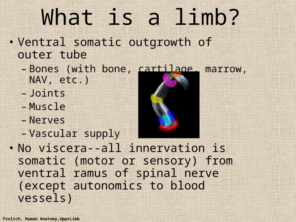
What is a limb?• Ventral somatic outgrowth of outer tube
– Bones (with bone, cartilage, marrow, NAV, etc.)
– Joints– Muscle– Nerves– Vascular supply
• No viscera--all innervation is somatic (motor or sensory) from ventral ramus of spinal nerve (except autonomics to blood vessels)
Frolich, Human Anatomy,UpprLimb
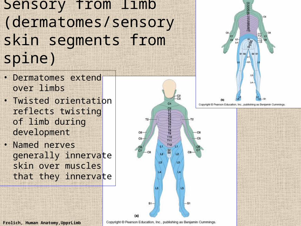
Sensory from limb (dermatomes/sensory skin segments from spine)
• Dermatomes extend over limbs
• Twisted orientation reflects twisting of limb during development
• Named nerves generally innervate skin over muscles that they innervate
Frolich, Human Anatomy,UpprLimb
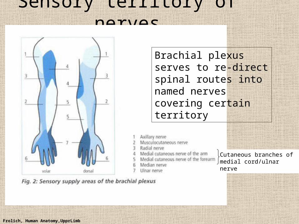
Sensory territory of nerves
Brachial plexus serves to re-direct spinal routes into named nerves covering certain territory
Cutaneous branches of medial cord/ulnar nerve
Frolich, Human Anatomy,UpprLimb
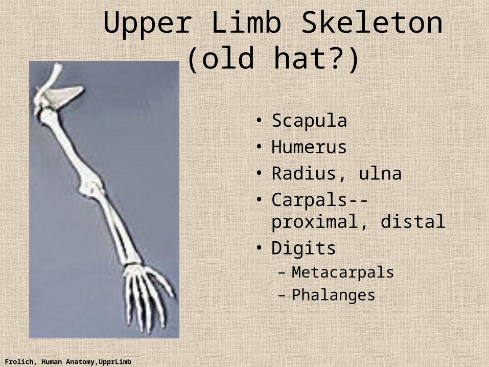
Upper Limb Skeleton (old hat?)
• Scapula• Humerus• Radius, ulna• Carpals--proximal,
distal• Digits
– Metacarpals
– Phalanges
Frolich, Human Anatomy,UpprLimb
JointsJOINT BETWEEN MOVEMENT TYPE
Frolich, Human Anatomy,UpprLimb
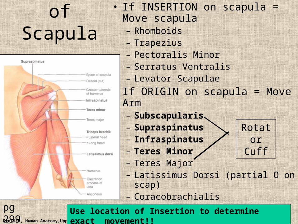
Muscles of Scapula
• If INSERTION on scapula = Move scapula– Rhomboids– Trapezius– Pectoralis Minor– Serratus Ventralis– Levator Scapulae
• If ORIGIN on scapula = Move Arm– Subscapularis– Supraspinatus– Infraspinatus– Teres Minor– Teres Major– Latissimus Dorsi (partial O on scap)– Coracobrachialis
pg 299
Rotator Cuff
Use location of Insertion to determine exact movement!!
Frolich, Human Anatomy,UpprLimb
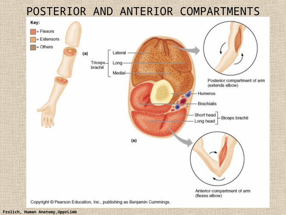
POSTERIOR AND ANTERIOR COMPARTMENTS
Frolich, Human Anatomy,UpprLimb
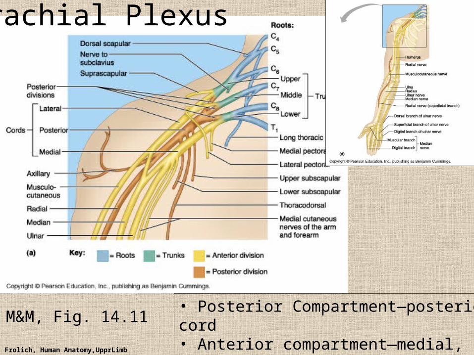
M&M, Fig. 14.11• Posterior Compartment—posterior cord• Anterior compartment—medial, lateral cords• Name of cord is relative to axillary artery
Brachial Plexus
Frolich, Human Anatomy,UpprLimb
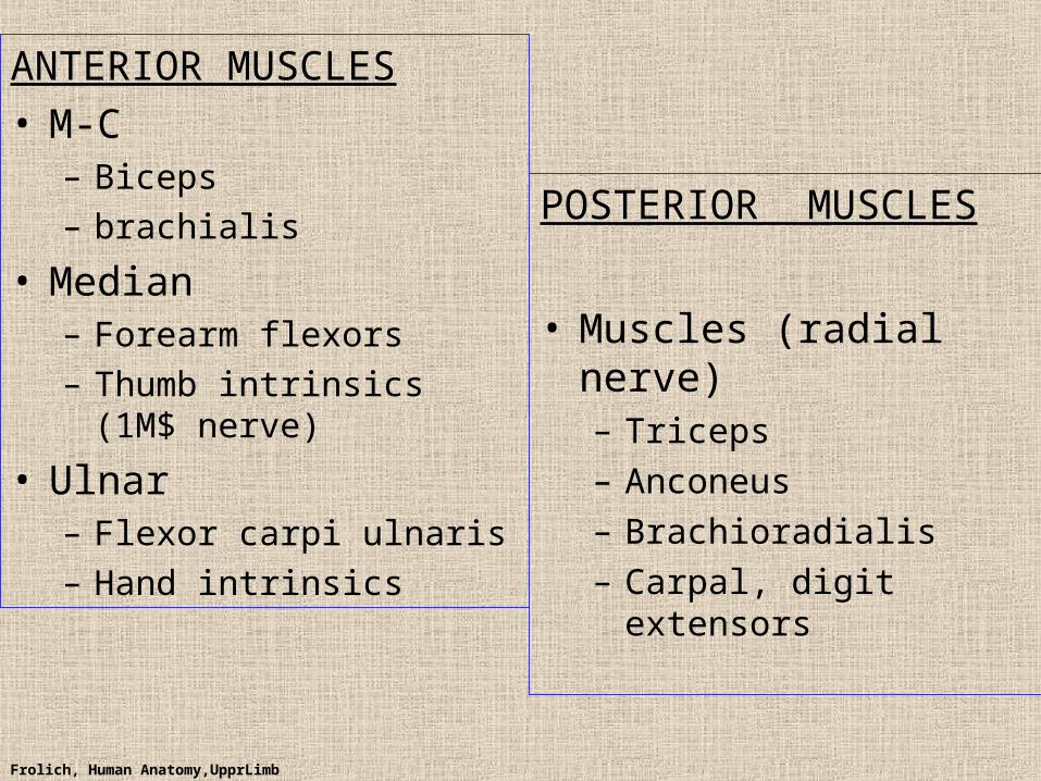
ANTERIOR MUSCLES• M-C
– Biceps
– brachialis
• Median– Forearm flexors
– Thumb intrinsics (1M$ nerve)
• Ulnar– Flexor carpi ulnaris
– Hand intrinsics
POSTERIOR MUSCLES
• Muscles (radial nerve)– Triceps
– Anconeus
– Brachioradialis
– Carpal, digit extensors
Frolich, Human Anatomy,UpprLimb
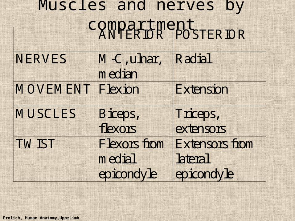
Muscles and nerves by compartment ANTERIOR POSTERIOR
NERVES M-C, ulnar, median
Radial
MOVEMENT Flexion Extension
MUSCLES Biceps, flexors
Triceps, extensors
TWIST Flexors from medial epicondyle
Extensors from lateral epicondyle
Frolich, Human Anatomy,UpprLimb
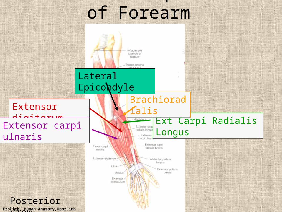
Posterior Compartment of Forearm
Extensor digitorum
Extensor carpi ulnaris Ext Carpi Radialis Longus
Brachioradialis
Lateral Epicondyle
Posterior View
Frolich, Human Anatomy,UpprLimb
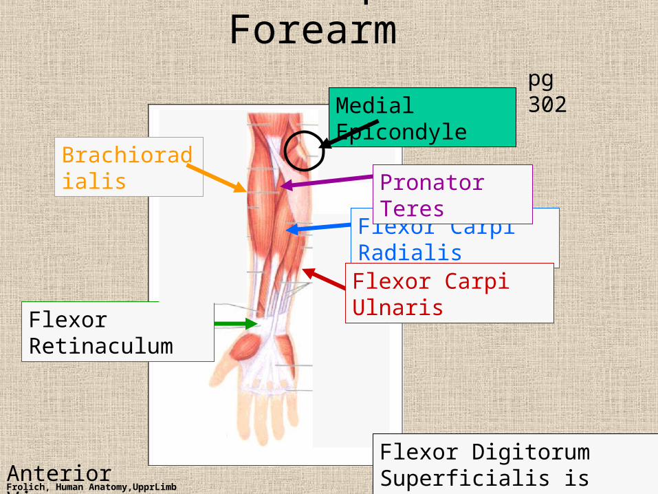
Anterior Compartment Forearm
Flexor Carpi Radialis
Flexor Retinaculum
Medial Epicondyle
Flexor Digitorum Superficialis is deep to other flexors
pg 302
Flexor Carpi Ulnaris
BrachioradialisPronator Teres
Anterior View
Frolich, Human Anatomy,UpprLimb
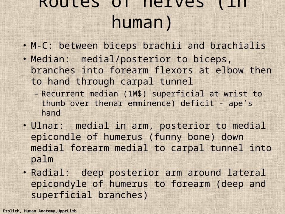
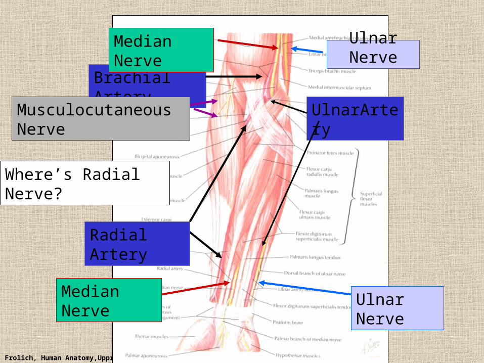
Routes of nerves (in human)
• M-C: between biceps brachii and brachialis• Median: medial/posterior to biceps, branches into
forearm flexors at elbow then to hand through carpal tunnel– Recurrent median (1M$) superficial at wrist to thumb over
thenar emminence) deficit - ape’s hand
• Ulnar: medial in arm, posterior to medial epicondle of humerus (funny bone) down medial forearm medial to carpal tunnel into palm
• Radial: deep posterior arm around lateral epicondyle of humerus to forearm (deep and superficial branches)
Frolich, Human Anatomy,UpprLimb
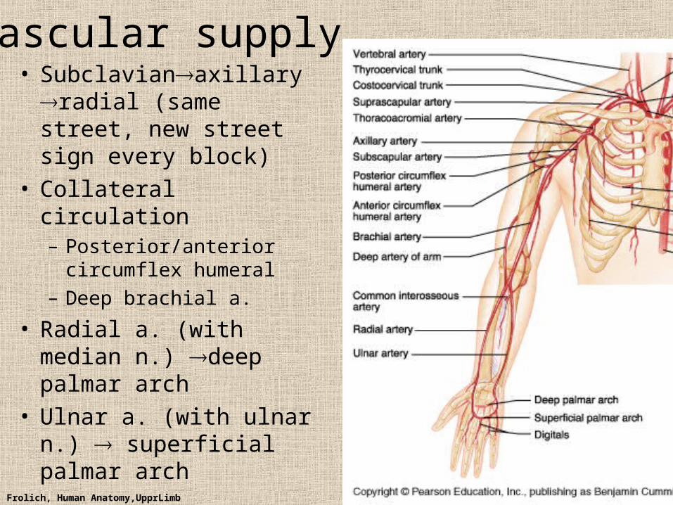
Vascular supply• Subclavianaxillary
radial (same street, new street sign every block)
• Collateral circulation– Posterior/anterior
circumflex humeral
– Deep brachial a.
• Radial a. (with median n.) deep palmar arch
• Ulnar a. (with ulnar n.) superficial palmar arch
Frolich, Human Anatomy,UpprLimb
Ulnar Nerve
Brachial Artery
Median Nerve
Ulnar NerveMedian Nerve
Radial Artery
Musculocutaneous Nerve UlnarArtery
Where’s Radial Nerve?
Frolich, Human Anatomy,UpprLimb
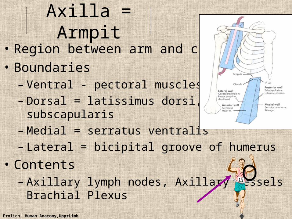
Axilla = Armpit
• Region between arm and chest
• Boundaries– Ventral - pectoral muscles– Dorsal = latissimus dorsi, teres major
subscapularis– Medial = serratus ventralis– Lateral = bicipital groove of humerus
• Contents– Axillary lymph nodes, Axillary vessels
Brachial Plexus
Frolich, Human Anatomy,UpprLimb
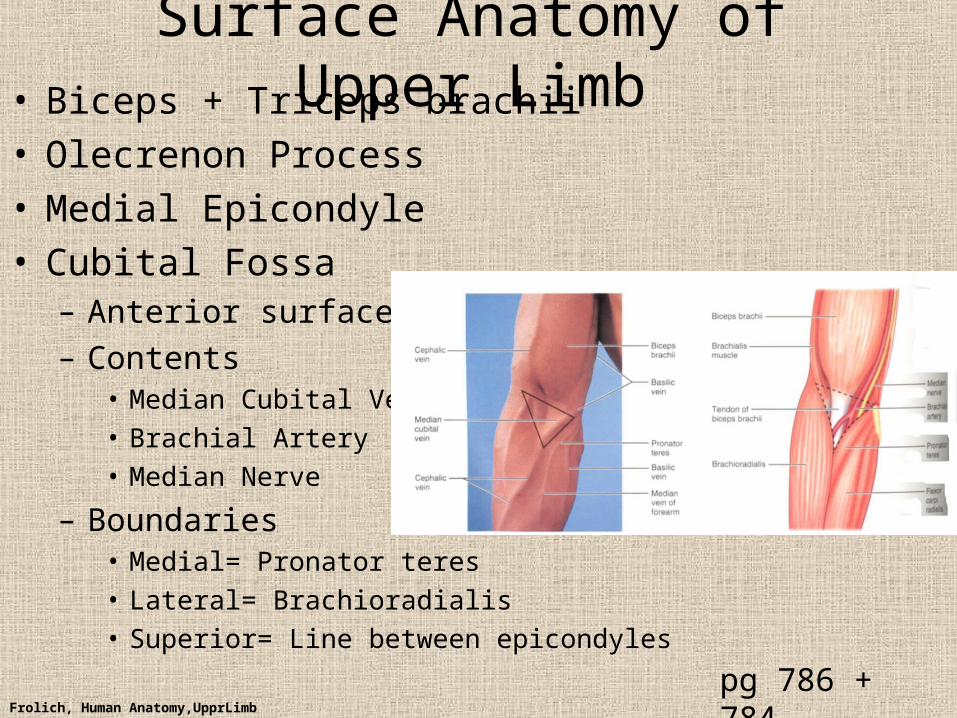
Surface Anatomy of Upper Limb• Biceps + Triceps brachii• Olecrenon Process• Medial Epicondyle• Cubital Fossa
– Anterior surface elbow
– Contents• Median Cubital Vein
• Brachial Artery
• Median Nerve
– Boundaries• Medial= Pronator teres
• Lateral= Brachioradialis
• Superior= Line between epicondyles
pg 786 + 784
Frolich, Human Anatomy,UpprLimb
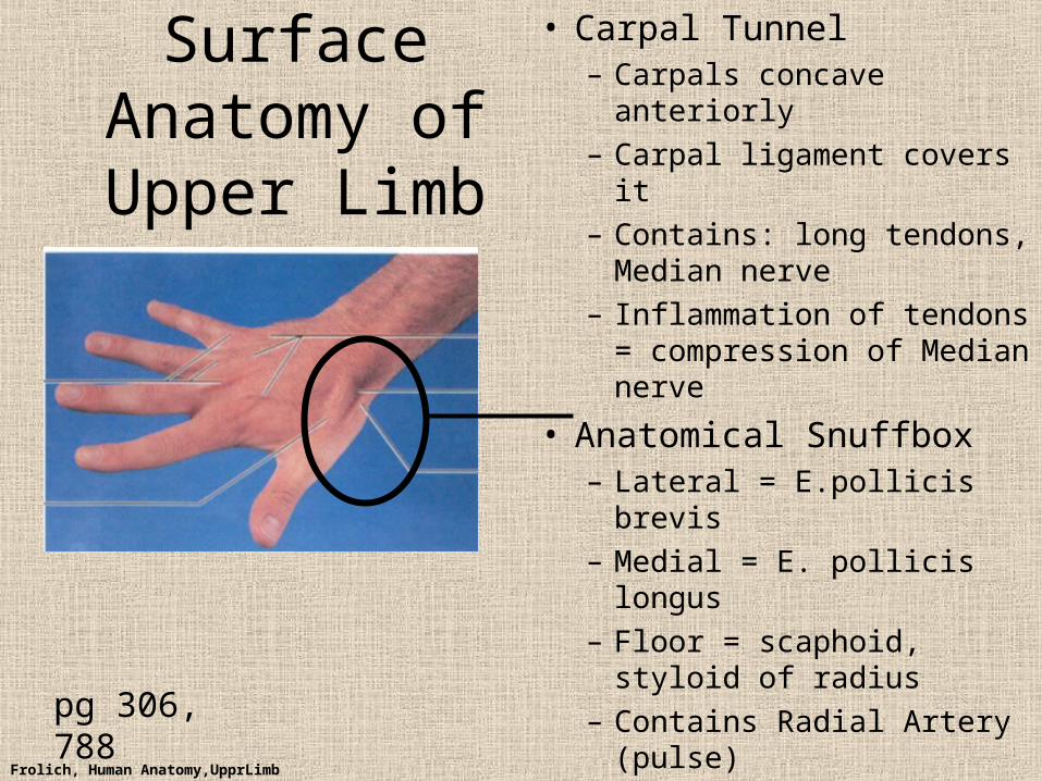
Surface Anatomy of Upper Limb
• Carpal Tunnel– Carpals concave anteriorly
– Carpal ligament covers it
– Contains: long tendons, Median nerve
– Inflammation of tendons = compression of Median nerve
• Anatomical Snuffbox– Lateral = E.pollicis brevis
– Medial = E. pollicis longus
– Floor = scaphoid, styloid of radius
– Contains Radial Artery (pulse)pg 306, 788
Frolich, Human Anatomy,UpprLimb
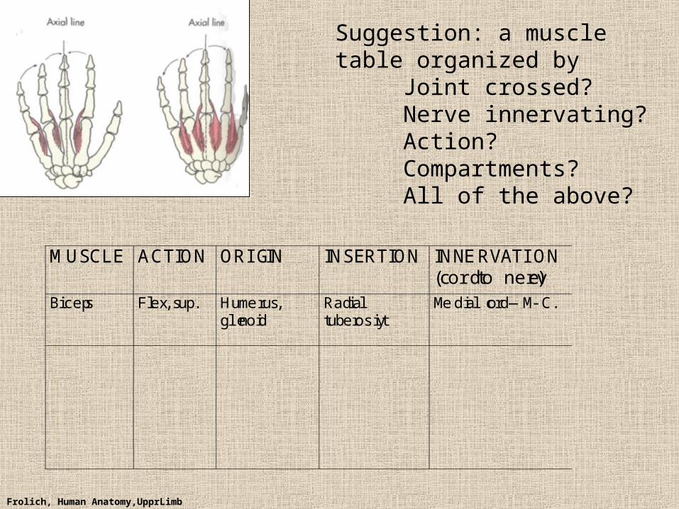
Suggestion: a muscle table organized by
Joint crossed?Nerve innervating?Action?Compartments?All of the above?
MUSCLE ACTION ORIGIN INSERTION INNERVATI ON(cord to nerve)
Biceps Flex, sup. Humerus,glenoid
Radialtuberosity
Medial cord—M-C.