Frizzled and LRP5 6 Receptors for Wnt b-Catenin...
24
Frizzled and LRP5/6 Receptors for Wnt/b-Catenin Signaling Bryan T. MacDonald and Xi He The F. M. Kirby Neurobiology Center, Boston Children’s Hospital, Boston, Massachusetts 02115; Department of Neurology, Harvard Medical School, Boston, Massachusetts 02115 Correspondence: [email protected] Frizzled and LRP5/6 are Wnt receptors that upon activation lead to stabilization of cytoplas- mic b-catenin. In this study, we review the current knowledge of these two families of receptors, including their structures and interactions with Wnt proteins, and signaling mech- anisms from receptor activation to the engagement of intracellular partners Dishevelled and Axin, and finally to the inhibition of b-catenin phosphorylation and ensuing b-catenin stabilization. T he Wnt/b-catenin pathway, or canonical Wnt pathway as it is often referred to, is an ancient and conserved signaling cascade involv- ing b-catenin acting as a transcriptional coac- tivator (Logan and Nusse 2004). The pathway is best understood when considered in a two-state model of OFF (without Wnt) and ON (with Wnt). In the OFF state, cytoplasmic b-catenin is constitutively targeted for degradation by two multidomain scaffolding proteins, Axin and Adenomatous polyposis coli (APC), which fa- cilitate the amino-terminal phosphorylation of b-catenin via the kinases GSK3 and CKIa. Phosphorylated b-catenin is recognized by the E3 ubiquitin ligase b-Trcp and is thus ubiquiti- nated and degraded by the proteasome, thereby maintaining low levels of free b-catenin in the cytoplasm and nucleus (MacDonald et al. 2009). In the ON state, a Wnt ligand binds to the seven-pass transmembrane receptor Friz- zled (FZD) and the single-pass low-density lipoprotein receptor-related protein 5 or 6 (LRP5/6) (He et al. 2004). The Wnt–FZD– LRP5/6 trimeric complex recruits Dishevelled (DVL) and Axin through the intracellular do- mains of FZD and LRP5/6, resulting in inhibi- tion of b-catenin phosphorylation and thus ensuing b-catenin stabilization. The rise of cy- toplasmic and nuclear levels of b-catenin levels promotes b-catenin partnering with the TCF/ LEF transcription factors for activation of Wnt- responsive gene expression. Wnt/b-catenin signaling controls cell pro- liferation and differentiation and is a key regu- latory mechanism for stem cells. As such, muta- tions in the Wnt pathway cause many diseases including cancer (Clevers 2006). All of the above “core” Wnt/b-catenin signaling components are present in vertebrates and the well-studied fruit fly Drosophila and are also encoded in se- quenced genomes of radial symmetric Cnidari- ans Hydra magnipapillata and Nematostella vec- tensis (Guder et al. 2006) and of the primitive metazoan sponge Amphimedon queenslandica Editors: Roel Nusse, Xi He, and Renee van Amerongen Additional Perspectives on Wnt Signaling available at www.cshperspectives.org Copyright # 2012 Cold Spring Harbor Laboratory Press; all rights reserved; doi: 10.1101/cshperspect.a007880 Cite this article as Cold Spring Harb Perspect Biol 2012;4:a007880 1 on July 18, 2018 - Published by Cold Spring Harbor Laboratory Press http://cshperspectives.cshlp.org/ Downloaded from
Transcript of Frizzled and LRP5 6 Receptors for Wnt b-Catenin...

Frizzled and LRP5/6 Receptors forWnt/b-Catenin Signaling
Bryan T. MacDonald and Xi He
The F. M. Kirby Neurobiology Center, Boston Children’s Hospital, Boston, Massachusetts 02115; Departmentof Neurology, Harvard Medical School, Boston, Massachusetts 02115
Correspondence: [email protected]
Frizzled and LRP5/6 are Wnt receptors that upon activation lead to stabilization of cytoplas-mic b-catenin. In this study, we review the current knowledge of these two families ofreceptors, including their structures and interactions with Wnt proteins, and signaling mech-anisms from receptor activation to the engagement of intracellular partners Dishevelled andAxin, and finally to the inhibition of b-catenin phosphorylation and ensuing b-cateninstabilization.
The Wnt/b-catenin pathway, or canonicalWnt pathway as it is often referred to, is an
ancient and conserved signaling cascade involv-ing b-catenin acting as a transcriptional coac-tivator (Logan and Nusse 2004). The pathway isbest understood when considered in a two-statemodel of OFF (without Wnt) and ON (withWnt). In the OFF state, cytoplasmic b-cateninis constitutively targeted for degradation by twomultidomain scaffolding proteins, Axin andAdenomatous polyposis coli (APC), which fa-cilitate the amino-terminal phosphorylation ofb-catenin via the kinases GSK3 and CKIa.Phosphorylated b-catenin is recognized by theE3 ubiquitin ligase b-Trcp and is thus ubiquiti-nated and degraded by the proteasome, therebymaintaining low levels of free b-catenin in thecytoplasm and nucleus (MacDonald et al.2009). In the ON state, a Wnt ligand binds tothe seven-pass transmembrane receptor Friz-zled (FZD) and the single-pass low-densitylipoprotein receptor-related protein 5 or 6
(LRP5/6) (He et al. 2004). The Wnt–FZD–LRP5/6 trimeric complex recruits Dishevelled(DVL) and Axin through the intracellular do-mains of FZD and LRP5/6, resulting in inhibi-tion of b-catenin phosphorylation and thusensuing b-catenin stabilization. The rise of cy-toplasmic and nuclear levels of b-catenin levelspromotes b-catenin partnering with the TCF/LEF transcription factors for activation of Wnt-responsive gene expression.
Wnt/b-catenin signaling controls cell pro-liferation and differentiation and is a key regu-latory mechanism for stem cells. As such, muta-tions in the Wnt pathway cause many diseasesincluding cancer (Clevers 2006). All of the above“core” Wnt/b-catenin signaling componentsare present in vertebrates and the well-studiedfruit fly Drosophila and are also encoded in se-quenced genomes of radial symmetric Cnidari-ans Hydra magnipapillata and Nematostella vec-tensis (Guder et al. 2006) and of the primitivemetazoan sponge Amphimedon queenslandica
Editors: Roel Nusse, Xi He, and Renee van Amerongen
Additional Perspectives on Wnt Signaling available at www.cshperspectives.org
Copyright # 2012 Cold Spring Harbor Laboratory Press; all rights reserved; doi: 10.1101/cshperspect.a007880
Cite this article as Cold Spring Harb Perspect Biol 2012;4:a007880
1
on July 18, 2018 - Published by Cold Spring Harbor Laboratory Press http://cshperspectives.cshlp.org/Downloaded from

(Adamska et al. 2010). Gene loss is prevalent inthe genome of nematodes, such as the popularmodel organism Caenorhabditis elegans, whichappears to lack an ortholog of LRP5/6 (Phillipsand Kimble 2009). As a result, the mechanismsof Wnt receptor activation are likely divergent innematodes, whose Wnt pathways are discussedin Jackson and Eisenmann (2012). We focuson the vertebrate (and Drosophila) Wnt/FZD/LRP5/6 pathway, which is characterized by itsabsolute requirement for both FZD and LRP5/6receptors. We note that FZD is also requiredfor other “noncanonical” or “alternative” Wntpathways that are independent ofb-catenin (vanAmerongen et al. 2008), and that in some casessuch as in planar cell polarity (PCP) signaling inDrosophila (Bayly and Axelrod 2011), FZD maysignal in a Wnt-independent manner.
WNT–RECEPTOR INTERACTIONS:OUTSIDE THE CELL
FZD Receptors
Calvin Bridges discovered a recessive fly mutant,which he called frizzled ( fz), with irregularlyorientated hairs and ommatidia (compoundeye) and an occasionally slight reduction inwing size (Bridges and Brehme 1944). It becameclear subsequently that fz plays an importantrole in planar cell polarity (PCP) (Bayly andAxelrod 2011). fz was shown to encode a proteinwith an extracellular cysteine-rich domain and apredicted topology of seven transmembrane he-lices resembling a G-protein-coupled receptor(GPCR) (Vinson et al. 1989). Epistasis and sim-ilarities between the fz and dishevelled (dsh) mu-tants (Wong and Adler 1993) suggested a link ofthe two genes, although in PCP signaling initial-ly. With the fledgling wingless (wg, Wnt)–dsh(DVL)–zeste white3 (zw3, GSK3)–armadillo(arm, b-catenin) signaling pathway importantfor embryonic patterning and wing develop-ment (Klingensmith and Nusse 1994; Krasnowet al. 1995), came along the identification of thesecond fly Frizzled gene Dfz2 and the findingthat Wg is a ligand for Dfz2 and Fz (Bhanotet al. 1996). fz(also called Dfz1) and Dfz2 haveredundant functions, although Dfz2 has a more
prominent role, in Wg signaling, whereas fz/Dfz1 has a unique role in the PCP pathway (Ken-nerdell and Carthew 1998; Chen and Struhl1999; Boutros et al. 2000; Rulifson et al. 2000).
In human, there are 10 FZD genes, num-bered FZD1 through 10. Phylogenetic analysisof the mature FZD proteins generates the follow-ing five subgroups: FZD1/2/7, FZD3/6, FZD5/8, FZD9/10, and FZD4 (Fig. 1A). Dfz2 groupsclosely with FZD5/8 and fz/Dfz1 with FZD3/6.The Hedgehog (another family of secreted sig-naling protein) pathway protein Smoothened(SMO) is distantly related to FZD (Schulte andBryja 2007) in their membrane topologies andamino-terminal cysteine-rich domains (CRDs);however, FZD proteins are distinguished fromSMO by a conserved juxtamembrane KTxxxWmotif in the carboxy tail region necessary forsignaling (Fig. 1B).
THE FZD EXTRACELLULAR DOMAIN ANDWNT BINDING: THE WNT8-FZD8CRDCO-CRYSTAL STRUCTURE
FZD contains a conserved 120-amino-acid cys-teine-rich domain (CRD) at the amino termi-nus, which is connected to the first transmem-brane helix through a variable 70- to 120-amino-acid linker region (Fig. 1B). Wnt/Wgligands bind to CRD with high affinity (Kd of1–10 nM) (Hsieh et al. 1999; Rulifson et al.2000; Wu and Nusse 2002), although fz/Dfz1PCP signaling may not involve any Wnt ligands.Deletion of the CRD prevents Wnt/Wg bind-ing, but the CRD can be replaced with otherheterologous Wnt-binding domains to generatea functional FZD receptor (Povelones andNusse 2005; Mulligan et al. 2012). Crystal struc-tures of CRDs from mouse FZD8 and Sfrp3(secreted FZD-related protein 3) reveal a com-pact, predominantly a-helical regions held inplace by disulfide bonds among 10 invariablecysteines (Fig. 1B,C) (Dann et al. 2001).
How a Wnt ligand engages the FZD receptoris revealed by the co-crystal structure of Xeno-pus Wnt8 in complex with mouse FZD8CRD(Fig. 1D) (Janda et al. 2012). In the complex,Wnt8 forms an unusual structure resembling ahuman hand with a central “palm” that extends
B.T. MacDonald and X. He
2 Cite this article as Cold Spring Harb Perspect Biol 2012;4:a007880
on July 18, 2018 - Published by Cold Spring Harbor Laboratory Press http://cshperspectives.cshlp.org/Downloaded from

1 2 3 4 5 6 7
DIX PDZ DEPDishevelled
C
C C CψECL1 ECL2 ECL3
ICL1 ICL2 ICL3 Carboxyl tail
GVCFVG?
(Polymerization)
+ + +
8
(Motif I) (Motif II) (Motif III)
FZD ICL3
FZD carboxyl tail
A
N term
C
C1
C3C4
C5
C6C7C8C9
C10
C
C
C2
ψ
ψ
Linker region
CRD
YNxT
N term
D
FZD1
FZD2
FZD7
FZD3
FZD6
FZD5
FZD8
FZD9
FZD10
FZD4
10/100 amino acidsubstitutions
Noncanonical
(ETxV)
(PTxV)
(ETxV)
(LSxV)
Site 1
Xwnt8
FZD8CRD
B
C term Site 1Site 2
N term
C term
Site 2
P
P
Key intracellularamino acids
Single mutationConserved
XX,*,
Bold,
Double-alaninescanningmutagenesis
RFxYPERP* * TWFLAGxKWGxEAIE**** IRxV* ** KLEKLMVR* * * KTxxxW* *
Figure 1. Frizzled (FZD) and Dishevelled (DVL). (A) Phylogeny comparing human FZD proteins. (B) Topologyof a generic FZD on the plasma membrane. The shape of CRD is shaded in green, with the 10 invariable cysteine(C) residues forming five disulfide bonds highlighted. (c) Potential N-glycosylation sites. Additional conservedcysteine and other residues in the linker and ECL1-3 in the extracellular space are indicated. Conserved residuesin ICL1-3 and the carboxy-terminal domain in the intracellular space are also indicated, with invariable residuesamong all FZD proteins in bold. (� above the letter) Missense mutations found in Drosophila Dfz1 (Poveloneset al. 2005) and human FZD4 (Robitaille et al. 2002); (underlined) residues tested via double alanine substi-tution scanning mutagenesis in FZD5 (Cong et al. 2004b). DVL protein is also shown schematically with DIX,PDZ, and DEP domains and their interaction partners highlighted. Two discontinuous regions of FZD ICL3(motif I [pink]; motif II [green]) bind to the carboxy-terminal region of DVL. The FZD carboxyl tail containingthe KTxxxW motif (blue) interacts strongly with the DVL PDZ domain and also the DEP domain, which alsoshows some interaction with motif II in ICL3. (See following page for legend.)
Wnt Receptors and Signaling Mechanisms
Cite this article as Cold Spring Harb Perspect Biol 2012;4:a007880 3
on July 18, 2018 - Published by Cold Spring Harbor Laboratory Press http://cshperspectives.cshlp.org/Downloaded from

a “thumb” plus an “index finger” to grab/pinchthe FZD8CRD globular structure on two oppo-site sides, without changing FZD8CRD confor-mation (Fig. 1D). The amino terminal two-thirds of Wnt8 give rise to the palm—whichconsists mostly of a-helixes and interveningloops—and the thumb that consists of twoanti-parallel b-strands and a connecting looprigidified by a pair of disulfide bonds (Fig.1D) (Janda et al. 2012). Most strikingly, a fattyacid adduct covalently attached to a serine at thetip of the thumb loop, likely a palmitoleic acidas seen in Wnt3a (Takada et al. 2006), insertsinto a hydrophobic groove of FZD8CRD, con-stituting much of the Wnt8-FZD8CRD binding“site 1” that features extensive hydrophobic in-teractions between the lipid and apolar residuesof FZD8CRD, “lipid-in-groove” fashion (Fig.1D). The remaining portion of site 1 is contrib-uted by protein–protein contacts between res-idues of the Wnt8 thumb loop and FZD8CRD(Fig. 1D). The carboxyl one-third of Wnt8makes up the index finger featuring two anti-parallel b-strands and a long intervening loop,which is also rigidified by several disulfide bondsand engages in hydrophobic contacts within adepression of FZD8CRD, “knob-in-hole” fash-ion (Fig. 1D). This constitutes Wnt8-FZD8CRDinteraction “site 2,” which roughly correspondsto a Wnt-binding surface encompassing resi-dues near the second cysteine (C2) of FZD8CRDas suggested by scanning mutagenesis (Fig.1B,C) (Hsieh et al. 1999; Dann et al. 2001).Both site 1 and site 2 are dominated by hydro-phobic contacts (lipid-in-groove and knob-in-hole, respectively), which are mostly mediatedby conserved residues of Wnt8 and FZD8CRD,suggesting an explanation for broad or relatively
promiscuous specificity of Wnt–FZD relation-ships, i.e., a single Wnt can often engage mul-tiple FZD proteins and vice versa. However, atboth site 1 and site 2, Wnt8 also exhibits pro-tein–protein interactions with FZD8CRD resi-dues that are conserved in some but altered inother FZD proteins, implying certain selectivityon top of broad specificity in Wnt-FZD inter-actions (Janda et al. 2012).
A YNxT motif at site 2 is conserved in allFZD proteins and is a predicted N-glycosylationsite, and is indeed glycosylated in the Wnt8CRDcrystal (Fig. 1B,C,D). Another predicted N-gly-cosylation site is at the end of the CRD in mostFZDs, with the exception of the FZD3/6 group,which has its own unique predicted glycosyla-tion site in extracellular loop 2 (ECL2) (Fig. 1B).N-glycans in both Wnt8 and FZD8CRD in thecrystal structure are solvent-exposed and do notappear to contribute directly to Wnt–FZD in-teraction (Fig. 1D) (Janda et al. 2012). FZD N-glycosylation appears to be required for receptormaturation and can be regulated by the ER-res-ident protein Shisa, which binds preferentiallyto the immature form of FZD for ER trappingand thus antagonizes Wnt signaling (Yamamo-to et al. 2005).
Beyond the Wnt–CRD interaction, little isknown regarding the function of other FZD ex-tracellular regions. FZD receptors are structur-ally analogous to GPCRs, and there is evidencethat FZDs can interact and signal through G-proteins (see below), although this issue re-mains debated. Nonetheless, some commonGPCR structural elements that are also foundin FZD may suggest other potentially impor-tant regions. Many small GPCR ligands bindto the extracellular loop regions or between
Figure 1. (Continued) (C) Crystal structure of FZD8CRD. Residues in red (and to a lesser degree those inorange) are suggested to be a Wnt-binding interface from an alanine scanning mutagenesis (Hsieh et al. 1999;Dann et al. 2001), which partially overlaps with site 2 indentified in the Wnt8–FZD8CRD co-crystal structure.Residues in green when altered did not affect Wnt activity and areas in gray were not tested. Site 1 and site 2,which mediate contacts between Wnt8 and FZD8CRD in the crystal structure, are labeled. A shade of Wnt8index finger contacting site 2 is sketched. (Panel C is derived from Fzd8 1IJY [PDB doi: 10.2210/pdb1ijy/pdb].)(D) Wnt8–FZD8CRD co-crystal structure, shown as a ribbon diagram superimposed on surface representation(Janda et al., 2012). Palmitoleic acid adduct from the Wnt8 thumb at site 1 (red); N-glycans of Wnt8 andFZD8CRD (yellow). Note that the FZD8CRD in C and D is viewed in different angles. N term, amino terminal;C term, carboxyl terminal.
B.T. MacDonald and X. He
4 Cite this article as Cold Spring Harb Perspect Biol 2012;4:a007880
on July 18, 2018 - Published by Cold Spring Harbor Laboratory Press http://cshperspectives.cshlp.org/Downloaded from

the transmembrane helices at the extracellularface (Tebben and Schnur 2011), causing a con-formation change for GPCR activation. Ligandbinding and relative orientation of the trans-membrane helices are influenced by inter- andintraloop disulfide bonds in the extracellularregions/loops of GPCRs. Aside from the 10 in-variable cysteines within the CRD, there are ad-ditional conserved cysteines in FZD extracellu-lar regions: two in the linker between the CRDand the first transmembrane helix, two in extra-cellular loop 1 (ECL1), one in ECL2, and two inECL3 (Fig. 1B). A common extracellular disul-fide bond in GPCRs is between the top of trans-membrane helix 3 and ECL2 and is thought tobe important for orienting and stabilizing thetransmembrane helices (Peeters et al. 2010).This disulfide bond is potentially present inall FZDs (Fig. 1B). In addition, the residuesflanking the cysteine in ECL2 are conserved(GVCFV) and are predicted to form a b-strand.There are also examples of an intraloop disul-fide bond in ECL3 and disulfide bonds connect-ing with the amino-terminal region (Wheatleyet al. 2012). Smo, a distant member of the FZDfamily, may share these GPCR features in thatmutations of cysteines in ECL1 and 2 or a partialdeletion of ECL2 renders a constitutively activeor less active receptor (Carroll et al. 2012). Fur-thermore, a fly fz allele with a mutation in theECL2 cysteine causes a partial loss of function(Povelones et al. 2005). Studies are required toelucidate the role of these ECLs in Wnt-inducedFZD activation and signaling.
LRP5/6 AND ARROW
Fly mutants for arrow were first reported byNusslein-Volhard and Wieschaus from the fa-mous genetic screen for embryonic lethal mu-tants (Nusslein-Volhard et al. 1984). Fly embryosegments normally develop a stripe of anteriordenticles that can be distinguished from the na-ked posterior region of the segment. Mutantsfor arrow contained extra bands of denticlesthat were more prominent in the midline, caus-ing the denticle stripes to look like arrows. Elim-ination of maternal and zygotic arrow resultedin a phenotype identical to that of wg mutants,
and molecular cloning revealed that Arrow ishomologous to LRP5 and LRP6 (Wehrli et al.2000), which had been cloned as members ofthe LDLR family (Brown et al. 1998; Hey et al.1998). With the evidence that the Lrp6 mousemutant phenotypically resembles a compositeof several Wnt mutants (Pinson et al. 2000),that LRP5 and LRP6 display critical roles inWnt/b-catenin signaling in Xenopus (Tamaiet al. 2000), and that Wnt1 can bridge a complexformation between the extracellular domains ofFZD and LRP6 (Tamai et al. 2000), LRP5/6 andArrow were established as coreceptors for theWnt/b-catenin pathway.
THE LRP5/6 EXTRACELLULAR DOMAIN
LRP5 and LRP6 have more than 1600 aminoacids and represent a unique group of theLDLR family. LRP5 and LRP6 proteins are70% identical, and each is 45% identical to Ar-row (He et al. 2004). These single transmem-brane receptors have an extracellular domaincontaining four tandem b-propeller/epidermalgrowth factor (EGF) repeats followed by threeLDLR type A repeats (Fig. 2A). The b-propellerdomain was first proposed by Springer, who pre-dicted a layout of a six-bladed propeller, witheach blade consisting of four short b-strandsand a YWTD motif in strand 2 that acts to sta-bilize the neighboring b-sheets and is key to theblade structure (Springer 1998). The structurefor the single LDLR b-propeller-EGF (PE) unitwas solved confirming the predicted six-bladedpropeller, which intimately interacts with theEGF repeat (of 50 amino acids) with six cyste-ines in a disulfide bond pattern of C1–C3, C2–C4, and C5–C6 (Jeon et al. 2001).
Earlier mapping studies divided the LRP5/6extracellular domain into three segments: b-propeller-EGFs 1 and 2 (P1E1-P2E2),b-propel-ler-EGFs 3 and 4 (P3E3–P4E4), and the threeLDLR type A repeats (Fig. 2A) (He et al. 2004).Deletion of a single b-propeller in LRP5/6 typ-ically interferes with receptor biogenesis, andbetter stability and recombinant protein pro-duction were achieved using tandem pairs ofP1E1–P2E2 and P3E3–P4E4, but not P2E2–P3E3 (Liu et al. 2009; Bourhis et al. 2010).
Wnt Receptors and Signaling Mechanisms
Cite this article as Cold Spring Harb Perspect Biol 2012;4:a007880 5
on July 18, 2018 - Published by Cold Spring Harbor Laboratory Press http://cshperspectives.cshlp.org/Downloaded from
Domain key
A
C F
G
D
E
B
EGF repeat,LDLR-A,
LDLR Arrow
PP P P
PP
PP
PP
PP
PP
P
LRP5 LRP6
Wnt1, 2, 2b, 6,8a, 9a, 9b, 10bSOST, DKK1
LRP6 P1
LRP5 P1
LRP6 P3
LRP5 P3
LRP5 P2
LRP5 P4
LRP6 P4
10/100amino acid
substitutions
LRP6 ECD—Top view(from plasma membrane)
LRP6 ECD—Side view(from plasma membrane)
Side viewP3Top
P4Top
E3
Top view
P3B4P3B3 P4B5
P4B4
P4B3
P4B2P4B1
P3B1
P4B6
P3B2
P3B6
Top view
Axin
DAXLRP6
PP
PP
P
DishevelledAPC
Affects Dkk1 binding onlyNo effect
Affects Wnt3a and Dkk1 bindingAffects Wnt3a binding only
GSK3 CK1
P3B5
E4
CLDLR-A
N
LRP5 P2
Wnt3, 3aDKK1
P
NPxY,PPPSPxS,
β-propeller,
β-catenin
1
2
3
4
1
2
3
4
and untested
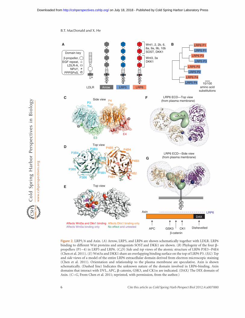
Figure 2. LRP5/6 and Axin. (A) Arrow, LRP5, and LRP6 are shown schematically together with LDLR. LRP6binding to different Wnt proteins and antagonists SOST and DKK1 are shown. (B) Phylogeny of the four b-propellers (P1–4) in LRP5 and LRP6. (C,D) Side and top views of the atomic structure of LRP6 P3E3–P4E4(Chen et al. 2011). (E) Wnt3a and DKK1 share an overlapping binding surface on the top of LRP6 P3. (F,G) Topand side views of a model of the entire LRP6 extracellular domain derived from electron microscopic staining(Chen et al. 2011). Orientation and relationship to the plasma membrane are speculative. Axin is shownschematically. (Dashed line) Indicates the unknown nature of the domain involved in LRP6-binding. Axindomains that interact with DVL, APC, b-catenin, GSK3, and CK1a are indicated. (DAX) The DIX domain ofAxin. (C–G, From Chen et al. 2011; reprinted, with permission, from the author.)
B.T. MacDonald and X. He
6 Cite this article as Cold Spring Harb Perspect Biol 2012;4:a007880
on July 18, 2018 - Published by Cold Spring Harbor Laboratory Press http://cshperspectives.cshlp.org/Downloaded from
Recent crystal structures of LRP6 P1E1–P2E2and P3E3–P4E4 highlighted the tandem natureof these repeating units (Fig. 2C,D) (Ahn et al.2011; Chen et al. 2011; Cheng et al. 2011). Al-though conforming to the prototypic PE struc-ture of LDLR, extensive interface interactionsbetween P1E1 and P2E2, and between P3E3and P4E4 were observed, for example, betweenP3 and P4 and between E3 and P4 (Fig. 2C,D),and these inter-PE interactions were shown tobe critical for LRP6 biogenesis and maturationto the plasma membrane. The interface betweenP2E2 and P3E3 is unresolved but is likely to bedifferent. A low-resolution electron microscop-ic structure of the entire LRP6 extracellular do-main suggested a compact horseshoe platformconfiguration (Fig. 2F,G), which may be consis-tent with the notion of a P2E2–P3E3 interfacethat is distinct from those of P1E1–P2E2 and ofP3E3–P4E4 (Chen et al. 2011). A platform con-figuration of a different kind was also suggestedfor the LRP6 extracellular domain (lacking theLDLR-A repeats) via a low-resolution small-an-gle X-ray scattering analysis (Ahn et al. 2011).
The endoplasmic reticulum chaperone pro-tein MESD is required for proper folding of theLRP5/6 extracellular domain (Culi and Mann2003; Hsieh et al. 2003). MESD also facilitatesthe folding of other LDLR family members andinteracts with multiple b-propellers of LRP5/6(Culi et al. 2004; Lighthouse et al. 2011). Exam-ination of LRP5/6 via western blotting oftenreveals two bands, a lower (faster migrating)immature form and a higher fully glycosylatedform. Mest (also known as Paternally expressedgene 1, or Peg1), a multispan transmembraneprotein that resides in the ER and contains ana/b hydrolase domain, modifies LRP6 glycosyla-tion resulting in less mature LRP6 at the plasmamembrane, thereby modulating LRP6 in a man-ner that appears to be analogous to Shisa inhi-bition of FZD receptors (Jung et al. 2011).
TWO OR MORE WNT-BINDINGSURFACES OF LRP6
Early studies suggested that Wnt1 bridges extra-cellular domains of a FZD (mFZD8CRD) andLRP6 into a receptor complex via direct binding
(Tamai et al. 2000). Consistent with this notion,an LRP6 mutant that lacks the cytoplasmic do-main (and is thus membrane bound) or lackstransmembrane plus cytoplasmic domains (andis thus secreted) behaves as a dominant-negativereceptor for Wnt/b-catenin signaling (Fig. 3A),presumably via binding to Wnt ligands (Tamaiet al. 2000; He et al. 2004). Wnt1 signaling viaLRP6 is mediated through the PE domains(P1E1–P4E4) (Mao et al. 2001a). A recent studyshowed a Kd between the LRP6 extracellulardomain and Wnt3a or Wnt9b to be �10 nM
(Bourhis et al. 2010). Unexpectedly, however,Wnt3a and Wnt9b were shown to preferentiallyinteract with P3E3–P4E4 and P1E1–P2E2, re-spectively, and a Wnt3a–Wnt9b–LRP6 (extra-cellular domain) complex could be detectedin vitro (Bourhis et al. 2010), suggesting a pos-sibility that a single LRP6 may engage two dif-ferent Wnt proteins simultaneously. Further-more, functional-blocking monoclonal anti-bodies (mAbs) against epitopes in P1 and P3,respectively, show a distinct inhibition profiletoward different Wnt proteins, presumably viablocking Wnt binding to either P1 or P3 (Etten-berg et al. 2010; Gong et al. 2010). These datainfer that many Wnts (Wnt1, Wnt2, Wnt2b,Wnt6, Wnt8a, Wnt9a, Wnt9b, and Wnt10a) in-teract with P1, whereas Wnt3 and Wnt3a preferP3 (Fig. 2A). Other Wnts (Wnt7a, Wnt7b, andWnt10a) could not be assigned into eithergroup because they were not inhibited by themAbs alone or in combination (Gong et al.2010), raising the possibility that these Wntsmay bind to regions outside P1 and P3. Theremaining Wnts have not been tested in theseassays. The top surfaces of P1 and P3 (and P2and P4 as well) do not harbor N-glycosylationsites (Ahn et al. 2011; Bourhis et al. 2011; Chenet al. 2011; Cheng et al. 2011) and are likely theWnt-binding interface. Indeed, missense sub-stitutions of multiple top surface residues ofP3 of LRP6, designed based on the crystal struc-ture, diminish or enhance Wnt3a binding andsignaling (Fig. 2E) but show minimal effect onsignaling by Wnt1 (Chen et al. 2011), whichprefers to bind to P1. Therefore, an emergingmodel is that LRP6, and likely LRP5, engagedifferent Wnts via multiple ligand interfaces
Wnt Receptors and Signaling Mechanisms
Cite this article as Cold Spring Harb Perspect Biol 2012;4:a007880 7
on July 18, 2018 - Published by Cold Spring Harbor Laboratory Press http://cshperspectives.cshlp.org/Downloaded from

(Fig. 2A). The LRP6 platform configurationseems to suit this model (Fig. 2F,G). Sequenceanalysis of LRP5 and LRP6 reveals that P1, P2,and P3 are most related by homology, but P4is more divergent (Fig. 2B). Further studies ofLRP5/6 will be needed to parse out differentbinding sites for Wnts and other ligands, in-cluding whether P2 or P4 also represents aWnt docking site. The role of three LDLR-Arepeats in LRP5/6 remains unknown.
THE WNT–FZD–LRP5/6 COMPLEX
A distinguishing feature of the Wnt/b-cateninpathway is the requirement for both FZD andLRP5/6 receptors (He et al. 2004). Extracellulardomains of LRP6 and FZD8 were shown to
form a complex in vitro in the presence ofWnt1 (Tamai et al. 2000). A similar trimericcomplex was confirmed using recombinantWnt3a and extracellular domains of FZD8 andLRP6 (Bourhis et al. 2010). These results suggestthat Wnts can interact with both receptors si-multaneously, perhaps via different parts of theWnt molecule, although this important ques-tion has not been addressed (because of theprior lack of Wnt structural information andthe difficulty in generating soluble Wnt proteinfragments necessary for mapping studies). TheWnt8–FZD8CRD structure (Janda et al. 2012)will help resolve this issue. Also not addressedis the stoichiometry of the putative Wnt–FZD–LRP6 (or LRP5) complex, which is com-monly drawn at a 1:1:1 ratio in models. The
Ligand-induced phosphorylation
FZD
A
B
LRP5/6 LRP6 ECD LRP6ΔC
CK1ε
Human LRP6Human LRP5
Drosophila arrowNematostella L5/6
Human LRP6Human LRP5
Drosophila arrowNematostella L5/6
Human LRP6Human LRP5
Drosophila arrowNematostella L5/6
CK1γ CK1GSK3 GSK3 CK1
GSK3 CK1 GSK3 CK1GSK3 CK1
PKAS/T cluster
S/T cluster A motif
C motif D motif E motif
B motif
CK1ε
Dominant negative Dominant active
Constitutively phosphorylated(ligand independent)
LDLRΔN-PPPSPLRP6ΔN
LRP6 m5(or m10)
PP
PP
P
Wnt
XXXX
X
PP
PP
P
CQRMLCPRMKGDGETMTNDYVV---HGPASVPLGYVPHPSSLSGSLPGMSRGKSMISSLSIMGGSSGPP-YDRAHVTGASSSCQRVVCQRYAGANGPFPHEYV----SGTPHVPLNFIAFGGSQHGPFTGIACGKSMMSSVSLMGGRGGVPLYDRNHVTGASSSYLLQFCRTRIGKSRTEFKDDQ-----ATD--P---LSPSTLSKSQRVSKIASVADAVRMSTLNSRNSMNSYDRNHITGASSSTPHTLSTEIGLVVTPVLHGHASSLSSSSSSRASICAALPVRNGSTTSGSNKRQKPLPGPTSLSGTSQTP-YDRNHLTGASSS
TTNGSSMVAYP---INPPPSPATRSRR-------------------PYRHYKIINQPPPPTPCSTDICDESDSNYTSKSNSNSSACTVISAYPREPLNPPPSPATERSLFTVRSRGYCESVMTPSTCPSHRYYRTPRMPPPPTPASTDAEGSVKQPSRHCRRSH
SSSSTKGTYFP-AILNPPPSPATERSHYTMEFGYSSNSPSTHRSY-SYRPYSYRHFAPPTTPCSTDVCDSDYAPSRRMTS--SSSSTKATLYP-PILNPPPSPATDPSLYNMDMFYSSNIPATVR---PYRPYIIRGMAPPTTPCSTDVCDSDYSASRW-----
-----------VATAKGYTSDLNYDSEPVPPPPTPRSQYLSAEENY--ESCPPSPYTERSYSHHLYPPPPSPCTDSS----
NS--NGGATKHSSSSAAACLQYGYDSEPYPPPPTPRSHYHSDVRIVPESSCPPSPSSRSSTYFSPLPPPPSPVQSPSRGFT
P
-------------KASKYYLDLNSDSDPYPPPPTPHSQYLSAED-----SCPPSPATERSY-FHLFPPPPSPCTDSS----
RCRPSRGTLSRYNGSVTESTDLAYESDLFAPPPTPNTNYLSEATHLSDQECPPSPTATERSFFQPYPPPPSPVTETPSLV-
Figure 3. LRP6 phosphorylation and phosphorylation sites. (A) Wnt induces LRP6 phosphorylation in theFZD–LRP6 complex. Dominant-negative LRP6 mutants are generated by deleting the cytoplasmic domain ormutating all five PPPSPxS motifs (S to A). Constitutively activated LRP6DN and LDLRDN-PPPSP are consti-tutively phosphorylated. (B) Alignment of the cytoplasmic domain of LRP6, LRP5, Arrow, and the NematostellaLRP5/6 homolog. Identified phosphorylation sites by various kinases in LRP6 are indicated.
B.T. MacDonald and X. He
8 Cite this article as Cold Spring Harb Perspect Biol 2012;4:a007880
on July 18, 2018 - Published by Cold Spring Harbor Laboratory Press http://cshperspectives.cshlp.org/Downloaded from

observation in vitro that an LRP6 can simulta-neously bind two different Wnt proteins sug-gests the possibility that various combinationsof Wnt–FZD–LRP6 complexes may exist invivo. An LRP6 platform model (Fig. 2F,G) spec-ulates thatb-propeller top surfaces/ligand bind-ing regions may position at roughly the sameheight relative to the plasma membrane, po-tentially facilitating engagement of the CRDdomain of a single FZD or more FZDs (Ahnet al. 2011; Chen et al. 2011). Interestingly, theWnt8–FZD8CRD structure suggests a possibil-ity of asymmetric Wnt–FZD oligomers that areformed to fully shield the palmitoleic acid ad-duct from aqueous solvent (Janda et al. 2012).Whether such asymmetric Wnt–FZD oligomerformation occurs in vivo and has a role in sig-naling deserves further investigation.
Another limiting factor for understandingthe Wnt–FZD–LRP5/6 complex is our poorknowledge of Wnt–FZD and Wnt–LRP5/6binding specificity and affinity, because mostWnts are not available in soluble forms (Mulli-gan et al. 2012). FZD8 appears to bind Wnt3awith 2� –3� stronger affinity than LRP6 does(Bourhis et al. 2010), seemingly consistent withsome anecdotal experience that Wnt–LRP6binding appears to be weaker than that ofWnt–FZD (He et al. 2004). In contrast, the re-verse is true for Wnt9b, which displays weakbinding to FZD8 but binds to LRP6 comparablyto how Wnt3a does (Bourhis et al. 2010). It re-mains possible, however, that Wnt9b may pre-fer a FZD or FZDs other than FZD8. Some ofthe questions on the Wnt–FZD–LRP6 com-plex may have to wait until more recombinantWnt proteins become available and a high-res-olution structure of a Wnt protein in complexwith an FZD plus LRP6 is achieved.
WNT RECEPTOR SIGNAL TRANSDUCTION:INSIDE THE CELL
Dishevelled and Axin
FZD and LRP5/6 transduce Wnt signal via en-gaging downstream cytoplasmic components,among which two scaffolding proteins, Dishev-elled and Axin, have prominent roles.
Dishevelled (DVL1-3 in human, Dsh inDrosophila, and Xdsh in Xenopus) is a multi-functional protein that serves as a hub for ca-nonical and noncanonical Wnt signaling. Firstidentified in Drosophila, most fly dsh mutantsare embryonic lethal because of their loss in wgsignaling (Perrimon and Mahowald 1987). DVLproteins are about 700 amino acids in lengthand contain three main domains of about 80–90 amino acids each: DIX (Dishevelled, Axin),PDZ (Postsynaptic density 95, discs large, zonaoccludens-1), and DEP (Dishevelled, Egl-10,Pleckstrin) (Fig. 1B). The PDZ domain is essen-tial for binding to a juxtamembrane KTxxxWmotif (Umbhauer et al. 2000) in the FZD car-boxyl cytoplasmic region (Wong et al. 2003).The first dsh mutant identified, dsh1, displaysa PCP phenotype (Fahmy and Fahmy 1959),and is a missense mutation (K417M) in theDEP domain (Axelrod et al. 1998; Boutros etal. 1998). Further studies have shown that theDEP domain has a positively charged surfacethat may interact with phospholipids in theplasma membrane (Wong et al. 2000; Simonset al. 2009) and additional surfaces for interact-ing proteins including the endocytic adaptorprotein 2 (AP-2) complex (Yu et al. 2010). Arecent study further suggests that DEP plusthe carboxyl region of DVL interact with FZDICL3 (via the so-called motif I and motif II)(Fig. 1B) and the KTxxxW region (so-calledmotif III) (Fig. 1B), thereby facilitating DVLassociation with a discontinuous cytoplasmicsurface of FZD (Tauriello et al. 2012). Thus,PDZ and DEP domains have roles in recruitingDVL to FZD at the plasma membrane (Fig. 1B).The DIX domain shows an interesting propertyof head-to-tail polymerization (Schwarz-Ro-mond et al. 2007a). This property correlateswell with DVL aggregates under the overexpres-sion condition, and it has been argued that theendogenous DVL may form such aggregates,which are highly dynamic (Schwarz-Romondet al. 2005). DIX oligomerization/polymeriza-tion is proposed to provide a DVL platformfor dynamic assembly of protein–protein inter-actions of low avidity, such as between DVLand Axin (Schwarz-Romond et al. 2007b), andis a main underpinning for the receptor
Wnt Receptors and Signaling Mechanisms
Cite this article as Cold Spring Harb Perspect Biol 2012;4:a007880 9
on July 18, 2018 - Published by Cold Spring Harbor Laboratory Press http://cshperspectives.cshlp.org/Downloaded from

“signalosome” hypothesis (Bilic et al. 2007) (seebelow). However, some have suggested thatthe DVL “aggregates” (or “dots” under micro-scopes) represent endocytic vesicles (Capellutoet al. 2002; Taelman et al. 2010). The simplestmodel states that DIX and PDZ domains, butnot DEP, are required for Wnt/b-catenin sig-naling, as seen in some overexpression studies.However, evidence exists that the DEP domaininfluences b-catenin signaling (Wong et al.2000; Simons et al. 2009; Tauriello et al. 2012).DVL associates with kinases such as CK11 andCK2, and becomes hyperphosphorylated uponWnt signaling (Willert et al. 1997; Peters et al.1999; Kishida et al. 2001; Cong et al. 2004a;Klein et al. 2006). These kinases, in particularCK11, have been documented to have activatingroles in Wnt/b-catenin signaling, but the roleof DVL phosphorylation remains unclear.
Axin was identified as the gene mutated inFused mice (Zeng et al. 1997), which display adominant phenotype of vertebral fusions orkinked tails, with homozygous mutants showingmore severe phenotypes including axis duplica-tion of the tail (Reed 1937). Axin has about 850amino acids and has a close homolog in ver-tebrates (Behrens et al. 1998; Yamamoto et al.1998). Axin is a key negative component ofthe b-catenin pathway, owing to its scaffoldingfunction in promoting b-catenin phosphoryla-tion and degradation (Behrens et al. 1998; Ikedaet al. 1998; Kishida et al. 1998; Liu et al. 2002).Axin directly binds to b-catenin, GSK3, CK1a,and APC (Fig. 2), and additional proteins inthe assembly of the “b-catenin destructioncomplex,” in which b-catenin phosphorylation(and degradation) is performed (MacDonaldet al. 2009; Stamos and Weis 2012). In relevanceto Wnt receptor and DVL functions, Axin has acarboxy-terminal DIX domain, which was alsocalled for convenience DAX to distinguish itfrom the DVL DIX domain (Schwarz-Romondet al. 2007b). DAX shows an oligomerization/polymerization property similar to but less dy-namic than DIX (Schwarz-Romond et al.2007a). Studies in mammalian and Drosophilamodels argue that DAX oligomerization is crit-ical for Axin function in b-catenin phosphory-lation/degradation (Kishida et al. 1999b; Fied-
ler et al. 2011). Axin binds directly with DVLand is recruited into DVL aggregates (underoverexpression) via, in part, DAX–DIX interac-tion (Kishida et al. 1999b; Schwarz-Romondet al. 2007b), which may represent one mecha-nism by which DVL inhibits Axin (Kishida et al.1999b; Fiedler et al. 2011). But it is unclearwhether/how Wnt signaling regulates DVL–Axin interaction.
FZD AND DVL INTERACTION
The characterized FZD–DVL binding involvesPDZ interaction with the juxtamembraneKTxxxW motif in the FZD carboxyl terminus(Umbhauer et al. 2000; Wong et al. 2003), DEPinteraction with the same or an overlappingKTxxxW motif, and the interaction of theDVL carboxyl region with FZD ICL3 (motif Iand motif II) (Fig. 1B) (Tauriello et al. 2012).Thus, multiple DVL domains/regions appearto engage a discontinuous cytoplasmic surfaceof FZD, and FZD–DVL association, whichderives from a sum of multiple relatively weakinteractions, may be further stabilized by theDEP–phospholipids (in the plasma membrane)interaction (Fig. 1B). It is unknown how PDZand DEP may bind to the same or overlappingKTxxxW region, or whether each binding maybe exclusive of the other and related to FZDactivation status. A common feature of GPCRsis an intracellular helical region in the carboxyltail, sometimes referred to as helix 8 taking intoaccount the seven transmembrane helices. Crys-tal structures commonly show helix 8 to be per-pendicular to TM7 and parallel with the mem-brane and often amphipathic to enable aninteraction with the lipid membrane. In thecase of FZD, a predicted helix 8 begins at theKTxxxW motif and extends for 14 amino acidsin most receptors (Fig. 1B). Given the impor-tance of the KTxxxWregion for DVL (PDZ and/or DEP) binding (Punchihewa et al. 2009; Taur-iello et al. 2012), one potential mechanism forFZD activation would be a Wnt-induced move-ment of TM7 to expose the key FZD–DVL in-teraction site, if we assume that Wnt regulatesFZD–DVL interaction. It should be noted thatFZD–DVL interaction has thus far been studied
B.T. MacDonald and X. He
10 Cite this article as Cold Spring Harb Perspect Biol 2012;4:a007880
on July 18, 2018 - Published by Cold Spring Harbor Laboratory Press http://cshperspectives.cshlp.org/Downloaded from

either in vitro or under overexpression condi-tions in vivo, and it remains unknown whetherthis interaction is under Wnt regulation. How-ever, because Dfz1/Dsh PCP signaling in Dro-sophila appears to be Wnt independent (Baylyand Axelrod 2011), it is conceivable that FZD–DVL interaction can occur without Wnt, at leastin some circumstances.
In addition to the helix 8 region, manyGPCRs use a region in intracellular loop (ICL)2 that serves as an interaction site for the Ga
protein. However, ICLs are divergent betweenGPCRs and FZD. Comparison of the 10 FZDsreveals conserved residues in the ICLs (Fig. 1B).The importance of these residues for signalinghas been shown by fly Fz/Dfz1 mutants, patientmutations in FZD4, and site-directed mutagen-esis studies (Robitaille et al. 2002; Cong et al.2004b; Povelones et al. 2005; Zeng et al. 2008;Nikopoulos et al. 2010). Some of these findingscan potentially be explained by DVL interactionwith FZD ICL3 (Tauriello et al. 2012). WhetherICL1 and ICL2 residues are also involved in in-teraction with DVL, or G-proteins (see below), orunidentified proteins remains to be investigated.
Most FZD receptors, except for the nonca-nonical FZD3/6 group, contain a carboxy-ter-minal S/TxV motif (Fig. 1A), which is com-monly involved in PDZ binding (Hering andSheng 2002) and may serve as an accessory siteto augment FZD clustering by perhaps PDZproteins. Phosphorylation of the FZD carboxylregion has been observed and is associated withdown-regulation of FZD/PCP signaling, suchas Fz/Dfz1 phosphorylation by aPKC aroundthe DVL-interacting SxKTxxSW motif in Dro-sophila (Djiane et al. 2005), and Dvl-dependentFZD3 phosphorylation, which inhibits FZD3endocytosis and signaling, in axon guidance(Shafer et al. 2011) and possibly in neural crestinduction (Yanfeng et al. 2006). However, it isunknown whether phosphorylation regulatesFZD function in b-catenin signaling.
LRP6 PHOSPHORYLATION ANDAXIN RECRUITMENT
The LRP5/6 cytoplasmic region has about200 amino acids and contains five signature
PPPSPxS motifs named from A to E, which areconserved from invertebrates to human (Fig.3B). LRP6 mutants that lack the entire cytoplas-mic region (LRP6DC), or have all five PPPSPxSmotifs changed to PPPAPxA (LRP6m10), orhave all five PPPSP motifs changed to PPPAP(LRP6m5), behave each as a loss-of-functionand, in fact, dominant-negative mutant (Fig.3A) (Tamai et al. 2000, 2004; Zeng et al. 2005).Conversely, LRP6 (and LRP5) mutants that lackthe extracellular domain (LRP6DN) behave asconstitutively activated Wnt receptors (Maoet al. 2001a,b; Tamai et al. 2004). Most tellingly,a single PPPSPxS motif is sufficient to transferWnt signaling activation function to a heterol-ogous receptor (Fig. 3A) (Tamai et al. 2004;MacDonald et al. 2008). These results highlightthe critical importance of the PPPSPxS motif.
The PPPSPxS motif is dually phosphorylat-ed in the endogenous LRP6 in response to Wntbut is constitutively phosphorylated in eitherLRP6DN or in any single PPPSPxS motif thatis transferred to a heterologous receptor (Fig.3A) (Tamai et al. 2004; Zeng et al. 2005; Mac-Donald et al. 2008), correlating fully with thesignaling activity. The phosphorylated PPPSPxSmotif, but not the unphosphorylated one, isa docking site for Axin (Tamai et al. 2004),explaining, in part, Wnt-induced recruitmentof Axin via LRP5/6 to the plasma membrane(Mao et al. 2001b). Axin binding to the phos-phorylated PPPSPxS motif is likely direct(Tamai et al. 2004; Zeng et al. 2005; MacDonaldet al. 2008, 2011), but it remains unclear whichpart of Axin is involved in the binding, with theearlier mapping studies implicating a broad seg-ment of Axin (Fig. 2) (Mao et al. 2001b). Thiscritical but unresolved issue has fueled an alter-native model in which LRP6–Axin interactionmay be indirectly mediated by other factorssuch as GSK3 (Piao et al. 2008).
LRP6 PHOSPHORYLATION: KINASESAND REGULATION
Of the kinases that have been implicated inLRP6 phosphorylation, the GSK3 and CK1families are the most prominent in their phos-phorylation of PPPSP and xS, respectively (Fig.
Wnt Receptors and Signaling Mechanisms
Cite this article as Cold Spring Harb Perspect Biol 2012;4:a007880 11
on July 18, 2018 - Published by Cold Spring Harbor Laboratory Press http://cshperspectives.cshlp.org/Downloaded from

3B) (Davidson et al. 2005; Zeng et al. 2005).PPPSP phosphorylation has been mostly stud-ied using Ab1490, which corresponds to motifA (Tamai et al. 2004), and studies of otherPPPSP motifs (C and E) suggested similar reg-ulation (MacDonald et al. 2008, 2011). PPPSP isphosphorylated by GSK3, which primes xSphosphorylation by CK1 (Zeng et al. 2005).This dual kinase model contrasts that for b-cat-enin, whose phosphorylation by CK1a primesphosphorylation by GSK3 (Liu et al. 2002). Inmany cases such as in b-catenin phosphoryla-tion, GSK3 requires a priming kinase, but inits PPPSP phosphorylation, there does notseem to be a priming requirement (Zeng et al.2005). GSK3 phosphorylation and activation ofLRP6, which also contrasts its phosphorylationand degradation of b-catenin, is supported bytwo key pieces of evidence: (1) Wnt-inducedLRP6 PPPSP phosphorylation does not occurin cells that lack both Gsk3a and Gsk3b; and (2)inhibition of GSK3 by a plasma membrane-tethered inhibitor blocks Wnt signaling, where-as the same inhibitor in the cytoplasm activatesWnt signaling (Zeng et al. 2005, 2008). Suchdichotomic roles of GSK3 are not without par-allel: in Drosophila, protein kinase A (PKA) an-tagonizes Hedgehog (Hh) signaling by phos-phorylating and inhibiting the Ci transcriptionfactor in the absence of Hh but activates Hhsignaling by phosphorylating and activatingSmo in response to Hh (Jiang and Hui 2008).
FZD is required for Wnt-induced PPPSPphosphorylation by GSK3, and forced FZD–LRP6 association is sufficient to induce PPPSPphosphorylation (Zeng et al. 2008), consistentwith the importance of Wnt-induced FZD–LRP6 complex formation in Wnt signaling.Mutations in FZD intracellular loops or the car-boxyl tail that prevent FZD–DVL interaction(Wong et al. 2003; Cong et al. 2004b) abolishthe ability of FZD to promote LRP6 phosphor-ylation, implying that DVL is required to medi-ate FZD regulation of LRP6 PPPSP phosphory-lation (Fig. 4), a notion that is supported by Dvldepletion experiments (Zeng et al. 2008). Therecent observation in Drosophila that Dsh actsupstream of Arrow further corroborates thisbiochemical relationship (Metcalfe et al. 2010).
Unexpectedly, Axin is also required forWnt-induced LRP6 PPPSP phosphorylation,and Axin overexpression enhances PPPSP phos-phorylation (Tamai et al. 2004; Yamamoto etal. 2006), but only if Axin is not impaired inGSK3 binding (Zeng et al. 2008). These findingsled to a model that DVL binding to and recruit-ment of Axin into the FZD–LRP6 complexinitiates GSK3 phosphorylation of LRP6 onPPPSP motifs (Zeng et al. 2008). This modelposits that the Axin–GSK3 complex phosphor-ylates b-catenin in the absence of Wnt butswitches to phosphorylate LRP6 under theFZD/DVL control. DIX/DAX-mediated DVLand Axin oligomerization is compatible withthis model.
Wnt-induced CK1 phosphorylation ofLRP6 presents a more complicated picture.Firstly, there are CK1 phosphorylation siteswithin and outside the PPPSPxS motif, such asthe conserved region that precedes the firstPPPSPxS motif and contains residues “YDRxH”and a block of serine and threonine residues,referred to as the S/T cluster (Fig. 3B). This re-gion is likely phosphorylated by CK1, includingthe nearby T1479 by CK1g (Davidson et al.2005). The functional significance of this S/Tcluster is unclear, although one study suggestsit as a GSK3-binding site upon CK1 phosphor-ylation (Piao et al. 2008). Secondly, there existseven mammalian CK1s (a,b, g1, g2, g3, d, and1) (Price 2006). CK1a and CK11/d are associ-ated with Axin and DVL, respectively, whereasCK1g is membrane tethered via isoprenylation(Price 2006; MacDonald et al. 2009), and there-fore these CK1s are each in proximity to LRP6 inthe receptor complex. xS phosphorylation byCK1 is proceeded/primed by PPPSP phosphor-ylation (Zeng et al. 2005), and therefore appearsto be secondary to regulation of PPPSP phos-phorylation by Wnt through FZD, DVL, andAxin, although independent layers of CK1 reg-ulation may exist. Indeed, the transmembraneprotein TMEM198 enhances LRP6 signalingand phosphorylation by CK1 through its bind-ing to CK1a, 1, and g (Liang et al. 2011). Effortshave been taken to distinguish roles of differ-ent CK1s in multiple steps in Wnt signaling(Del Valle-Perez et al. 2011), but the available
B.T. MacDonald and X. He
12 Cite this article as Cold Spring Harb Perspect Biol 2012;4:a007880
on July 18, 2018 - Published by Cold Spring Harbor Laboratory Press http://cshperspectives.cshlp.org/Downloaded from

evidence suggests overlapping functions of CK1sin LRP6 phosphorylation.
LRP6 PHOSPHORYLATION: ROLES OFPIP2, G-PROTEIN, AND OTHER KINASES
Two lipid kinases, phosphatidylinositol 4-ki-nase type II (PI4KIIa) and phosphatidylino-sitol-4-phosphate 5-kinase type I (PIP5KI),which are bound and activated by DVL, mediateWnt-induced PIP2 (phosphatidylinositol 4,5-
bisphosphate) production that promotes LRP6phosphorylation by GSK3 and CK1 (Pan et al.2008; Qin et al. 2009). One model suggests thatPIP2 enhances LRP6 aggregation/signalosomeformation (see below) and thereby phosphory-lation (Pan et al. 2008). An alternative mecha-nism has been suggested by a study of WTX/Amer1, which is an X-linked tumor suppressorin Wilms tumor. WTX binds to Axin and pro-motes b-catenin degradation in absence ofWnt (Major et al. 2007). However, upon Wnt
DAX
Wnt
Wnt
Wnt WntWntWnt
Wnt
Wnt
Wnt
Wnt
LRP5/6
A
B
C
Frizzled
Dishevelled DIX–DIX polymerization Axin recruitment through DAX–DIX
Initiation Amplification
Axin
Axin
PP
GS
K3
GS
K3 C
K1
CK
1
DAX
AxinAPC
β-catenin
β-catenin
β-catenin
β-catenin
DAX
DIXDishevelled
DIXDishevelled
DIXDishevelled
DIXDishevelled
DIXDishevelled
DIX Dishevelled
DIX Dishevelled
DIX Dishevelled
DIX
Dis
heve
lled
Dishevelled
DIX
DAX
DAX
Axin
Axin
Axin
GSK3
GS
K3
GS
K3
CK
1
CK1
CK
1
CK
1
Axin Dishevelled
P
Wnt
DIX Dishevelled
DAX
GS
K3
CK
1γ
β-ca
teni
nC
K1
P
PP
P
DIXDishevelled
PP
P
GS
K3
CK
1
Axin DAX
PPP
PP
PP
PP
PP
PP
PP
WntWntWnt
DIX Dishevelled
GS
K3 DAX
DIX
P PP P
P
PPP
PP
PP
PPP
P PP P
P
Figure 4. Models for LRP6 phosphorylation and signaling. (A) Initiation-amplification model. (B) Signalosomemodel. Only one Axin molecule is drawn for clarity. (C) Endosomal signaling model.
Wnt Receptors and Signaling Mechanisms
Cite this article as Cold Spring Harb Perspect Biol 2012;4:a007880 13
on July 18, 2018 - Published by Cold Spring Harbor Laboratory Press http://cshperspectives.cshlp.org/Downloaded from

stimulation, WTX via its PIP2-binding motifrecruits Axin into the Wnt receptor complex,where PIP2 is high, thereby facilitating LRP6phosphorylation by GSK3 and CK1g (Regim-bald-Dumas and He 2011; Tanneberger et al.2011). WTX, which is vertebrate specific, thushas dichotomic roles in Wnt signaling, analo-gous to those for Axin and GSK3.
The FZD topology invites GPCR compari-sons, and FZD coupling to trimeric G-proteinsin Wnt/b-catenin signaling has received cer-tain support from pharmacological and geneticstudies (Ahumada et al. 2002; Katanaev et al.2005; Liu et al. 2005; Schulte and Bryja 2007),although this remains a debated issue. AlthoughGao and Gaq are suggested to regulate Axin–GSK3 interaction (Liu et al. 2005), Gbg is shownto be in complex with LRP6, DVL, and Axinand to recruit GSK3 to phosphorylate LRP6(Jernigan et al. 2010). Thus, multiple routesthrough DVL, G-protein, and WTX promoteLRP6 phosphorylation, and DVL is essential inthis capacity (Bilic et al. 2007; Zeng et al. 2008).
The SP motif is preferred by the so-calledproline-directed kinases, which include GSK3,MAPK, and CDK subfamilies (Manning etal. 2002). PFTK, a CDK, and several MAPKshave been suggested to phosphorylate PPPSPin LRP6 in contexts in which cross-regulationwith Wnt signaling may occur. A study showedthat the PFTK/CyclinY pair phosphorylatesLRP6 in G2/M of the cell cycle, and thus “pois-es” LRP6 for Wnt signaling to peak at G2/M(Davidson et al. 2009). This notion, however,contrasts the conventional view that prolifera-tive signaling pathways commonly act duringG1/S transition (to regulate DNA synthesis).Indeed, a recent study suggests that Wnt signal-ing is highest at G1/S but lowest at G2/M (Had-jihannas et al. 2012). MAPK phosphorylation ofLRP6 PPPSP was also suggested and may un-derlie synergy between FGF and Wnt signaling(Cervenka et al. 2011). This is likely contextdependent, however, because antagonism be-tween Wnt and MAPK signaling pathways hasbeen observed in development (Szuts et al.1997; Freeman and Bienz 2001) and in cancersuch as melanoma (Biechele et al. 2012).GRK5/6 kinases, which are mostly associated
with down-regulation of GPCR signaling, canphosphorylate LRP6 on PPPSP motifs and oth-er sites in vitro (Chen et al. 2009), although thesignificance in vivo is unclear. Finally, evidencealso exists that PKA can phosphorylate LRP6 ata site between PPPSPxS motifs (Fig. 3B) in re-sponse to activation of PTHR, a GPCR, and thisevent correlates with b-catenin signaling (Wanet al. 2011).
LRP6 SIGNAL INITIATION-AMPLIFICATION,SIGNALOSOMES, AND ENDOSOMALSIGNALING
Several models have been elaborated based onWnt-induced LRP6 phosphorylation/activa-tion. FZD/DVL recruitment of Axin–GSK3 in-itiates PPPSPxS phosphorylation, which, inturn, reinforces Axin interaction, thereby form-ing a local positive feed-forward loop that pro-motes further PPPSPxS phosphorylation andenhances further Axin recruitment (Fig. 4A)(MacDonald et al. 2008; Zeng et al. 2008). Thisfeed-forward loop between LRP6 and Axin mayact in cis (among five PPPSPxS motifs within anLRP6 molecule) or in trans (among PPPSPxSmotifs of different LRP6 molecules if LRP6forms higher orders of oligomers), and has in-spired the Wnt receptor “initiation- amplifi-cation” model (MacDonald et al. 2008; Zenget al. 2008), which is consistent with transgenicexperiments in Drosophila suggesting that Dfz2and Arrow initiate whereas Arrow amplifies Wgsignaling (Baig-Lewis et al. 2007). Supportingthis model, phosphorylation of an individualPPPSP motif depends profoundly on neighbor-ing PPPSP motifs, and LRP6 requires minimallyfour PPPSPxS motifs to be quasi-competent forsignaling (and LRP6 mutants with combina-tions of three PPPSPxS motifs are mostly in-active or even dominant negative) (MacDonaldet al. 2008; Wolf et al. 2008). Therefore, fivePPPSPxS motifs in LRP6 act like an in cis ampli-fier for signaling. Why then is a single PPPSPxSmotif sufficient to transfer signaling to a heter-ologous receptor, LDLRDN (Tamai et al. 2004;Zeng et al. 2005; MacDonald et al. 2008)?LDLRDN, and therefore LDLRDN-PPPSP, for-tuitously form on membrane large aggregates
B.T. MacDonald and X. He
14 Cite this article as Cold Spring Harb Perspect Biol 2012;4:a007880
on July 18, 2018 - Published by Cold Spring Harbor Laboratory Press http://cshperspectives.cshlp.org/Downloaded from

(Bilic et al. 2007; X-J Zhang and X He, unpubl.),which likely constitute in trans amplification.
The LRP6 “signalosome” model is basedon observations that Wnt induces large-sizedLRP6 aggregates in fractionation and immu-nostaining (Bilic et al. 2007), and that DVL isrequired for LRP6 phosphorylation in these ag-gregates in a manner that correlates with DIX-mediated polymerization (Schwarz-Romondet al. 2007a). When LRP6 is overexpressed to-gether with FZD, DVL, Axin, and GSK3, LRP6aggregates become particularly prominent andlikely contain these other components (Fig. 4B)(Bilic et al. 2007; Schwarz-Romond et al. 2007a).Note that these results are consistent withthose that led to the “initiation-amplification”model, suggesting that the two models may de-scribe the same receptor activation events fromdifferent temporal/molecular and spatial/cel-lular perspectives, with DVL polymerizationunderlying and being emphasized in the signal-osome model.
Another model, in which phosphorylated/activated LRP6 signals in an endosomal plat-form, posits that LRP6 and phosphorylationin lipid rafts (a cholesterol-rich microdomainin the plasma membrane) and its subsequentendocytosis through caveolin are both requiredfor signaling (Yamamoto et al. 2006, 2008).Caveolin binds to LRP6 upon Wnt stimulationand is required for phosphorylated LRP6 torecruit Axin (Yamamoto et al. 2006). One maynotice significant similarities between lipidraft/endosomal LRP6 versus signalosomes, be-cause methods (gradient fractionation and im-munostaining) used to define them are similar,and signalosomes indeed contain caveolin (Ya-mamoto et al. 2006; Bilic et al. 2007), suggestingthat these two models overlap despite differentemphases. PIP2 is suggested to promote LRP6signalosome formation and thus phosphoryla-tion (Pan et al. 2008), but its key roles in recep-tor endocytosis should be noted (Di Paolo andDe Camilli 2006). A new transmembrane Wntinhibitor, Waif1, binds to LRP6 and preventsLRP6 endocytosis but not its phosphorylation(Kagermeier-Schenk et al. 2011), lending sup-port to this “endosomal signaling” model. An-other transmembrane protein, PRR (prorenin
receptor), which binds to FZD and LRP6 andalso to the vacuolar Hþ-adenosine triphospha-tase (V-ATPase) complex that acidifies vesicles,is required for LRP6 phosphorylation by CK1(Cruciat et al. 2010), consistent with the possi-bility of a vesicular LRP6 signalosome (or en-dosome) where LRP6 phosphorylation occurs.
An extension of the endosomal signalingmodel posits that LRP6 endocytosis via caveolinversus clathrin activates and inhibits signaling,respectively (Yamamoto et al. 2008). However,different results exist regarding this issue (Blit-zer and Nusse 2006). Another obstacle is thatcaveolin12/2 mice show apparently higher b-catenin signaling in mammary and intestinalstem cells (Li et al. 2005; Sotgia et al. 2005). Inaddition, caveolin does not exist in Drosophila,and therefore a different mechanism has to beenvisioned for Wg signaling. Thus, some cau-tions are warranted, particularly when pertur-bations of general endocytic pathways are per-formed and thus broad cellular events beyondWnt signaling are affected (Gagliardi et al.2008). Indeed, manipulating endocytic mole-cules in Drosophila has also yielded contradicto-ry results regarding Wg signaling (Piddini et al.2005; Rives et al. 2006; Seto and Bellen 2006).Finally, some of the cell biological studies ofLRP6 and Wnt signaling have relied on over-expression of LRP6 and other components.Whether the endogenous LRP6 and other Wntsignaling proteins behave similarly is a majorcaveat that needs to be taken into account.
INHIBITION OF b-CATENINPHOSPHORYLATION POST RECEPTORACTIVATION
How activation of the Wnt receptor complex, inparticular phosphorylation of LRP6, leads toinhibition of b-catenin phosphorylation re-mains not fully understood and debated. Anearlier model (Fig. 5A), prior to knowledge ofLRP6 phosphorylation, suggests that Wnt sig-naling via DVL leads to disruption of the Axincomplex (Kimelman and Xu 2006; MacDonaldet al. 2009). Indeed, upon Wnt stimulation, re-duced association of Axin with b-catenin andGSK3 is observed (Kishida et al. 1999a; Li et al.
Wnt Receptors and Signaling Mechanisms
Cite this article as Cold Spring Harb Perspect Biol 2012;4:a007880 15
on July 18, 2018 - Published by Cold Spring Harbor Laboratory Press http://cshperspectives.cshlp.org/Downloaded from

1999; Itoh et al. 2000; Liu et al. 2005), and this isaccompanied by hypophosphorylation of Axin(Willert et al. 1999; Yamamoto et al. 1999),which itself is a substrate for GSK3 (and CK1)(Ikeda et al. 1998; Jho et al. 1999). This modelsuggests that displacement from or inhibition ofGSK3 in the Axin complex causes Axin hypo-phosphorylation, which further results in re-duced b-catenin binding to Axin and thus b-catenin phosphorylation (Kimelman and Xu
2006). Some of these earlier studies includeda role for FRAT/GBP (Frequently Rearrangedin Advanced T-cell lymphoma/GSK3-BindingProtein), which is a family of GSK3-bindingproteins that displace GSK3 from Axin andthus inhibit GSK3 (Yost et al. 1998; Ferkey andKimelman 2002; Dajani et al. 2003). BecauseFRAT also binds to DVL, an extension of themodel is that DVL through FRAT displacesGSK3 in the Axin complex, leading to inhibition
Disheve
lled
Wnt
PP
DIX DishevelledP
PP
AxinAPC
DAX
Complex disassembly
GS
K3
GSK3
β-ca
teni
n
CK
1
P PAxin
CK
1 DAX
Axin DAX
β-catenin
β-catenin
β-catenin
β-catenin
β-catenin
β-catenin
B
A
D
CWnt-stimulatedAxin degradation
1. Internalized vesicle(early endosome)
2. MVB formation
Multivesicularbodies
Direct inhibition of GSK3through pLRP6
Wnt
Wnt
Axin
PP
GS
K3
CK
1
DAXDIX
DishevelledP
P
P
DAX
Axin
DAX
AxinPPP
P P
β-cateninDAX
AxinG
SK3
Axin
Wnt
GS
K3
CK
1
DIX
Wnt
GS
K3
CK
1
DIXDishevelled
P PPPP CK1
Dishevelled
PPP
P
P
Wnt
DIXWnt
GS
K3
CK
1
PPP
P
P
DAX DishevelledDIX
Figure 5. Models for Wnt-induced inhibition of b-catenin phosphorylation. (A) Axin complex disassembly. (B)Axin protein degradation. (C) GSK3 inhibition by phosphorylated PPPSPxS motifs. (D) GSK3 inclusion bymultivesicular bodies.
B.T. MacDonald and X. He
16 Cite this article as Cold Spring Harb Perspect Biol 2012;4:a007880
on July 18, 2018 - Published by Cold Spring Harbor Laboratory Press http://cshperspectives.cshlp.org/Downloaded from
of b-catenin phosphorylation. However, tripleknockouts of all three Frat genes produced via-ble and fertile mice, arguing that FRAT is dis-pensable for Wnt signaling (van Amerongenet al. 2005). This is also true for Wg signaling,because Drosophila does not have a FRAT ho-molog. Although a FRAT-based model is nolonger favored, the core part of the “complexdisruption” model and data leading to it remainvalid and should not be discounted. A recentstudy showed that protein phosphatase 1 (PP1)is required for Wnt signaling through Axindephosphorylation that disrupts Axin–GSK3interaction (Luo et al. 2007), consistent withthe suggestion that active Axin dephosphoryla-tion together with inhibition of Axin phosphor-ylation by GSK3 are involved in Axin complexdisruption (Willert et al. 1999).
A study suggests that APC, via an unknownmechanism, is broadly required for GSK3 activ-ity and that Wnt stimulation results in dissoci-ation of APC from the Axin–GSK3 complex,resulting in down-regulation of GSK3 (Valvezanet al. 2012), again consistent with the complexdisruption model.
Earlier results showed that overexpressionof constitutively activated LRP5 or Arrow re-sulted in decreased Axin protein levels in mam-malian and Drosophila systems (Fig. 5B) (Maoet al. 2001b; Tolwinski et al. 2003). However, theWnt-induced decrease in Axin protein levels lagsbehind the increase in b-catenin protein levels(Willert et al. 1999; Yamamoto et al. 1999), andthus is unlikely to be the primary mechanism forb-catenin stabilization. However, Axin degrada-tion may be important for long-term or chronicWnt stimulation. It is worth noting that Tank-yrase has been shown to promote Axin PARsy-lation and degradation (Huang et al. 2009), butTankyrase-induced Axin degradation does notseem to be under Wnt regulation.
To integrate recent findings of LRP6 phos-phorylation and DVL acting primarily throughLRP6 phosphorylation, a newer model hasemerged in which phosphorylated PPPSPxSmotifs directly inhibit GSK3 (Cselenyi et al.2008; Piao et al. 2008; Wu et al. 2009). Thismodel is based on findings that LRP6 bindsto GSK3 (Zeng et al. 2005; Mi et al. 2006) and
that phosphorylated recombinant LRP6 cyto-plasmic domain and, in fact, phosphorylatedPPPSPxS peptides bind to and inhibit b-cate-nin phosphorylation by GSK3 in vitro and ac-tivate b-catenin signaling in vivo (Cselenyi etal. 2008; Piao et al. 2008; Wu et al. 2009). Thisinhibition mechanism bears apparent similarityto GSK3 inhibition during insulin signalingby its own amino-terminal region, whose phos-phorylation (at S21 in GSK3a or S9 in GSK3bby the AKT kinase) creates an inhibitory andbinding pseudosubstrate for GSK3 inhibition(Dajani et al. 2001; Frame et al. 2001). Thismodel fits well with the requirement of LRP6PPPSPxS phosphorylation in signaling and thehypothesis of DVL-dependent signalosomes inrecruitment of Axin–GSK3 to the proximity ofLRP6 and can account for the specificity of in-hibition of GSK3 phosphorylation of b-catenin(but not of other GSK3 substrates outside of theAxin complex). This model is also compatiblewith the “complex disruption” model, becauseLRP6 inhibition of GSK3 will also result in Axinhypophosphorylation and thus Axin complexdisruption. Implicit in this model is that phos-phorylated PPPSPxS motifs may have multipleroles in Wnt signaling: binding to Axin, enhanc-ing mutual PPPSPxS phosphorylation, andbinding to and inhibition of GSK3. In this con-text, it is noted that five PPPSPxS motifs inLRP5/6 are spaced tightly within a region of120 residues (Fig. 3B), leading to speculationthat an LRP6 may only interact with a singleAxin molecule at any given time, that is, onlyone phosphorylated PPPSPxS motif is occupiedby Axin, while the remaining ones provide a highlocal concentration to sustain LRP6–Axin inter-action (Wu et al. 2009). In this scenario, LRP6via its five phosphorylated PPPSPxS motifs canengage Axin and inhibit GSK3 simultaneously,a possibility that is also conceivable if LRP6 ag-gregates in trans in signalosomes. Related to thisissue is whether Axin binds to LRP6 indirectlyvia GSK3 (Piao et al. 2008), but recent evidencedoes not support the view of indirect LRP6–Axin interaction (MacDonald et al. 2011). Fur-ther studies will be needed to clarify the mech-anism of phosphorylated PPPSPxS motifs inAxin and GSK3 binding and GSK3 inhibition.
Wnt Receptors and Signaling Mechanisms
Cite this article as Cold Spring Harb Perspect Biol 2012;4:a007880 17
on July 18, 2018 - Published by Cold Spring Harbor Laboratory Press http://cshperspectives.cshlp.org/Downloaded from
A drastically different model was proposedrecently, in which multivesicular body (MVB)formation after endocytosis of the Wnt receptorwraps GSK3 into the deep interior of the MVB,thereby physically separating GSK3 from its cy-toplasmic substrates including b-catenin (Fig.5D) (Taelman et al. 2010). This “MVB inclu-sion” model has several features: (1) It requiresMVBs to round up most of the cellular GSK3proteins upon Wnt signaling, because gene de-letions of Gsk3a and/or Gsk3b suggest thatcells can perform Wnt signaling when two (orless than three) of the four Gsk3 alleles are de-leted (Doble et al. 2007). (2) Because GSK3 levelsdo not overtly decrease during Wnt signaling, itrequires a mechanism for GSK3 subsequently toescape MVBs, which normally shuttles the cargoto lysosomes for degradation. (3) It suggests thatgenes involved in MVB formation are requiredfor Wnt signaling, for which supporting evi-dence exists in Xenopus but not in Drosophila(Piddini et al. 2005; Rives et al. 2006; Seto andBellen 2006; Taelman et al. 2010). (4) It predictsthat most GSK3 substrates (in fact, �20% ofcellular proteins) in addition to b-catenin areunder Wnt regulation (Taelman et al. 2010),thus departing from the prevailing view thatroles of GSK3 in Wnt and other signaling(such as insulin) pathways are insulated fromone another. Another unexpected finding isthat b-catenin itself promotes inclusion ofGSK3 into MVBs (Taelman et al. 2010), imply-ing a newb-catenin function downstream of theWnt receptors, acting before its role as a tran-scription coactivator in the nucleus. How tointegrate/reconcile this provocative MVB mod-el with the existing data and models remains tobe considered (Metcalfe and Bienz 2011), al-though the possibility of MVB actions in thelonger time course of Wnt treatment has beendiscussed.
CONCLUDING REMARKS
Our understanding of the Wnt receptor com-plex has progressed tremendously but remainsrudimentary. Many unanswered questions lin-ger, including the specificity and stoichiometryof the receptors with Wnt ligands and their an-
tagonists, newly and yet-to-be identified com-ponents of the receptor complex, molecular anddynamic interactions that assemble and disas-semble the receptor complex and downstreamsignaling components, and ultimately theatomic structures of some or all of these com-ponents in Wnt-OFF and Wnt-ON states. Thesewill represent huge challenges for generations ofresearchers to accomplish.
ACKNOWLEDGMENTS
We thank members of the Xi He laboratory fortheir critical reading and comments on themanuscript, and Claudia Janda and Chris Gar-cia for providing the Figure 1D template. X.H.acknowledges grant support from the NationalInstitutes of Health (NIH) (RO1 GM057603,RO1 GM057603S1, RO1 GM074241, and RO1AR060359) and Boston Children’s Hospital In-tellectual and Developmental Disabilities Re-search Center (P30 HD-18655).
REFERENCES�Reference is also in this collection.
Adamska M, Larroux C, Adamski M, Green K, Lovas E,Koop D, Richards GS, Zwafink C, Degnan BM. 2010.Structure and expression of conserved Wnt pathwaycomponents in the demosponge Amphimedon queens-landica. Evol Dev 12: 494–518.
Ahn VE, Chu ML, Choi HJ, Tran D, Abo A, Weis WI. 2011.Structural basis of Wnt signaling inhibition by Dickkopfbinding to LRP5/6. Dev Cell 21: 862–873.
Ahumada A, Slusarski DC, Liu X, Moon RT, Malbon CC,Wang HY. 2002. Signaling of rat Frizzled-2 through phos-phodiesterase and cyclic GMP. Science 298: 2006–2010.
Axelrod JD, Miller JR, Shulman JM, Moon RT, Perrimon N.1998. Differential recruitment of Dishevelled providessignaling specificity in the planar cell polarity and Wing-less signaling pathways. Genes Dev 12: 2610–2622.
Baig-Lewis S, Peterson-Nedry W, Wehrli M. 2007. Wingless/Wnt signal transduction requires distinct initiation andamplification steps that both depend on Arrow/LRP. DevBiol 306: 94–111.
Bayly R, Axelrod JD. 2011. Pointing in the right direction:New developments in the field of planar cell polarity. NatRev Genet 12: 385–391.
Behrens J, Jerchow BA, Wurtele M, Grimm J, Asbrand C,Wirtz R, Kuhl M, Wedlich D, Birchmeier W. 1998. Func-tional interaction of an axin homolog, conductin, withb-catenin, APC, and GSK3b. Science 280: 596–599.
Bhanot P, Brink M, Samos CH, Hsieh JC, Wang Y, Macke JP,Andrew D, Nathans J, Nusse R. 1996. A new member of
B.T. MacDonald and X. He
18 Cite this article as Cold Spring Harb Perspect Biol 2012;4:a007880
on July 18, 2018 - Published by Cold Spring Harbor Laboratory Press http://cshperspectives.cshlp.org/Downloaded from

the frizzled family from Drosophila functions as a Wing-less receptor. Nature 382: 225–230.
Biechele TL, Kulikauskas RM, Toroni RA, Lucero OM, SwiftRD, James RG, Robin NC, Dawson DW, Moon RT, ChienAJ. 2012. Wnt/b-catenin signaling and AXIN1 regulateapoptosis triggered by inhibition of the mutant kinaseBRAFV600E in human melanoma. Sci Signal 5: ra3.
Bilic J, Huang YL, Davidson G, Zimmermann T, CruciatCM, Bienz M, Niehrs C. 2007. Wnt induces LRP6 signal-osomes and promotes Dishevelled-dependent LRP6phosphorylation. Science 316: 1619–1622.
Blitzer JT, Nusse R. 2006. A critical role for endocytosis inWnt signaling. BMC Cell Biol 7: 28.
Bourhis E, Tam C, Franke Y, Bazan JF, Ernst J, Hwang J, CostaM, Cochran AG, Hannoush RN. 2010. Reconstitution ofa Frizzled8–Wnt3a–LRP6 signaling complex revealsmultiple Wnt and Dkk1 binding sites on LRP6. J BiolChem 285: 9172–9179.
Bourhis E, Wang W, Tam C, Hwang J, Zhang Y, Spittler D,Huang OW, Gong Y, Estevez A, Zilberleyb I, et al. 2011.Wnt antagonists bind through a short peptide to the firstb-propeller domain of LRP5/6. Structure 19: 1433–1442.
Boutros M, Paricio N, Strutt DI, Mlodzik M. 1998. Dishev-elled activates JNK and discriminates between JNK path-ways in planar polarity and wingless signaling. Cell 94:109–118.
Boutros M, Mihaly J, Bouwmeester T, Mlodzik M. 2000.Signaling specificity by Frizzled receptors in Drosophila.Science 288: 1825–1828.
Bridges CB, Brehme KS. 1944. The mutants of Drosophilamelanogaster. Carnegie Institution of Washington,Washington, DC.
Brown SD, Twells RC, Hey PJ, Cox RD, Levy ER, SodermanAR, Metzker ML, Caskey CT, Todd JA, Hess JF. 1998.Isolation and characterization of LRP6, a novel memberof the low density lipoprotein receptor gene family. Bio-chem Biophys Res Commun 248: 879–888.
Capelluto DG, Kutateladze TG, Habas R, Finkielstein CV,He X, Overduin M. 2002. The DIX domain targets Di-shevelled to actin stress fibres and vesicular membranes.Nature 419: 726–729.
Carroll CE, Marada S, Stewart DP, Ouyang JX, Ogden SK.2012. The extracellular loops of Smoothened play a reg-ulatory role in control of Hedgehog pathway activation.Development 139: 612–621.
Cervenka I, Wolf J, Masek J, Krejci P, Wilcox WR, Kozubik A,Schulte G, Gutkind JS, Bryja V. 2011. Mitogen-activatedprotein kinases promote WNT/b-catenin signaling viaphosphorylation of LRP6. Mol Cell Biol 31: 179–189.
Chen CM, Struhl G. 1999. Wingless transduction by theFrizzled and Frizzled2 proteins of Drosophila. Develop-ment 126: 5441–5452.
Chen M, Philipp M, Wang J, Premont RT, Garrison TR,Caron MG, Lefkowitz RJ, Chen W. 2009. G protein-cou-pled receptor kinases phosphorylate LRP6 in the Wntpathway. J Biol Chem 284: 35040–35048.
Chen S, Bubeck D, MacDonald BT, Liang WX, Mao JH,Malinauskas T, Llorca O, Aricescu AR, Siebold C, He X,et al. 2011. Structural and functional studies of LRP6
ectodomain reveal a platform for Wnt signaling. DevCell 21: 848–861.
Cheng Z, Biechele T, Wei Z, Morrone S, Moon RT, Wang L,Xu W. 2011. Crystal structures of the extracellular domainof LRP6 and its complex with DKK1. Nat Struct Mol Biol18: 1204–1210.
Clevers H. 2006. Wnt/b-catenin signaling in developmentand disease. Cell 127: 469–480.
Cong F, Schweizer L, Varmus H. 2004a. Casein kinase I1modulates the signaling specificities of Dishevelled. MolCell Biol 24: 2000–2011.
Cong F, Schweizer L, Varmus H. 2004b. Wnt signals acrossthe plasma membrane to activate the b-catenin pathwayby forming oligomers containing its receptors, Frizzledand LRP. Development 131: 5103–5115.
Cruciat CM, Ohkawara B, Acebron SP, Karaulanov E, Rein-hard C, Ingelfinger D, Boutros M, Niehrs C. 2010.Requirement of prorenin receptor and vacuolar Hþ-ATPase-mediated acidification for Wnt signaling. Science327: 459–463.
Cselenyi CS, Jernigan KK, Tahinci E, Thorne CA, Lee LA,Lee E. 2008. LRP6 transduces a canonical Wnt signalindependently of Axin degradation by inhibitingGSK3’s phosphorylation of b-catenin. Proc Natl AcadSci 105: 8032–8037.
Culi J, Mann RS. 2003. Boca, an endoplasmic reticulumprotein required for Wingless signaling and traffickingof LDL receptor family members in Drosophila. Cell112: 343–354.
Culi J, Springer TA, Mann RS. 2004. Boca-dependent mat-uration of b-propeller/EGF modules in low-density li-poprotein receptor proteins. EMBO J 23: 1372–1380.
Dajani R, Fraser E, Roe SM, Young N, Good V, Dale TC,Pearl LH. 2001. Crystal structure of glycogen synthasekinase 3b: Structural basis for phosphate-primed sub-strate specificity and autoinhibition. Cell 105: 721–732.
Dajani R, Fraser E, Roe SM, Yeo M, Good VM, Thompson V,Dale TC, Pearl LH. 2003. Structural basis for recruitmentof glycogen synthase kinase 3b to the Axin-APC scaffoldcomplex. EMBO J 22: 494–501.
Dann CE, Hsieh JC, Rattner A, Sharma D, Nathans J, LeahyDJ. 2001. Insights into Wnt binding and signalling fromthe structures of two Frizzled cysteine-rich domains. Na-ture 412: 86–90.
Davidson G, Wu W, Shen J, Bilic J, Fenger U, Stannek P,Glinka A, Niehrs C. 2005. Casein kinase 1g couplesWnt receptor activation to cytoplasmic signal transduc-tion. Nature 438: 867–872.
Davidson G, Shen J, Huang YL, Su Y, Karaulanov E, Bart-scherer K, Hassler C, Stannek P, Boutros M, Niehrs C.2009. Cell cycle control of wnt receptor activation. DevCell 17: 788–799.
Del Valle-Perez B, Arques O, Vinyoles M, de Herreros AG,Dunach M. 2011. Coordinated action of CK1 isoforms incanonical Wnt signaling. Mol Cell Biol 31: 2877–2888.
Di Paolo G, De Camilli P. 2006. Phosphoinositides in cellregulation and membrane dynamics. Nature 443: 651–657.
Djiane A, Yogev S, Mlodzik M. 2005. The apical determi-nants aPKC and dPatj regulate Frizzled-dependent planarcell polarity in the Drosophila eye. Cell 121: 621–631.
Wnt Receptors and Signaling Mechanisms
Cite this article as Cold Spring Harb Perspect Biol 2012;4:a007880 19
on July 18, 2018 - Published by Cold Spring Harbor Laboratory Press http://cshperspectives.cshlp.org/Downloaded from

Doble BW, Patel S, Wood GA, Kockeritz LK, Woodgett JR.2007. Functional redundancy of GSK-3a and GSK-3b inWnt/b-catenin signaling shown by using an allelic seriesof embryonic stem cell lines. Dev Cell 12: 957–971.
Ettenberg SA, Charlat O, Daley MP, Liu S, Vincent KJ, StuartDD, Schuller AG, Yuan J, Ospina B, Green J, et al. 2010.Inhibition of tumorigenesis driven by different Wnt pro-teins requires blockade of distinct ligand-binding regionsby LRP6 antibodies. Proc Natl Acad Sci 107: 15473–15478.
Fahmy OG, Fahmy M. 1959. New mutants report. DrosophInf Serv 33: 82–94.
Ferkey DM, Kimelman D. 2002. Glycogen synthase kinase-3bmutagenesis identifies a common binding domain forGBP and Axin. J Biol Chem 277: 16147–16152.
Fiedler M, Mendoza-Topaz C, Rutherford TJ, MieszczanekJ, Bienz M. 2011. Dishevelled interacts with the DIX do-main polymerization interface of Axin to interfere withits function in down-regulatingb-catenin. Proc Natl AcadSci 108: 1937–1942.
Frame S, Cohen P, Biondi RM. 2001. A common phosphatebinding site explains the unique substrate specificity ofGSK3 and its inactivation by phosphorylation. Mol Cell7: 1321–1327.
Freeman M, Bienz M. 2001. EGF receptor/Rolled MAP ki-nase signalling protects cells against activated Armadilloin the Drosophila eye. EMBO Rep 2: 157–162.
Gagliardi M, Piddini E, Vincent JP. 2008. Endocytosis: Apositive or a negative influence on Wnt signalling? Traffic9: 1–9.
Gong Y, Bourhis E, Chiu C, Stawicki S, DeAlmeida VI, LiuBY, Phamluong K, Cao TC, Carano RA, Ernst, et al. 2010.Wnt isoform-specific interactions with coreceptor spec-ify inhibition or potentiation of signaling by LRP6 anti-bodies. PLoS ONE 5: e12682.
Guder C, Philipp I, Lengfeld T, Watanabe H, Hobmayer B,Holstein TW. 2006. The Wnt code: Cnidarians signal theway. Oncogene 25: 7450–7460.
Hadjihannas MV, Bernkopf DB, Bruckner M, Behrens J.2012. Cell cycle control of Wnt/b-catenin signalling byconductin/axin2 through CDC20. EMBO Rep 13: 347–354.
He X, Semenov M, Tamai K, Zeng X. 2004. LDL receptor-related proteins 5 and 6 in Wnt/b-catenin signaling: Ar-rows point the way. Development 131: 1663–1677.
Hering H, Sheng M. 2002. Direct interaction of Frizzled-1,-2, -4, and -7 with PDZ domains of PSD-95. FEBS Lett521: 185–189.
Hey PJ, Twells RC, Phillips MS, Yusuke N, Brown SD, Ka-waguchi Y, Cox R, Guochun X, Dugan V, Hammond H,et al. 1998. Cloning of a novel member of the low-densitylipoprotein receptor family. Gene 216: 103–111.
Hsieh JC, Rattner A, Smallwood PM, Nathans J. 1999. Bio-chemical characterization of Wnt–Frizzled interactionsusing a soluble, biologically active vertebrate Wnt pro-tein. Proc Natl Acad Sci 96: 3546–3551.
Hsieh JC, Lee L, Zhang L, Wefer S, Brown K, DeRossi C,Wines ME, Rosenquist T, Holdener BC. 2003. mesd en-codes an LRP5/6 chaperone essential for specification ofmouse embryonic polarity. Cell 112: 355–367.
Huang SM, Mishina YM, Liu S, Cheung A, Stegmeier F,Michaud GA, Charlat O, Wiellette E, Zhang Y, WiessnerS, et al. 2009. Tankyrase inhibition stabilizes axin andantagonizes Wnt signalling. Nature 461: 614–620.
Ikeda S, Kishida S, Yamamoto H, Murai H, Koyama S, Ki-kuchi A. 1998. Axin, a negative regulator of the Wntsignaling pathway, forms a complex with GSK-3b andb-catenin and promotes GSK-3b-dependent phosphor-ylation of b-catenin. EMBO J 17: 1371–1384.
Itoh K, Antipova A, Ratcliffe MJ, Sokol S. 2000. Interactionof Dishevelled and XenopusAxin-related protein is re-quired for Wnt signal transduction. Mol Cell Biol 20:2228–2238.
� Jackson BM, Eisenmann DM. 2012. b-Catenin-dependentWnt signaling in C. elegans: Teaching an old dog a newtrick. Cold Spring Harb Perspect Biol doi: 10.1101/cshper-spect.a007948.
Janda CY, Waghray D, Levin AM, Thomas C, Garcia KC.2012. Structural basis of Wnt recognition by Frizzled.Science 337: 59–64.
Jeon H, Meng W, Takagi J, Eck MJ, Springer TA, BlacklowSC. 2001. Implications for familial hypercholesterolemiafrom the structure of the LDL receptor YWTD-EGF do-main pair. Nat Struct Biol 8: 499–504.
Jernigan KK, Cselenyi CS, Thorne CA, Hanson AJ, TahinciE, Hajicek N, Oldham WM, Lee LA, Hamm HE, Hepler,et al. 2010. Gbg activates GSK3 to promote LRP6-medi-ated b-catenin transcriptional activity. Sci Signal 3: ra37.
Jho E, Lomvardas S, Costantini F. 1999. A GSK3b phosphor-ylation site in axin modulates interaction with b-cateninand Tcf-mediated gene expression. Biochem Biophys ResCommun 266: 28–35.
Jiang J, Hui CC. 2008. Hedgehog signaling in developmentand cancer. Dev Cell 15: 801–812.
Jung H, Lee SK, Jho EH. 2011. Mest/Peg1 inhibits Wntsignalling through regulation of LRP6 glycosylation. Bio-chem J 436: 263–269.
Kagermeier-Schenk B, Wehner D, Ozhan-Kizil G, Yamamo-to H, Li J, Kirchner K, Hoffmann C, Stern P, Kikuchi A,Schambony A, et al. 2011. Waif1/5T4 inhibits Wnt/b-catenin signaling and activates noncanonical Wnt path-ways by modifying LRP6 subcellular localization. DevCell 21: 1129–1143.
Katanaev VL, Ponzielli R, Semeriva M, Tomlinson A. 2005.Trimeric G protein-dependent Frizzled signaling in Dro-sophila. Cell 120: 111–122.
Kennerdell JR, Carthew RW. 1998. Use of dsRNA-mediatedgenetic interference to demonstrate that Frizzled andFrizzled 2 act in the Wingless pathway. Cell 95: 1017–1026.
Kimelman D, Xu W. 2006. b-Catenin destruction complex:Insights and questions from a structural perspective. On-cogene 25: 7482–7491.
Kishida S, Yamamoto H, Ikeda S, Kishida M, Sakamoto I,Koyama S, Kikuchi A. 1998. Axin, a negative regulator ofthe Wnt signaling pathway, directly interacts with adeno-matous polyposis coli and regulates the stabilization ofb-catenin. J Biol Chem 273: 10823–10826.
Kishida M, Koyama S, Kishida S, Matsubara K, NakashimaS, Higano K, Takada R, Takada S, Kikuchi A. 1999a. Axin
B.T. MacDonald and X. He
20 Cite this article as Cold Spring Harb Perspect Biol 2012;4:a007880
on July 18, 2018 - Published by Cold Spring Harbor Laboratory Press http://cshperspectives.cshlp.org/Downloaded from

prevents Wnt-3a-induced accumulation of b-catenin.Oncogene 18: 979–985.
Kishida S, Yamamoto H, Hino S, Ikeda S, Kishida M, Kiku-chi A. 1999b. DIX domains of Dvl and axin are necessaryfor protein interactions and their ability to regulate b-catenin stability. Mol Cell Biol 19: 4414–4422.
Kishida M, Hino S, Michiue T, Yamamoto H, Kishida S,Fukui A, Asashima M, Kikuchi A. 2001. Synergistic acti-vation of the Wnt signaling pathway by Dvl and caseinkinase I1. J Biol Chem 276: 33147–33155.
Klein TJ, Jenny A, Djiane A, Mlodzik M. 2006. CKI1/discsovergrown promotes both Wnt-Fz/b-catenin and Fz/PCP signaling in Drosophila. Curr Biol 16: 1337–1343.
Klingensmith J, Nusse R. 1994. Signaling by Wingless inDrosophila. Dev Biol 166: 396–414.
Krasnow RE, Wong LL, Adler PN. 1995. Dishevelled is acomponent of the Frizzled signaling pathway in Droso-phila. Development 121: 4095–4102.
Li L, Yuan H, Weaver CD, Mao J, Farr GH III, Sussman DJ,Jonkers J, Kimelman D, Wu D. 1999. Axin and Frat1interact with Dvl and GSK, bridging Dvl to GSK inWnt-mediated regulation of LEF-1. EMBO J 18: 4233–4240.
Li J, Hassan GS, Williams TM, Minetti C, Pestell RG, Tano-witz HB, Frank PG, Sotgia F, Lisanti MP. 2005. Loss ofcaveolin-1 causes the hyper-proliferation of intestinalcrypt stem cells, with increased sensitivity to wholebody g-radiation. Cell Cycle 4: 1817–1825.
Liang J, Fu Y, Cruciat CM, Jia S, Wang Y, Tong Z, Tao Q,Ingelfinger D, Boutros M, Meng A, et al. 2011. Trans-membrane protein 198 promotes LRP6 phosphorylationand Wnt signaling activation. Mol Cell Biol 31: 2577–2590.
Lighthouse JK, Zhang L, Hsieh JC, Rosenquist T, HoldenerBC. 2011. MESD is essential for apical localization ofmegalin/LRP2 in the visceral endoderm. Dev Dyn 240:577–588.
Liu C, Li Y, Semenov M, Han C, Baeg GH, Tan Y, Zhang Z,Lin X, He X. 2002. Control of b-catenin phosphoryla-tion/degradation by a dual-kinase mechanism. Cell 108:837–847.
Liu X, Rubin JS, Kimmel AR. 2005. Rapid, Wnt-inducedchanges in GSK3b associations that regulate b-cateninstabilization are mediated by Ga proteins. Curr Biol 15:1989–1997.
Liu CC, Pearson C, Bu G. 2009. Cooperative folding andligand-binding properties of LRP6 b-propeller domains.J Biol Chem 284: 15299–15307.
Logan CY, Nusse R. 2004. The Wnt signaling pathway indevelopment and disease. Annu Rev Cell Dev Biol 20:781–810.
Luo W, Peterson A, Garcia BA, Coombs G, Kofahl B, Hein-rich R, Shabanowitz J, Hunt DF, Yost HJ, Virshup DM.2007. Protein phosphatase 1 regulates assembly and func-tion of the b-catenin degradation complex. EMBO J 26:1511–1521.
MacDonald BT, Yokota C, Tamai K, Zeng X, He X. 2008.Wnt signal amplification via activity, cooperativity, andregulation of multiple intracellular PPPSP motifs in theWnt co-receptor LRP6. J Biol Chem 283: 16115–16123.
MacDonald BT, Tamai K, He X. 2009. Wnt/b-catenin sig-naling: Components, mechanisms, and diseases. Dev Cell17: 9–26.
MacDonald BT, Semenov MV, Huang H, He X. 2011. Dis-secting molecular differences between Wnt coreceptorsLRP5 and LRP6. PLoS ONE 6: e23537.
Major MB, Camp ND, Berndt JD, Yi X, Goldenberg SJ,Hubbert C, Biechele TL, Gingras AC, Zheng N, MaccossMJ, et al. 2007. Wilms tumor suppressor WTX negativelyregulates WNT/b-catenin signaling. Science 316: 1043–1046.
Manning G, Whyte DB, Martinez R, Hunter T, SudarsanamS. 2002. The protein kinase complement of the humangenome. Science 298: 1912–1934.
Mao B, Wu W, Li Y, Hoppe D, Stannek P, Glinka A, Niehrs C.2001a. LDL-receptor-related protein 6 is a receptor forDickkopf proteins. Nature 411: 321–325.
Mao J, Wang J, Liu B, Pan W, Farr GH III, Flynn C, Yuan H,Takada S, Kimelman D, Li L, et al. 2001b. Low-densitylipoprotein receptor-related protein-5 binds to Axin andregulates the canonical Wnt signaling pathway. Mol Cell7: 801–809.
Metcalfe C, Bienz M. 2011. Inhibition of GSK3 by Wntsignalling—Two contrasting models. J Cell Sci 124:3537–3544.
Metcalfe C, Mendoza-Topaz C, Mieszczanek J, Bienz M.2010. Stability elements in the LRP6 cytoplasmic tailconfer efficient signalling upon DIX-dependent poly-merization. J Cell Sci 123: 1588–1599.
Mi K, Dolan PJ, Johnson GV. 2006. The low density lipo-protein receptor-related protein 6 interacts with glycogensynthase kinase 3 and attenuates activity. J Biol Chem 281:4787–4794.
Mulligan KA, Fuerer C, Ching W, Fish M, Willert K, NusseR. 2012. Secreted Wingless-interacting molecule (Swim)promotes long-range signaling by maintaining Winglesssolubility. Proc Natl Acad Sci 109: 370–377.
Nikopoulos K, Venselaar H, Collin RW, Riveiro-Alvarez R,Boonstra FN, Hooymans JM, Mukhopadhyay A, ShearsD, van Bers M, de Wijs IJ, et al. 2010. Overview of themutation spectrum in familial exudative vitreoretinop-athy and Norrie disease with identification of 21 novelvariants in FZD4, LRP5, and NDP. Hum Mutat 31:656–666.
Nusslein-Volhard C, Wieschaus E, Kluding H. 1984. Muta-tions affecting the pattern of the larval cuticle in Droso-phila melanogaster. I. Zygotic loci on the second chromo-some. Rouxs Arch Dev Biol 193: 267–282.
Pan W, Choi SC, Wang H, Qin Y, Volpicelli-Daley L, Swan L,Lucast L, Khoo C, Zhang X, Li L, et al. 2008. Wnt3a-mediated formation of phosphatidylinositol 4,5-bi-sphosphate regulates LRP6 phosphorylation. Science321: 1350–1353.
Peeters MC, van Westen GJ, Li Q, AP IJ. 2010. Importance ofthe extracellular loops in G protein-coupled receptors forligand recognition and receptor activation. Trends Phar-macol Sci 32: 35–42.
Perrimon N, Mahowald AP. 1987. Multiple functions of seg-ment polarity genes in Drosophila. Dev Biol 119: 587–600.
Wnt Receptors and Signaling Mechanisms
Cite this article as Cold Spring Harb Perspect Biol 2012;4:a007880 21
on July 18, 2018 - Published by Cold Spring Harbor Laboratory Press http://cshperspectives.cshlp.org/Downloaded from

Peters JM, McKay RM, McKay JP, Graff JM. 1999. Caseinkinase I transduces Wnt signals. Nature 401: 345–350.
Phillips BT, Kimble J. 2009. A new look at TCF andb-cateninthrough the lens of a divergent C. elegans Wnt pathway.Dev Cell 17: 27–34.
Piao S, Lee SH, Kim H, Yum S, Stamos JL, Xu Y, Lee SJ, Lee J,Oh S, Han JK, et al. 2008. Direct inhibition of GSK3b bythe phosphorylated cytoplasmic domain of LRP6 inWnt/b-catenin signaling. PLoS ONE 3: e4046.
Piddini E, Marshall F, Dubois L, Hirst E, Vincent JP. 2005.Arrow (LRP6) and Frizzled2 cooperate to degrade Wing-less in Drosophila imaginal discs. Development 132:5479–5489.
Pinson KI, Brennan J, Monkley S, Avery BJ, Skarnes WC.2000. An LDL-receptor-related protein mediates Wntsignalling in mice. Nature 407: 535–538.
Povelones M, Nusse R. 2005. The role of the cysteine-richdomain of Frizzled in Wingless–Armadillo signaling.EMBO J 24: 3493–3503.
Povelones M, Howes R, Fish M, Nusse R. 2005. Geneticevidence that Drosophila Frizzled controls planar cellpolarity and Armadillo signaling by a common mecha-nism. Genetics 171: 1643–1654.
Price MA. 2006. CKI, there’s more than one: Casein kinase Ifamily members in Wnt and Hedgehog signaling. GenesDev 20: 399–410.
Punchihewa C, Ferreira AM, Cassell R, Rodrigues P, Fujii N.2009. Sequence requirement and subtype specificity inthe high-affinity interaction between human Frizzledand Dishevelled proteins. Protein Sci 18: 994–1002.
Qin Y, Li L, Pan W, Wu D. 2009. Regulation of phosphati-dylinositol kinases and metabolism by Wnt3a and Dvl.J Biol Chem 284: 22544–22548.
Reed SC. 1937. The inheritance and expression of Fused, anew mutation in the house mouse. Genetics 22: 1–13.
Regimbald-Dumas Y, He X. 2011. Wnt signalling: What theX@# is WTX? EMBO J 30: 1415–1417.
Rives AF, Rochlin KM, Wehrli M, Schwartz SL, DiNardo S.2006. Endocytic trafficking of Wingless and its receptors,Arrow and DFrizzled-2, in the Drosophila wing. Dev Biol293: 268–283.
Robitaille J, MacDonald ML, Kaykas A, Sheldahl LC, ZeislerJ, Dube MP, Zhang LH, Singaraja RR, Guernsey DL,Zheng B, et al. 2002. Mutant frizzled-4 disrupts retinalangiogenesis in familial exudative vitreoretinopathy. NatGenet 32: 326–330.
Rulifson EJ, Wu CH, Nusse R. 2000. Pathway specificity bythe bifunctional receptor Frizzled is determined by affin-ity for Wingless. Mol Cell 6: 117–126.
Schulte G, Bryja V. 2007. The Frizzled family of unconven-tional G-protein-coupled receptors. Trends PharmacolSci 28: 518–525.
Schwarz-Romond T, Merrifield C, Nichols BJ, Bienz M.2005. The Wnt signalling effector Dishevelled forms dy-namic protein assemblies rather than stable associationswith cytoplasmic vesicles. J Cell Sci 118: 5269–5277.
Schwarz-Romond T, Fiedler M, Shibata N, Butler PJ, Kiku-chi A, Higuchi Y, Bienz M. 2007a. The DIX domain ofDishevelled confers Wnt signaling by dynamic polymer-ization. Nat Struct Mol Biol 14: 484–492.
Schwarz-Romond T, Metcalfe C, Bienz M. 2007b. Dynamicrecruitment of axin by Dishevelled protein assemblies.J Cell Sci 120: 2402–2412.
Seto ES, Bellen HJ. 2006. Internalization is required forproper Wingless signaling in Drosophila melanogaster.J Cell Biol 173: 95–106.
Shafer B, Onishi K, Lo C, Colakoglu G, Zou Y. 2011. Vangl2promotes Wnt/planar cell polarity-like signaling by an-tagonizing Dvl1-mediated feedback inhibition in growthcone guidance. Dev Cell 20: 177–191.
Simons M, Gault WJ, Gotthardt D, Rohatgi R, Klein TJ,Shao Y, Lee HJ, Wu AL, Fang Y, Satlin LM, et al. 2009.Electrochemical cues regulate assembly of the Frizzled/Dishevelled complex at the plasma membrane duringplanar epithelial polarization. Nat Cell Biol 11: 286–294.
Sotgia F, Williams TM, Cohen AW, Minetti C, Pestell RG,Lisanti MP. 2005. Caveolin-1-deficient mice have an in-creased mammary stem cell population with upregula-tion of Wnt/b-catenin signaling. Cell Cycle 4: 1808–1816.
Springer TA. 1998. An extracellularb-propeller module pre-dicted in lipoprotein and scavenger receptors, tyrosinekinases, epidermal growth factor precursor, and extracel-lular matrix components. J Mol Biol 283: 837–862.
� Stamos JL, Weis WI. 2012. The b-catenin destruction com-plex. Cold Spring Harb Perspect Biol doi: 10.1101/cshper-spect.a007898.
Szuts D, Freeman M, Bienz M. 1997. Antagonism betweenEGFR and Wingless signalling in the larval cuticle ofDrosophila. Development 124: 3209–3219.
Taelman VF, Dobrowolski R, Plouhinec JL, Fuentealba LC,Vorwald PP, Gumper I, Sabatini DD, De Robertis EM.2010. Wnt signaling requires sequestration of glycogensynthase kinase 3 inside multivesicular endosomes. Cell143: 1136–1148.
Takada R, Satomi Y, Kurata T, Ueno N, Norioka S, KondohH, Takao T, Takada S. 2006. Monounsaturated fatty acidmodification of Wnt protein: Its role in Wnt secretion.Dev Cell 11: 791–801.
Tamai K, Semenov M, Kato Y, Spokony R, Liu C, KatsuyamaY, Hess F, Saint-Jeannet JP, He X. 2000. LDL-receptor-related proteins in Wnt signal transduction. Nature407: 530–535.
Tamai K, Zeng X, Liu C, Zhang X, Harada Y, Chang Z, He X.2004. A mechanism for Wnt coreceptor activation. MolCell 13: 149–156.
Tanneberger K, Pfister AS, Brauburger K, Schneikert J,Hadjihannas MV, Kriz V, Schulte G, Bryja V, Behrens J.2011. Amer1/WTX couples Wnt-induced formation ofPtdIns4,5P2 to LRP6 phosphorylation. EMBO J 30:1433–1443.
Tauriello DV, Jordens I, Kirchner K, Slootstra JW, Kruitwa-gen T, Bouwman BA, Noutsou M, Rudiger SG, Schwam-born K, Schambony A, et al. 2012. Wnt/b-catenin sig-naling requires interaction of the Dishevelled DEPdomain and C terminus with a discontinuous motif inFrizzled. Proc Natl Acad Sci 109: E812–E820.
Tebben AJ, Schnur DM. 2011. Beyond rhodopsin: G pro-tein-coupled receptor structure and modeling incorpo-rating the b2-adrenergic and adenosine A(2A) crystalstructures. Methods Mol Biol 672: 359–386.
B.T. MacDonald and X. He
22 Cite this article as Cold Spring Harb Perspect Biol 2012;4:a007880
on July 18, 2018 - Published by Cold Spring Harbor Laboratory Press http://cshperspectives.cshlp.org/Downloaded from

Tolwinski NS, Wehrli M, Rives A, Erdeniz N, DiNardo S,Wieschaus E. 2003. Wg/Wnt signal can be transmittedthrough arrow/LRP5,6 and Axin independently of Zw3/Gsk3b activity. Dev Cell 4: 407–418.
Umbhauer M, Djiane A, Goisset C, Penzo-Mendez A, RiouJF, Boucaut JC, Shi DL. 2000. The C-terminal cytoplas-mic Lys-thr-X-X-X-Trp motif in Frizzled receptors me-diates Wnt/b-catenin signalling. EMBO J 19: 4944–4954.
Valvezan AJ, Zhang F, Diehl JA, Klein PS. 2012. Adenoma-tous polyposis coli (APC) regulates multiple signalingpathways by enhancing glycogen synthase kinase-3(GSK-3) activity. J Biol Chem 287: 3823–3832.
van Amerongen R, Nawijn M, Franca-Koh J, Zevenhoven J,van der Gulden H, Jonkers J, Berns A. 2005. Frat is dis-pensable for canonical Wnt signaling in mammals. GenesDev 19: 425–430.
van Amerongen R, Mikels A, Nusse R. 2008. Alternative wntsignaling is initiated by distinct receptors. Sci Signal 1:re9. doi: 10.1126/scisignal.135re9.
Vinson CR, Conover S, Adler PN. 1989. A Drosophila tissuepolarity locus encodes a protein containing seven poten-tial transmembrane domains. Nature 338: 263–264.
Wan M, Li J, Herbst K, Zhang J, Yu B, Wu X, Qiu T, Lei W,Lindvall C, Williams BO, et al. 2011. LRP6 mediatescAMP generation by G protein-coupled receptorsthrough regulating the membrane targeting of Gas. SciSignal 4: ra15.
Wehrli M, Dougan ST, Caldwell K, O’Keefe L, Schwartz S,Vaizel-Ohayon D, Schejter E, Tomlinson A, DiNardo S.2000. arrow encodes an LDL-receptor-related protein es-sential for Wingless signalling. Nature 407: 527–530.
Wheatley M, Wootten D, Conner M, Simms J, Kendrick R,Logan R, Poyner D, Barwell J. 2012. Lifting the lid onGPCRs: The role of extracellular loops. Br J Pharmacol165: 1688–1703.
Willert K, Brink M, Wodarz A, Varmus H, Nusse R. 1997.Casein kinase 2 associates with and phosphorylates di-shevelled. EMBO J 16: 3089–3096.
Willert K, Shibamoto S, Nusse R. 1999. Wnt-induced de-phosphorylation of Axin releases b-catenin from theAxin complex. Genes Dev 13: 1768–1773.
Wolf J, Palmby TR, Gavard J, Williams BO, Gutkind JS. 2008.Multiple PPPS/TP motifs act in a combinatorial fashionto transduce Wnt signaling through LRP6. FEBS Lett 582:255–261.
Wong LL, Adler PN. 1993. Tissue polarity genes of Droso-phila regulate the subcellular location for prehair initia-tion in pupal wing cells. J Cell Biol 123: 209–221.
Wong HC, Mao J, Nguyen JT, Srinivas S, Zhang W, Liu B, LiL, Wu D, Zheng J. 2000. Structural basis of the recogni-tion of the dishevelled DEP domain in the Wnt signalingpathway. Nat Struct Biol 7: 1178–1184.
Wong HC, Bourdelas A, Krauss A, Lee HJ, Shao Y, Wu D,Mlodzik M, Shi DL, Zheng J. 2003. Direct binding of the
PDZ domain of Dishevelled to a conserved internal se-quence in the C-terminal region of Frizzled. Mol Cell 12:1251–1260.
Wu CH, Nusse R. 2002. Ligand receptor interactions in theWnt signaling pathway in Drosophila. J Biol Chem 277:41762–41769.
Wu G, Huang H, Garcia Abreu J, He X. 2009. Inhibition ofGSK3 phosphorylation of b-catenin via phosphorylatedPPPSPXS motifs of Wnt coreceptor LRP6. PLoS ONE 4:e4926. doi: 10.1371/journal.pone.0004926.
Yamamoto H, Kishida S, Uochi T, Ikeda S, Koyama S, Asa-shima M, Kikuchi A. 1998. Axil, a member of the Axinfamily, interacts with both glycogen synthase kinase 3band b-catenin and inhibits axis formation of Xenopusembryos. Mol Cell Biol 18: 2867–2875.
Yamamoto H, Kishida S, Kishida M, Ikeda S, Takada S,Kikuchi A. 1999. Phosphorylation of Axin, a Wnt signalnegative regulator, by glycogen synthase kinase-3b regu-lates its stability. J Biol Chem 274: 10681–10684.
Yamamoto A, Nagano T, Takehara S, Hibi M, Aizawa S. 2005.Shisa promotes head formation through the inhibition ofreceptor protein maturation for the caudalizing factors,Wnt and FGF. Cell 120: 223–235.
Yamamoto H, Komekado H, Kikuchi A. 2006. Caveolin isnecessary for Wnt-3a-dependent internalization of LRP6and accumulation of b-catenin. Dev Cell 11: 213–223.
Yamamoto H, Sakane H, Yamamoto H, Michiue T, KikuchiA. 2008. Wnt3a and Dkk1 regulate distinct internaliza-tion pathways of LRP6 to tune the activation ofb-cateninsignaling. Dev Cell 15: 37–48.
Yanfeng WA, Tan C, Fagan RJ, Klein PS. 2006. Phosphory-lation of frizzled-3. J Biol Chem 281: 11603–11609.
Yost C, Farr GH, Pierce SB III, Ferkey DM, Chen MM, Ki-melman D. 1998. GBP, an inhibitor of GSK-3, is impli-cated in Xenopus development and oncogenesis. Cell 93:1031–1041.
Yu A, Xing Y, Harrison SC, Kirchhausen T. 2010. Structuralanalysis of the interaction between Dishevelled2 and cla-thrin AP-2 adaptor, a critical step in noncanonical Wntsignaling. Structure 18: 1311–1320.
Zeng L, Fagotto F, Zhang T, Hsu W, Vasicek TJ, Perry WL,Lee JJ III, Tilghman SM, Gumbiner BM, Costantini F.1997. The mouse Fused locus encodes Axin, an inhibitorof the Wnt signaling pathway that regulates embryonicaxis formation. Cell 90: 181–192.
Zeng X, Tamai K, Doble B, Li S, Huang H, Habas R, Oka-mura H, Woodgett J, He X. 2005. A dual-kinase mecha-nism for Wnt co-receptor phosphorylation and activa-tion. Nature 438: 873–877.
Zeng X, Huang H, Tamai K, Zhang X, Harada Y, Yokota C,Almeida K, Wang J, Doble B, Woodgett J, et al. 2008.Initiation of Wnt signaling: Control of Wnt coreceptorLrp6 phosphorylation/activation via Frizzled, Dishev-elled and Axin functions. Development 135: 367–375.
Wnt Receptors and Signaling Mechanisms
Cite this article as Cold Spring Harb Perspect Biol 2012;4:a007880 23
on July 18, 2018 - Published by Cold Spring Harbor Laboratory Press http://cshperspectives.cshlp.org/Downloaded from
2012; doi: 10.1101/cshperspect.a007880Cold Spring Harb Perspect Biol Bryan T. MacDonald and Xi He
-Catenin SignalingβFrizzled and LRP5/6 Receptors for Wnt/
Subject Collection Wnt Signaling
Wnt Signaling in Vertebrate Axis SpecificationHiroki Hikasa and Sergei Y. Sokol
-Catenin Destruction ComplexβThe Jennifer L. Stamos and William I. Weis
ActivatorsSecreted and Transmembrane Wnt Inhibitors and
Cristina-Maria Cruciat and Christof Niehrsand DiseaseWnt Signaling in Skin Development, Homeostasis,
Xinhong Lim and Roel Nusse
HematopoiesisWnt Signaling in Normal and Malignant
et al.William Lento, Kendra Congdon, Carlijn Voermans,
Making Stronger Bone with WntsWnt Signaling in Bone Development and Disease:
Williams, et al.Jean B. Regard, Zhendong Zhong, Bart O.
Signaling-CateninβFrizzled and LRP5/6 Receptors for Wnt/
Bryan T. MacDonald and Xi He
Targeting Wnt Pathways in Disease
J. ChienZachary F. Zimmerman, Randall T. Moon and Andy
TCF/LEFs and Wnt Signaling in the NucleusKen M. Cadigan and Marian L. Waterman Fates and Combinatorial Signaling
Wnt Signaling in Mammary Glands: Plastic Cell
Fakhraldeen, et al.Caroline M. Alexander, Shruti Goel, Saja A.
Alternative Wnt Pathways and ReceptorsRenée van Amerongen
Wnt Signaling and Injury Repair
HelmsJemima L. Whyte, Andrew A. Smith and Jill A.
Teaching an Old Dog a New Trick:C. elegans-Catenin-Dependent Wnt Signaling in β
Belinda M. Jackson and David M. Eisenmann
Wnt Signaling and Forebrain DevelopmentSusan J. Harrison-Uy and Samuel J. Pleasure
The Evolution of the Wnt PathwayThomas W. Holstein Development
Wnt Signaling in Neuromuscular Junction
Kate Koles and Vivian Budnik
http://cshperspectives.cshlp.org/cgi/collection/ For additional articles in this collection, see
Copyright © 2012 Cold Spring Harbor Laboratory Press; all rights reserved
on July 18, 2018 - Published by Cold Spring Harbor Laboratory Press http://cshperspectives.cshlp.org/Downloaded from