Frequency of MecA gene and Susceptibility Pattern in ... · Frequency of MecA gene and...
8
65 Sebha Medical Journal, Vol. 12(2), 2013. Frequency of MecA gene and Susceptibility Pattern in Airborne Methicillin Resistant Staphylococcus aureus Isolated from Different Hospitals in North of Jordan. Ismail Saadoun,* Ibrahim Ali Al Tayyar, ** Ziad Elnasser,*** Qotaiba Ababneh,* Summary: Methicillin-resistant Staphylococcus aureus (MRSA) is seen with increasing frequency in hospitals and considered as a major cause of hospital acquired infection. Our objective is to evaluate the presence of airborne MRSA in the operating theatres (OT), intensive care units (ICU) and nursery intensive care units (NICU) of six Hospitals, North of Jordan, their susceptibility to different antibiotics and their analysis for the presence of methicillin resistant gene ( mecA) by PCR. Thirty four air samples of 100 liters volume/min were collected by a microbiological air sampler from the above units during the period June-December/2005. Air samples were impacted on trypticase soy agar (TSA) then incubated at 37 °C for 48 h. Each bacterial colony appeared on agar plates were sub-cultured on TSA or blood agar with incubation at 37 °C for 24-48 h, and then identified by standard methods. From all S. aureus, 17.2 % were identified as MRSA which comprised 5.8%, 3.9% and 7.5 % in the ICU, NICU and OT, respectively. MRSA isolates were remarkably susceptible (>80%) to each of amikacin, chloramphenicol, imipenem and rifampin. MRSA isolates were shown in all units with MIC value between 4 to >256μg/l in hospitals units. PCR analysis of 96.8 % MRSA isolates indicated the amplification of the mecA gene with size band of 220 bp. Key Words: Airborne, Antibiotic, MRSA, mecA gene Introduction: The presence of airborne microorganisms within the indoor environments of hospitals is of increasing concern with respect to many acute diseases, infections, allergies, and types of microorganisms. 1,2 Different types of Gram positive cocci were reported to be transmitted in hospital environments though the air and cause the infection of the patients. They include methicillin-resistant Staphylococcus aureus (MRSA), coagulase-positive staphylococci and beta-hemolytic streptococci. 3,4 Methicillin-resistant Staphylococcus aureus (MRSA) is of a special concern because they are resistant to numerous antibiotics and associated with community-acquired infection and considered as a major nosocomial pathogen that can have serious consequences in terms of increased patient mortality, morbidity, length of hospital stay and overall costs. 5,6 MRSA was isolated from hospital air, medical equipment and devices 7-9 it also has been reported in ICU, NICU and OT especially in cases where MRSA has colonized the respiratory tract of patients and carriers and transmission among persons within hospitals or the community. 10-12 MRSA was found to comprise 16 % of all bacterial isolates sampled from the air of hospitals 11 and 12.98% from all isolated bacteria from air of ICU. 13 Most MRSA resistance commonly mediated by the mecA gene which is chromosomally encoded. mecA is a large element called a resistance island that is only rarely transferred horizontally from one organism to another because of its size and complexity. Harboring mecA gene is not sufficient for methicillin resistance; some S. aureus (<2%) strains containing the mecA gene are susceptible to methicillin. 14,15 The mecA gene is widely distributed in both coagulase positive and coagulase negative staphylococci, is carried on a transposon, and appears to integrate into a single site in the staphylococcal chromosome along with an additional 30 kb of DNA, the mec locus. 16 Identification of MRSA by molecular methods is widely used and based in general on the detection of S. aureus specific gene target the mecA gene. 17 *) Dept. of Applied Biology, Jordan University of Science and Technology, Irbid, Jordan. **) Dept. of Medical Laboratory Science, Faculty of Engineering Technology, Sebha University, Sebha, Libya. ***) Dept. of Pathology and Microbiology, Jordan University of Science and Technology, Irbid, Jordan.
-
Upload
phungtuyen -
Category
Documents
-
view
218 -
download
0
Transcript of Frequency of MecA gene and Susceptibility Pattern in ... · Frequency of MecA gene and...

65 Sebha Medical Journal, Vol. 12(2), 2013.
Frequency of MecA gene and Susceptibility Pattern in Airborne
Methicillin Resistant Staphylococcus aureus Isolated from Different
Hospitals in North of Jordan.
Ismail Saadoun,* Ibrahim Ali Al Tayyar,** Ziad Elnasser,*** Qotaiba Ababneh,*
Summary:
Methicillin-resistant Staphylococcus aureus (MRSA) is seen with increasing frequency in hospitals and
considered as a major cause of hospital acquired infection. Our objective is to evaluate the presence of
airborne MRSA in the operating theatres (OT), intensive care units (ICU) and nursery intensive care
units (NICU) of six Hospitals, North of Jordan, their susceptibility to different antibiotics and their
analysis for the presence of methicillin resistant gene (mecA) by PCR. Thirty four air samples of 100
liters volume/min were collected by a microbiological air sampler from the above units during the
period June-December/2005. Air samples were impacted on trypticase soy agar (TSA) then incubated
at 37 °C for 48 h. Each bacterial colony appeared on agar plates were sub-cultured on TSA or blood
agar with incubation at 37 °C for 24-48 h, and then identified by standard methods. From all S. aureus,
17.2 % were identified as MRSA which comprised 5.8%, 3.9% and 7.5 % in the ICU, NICU and OT,
respectively. MRSA isolates were remarkably susceptible (>80%) to each of amikacin,
chloramphenicol, imipenem and rifampin. MRSA isolates were shown in all units with MIC value
between 4 to >256μg/l in hospitals units. PCR analysis of 96.8 % MRSA isolates indicated the
amplification of the mecA gene with size band of 220 bp.
Key Words: Airborne, Antibiotic, MRSA, mecA gene
Introduction:
The presence of airborne microorganisms
within the indoor environments of hospitals is
of increasing concern with respect to many
acute diseases, infections, allergies, and types
of microorganisms.1,2
Different types of Gram
positive cocci were reported to be transmitted
in hospital environments though the air and
cause the infection of the patients. They
include methicillin-resistant Staphylococcus
aureus (MRSA), coagulase-positive
staphylococci and beta-hemolytic
streptococci.3,4
Methicillin-resistant Staphylococcus aureus
(MRSA) is of a special concern because they
are resistant to numerous antibiotics and
associated with community-acquired infection
and considered as a major nosocomial
pathogen that can have serious consequences
in terms of increased patient mortality,
morbidity, length of hospital stay and overall
costs.5,6
MRSA was isolated from hospital air,
medical equipment and devices7-9
it also has
been reported in ICU, NICU and OT especially
in cases where MRSA has colonized the
respiratory tract of patients and carriers and
transmission among persons within hospitals
or the community.10-12
MRSA was found to
comprise 16 % of all bacterial isolates sampled
from the air of hospitals11
and 12.98% from all
isolated bacteria from air of ICU.13
Most MRSA resistance commonly mediated by
the mecA gene which is chromosomally
encoded. mecA is a large element called a
resistance island that is only rarely transferred
horizontally from one organism to another
because of its size and complexity. Harboring
mecA gene is not sufficient
for methicillin
resistance; some S. aureus (<2%) strains
containing the mecA gene are susceptible to
methicillin.14,15
The mecA
gene is widely
distributed in both coagulase positive and
coagulase negative staphylococci, is carried on
a transposon, and appears to integrate into a
single site in the staphylococcal chromosome
along with an additional 30 kb of DNA, the
mec locus.16
Identification of MRSA by molecular methods
is widely used and based in general on the
detection of S. aureus specific gene target the
mecA gene.17
*) Dept. of Applied Biology, Jordan University of Science and Technology, Irbid, Jordan.
**) Dept. of Medical Laboratory Science, Faculty of Engineering Technology, Sebha University, Sebha, Libya.
***) Dept. of Pathology and Microbiology, Jordan University of Science and Technology, Irbid, Jordan.

Frequency of MecA gene and susceptibility pattern …… Ismail Saadoun, et al.
66 Sebha Medical Journal, Vol. 12(2), 2013.
Several methods have been used to identify
and detect the MRSA isolates including the
1µg of oxacillin disk diffusion test, agar plate
screen, the micro broth dilution and
Epsilometer test (E-test) to evaluated MIC and
polemerase chain reaction (PCR) amplification
of mecA gene. For typically detection of
MRSA isolates, most of these methods include
media supplemented with NaCl.18,19
This article reports the findings of a study on
the measurements of airborne Gram positive
cocci present in the indoor environments of the
operating theatres (OT), intensive care units
(ICU) and nursery intensive care units (NICU)
of six Hospitals, North of Jordan. In addition
the presence of airborne MRSA in the above
units, their susceptibility to different antibiotics
and their content of mecA gene were evaluated.
Materials and Methods:
Collection of air samples
During the period between June to December
/2005, a total of 258 air samples were collected
from operating theatres (OT), intensive care
units (ICU) and nursery intensive care units
(NICU) of six different hospitals serving a
population of about 1.3 million. 128 separated
samples were collected from the OT, and 68,
62 other samples were collected from the ICU
and the NICU, respectively. Each air sample
was collected manually or by a microbiological
air sampler (M.A.Q.S.II-90)/ OXOID, UK)
that holds 90 mm Petri dishes within an
autoclavable anodized aluminum head of 380
holes. The sampler was set at an air-sampling
rate 100 L/min for two minutes per sample.
Duplicated air samples were collected from
each visit at different sites of each unit with a
one m elevation from the floor (i.e. the same
level of the patient’s bed). Samples from the
OT were collected before the operation when
the room is empty.
Sample processing
After impacting the air samples on Trypticas
Soy Agar (TSA) media plates, they were
transported to the laboratory and immediately
incubated at 37 °C for 48 h to determine the
total Gram positive cocci bacterial count.
Diversity and total counts of bacteria on TSA
plates were recorded by using the colony
counter (560, Suntex, Labolan).
Bacterial identification
Based on Gram stain results, each Gram
positive cocci bacterial colony was sub-
cultured under aseptic conditions on different
culture media for isolation pure form,
identification, and testing the susceptibility of
the isolates for common used antibiotics. The
culture media that have been used were: blood
agar with 5-7% defibrinized blood, Mueller-
Hinton agar, mannitol salt agar, and nutrient
agar. All of the above agar or broth media were
obtained from Oxoid Company, UK.
Identification of Staphylococcus aureus
Identification of Staphylococcus was based on
the catalase test which was performed to the
standard methods (20) and on the morphology
of colonies. They can grow on mannitol and
their coagulation of plasma indicates positive
results of coagulase production especially for
S. aureus.
Antimicrobial susceptibility test
Plates and inoculums preparation
Test organism from isolated culture was
activated by inoculation into Mueller-Hinton
broth or trypticase soy broth (Oxoid, UK) then
incubated at 37 °C for 24 h. Turbidity of
organisms in the broth was adjusted to be equal
to or greater than 0.5 McFarland turbidity
standards (1.5 x 10⁸ cfu/ml). Mueller-Hinton
agar plates and 0.5 McFarland standards were
prepared according to the recommendation of
Kirby-Bauer disc diffusion procedure (Bauer et
al., 1966) and NCCLS.21
The test organisms
were homogeneously inoculated by a sterile
cotton swab on the surface of two freshly
prepared Mueller-Hinton agar plates. Plates
were incubated at 37 °C for 24 h.
Application of disks and incubation
Eighteen different antibiotic discs (Oxoid, UK)
(Table 1) were used for susceptibility test of
Staphylococcus aureus. Antibiotic disks were
applied to the agar surface under aseptic
conditions. Plates were then inverted and
incubated at 35°C for 16-18 h. The results of
susceptibility test (inhibition zone diameter)
for each isolate were measured in (mm) and
interpreted according to NCCLS standards on a
special data sheet.
Identification of methicillin resistant S.
aureus (MRSA)
The isolated MRSA were confirmed by widely
used standard methods including the positive
coagulase reaction and growth on mannitol salt
agar for isolation S. aureus, disk diffusion (1
μg oxacillin and cefoxitin 30 μg) on Mueller
Hinton agar and E-test.22
Any bacterium
identified as coagulase positive Gram positive
cocci and was able to ferment mannitol by
growing on mannitol salt agar was described as
S. aureus. Methicillin resistant S. aureus
(MRSA) was detected from all isolated S.

Frequency of MecA gene and susceptibility pattern …… Ismail Saadoun, et al.
67 Sebha Medical Journal, Vol. 12(2), 2013.
aureus by the inoculation of the tested
organisms on 4% NaCl Mueller-Hinton agar
and then a disc of Oxacillin (OX, 1 μg) and
cefoxitin (FOX, 30 μg) was applied to the
plates. The inoculated plates were incubated at
37 °C for 24 h, the diameter of the inhibition
zone around the Oxacillin disc was measured.
The results were interpreted according to
NCCLS standards on a special data sheet.22
E- Test method
The E-test method was performed according to
Carolyn et al., (1994) on Mueller Hinton agar
plates.23
Handling and applying of E-test strips
E-test strip (AB Biodisk/Sweden) was applied
to the central of the inoculated agar plates by a
sterile forceps with complete contact with the
agar surface and the MIC scale facing
upwards. The plates were incubated at 37 °C
for 24 h then the MIC was visually observed
by detection of the inhibition ellipse intersects
the scale (Fig. 1). The results of MIC of
oxacillin and susceptibility were reported
according to NCCLS (2005).
Detection of mecA gene by PCR
Growth conditions All of the 31 MRSA isolates were cultured on
nutrient broth (Oxoid) at 37 C with shaking at
140 rev min-1
for 24 h. Purity of the cultures
was confirmed by plating 0.1 ml from the
broth on TSA plates and incubated at 37 °C for
48 hours.
Extraction of genomic DNA frommethicillin-
resistant Staphyolococcus aureus (MRSA)
isolates.
Genomic DNA extraction was conducted using
Wizard Genomic DNA Purification Kit
(Promega, USA). One ml of a 24 h broth
growth was used. All DNA manipulation,
handling and PCR work was conducted using
DNase, RNsae-Free barrier tips.24
Estimation of the purity and quantity of the
extracted DNA.
The isolated DNA was checked for purity and
quantitated by spectrophotometric method.25
In
each extraction group one sample (selected
randomly) was extracted twice, and was used
in the estimation process. One sample (50 l)
was diluted in 950 l TE buffer and measured
at 260 and 280 nm wavelengths (Genesys 2,
Milton Roy, USA). The readings at 260 nm
were used to calculate the DNA concentration
in the original sample, where 1 OD
corresponds to 50 g/ml of double-stranded
DNA. The ratio between the readings at 260
nm and 280 nm (OD260 /OD280) was used to
estimate the purity of the DNA.
Primers for detection of mecA gene (mecA).
Two primers (Operon Technologies, USA)
were selected,22,24
were used in the PCR study.
These primers were forward primer (mecA1) of
15 mer long: 5´-GCAATCGCTAAAGAA-3´,
and reverse primer (mecA2) of 20 mer long: 5´-
GGGACCAACATAACCTAATA-3´.
PCR amplification
Amplification reactions were performed in
volumes of 20 µl containing 7.76 µl of master
mix (Promega, USA), 0.9 µl of each forward
and reverse primer (200 pmol/µl), 10.24 µl
nuclease free water and 2 µl of template DNA.
PCR amplification was carried out in 0.2 ml
thin walled, nucleases free water PCR tubes
(Treff lab, Switzerland) using iCycler
theromocycler (Bio-Rad, USA) programmed
as followed: initial denaturizing step at 94°C
for 3 min for the first cycle, 94 °C for 10 sec
and 53 °C for 20 sec for the next 30 cycles, and
72 °C for 5 min in the last cycle. After that, the
tubes were held at 4 °C for direct use, or stored
at -20 °C.
Electrophoresis and photography
Electrophoresis was carried out at 100 Volts
for 1.5 hours. The size of the PCR products
were estimated using 100 bp DNA ladder
(Promega, USA) and viewed on 2% agarose
(Promega, USA) in 1X TBE buffer and
detected by staining with ethidium bromide
(EB) (Acros Organic, USA) at 0.5 µl ml-1
final
concentration. DNA bands containing-gels
were viewed using Gel Doc (Bio-Rad, USA)
and photographed using the same instrument.
Results and Discussion:
In this study bacterial count and diversity were
variable with wide range in OT, ICU and
NICU between the different hospitals this may
due to many factors including the number of
visitors, cases of patients, opened doors and
the amount of material brought from outside,
such as flowers and fruits.1,2,26
These were
recognized as sources of hospital
contamination.27
Bacterial counts in OT were
less than ICU and NICU. This is due to the
high sanitary standards in OT, as compared to
other hospital areas.
Diversity of bacteria is usually related to the
count. In this study the bacterial diversity was
similar in units of each hospital. Ross &
Menezes (2004) approved that the high
microbial biodiversity is associated with high
temperature and relative air humidity that
favors microbial growth.28
Frequency of MecA gene and susceptibility pattern …… Ismail Saadoun, et al.
68 Sebha Medical Journal, Vol. 12(2), 2013.
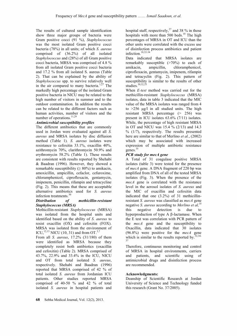
The results of cultured sample identification
show three major groups of bacteria were
Gram positive cocci (91 %), Staphylococcus
was the most isolated Gram positive cocci
bacteria (78%) in all units; of which S. aureus
comprised of (36.2%) of all isolated
Staphylococcus and (28%) of all Gram positive
cocci bacteria, MRSA was comprised of 4.8 %
from all isolated Gram positive cocci bacteria
and 17.2 % from all isolated S. aureus (Table
2). That can be explained by the ability of
Staphylococcus spp. to survive relatively well
in the air compared to many bacteria.1,27
The
markedly high percentage of the isolated Gram
positive bacteria in NICU may be related to the
high number of visitors in summer and to the
outdoor contamination. In addition the results
can be related to the different factors such as
human activities, number of visitors and the
number of operations.11,29
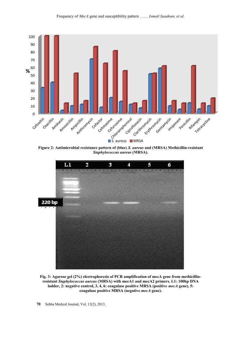
Antimicrobial susceptibility profiles
The different antibiotics that are commonly
used in Jordan were evaluated against all S.
aureus and MRSA isolates by disc diffusion
method (Table 1). S. aureus isolates were
resistance to cefoxitin 33.1%, oxacillin 40%,
azithromycin 70%, clarithromycin 50.9% and
erythromycin 58.2% (Table 1). These results
are consistent with results reported by Shehabi
& Baadran (1996). However, they showed a
remarkable susceptibility (≥ 80%) to amikacin,
amoxicillin, ampicillin, cefaclor, cefuroxime,
chloramphenicol, ciprofloxacin, gentamycin,
imipenem, penicillin, rifampin and tetracycline
(Fig. 2). This means that these are acceptable
alternative antibiotics used for S. aureus
infection treatments.30
Distribution of methicillin-resistant
Staphylococcus (MRSA)
Methicillin-resistant Staphylococcus (MRSA)
was isolated from the hospital units and
identified based on the ability of S. aureus to
resist oxacillin (OX) and cefoxitin (FOX).
MRSA was isolated from the environment of
ICU,12,13
NICU (10, 31) and from OT.3,7
From all S. aureus, 17.2% (31/180) of them
were identified as MRSA because they
completely resist both antibiotics (oxacillin
and cefoxitin) (Table 2). MRSA comprised of
43.7%, 22.9% and 33.4% in the ICU, NICU
and OT from total isolated S. aureus,
respectively. Shehabi and Baadran (1996)
reported that MRSA comprised of 42 % of
total isolated S. aureus from Jordanian ICU
patients. Other studies reported MRSA
comprised of 40-50 % and 42 % of total
isolated S. aureus in hospital patients and
hospital staff, respectively,27
and 38 % in those
hospitals with more than 500 beds.
10 The high
percentages of MRSA in OT and ICU than the
other units were correlated with the excess use
of disinfection process antibiotics and patient
infection.10,32-34
Data indicated that MRSA isolates are
remarkably susceptible (>70%) to each of
amikacin, ampicillin, chloramphenicol,
ciprofloxacin, gentamycin, imipenem, rifampin
and tetracyclin (Fig. 2). This pattern of
susceptibility is similar to the results of other
studies.30,32,33
When E-test method was carried out for the
methicillin-resistant Staphylococcus (MRSA)
isolates, data in table 3 indicated that the MIC
value of the MRSA isolates was ranged from 4
to >256 μg/l in all studied units. The high
resistant MRSA percentage (> 256) was
present in ICU isolates 63.6% (7/11) isolates.
While, the percentage of high resistant MRSA
in OT and NICU was 15.4 % (2/13) and 14.3
% (1/7), respectively. The results presented
here are similar to that of Merlino et al., (2002)
which may be associated with increased
expression of multiple antibiotic resistance
genes.35
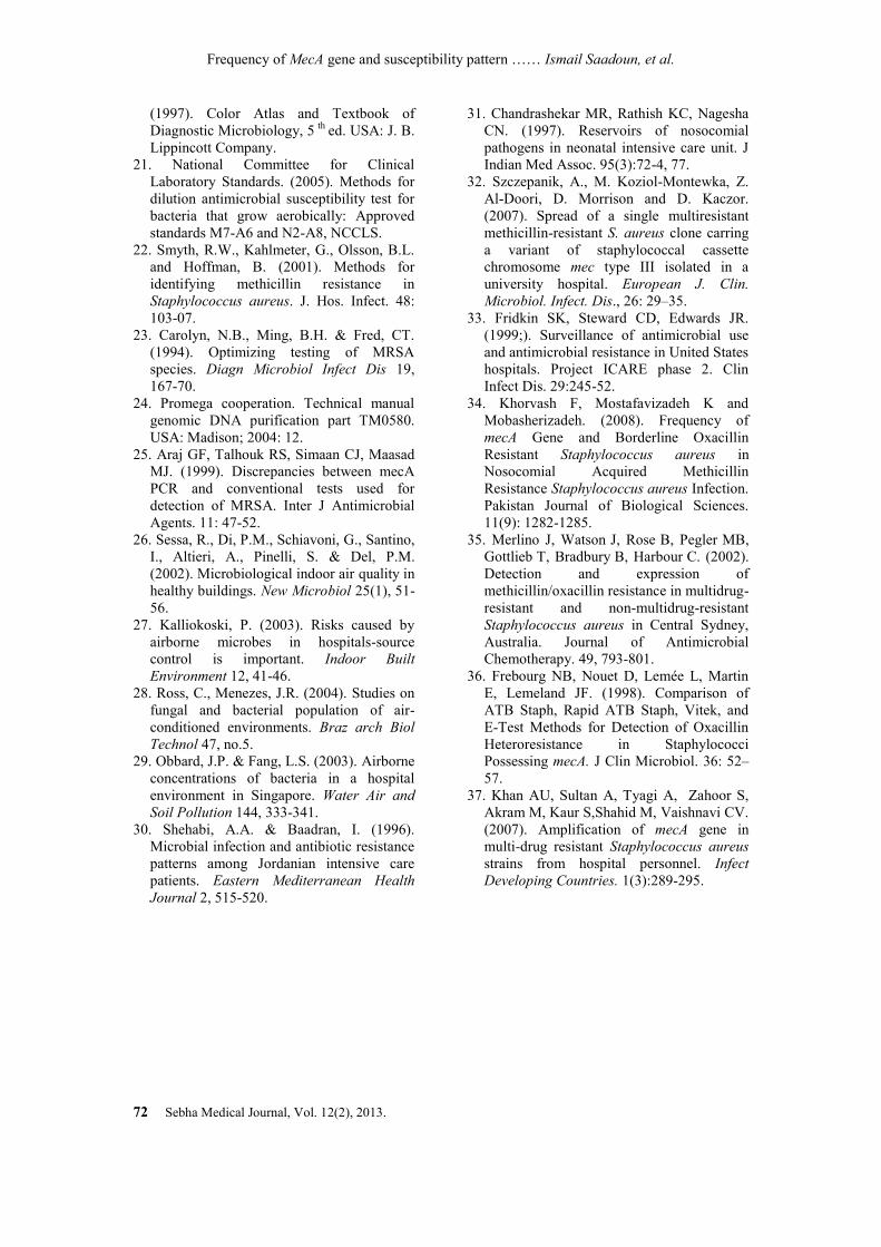
PCR study for mecA gene
A Total of 31 coagulase positive MRSA
isolates (table 3) were tested for the presence
of mecA gene. A DNA fragment of 220 bp was
amplified from DNA of all of the tested MRSA
isolates (Fig. 3). When the presence of the
mecA gene is correlated with the resistance
level in the aerosol isolates of S. aureus and
the MIC of oxacillin and cefoxitin data
indicated that one (3.2%) of 31 methicillin-
resistant S. aureus was classified as mecA gene
negative S. aureus according to Merlino et al,35
this negative detection is due to
hyperproduction of type A β-lactamase. When
the E test was correlation with PCR pattern of
the mecA gene and the susceptibility to
Oxacillin, data indicated that 30 isolates
(96.8%) were positive for the mecA gene
which is similar to the results reported by.32,34-
37
Therefore, continuous monitoring and control
of MRSA in hospital environments, carriers
and patients, and scientific using of
antimicrobial drugs and disinfection process
are recommended.
Acknowledgments:
Deanship of Scientific Research at Jordan
University of Science and Technology funded
this research (Grant No. 37/2005).

Frequency of MecA gene and susceptibility pattern …… Ismail Saadoun, et al.
69 Sebha Medical Journal, Vol. 12(2), 2013.
Tables and figures:
Table 1: Antibiotic panel used for S. aureus and methicillin-resistant Staphylococcus aureus
(MRSA) and the percentage resistance of these bacteria to different antibiotics.
Antibiotic Symbol Concentration (ug) S. aureus MRSA
Amikacin AK 3 3.5% 12.9%
Amoxicillin AX 25 9.4% 51.6%
Ampicillin AM 10 11.3% 16.1%
Azithromycin AZM 15 70% 86%
Cefaclor CEC 30 7.5% 64.5%
Cefotaxime CTX 30 20% 80.6%
Cefoxitin FOX 30 33.1% 100%
Cefuroxime CXM 30 15% 54.8%
Chloramphenicol C 30 11.3% 12.9%
Ciprofloxacin CIP 5 6.6% 16.1%
Clarithromcin CLR 15 50.9% 51.6%
Erythromycin E 15 58.2% 61.2%
Gentamicin GN 10 9.4% 16.1%
Imipenem IPM 10 4.7% 12.9%
Oxacillin OX 1 40% 100%
Penicillin P 10 13.2% 61.3%
Rifampin RA 10 5.2% 12.9%
Tetracycline TE 30 9.4% 19.4%
Total 18 18
B A
Figure 1: E-Test Method for a resistant (A) and susceptible (B) S. aureus isolate to Oxacillin.
Table 2: the distribution percentages of isolated bacteria from the total isolates.
*OT (%) *ICU (%) *NICU (%) *T (%)
Gram positiveve cocci 39 25 27 91
Staphylococcus spp 28 24.5 25.5 78
S. aureus 10.5 8.5 9 28
MRSA 2.1 1.6 1.1 4.8
MRSA from all isolated S. aureus 7.5 5.8 3.9 17.2
*OT: operating theatres, ICU: intensive care units, NICU: nursery intensive care units, T: Percentage
from the total isolated bacteria.
Frequency of MecA gene and susceptibility pattern …… Ismail Saadoun, et al.
70 Sebha Medical Journal, Vol. 12(2), 2013.
0
10
20
30
40
50
60
70
80
90
100
S. aureus MRSA
%
Figure 2: Antimicrobial resistance pattern of (blue) S. aureus and (MRSA) Methicillin-resistant
Staphylococcus aureus (MRSA).
Fig. 3: Agarose gel (2%) electrophoresis of PCR amplification of mecA gene from methicillin-
resistant Staphylococcus aureus (MRSA) with mecA1 and mecA2 primers. L1: 100bp DNA
ladder, 2: negative control, 3, 4, 6: coagulase positive MRSA (positive mecA gene), 5:
coagulase positive MRSA (negative mecA gene).

Frequency of MecA gene and susceptibility pattern …… Ismail Saadoun, et al.
71 Sebha Medical Journal, Vol. 12(2), 2013.
Table 3: the distribution of MIC values and MecA gene according to hospital units.
E-Test/MIC value (μg/l ) 4 6 8 16 24 32 64 96 128 >256 TOTAL
MRSA IN ICU 1 0 0 2 1 0 0 0 0 7 11
MRSA IN OT 4 2 1 0 1 2 0 0 1 2 13
MRSA IN NICU 0 1 0 0 2 1 1 1 0 1 7
MecA gene psitive 4 3 1 2 4 3 1 1 1 10 30/31
References:
1. Jaffal, A.A., Nsanze, H., Banat, I.M.,
Mogheth, A.A., Bener, A. & Ameen, A.S.
(1997). Hospital airborne microbial
pollution in a desert country. Environ Int 23,
167-172
2. Saad, S.G. (2003). Integrated Environmental
Management for Hospitals. Indoor Built Environment 12, 93-98.
3. Javed I, Hafeez R, Zubair M, Anwar MS,
Tayyib M, Husnain S. (2008).
Microbiological Surveillance of Operation
Theatres and ICUs of a Tertiary Care
Hospital, Lahore. African Journal of
Biotechnology, 7(20): 3535-3539.
4. Newman, M.J. (2002). Neonatal intensive
care unit: reservoirs of nosocomial
pathogens. West Afr J Med 21(4), 310-312.
5. Karabay O, Kocoglu E, Tahtaci M. (2007).
The role of mobile phones in the spread of
bacteria associated with nosocomial
infections. J Infect Developing Countries,
1:72–73.
6. Hoet AE, Johnson A, Nava-Hoet RC,
Bateman S, Hillier A, Dyce J, Gebreyes
WA, Wittum TE. (2011). Environmental
methicillin-resistant Staphylococcus aureus
in a veterinary teaching hospital during a
nonoutbreak period. Vect Zoonot Dis,
11:609–615.
7. Buemi, M., Floccari, F. & Netto, M. (2000).
Environmental air pollution in an intensive
care unit for nephrology and dialysis. J
Nephrol 13(6), 433-6.
8. Nzeako BC, Alrashiedi S, Neilson F,
Albalkhair A. (2010). Type of Bacteria on
Some Medical Devices Used in Sultan
Qaboos University Hospital Wards. Middle-
East J. of Scientific Resarch. 5(6): 449-453.
9. Ekhaise FO, Ighosewe OU, Ajakpovi OD.
(2008). Hospital Indoor airborne Microflora
in Private and Government Owned
Hospitals in Benin City. Nigeria. World J.
of Medical Science, 3 (1): 19-23.
10. Andersen, B.M., Lindemann, R., Bergh,
K., Nesheim, B.I., Syyversen, G., Solheim,
N. & Laugeurd, F. (2002). Spread of
methicillin-resistant Staphylococcus aureus
in a neonatal intensive unit associated with
understaffing, overcrowding and mixing of
patients. J Hosp Infect 50, 18-24.
11. Beggs, C.B. (2003). The airborne
transmission of infection in hospital
buildings: fact or fiction? Indo Built
Environ 12, 9-18.
12. Meyer E, Schwab F, Gastmeier P, Rueden
H, Daschner FD. (2006). Surveillance of
antimicrobial use and antimicrobial
resistance in German intensive care units
(SARI): a summary of the data from 2001
through 2004. Infection, 34:303-309.
13. Cavalcanti, S.M., Franca, E.R., Cabral, C.,
Vilela, M.A., Montenegro, F., Menezes, D.
& Medeiros, A.C. (2005). Prevalence of
Staphylococcus aureus introduced into
intensive care units of a University Hospital.
Braz J Infect Dis 9(1), 56-63.
14. Hiramatsu K. (1995). Molecular evolution
of MRSA. Microbiol Immunol. 39:531.
15. Niemeyer DM, Pucci MJ, Thanassi JA, et
al. (1996). Role of mecA transcriptional
regulation in the phenotypic expression of
methicillin resistance in Staphylococcus
aureus. J Bacteriol. 178:5464–71.
16. Archer GL, Niemeyer DM, Thanassi JA.
(1997). Dissemination among staphylococci
of DNA sequences associated with
methicillin resistance. Antimicrob Agents
Chenother. 38:447–54.
17. Ahmed S, Edet E. Tulsi DC. (2000).
Molecular diagnosis of antimicrobial
resistance. Lebanese Medical Journal. 48
(4): 203-7.
18. Araj GF, Talhouk RS, Simaan CJ, Maasad
MJ. (1999;). Discrepancies between mecA
PCR and conventional tests used for
detection of MRSA. Inter J Antimicrobial
Agents. 11: 47-52.
19. Betty AF, Danial FS, Alice SW. Diagnostic
Microbiology 10th
ed. Mospy: Don Ladig;
1998:608.
20. Koneman, E.W., Allin, S.D., Janad, W.M.,
Schreckenberger, P.C. & Winn, W.C.
Frequency of MecA gene and susceptibility pattern …… Ismail Saadoun, et al.
72 Sebha Medical Journal, Vol. 12(2), 2013.
(1997). Color Atlas and Textbook of
Diagnostic Microbiology, 5 th
ed. USA: J. B.
Lippincott Company.
21. National Committee for Clinical
Laboratory Standards. (2005). Methods for
dilution antimicrobial susceptibility test for
bacteria that grow aerobically: Approved
standards M7-A6 and N2-A8, NCCLS.
22. Smyth, R.W., Kahlmeter, G., Olsson, B.L.
and Hoffman, B. (2001). Methods for
identifying methicillin resistance in
Staphylococcus aureus. J. Hos. Infect. 48:
103-07.
23. Carolyn, N.B., Ming, B.H. & Fred, CT.
(1994). Optimizing testing of MRSA
species. Diagn Microbiol Infect Dis 19,
167-70.
24. Promega cooperation. Technical manual
genomic DNA purification part TM0580.
USA: Madison; 2004: 12.
25. Araj GF, Talhouk RS, Simaan CJ, Maasad
MJ. (1999). Discrepancies between mecA
PCR and conventional tests used for
detection of MRSA. Inter J Antimicrobial
Agents. 11: 47-52.
26. Sessa, R., Di, P.M., Schiavoni, G., Santino,
I., Altieri, A., Pinelli, S. & Del, P.M.
(2002). Microbiological indoor air quality in
healthy buildings. New Microbiol 25(1), 51-
56.
27. Kalliokoski, P. (2003). Risks caused by
airborne microbes in hospitals-source
control is important. Indoor Built
Environment 12, 41-46.
28. Ross, C., Menezes, J.R. (2004). Studies on
fungal and bacterial population of air-
conditioned environments. Braz arch Biol
Technol 47, no.5.
29. Obbard, J.P. & Fang, L.S. (2003). Airborne
concentrations of bacteria in a hospital
environment in Singapore. Water Air and
Soil Pollution 144, 333-341.
30. Shehabi, A.A. & Baadran, I. (1996).
Microbial infection and antibiotic resistance
patterns among Jordanian intensive care
patients. Eastern Mediterranean Health
Journal 2, 515-520.
31. Chandrashekar MR, Rathish KC, Nagesha
CN. (1997). Reservoirs of nosocomial
pathogens in neonatal intensive care unit. J
Indian Med Assoc. 95(3):72-4, 77.
32. Szczepanik, A., M. Koziol-Montewka, Z.
Al-Doori, D. Morrison and D. Kaczor.
(2007). Spread of a single multiresistant
methicillin-resistant S. aureus clone carring
a variant of staphylococcal cassette
chromosome mec type III isolated in a
university hospital. European J. Clin.
Microbiol. Infect. Dis., 26: 29–35.
33. Fridkin SK, Steward CD, Edwards JR.
(1999;). Surveillance of antimicrobial use
and antimicrobial resistance in United States
hospitals. Project ICARE phase 2. Clin
Infect Dis. 29:245-52.
34. Khorvash F, Mostafavizadeh K and
Mobasherizadeh. (2008). Frequency of
mecA Gene and Borderline Oxacillin
Resistant Staphylococcus aureus in
Nosocomial Acquired Methicillin
Resistance Staphylococcus aureus Infection.
Pakistan Journal of Biological Sciences.
11(9): 1282-1285.
35. Merlino J, Watson J, Rose
B, Pegler
MB,
Gottlieb T, Bradbury
B, Harbour
C. (2002).
Detection and expression of
methicillin/oxacillin resistance in multidrug-
resistant and non-multidrug-resistant
Staphylococcus aureus in Central Sydney,
Australia. Journal of Antimicrobial
Chemotherapy. 49, 793-801.
36. Frebourg NB, Nouet D, Lemée L, Martin
E, Lemeland JF. (1998). Comparison of
ATB Staph, Rapid ATB Staph, Vitek, and
E-Test Methods for Detection of Oxacillin
Heteroresistance in Staphylococci
Possessing mecA. J Clin Microbiol. 36: 52–
57.
37. Khan AU, Sultan A, Tyagi A, Zahoor S,
Akram M, Kaur S,Shahid M, Vaishnavi CV.
(2007). Amplification of mecA gene in
multi-drug resistant Staphylococcus aureus
strains from hospital personnel. Infect
Developing Countries. 1(3):289-295.
http://www.ncbi.nlm.nih.gov/entrez/query.fcgi?db=pubmed&cmd=Search&term=%22Rathish+KC%22%5BAuthor%5D