Frequency and antimicrobial susceptibility of bacterial ... · Peritonitis is the main cause of PD...
12
Montelli et al., Cogent Medicine (2016), 3: 1242246 http://dx.doi.org/10.1080/2331205X.2016.1242246 NEPHROLOGY & UROLOGY | RESEARCH ARTICLE Frequency and antimicrobial susceptibility of bacterial agents causing peritoneal dialysis- peritonitis in a Brazilian single center over 20 years Augusto C. Montelli 1,2 , Terue Sadatsune 2 , Alessandro L. Mondelli 1 , Maria L.R.S. Cunha 2 , Jacqueline C.T. Caramori 1 , Pasqual Barretti 1 and Carlos H. Camargo 1,2 * Abstract: Peritonitis remains the main complication of peritoneal dialysis (PD). Antimicrobial therapy may be threatened by development of bacterial resistance, demanding continuous surveillance of infectious agents to improve empirical treatment. The aims of this study were to evaluate the bacterial agents caus- ing peritonitis and their antimicrobial susceptibility in patients undergone PD in a Brazilian center, between 1993 and 2013. Strains were recovered from peritoneal fluid, identified by usual methods and submitted to Etest antimicrobial susceptibil- ity. A total of 400 strains were studied, of which 65.8% were Staphylococcus spp.: S. epidermidis, the main species (22.8%), followed by S. aureus (21.3%). Over time, we verified a decrease in overall peritonitis occurrence, and an increase in gram-nega- tive bacteria proportion, attributed, mainly, to a decrease in gram-positive agents. Vancomycin was effective against all Staphylococcus strains, and oxacillin resistance was higher in coagulase-negative Staphylococcus compared to S. aureus (p < 0.01). Regarding gram-negative bacteria, Enterobacteriaceae species accounted for 20.3% of the strains, but P. aeruginosa, Acinetobacter species, S. maltophilia, B. cepacia, *Corresponding author: Carlos H. Camargo, Department of Internal Medicine, Botucatu Medical School, UNESP—Universidade Estadual Paulista, Botucatu Campus, Sao Paulo, Brazil; Department of Microbiology and Immunology, Biosciences Institute of Botucatu, UNESP—Universidade Estadual Paulista, Distrito de Rubião Jr, s/n, CEP 18618-970, Botucatu Campus, São Paulo, Brazil E-mail: [email protected] Reviewing editor: Tsai-Ching Hsu, Chung Shan Medical University, Taiwan Additional information is available at the end of the article ABOUT THE AUTHORS After the introduction of peritoneal dialysis in Botucatu Medical School (BMS) on 1990s, a great number of patients presented peritonitis. In 1993, a multidisciplinary team of medicine doctors from BMS and biologists from Botucatu Biosciences Institute, UNESP, was structured to investigate the clinical, epidemiological and microbiological aspects of peritonitis and to reduce the infection rates in our center, composed by clinical nephrologists (Pasqual Barretti, Jacqueline Caramori), clinical pathologists (Augusto Montelli, Alessandro Mondelli), microbiologists (Terue Sadatsune, Maria Cunha) and molecular microbiologist (Carlos Camargo). The ongoing work of the group over the last 20 years generated numerous national and international publications and academic thesis about peritonitis, with microbiological investigations in all of them. In this paper we present a synthesis of all microbiological information obtained from 400 bacterial strains studied from 1993 to 2013, including identification and antimicrobial susceptibility. The use of our results for improving patient care is suggested. PUBLIC INTEREST STATEMENT Patient with chronic kidney disease often progress to renal failure. Peritoneal dialysis (PD) represents an option in treating these cases before hemodialysis or renal transplantation. PD uses peritoneum as a filter, being a practical alternative with comparable survival rates to hemodialysis. Peritonitis is the main cause of PD interruption, with known impact on morbidity and mortality. It is an infection that can be caused by several microorganisms, presenting variable pathogenic capacity and antimicrobial resistance. Serious problems for the practice of rational antibiotic therapy can occur. In this study we investigated, over 20 years (1993–2013), the frequency and the antimicrobial sensibility of 400 bacterial strains isolated from peritonitis, in order to analyze the impact of practice changes adopted in one center and to provide data for improving patient care. Our microbiological data are in accordance to the current international guidelines, which recommends retrospective local results to define the optimal empiric therapy. Received: 09 June 2016 Accepted: 25 September 2016 First Published: 28 September 2016 © 2016 The Author(s). This open access article is distributed under a Creative Commons Attribution (CC-BY) 4.0 license. Page 1 of 12
-
Upload
duongthien -
Category
Documents
-
view
221 -
download
0
Transcript of Frequency and antimicrobial susceptibility of bacterial ... · Peritonitis is the main cause of PD...

Montelli et al., Cogent Medicine (2016), 3: 1242246http://dx.doi.org/10.1080/2331205X.2016.1242246
NEPHROLOGY & UROLOGY | RESEARCH ARTICLE
Frequency and antimicrobial susceptibility of bacterial agents causing peritoneal dialysis-peritonitis in a Brazilian single center over 20 yearsAugusto C. Montelli1,2, Terue Sadatsune2, Alessandro L. Mondelli1, Maria L.R.S. Cunha2, Jacqueline C.T. Caramori1, Pasqual Barretti1 and Carlos H. Camargo1,2*
Abstract: Peritonitis remains the main complication of peritoneal dialysis (PD). Antimicrobial therapy may be threatened by development of bacterial resistance, demanding continuous surveillance of infectious agents to improve empirical treatment. The aims of this study were to evaluate the bacterial agents caus-ing peritonitis and their antimicrobial susceptibility in patients undergone PD in a Brazilian center, between 1993 and 2013. Strains were recovered from peritoneal fluid, identified by usual methods and submitted to Etest antimicrobial susceptibil-ity. A total of 400 strains were studied, of which 65.8% were Staphylococcus spp.: S. epidermidis, the main species (22.8%), followed by S. aureus (21.3%). Over time, we verified a decrease in overall peritonitis occurrence, and an increase in gram-nega-tive bacteria proportion, attributed, mainly, to a decrease in gram-positive agents. Vancomycin was effective against all Staphylococcus strains, and oxacillin resistance was higher in coagulase-negative Staphylococcus compared to S. aureus (p < 0.01). Regarding gram-negative bacteria, Enterobacteriaceae species accounted for 20.3% of the strains, but P. aeruginosa, Acinetobacter species, S. maltophilia, B. cepacia,
*Corresponding author: Carlos H. Camargo, Department of Internal Medicine, Botucatu Medical School, UNESP—Universidade Estadual Paulista, Botucatu Campus, Sao Paulo, Brazil; Department of Microbiology and Immunology, Biosciences Institute of Botucatu, UNESP—Universidade Estadual Paulista, Distrito de Rubião Jr, s/n, CEP 18618-970, Botucatu Campus, São Paulo, Brazil E-mail: [email protected]
Reviewing editor:Tsai-Ching Hsu, Chung Shan MedicalUniversity, Taiwan
Additional information is available at the end of the article
ABOUT THE AUTHORSAfter the introduction of peritoneal dialysis in Botucatu Medical School (BMS) on 1990s, a great number of patients presented peritonitis. In 1993, a multidisciplinary team of medicine doctors from BMS and biologists from Botucatu Biosciences Institute, UNESP, was structured to investigate the clinical, epidemiological and microbiological aspects of peritonitis and to reduce the infection rates in our center, composed by clinical nephrologists (Pasqual Barretti, Jacqueline Caramori), clinical pathologists (Augusto Montelli, Alessandro Mondelli), microbiologists (Terue Sadatsune, Maria Cunha) and molecular microbiologist (Carlos Camargo). The ongoing work of the group over the last 20 years generated numerous national and international publications and academic thesis about peritonitis, with microbiological investigations in all of them. In this paper we present a synthesis of all microbiological information obtained from 400 bacterial strains studied from 1993 to 2013, including identification and antimicrobial susceptibility. The use of our results for improving patient care is suggested.
PUBLIC INTEREST STATEMENTPatient with chronic kidney disease often progress to renal failure. Peritoneal dialysis (PD) represents an option in treating these cases before hemodialysis or renal transplantation. PD uses peritoneum as a filter, being a practical alternative with comparable survival rates to hemodialysis. Peritonitis is the main cause of PD interruption, with known impact on morbidity and mortality. It is an infection that can be caused by several microorganisms, presenting variable pathogenic capacity and antimicrobial resistance. Serious problems for the practice of rational antibiotic therapy can occur. In this study we investigated, over 20 years (1993–2013), the frequency and the antimicrobial sensibility of 400 bacterial strains isolated from peritonitis, in order to analyze the impact of practice changes adopted in one center and to provide data for improving patient care. Our microbiological data are in accordance to the current international guidelines, which recommends retrospective local results to define the optimal empiric therapy.
Received: 09 June 2016Accepted: 25 September 2016First Published: 28 September 2016
© 2016 The Author(s). This open access article is distributed under a Creative Commons Attribution (CC-BY) 4.0 license.
Page 1 of 12

Page 2 of 12
Montelli et al., Cogent Medicine (2016), 3: 1242246http://dx.doi.org/10.1080/2331205X.2016.1242246
P. fluorescens and A. xylosoxidans were also detected (14.3%). Non-fermentative gram-Negative bacilli presented increased antimicrobial resistance compared to Enterobacteriaceae, and imipenem was the most active antimicrobial drug. Over the 20 year period, increasing or decreasing in the antimicrobial susceptibilities were not observed, despite the occurrence of punctual oscillations. In summary, we veri-fied peritonitis occurrence declining over years, gram-positive infections proportion dropping, and, inversely, gram-negative pathogens proportion increasing. Our find-ing reinforces recommendations regarding retrospective evaluation of antimicrobial susceptibility to define the optimal empirical therapy in each center.
Subjects: Dialysis; Infectious Diseases; Microbiology
Keywords: gram-positive; gram-negative; MIC; peritoneal infection; peritonitis; antimicrobial resistance; gentamicin resistance; Etest
1. IntroductionPeritoneal dialysis (PD) represents an option in treating chronic renal diseases. This method presents the same survival rates as hemodialysis in treating end-stage renal disease, but has an important advantage in patient satisfaction and life quality reports (Tokgoz, 2009). Advances in this treatment option resulted in reducing peritonitis rates in recent decades, but infective events are still the main adverse cause of technique failure, with significant impacts on morbidity and mortality rates (Pérez Fontan, et al., 2005). Despite geographic specificities, the main etiological agents of infections are Staphylococcus species (Nishina et al., 2014; van Esch, Krediet, & Struijk, 2014). We have shown that outcomes of peritonitis due to Staphylococcus are influenced by implicated species (Barretti, Montelli, Batalha, Caramori, & Maria de Lourdes, 2009; Camargo et al., 2014), virulence factors (Barretti, Montelli, et al., 2009; Barretti et al., 2012; Cunha et al., 2004, 2005), antimicrobial resistance (Camargo et al., 2014; Oliveira et al., 2012) besides some specific host features (Barretti et al., 2012). Indeed, peritonitis due to gram-negative rods are milder when caused by Enterobacteriaceae strains compared to non-fermentative gram-negative bacilli (NFGNB) (Oliveira et al., 2012); very frequent catheter removal and hospitalization rates are reported for patients with P. aeruginosa infections (Borràs, 2009; Siva et al., 2009). The International Society for Peritoneal Dialysis (ISPD) recommends that empirical treatment must be initiated based on retrospective data of antimicrobial susceptibil-ity of each center (Li et al., 2010; Piraino et al., 2005). Changes in the practices, in the agents impli-cated in peritonitis and in their antimicrobial susceptibility, warrant continuous surveillance of infections in PD (Szeto, 2014). In this study, we aimed to evaluate the frequency and the antimicro-bial susceptibility of the bacterial agents causing PD-infections in a single Brazilian center over the last two decades (1994–2013), in order to analyze the impact of practice changes adopted in this center and to provide data for improving patient care.
2. Materials and methodsThis study evaluated the most prevalent bacterial isolates (Staphylococcus and gram-negative ba-cilli) recovered from peritoneal fluid of patients attending a single university center in Brazil, continu-ously, over a period of 20 years (1993–2013). Patients were diagnosed with peritonitis by the presence of at least two of the following criteria: (1) cloudy peritoneal effluent, (2) abdominal pain, (3) a dialysate white-cell count higher than 100/μL with at least 50% polymorphonuclear cells, and (4) positive culture of peritoneal effluent (Li et al., 2010; Piraino et al., 2005). Dialysis practices changed over the evaluated period (Figure 1): a Y-set system was introduced in 1994; automated PD (APD) in 1998; a twin bag was introduced in 1999. Daily mupirocin cream application at the exit site has been recommended since 2003 and gentamicin, since 2007.
The initial cultures from centrifuged peritoneal fluid samples were performed with the Bactec® Automated System (Becton Dickinson Company, Sparks, Maryland, USA), and then seeded onto blood agar and MacConkey Agar (Oxoid, Basingstoke, United Kingdom). The isolates were gram

Page 3 of 12
Montelli et al., Cogent Medicine (2016), 3: 1242246http://dx.doi.org/10.1080/2331205X.2016.1242246
stained to confirm purity and determine morphology and specific color. Bacteria identification was performed by classical phenotypic tests (Koneman & Winn, 2006) and by Vitek or Vitek 2 (bioMé-rieux, Inc., Durham, NC, USA) automated systems.
The in vitro susceptibilities to antimicrobial agents, chosen according to the bacterial agent and the Clinical and Laboratory Standards Institute (CLSI) recommendations, were evaluated based on minimal inhibitory concentrations (MIC) determined by the Etest (bioMérieux, Inc., Durham, NC, USA), according to the manufacturer’s instructions. The following antimicrobial agents were evalu-ated: penicillin G, oxacillin, cephalothin, levofloxacin, netilmicin, vancomycin, for gram-positive agents, and gentamicin, ceftazidime, cefepime, gatifloxacin, ofloxacin and imipenem for gram-neg-ative ones. MIC was categorized in susceptible or resistant (strains with intermediate susceptibility were considered resistant) according to CLSI breakpoints (Clinical & Laboratory Standards Institute, 2015). Quality control strains (S. aureus ATCC 29213; E. coli ATCC 25922; P. aeruginosa ATCC 27853) were used to ensure test performance. MIC50 and MIC90 values were calculated based on the MIC value distribution: MIC50 is defined as the median value of MIC distribution; that is, the MIC value that can inhibit the growth of 50% of the bacterial population; MIC90 is the MIC value that can inhibit the growth of 90% of the bacterial population.
Resistance pattern classes were created to compare different bacterial species and were deter-mined according to the number of resistance presented for each strain: susceptible (zero resistance), simple resistant (resistance to only one drug) or multiple resistant (resistance to two or more drugs).
Chi-squared or Fisher’s exact tests were used to compare frequencies (www.openepi.com); p values below 0.05 were considered to be significant.
The Research Ethics Committee of the Faculty of Medicine of Botucatu, Brazil (OF. 028/08-CEP) ap-proved this study. We were granted an exemption from the requirement to obtain written informed-consent from the participants and/or their legal guardians because the strains included in the study had already been isolated and stored, on an ongoing basis, in the Culture Collection of the Department of Microbiology and Immunology, UNESP, Botucatu, São Paulo, Brazil.
Figure 1. Temporal distribution of bacterial causative organisms of peritoneal dialysis-related peritonitis in a Brazilian center over two decades. Left axis (columns) represents the proportion (percentage) of each bacterial group, according to the 3-year periods evaluated. Right axis (line) indicates the overall number of isolates in each of the same 3-year periods. Changes in peritoneal dialysis practice are named above the bars, as well as the year of their implementation.
Notes: APD: automated peritoneal dialysis; CoNS: coagulase-negative Staphylococcus; GNB: gram-negative bacilli.
Page 4 of 12
Montelli et al., Cogent Medicine (2016), 3: 1242246http://dx.doi.org/10.1080/2331205X.2016.1242246
3. Results
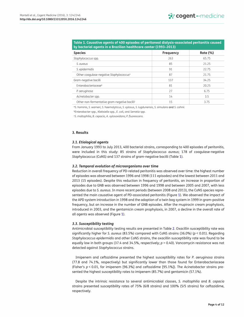
3.1. Etiological agentsFrom January 1993 to July 2013, 400 bacterial strains, corresponding to 400 episodes of peritonitis, were included in this study: 85 strains of Staphylococcus aureus; 178 of coagulase-negative Staphylococcus (CoNS) and 137 strains of gram-negative bacilli (Table 1).
3.2. Temporal evolution of microorganisms over timeReduction in overall frequency of PD-related peritonitis was observed over time: the highest number of episodes was observed between 1996 and 1998 (111 episodes) and the lowest between 2011 and 2013 (15 episodes). Despite this reduction in frequency of peritonitis, an increase in proportion of episodes due to GNB was observed between 1996 and 1998 and between 2005 and 2007, with less episodes due to S. aureus. In more recent periods (between 2008 and 2013), the CoNS species repre-sented the main causative agent of PD-associated peritonitis (Figure 1). We observed the impact of the APD system introduction in 1998 and the adoption of a twin bag system in 1999 in gram-positive frequency, but an increase in the number of GNB episodes. After the mupirocin cream prophylaxis, introduced in 2003, and the gentamicin cream prophylaxis, in 2007, a decline in the overall rate of all agents was observed (Figure 1).
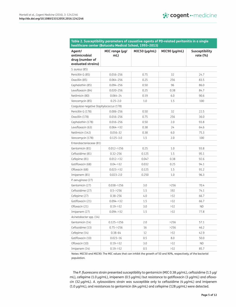
3.3. Susceptibility testingAntimicrobial susceptibility testing results are presented in Table 2. Oxacillin susceptibility rate was significantly higher for S. aureus (83.5%) compared with CoNS strains (36.0%) (p < 0.01). Regarding Staphylococcus epidermidis and other CoNS strains, the oxacillin susceptibility rate was found to be equally low in both groups (37.4 and 34.5%, respectively; p = 0.40). Vancomycin resistance was not detected against Staphylococcus strains.
Imipenem and ceftazidime presented the highest susceptibility rates for P. aeruginosa strains (77.8 and 74.1%, respectively) but significantly lower than those found for Enterobacteriaceae (Fisher’s p < 0.01, for imipenem [96.3%] and ceftazidime [95.1%]). The Acinetobacter strains pre-sented the highest susceptibility rates to imipenem (85.7%) and gentamicin (57.1%).
Despite the intrinsic resistance to several antimicrobial classes, S. maltophilia and B. cepacia strains presented susceptibility rates of 75% (6/8 strains) and 100% (5/5 strains) for ceftazidime, respectively.
Table 1. Causative agents of 400 episodes of peritoneal dialysis-associated peritonitis caused by bacterial agents in a Brazilian healthcare center (1993–2013)
aS. hominis, S. warneri, S. haemolyticus, S. xylosus, S. lugdunensis, S. simulans and S. cohnii.bEnterobacter spp., Klebsiella spp., E. coli, and Serratia spp.cS. maltophilia, B. cepacia, A. xylosoxidans; P. fluorescens.
Species Frequency Rate (%)Staphylococcus spp. 263 65.75
S. aureus 85 21.25
S. epidermidis 91 22.75
Other coagulase-negative Staphylococcusa 87 21.75
Gram-negative bacilli 137 34.25
Enterobacteriaceaeb 81 20.25
P. aeruginosa 27 6.75
Acinetobacter spp. 14 3.5
Other non-fermentative gram-negative bacillic 15 3.75
Page 5 of 12
Montelli et al., Cogent Medicine (2016), 3: 1242246http://dx.doi.org/10.1080/2331205X.2016.1242246
The P. fluorescens strain presented susceptibility to gentamicin (MIC 0.38 μg/mL), ceftazidime (1.5 μg/mL), cefepime (1.0 μg/mL), imipenem (0.5 μg/mL) but resistance to gatifloxacin (3 μg/mL) and ofloxa-cin (32 μg/mL). A. xylosoxidans strain was susceptible only to ceftazidime (4 μg/mL) and imipenem (1.0 μg/mL), and resistances to gentamicin (64 μg/mL) and cefepime (128 μg/mL) were detected.
Table 2. Susceptibility parameters of causative agents of PD-related peritonitis in a single healthcare center (Botucatu Medical School, 1993–2013)
Notes: MIC50 and MIC90: The MIC values that can inhibit the growth of 50 and 90%, respectively, of the bacterial population.
Agent/antimicrobial drug (number of evaluated strains)
MIC range (μg/mL)
MIC50 (μg/mL) MIC90 (μg/mL) Susceptibility rate (%)
S. aureus (85)
Penicillin G (85) 0.016–256 0.75 32 24.7
Oxacillin (85) 0.064–256 0.25 256 83.5
Cephalothin (85) 0.094–256 0.50 96 86.0
Levofloxacin (84) 0.020–256 0.25 0.38 84.7
Netilmicin (80) 0.064–24 0.19 6.0 90.6
Vancomycin (85) 0.25–2.0 1.0 1.5 100
Coagulase negative Staphylococcus (178)
Penicillin G (178) 0.006–256 0.50 32 22.5
Oxacillin (178) 0.016–256 0.75 256 36.0
Cephalothin (178) 0.016–256 0.50 2.0 93.8
Levofloxacin (63) 0.064–>32 0.38 24 64.6
Netilmicin (142) 0.016–32 0.38 6.0 75.3
Vancomycin (178) 0.125–3.0 1.5 2.0 100
Enterobacteriaceae (81)
Gentamicin (81) 0.012–>256 0.25 1.0 93.8
Ceftazidime (81) 0.32–256 0.125 1.5 95.1
Cefepime (81) 0.012–>32 0.047 0.38 92.6
Gatifloxacin (68) 0.04–>32 0.032 0.25 94.1
Ofloxacin (68) 0.023–>32 0.125 1.5 91.2
Imipenem (81) 0.023–2.0 0.250 1.0 96.3
P. aeruginosa (27)
Gentamicin (27) 0.038–>256 3.0 >256 70.4
Ceftazidime (27) 0.5–>256 1.5 192 74.1
Cefepime (27) 0.38–256 4.0 >32 66.7
Gatifloxacin (21) 0.094–>32 1.5 >32 66.7
Ofloxacin (21) 0.19–>32 3.0 >32 ND
Imipenem (27) 0.094–>32 1.5 >32 77.8
Acinetobacter spp. (14)
Gentamicin (14) 0.125–>256 2.0 >256 57.1
Ceftazidime (13) 0.75–>256 16 >256 46.2
Cefepime (14) 0.38–64 12 >32 42.9
Gatifloxacin (10) 0.023–16 0.5 8.0 50.0
Ofloxacin (10) 0.19–>32 3.0 >32 ND
Imipenem (14) 0.19–>32 0.5 >32 85.7

Page 6 of 12
Montelli et al., Cogent Medicine (2016), 3: 1242246http://dx.doi.org/10.1080/2331205X.2016.1242246
3.4. Resistance patterns: Susceptible, simple and multiple resistancesResistance patterns were defined according to the number of resistance presented for each strain: susceptible (zero resistance), simple resistant (resistance to only one drug) or multiple resistant (resistance to two or more drugs) (Table 3). Overall CoNS presented higher proportion of multiple resistant strains (61%) compared to S. aureus (17%) (p < 0.01), but similar rates of multiple resistant between S. epidermidis (62%) and the other CoNS staphylococci (60%) (p = 0.46). Regarding gram-negative bacilli, Enterobacteriaceae species presented a lower proportion of multiple resistant strains (10%) compared to P. aeruginosa (33%; Fisher’s p < 0.01) or Acinetobacter spp. (64%; Fisher’s p < 0.01).
3.5. Evolution of antimicrobial susceptibility over timeStaphylococcus spp. presented fluctuating values of penicillin G susceptibility rates, but constant rates of oxacillin (lower rates for CoNS), cephalothin, and vancomycin (always 100%) susceptibilities. Among Enterobacteriaceae strains, high susceptibility rates (about 90%) were verified over the peri-ods for the drugs ceftazidime and imipenem; a statistically insignificant decrease in susceptibility rates of gentamicin (from 92.9% to 66.7%, Fisher’s p = 0.14) and cefepime (from 92.9% to 55.6%, Fisher’s p = 0.06) was observed in 2008–2010 compared with 2005–2007 period, respectively. For P. aeruginosa and Acinetobacter spp., imipenem showed the highest activity, but the remaining drug susceptibility rates oscillated over time (Table 4).
Over time, it was possible to observe a fluctuation in resistant pattern categories, as demonstrat-ed in Figure 2, with a slight (non-significant) trend of increasing strains susceptible to all evaluated drugs. The period of 1993–1995 presented the lowest value of susceptibility (19%) and the highest rate of multiple resistance strains (58%). From this period on, susceptibility ranged from 27% in 1996–1998 to the peak 53% in 2011–2013. Contrasting to emerging gentamicin resistance observed from 2007, increase in the susceptibility class was mainly influenced by Enterobacteriaceae strains.
Table 3. Distribution of resistance pattern classes [frequency (%)] of bacterial agents of peritoneal dialysis-associated peritonitis caused by bacterial agents in a Brazilian healthcare center (1993–2013)
Notes: Susceptible: zero resistance; simple resistant: resistance to only one drug; multiple resistant: resistance to two or more drugs.aS. hominis, S. warneri, S. haemolyticus, S. xylosus, S. lugdunensis, S. simulans and S. cohnii.bEnterobacter spp., Klebsiella spp., E. coli, and Serratia spp.cS. maltophilia, B. cepacia, A. xylosoxidans; P. fluorescens.
Agent Susceptible Simple resistant Multiple resistant TotalStaphylococcus spp. 50 (19) 91 (35) 122 (46) 263 (100)
S. aureus 21 (25) 50 (59) 14 (17) 85 (100)
S. epidermidis 11 (12) 24 (26) 56 (62) 91 (100)
Other coagulase-negative Staphylococcusa
18 (21) 17 (20) 52 (60) 87 (100)
Gram-negative bacilli 101 (74) 8 (6) 28 (20) 137 (100)
Enterobacteriaceaeb 68 (84) 5 (6) 8 (10) 81 (100)
P. aeruginosa 17 (63) 1 (4) 9 (33) 27 (100)
Acinetobacter spp. 5 (36) 0 (0) 9 (64) 14 (100)
Other non-fermentative gram-negative bacillic
11 (73) 2 (13) 2 (13) 15 (100)
Total 151 (38) 99 (25) 150 (37) 400 (100)

Page 7 of 12
Montelli et al., Cogent Medicine (2016), 3: 1242246http://dx.doi.org/10.1080/2331205X.2016.1242246
Tabl
e 4.
Dis
trib
utio
n of
sus
cept
ibili
ty ra
tes
for t
he m
ain
antim
icro
bial
age
nts
in p
reva
lent
cau
sativ
e ag
ents
of p
erito
neal
-dia
lysi
s ba
cter
ial r
elat
ed p
erito
nitis
S.
aure
us; c
oagu
lase
neg
ativ
e St
aphy
loco
ccus
(CoN
S); E
nter
obac
teria
ceae
; P. a
erug
inos
a; A
cine
toba
cter
spp
.
Not
e: N
T–no
t tes
ted.
Agen
t/an
timic
robi
al19
93–1
995
1996
–199
819
99–2
001
2002
–200
420
05–2
007
2008
–201
020
11–2
013
S. a
ureu
s (N
= 8
5)N
= 0
N =
52N
= 16
N =
11N
= 1
N =
5N
= 0
Pe
nici
llin
GNT
3313
00
40NT
O
xaci
llin
NT87
7582
100
80NT
Ce
phal
otin
NT87
8182
100
100
NT
Va
ncom
ycin
NT10
010
010
010
010
0NT
CoNS
(N =
178
)N
= 52
N =
53N
= 19
N =
15N
= 8
N =
21N
= 10
Pe
nici
llin
G25
1716
1325
2950
O
xaci
llin
4038
3213
2529
70
Ce
phal
otin
8994
9010
010
010
010
0
Va
ncom
ycin
100
100
100
100
100
100
100
Ente
roba
cter
iace
ae (N
= 8
1)N
= 0
N =
6N
= 21
N =
29N
= 14
N =
9N
= 2
Ge
ntam
icin
NT10
010
097
9367
100
Ce
ftaz
idim
eNT
100
100
9393
8910
0
Ce
fepi
me
NT10
010
097
9356
100
Im
ipen
emNT
100
100
9793
8910
0
P. ae
rugi
nosa
(N =
27)
N =
0N
= 0
N =
6N
= 7
N =
9N
= 3
N =
2
Ge
ntam
icin
NTNT
8386
4467
100
Ce
ftaz
idim
eNT
NT10
086
4467
100
Im
ipen
emNT
NT10
086
4410
010
0
Acin
etob
acte
r spp
. (N
= 14
)N
= 0
N =
0N
= 3
N =
5N
= 2
N =
3N
= 1
Ge
ntam
icin
NTNT
3360
5067
100
Ce
ftaz
idim
eNT
NT67
400
670
Im
ipen
emNT
NT10
010
050
100
0

Page 8 of 12
Montelli et al., Cogent Medicine (2016), 3: 1242246http://dx.doi.org/10.1080/2331205X.2016.1242246
4. Discussion
4.1. Etiological agentsIn this largest series of PD-related peritonitis in a single Brazilian healthcare center, we evaluated the frequency of etiological agents and their antimicrobial susceptibility over 20 years. We verified the overall predominance of Staphylococcus species and a global reduction in the number of bacte-rial peritonitis over the years.
Staphylococcus spp. are currently the main etiological agents of PD infections (Nieto-Ríos et al., 2014; Nishina et al., 2014; van Esch et al., 2014), and nasal colonization status seems to play an important role in the development of S. aureus peritonitis. Indeed, infection due to S. aureus were shown to increase the unfavorable outcomes compared to CoNS in our center (Camargo et al., 2014) with virulence factors such as beta-hemolysin and biofilm production (Barretti, Montelli, et al., 2009; Barretti et al., 2012) impacting negatively on outcomes. On the other hand, but in line with the larg-est series (Fahim et al., 2010; Szeto et al., 2008), infections due to CoNS were milder and with a fa-vorable outcome. In our center, a favorable outcome was influenced by oxacillin susceptibility and initial treatment with vancomycin (Camargo et al., 2014), underscoring the importance of longitudi-nal analyses of antimicrobial susceptibility data towards optimizing empirical strategies and avoid-ing the emergence of antimicrobial resistance (Szeto, 2014). A high and constant frequency rate of oxacillin resistance CoNS required changes in the empirical treatment in our center, which currently is based on IP vancomycin and amikacin (Barretti et al., 2012). Contrasting to the reduction in the overall frequency of gram-positive pathogens, infection due to gram-negative pathogens increased in number, and consequently, in its proportion, in the early 2000s (Borràs, 2009). The same phenom-enon was also reported earlier (Huang et al., 2011; Szeto et al., 2005), mainly attributed to improve-ments in connectology methodology that reduced the incidence of skin contaminating pathogens (Szeto, 2014). Importance of gram-negative pathogens have increased over the last years, particu-larly due to the emergence of different antimicrobial resistance determinants, such as extended spectrum beta-lactamase in Enterobacteriaceae species (Borràs, 2009; Feng et al., 2014) and car-bapenemases in P. aeruginosa and Acinetobacter spp. (Prasad et al., 2014; Zhang et al., 2014).
Overall, a reduction in the number of infections in this series might be affected by changes in peri-toneal practices over the years, initiated by the introduction of the Y-set system in the early 1990s;
Figure 2. Temporal evolution of resistant pattern categories and changes in peritoneal dialysis practice observed in peritoneal dialysis agents responsible for peritonitis.

Page 9 of 12
Montelli et al., Cogent Medicine (2016), 3: 1242246http://dx.doi.org/10.1080/2331205X.2016.1242246
APD introduction in 1998 contributed to this decline, which was reinforced by the implementation of other measures (Figure 1). Mupirocin cream prophylaxis was shown to be associated with the reduc-tion in the S. aureus (Barretti et al., 2012) but not with CoNS peritonitis incidence in this center (Camargo et al., 2014). An absence of influence of mupirocin on PD-peritonitis pathogens other than S. aureus was verified by Xu, Tu, and Xu (2010), analyzing three randomized controlled trials. Still CoNS have presented high oxacillin resistance rates in that casuistry; cross resistance to mupirocin was not investigated; the literature, however, does not confirm cross resistance between oxacillin and mupirocin (Cookson, 1998), likely ruling out this hypothesis and instigating the real role of mupi-rocin prophylaxis on S. aureus and coagulase-negative Staphylococcus peritonitis. Indeed, the role of mupirocin on peritonitis episodes seems to be less impacting in the daily practice than in the rand-omized trials, maybe due to the adherence to this practice (Szeto, 2014).
4.2. Antimicrobial susceptibilityComprehensive evaluation of antimicrobial susceptibility was performed in all 400 bacterial strains isolated from PD-peritonitis. In line with a previous contemporary series (McGuire, Carson, Inglis, & Chakera, 2015; van Esch et al., 2014), vancomycin was the most potent antimicrobial agent against gram-positive bacteria (100% susceptible), even for CoNS strains more prone to present resistance to this drug (Srinivasan, Dick, & Perl, 2002). Oxacillin resistance was remarkably lower in S. aureus compared to CoNS. Oxacillin resistance is mainly mediated by mecA gene, which is allocated into the Staphylococcal chromosomal cassette mec (SCCmec), a transferable genetic element (Martins & Cunha, 2007). Higher frequency of oxacillin resistance in CoNS may be the result of the spread of those species and to the increased interaction among negative- and positive-mecA carrying strains, besides the higher diversity of SCCmec elements in CoNS, favoring the transmissibility of this resist-ance determinant (Zong, Peng, & Lü, 2011). In relation to gram-negative antimicrobial susceptibility, imipenem presented the best results but beta-lactams also achieved more than 90% activity. Imipenem was the most efficient drug against P. aeruginosa and Acinetobacter species, but pre-sented consistently lower susceptibility rates than Enterobacteriaceae. For B. cepacia and S. malt-ophilia, ceftazidime presented good results, inhibiting all but two evaluated strains (Table 2).
Susceptibility categories defined by the number of resistances demonstrated the high frequency of multiple resistant CoNS and the predominance of susceptible Enterobacteriaceae strains. CoNS are recognized as a reservoir of resistance determinants in Staphylococcus (Martins & Cunha, 2007; Zhang, Agidi, & LeJeune, 2009; Zong et al., 2011) and yet this pathogen is associated with milder infections, virulence determinants, such as toxins and biofilm, may be present and to contribute to its pathogenicity, and, consequently, to unfavorable outcomes (Barretti, Montelli, et al., 2009; Cunha et al., 2005), underscoring the need for monitoring the antimicrobial resistance of these emerging causative organisms of PD infections. Kim et al. (2004) reported an increase in MRCoNS among pa-tients suffering from PD-peritonitis in a Korean healthcare center over 10 years. A recent German report also described the increase in Methicillin-resistant S. epidermidis in a single-center longitudi-nal study (Kitterer et al., 2015).
On the other hand, peritonitis due to Enterobacteriaceae presented better outcomes (Oliveira et al., 2012) and lower resistance rates (Barretti, Pereira, et al., 2009) than NFGNB. It is reasonable to attribute the worst outcomes in NFGNB peritonitis to antimicrobial resistance compared to Enterobacteriaceae infections. Pathogens enrolled into the NFGNB group are naturally more resist-ant than other GNB due to intrinsic and multiple acquired resistance mechanisms (Livermore, Winstanley, & Shannon, 2001). Differences in the membrane composition and permeability, and production of chromosomally encoded antibiotic degrading enzymes, most of the time plasmid-mediated, figure as the main resistance mechanisms in NFGNB. In more recent years, however, viru-lence (despite the antimicrobial susceptibility) was appointed as an important factor associated with worst outcome of E. coli peritonitis (Valdes-Sotomayor et al., 2003). These findings indicate that comprehensive evaluation of etiological agents and antimicrobial susceptibility testing must be |performed to support adequate management of peritonitis after laboratory results are available.

Page 10 of 12
Montelli et al., Cogent Medicine (2016), 3: 1242246http://dx.doi.org/10.1080/2331205X.2016.1242246
As a post-hoc result, we observed a decrease in gentamicin susceptibility rates in Enterobacteriaceae, which led us to investigate the impact of introduction of gentamicin prophylaxis practice (initiated in 2007). Gentamicin susceptibility was determined in 165 strains (93 up to 2006 and 72 from 2007 on). Overall, changes in the percentage of gentamicin resistant strains was observed after gentamicin prophylaxis implementation, increasing from 12.9% in the strains isolated up to 2006, to 27.8% in the strains recovered from 2007 on (p = 0.01). This change is likely due to increasing in the resistance rate of Enterobacteriaceae strains (which rises from 1.6% to 21.1%, p < 0.01, in the periods before and after gentamicin prophylaxis implementation, respectively) whilst other pathogens did not show in-creases in the gentamicin resistance rates (p > 0.05). Although this finding deserves further studies, fortunately, the gentamicin prophylaxis is not widely employed (Pierce, Williamson, Mauck, Russell, & Palavecino, 2012). Differently, McGuire and colleagues (2015) reported no increase in gentamicin re-sistance in their center, over a 5-year period in recent years but the gentamicin prophylaxis is not reported. On a practical point of view, the present utilization of vancomycin plus the aminoglycoside amikacin as empiric treatment of PD-peritonitis in our center maybe should be revised.
Finally, this study has several strengths but also potential limitations. The extrapolation of our results is limited because we used data from a single center; a large temporal series would be able to evaluate the impact of agents on mortality and morbidity, as we have already explored in earlier studies (Barretti, Montelli, et al., 2009; Barretti et al., 2012; Barretti, Pereira, et al., 2009; Camargo et al., 2014; Cunha et al., 2004, 2005; Oliveira et al., 2012). The strengths of this study, on the other hand, include the consistency in the presence of the same clinical staff in peritonitis diagnosis and treatments; highly meticulous laboratory work in strain maintenance; antimicrobial susceptibility testing done by the same laboratorial staff, which, for 20 years, provided quantitative results related to etiologic agents involved in PD-related peritonitis.
5. ConclusionIn summary, our results show that PD-peritonitis frequency declined over years; gram-positive infec-tion proportion has dropped off and gram-negative pathogens proportion arise. Changes in the etio-logical agents are likely related to changes in PD-practices. Longitudinal antimicrobial susceptibility testing ensures that current empirical therapy covers the majority of prevalent agents of PD-related peritonitis in this center, although detection of gentamicin-resistant Enterobacteriaceae strains mainly after introduction of topic gentamicin prophylaxis alerts for the occurrence of aminoglyco-side resistance. This study also supports the current recommendations regarding retrospective eval-uation of antimicrobial susceptibility in order to define the optimal empiric therapy in each center (Barretti, Doles, Pinotti, & El Dib, 2014; Li et al., 2010; Piraino et al., 2005).
AcknowledgmentsThe authors are thankful to staff of the Clinical Laboratory Division and of Dialysis Unit of the Botucatu Medical School for their routine procedures. Authors thank Dra. Erica Chimara for the manuscript revision and suggestions.
FundingThe authors received no direct funding for this research.
Competing InterestsThe authors declare no competing interest.
Author detailsAugusto C. Montelli1,2
E-mail: [email protected] Sadatsune2
E-mail: [email protected] L. Mondelli1
E-mail: [email protected] L.R.S. Cunha2
E-mail: [email protected]
Jacqueline C.T. Caramori1
E-mail: [email protected] Barretti1
E-mail: [email protected] H. Camargo1,2
E-mail: [email protected] Department of Internal Medicine, Botucatu Medical School,
UNESP—Universidade Estadual Paulista, Botucatu Campus, Brazil.
2 Department of Microbiology and Immunology, Biosciences Institute of Botucatu, UNESP—Universidade Estadual Paulista, Distrito de Rubião Jr, s/n, CEP 18618-970, Botucatu Campus, Sao Paulo, Brazil.
Citation informationCite this article as: Frequency and antimicrobial susceptibility of bacterial agents causing peritoneal dialysis-peritonitis in a Brazilian single center over 20 years, Augusto C. Montelli, Terue Sadatsune, Alessandro L. Mondelli, Maria L.R.S. Cunha, Jacqueline C.T. Caramori, Pasqual Barretti & Carlos H. Camargo, Cogent Medicine (2016), 3: 1242246.
Page 11 of 12
Montelli et al., Cogent Medicine (2016), 3: 1242246http://dx.doi.org/10.1080/2331205X.2016.1242246
ReferencesBarretti, P., Doles, J. V. P., Pinotti, D. G., & El Dib, R. (2014).
Efficacy of antibiotic therapy for peritoneal dialysis-associated peritonitis: A proportional meta-analysis. BMC Infectious Diseases, 14, 445. doi:10.1186/1471-2334-14-445
Barretti, P., Montelli, A. C., Batalha, J. E., Caramori, J. C., & Maria de Lourdes, R. S. (2009). The role of virulence factors in the outcome of staphylococcal peritonitis in CAPD patients. BMC Infectious Diseases, 9, 297. doi:10.1186/1471-2334-9-212
Barretti, P., Moraes, T. M., Camargo, C. H., Caramori, J. C., Mondelli, A. L., Montelli, A. C., & Maria de Lourdes, R. S. (2012). Peritoneal dialysis-related peritonitis due to staphylococcus aureus: A single-center experience over 15 years. PLoS ONE, 7, e31780. doi:10.1371/journal.pone.0031780
Barretti, P., Pereira, D., Brasil, M. A., de Lourdes Cunha, M., Caramori, J., & Montelli, A. C. (2009). Evolution of gram-negative bacilli susceptibility in peritoneal dialysis-related peritonitis in Brazil: A single center’s experience over nine years. Peritoneal Dialysis International, 29, 230–233.
Borràs, M. (2009). Antibiotic resistance in gram-negative peritonitis. Peritoneal Dialysis International, 29, 274–276.
Camargo, C. H., da Cunha, M. D. L. R. D. S., Caramori, J. C., Mondelli, A. L., Montelli, A. C., & Barretti, P. (2014). Peritoneal dialysis-related peritonitis due to coagulase-negative Staphylococcus: A review of 115 cases in a Brazilian center. Clinical Journal of the American Society of Nephrology, 9, 1074–1081. doi:10.2215/CJN.09280913
Clinical and Laboratory Standards Institute. (2015). Performance standards for antimicrobial susceptibility testing (M100–S25). Wayne, PA: Author.
Cookson, B. D. (1998). The emergence of mupirocin resistance: A challenge to infection control and antibiotic prescribing practice. Journal of Antimicrobial Chemotherapy, 41, 11–18. http://dx.doi.org/10.1093/jac/41.1.11
Cunha, M. L. R. S., Caramori, J. C., Fioravante, A. M., Batalha, J. E., Montelli, A. C., & Barretti, P. (2004). Significance of slime as virulence factor in coagulase-negative staphylococcus peritonitis in CAPD. Peritoneal Dialysis International, 24, 191–193.
Cunha, M. L. R. S., Montelli, A. C., Fioravante, A. M., Batalha, J. E. N., Caramori, J. C. T., & Barretti, P. (2005). Predictive factors of outcome following staphylococcal peritonitis in continuous ambulatory peritoneal dialysis. Clinical Nephrology, 64, 378–382. http://dx.doi.org/10.5414/CNP64378
Fahim, M., Hawley, C. M., McDonald, S. P., Brown, F. G., Rosman, J. B., Wiggins, K. J., ... Johnson, W. D. (2010). Coagulase-negative staphylococcal peritonitis in Australian peritoneal dialysis patients: Predictors, treatment and outcomes in 936 cases. Nephrology Dialysis Transplantation, 25, 3386–3392. doi:10.1093/ndt/gfq222
Feng, X., Yang, X., Yi, C., Guo, Q., Mao, H., Jiang, Z., ... Yu, X. (2014). Escherichia coli peritonitis in peritoneal dialysis: The prevalence, antibiotic resistance and clinical outcomes in a South China dialysis center. Peritoneal Dialysis International, 34, 308–316. doi:10.3747/pdi.2013.00012
Huang, S. T., Chuang, Y. W., Cheng, C. H., Wu, M. J., Chen, C. H, Wu, T. M., & Shu, K. H. (2011). Evolution of microbiological trends and treatment outcomes in peritoneal dialysis-related peritonitis. Clinical Nephrology, 75, 416–425.
Kim, D. K., Yoo, T. H., Ryu, D. R., Xu, Z. G., Kim, H. J., Choi, K. H., … Kang, S. W. (2004). Changes in causative organisms and their antimicrobial susceptibilities in CAPD peritonitis: A single center’s experience over one decade. Peritoneal Dialysis International, 24, 424–432.
Kitterer, D., Latus, J., Pöhlmann, C., Alscher, M. D., Kimmel, M., & Al-Ahmad, A. (2015). Microbiological surveillance of
peritoneal dialysis associated peritonitis: Antimicrobial susceptibility profiles of a referral center in Germany over 32 years. PLOS ONE, 10, e0135969. doi:10.1371/journal.pone.0135969
Koneman, E. W., & Winn, W. C. (2006). Koneman’s color atlas and textbook of diagnostic microbiology (6th ed.). Philadelphia, PA: Lippincott Williams & Wilkins.
Li, P. K., Szeto, C. C., Piraino, B., Bernardini, J., Figueiredo, A. E., Gupta, A., ... Struijk, D. G. (2010). Peritoneal dialysis-related infections recommendations: 2010 update. Peritoneal Dialysis International, 30, 393–423. doi:10.3747/pdi.2010.00049
Livermore, D. M., Winstanley, T. G., & Shannon, K. P. (2001). Interpretative reading: Recognizing the unusual and inferring resistance mechanisms from resistance phenotypes. Journal of Antimicrobial Chemotherapy, 48, 87–102. http://dx.doi.org/10.1093/jac/48.suppl_1.87
Martins, A., & Cunha, M. (2007). Methicillin resistance in Staphylococcus aureus and coagulase-negative staphylococci: Epidemiological and molecular aspects. Microbiology and Immunology, 51, 787–795. doi: JST.JSTAGE/mandi/51.787
McGuire, A. L., Carson, C. F., Inglis, T. J. J., & Chakera, A. (2015). Effects of a Statewide Protocol for the Management of Peritoneal Dialysis-Related Peritonitis on Microbial Profiles and Antimicrobial Susceptibilities: A Retrospective Five-Year Review. Peritoneal Dialysis International, 35, 722–728. doi:10.3747/pdi.2014.00117
Nieto-Ríos, J. F., Díaz-Betancur, J. S., Arbeláez-Gómez, M., García-García, A., Rodelo-Ceballos, J., Reino-Buelvas, A., ... Henao-Sierra, J. E. (2014). Peritoneal dialysis-related peritonitis: Twenty-seven years of experience in a Colombian medical center. Nefrologia, 34, 88–95. doi:10.3265/Nefrologia.pre2013.Nov.12002
Nishina, M., Yanagi, H., Kakuta, T., Endoh, M., Fukagawa, M., & Takagi, A. (2014). A 10-year retrospective cohort study on the risk factors for peritoneal dialysis-related peritonitis: A single-center study at Tokai University hospital. Clinical and Experimental Nephrology, 18, 649–654. doi:10.1007/s10157-013-0872-y
Oliveira, L. G., Luengo, J., Caramori, J. C., Montelli, A. C., Maria de Lourdes, R. S., & Barretti, P. (2012). Peritonitis in recent years: Clinical findings and predictors of treatment response of 170 episodes at a single Brazilian center. International Urology and Nephrology, 44, 1529–1537. doi:10.1007/s11255-011-0107-7
Pérez Fontan, M., Rodríguez-Carmona, A., García-Naveiro, R., Rosales, M., Villaverde, P., & Valdés, F. (2005). Peritonitis-related mortality in patients undergoing chronic peritoneal dialysis. Peritoneal Dialysis International, 25, 274–284.
Pierce, D. A., Williamson, J. C., Mauck, V. S., Russell, G. B., Palavecino, E., & Burkart, J. M. (2012). The effect on peritoneal dialysis pathogens of changing topical antibiotic prophylaxis. Peritoneal Dialysis International, 32, 525–530. doi:10.3747/pdi.2011.00183
Piraino, B., Bailie, G. R., Bernardini, J., Boeschoten, E., Gupta, A., Holmes, C., … ISPD Ad Hoc Advisory Committee. (2005). Peritoneal dialysis-related infections recommendations: 2005 update. Peritoneal Dialysis International, 25, 107–131.
Prasad, K. N., Singh, K., Rizwan, A., Mishra, P., Tiwari, D., Prasad, N., & Gupta, A. (2014). Microbiology and outcomes of peritonitis in Northern India. Peritoneal Dialysis International, 34, 188–194. doi:10.3747/pdi.2012.00233
Siva, B., Hawley, C. M., McDonald, S. P., Brown, F. G., Rosman, J. B., Wiggins, K. J., ... Johnson, D. W. (2009). Pseudomonas peritonitis in Australia: Predictors, treatment, and outcomes in 191 cases. Clinical Journal of the American Society of Nephrology, 4, 957–964. doi:10.2215/CJN.00010109

Page 12 of 12
Montelli et al., Cogent Medicine (2016), 3: 1242246http://dx.doi.org/10.1080/2331205X.2016.1242246
© 2016 The Author(s). This open access article is distributed under a Creative Commons Attribution (CC-BY) 4.0 license.You are free to: Share — copy and redistribute the material in any medium or format Adapt — remix, transform, and build upon the material for any purpose, even commercially.The licensor cannot revoke these freedoms as long as you follow the license terms.
Under the following terms:Attribution — You must give appropriate credit, provide a link to the license, and indicate if changes were made. You may do so in any reasonable manner, but not in any way that suggests the licensor endorses you or your use. No additional restrictions You may not apply legal terms or technological measures that legally restrict others from doing anything the license permits.
Cogent Medicine (ISSN: 2331-205X) is published by Cogent OA, part of Taylor & Francis Group. Publishing with Cogent OA ensures:• Immediate, universal access to your article on publication• High visibility and discoverability via the Cogent OA website as well as Taylor & Francis Online• Download and citation statistics for your article• Rapid online publication• Input from, and dialog with, expert editors and editorial boards• Retention of full copyright of your article• Guaranteed legacy preservation of your article• Discounts and waivers for authors in developing regionsSubmit your manuscript to a Cogent OA journal at www.CogentOA.com
Srinivasan, A., Dick, J. D., & Perl, T. M. (2002). Vancomycin resistance in staphylococci. Clinical Microbiology Reviews, 15, 430–438. http://dx.doi.org/10.1128/CMR.15.3.430-438.2002
Szeto, C. C. (2014). Peritonitis rates of the past thirty years: From improvement to stagnation. Peritoneal Dialysis International, 34, 151–153. doi:10.3747/pdi.2014.00007
Szeto, C. C., Kwan, B. C., Chow, K. M., Lau, M. F., Law, C. M., Chung, K. Y., ... Li, P. K. T. (2008). Coagulase negative staphylococcal peritonitis in peritoneal dialysis patients: Review of 232 consecutive cases. Clinical Journal of the American Society of Nephrology, 3, 91–97. doi:10.2215/CJN.03070707
Szeto, C. C., Leung, C. B., Chow, K. M., Kwan, B. S.-H., Law, C. M., Wang, A. Y.-M., ... Li, P. K.-T. (2005). Change in bacterial aetiology of peritoneal dialysis-related peritonitis over 10 years: Experience from a centre in South-East Asia. Clinical Microbiology and Infection, 11, 837–839. doi:10.1111/j.1469-0691.2005.01222.x
Tokgoz, B. (2009). Clinical advantages of peritoneal dialysis. Peritoneal Dialysis International, 29, S59–S61.
Valdes-Sotomayor, J., Cirugeda, A., Bajo, M. A., del Peso, G., Escudero, E., Sánchez-Tomero, J. A., … Grupo de Estudios Peritoneales de Madrid. (2003). Increased severity of Escherichia coli peritonitis in peritoneal dialysis patients
independent of changes in in vitro antimicrobial susceptibility testing. Peritoneal Dialysis International, 23, 450–455.
van Esch, S., Krediet, R. T., & Struijk, D. G. (2014). 32 years’ experience of peritoneal dialysis-related peritonitis in a university hospital. Peritoneal Dialysis International, 34, 162–170. doi:10.3747/pdi.2013.00275
Xu, G., Tu, W., & Xu, C. (2010). Mupirocin for preventing exit-site infection and peritonitis in patients undergoing peritoneal dialysis. Nephrology Dialysis Transplantation, 25), 587–592. doi:10.1093/ndt/gfp411
Zhang, Y., Agidi, S., & LeJeune, J. T. (2009). Diversity of staphylococcal cassette chromosome in coagulase-negative staphylococci from animal sources. Journal of Applied Microbiology, 107, 1375–1383. doi:10.1111/j.1365-2672.2009.04322.x
Zhang, W., Wu, Y. G., Qi, X. M., Dai, H., Lu, W., & Zhao, M. (2014). Peritoneal dialysis-related peritonitis with acinetobacter baumannii: A review of seven cases. Peritoneal Dialysis International, 34, 317–321. doi:10.3747/pdi.2012.00198
Zong, Z., Peng, C., & Lü, X. (2011). Diversity of SCCmec elements in methicillin-resistant coagulase-negative staphylococci clinical isolates. PLoS ONE, 6, e20191. doi:10.1371/journal.pone.0020191