Fracture Toughness of High Alumina Core Dental Ceramics
7
Francis K. Mante. BOS, MS, PhD' University ¡if Pennsylvania Fracture Toughness of High Alumina Core Dental Ceramics: The Effect of Water and Artificial Saliva William A. Brantley, PhD" The Ohio State University Virendra B. Dhuru, BDS, MSc' Gerald /. Ziebert, DDS, MS"" Marquette University The fracture toughnesses of two high alumina core ceramics were determined by using the single edge notched specimen method. Measurements were made in air, water, and artiticial saliva. Fracture toughnesses of 1.89 and 1.75 MPa'm'' were obtained for Cerestore and Vitadur N samples, respectively, tested in air. Values obtained from Vitadur N samples Ihat were tested in deionized water were significantly lower than those that were measured in air. Values for samples ot both ceramics that were tested in artificial saliva were not significantly different from those that were tested in air. The fracture mode was a combination of intergranular and transgranular tor Cerestore and predominantly transgranular tor Vitadur N. Inl i Prosthodont 1993;6:546-552. F racture toughness bas gained wide acceptance as a technique for characterizing the fracture behavior of brittle dental materials.'' Lloyd and lannetta' and Morena et al' have respectively re- viewed the principles involved in using single edge notch (SEN) and indentation techniques for measur- ing fracture toughness. Morena et al' used tbe indentation techniques formulated by Evans and Charles' lo measure tbe fracture tougbness of den- tal ceramics and obtained values ranging from 0.90 to1.56MPa-m'", The presence of water has been shown to be detrimental to the mechanical properties of ce- ramics. The influence of moisture on the static fatigue of ceramics is attributed to a stress corro- sion process and a lowering of the surface energy by the absorption of surface active species. '• Sherrill and O'Brien' obtained a 27% decrease in the trans- verse strength of both feldspatbic and aluminous 'Department of Restorative Dentistry, School ol Dental Medicine "Section of Restorative and Prosthetic Dentistry, College of Dentistry. ' "School of Dentistry. Reprint requests: Di Francis Mante, Department of Rçstotative Dentistry, School of Dental Medicine, University of Pennsylva- nia, 4001 .Spruce Street, Philadelphia, Pennsylvania 19104. porcelains tbat were tested in water compared to samples that were tested inair. A study of fatigue in dental ceramics'" has also shown a similar reduction in fatigue strength in water. Tbe purpose of this investigation was to compare tbe fracture toughness of two high-alumina dentai ceramics when tested in air, water, and artificial sa- liva. It was intended tbat samples should be as close to clinical sizes as possible but satisfy plain strain conditions as specified byASTM (1983] standards/ Materials and Methods The ceramic materials studied in this investiga- tion were an aiuminous core porcelain (Vitadur N, Vident, Baldwin Park, CA) and a low-shrinkage ce- ramic that was originally marketed under the trade name of Cerestore (Coors Biomédical, Lakewood, CO). Cerestore, a high alumina core ceramic, was introduced in 1983, withdrawn from the market in 1988, and reintroduced in 1989 as Alceram (Innotek, Lakewood, CO). The characteristics of tbis material were described by Sozio and Riley." Vitadur N, a high-alumina porcelain, is used for forming the core of aluminous porcelain crowns, and its charac- teristics have been described by McLean.' Rectangular bar specimens 18 mm long (L), 2 mm thick (B), and 4 mm wide (W) were prepared for both ceramics following the recommendations of Tbe (nternalional journal of Prosihodontif, 546 Volii
Transcript of Fracture Toughness of High Alumina Core Dental Ceramics
Francis K. Mante. BOS, MS, PhD'University ¡if Pennsylvania
Fracture Toughness ofHigh Alumina Core Dental
Ceramics: The Effect ofWater and Artificial Saliva
William A. Brantley, PhD"The Ohio State University
Virendra B. Dhuru, BDS, MSc'
Gerald /. Ziebert, DDS, MS""
Marquette University
The fracture toughnesses of two high alumina core ceramics weredetermined by using the single edge notched specimen method.Measurements were made in air, water, and artiticial saliva. Fracturetoughnesses of 1.89 and 1.75 MPa'm' ' were obtained for Cerestore andVitadur N samples, respectively, tested in air. Values obtained from VitadurN samples Ihat were tested in deionized water were significantly lower thanthose that were measured in air. Values for samples ot both ceramics thatwere tested in artificial saliva were not significantly different from those thatwere tested in air. The fracture mode was a combination of intergranularand transgranular tor Cerestore and predominantly transgranular tor VitadurN. Inl i Prosthodont 1993;6:546-552.
F racture toughness bas gained wide acceptanceas a technique for characterizing the fracture
behavior of brittle dental materials.'' Lloyd andlannetta' and Morena et al' have respectively re-viewed the principles involved in using single edgenotch (SEN) and indentation techniques for measur-ing fracture toughness. Morena et al' used tbeindentation techniques formulated by Evans andCharles' lo measure tbe fracture tougbness of den-tal ceramics and obtained values ranging from 0.90to1.56MPa-m'",
The presence of water has been shown to bedetrimental to the mechanical properties of ce-ramics. The influence of moisture on the staticfatigue of ceramics is attributed to a stress corro-sion process and a lowering of the surface energyby the absorption of surface active species. '• Sherrilland O'Brien' obtained a 27% decrease in the trans-verse strength of both feldspatbic and aluminous
'Department of Restorative Dentistry, School ol DentalMedicine
"Section of Restorative and Prosthetic Dentistry, College ofDentistry.
' "School of Dentistry.
Reprint requests: Di Francis Mante, Department of RçstotativeDentistry, School of Dental Medicine, University of Pennsylva-nia, 4001 .Spruce Street, Philadelphia, Pennsylvania 19104.
porcelains tbat were tested in water compared tosamples that were tested inair. A study of fatigue indental ceramics'" has also shown a similar reductionin fatigue strength in water.
Tbe purpose of this investigation was to comparetbe fracture toughness of two high-alumina dentaiceramics when tested in air, water, and artificial sa-liva. It was intended tbat samples should be as closeto clinical sizes as possible but satisfy plain strainconditions as specified byASTM (1983] standards/
Materials and Methods
The ceramic materials studied in this investiga-tion were an aiuminous core porcelain (Vitadur N,Vident, Baldwin Park, CA) and a low-shrinkage ce-ramic that was originally marketed under the tradename of Cerestore (Coors Biomédical, Lakewood,CO). Cerestore, a high alumina core ceramic, wasintroduced in 1983, withdrawn from the market in1988, and reintroduced in 1989 as Alceram (Innotek,Lakewood, CO). The characteristics of tbis materialwere described by Sozio and Riley." Vitadur N, ahigh-alumina porcelain, is used for forming thecore of aluminous porcelain crowns, and its charac-teristics have been described by McLean.'
Rectangular bar specimens 18 mm long (L), 2 mmthick (B), and 4 mm wide (W) were prepared forboth ceramics following the recommendations of
Tbe (nternalional journal of Prosihodontif, 546 Volii
Toughness of High Alumina Core Ceramics
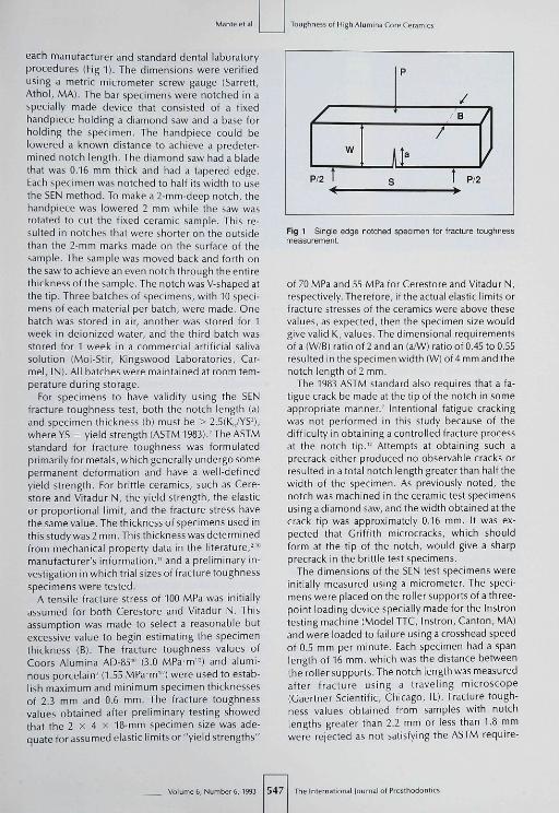
each manufacturer and standard dental laboratoryprocedures (Fig 1), The dimensions were verifiedusing a metric micrometer screw gauge (Sarrett,Athol, MA), The bar specimens were notched in aspecially made device that consisted of a fixedhandpiece holding a diamond saw and a base forholding the specimen. The handpiece could belowered a known distance to achieve a predeter-mined notch length. The diamond saw had a bladethat was 0,16 mm thick and had a tapered edge.Each specimen was notched to half its width to usethe SEN method. To make a 2-mm-decp notch, thehandpiece was lowered 2 mm while the saw wasrotated to cut the fixed ceramic sample. This re-sulted in notches that were shorter on the outsidethan the 2-mm marks made on fhe surface of fhesample. The sample was moved back and forth onthe saw to achieve an even notch through the entirethickness of the sample. The notch was V-shaped atthe tip. Three batches of specimens, with 10 speci-mens of each material per batch, were made. Onebatch was stored in air, another was stored for 1week in deionized water, and the third batch wasstored for 1 week in a commercial artificial salivasolution (Moi-Stir, Kingswood Laboratories, Car-mel, IN), All batches were maintained at room tem-perature during storage.
For specimens to have validity using the SLNfracture toughness test, both the notch length (a)and specimen thickness (b) must be > 2,5(Kn/YS-),where YS = yield strength (ASTM 1983).' The ASTMstandard for fracture toughness was formulatedprimarily for metals, which generally undergo somepermanent deformation and have a well-definedyield strength. For brittle ceramics, such as Cere-store and Vitadur N, the yield strength, the elasticor proportional limit, and the fracture stress havethe same value. The thickness of specimens used inthis study was 2 mm. This thickness was determinedfrom mechanical property data in fhe literature,-'"manufacturer's information," and a preliminary in-vestigation in which trial sizes of fracfure toughnessspecimens were tested,
A tensile fracture stress of 100 MPa was initiallyassumed for both Cerestore and Vitadur N, Thisassumption was made to select a reasonable butexcessive value to begin estimating the specimenthickness (B), The fracture toughness values ofCoors Alumina AD-S.'i"' (3.0 MPa-m''̂ ) and alumi-nous porcelain-(1,55MPa-m'-) were used to estab-lish maximum and minimum specimen thicknessesof 2,3 mm and 0,6 mm. The fracfure toughnessvalues obtained after preliminary testing showedthat the 2 X 4 X 18-mm specimen size was ade-quate for assumed elastic limits or "yield strengths"
/
p/2 tW
\
S
p
B
t
A
p/2
)
Fig 1 Single edge notched specimen for fracture toughnessmeasurement.
of 70 MPa and 55 MPafor Cerestore and Vitadur N,respectively. Therefore, if the actual elastic limits orfracture stresses of the ceramics were above thesevalues, as expected, then the specimen size wouldgive valid K,, values. The dimensional requirementsof a (W/B) rafio of 2 and an (a/W) ratio of 0,45 to 0.55resultedin the specimen width (W) of4mmandthenotch length of 2 mm.
The 1983 ASTM standard also requires that a fa-tigue crack be made at the tip of fhe notch in someappropriate manner," Intentional fatigue crackingwas not performed in this study because of thedifficulty in obtaining a controlled fracture processat the notch tip," Attempts at obtaining such aprecrack either produced no obsen/able cracks orresulted in a total notch length greater than half thewidth of the specimen. As previously noted, thenotch was machined in the ceramic test specimensusing a diamond saw, and the width obtained atthecrack tip was approximately 0,16 mm. It was ex-pected that Griffith microcracks, which shouldform at the tip of the notch, would give a sharpprecrack in the brittle test specimens.
The dimensions of the SFN test specimens wereinitially measured using a micrometer. The speci-mens were placed on the roller supports of a three-poinf loading device specially made for the Instrontesting machine (Model TTC, Instron, Canton, MA)and were loaded to failure using a crosshead speedof 0.5 mm per minute. Each specimen had a spanlength of 16 mm, which was the distance betweenthe rollersupports. The notch length was measuredaffer fracture using a traveling microscope(Gaertner Scientific, Chicago, IL), Fracfure tough-ness values obtained from samples with notchlengths greater than 2,2 mm or less than 1.8 mmwere rejected as not satisfying the ASTM require-
Voliime6,Number6,1993 547 l of Pro5thodontic5
Toughness of High Aiumina Core Ceramics
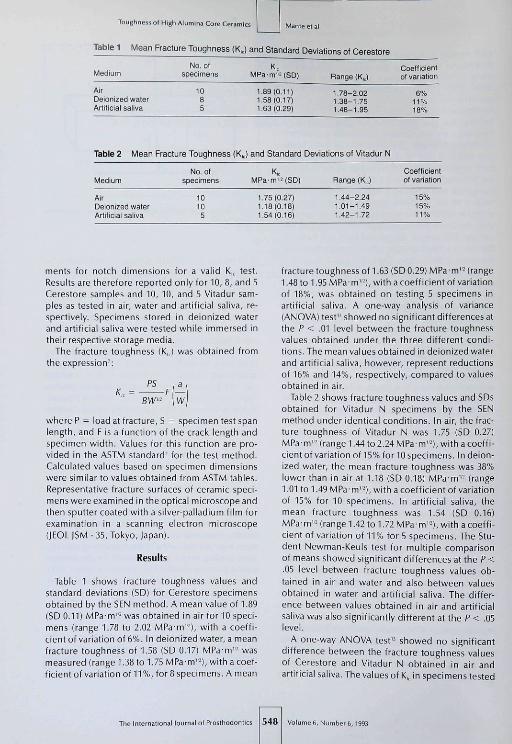
Table 1 Mean Fracture Tougfiness (K«;
Medium
AirDe ionized waterArtilicial saliva
No. olspecimens
1085
) and Standard Deviations of Cerestore
MPa-m'MSD)
1.89(0.11]1.5B(O,17)1.63(0.29)
Bange (KJ
1.78-2.021.38-1.751.48-1.95
Coetticientot variation
11%16%
Table 2 Mean Fracture Toughness {KJ and Standanj Deviations of Vitadur N
Medium
AirDeionized waterArtille lal saliva
No.otspecimens
1010
5
KMPa'm'MSD)
1.75(0 27)1.18(0.16)1.54(0.16)
Range(KJ
1.44-2.241.01-1.491.42-1.72
Coefficientol variation
15%15%11%
menfs for notch dimensions for a valid K,, test.Results are therefore reported only for 10, 8, and 5Cerestore samples and 10, 10, and 5 Vitadur sam-ples as tested in air, water and artificial saliva, re-spectively. Specimens stored in deionized waterand artificial saliva were tested while immersed intheir respective storage media.
The fracture foughness [KJ was obtained fromthe expression':
PS
BW' W
where P = load at fracture, S = specimen tesf spanlength, and F is a function of the crack length andspecimen width. Values for this function are pro-vided in the ASTM standard" for the test method.Calculated values based on specimen dimensionswere similar to values obtained from ASTM tables.Representative fracture surfaces of ceramic speci-mens were examined in the optical microscope andthen sputter coated with a silver-palladium film forexamination in a scanning electron microscope(lEOLiSM-35, Tokyo, Japan).
Results
Table 1 shows fracture toughness values andstandard deviations (SD) for Cerestore specimensobtained by the SEN method. A mean value of 1.89(SD 0.11) MPa-m'- was obtained in air for 10 speci-mens (range T.78 to 2.02 MPa-m''-), with a coeffi-cient of variation of 6%. In deionized water, a meanfracture toughness of 1.58 (SD 0.17) MPa-m'- wasmeasured (range 1.38 fo 1.75 MPa-m'-), wifh a coef-ficient of variation of 11%, for8 specimens. A mean
fracture toughness of 1.63 (SD 0.29) MPa m' - (range1.48 to 1.95 MPa-m'-'), with a coefficient of variaf ionof 18%, was obfained on testing 5 specimens inartificial saliva. A one-way analysis of variance(ANOVA) test" showed no significant differences afthe P < .01 level between fhe fracture toughnessvalues obtained under the three different condi-tions. The mean values obtained in deionized waterand artificial saliva, however, represent reductionsof 16% and 14%, respectively, compared to valuesobtained in air.
Table 2 shows fracture toughness values and SDsobtained for Vitadur N specimens by the SENmethod under identical conditions. In air, the frac-ture toughness of Vitadur N was 1.75 (SD 0.27)MPa'm"- (range 1.44 to 2.24 MPa-m"-), with a coeffi-cient of variaf ion of 15% for 10 specimens. In deion-ized water, the mean fracture foughness was 38%lower than in air at 1.18 (SD 0,18) MPa-m'- (range1.01 to 1.49 MPa- m'-), with a coefficient of variationof 15% for 10 specimens. In artificial saliva, fhemean fracfure toughness was 1.54 (SD 0.16)MPa-m'- (range 1.42 to 1.72 MPa-m'-), with a coeffi-cient of variafion of 11% for 5 specimens. The Stu-dent Newman-Keuls test for multiple comparisonof means showed significant differences at the P <.05 level between fracture toughness values ob-tained in air and water and also between valuesobtained in water and artificial saliva. The differ-ence between values obtained in air and artificialsaliva was also significantly different at the P < .05level.
A one-way ANOVA test'' showed no significanfdifference between the fracture foughness valuesof Cerestore and Viiadur N obtained in air andartificial saliva. The values of K,, in specimens tested
The International Journal of Prosthodortics 548 Volumes, Number6,1993
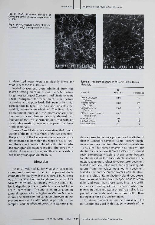
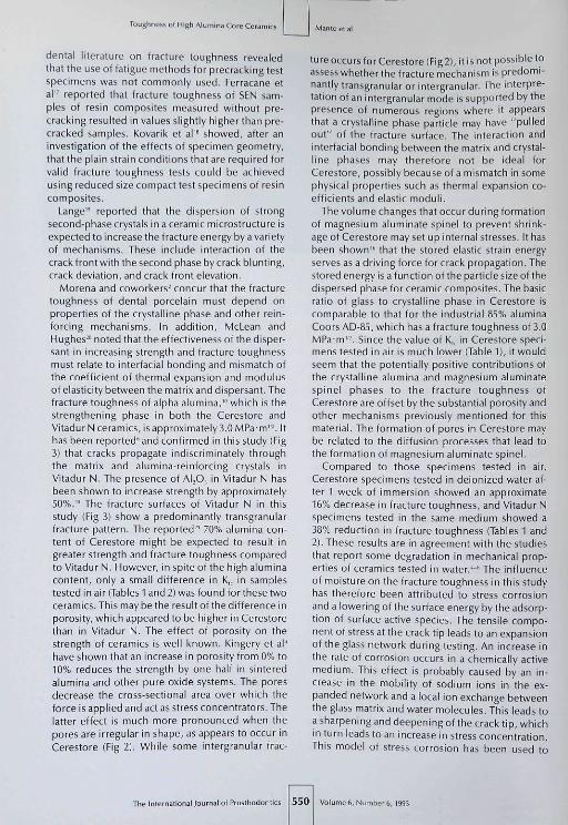
Fig 2 (Left) Fracture surface ofCerestore ceramic (original magnificationX 500).
Fig 3 (Right) Fracture surface of VitadurN ceramic (original magnification x 500).
Toughness of High Alumina Core Ceramics
in deionized water were significantly lower forVitadur N at the P < .01 level.
Load-displacement plots obtained from theInstron testing machine during the SEN fracturetoughness testing of Cerestore and Vitadur N werelinear throughout the experiment, with fractureoccurring at the peak load. This type of behaviorcorresponds to Type III curves^ and indicates thatvalid K|, values were obtained. The linear load-displacement plots and the macroscopically flatfracture surfaces observed visually showed thatfracture of the test specimens occurred with noplastic deformation, as was anticipated for thesebrittle materials.
Figures 2 and 3 show representative SEM photo-graphs of the fracture surfaces of the two ceramics.The porosity of the Cerestore specimens was visu-ally estimated to be within the range of 5% to 10%,and these specimens exhibited both intergranularand transgranular fracture modes. The porosity inVitadur N was much lower, and this ceramic exhib-ited mainly transgranular fracture.
Discussion
The mean value of K,, for Vitadur N specimensstored and measured in air in the present studycompares favorably with that reported by Morenaet al.- The SEN fracture toughness in air of 1.75MPa-m'-for Vitadur N is nearly twice the value of K,,for feldspathic porcelain, which is reported to be0.9 to 1.0 MPa m'-.'The coefficient of variation, ingeneral, appears to be higher in Vitadur N speci-mens. Tbe coefficient of variation observed in thepresent test can be attributed to porosity in thesamples, and the effect of porosity in scattering the
Table 3 Fracture Tougfiness of Some Brittle DentalMaterials
Material
Dental amalgam(Sybraloy)
Silicate cement(Brotrey)
Composite resin(Occlusin)
Giass-ionomer cement(Ketac Silver)
¡X aluminaHuman enamelHuman dentin
KMPa-m"
0.91
0.30
2.06
0.42
3.01.03.1
Bete re nee
16
26
16
16
101415
data appears to be more pronounced in Vitadur Nthan in Cerestore samples. Some fracture tough-ness values reported for other dental materials are1.0 MPa-m'- for human enamel," 3.1 MPa-m"- fordentin,''and a range of 0.7 to 1.7 MPa-m'-for dentalresin composites.'" Table 3 shows some fracturetoughness values for various dental materials. Thefracture toughness values for Cerestore specimenstested in artificial saliva were not significantly dif-ferent from the values obtained in specimenstested in air and deionized water (Table 1). How-ever, the value of K,, for Vitadur N aluminous porce-lain was significantly lower for specimens tested indeionized water than those tested in air and artifi-cial saliva. Loading of the specimen while im-mersed in deionized water or artificial saliva is ex-pected to simulate oral conditions better thantesting in air or a dry environment.
No fatigue precracking was performed on SENtest specimens used in this study. A search of the
549 The Inlernatiora I of Prosthodontics
Toughness of High Alumina Core Ceran
dental literature on fracture toughness revealedthat the use of fatigue methods for precracking testspecimens was not commonly used. Ferracane etal" reported that fracture toughness of SEN sam-ples of resin composites measured without pre-cracking resulted in values slightly higher than pre-cracked samples. Kovarik et al'" showed, after aninvestigation of the effects of specimen geometry,that the plain strain conditions that are required forvalid fracture toughness tests could be achievedusing reduced size compact test specimens of resincomposites.
Lange" reported that the dispersion of strongsecond-phase crystals in a ceramic microstructure isexpected to increase the fracture energy by a varietyof mechanisms. These include interaction of thecrack front with the second phase by crack blunting,crack deviation, and crack front elevation.
Morena and coworkers- concur that the fracturetoughness of dental porcelain must depend onproperties of the crystalline phase and other rein-forcing mechanisms. In addition, McLean andHughes'" noted that the effectiveness of the disper-sant in increasing strength and fracture toughnessmust relate to interfacial bonding and mismatch ofthe coefficient of thermal expansion and modulusof elasticity between the matrix and dispersant. Thefracture toughness of alpha alumina,'" which is thestrengthening phase in both the Cerestore andVitadurN ceramics, is approximately 3.0 MPa m'-'. Ithas been reported' and confirmed in this study (Fig3) that cracks propagate indiscriminately throughthe matrix and alumina-reinforcing crystals inVitadur N. The presence of ALO, in Vitadur N hasbeen shown to increase strength by approximately50%.-° The fracture surfaces of Vitadur N in thisstudy (Fig 3) show a predominantly transgranularfracture pattern. The reported-^ 70% alumina con-tent of Cerestore might be expected to resuit ingreater strength and fracture toughness comparedto Vitadur N. However, in spite of the high aluminacontent, only a small difference in K̂ in samplestested in air (Tables 1 and 2) was found for these twoceramics. This may be the result of the difference inporosity, which appeared fo be higher in Cerestorethan in Vitadur N. The effect of porosity on thestrength of ceramics is well known. Kingery et al'have shown that an increase in porosity from 0% to10% reduces the strength by one half in sinteredalumina and other pure oxide systems. The poresdecrease the cross-sectional area over which theforce is applied and act as stress concentrators. Thelatter effect is much more pronounced when thepores are irregular in shape, as appears to occur inCerestore (Fig 2). While some intergranular frac-
ture occurs for Cerestore (Fig 2), it is not possible toassess whether the fracture mechanism is predomi-nantly transgranular or infergranular. The interpre-tation of an intergranular mode is supported by thepresence of numerous regions where it appearsthat a crystalline phase particle may have "pulledout" of the fracture surface. The interaction andinterfacial bonding between tbe matrix and crystal-line phases may therefore not be ideal forCerestore, possibly because oí a mismatch in somephysical properties such as thermal expansion co-efficients and elastic moduli.
The volume changes that occur during formafionof magnesium aluminate spinel fo prevent shrink-age of Cerestore may set up internal stresses. It hasbeen shown'" that the stored elastic strain energyserves as a driving force for crack propagation. Thestored energy is a function of the particle size of thedispersed phase for ceramic composites. The basicratio of glass to crystalline phase in Cerestore iscomparable to that for the industrial 85% aluminaCoors AD-85, which has a fracture toughness of 3.0MPa m'-. Since the value of K,, in Cerestore speci-mens tested in air is much lower (Table 1), it wouldseem that the potentially positive contributions ofthe crystalline alumina and magnesium aluminatespinel phases to the fracture toughness ofCerestore are offset by the substantial porosity andother mechanisms previously mentioned for thismaterial. The formation of pores in Cerestore maybe related to the diffusion processes that lead tothe formation of magnesium aluminate spinel.
Compared to those specimens tested in air,Cerestore specimens tested in deionized water af-ter 1 week of immersion showed an approximate16% decrease in fracture toughness, and Vitadur Nspecimens tested in the same medium showed a38% reduction in fracture toughness (Tables 1 and2). These results are in agreement with the studiesthat report some degradation in mechanical prop-erties of ceramics tested in water.''• The influenceof moisture on the fracture toughness in this studyhas therefore been attributed to stress corrosionand a lowering of the surface energy by the adsorp-tion of surface active species. The tensile compo-nent of stress at the crack tip leads to an expansionof the glass network during testing. An increase inthe rate of corrosion occurs in a chemically activemedium. This effect is probably caused by an in-crease in the mobility of sodium ions in the ex-panded network and a local ion exchange betweenthe glass matrix and water molecules. This leads toa sharpening and deepening of the crack tip, whichin turn leads to an increase in stress concentration.This model of sfress corrosion has been used to
The International lournal of Prosthodontii 550 VolLme6, Number 6,1993
i ot High Alumina Core Ceran
explain the linear variation in crack velocity withrelative humidity for soda-lime-silica glass.'
The difference in the reduction of K,, forCereslore and Vitadur N specimens tested in watermay be related lo a difference in solubility and,therefore, in interaction with water at the crackfront. In spite of the well-known chemical stabilityof ceramics compared to other dental restorativematerials, they have been shown to dissolve anddiscolor in use.-' Solubilities of 0.296 mg/cm- foraluminous enamel porcelains and 0.252 mg/cm- foraluminous body porcelains in various media havebeen reported.-' McLean' reported a solubilityrange of 0.01% to 0.07% for commercial porcelainsin 0.25% HCI.
The dissolution of dental porcelain has been clas-sified into leaching, in which only the metallic ionsare dissolved, and etching, in which the entire glassundergoes dissolution and leaves a new surface."The degree lo which each mechanism occurs de-pends on the pH of the solution and the composi-tion of the glass. Dissolving elements were re-ported to include Si, B, Na, K, and Al; Si, Na and Kdissolved to a greater extent, while B and Al dis-solved in smaller amounts. If the stress corrosionmodel is an acceptable explanation for the differ-ence in fracture toughness for testing in an aque-ous environment vs air, then Cerestore may bemore stable in deionized water than aluminousporcelain because of the bigher values for K,̂ inwater. An explanation for Ihe greater chemical sta-bility of Cerestore may be the presence of a sub-stantially crystalline structure,'- which contains thealumina, magnesium aluminate spinel, and possi-bly MgO phases.
DeRijk et aH' have investigated the solubility ofdental porcelains using the standardized Interna-tional Standards Organization methods. Speci-mens of porcelain are shaken in 4% acetic acid with15-to20-minutecyclesfor 16 hours, after which thevalues ofweight loss are measured. They found thatthe loss of dental porcelain surface that occurs inacetic acid solution, artificial saliva, and distilledwater is linear with time. The ranking for dissolu-tion of different porcelains by artificial saliva andacetic acid was the same; however, the amount ofdissolution that occurred in acetic acid after 6weeks was equivalent to the amount of dissolutionthat would occur in artificial saliva after 20 years.The solubility of dental porcelain in artificial salivais thus very low and may account for the lowerreductionof fracture toughness inartificial saliva ascompared to that in deionized water for Cerestoreand Vitadur N. The presence of an abundant supplyof fhe active species of smaller molecular size of
water may be more effective in sharpening thecrack than artificial saiiva, in which water moleculesmay be bound to larger organic molecules.
Using lifetime curves obtained by fatigue testingof high alumina core ceramics, Morena et al" pre-dicted that long-term failure should not be a prob-lem when aluminous porcelains and fine-grain ce-ramics, such as Vitadur N and Cerestore, are usedfor anterior crowns. Although a 5-year failure stressof 95 MPa was obtained for Cerestore, its fatiguebehavior indicated that residual tensile mismatchstresses could lead to rapid failure when it ¡s usedfor posterior restorations.
With the increase in the number of ceramic re-storative materials available, there is a need to in-vestigate their fracture behavior before large-scaleclinical use. Fracture toughness determined withspecimens fabricated using clinical techniqueswould give clinically relevant information about thefracture behavior of fhese ceramics. The scientificprinciples outlined in this study will have generalapplicability and should be useful in investigationsof the fracture behavior of the new generation ofhigh-strength and machinable ceramics.
Conclusion
Two reinforced alumina ceramic materials weretested for fracture toughness in air, deionized wa-ter, and artificial saliva. Within the design of thestudy and the materials used, the following conclu-sions can be made;
1. Mean fracture toughness values in this studywere 1.89 MPa-m'- for Cerestore specimenstested in air and 1.75 MPa-m" for Vitadur Nspecimens tested in air. These fracture tough-ness values were not significantiy different.
2. The mode of fracture was both intergranular andtransgranular for Ccrestore and predominantlytransgranular for Vitadur N.
3. The fracture toughness values obtained by test-ing Cerestore in deionized water and artificialsaliva were not significantly different from thoseobtained by testing Cerestore specimens in air.
4. Thefracture toughness values of Vitadur N weresignificantly lower when specimens were testedin deionized water and artificial saliva thanwhen specimens were tested in air.
References
1. Lloyd CH, lannetta RV. The fracture touginness ot dentalcomposites. I. The development of strength and fracturetoughrieis. | Orai Rehabil 1982:9:55-66.
VolLme6, Number 6, t993 551 Ttie International Journal of Prosthodortics
Toughness of High Alumina Core Ce
2. Morena R, Lockwood PE, Fairhurst CW. Fiaclure toughnessof commercial dental porcelains. Dent Maler 1986;2;58-62.
3. Evans AC, Charles EA. Eracture toughness determinations hyindentations. I Am Ceram Soc 197fi;59:37l-372.
4. Kingery WD, Bowen HK, Uhlmann DR. Introduction to Ce-ramics, ed 2, New Vork: lohn Wiley, 1976;97-98.307,797-806.
5. Sherrill CA, O'Brien W|. Transverse strength of aluminousand feldspathic porcelain. | Dent Res 1974;53:6B3-690.
6. Morena R, Beaudreau CM, Lockwood PE, Evans AL, FairhurstCW. Fatigue of denial cerarnics in a simulated oral environ-ment. I Dent Res 1986; 65:993-997.
7. American Society ol Testing and Materials (ASTM). StandardNo. E-.Î99-83. Philadelphia, 1983.
8. Sozio RB, Riley E). The shrink-free ceramic crown. ) ProsthetDent 1983:49:182-187.
9. McLean JW. The Science and Art of Dental Ceramics, vol. 1.The Nature of Dental Ceramics and Their Clinical Use. Chi-cago: Quintessence, 1979:23-51.
10. Barker LM. Short rod K,; measurements of AijOj. In: BrandtRC, Hasselman DPH, Lange FE (eds). Fracture Mechanics ofCeramics, vol. 3, New York: Plenum, 1978:483-494.
11. Product information literature. A posterior coping strengthstudy comparing Cerestore and Vitadur N core materials.East Windsor, New lersey: lohnson and lohnson DenialProducts Co.
12. Mante E, Brantley WA, Dhuru VB, Ziebert CJ. Characteriza-tion and fracture toughness of the high alumina shrink-freecore ceramic labstractl. J Dent Res 19B7;66(special issue).
13. Chihon NW. Design and Analysis in Dental and Oral Re-search,ed2. New York: Praeger.19B2:99-162.
14. Hassan R, Caputo AA, Eunshah RF. Fracture toughness ofhuman enamel. | Denl Res 1981,60:820-827.
15. ElMowafy OM, Watts DC. Fracture toughness of humandenlin. | Dent Res 1986;65:677-681
16. Lloyd CH, Adamson M. The development of fracture lough-ness and fracture strength in posterior restorative materials.Dent Maler 1987;3:225-231.
17. Ferracane |L, Antonio RC, Matsumoto H. Variables affectingKit of dental composites, 1 Denl Res 19B7;66:1140-1145.
18. Kovarik RE, Ergle IW, Fairhurst CW. Effects of specimengeometry on the measurement of fracture toughness. DentMaterl991;7:166-169.
19. Lange FF. Interaction of crack front with second phase dis-persion. Phil Mag 1970; 179:983-992.
20. McLean |W, Hughes TH. The reinforcement of dental porce-lain with ceramic oxides. Br Dent 1196S;1l9:251-267.
21. Technical Manual. Cerestore crown alumina ceramic corematerial. Lakewood. Colorado: Coors Biomédical. 19B5.
22. Davidge RN, Green TJ. The strength of 2 phase ceramic-glassmaterials. | Mater Sei 1968; 3:625-634.
23. Menis DL. Investigation of experimental dental porcelainsand their fusion to cast titanium [Masters thesis]. Evanston,Illinois: Northwestern University, 1987.
24. Nishiyama M. Makoto T, Masayoshi 0 . A sludy of dentalporcelain solubility —Discoloring of component elementsand resultant surface roughness and abrasion. J Nihon UnivSch Dent 1983,25:262-276.
25. DeRi|k WG, Jennings KA, Menis DL. A comparison of chemi-cal durahility test solutions for dental porcelains. In: SauerBW {ed). Biomédical Engineering IV, Recent Developments.Proceedings of the Fourth Southern Biomédical EngineeringConference. New York: Pergamon Press, 1985:152-155.
26. Lloyd CH, Mitchell L. Eracture toughness of tooth coloredrestorative materials. J Oral Rehabil 1984;11:257-272.
Uteralure Abstract _
Ora! Hygiene, Periodontal Conditions and Carious
Lesions in Patients Treated With Dental Bridges
The potentially detrimental reaction of the periodontal tissues to the placement of fixed partialdentures IFPDs) has been observed clinically and is well documented. The present investigationdetermined the level of oral hygiene, periodontal condition, alteration of alveolar bone level, andcaries prevalence in patients who had received regular oral prophylaxis following the placement ofFPDs. The location of restorative margin placement was compared relative to the above indices.Patients [n = 102, mean age 40 years) receiving 108 FPDs were studied, and the patients' remainingnatural teeth in each arch served as controls. Retainers were distributed as follows: 77% were of TypeIII gold veneered with acrylic resin; 12% were partial veneer gold, and 11% were complete goldcrowns. Periodontal conditions and oral hygiene levels were assessed annually during the first 10 yearspostplacement and then at a 15-year examination. Results suggest that (7) vvhen compared to controls,abutment teeth had identical Plaque Index scores but a tendency to higher Gingival Index scores anddeeper pocket depths, although differences were not statistically significant; (2) the incidence ofGingival Index scores of 2 and 3 was higher when restorative margins were located subgingivally; and131 no statistically significant difference in bone loss, which was not related to restorative marginlocation, was recorded between control and abutment teeth. The authors conclude that the favorable15-year results presented in this report are likely to be the result of (he regular oral prophylaxis that thepatients received every 6 months during the observation period.
Valderhiug |, Ellingsen |E, |o!istad A. ( Ctin Penodontol 1993;20:482-.469. References: 42. Reprints; |akoh Valdcrhaug.Department of Prosthetic Dentistry and Stomatognathic Physiology, Dental Faculty, University of Osio, Box 1109,Blindern, 0317 Osio, Norway. —DacWS Cagna, DMD, Department oíPiosthoàontiçs, The University olTexasHesllh Science Ceiiiemt San Antonio, San Antonio, Texas
The international lournal of Proslhodontics 552 Volume 6, Number 6,1993