Foundations - Cells, organelles and cell boundaries ... · Foundations - Cells, organelles and cell...
14
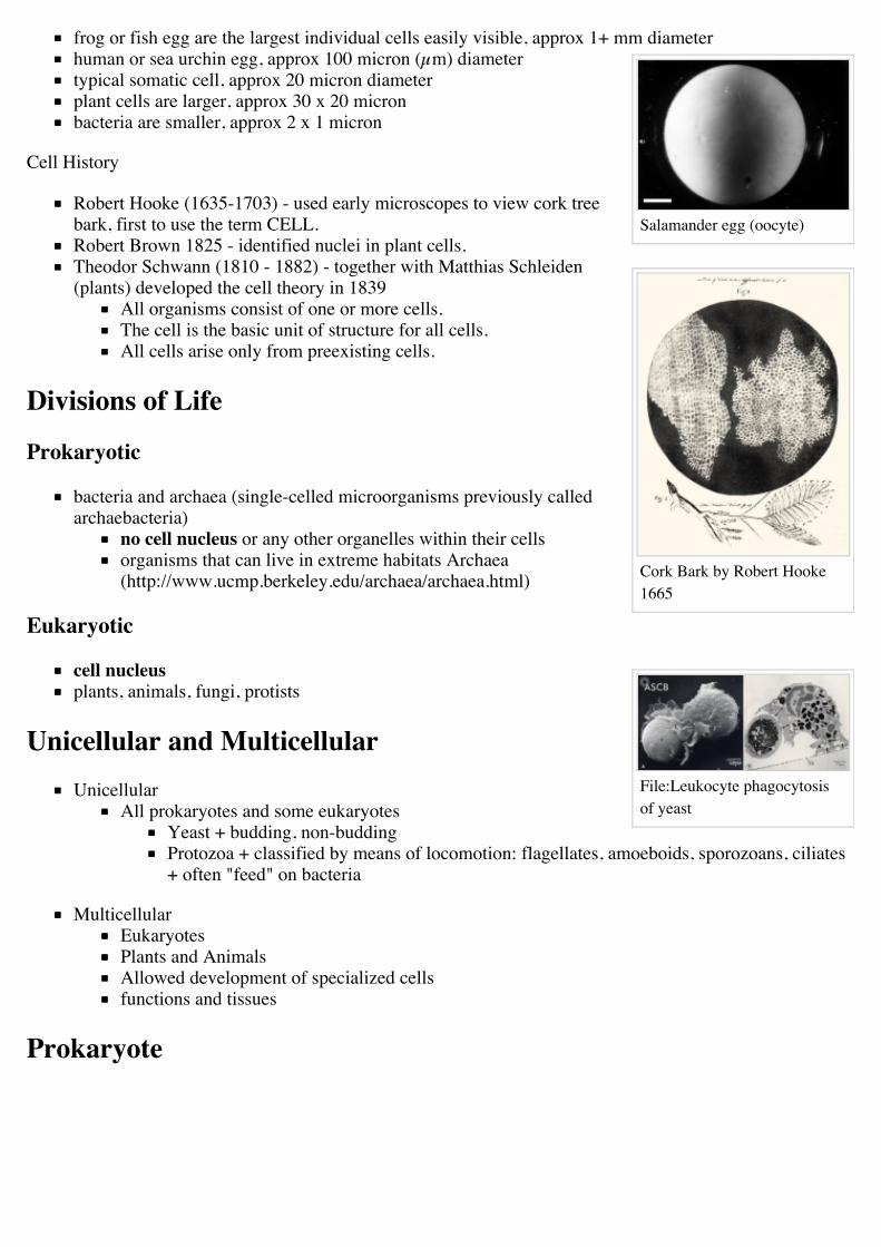
Typical cell membrane bound compartments Cells are dynamic Watch Movie (http://php.med.unsw.edu.au/cellbiology/images/b/b0/ Foundations - Cells, organelles and cell boundaries From Cellbiology Introduction 2012 Lecture Slides: The 2 major classes of cells are defined by the presence or absence of a nucleus; Eukaryotic (with nucleus) and Prokaryotic (without nucleus). Eukaryotes can be further divided into unicellular (only one cell, like prokaryotes) and multicellular (like us) organisms. A major difference between eukayotes and prokaryotes is the presence of physical compartments (membrane bound) and organelles within the cell. These compartments allow the separation/specialization of processes within the cell. There also exist within each of these physical compartments, functional compartments where specific processes may occur or are restricted. This lecture is also an introduction to cell compartments and describes the structure of membranes forming these compartments. About Human Body Human Cells 210+ cell types in body total number of estimated cells in the body - 10 13 (American Ten trillion/British Ten billion) Flora bacteria, fungi and archaea found on all surfaces exposed to the environment skin and eyes, in the mouth, nose, small intestine most bacteria live in the large intestine 500 to 1000 species of bacteria live in the human gut total number of estimated flora ten times as many bacteria 10 14 (American One hundred trillion/British One hundred billion) Cell Sizes
Transcript of Foundations - Cells, organelles and cell boundaries ... · Foundations - Cells, organelles and cell...
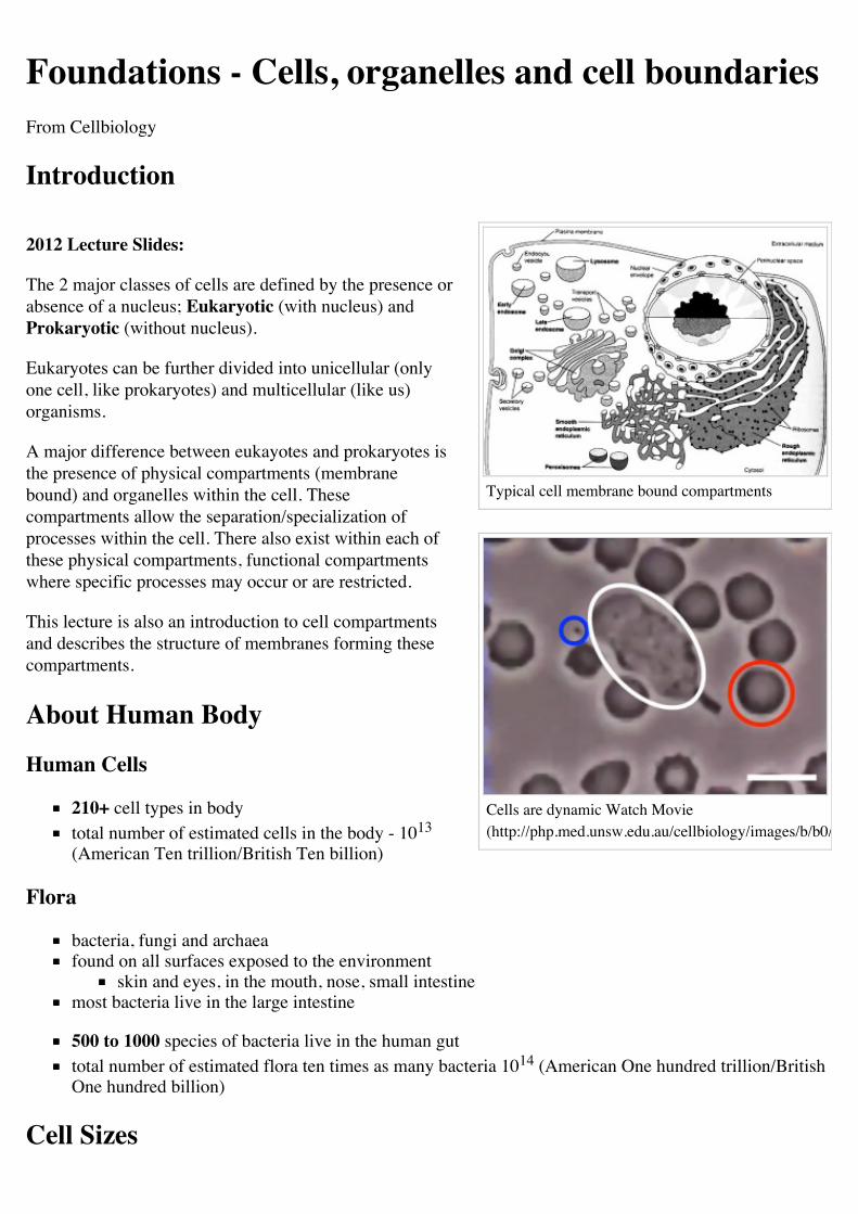
Typical cell membrane bound compartments
Cells are dynamic Watch Movie(http://php.med.unsw.edu.au/cellbiology/images/b/b0/Neutrophil_Rogers1950s.mov)
Foundations - Cells, organelles and cell boundariesFrom Cellbiology
Introduction
2012 Lecture Slides:
The 2 major classes of cells are defined by the presence orabsence of a nucleus; Eukaryotic (with nucleus) andProkaryotic (without nucleus).
Eukaryotes can be further divided into unicellular (onlyone cell, like prokaryotes) and multicellular (like us)organisms.
A major difference between eukayotes and prokaryotes isthe presence of physical compartments (membranebound) and organelles within the cell. Thesecompartments allow the separation/specialization ofprocesses within the cell. There also exist within each ofthese physical compartments, functional compartmentswhere specific processes may occur or are restricted.
This lecture is also an introduction to cell compartmentsand describes the structure of membranes forming thesecompartments.
About Human Body
Human Cells
210+ cell types in bodytotal number of estimated cells in the body - 1013
(American Ten trillion/British Ten billion)
Flora
bacteria, fungi and archaeafound on all surfaces exposed to the environment
skin and eyes, in the mouth, nose, small intestinemost bacteria live in the large intestine
500 to 1000 species of bacteria live in the human guttotal number of estimated flora ten times as many bacteria 1014 (American One hundred trillion/BritishOne hundred billion)
Cell Sizes
Salamander egg (oocyte)
Cork Bark by Robert Hooke1665
File:Leukocyte phagocytosisof yeast
frog or fish egg are the largest individual cells easily visible, approx 1+ mm diameterhuman or sea urchin egg, approx 100 micron (µm) diametertypical somatic cell, approx 20 micron diameterplant cells are larger, approx 30 x 20 micronbacteria are smaller, approx 2 x 1 micron
Cell History
Robert Hooke (1635-1703) - used early microscopes to view cork treebark, first to use the term CELL.Robert Brown 1825 - identified nuclei in plant cells.Theodor Schwann (1810 - 1882) - together with Matthias Schleiden(plants) developed the cell theory in 1839
All organisms consist of one or more cells.The cell is the basic unit of structure for all cells.All cells arise only from preexisting cells.
Divisions of Life
Prokaryotic
bacteria and archaea (single-celled microorganisms previously calledarchaebacteria)
no cell nucleus or any other organelles within their cellsorganisms that can live in extreme habitats Archaea(http://www.ucmp.berkeley.edu/archaea/archaea.html)
Eukaryotic
cell nucleusplants, animals, fungi, protists
Unicellular and MulticellularUnicellular
All prokaryotes and some eukaryotesYeast + budding, non-buddingProtozoa + classified by means of locomotion: flagellates, amoeboids, sporozoans, ciliates+ often "feed" on bacteria
MulticellularEukaryotesPlants and AnimalsAllowed development of specialized cellsfunctions and tissues
Prokaryote
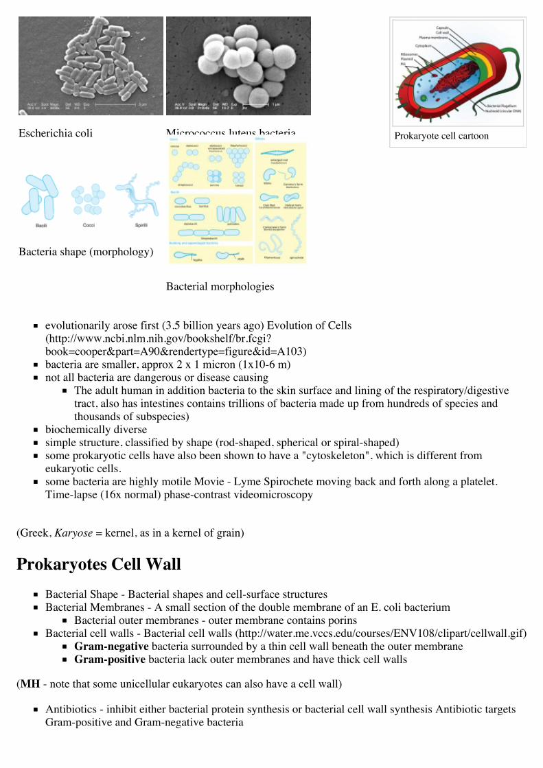
Prokaryote cell cartoonEscherichia coli Micrococcus luteus bacteria
Bacteria shape (morphology)
Bacterial morphologies
evolutionarily arose first (3.5 billion years ago) Evolution of Cells(http://www.ncbi.nlm.nih.gov/bookshelf/br.fcgi?book=cooper&part=A90&rendertype=figure&id=A103)bacteria are smaller, approx 2 x 1 micron (1x10⁻⁶ m)not all bacteria are dangerous or disease causing
The adult human in addition bacteria to the skin surface and lining of the respiratory/digestivetract, also has intestines contains trillions of bacteria made up from hundreds of species andthousands of subspecies)
biochemically diversesimple structure, classified by shape (rod-shaped, spherical or spiral-shaped)some prokaryotic cells have also been shown to have a "cytoskeleton", which is different fromeukaryotic cells.some bacteria are highly motile Movie - Lyme Spirochete moving back and forth along a platelet.Time-lapse (16x normal) phase-contrast videomicroscopy
(Greek, Karyose = kernel, as in a kernel of grain)
Prokaryotes Cell WallBacterial Shape - Bacterial shapes and cell-surface structuresBacterial Membranes - A small section of the double membrane of an E. coli bacterium
Bacterial outer membranes - outer membrane contains porinsBacterial cell walls - Bacterial cell walls (http://water.me.vccs.edu/courses/ENV108/clipart/cellwall.gif)
Gram-negative bacteria surrounded by a thin cell wall beneath the outer membraneGram-positive bacteria lack outer membranes and have thick cell walls
(MH - note that some unicellular eukaryotes can also have a cell wall)
Antibiotics - inhibit either bacterial protein synthesis or bacterial cell wall synthesis Antibiotic targetsGram-positive and Gram-negative bacteria
Bacterial Replication - DNA replication and cell division in a prokaryote MCB - DNA replication andcell division in a prokaryote (http://www.ncbi.nlm.nih.gov/bookshelf/br.fcgi?book=mcb&part=A3163&rendertype=figure&id=A3176)
File:Bacterial cell division.mov
Bacterial Growth Movie
Molecular Biology of the CellFigure 25-4. Bacterial shapes and cell-surface structures(http://www.ncbi.nlm.nih.gov:80/books/bv.fcgi?db=Books&rid=mboc4.figgrp.4620)Figure 11-17. A small section of the double membrane of an E. coli bacterium(http://www.ncbi.nlm.nih.gov:80/books/bv.fcgi?db=Books&rid=mboc4.figgrp.2023)
Medical MicrobiologyFigure 2-6. Comparison of the thick cell wall of Gram-positive bacteria with the comparativelythin cell wall of Gram-negative bacteria (http://www.ncbi.nlm.nih.gov:80/books/bv.fcgi?db=Books&rid=mmed.figgrp.294)
Prokaryote Mycoplasmassmallest self-replicating organismssmallest genomes (approx 500 to 1000 genes)spherical to filamentous cellsno cell wallssurface parasites of the human respiratory and urogenital tracts
Mycoplasma pneumoniae infect the upper and lower respiratory tractMycoplasma genitalium a prevalent sexually transmitted infectionMycoplasma hyorhinis found in patients with AIDS
Prokaryotic and Eukaryotic Cells
The following links describe the major differences between prokaryotic and eukaryotic cells, the way theydivide and the way in which antibiotics have their action on prokaryotic cells.
The Cell- A Molecular Approach | Table 1.1. Prokaryotic and Eukaryotic Cells |[http://www.ncbi.nlm.nih.gov:80/books/bv.fcgi?db=Books&rid=cooper.table.1198 Antibiotic Inhibitorsof Protein Synthesis (http://www.ncbi.nlm.nih.gov:80/books/bv.fcgi?db=Books&rid=cooper.table.91)Molecular Cell Biology Figure 12-6. DNA replication and cell division in a prokaryote(http://www.ncbi.nlm.nih.gov:80/books/bv.fcgi?db=Books&rid=mcb.figgrp.3176)Biochemistry Figure 28.15. Transcription and Translation(http://www.ncbi.nlm.nih.gov:80/books/bv.fcgi?db=Books&rid=stryer.figgrp.3980) two processes areclosely coupled in prokaryotes, whereas they are spacially and temporally separate in eukaryotes.
Virus
single compartment, no membranes
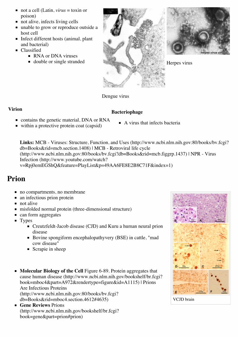
VCJD brain
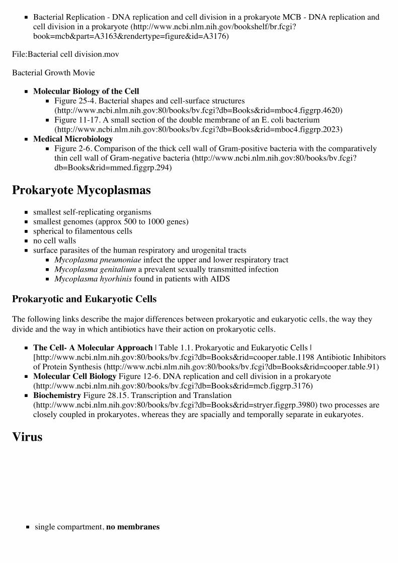
not a cell (Latin, virus = toxin orpoison)not alive, infects living cellsunable to grow or reproduce outside ahost cellInfect different hosts (animal, plantand bacterial)Classified
RNA or DNA virusesdouble or single stranded
Dengue virus
Herpes virus
Virion
contains the genetic material, DNA or RNAwithin a protective protein coat (capsid)
Bacteriophage
A virus that infects bacteria
Links: MCB - Viruses: Structure, Function, and Uses (http://www.ncbi.nlm.nih.gov:80/books/bv.fcgi?db=Books&rid=mcb.section.1408) | MCB - Retroviral life cycle(http://www.ncbi.nlm.nih.gov:80/books/bv.fcgi?db=Books&rid=mcb.figgrp.1437) | NPR - VirusInfection (http://www.youtube.com/watch?v=Rpj0emEGShQ&feature=PlayList&p=49AA6FE8E2B8C71F&index=1)
Prionno compartments, no membranean infectious prion proteinnot alivemisfolded normal protein (three-dimensional structure)can form aggregatesTypes
Creutzfeldt-Jacob disease (CJD) and Kuru a human neural priondiseaseBovine spongiform encephalopathyvery (BSE) in cattle, "madcow disease"Scrapie in sheep
Molecular Biology of the Cell Figure 6-89. Protein aggregates thatcause human disease (http://www.ncbi.nlm.nih.gov/bookshelf/br.fcgi?book=mboc4&part=A972&rendertype=figure&id=A1115) | PrionsAre Infectious Proteins(http://www.ncbi.nlm.nih.gov:80/books/bv.fcgi?db=Books&rid=mboc4.section.4612#4635)Gene Reviews Prions(http://www.ncbi.nlm.nih.gov/bookshelf/br.fcgi?book=gene&part=prion#prion)
Typical cell membrane boundcompartments
Neuroscience Prion Disease (http://www.ncbi.nlm.nih.gov:80/books/bv.fcgi?db=Books&rid=neurosci.box.1305)
Biological LevelsCells can be "broken down" into smaller and smaller constituent "parts"
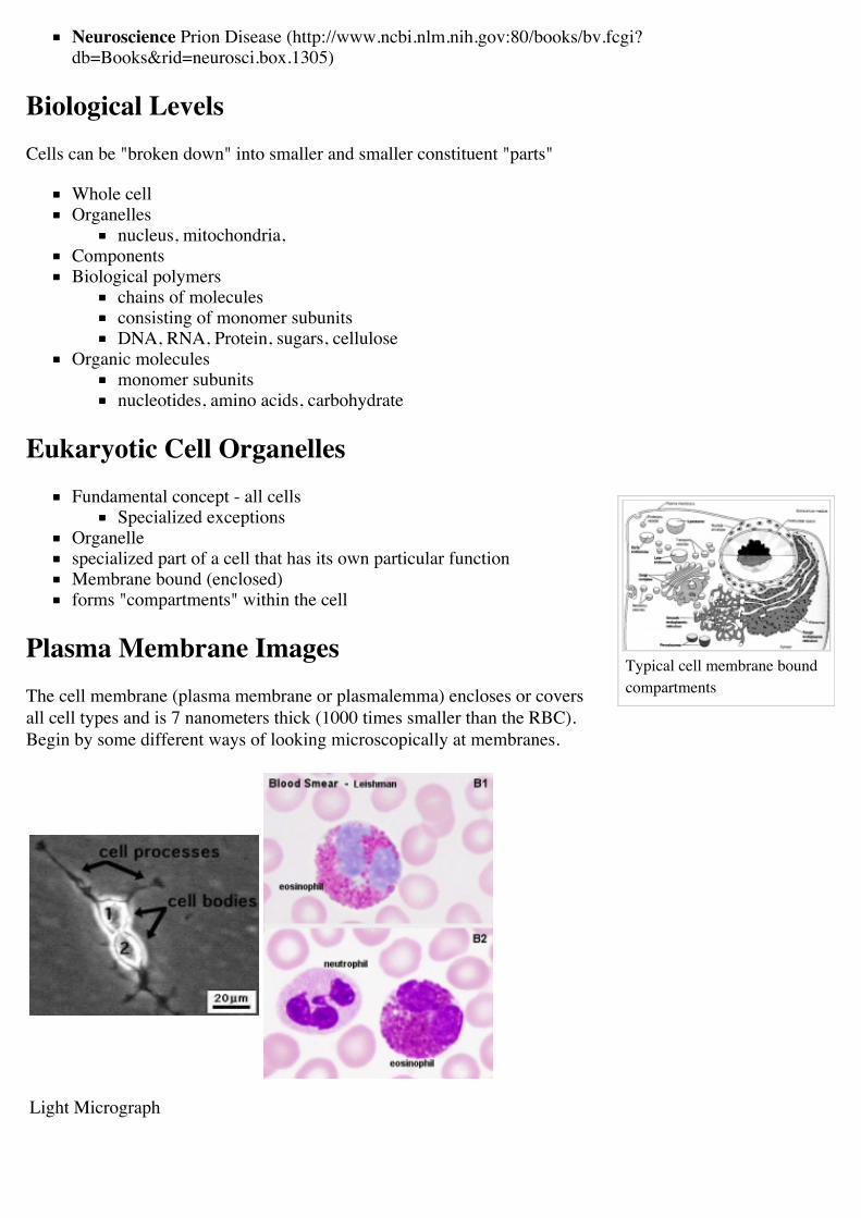
Whole cellOrganelles
nucleus, mitochondria,ComponentsBiological polymers
chains of moleculesconsisting of monomer subunitsDNA, RNA, Protein, sugars, cellulose
Organic moleculesmonomer subunitsnucleotides, amino acids, carbohydrate
Eukaryotic Cell OrganellesFundamental concept - all cells
Specialized exceptionsOrganellespecialized part of a cell that has its own particular functionMembrane bound (enclosed)forms "compartments" within the cell
Plasma Membrane ImagesThe cell membrane (plasma membrane or plasmalemma) encloses or coversall cell types and is 7 nanometers thick (1000 times smaller than the RBC).Begin by some different ways of looking microscopically at membranes.
Light Micrograph
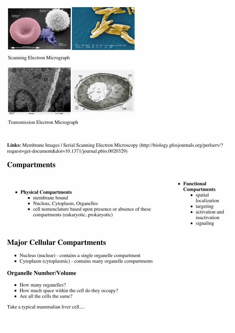
Scanning Electron Micrograph
Transmission Electron Micrograph
Links: Membrane Images | Serial Scanning Electron Microscopy (http://biology.plosjournals.org/perlserv/?request=get-document&doi=10.1371/journal.pbio.0020329)
Compartments
Physical Compartmentsmembrane boundNucleus, Cytoplasm, Organellescell nomenclature based upon presence or absence of thesecompartments (eukaryotic, prokaryotic)
FunctionalCompartments
spatiallocalizationtargetingactivation andinactivationsignaling
Major Cellular CompartmentsNucleus (nuclear) - contains a single organelle compartmentCytoplasm (cytoplasmic) - contains many organelle compartments
Organelle Number/Volume
How many organelles?How much space within the cell do they occupy?Are all the cells the same?
Take a typical mammalian liver cell....
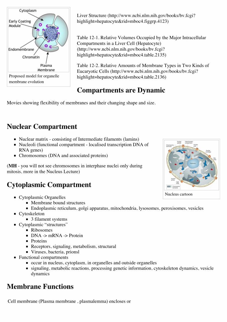
Proposed model for organellemembrane evolution
Nucleus cartoon
Liver Structure (http://www.ncbi.nlm.nih.gov/books/bv.fcgi?highlight=hepatocyte&rid=mboc4.figgrp.4123)
Table 12-1. Relative Volumes Occupied by the Major IntracellularCompartments in a Liver Cell (Hepatocyte)(http://www.ncbi.nlm.nih.gov/books/bv.fcgi?highlight=hepatocyte&rid=mboc4.table.2135)
Table 12-2. Relative Amounts of Membrane Types in Two Kinds ofEucaryotic Cells (http://www.ncbi.nlm.nih.gov/books/bv.fcgi?highlight=hepatocyte&rid=mboc4.table.2136)
Compartments are DynamicMovies showing flexibility of membranes and their changing shape and size.
Nuclear CompartmentNuclear matrix - consisting of Intermediate filaments (lamins)Nucleoli (functional compartment - localised transcription DNA ofRNA genes)Chromosomes (DNA and associated proteins)
(MH - you will not see chromosomes in interphase nuclei only duringmitosis, more in the Nucleus Lecture)
Cytoplasmic CompartmentCytoplasmic Organelles
Membrane bound structuresEndoplasmic reticulum, golgi apparatus, mitochondria, lysosomes, peroxisomes, vesicles
Cytoskeleton3 filament systems
Cytoplasmic “structures”RibosomesDNA -> mRNA -> ProteinProteinsReceptors, signaling, metabolism, structuralViruses, bacteria, prionsl
Functional compartmentsoccur in nucleus, cytoplasm, in organelles and outside organellessignaling, metabolic reactions, processing genetic information, cytoskeleton dynamics, vesicledynamics
Membrane Functions
Cell membrane (Plasma membrane , plasmalemma) encloses or
EM - Cell (Plasma) andOrganelle Membranes
Phospholipid Bilayer
Phospholipid Orientation
covers all cell types.
Regulation of transportDetection of signalsCell-cell communicationCell Identity
Internal membranes
form compartmentsAllow “specialisation” - metabolicand biochemicalLocalization of function
Membrane Components
phospholipids, proteins and cholesterolfirst compartment formedprokaryotes (bacteria) just this 1 compartmenteukaryotic cells many different compartments
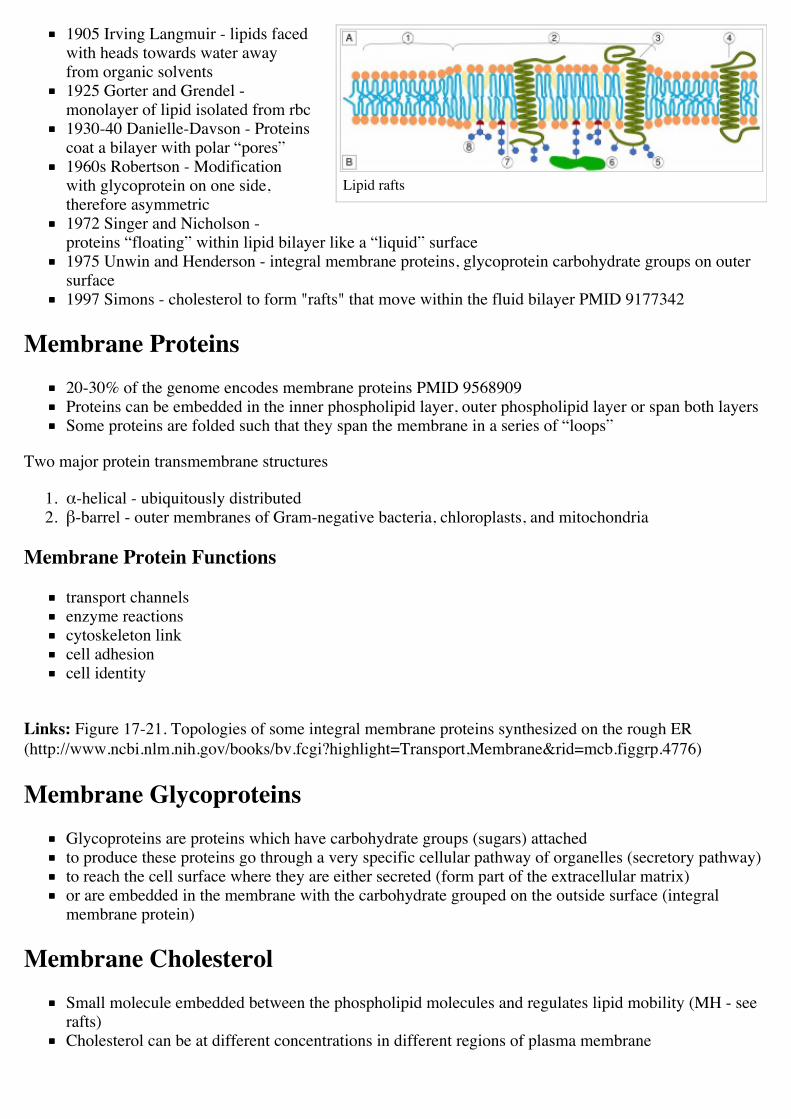
Phospholipidsmembranes contain phospholipids, glycolipids, and steroidsThe main lipid components include: phosphatidylcholine (~50%),phosphatidylethanolamine (~10%), phosphatidylserine (~15%),sphingolipids (~10%),cholesterol (~10%), phosphatidylinositol (1%).
Phospholipid Orientation
A liposome (lipid vesicle) is a small aqueous compartment surroundedby a lipid bilayer.A micelle is a small compartment surrounded by a single lipid layer.
Links: MBoC - Three views of a cell membrane(http://www.ncbi.nlm.nih.gov:80/books/bv.fcgi?db=Books&rid=mboc4.figgrp.1862) | MBoC - Phospholipid structure andthe orientation in membranes (http://www.ncbi.nlm.nih.gov/books/bv.fcgi?&rid=mboc4%2Efiggrp%2E211)
Membranes History(Background only, you do not need to know the details)
1890 Charles Overton - selective permeation of membranes, non-polarpass through (lipid soluble), polar refractory
Lipid rafts
1905 Irving Langmuir - lipids facedwith heads towards water awayfrom organic solvents1925 Gorter and Grendel -monolayer of lipid isolated from rbc1930-40 Danielle-Davson - Proteinscoat a bilayer with polar “pores”1960s Robertson - Modificationwith glycoprotein on one side,therefore asymmetric1972 Singer and Nicholson -proteins “floating” within lipid bilayer like a “liquid” surface1975 Unwin and Henderson - integral membrane proteins, glycoprotein carbohydrate groups on outersurface1997 Simons - cholesterol to form "rafts" that move within the fluid bilayer PMID 9177342
Membrane Proteins20-30% of the genome encodes membrane proteins PMID 9568909Proteins can be embedded in the inner phospholipid layer, outer phospholipid layer or span both layersSome proteins are folded such that they span the membrane in a series of “loops”
Two major protein transmembrane structures
1. α-helical - ubiquitously distributed2. β-barrel - outer membranes of Gram-negative bacteria, chloroplasts, and mitochondria
Membrane Protein Functions
transport channelsenzyme reactionscytoskeleton linkcell adhesioncell identity
Links: Figure 17-21. Topologies of some integral membrane proteins synthesized on the rough ER(http://www.ncbi.nlm.nih.gov/books/bv.fcgi?highlight=Transport,Membrane&rid=mcb.figgrp.4776)
Membrane GlycoproteinsGlycoproteins are proteins which have carbohydrate groups (sugars) attachedto produce these proteins go through a very specific cellular pathway of organelles (secretory pathway)to reach the cell surface where they are either secreted (form part of the extracellular matrix)or are embedded in the membrane with the carbohydrate grouped on the outside surface (integralmembrane protein)
Membrane CholesterolSmall molecule embedded between the phospholipid molecules and regulates lipid mobility (MH - seerafts)Cholesterol can be at different concentrations in different regions of plasma membrane
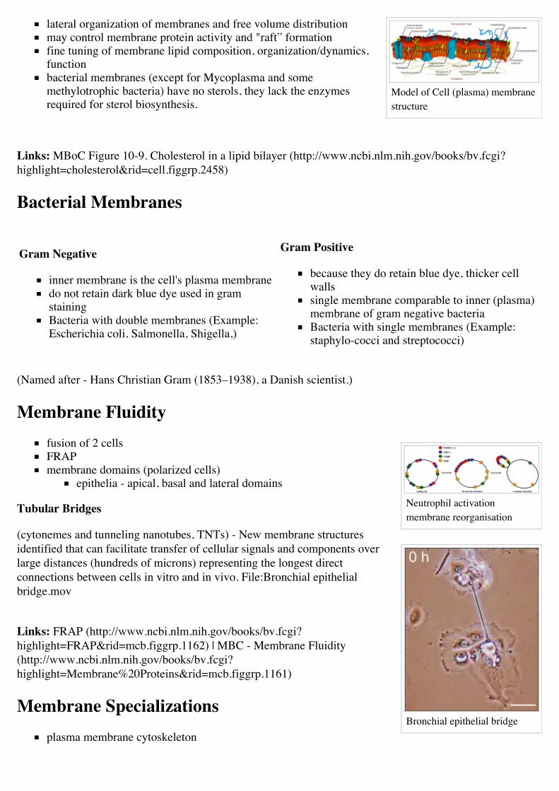
Model of Cell (plasma) membranestructure
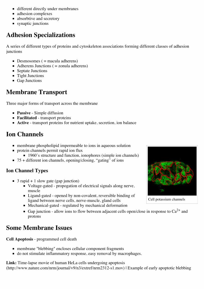
Neutrophil activationmembrane reorganisation
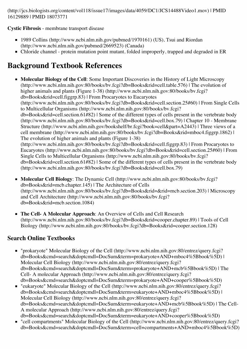
Bronchial epithelial bridge
lateral organization of membranes and free volume distributionmay control membrane protein activity and "raft” formationfine tuning of membrane lipid composition, organization/dynamics,functionbacterial membranes (except for Mycoplasma and somemethylotrophic bacteria) have no sterols, they lack the enzymesrequired for sterol biosynthesis.
Links: MBoC Figure 10-9. Cholesterol in a lipid bilayer (http://www.ncbi.nlm.nih.gov/books/bv.fcgi?highlight=cholesterol&rid=cell.figgrp.2458)
Bacterial Membranes
Gram Negative
inner membrane is the cell's plasma membranedo not retain dark blue dye used in gramstainingBacteria with double membranes (Example:Escherichia coli, Salmonella, Shigella,)
Gram Positive
because they do retain blue dye, thicker cellwallssingle membrane comparable to inner (plasma)membrane of gram negative bacteriaBacteria with single membranes (Example:staphylo-cocci and streptococci)
(Named after - Hans Christian Gram (1853–1938), a Danish scientist.)
Membrane Fluidityfusion of 2 cellsFRAPmembrane domains (polarized cells)
epithelia - apical, basal and lateral domains
Tubular Bridges
(cytonemes and tunneling nanotubes, TNTs) - New membrane structuresidentified that can facilitate transfer of cellular signals and components overlarge distances (hundreds of microns) representing the longest directconnections between cells in vitro and in vivo. File:Bronchial epithelialbridge.mov
Links: FRAP (http://www.ncbi.nlm.nih.gov/books/bv.fcgi?highlight=FRAP&rid=mcb.figgrp.1162) | MBC - Membrane Fluidity(http://www.ncbi.nlm.nih.gov/books/bv.fcgi?highlight=Membrane%20Proteins&rid=mcb.figgrp.1161)
Membrane Specializationsplasma membrane cytoskeleton
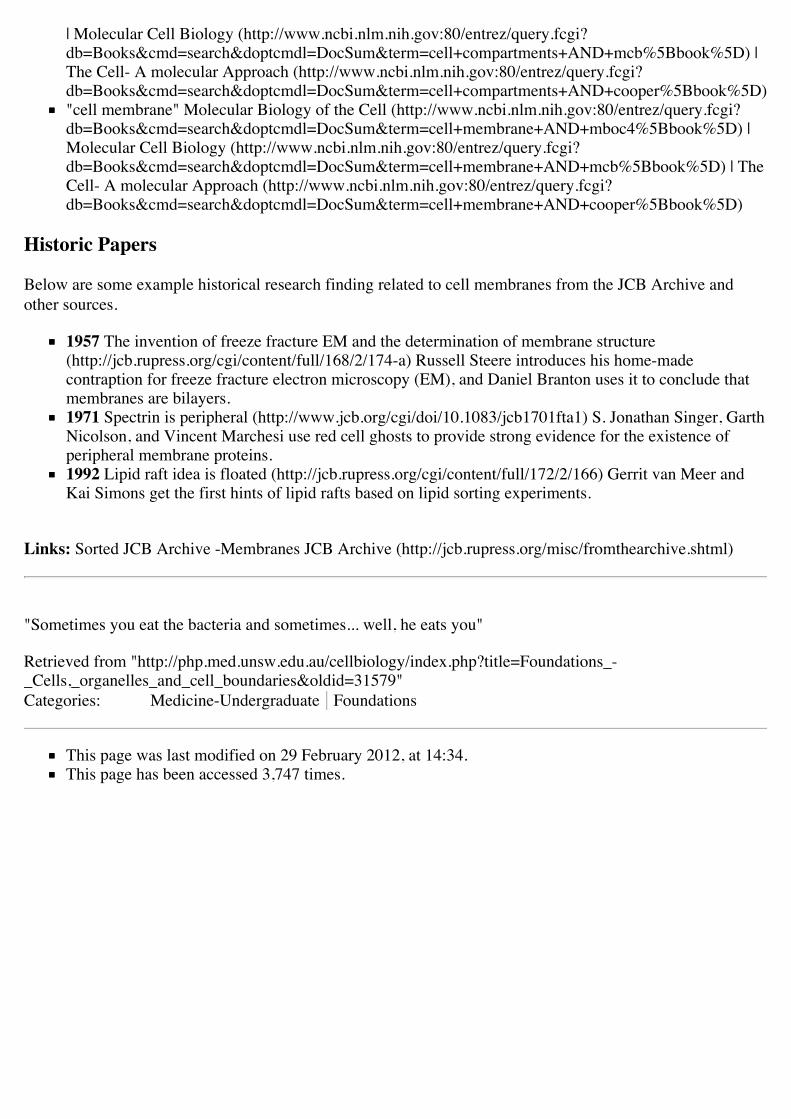
Cell potassium channels
different directly under membranesadhesion complexesabsorbtive and secretorysynaptic junctions
Adhesion SpecializationsA series of different types of proteins and cytoskeleton associations forming different classes of adhesionjunctions
Desmosomes ( = macula adherens)Adherens Junctions ( = zonula adherens)Septate JunctionsTight JunctionsGap Junctions
Membrane TransportThree major forms of transport across the membrane
Passive - Simple diffusionFacilitated - transport proteinsActive - transport proteins for nutrient uptake, secretion, ion balance
Ion Channelsmembrane phospholipid impermeable to ions in aqueous solutionprotein channels permit rapid ion flux
1960’s structure and function, ionophores (simple ion channels)75 + different ion channels, opening/closing, “gating” of ions
Ion Channel Types
3 rapid + 1 slow gate (gap junction)Voltage-gated - propogation of electrical signals along nerve,muscleLigand-gated - opened by non-covalent, reversible binding ofligand between nerve cells, nerve-muscle, gland cellsMechanical-gated - regulated by mechanical deformationGap junction - allow ions to flow between adjacent cells open/close in response to Ca2+ andprotons
Some Membrane IssuesCell Apoptosis - programmed cell death
membrane "blebbing" encloses cellular component fragmentsdo not stimulate inflammatory response, easy removal by macrophages.
Link: Time-lapse movie of human HeLa cells undergoing apoptosis(http://www.nature.com/nrm/journal/v9/n3/extref/nrm2312-s1.mov) | Example of early apoptotic blebbing
(http://jcs.biologists.org/content/vol118/issue17/images/data/4059/DC1/JCS14488Video1.mov) | PMID16129889 | PMID 18073771
Cystic Fibrosis - membrane transport disease
1989 Collins (http://www.ncbi.nlm.nih.gov/pubmed/1970161) (US), Tsui and Riordan(http://www.ncbi.nlm.nih.gov/pubmed/2669523) (Canada)Chloride channel - protein mutation point mutant, folded improperly, trapped and degraded in ER
Background Textbook ReferencesMolecular Biology of the Cell: Some Important Discoveries in the History of Light Microscopy(http://www.ncbi.nlm.nih.gov:80/books/bv.fcgi?db=Books&rid=cell.table.576) | The evolution ofhigher animals and plants (Figure 1-38) (http://www.ncbi.nlm.nih.gov:80/books/bv.fcgi?db=Books&rid=cell.figgrp.83) | From Procaryotes to Eucaryotes(http://www.ncbi.nlm.nih.gov:80/books/bv.fcgi?db=Books&rid=cell.section.25#60) | From Single Cellsto Multicellular Organisms (http://www.ncbi.nlm.nih.gov:80/books/bv.fcgi?db=Books&rid=cell.section.61#82) | Some of the different types of cells present in the vertebrate body(http://www.ncbi.nlm.nih.gov:80/books/bv.fcgi?db=Books&rid=cell.box.79) | Chapter 10 - MembraneStructure (http://www.ncbi.nlm.nih.gov/bookshelf/br.fcgi?book=cell&part=A2443) | Three views of acell membrane (http://www.ncbi.nlm.nih.gov:80/books/bv.fcgi?db=Books&rid=mboc4.figgrp.1862) |The evolution of higher animals and plants (Figure 1-38)(http://www.ncbi.nlm.nih.gov:80/books/bv.fcgi?db=Books&rid=cell.figgrp.83) | From Procaryotes toEucaryotes (http://www.ncbi.nlm.nih.gov:80/books/bv.fcgi?db=Books&rid=cell.section.25#60) | FromSingle Cells to Multicellular Organisms (http://www.ncbi.nlm.nih.gov:80/books/bv.fcgi?db=Books&rid=cell.section.61#82) | Some of the different types of cells present in the vertebrate body(http://www.ncbi.nlm.nih.gov:80/books/bv.fcgi?db=Books&rid=cell.box.79)
Molecular Cell Biology: The Dynamic Cell (http://www.ncbi.nlm.nih.gov:80/books/bv.fcgi?db=Books&rid=mcb.chapter.145) | The Architecture of Cells(http://www.ncbi.nlm.nih.gov:80/books/bv.fcgi?db=Books&rid=&rid=mcb.section.203) | Microscopyand Cell Architecture (http://www.ncbi.nlm.nih.gov:80/books/bv.fcgi?db=Books&rid=mcb.section.1084)
The Cell- A Molecular Approach: An Overview of Cells and Cell Research(http://www.ncbi.nlm.nih.gov:80/books/bv.fcgi?db=Books&rid=cooper.chapter.89) | Tools of CellBiology (http://www.ncbi.nlm.nih.gov:80/books/bv.fcgi?db=Books&rid=cooper.section.128)
Search Online Textbooks
"prokaryote" Molecular Biology of the Cell (http://www.ncbi.nlm.nih.gov:80/entrez/query.fcgi?db=Books&cmd=search&doptcmdl=DocSum&term=prokaryote+AND+mboc4%5Bbook%5D) |Molecular Cell Biology (http://www.ncbi.nlm.nih.gov:80/entrez/query.fcgi?db=Books&cmd=search&doptcmdl=DocSum&term=prokaryote+AND+mcb%5Bbook%5D) | TheCell- A molecular Approach (http://www.ncbi.nlm.nih.gov:80/entrez/query.fcgi?db=Books&cmd=search&doptcmdl=DocSum&term=prokaryote+AND+cooper%5Bbook%5D)"eukaryote" Molecular Biology of the Cell (http://www.ncbi.nlm.nih.gov:80/entrez/query.fcgi?db=Books&cmd=search&doptcmdl=DocSum&term=eukaryote+AND+mboc4%5Bbook%5D) |Molecular Cell Biology (http://www.ncbi.nlm.nih.gov:80/entrez/query.fcgi?db=Books&cmd=search&doptcmdl=DocSum&term=eukaryote+AND+mcb%5Bbook%5D) | The Cell-A molecular Approach (http://www.ncbi.nlm.nih.gov:80/entrez/query.fcgi?db=Books&cmd=search&doptcmdl=DocSum&term=eukaryote+AND+cooper%5Bbook%5D)"cell compartments" Molecular Biology of the Cell (http://www.ncbi.nlm.nih.gov:80/entrez/query.fcgi?db=Books&cmd=search&doptcmdl=DocSum&term=cell+compartments+AND+mboc4%5Bbook%5D)
| Molecular Cell Biology (http://www.ncbi.nlm.nih.gov:80/entrez/query.fcgi?db=Books&cmd=search&doptcmdl=DocSum&term=cell+compartments+AND+mcb%5Bbook%5D) |The Cell- A molecular Approach (http://www.ncbi.nlm.nih.gov:80/entrez/query.fcgi?db=Books&cmd=search&doptcmdl=DocSum&term=cell+compartments+AND+cooper%5Bbook%5D)"cell membrane" Molecular Biology of the Cell (http://www.ncbi.nlm.nih.gov:80/entrez/query.fcgi?db=Books&cmd=search&doptcmdl=DocSum&term=cell+membrane+AND+mboc4%5Bbook%5D) |Molecular Cell Biology (http://www.ncbi.nlm.nih.gov:80/entrez/query.fcgi?db=Books&cmd=search&doptcmdl=DocSum&term=cell+membrane+AND+mcb%5Bbook%5D) | TheCell- A molecular Approach (http://www.ncbi.nlm.nih.gov:80/entrez/query.fcgi?db=Books&cmd=search&doptcmdl=DocSum&term=cell+membrane+AND+cooper%5Bbook%5D)
Historic Papers
Below are some example historical research finding related to cell membranes from the JCB Archive andother sources.
1957 The invention of freeze fracture EM and the determination of membrane structure(http://jcb.rupress.org/cgi/content/full/168/2/174-a) Russell Steere introduces his home-madecontraption for freeze fracture electron microscopy (EM), and Daniel Branton uses it to conclude thatmembranes are bilayers.1971 Spectrin is peripheral (http://www.jcb.org/cgi/doi/10.1083/jcb1701fta1) S. Jonathan Singer, GarthNicolson, and Vincent Marchesi use red cell ghosts to provide strong evidence for the existence ofperipheral membrane proteins.1992 Lipid raft idea is floated (http://jcb.rupress.org/cgi/content/full/172/2/166) Gerrit van Meer andKai Simons get the first hints of lipid rafts based on lipid sorting experiments.
Links: Sorted JCB Archive -Membranes JCB Archive (http://jcb.rupress.org/misc/fromthearchive.shtml)
"Sometimes you eat the bacteria and sometimes... well, he eats you"
Retrieved from "http://php.med.unsw.edu.au/cellbiology/index.php?title=Foundations_-_Cells,_organelles_and_cell_boundaries&oldid=31579"Categories: Medicine-Undergraduate Foundations
This page was last modified on 29 February 2012, at 14:34.This page has been accessed 3,747 times.