Foundation year BIOLOGY-BIOL (101) Animal Histology Cells & Tissues Dr. Huda Kassem.
36
Foundation year BIOLOGY-BIOL (101) Animal Histology Cells & Tissues Dr. Huda Kassem
-
Upload
annice-cathleen-willis -
Category
Documents
-
view
217 -
download
1
Transcript of Foundation year BIOLOGY-BIOL (101) Animal Histology Cells & Tissues Dr. Huda Kassem.
Foundation year
BIOLOGY-BIOL (101)
Animal HistologyCells & Tissues
Dr. Huda Kassem
Tissues
• How do we define tissue? Tissues are groups of specialized cells that work
together for a particular function. • There are four types of tissue.
• Epithelial (covering)• Connective (support)• Muscle (movement)• Nervous (control)

Epithelial Tissues
• Epithelial tissues cover body surfaces. – Outer layer of skin and the lining of organs – Also found in glandular tissue– Play roles in absorption, filtration, secretion, and protection against foreign substances
Special Characteristics of Epithelium
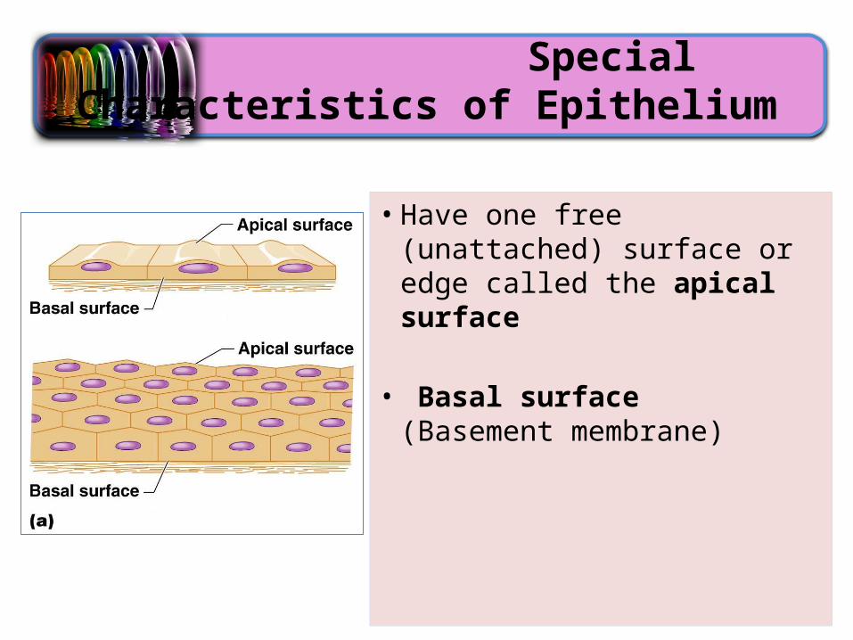
• Have one free (unattached) surface or edge called the apical surface
• Basal surface (Basement membrane)

Classification of Epithelium
• According to the number of cell layers:– Simple (one layer
of cells)– Stratified (more
than one cell layer)
Classification of Epithelium
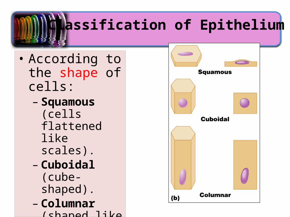
• According to the shape of cells:– Squamous (cells
flattened like scales).
– Cuboidal (cube-shaped).
– Columnar (shaped like columns).

Simple Squamous Epithelium
• It lines the walls of blood vessels, and the lining of the heart, lung, and peritoneal cavities.
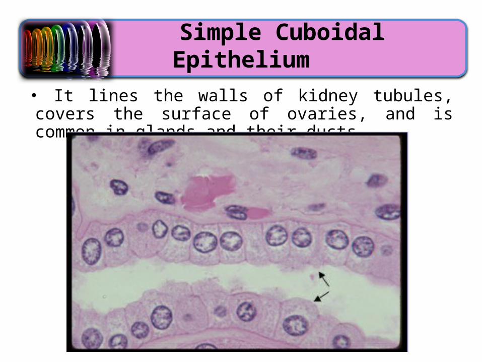
Simple Cuboidal Epithelium
• It lines the walls of kidney tubules, covers the surface of ovaries, and is common in glands and their ducts.

Glandular Epithelium

Simple Columnar Epithelium
• It often includes mucus-producing goblet cells.• It often lines the digestive tract.
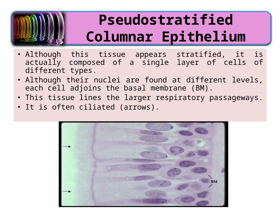
Pseudostratified Columnar Epithelium
• Although this tissue appears stratified, it is actually composed of a single layer of cells of different types.
• Although their nuclei are found at different levels, each cell adjoins the basal membrane (BM).
• This tissue lines the larger respiratory passageways. • It is often ciliated (arrows).
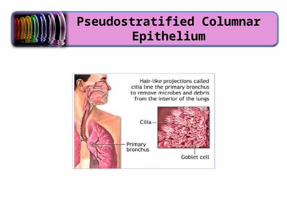
Pseudostratified Columnar Epithelium

Pseudostratified Columnar Epithelium
• Main functions: absorption and secretion• Ciliated variety lines respiratory tract– Mucus produced by goblet cells traps dust and other
debris– Cilia propel mucus upward and away from the lungs
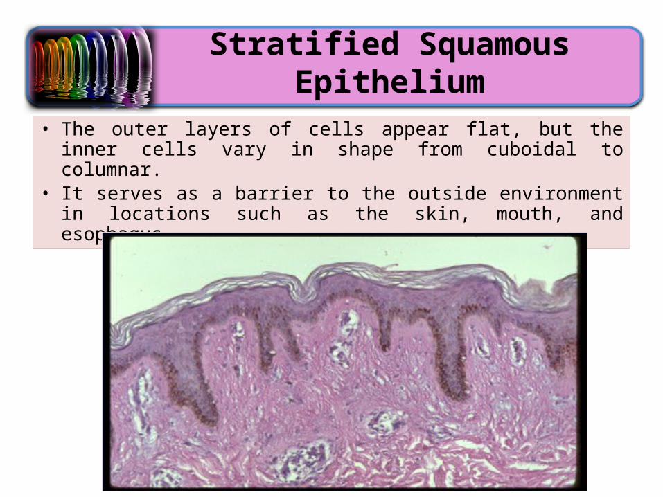
Stratified Squamous Epithelium
• The outer layers of cells appear flat, but the inner cells vary in shape from cuboidal to columnar.
• It serves as a barrier to the outside environment in locations such as the skin, mouth, and esophagus.
Connective Tissue
• Connective tissue differs from other tissues in that it contains large amounts of extracellular matrix.• Extracellular matrix is nonliving material that surrounds living
cells• It is found everywhere and includes the most abundant and
widely distributed tissue
Connective Tissue
• Connective tissues function to • bind other tissues together• provide support• provide nourishment• store wastes• repair damaged tissues
• These tissues are generally well vascularized• Exceptions: tendons, ligaments, cartilage• The exceptions are avascular
Extracellular Matrix
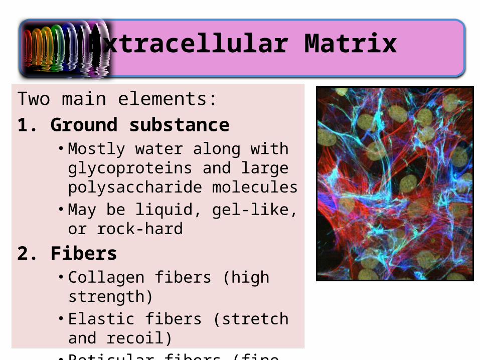
Two main elements:1. Ground substance
• Mostly water along with glycoproteins and large polysaccharide molecules• May be liquid, gel-like, or rock-hard
2. Fibers• Collagen fibers (high strength)• Elastic fibers (stretch and recoil)• Reticular fibers (fine fibers, internal
‘skeleton’)
Types of Connective Tissue
• Bone• Various types of cartilage• Adipose tissue• Dense and loose connective tissue• Blood
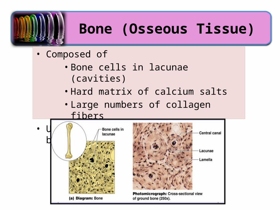
Bone (Osseous Tissue)
• Composed of• Bone cells in lacunae (cavities)• Hard matrix of calcium salts• Large numbers of collagen fibers
• Used to protect and support the body
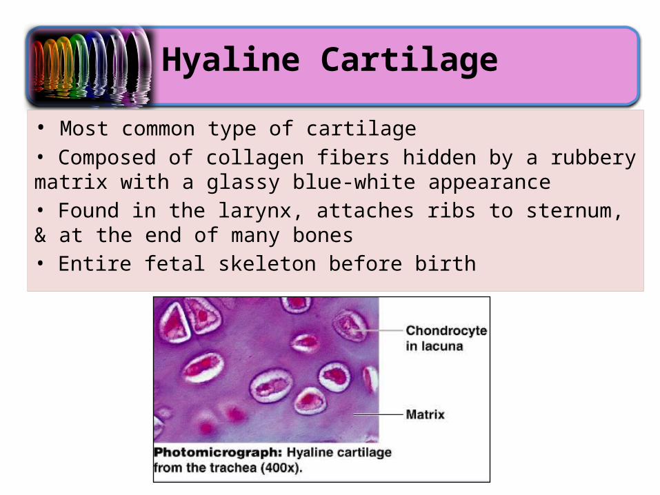
Hyaline Cartilage
• Most common type of cartilage• Composed of collagen fibers hidden by a rubbery matrix with a glassy blue-white appearance• Found in the larynx, attaches ribs to sternum, & at the end of many bones• Entire fetal skeleton before birth
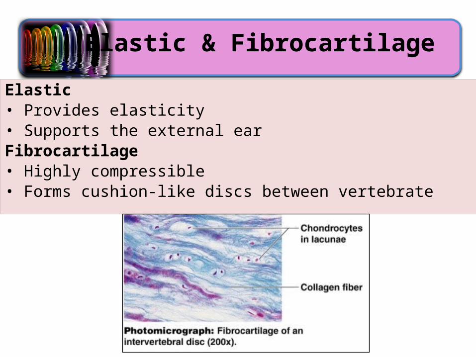
Elastic & Fibrocartilage
Elastic• Provides elasticity• Supports the external earFibrocartilage• Highly compressible• Forms cushion-like discs between vertebrate

Dense Connective Tissue
• Dense connective tissue contains a large number of fibers with only a few cells.
• Fibers shown here are all running parallel to each other, and no cells are present.
• Tendons (muscle to bone) and ligaments (bone to bone) are composed of dense connective tissue.
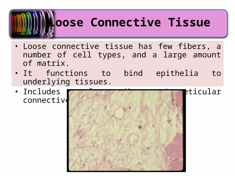
Loose Connective Tissue
• Loose connective tissue has few fibers, a number of cell types, and a large amount of matrix.
• It functions to bind epithelia to underlying tissues.• Includes areolar, adipose, & reticular connective

Areolar Tissue
• Most widely distributed connective tissue
• Soft, pliable “cobwebby” tissue that cushions and protects the body’s organs it wraps
• Holds internal organs together and in their proper positions
• Under microscope: matrix appears as empty space, reservoir of water and salts

Edema
• When a body region is inflamed, the areolar tissue in the area soaks up the excess fluid like a sponge, and the area swells and becomes puffy.

Adipose Tissue
• Adipose cells are bundled together by connective tissue.• Each cell appears as a clear space, representing the site of the
large drop of lipid (fat) before it dissolved during preparation of the microscope slide.
• The nuclei appear as small disks on the periphery of cells. • Functions to insulate the body, protect organs, and fuel storage

Reticular Connective Tissue
• Consists of a delicate network of interwoven reticular fibers • Forms the stroma (internal framework) which can support free
blood cells in lymphoid organs (lymph nodes, spleen, & bone marrow)

Blood (Vascular Tissue)
• Consists of blood cells surrounded by nonliving, fluid matrix called blood plasma
• ‘Fibers’ only visible during blood clotting• Functions as a transport medium for materials
Muscle Tissue
• Muscle is a contractile tissue. • There are three types of muscle:– Skeletal–Cardiac– Smooth
• Main function is to produce movement.

Skeletal Muscle
• Under voluntary control• Contracts to pull on bones or skin• Produces gross body movements or facial expressions• Characteristics of skeletal muscle cells– Striated (stripe-like pattern)– Multinucleate (more than one nucleus)– Long, cylindrical

Cardiac Muscle
• Under involuntary control• Found only in the heart• Function is to pump blood• Characteristics of cardiac muscle cells– Cells are attached to other cardiac muscle cells at
intercalated disks– Striated– One nucleus/cell
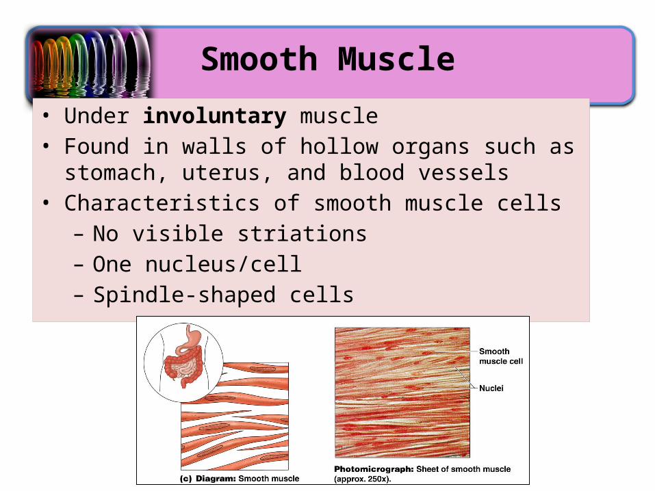
Smooth Muscle
• Under involuntary muscle• Found in walls of hollow organs such as stomach, uterus,
and blood vessels• Characteristics of smooth muscle cells– No visible striations– One nucleus/cell– Spindle-shaped cells

Nervous Tissue
• Nervous tissue, which occurs throughout the body, receives and transmits stimuli.
• It converts a stimulus, whether chemical or physical in nature, into an electrical impulse that is conducted by neurons.
• Nervous tissue also consists of glia, which are the various types of supporting cells in the nervous system.
Cerebellum
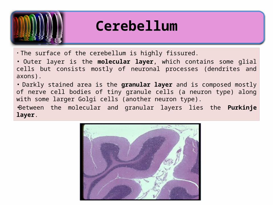
• The surface of the cerebellum is highly fissured. • Outer layer is the molecular layer, which contains some glial cells but consists mostly of neuronal processes (dendrites and axons). • Darkly stained area is the granular layer and is composed mostly of nerve cell bodies of tiny granule cells (a neuron type) along with some larger Golgi cells (another neuron type). •Between the molecular and granular layers lies the Purkinje layer.

How Histology Can Be Used…
Immunohistochemistry is used to reveal BrdU (brown), a thymidine analog that is incorporated in cells undergoing S phase. Cells stained brown were caught in the act of DNA synthesis. These cells are found in the hippocampus, one of the only brain regions where new neurons are formed.
(One example)
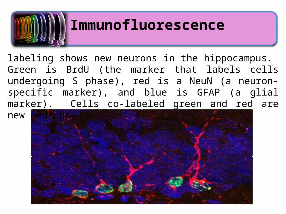
Immunofluorescence
labeling shows new neurons in the hippocampus. Green is BrdU (the marker that labels cells undergoing S phase), red is a NeuN (a neuron-specific marker), and blue is GFAP (a glial marker). Cells co-labeled green and red are new neurons.