FOOT AND ANKLE RETROSPECTIVE STUDY - Orthofix
19
FOOT AND ANKLE RETROSPECTIVE STUDY Retrospective Studies and Results in Foot and Ankle Reconstruction
Transcript of FOOT AND ANKLE RETROSPECTIVE STUDY - Orthofix

FOOT AND ANKLE RETROSPECTIVE STUDY
Retrospective Studies andResults in Foot and AnkleReconstruction

2 CASE STUDY 1
6 CASE STUDY 2
9 CASE STUDY 3
12 CASE STUDY 4
Ankle Distraction with Arthroplasty as an Alternative Treatment for Severe Ankle Arthritis
Retrospective Comparative Analysis of Intra-Articular Calcaneal Fractures
Charcot Foot Reconstruction Utilizing Multiplanar External Ring Fixation
Treatment of Osteochondral Lesions of the Taluswith Cryopreserved Talar Allograft and AnkleDistraction with External Fixation

CASE STUDIES 1
Orthofix wishes to thank the following surgeons for their contribution to the development of the technique:
Edgardo R. Rodriguez, DPM Clinical InstructorDirector: Chicago Foot & Ankle Deformity Correction Center
Other Key Contributors:Paul Cannon, DPMJeffrey Hall, DPMClinton F. Holland, DPMTravis S. Jensen, DPMJohn P. Rachoy, DPMRobert Sheffey, DPMRaymond L. Smith, DPMTomasz Szmyd, DPMGeorge Vito, DPM

2
PURPOSE
A retrospective review was performed to determine thefeasibility of utilizing ankle distraction with arthroplastyas an alternative to more aggressive traditional treatment modalities, such as fusion or total joint implant, for severe ankle arthritis. Outcomes fortwenty-five patients who underwent ankle arthroplastywith distraction were reviewed and graded utilizing theMaryland foot score.
METHOD
1. A total of 25 patients, 12 men and 13 women, witha mean age of 44, ranging from 21 to 71 years ofage were reviewed.
2. All 25 patients underwent open ankle arthroplastywith distraction.
3. 20 patients underwent ankle distraction utilizing amultiplanar external fixation system.
4. 5 patients underwent ankle distraction utilizing amonorail external fixator.
5. Each ankle was distracted 4-6 millimeters.6. Distraction times ranged from 4-10 weeks, with an
average distraction time of 8 weeks.7. In all cases, patients were allowed to bear weight as
tolerated one week post-operatively.
CASE STUDY 1 Ankle Distraction with Arthroplasty as an Alternative Treatment for Severe Ankle Arthritis
PROCEDURES
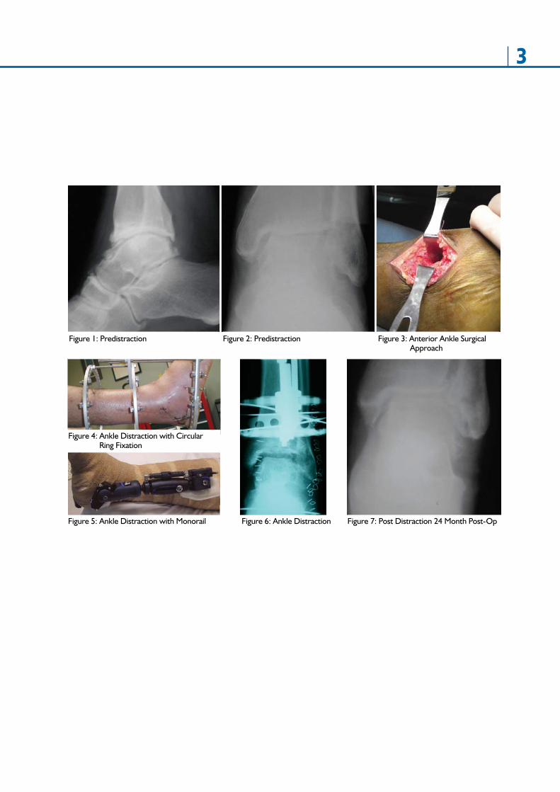
The procedure was performed with the patient in the supine position under general anesthesia. A thightourniquet was typically utilized for hemostasis. Theankle was exposed via a medial longitudinal incision.Care was taken to avoid the tendon of the tibialis anterior as well as the great saphenous vein. A longitudinal joint capsulotomy was performed and a capsular flat was created in order to adequately visualize the ankle mortise (Figure 3). Areas of hyper-trophic bone and soft tissue were extensively debrided.The ankle was copiously irrigated with antibiotic impregnated saline and closed in an appropriate manner. Following closure the thigh tourniquet was released and the ankles were distracted utilizing external fixation.
Twenty patients underwent ankle distraction utilizing a multiplanar external fixation system. These systemsconsisted of two proximal rings, each attached to thetibia by two smooth percutaneous transosseous wires,and a distal foot plate or one-third ring, connected tothe calcaneus with two smooth percutaneous transosseous wires and the midfoot with two smoothpercutaneous transosseous wires (Figure 6); no talarwire was used. Five patients underwent ankle distractionutilizing a monorail external fixator attached to theproximal tibia with two half pins and both the talusand calcaneus with one half pin each (Figure 5).Each ankle was acutely distracted 4-6 millimeters (Figure 6 ). Care was taken to release any excess skintension at pin sites in order to avoid local necrosis.Achilles tendon lengthening procedures were not performed. All patients were allowed to weight bear as tolerated one week postoperatively. Pin sites werecleansed with hydrogen peroxide or isopropyl alcoholfrom daily to weekly.
Before Ankle Distraction After Ankle Distraction
3
Figure 1: Predistraction Figure 2: Predistraction Figure 3: Anterior Ankle Surgical Approach
Figure 5: Ankle Distraction with Monorail Figure 6: Ankle Distraction Figure 7: Post Distraction 24 Month Post-Op
Figure 4: Ankle Distraction with Circular Ring Fixation

4
CASE STUDY 1 continuedAnkle Distraction with Arthroplasty as an Alternative Treatment for Severe Ankle Arthritis
RESULTS
1. Increased dorsiflexion ROM: 1° to 15° Average increase of 5.2°.
2. Increased plantarflexion ROM: 0° to 13° Average increase of 4.3°.
3. Patients with minimal increase in ROM related a significant reduction in symptoms.
4. Patients were followed up for a mean of 50 weeks,with follow up time ranging from 29 to 82 weeks.
CONCLUSION
Although no single treatment is appropriate for everypatient, the authors considered the use of ankle distraction with ankle arthroplasty a viable alternativeto previously accepted treatments for severe anklearthritis.

5
Number Age Sex Etiology Distraction Modality / Weeks
AVERAGE 44 8.12
6
CASE STUDY 2 Retrospective Comparative Analysis of Intra-Articular Calcaneal Fractures
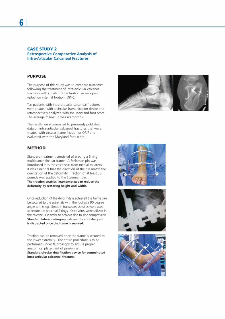
PURPOSE
The purpose of this study was to compare outcomes following the treatment of intra-articular calcanealfractures with circular frame fixation versus open reduction internal fixation (ORIF).
Ten patients with intra-articular calcaneal fractures were treated with a circular frame fixation device and retrospectively analyzed with the Maryland foot score.The average follow up was 48 months.
The results were compared to previously publisheddata on intra-articular calcaneal fractures that weretreated with circular frame fixation or ORIF and evaluated with the Maryland foot score.
METHOD
Standard treatment consisted of placing a 3 ring multiplanar circular frame. A Steinman pin was introduced into the calcaneus from medial to lateral. It was essential that the direction of the pin match theorientation of the deformity. Traction of at least 30pounds was applied to the Steinman pin.The traction enables ligamentotaxis to reduce the deformity by restoring height and width.
Once reduction of the deformity is achieved the frame canbe secured to the extremity with the foot at a 90 degreeangle to the leg. Smooth transosseous wires were used to secure the proximal 2 rings. Olive wires were utilized inthe calcaneus in order to achieve side to side compression. Standard lateral radiograph shows the subtalar joint is distracted once the frame is secured.
Traction can be removed once the frame is secured tothe lower extremity. The entire procedure is to be performed under fluoroscopy to ensure properanatomical placement of pins/wires.Standard circular ring fixation device for comminutedintra-articular calcaneal fracture.

7
calcaneus is restored via manual side to side compression while the traction device is in place. Next utilizing intra-operative fluoroscopy, the angles ofGissiane and Bohlers are addressed. A Brodens view isobtained to assess the posterior facet. If necessary, asmall incision is made inferior to the lateral malleolusinto which a thin osteotome is utilized under fluoroscopicguidance to raise the posterior facet, thus facilitating amore anatomically correct position of the calcaneus.
The Sheffield Ring Fixator is then attached to the extremity in the following manner. A frontal planesmooth wire is placed into the tibia at the level of theproximal ring. With the frame eccentrically positionedon the leg and foot, such that the frame is approximately two finger widths from the leg anteriorly, the wire is properly fixated to the ring andtensioned to 130kg. Another smooth wire is placedtransversely into the calcaneus, properly fixated to thefootplate and tensioned to 60kg. Three additionalsmooth tibial wires are added, as above, to secure the ring construct to the leg. The purpose of thesetransosseous wires is to maintain the foot in a 90 degree position to the leg. As needed, additionalwires (including olive wires) are placed from opposingdirections to further aid in reduction and maintenanceof calcaneal width. Distraction of the subtalar joint isthen accomplished by increasing the distance betweenthe footplate and the distal tibial ring. This is achievedby loosening the frame hardware on the dorsal side of the ring and tightening the frame hardware on theplantar side. Fluoroscopic evaluation of the final fracture reduction is then made. If satisfactory reduction is appreciated, skeletal traction is then removed from the extremity.
PROCEDURES
Following medical as well as anesthesia clearance, the surgeon should assess severity of fracture via evaluation of plain and CT films. Since external fixation does not involve extensive tissue dissection, the condition of the soft tissue does not prevent delayof the procedure. General indications for surgicalmanagement include a greater than 2 mm displacementof the posterior facet with disruption of the width,length, and height of the calcaneus. Fibulo-calcanealimpingement may result if the width of the calcaneusis not addressed.
The extremity is prepped and draped to the knee. A pre-assembled, circular Orthofix Sheffield Ring Fixator consisting of two leg rings and a foot plate areplaced about the extremity. The authors have recentlybegun utilizing a low profile external fixator consistingof two leg rings and a half ring instead of a foot plate.This gives the patient a more cosmetically pleasing device about the extremity as well as allows greaterease of ambulation. Following application of theframe to the extremity a half pin is introduced into theposterior-plantar aspect of the calcaneus from a lateralto medial orientation. Care should be taken to avoidthe critical neuro-vascular bundle on the medial aspect,as well as to allow placement of the trans-osseouswires. The Steiman pin is used for application of skeletal traction. The device consists of 30 lbs ofweight which is placed and secured to the Steiman pin transecting the calcaneus. The force of the fracturereduction should be directed according to the mechanism of the break. The goal of traction is to utilize the principles of ligamentous taxity to reestablish, anatomically and functionally, the length and height of the calcaneus. The width of the
ADVERSE EVENTS
Complications were limited to superficial skin infections at the level of the wires, transient sural neuritis and transient peroneal tendonitis. One patientexperienced a collapse of the posterior facet 2 weeksafter the frame removal.

8
CONCLUSION
The results of the treatment of displaced intra-articularcalcaneal fractures tend to be good-excellent regardlessof whether ORIF or circular ring fixation is utilized except in Sanders Type IV. However, there is a distinctadvantage to utilizing a circular frame in the treatmentof Sanders Type IV due to the fact that the height andwidth are restored. By addressing the height andwidth with the initial procedure it allows a subsequentfusion to be performed more efficiently.
10-12 weeks post-operative result of reduction andstabilization of displaced intra-articular calcaneal fracture via 3-ring circular frame.
CASE STUDY 2 continuedRetrospective Comparative Analysis of Intra-Articular Calcaneal Fractures

CASE STUDY 3 Charcot Foot Reconstruction Utilizing Multiplanar External Ring Fixation
9
PURPOSE
A retrospective case series of 58 patients with limbthreatening Charcot deformity was performed. All patients were treated using multiplanar externalring fixation. There were 41 males and 17 femaleswith a mean age of 55.7 years. 62% presented withType 11 Diabetes and 38% with Type 1. The mean period of follow up was 6.1 years
METHOD
In this series, external ring fixation was used to correctmultiplanar deformities in patients with Charcot Foot.Because of the poor quality of bone and soft tissue, external fixation was utilized to minimize hardware inthe affected limbs, and reduce the amount of periostealstripping in already compromised blood supply.
Proximal rings stabilizethe Tibia and Fibula
3-Ring Fixator
2-3 wires usedper ring, depending
on patient weight

CASE STUDY 3 continuedCharcot Foot Reconstruction Utilizing Multiplanar External Ring Fixation
10
ADVERSE EVENTS
The complication rate is high owing to the combinationof inherent complications that accompany external fixation and the complications associated with limb salvage procedures in Charcot patients who often havemultiple comorbidities. The division into minor andmajor complications attempts to stratify the results into clinically manageable complications and those complications involving more extensive treatment.
RESULTS
Limb salvage was successful in 53 of 58 patients (91%).Union was accomplished in 70% of those salvaged(37/53).
Outcomes were considered successful based on theachievement of a plantegrade foot that was braceableand/or shoeable, regardless of whether there was osseus union or not. This resulted in 48 of 58 patients(82.8%) achieving a “successful” result during thepost-operative period, as described above.
Goal of correction is to achieve a plantegrade, shoeable, and/or braceable foot that allows patient to return to normal daily activities.
Patient with mid-foot Charcot deformity correctedand stabilized with TrueLok ring fixation system.
Post Triple Arthrodesis, 10 weeks post-op, fusion of all joints

TRIPLE ARTHRODESIS/CHARCOT SURGICAL NOTES
• The patient is placed in a supine position.
• If the patient has a posterior compartment contracture, a tendo-achilles lengthening is recommended to increase dorsiflexion of the foot.
• A medial incision over the talonavicular joint and another incision over the lateral aspect of the foot isperformed to obtain access to the talocalcaneal andcalcaneocuboid joints.
• Curettage and prep all three joints. Upon satisfactory apposition and alignment, temporary fixation is applied.
• Three .062 k-wires are utilized for temporary fixation:one for the subtalar joint, one for the talonavicularjoint and one for the calcanealcuboid joint.
• The incisions are closed with the surgeons preferredmethod.
• The Sheffield Ring Fixator consists of two rings on thetibia and one ring around the posterior aspect of thecalcaneus (foot-ring).
• Allow about an inch between the posterior aspect of the calcaneus and the foot-ring.
• The first fine 2.0 mm wire is inserted in the calcaneusparallel to the ankle joint. This wire is attached tothe foot plate. The second wire is inserted on themost proximal ring parallel to the first wire. This willallow the surgeon proper foot manipulation prior tothe insertion of the wires through the midfoot andhindfoot.
• From this point, the additional 2.0 wires are insertedin the tibia at the preferred order of the surgeon.
RECOMMENDATION USE OF THE K-WIRE CLAMP KITS (81541) FOR THE FOLLOWING WIRE ATTACHMENT.
• A 2.0 k-wire is introduced through the navicular andcuboid. This wire will be bent posterior on the footring to compress the midtarsal joint.
• A second 2.0 k-wire is introduced at the talar neck,avoiding penetration of the wire through the fibulaor medial malleolus. This wire will be bent posteriorand inferior to compress the subtalar joint.
• X-Rays are taken to ensure proper placement of wiresand compression.
• A drain is applied for 24 hours post-op.
• Antibiotics are prescribed for a period of one week.
• If the patient has an underlying deformity at the levelof the ankle, midfoot or hindfoot, this must be addressed prior to distraction.
• The patient must be encouraged to ambulate as tolerated to increase micro-motion and bone formation.
• Staples or sutures must not be removed until complete certainty of would healing has been obtained.
• Recommendation of suture removal is approximately3 weeks.
• External fixator should only be removed when radiographic findings indicate complete bone healing.
11

12
CASE STUDY 4 Treatment of Osteochondral Lesions of theTalus with Cryopreserved Talar Allograft andAnkle Distraction with External Fixation
PURPOSE
Osteochondral lesions of the talus are relatively frequentand are often associated with significant morbidity. These lesions represent a separated fragment of cartilage,with or without subchondral bone, typically arising secondary to trauma, with the incidence increased in patients with recurrent ankle injuries and significant ligamentous injuries. Treatment of osteochondral lesionsdepends on the extent, or classification of the lesion.
This case series presents the results of a retrospective review of 6 osteochondral lesions in 6 patients treatedwith transplantation of cryopreserved talar allograft andankle joint distraction.
All patients complained of long standing ankle pain secondary to a traumatic episode confirmed through MRI(Figure 1). All surgeries were performed between 2002and 2004. The average follow up time was 24 months.
METHOD AND PROCEDURES
• A retrospective review was performed of six patientstreated for symptomatic osteochondral lesions.
• All patients underwent talar dome transplant withcryopreserved talar allograft as well as ankle joint distraction.
• The ankle was exposed via a medial longitudinal incision in patients with a medial gutter lesion and through a lateral longitudinal incision in patientswith a lateral gutter lesion.
• In order to provide adequate exposure of the talardome, a transverse osteotomy was performedthrough the medial malleolus at the level of the tibialplafond for medial gutter lesions and a transverse osteotomy was performed through the fibula abovethe level of the tibial plafond for lateral gutter lesions.
• The talar dome lesions were then visualized and resected utilizing an osteotome and mallet (Figure 1).
• The talus was fenestrated to enhance bony ingrowthinto the allograft. An osteotome and mallet was utilized to obtain a graft from the cadaveric Talus of the same size, shape, configuration and anatomical locations the talar dome lesion of the patient (Figure 2).
• The talar graft was then implanted into the operativesite and fixated into place utilizing the Magic PinFracture Fixation System. (Figure 3 and 4).
Figure 1
Figure 2
Figure 3
Figure 4 Figure 5 Figure 6
13
• The medial malleolar osteotomies were also fixatedutilizing the Magic Pin Fracture Fixation System. One of the fibular osteotomies was fixated utilizing a five-hole plate and two of the fibular osteotomieswere fixated with olive wires attached to the externalfixator.
• Refer to Case Study #1 Operative Technique forankle distraction.
• Each ankle was acutely distracted 4-6 millimeters.All patients were allowed to bear partial weight as tolerated one week postoperatively. All patientswere removed from the external fixator at eightweeks and placed in a removable walker boot for another 8 weeks.
RESULTS
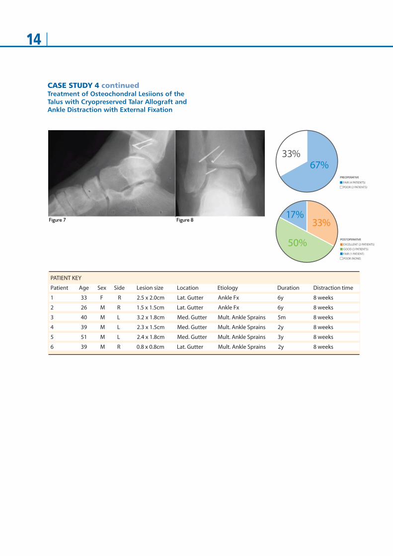
1. Serial Radiographs were taken throughout the postoperative course to assess appropriate healing andconsolidation of the graft. Healing was gauged by trabeculation across the graft site as well as decreasedpain and swelling at the affected ankle. All graftsshowed complete consolidation within 16 weeks (figures 7 and 8).
2. All patients related subjective improvement in symptomsfollowing distraction.
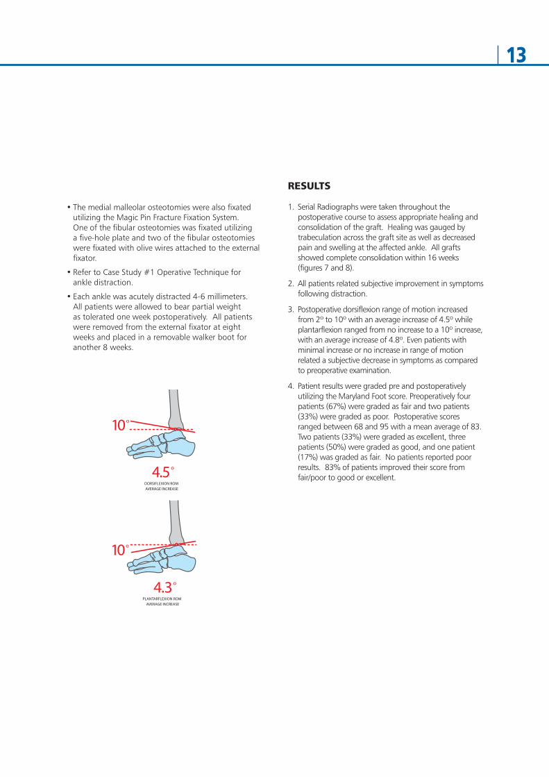
3. Postoperative dorsiflexion range of motion increasedfrom 2º to 10º with an average increase of 4.5º whileplantarflexion ranged from no increase to a 10º increase,with an average increase of 4.8º. Even patients with minimal increase or no increase in range of motion related a subjective decrease in symptoms as comparedto preoperative examination.
4. Patient results were graded pre and postoperatively utilizing the Maryland Foot score. Preoperatively four patients (67%) were graded as fair and two patients(33%) were graded as poor. Postoperative scoresranged between 68 and 95 with a mean average of 83.Two patients (33%) were graded as excellent, three patients (50%) were graded as good, and one patient(17%) was graded as fair. No patients reported poor results. 83% of patients improved their score fromfair/poor to good or excellent.
14
Figure 7 Figure 8
CASE STUDY 4 continuedTreatment of Osteochondral Lesiions of theTalus with Cryopreserved Talar Allograft andAnkle Distraction with External Fixation

15
CONCLUSION
Talar dome lesions pose a difficult treatment dilemma tothe foot and ankle specialist. The frequency of missed orincorrect diagnosis often creates a severe debilitatingarthritic condition of the ankle joint. Talar allografts areparticularly useful in large medial and lateral gutter lesions,allowing implantation with viable chondrocytes and intacthyaline cartilage. The author’s distraction modality ofchoice is a three ring multiplanar external fixator. This configuration provides an extremely stable fixation deviceand allows for early weight bearing.
Distraction of the ankle allows neovascularization and consolidation of the talar dome into the body of the talus.Without distraction the talar dome could collapse underthe weight of the ambulating patient. Distraction also produced a return to control levels of abnormal cartilage proteoglycan as well as a decrease in local inflammation of the ankle joint. Although technically challenging, talardome transplantation with ankle distraction may allow patient to avoid other end stage procedures such as implant arthroplasty or ankle arthrodesis. We believe further investigation is warranted based on these initialfindings.


Your Distributor is:
www.orthofix .comTL-0906-PL-E0 A 11/11
Deformi ty Cor rect ion I Trauma I Pediat r i cs I Bone Growth St imulat ion
Manufactured by: ORTHOFIX SrlVia Delle Nazioni 937012 Bussolengo (Verona)Italy
Telephone +39 045 6719000Fax +39 045 6719380
0123