
FOH UHJXODWLRQ RI 2LNRSOHXUD GLRLFD
93
Dissertation for the degree philosophiae doctor (PhD) at the University of Bergen Dissertation date: &HOO F\FOH UHJXODWLRQ RI 2LNRSOHXUD GLRLFD $ VWXG\ RI WKH F\FOLQ &'.FRPSOHPHQW -DQ ,QJH YUHE。
Transcript of FOH UHJXODWLRQ RI 2LNRSOHXUD GLRLFD

Dissertation for the degree philosophiae doctor (PhD)
at the University of Bergen
Dissertation date:


Acknowledgements
This project was carried out and funded by the department of biology and the Sars
centre at University of Bergen.
Foremost my sincere thanks go to Professor Eric M. Thompson for accepting me as a
PhD student in his group and for providing me with invaluable guidance through my
project, through encouraging discussion and advice. I am also grateful for adopting his
excellent attitude towards science.
I would also like to express my gratitude for having Coen Campsteijn as my co-
supervisor, who through his contagious enthusiasm for science and fruitful discussions
has inspired me greatly.
I am also thankful for my second co-supervisor Christofer Troedsson, who has
provided me with some of the biology perspective of my project, as well as helping me
with statistical analyses.
Sincere gratitude also goes to Harald Hausen and John Courtesis, which has done an
excellent job with sample preparation and TEM imaging for my project, and for all the
fruitful discussions we have had concerning all of the peculiar structures and details
we have found interesting.
My thanks also go to former and present members of the S3 group at the Sars center,
as well as the MDB group at the department of biology who creates the great working
environment in the lab and at the office. A special thanks go to Martina Raasholm,
who maintains order in the lab, making the lab work as well as it does. I am also
grateful for the help you have provided me during long days of micro injection.
I would also like to thank my summer internship student Marine Gueydan for choosing
my topic for her project, which has helped me develop some “boss” skills as well as
providing me with some interesting results.

Sincere thanks also go to the “Appypark” staff who perform a remarkable job
maintaining generations after generations of appendicularia, which my work depends
on.
Especially my parents and my brother, and his family, I would like to thank for
supporting my journey as a student in Bergen. Their love and support is more
important to me then they may realize.
Finally my thanks go to my beloved fiancée, Liv Gansmo, with whom I have shared
my journey. As well as being my loving partner she has also inspired my academic
skills and provided me support in abundance. Realistically words can’t describe my
gratitude.

i
Table of contents
Abstract........................................................................................................................ iii
1. Introduction .............................................................................................................. 1
1.1 The eukaryotic cell cycle...................................................................................... 1
1.2 Cyclins and CDKs ................................................................................................ 3
1.2.1 Cyclin-CDK structure and activation............................................................. 4
1.2.2 The PSTAIRE motif....................................................................................... 5
1.3 The mitotic cell cycle ........................................................................................... 6
1.3.1 The G1-S transition ........................................................................................ 6
1.3.2 The G2-M transition..................................................................................... 10
1.4 Oogenesis and the meiotic cell cycle.................................................................. 13
1.5 Endocycling ........................................................................................................ 17
1.5.1 Endocycle entry............................................................................................ 18
1.5.2 Maintaining endocycles................................................................................ 20
1.6 The urochordate Oikopleura dioica.................................................................... 22
1.6.1 Life cycle of O. dioica.................................................................................. 22
1.6.2 O. dioica oogenesis ...................................................................................... 24
1.7 A perspective on cell cycle evolution................................................................. 26
2. Aims of study .......................................................................................................... 31
3. List of papers .......................................................................................................... 33
4. Summary of results ................................................................................................ 35
4.1 Expansion of Cyclin D and CDK1 paralogs in Oikopleura, a chordate employing diverse cell cycle variants (Paper I).......................................................................... 35
4.2 Functional specialization of chordate CDK1 paralogs during oogenic meiosis (Paper II) ................................................................................................................... 36
5. General discussion ................................................................................................. 37
5.1 Specialized function amongst the amplified CDK1 paralogs in O. dioica ........ 38
5.2 Amplified CDK1 paralogs in O. dioica display variations in the Cyclin interaction motif. ...................................................................................................... 40
5.3 Cyclin Ds and odCDK1 - a possible partnership?.............................................. 41
5.4 Multiple Cyclin Bs: What are they used for? ..................................................... 43
ii
5.5 Cell cycle regulation within the coenocyst......................................................... 45
5.6 Meiotic regulation - Functions of odCDK1a and odCDK1d ............................. 47
5.7 O. dioica and evolution of the cell cycle ............................................................ 50
5.8 Endocycling and cancer...................................................................................... 52
5.9 Future perspectives ............................................................................................. 54
6. Appendix ................................................................................................................. 57
Appendix 1: Mitotic regulation by multiple odCDK1 paralogs in Oikopleura dioica................................................................................................................................... 57
Appendix 2: No observed effect on ring canal (RC) constriction upon odCDK1a knockdown ................................................................................................................ 65
Appendix 3: Supplementary figures ......................................................................... 69
7. References ............................................................................................................... 71
8. Paper I and II ......................................................................................................... 85
iii
Abstract
Regulation of the eukaryotic cell cycle is a fundamental biological process which
controls proliferation of all eukaryote cells. Progression through the cell cycle is
highly dependent on its core regulators; Cyclins and associated Cyclin-dependent
kinases (CDKs), which orchestrate a coordinated series of events through growth in
the first gap phase (G1), initiation of DNA synthesis (S), the second gap phase (G2)
and mitosis (M). Variations of the cell cycle include the canonical mitotic cell cycle,
giving rise to identical sister cells, meiosis, giving rise to haploid gametes, and various
endoreduplicative cycles, which increase ploidy of cells through repetitive S-phases
without intervening cytokinesis. Although limited to a very few specialized cell types
in vertebrates, endoreduplication is widespread amongst invertebrates. The marine
urochordate Oikopleura dioica, deploys somatic endocycling as a main developmental
strategy, which facilitates rapid growth during a very short life cycle. O. dioica
females also take advantage of the elevated transcriptional capacity of endocycling
nurse nuclei within the coenocyst; a single cell compartment shared by hundreds of
nurse and meiotic nuclei. Being a large transparent ovary, the coenocyst provides a
unique model to study both endocycling and meiosis within a shared cytoplasm. The
urochordates also belong to the closest sister group to vertebrates, which places
knowledge about the O. dioica cell cycle in an interesting evolutionary context.
By searching the fully sequenced genome of O. dioica we annotated the Cyclin-
CDK complement of O. dioica, which revealed amplified Cyclin D and Cyclin B
complements. We also identified a surprising amplification of CDK1, an important M-
phase regulator, which is highly conserved from yeast to vertebrates. Interestingly, the
majority of somatic cells grow through endocycling during O. dioica development,
which should favor conditions with low CDK1 activity. This observation therefore
raised the question; why does an organism that develops mainly through a mechanism
favoring reduced CDK1 activity have several paralogs of this particular cell cycle
regulator? In order to dissect possible explanations, we analyzed expression of
odCDK1 paralogs throughout O. dioica development revealing diverse expression
throughout mitotic and endocycling proliferation, in addition to male- and female-
iv
specific expression during gametogenesis. We also assessed functions amongst the
odCDK1 paralogs, which displayed variations within the highly conserved PSTAIRE
motif. Because the PSTAIRE motif is decisive in Cyclin interaction and thus indirectly
affects substrate specificity, functional variation amongst odCDK1 paralogs might
occur. Targeted knockdown of odCDK1 expression by injection of double stranded
RNA (dsRNA) revealed non-redundant and essential functions for two odCDK1
paralogs in producing viable oocytes, representing the first known case in metazoan
models where CDK1 paralogs have sub-functionalized in the control of meiosis.

v
Abbreviations
APC/C Anaphase promoting complex/cyclosome
ATM Ataxia telangiectasia mutated
ATR Ataxia telangiectasia and Rad3 related
CAK CDK activating kinase
cAMP cyclic adenosine 3’, 5’-monophosphate
CDC Cell division cycle
CDK Cyclin dependent kinase
CDT11 Chromatin licensing and DNA replication factor 1
CDH1 CDC20 homolog 1
CKI CDK inhibitor
cmRNA Capped messenger RNA
dsRNA Double stranded RNA
FZR fizzy related
G0 Quiescent phase
G1 First gap phase
G2 Second gap phase
M Mitotic phase
MAPK Mitogen-activated protein kinase
MCM Minichromosome maintenance protein
mRNA Messenger RNA
MPF Mitosis/Maturation promoting factor
MTOC Microtubule organizing center
MYPT Myosin phosphatase targeting protein
NEB Nuclear envelope breakdown
NPC Nuclear pore complex
OC Organizing center
ORC Origin recognition complex
PKA protein kinase A
PLK1 Polo-like kinase 1

vi
Pre-RC Pre-replication complex
PCNA Proliferating cell nuclear antigen
Rb Retinoblastoma protein
S DNA synthesis phase
TALEN Transcription activator-like effector nuclease
TEM Transmission electron microscopy
TGC Trophoblast giant cells
TS Trophoblast stem cells
1. Introduction
1
1. Introduction1.1 The eukaryotic cell cycle
One of the central concepts of biology is the replication/multiplication of the basic unit
of life; the cell. The core process of cell proliferation is controlled by the cell cycle
machinery. The canonical eukaryotic mitotic cell cycle passes through four phases: the
first gap phase (G1), synthesis phase (S), second gap phase (G2) and mitosis (M)
(Figure 1). After a cell has completed these four phases it can either repeat the cycle or
enter a quiescent state (G0), which is commonly found in terminally differentiated
cells. In G1 the cell accumulates nutrients, grows, makes organelles and produces
proteins in order to reach the minimal required size for cell cycle entry. From G1, the
cell can enter several fates; such as quiescence, apoptosis and senescence in response
to unresolved DNA damage, or enter S-phase in response to growth hormones and
nutrition. In S-phase the cell initiates replication of its genome resulting in duplication
of chromatids, making the cell transiently tetraploid. After completion of S-phase the
cell enters G2, where it resumes growth in preparation for M-phase. The duration of
G2 varies amongst cell types and is often absent during embryogenesis of several
species. As the cell enters the first sub-phase of M-phase; the prophase, chromatin
starts to condense and nuclear envelope breakdown (NEB) initiates, and mitosis
continues by mitotic spindle assembly and alignment of chromatin at the metaphase
plate. Chromatid segregation into two identical sister cells during anaphase is triggered
by activity of the anaphase-promoting complex/cyclosome (APC/C) which culminates
in a decline in CDK1 activity and cleavage of cohesion complexes; the protein ring
structures that keep newly replicated sister chromatids paired from S-phase onwards.
This results in segregation of sister chromatids towards opposite poles during
telophase, reassembly of the nuclear envelopes and the division of the cytoplasm
during cytokinesis, giving rise to two identical sister cells (Morgan, 2007). Another
important aspect of the cell cycle is the cycle of the centrosomes, the microtubule
organizing centers (MTOC) of the mitotic spindles, which duplicate and separate in
parallel with chromatin.
The well-known mitotic cell cycle is, however, one of several alternative
variations of the cell cycle. Additional variants include meiosis where two rounds of
1. Introduction
2
…………………………………………………………………………………………..
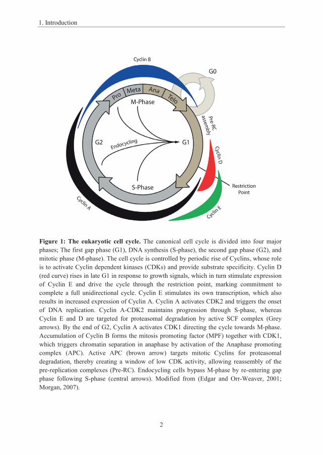
Figure 1: The eukaryotic cell cycle. The canonical cell cycle is divided into four major phases; The first gap phase (G1), DNA synthesis (S-phase), the second gap phase (G2), and mitotic phase (M-phase). The cell cycle is controlled by periodic rise of Cyclins, whose role is to activate Cyclin dependent kinases (CDKs) and provide substrate specificity. Cyclin D (red curve) rises in late G1 in response to growth signals, which in turn stimulate expression of Cyclin E and drive the cycle through the restriction point, marking commitment to complete a full unidirectional cycle. Cyclin E stimulates its own transcription, which also results in increased expression of Cyclin A. Cyclin A activates CDK2 and triggers the onset of DNA replication. Cyclin A-CDK2 maintains progression through S-phase, whereas Cyclin E and D are targeted for proteasomal degradation by active SCF complex (Grey arrows). By the end of G2, Cyclin A activates CDK1 directing the cycle towards M-phase. Accumulation of Cyclin B forms the mitosis promoting factor (MPF) together with CDK1, which triggers chromatin separation in anaphase by activation of the Anaphase promoting complex (APC). Active APC (brown arrow) targets mitotic Cyclins for proteasomal degradation, thereby creating a window of low CDK activity, allowing reassembly of the pre-replication complexes (Pre-RC). Endocycling cells bypass M-phase by re-entering gap phase following S-phase (central arrows). Modified from (Edgar and Orr-Weaver, 2001; Morgan, 2007).

1. Introduction
3
cell division occur without an intervening S-phase, reducing the original cell ploidy,
giving rise to haploid gametes. Another cell cycle variant is endoreplication, a strategy
to increase cellular genomic copies (ploidy) through repetitive rounds of DNA
replication without intervening cytokinesis (Edgar and Orr-Weaver, 2001; Zielke et
al., 2013). In mammals endocycling is limited to a few cell types, including
trophoblast giant cells (TGCs) and hepatocytes, but there are several organisms where
endocycling is a more widespread mechanism. In Drosophila endocycling is found in
the salivary gland and in the nurse nuclei, which are polyploid nuclei with high
transcriptional activity supporting oocyte development. In the marine urochordate
Oikopleura dioica, most somatic cells switch to endocycling shortly after hatching,
and analogous to Drosophila, O. dioica also possesses nurse nuclei, undergoing
endocycling, supporting oogenesis (Ganot et al., 2007a; Ganot and Thompson, 2002).
1.2 Cyclins and CDKs
Regulation of the cell cycle is enormously complex with a vast number of interacting
molecules, but can be generalized to be controlled by oscillations of kinase activity,
which is also the case in the prokaryotic cell cycle. The Cyclin dependent kinases
(CDKs) are considered the basic regulators of the eukaryotic cell cycle because they
activate critical components of the cell cycle engine (Morgan, 1997). CDKs constitute
a family of protein kinases capable of phosphorylating serine and threonine residues of
target proteins. Amongst the mammalian CDKs, four are directly involved in the cell
cycle; CDK1, CDK2, CDK4 and CDK6. Activation of CDKs is achieved through
association with their activating Cyclin subunit, which induces conformational
changes to reveal the catalytic site (Jeffrey et al., 1995) and to modulate the substrate
specificity of CDKs (Loog and Morgan, 2005; Roberts, 1999). Each Cyclin binds to
specific CDK partners so that the levels of the different Cyclins control which CDKs
are active. CDK4 and CDK6 are activated by Cyclin D during G1 phase (Sherr, 1993,
1995) in response to growth factors that trigger a kinase cascade activating
transcription of early and late response genes including; Cyclin D, Cyclin E, CDK2,
CDK4 and CDK6. During late G1, CDK4 and CDK6 induce expression of Cyclin E
through activation of the E2F transcription factors. Cyclin E-dependent activation of
1. Introduction
4
CDK2 further induces E2F activity, stimulating Cyclin A accumulation and S phase
entry (Kato et al., 1993). Cyclin A activates CDK2 and triggers onset and maintenance
of S-phase until G2, when Cyclin A activates CDK1 and initiates the path to M-phase
entry. Finally, Cyclin B controls CDK1 activation, forming the complex known as the
mitosis promoting factor (MPF), leading to initiation of and progression through M-
phase (Labbe et al., 1989).
1.2.1 Cyclin-CDK structure and activation
Activation of CDKs is highly dependent on Cyclin binding and structural changes
involving altered accessibility to the conserved catalytic site (Echalier et al., 2010).
CDKs are composed of an N-terminal lobe mainly comprising beta-sheets and a single
alpha-helix, known as the PSTAIRE helix (Figure 2), while the C-terminal lobe is
mainly arranged by alpha-helices (De Bondt et al., 1993; Schulze-Gahmen et al.,
1996). Within the cleft, created by the two lobes, lies the catalytic site containing the
ATP binding site, orienting phosphate groups outwards from the cleft. When inactive,
the catalytic site is blocked through steric hindrance by the activation loop, preventing
physical access to substrates. Disruption of the activation loop and access to the
catalytic site is mediated through two important mechanisms. Firstly, activation loop
phosphorylation of Thr160 (pThr160), in human CDK2, causes removal of the
activation loop from the catalytic site through interaction of pThr160 with a cationic
binding pocket on CDK, thus CDK activation requires phosphorylation by a CDK
activating kinase (CAK) (Jeffrey et al., 1995). Secondly, full CDK activation requires
binding of a Cyclin partner that is characterized by two domains each containing five
alpha helices termed Cyclin folds, also known as Cyclin boxes. The conserved N-
terminal Cyclin box possesses an MRAIL amino acid sequence and a hydrophobic
patch, which contributes to substrate specificity. Cyclin binding pushes the PSTAIRE
helix towards CDK, which allows the Glu51 within the PSTAIRE to interact with and
change conformation of the catalytic site, adjusting ATP into an optimal position for
catalytic activity.

1. Introduction
5
1.2.2 The PSTAIRE motif
The “PSTAIRE” protein sequence of the PSTAIRE helix is highly conserved amongst
CDK1 and CDK2 homologs, and has been invariable from the yeast CDK1 homolog,
CDC28/CDC2, to human CDK1 and CDK2. The PSTAIRE sequence is however not
retained amongst the Cyclin D interacting CDK4/6 homologs (Figure 3), although the
PxxxxRE consensus is conserved in all CDKs. The proline (P) residue has been
reported to maintain helix structure, and is important for Cyclin binding (Child et al.,
2010), whereas Arginine (R) and Glutamate (E) are important for CDK activation by
adjusting ATP position within the catalytic site. In metazoans the G2/M transition is
typically regulated by PSTAIRE CDKs, which can also rescue mutants of the yeast
CDK1 homologue CDC28 (Sherr, 1993), whereas the PI/LSTV/IRE CDKs are
involved in G1/S transition. Amongst the plant CDK1 paralogs however only CDKA
Figure 2. The Cyclin-CDK complex. The catalytic site of inactive CDKs is blocked through steric hindrance by the T-loop (red loop). CDKs are activated through Cyclin (blue molecule) interaction by repositioning the PSTAIRE helix (green helix), causing conformational changes including liberating the catalytic site by repositioning the T-loop (green loop).

1. Introduction
6
possesses a perfect PSTAIRE motif, but is normally involved in G1/S transition
through interaction with Cyclin D, whereas CDKB1, which possesses a PPTALRE
motif is involved in G2/M transition through interaction with A- and B-type Cyclins
(Nowack et al., 2012; Van Leene et al., 2010). Although the PSTAIRE CDKs have
been conserved from yeast to plants and metazoans, they have apparently sub-
functionalized towards regulation of different sub-phases of the cell cycle.
1.3 The mitotic cell cycle
1.3.1 The G1 – S transition
Essential for cell proliferation is replication of chromosomes in order to generate two
identical sets of the genome to be distributed between the two daughter cells following
mitotic cell division. An important consideration regarding entry into, and
maintenance of, S-phase is to prevent unresolved DNA damage and incomplete or
over-replication of the genome. Persistence of unresolved DNA problems is the
leading cause of genomic instability, which may ultimately cause oncogenic
transformation (Holland and Cleveland, 2009). Cancer cells accumulate mutations
providing growth advantages above the native cell population, which may prove fatal
to the organism as a whole. In order to prevent such developments there are several
checkpoints that monitor genome integrity, which arrest the cell cycle in response to
DNA damage until the damage is resolved, or if not; induce apoptosis or senescence.
Another important mechanism in maintenance of genomic integrity is to ensure that
the genome is completely replicated once, and only once, for each mitotic cell cycle.
hsCDK1 …GQVVAMKKIRLESEEEGVPSTAIREISLLKELRHPNIVSL…hsCDK2 …GEVVALKKIRLDTETEGVPSTAIREISLLKELNHPNIVKL…hsCDK4 …VALKSVRVPNGGGGGGGLPISTVREVALLRRLEAFEHPNV…hsCDK6 …GRFVALKRVRVQTGEEGMPLSTIREVAVLRHLETFEHPNV…
Figure 3. CDK PSTAIRE motifs. The Cyclin interacting PSTAIRE motif is conserved in vertebrate CDK1 and CDK2, whereas CDK4/6 share a conserved Proline, Arginine and Glutamate in the 1st, 6th and 7th positions of the motif, respectively. Conserved residues are in bold.
1. Introduction
7
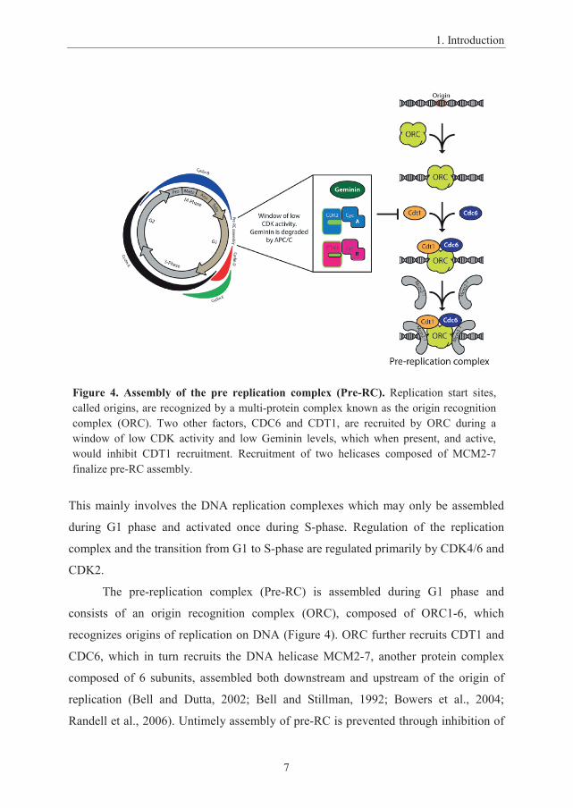
Figure 4. Assembly of the pre replication complex (Pre-RC). Replication start sites, called origins, are recognized by a multi-protein complex known as the origin recognition complex (ORC). Two other factors, CDC6 and CDT1, are recruited by ORC during a window of low CDK activity and low Geminin levels, which when present, and active, would inhibit CDT1 recruitment. Recruitment of two helicases composed of MCM2-7finalize pre-RC assembly.
This mainly involves the DNA replication complexes which may only be assembled
during G1 phase and activated once during S-phase. Regulation of the replication
complex and the transition from G1 to S-phase are regulated primarily by CDK4/6 and
CDK2.
The pre-replication complex (Pre-RC) is assembled during G1 phase and
consists of an origin recognition complex (ORC), composed of ORC1-6, which
recognizes origins of replication on DNA (Figure 4). ORC further recruits CDT1 and
CDC6, which in turn recruits the DNA helicase MCM2-7, another protein complex
composed of 6 subunits, assembled both downstream and upstream of the origin of
replication (Bell and Dutta, 2002; Bell and Stillman, 1992; Bowers et al., 2004;
Randell et al., 2006). Untimely assembly of pre-RC is prevented through inhibition of
1. Introduction
8
CDT1 by Geminin and CDK-dependent phosphorylation of CDT1, which prevents
recruitment by ORC, and thus low CDK activity and degradation of Geminin are
required for pre-RC assembly and licensing of replication (Li and Blow, 2004;
McGarry and Kirschner, 1998). Since CDK activity is also required to trigger and
activate replication once pre-RCs are assembled, CDKs ensure that DNA replication is
activated while preventing premature re-initiation (Bell and Dutta, 2002; Symeonidou
et al., 2012).
In order to reach minimum size required for cell cycle entry the mitotic cell
cycle does not progress continuously as the cell requires time to grow following cell
division. Entry into S-phase therefore depends on nutritional cues and growth signals
that activate the MAPK pathway responsible for Myc dependent expression of Cyclin
D (Adhikary and Eilers, 2005; Bouchard et al., 1999; Hermeking et al., 2000), which
in turn binds and activates CDK4/6 (Figure 5). Cyclin D however binds only weakly to
CDK4/6 and thus requires assistance from the Cip/Kip family of CDK inhibitors
(CKIs), p27 and p21, while facilitating assembly of an active Cyclin-p21/p27-CDK4/6
complex, as p27/p21 simultaneously inhibit CDK2 activity (Blain, 2008; Cheng et al.,
1999; LaBaer et al., 1997; Sherr and Roberts, 1999). Eventually, accumulation of
Cyclin D will titrate away enough p27/p21 from CDK2 to allow activation of the latter
and progression of the G1/S transition. Nuclear CDK4/6 phosphorylates the
Retinoblastoma protein (Rb), a proto-typical tumor suppressor and inhibitor of the
E2F1-3 transcription factors, which further leads to E2F-dependent transcriptional
activation (Dyson, 1998; Lees et al., 1993; Rubin et al., 2005; Weinberg, 1995) of
several cell cycle regulators such as Cyclin E, Cyclin A and Cyclin B (Blais and
Dynlacht, 2004; Cam and Dynlacht, 2003), and also E2F7-8, which antagonize E2F1-3
dependent transcription (de Bruin et al., 2003). Translation and accumulation of Cyclin
E leads to activation of CDK2, which again amplifies its own activation through
further phosphorylation of Rb, in addition to phosphorylation of p27/p21 (Akamatsu et
al., 1998; Ohtani et al., 1995). This positive feedback loop creates a rapid elevation of
cell cycle regulators, including Cyclin A, causing a high level of Cyclin A-CDK2
activity, which triggers firing of pre-RC and onset of S-phase.

1. Introduction
9

1. Introduction
10
1.3.2 The G2 – M transition
Complete and faithful replication of the genome prior to mitotic cell division is
essential to ensure production of two identical copies of the genome. Premature entry
into M-phase in the presence of DNA damage, or uneven duplication of sister
chromatids would lead to genomic instability and could culminate in cancer (Holland
and Cleveland, 2009). Regulation of M-phase entry must therefore ensure that DNA
replication is complete and that DNA damage is resolved before proceeding.
When S-phase is complete, newly duplicated sister-chromatids will remain
associated through sister chromatid cohesion, which helps to ensure symmetric bi-
polar separation of sister chromatids during cell division (Hopfner, 2003; Nasmyth,
2002). The responsible CDK for M-phase entry is CDK1, whose main Cyclin partners
are Cyclin A and Cyclin B. Cyclin B and CDK1 remain cytoplasmic during interphase
and CDK1 activity is held in check through inhibitory phosphorylation, on Thr14 and
Tyr15, by the protein kinases Wee1 and Myt1 (Figure 6) (Boutros et al., 2007; Gavet
and Pines, 2010). When entering M-phase, Cyclin B-CDK1 localization focuses on the
centrosomes where CDK1 becomes activated through de-phosphorylation of Thr14
and Tyr15 by the protein phosphatase CDC25. CDK1 further amplifies its own activity
through a positive feedback loop, by activating phosphorylation of CDC25, and
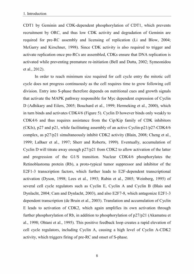
Figure 5: The G1-S transition. CDK4/6 control cell cycle entry from G1 to S-phase and are inhibited by the INK4 family of CDK inhibitors (CKIs), in response to anti-proliferative signals. Once the cell is stimulated to undergo cell division, growth signals activate transcription of Cyclin D. Cyclin D alone binds weakly to CDK4/6, but the CKI p27 stimulates binding between CDK4/6 and Cyclin D and activates CDK4/6 rather than inhibiting the complex. Cyclin D-CDK4/6 in turn phosphorylates the Retinoblastoma protein (Rb) and release inhibition of the transcription factors E2F1/2/3. E2Fs activate transcription of several Cyclins including Cyclin E and Cyclin A. Cyclin E forms an active complex with CDK2, which further phosphorylates Rb, creating a positive feedback loop which also causes an increased level of Cyclin A. Increased Cyclin A levels lead to Cyclin A-dependent activation of CDK2, which phosphorylates CDT1 of pre-replication complexes (Pre-RC) causing activation of DNA replication and the onset of S-phase.

1. Introduction
11
…………………………………………………………………………………………...

1. Introduction
12
inhibitory phosphorylation of Wee1/Myt1 (Lindqvist et al., 2005; Mailand et al.,
2002). Activation of CDK1 leads to rapid increase of CDK1 activity in an all or none
mechanism (bi-stable switch), which can be inhibited by DNA damage through the
ATM/ATR pathway (Zhou and Elledge, 2000), and thus CDK1 activation marks
passage of the M-phase entry checkpoint.
As Cyclin B-CDK1 complexes become active, they translocate to the nuclei
(Gavet and Pines, 2010) in late prophase where they promote NEB (Gong et al., 2007),
through phosphorylation of Lamins amongst others, and they are also responsible for
completion of chromatin condensation (Abe et al., 2011; Kimura et al., 2001). Another
important target for CDK1 is the anaphase promoting complex/cyclosome (APC/C), a
multi-protein Ubiquitin ligase complex that requires an activating subunit, CDC20,
which also contributes to substrate recognition. Upon activation by CDK1, APC/C can
ubiquitinylate and target several proteins, possessing destruction box motifs, for
proteasomal degradation, including Cyclin A, Cyclin B, Geminin and Securin
(Hershko, 1999). Securin destruction relieves Separase inhibition and leads to Cohesin
cleavage and onset of anaphase, whereas Cyclin destruction ensures low CDK activity,
which combined with Geminin destruction, allows reassembly of the pre-RC
(Vodermaier, 2004). As CDK activity drops, CDC20 is replaced by CDH1 as the
Figure 6: The G2-M transition. CDK1 is targeted by inhibitory phosphorylation by Wee1 and Myt1 . When the cell is ready to enter mitosis, the protein phosphatase CDC25 removes Wee1/Myt1 dependent inhibitory phosphorylations thus activating Cyclin B-CDK1 . Cyclin B-CDK1 creates a positive feedback loop by phosphorylation-dependent activation and inactivation of CDC25 and Wee1/Myt1 respectively . Cyclin B-CDK1 also prevents de-phosphorylation of its own substrates by inactivation of PP2A . Cyclin B-CDK1 phosphorylates several targets required for M-phase progression, which include nuclear lamins, causing nuclear envelope breakdown, and CDC20, a component of the anaphase promoting complex/cyclosome (APC/C) . APC/CCDC20 is an ubiquitin ligase that targets Securin, an inhibitor of Cohesin cleavage, and Geminin, an inhibitor of pre-RCassembly, for proteasomal degradation. These events trigger separation of sister chromatid and onset of anaphase, as well as degradation of mitotic Cyclins, which resets the cell cycle by creating low CDK activity . Low CDK activity is later maintained by APC/CCdh1,which allows reassembly of pre-RC and licensing for another entry into S-phase .

1. Introduction
13
APC/C activating subunit, which shares several targets with CDC20, but CDH1 does
not target Securin and is therefore not able to induce chromatin separation (Morgan,
2007). APC/CCDH1 maintains low CDK activity through M-phase until G1, in order to
allow pre-RC assembly. APC/CCDH1 is also involved in maintenance of prophase arrest
and the MI-MII transition in the meiotic cell cycle (Homer, 2013).
1.4 Oogenesis and the meiotic cell cycle
Sexual reproduction requires fusion of two haploid gametes, a single sperm cell from
the male and an oocyte from the female, merging two sets of chromosomes in order to
generate a diploid zygote containing genomic information from both parents. In
addition sexual reproduction allows exchange of genomic information between
homologous chromosomes through homologous recombination, an important source of
genetic variation (Cole et al., 2012). Differentiation and maturation of germ cells thus
rely on meiosis and ploidy reduction in order to produce haploid gametes. Germline
cells of insects and vertebrates initially proliferate synchronously through mitosis in
order to produce a cluster of cells, interconnected through ring canals, known as
germline cysts. In vertebrate females, the cyst phase exists only in juvenile individuals,
whereas the cyst phase persists through most of meiosis amongst insects and
appendicularians (Ganot et al., 2007a; Pepling et al., 1999). Initiation of the meiotic
program starts with entry into pre-meiotic S-phase in order to replicate the genome,
where sister chromatids are tightly joined by Cohesin rings before entering meiosis.
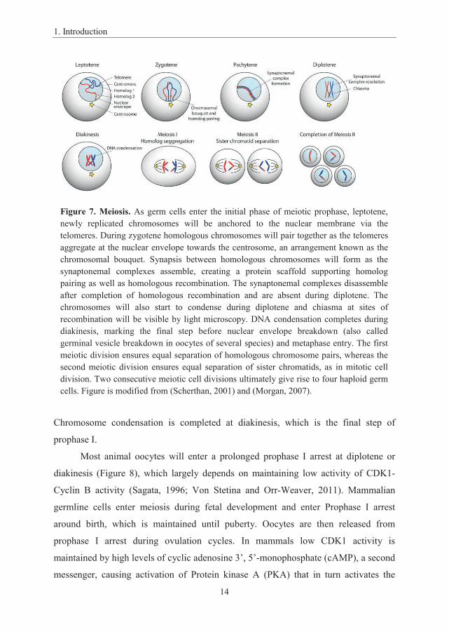
During the initial steps of meiotic prophase I, homologous chromosomes will find
each other and be paired together during zygotene (Figure 7.) (Klutstein and Cooper,
2014; Scherthan, 2001). Zygotene is characterized by clustering of telomeres at the
nuclear membrane towards the centrosome, a conformation defined as the
chromosomal bouquet. Synapsis between homologous chromosomes is further
strengthened through assembly of protein scaffolds known as synaptonemal
complexes, followed by resolution of the chromosomal bouquet during pachytene. The
synaptonemal complexes is then disassembled in diplotene, which also marks the
completion of homologous recombination, characterized by overlapping regions of
condensed chromosomes known as chiasma (Morgan, 2007; Scherthan, 2001).
1. Introduction
14
Chromosome condensation is completed at diakinesis, which is the final step of
prophase I.
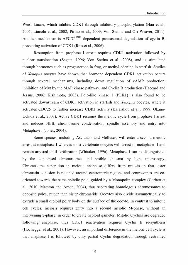
Most animal oocytes will enter a prolonged prophase I arrest at diplotene or
diakinesis (Figure 8), which largely depends on maintaining low activity of CDK1-
Cyclin B activity (Sagata, 1996; Von Stetina and Orr-Weaver, 2011). Mammalian
germline cells enter meiosis during fetal development and enter Prophase I arrest
around birth, which is maintained until puberty. Oocytes are then released from
prophase I arrest during ovulation cycles. In mammals low CDK1 activity is
maintained by high levels of cyclic adenosine 3’, 5’-monophosphate (cAMP), a second
messenger, causing activation of Protein kinase A (PKA) that in turn activates the
Figure 7. Meiosis. As germ cells enter the initial phase of meiotic prophase, leptotene, newly replicated chromosomes will be anchored to the nuclear membrane via the telomeres. During zygotene homologous chromosomes will pair together as the telomeres aggregate at the nuclear envelope towards the centrosome, an arrangement known as the chromosomal bouquet. Synapsis between homologous chromosomes will form as the synaptonemal complexes assemble, creating a protein scaffold supporting homolog pairing as well as homologous recombination. The synaptonemal complexes disassemble after completion of homologous recombination and are absent during diplotene. The chromosomes will also start to condense during diplotene and chiasma at sites of recombination will be visible by light microscopy. DNA condensation completes during diakinesis, marking the final step before nuclear envelope breakdown (also called germinal vesicle breakdown in oocytes of several species) and metaphase entry. The first meiotic division ensures equal separation of homologous chromosome pairs, whereas the second meiotic division ensures equal separation of sister chromatids, as in mitotic cell division. Two consecutive meiotic cell divisions ultimately give rise to four haploid germ cells. Figure is modified from (Scherthan, 2001) and (Morgan, 2007).
1. Introduction
15
Wee1 kinase, which inhibits CDK1 through inhibitory phosphorylation (Han et al.,
2005; Lincoln et al., 2002; Pirino et al., 2009; Von Stetina and Orr-Weaver, 2011).
Another mechanism is APC/CCDH1 dependent proteasomal degradation of cyclin B,
preventing activation of CDK1 (Reis et al., 2006).
Resumption from prophase I arrest requires CDK1 activation followed by
nuclear translocation (Sagata, 1996; Von Stetina et al., 2008), and is stimulated
through hormones such as progesterone in frog, or methyl adenine in starfish. Studies
of Xenopus oocytes have shown that hormone dependent CDK1 activation occurs
through several mechanisms, including down regulation of cAMP production,
inhibition of Myt by the MAP kinase pathway, and Cyclin B production (Haccard and
Jessus, 2006; Kishimoto, 2003). Polo-like kinase 1 (PLK1) is also found to be
activated downstream of CDK1 activation in starfish and Xenopus oocytes, where it
activates CDC25 to further increase CDK1 activity (Karaiskou et al., 1999; Okano-
Uchida et al., 2003). Active CDK1 resumes the meiotic cycle from prophase I arrest
and induces NEB, chromosome condensation, spindle assembly and entry into
Metaphase I (Jones, 2004).
Some species, including Ascidians and Molluscs, will enter a second meiotic
arrest at metaphase I whereas most vertebrate oocytes will arrest in metaphase II and
remain arrested until fertilization (Whitaker, 1996). Metaphase I can be distinguished
by the condensed chromosomes and visible chiasma by light microscopy.
Chromosome separation in meiotic anaphase differs from mitosis in that sister
chromatin cohesion is retained around centromeric regions and centrosomes are co-
oriented towards the same spindle pole, guided by a Monopolin complex (Corbett et
al., 2010; Marston and Amon, 2004), thus separating homologous chromosomes to
opposite poles, rather than sister chromatids. Oocytes also divide asymmetrically to
extrude a small diploid polar body on the surface of the oocyte. In contrast to mitotic
cell cycles, meiosis requires entry into a second meiotic M-phase, without an
intervening S-phase, in order to create haploid gametes. Mitotic Cyclins are degraded
following anaphase, thus CDK1 reactivation requires Cyclin B re-synthesis
(Hochegger et al., 2001). However, an important difference in the meiotic cell cycle is
that anaphase I is followed by only partial Cyclin degradation through restrained

1. Introduction
16
………………………………………………………………………………………….
1. Introduction
17
APC/C activity (Iwabuchi et al., 2000; Taieb et al., 2001). CDK1 activity can thus be
kept low enough to allow meiotic spindle disassembly and nuclear envelope re-
formation, but high enough to inhibit Wee1 and prevent pre-RC assembly (Nakajo et
al., 2000), allowing immediate entry into second meiotic M-phase without intervening
S-phase (Marston and Amon, 2004). Chromatid separation in meiosis II resembles
mitotic anaphase, and cleavage of cohesin on centromeres allow sister chromatid
separation creating a second, haploid, polar body and a haploid pro-nucleus.
1.5 Endocycling
Another widespread variant of the cell cycle referred to as endoreduplication or
endocycling is quite different from mitosis and meiosis in that such cells no longer
complete cytokinesis, and in many cases even lack M-phases all together. Instead of
duplication, these cells continue to grow by increasing their ploidy, through repetitive
cycles of S-phases, above the diploid state of mitotic cells. Because cells entering
endocycles becomes polyploid and cease cell division, endocyling normally occur only
in terminally differentiated cells. Endocycling is ubiquitous amongst eukaryotes (De
Clercq and Inze, 2006; Yin et al., 2010), but is most widespread in plants and
polyploid cells as such as it may contribute up to half of the earth’s biomass
(Sugimoto-Shirasu and Roberts, 2003; Whitman et al., 1998; Zielke et al., 2011).
Amongst metazoans, endocycling cells are abundant in insects, especially during rapid
Figure 8. The meiotic cycle: oogenesis. Mitotic germline cells commit to meiosis when entering the pre-meiotic S-phase, which is controlled by G1/S Cyclin-CDKs. After completing S-phase the cell enters the meiotic M-phase followed by a prolonged primary arrest in meiotic prophase I, a feature common to most Metazoan oocytes. This is maintained by low CDK1 activity. An increase in CDK1 activity triggers meiosis resumption and nuclear envelope breakdown leading to chromosome separation in anaphase. As with meiosis, anaphase is followed by degradation of M-phase Cyclins, causing low CDK activity, though in meiosis CDK activity is only partially inactivated.Partial inactivation of CDK1 allows the cell to enter meiotic M-phase II, immediately after telophase and extrusion of the first polar body. In most mammals, germline cells enter a secondary arrest in metaphase II, which is maintained by high CDK1 activity. Completion of meiosis II is triggered by fertilization, promoting anaphase and extrusion of a second polar body, creating a haploid nucleus which becomes diploid when fusing with the male sperm nucleus.
1. Introduction
18
growth of larvae, where endocycles have been well described in Drosophila salivary
glands and nurse nuclei of adult female ovaries (Zielke et al., 2013). Endocycling cells
also exist in mammals, where they are found in megakaryocytes, hepatocytes and
TGCs of the placenta (Hu and Cross, 2010). Altered features of polyploid cells
compared to diploid cells include the ability to maintain a larger cytoplasmic volume
due to increased transcription from the amplified genomic content and also increased
metabolic activity (Calvi and Spradling, 1999). Mitosis is a rather energy demanding
process, considering the structural reorganizations and massive surge of
phosphorylation activity associated with M-phase (Ma and Poon, 2011). Endocycling
cells, however, can maintain continuous transcriptional activity while maintaining
continuous growth, in contrast to mitotic cells whose transcription halts during mitosis,
endocycling provides a more efficient strategy facilitating rapid cellular growth (Edgar
and Orr-Weaver, 2001; Zielke et al., 2013). In addition, due to multiple gene copies,
polyploid cells are considered to be less susceptible to genetic instability due to a
dampened effect of mutations (Comai, 2005; Lee et al., 2009), as well as being more
tolerant to genotoxic stress (Mehrotra et al., 2008). On the other hand, once committed
to endocycles, cells would normally never revert to mitosis as the mitotic apparatus
would be unable to properly separate the polyploid genome. When this does happen,
cells can move towards the path of aneuploidy, genomic instability, tumourigenesis
and cancer development (Storchova and Pellman, 2004).
1.5.1 Endocycle entry
Because endocycles do not include an M-phase they are composed of repetitive S-
phases, doubling the genomic content for each cycle, separated by gap-phases
allowing time for growth and pre-RC assembly (Edgar and Orr-Weaver, 2001; Edgar
et al., 2014). In order to switch from a mitotic cell cycle to an endocycle, the cell needs
to establish two fundamental alterations. First, the cell needs to restrict M-phase entry,
which primarily involves down regulation of CDK1 activity in endocycling cells
studied so far. In Drosophila follicle cells CDK1 deregulation occurs through
transcriptional repression of String/CDC25 and an inhibitor of Fizzy related
(FZR)/CDH1 named Cut, which prevents removal of CDK1 inhibitory
1. Introduction
19
phosphorylation and causes destruction of mitotic Cyclins respectively, both controlled
by the Notch signaling pathway (Deng et al., 2001). The endocycle switch in
Drosophila also involves transcriptional and/or post-transcriptional (cell type
dependent) down regulation of mitotic Cyclins (Maqbool et al., 2010; Zielke et al.,
2008). In mammalian trophoblast stem cells (TS), endocycle transition is prevented by
mitogenic activation of CHK1 (Ullah et al., 2011), a kinase that is also involved in the
ATM/ATR- dependent DNA damage response. CHK1 maintains low levels of p57 and
p21 through phosphorylation, which targets them for poly-ubiquitinylation and
proteasomal degradation. Loss of mitogenic signal will therefore act to stabilize p57
and p21 through inactivation of CHK1 (Ullah et al., 2011). Increased levels of p57/p21
will then stimulate the transition from mitosis to endocycling by inhibiting CDK1
activity, causing G2 arrest followed by endocycle onset (Ullah et al., 2011). In plants,
endocycle entry is positively regulated by CKIs: the p57KIP related protein named KRP
(KIP related protein) and plant specific CKIs named Siamese (SIM) and Siamese-
related (SMR), which inhibit the activity of M-phase CDK activity (Churchman et al.,
2006; Roeder et al., 2010; Walker et al., 2000). On the other hand, plant endocycling
entry is negatively regulated by DEL1, an atypical E2F, which represses transcription
of CCS52, a CDH1/FZR homolog (Lammens et al., 2008), thus down-regulation of
DEL1 causes an up-regulation of CCS52, which inactivates CDK1 through
proteasomal degradation of mitotic Cyclins.
Though not completing a normal mitosis, endocycling cells still need to reset
the cell cycle following S-phase in order to allow reassembly of pre-RC and entry into
another S-phase. This usually requires degradation of Geminin and low CDK activity,
which are two requirements of pre-RC assembly (Bell and Dutta, 2002). Obtaining low
CDK activity and destruction of Geminin are achieved by proteasomal degradation by
APC/CCDC20/CDH1, and this is utilized to reset the mitotic cell cycle following M-phase.
Because mitotic CDK activity is specifically targeted for down-regulation when
switching to endocycles in plants, insects and mammals, Fzy/CDC20, whose activity
depends on mitotic kinase activity, will no longer be able to activate APC/C. However,
CCS52/FZR/CDH1, activated by low mitotic kinase activity, will remain fully capable
of activating the APC/C during the endocycle switch (Listovsky et al., 2000; Morgan,

1. Introduction
20
2007; Takahashi et al., 2013). In both Drosophila and plants, CCS52/FZR activity is
up-regulated through known mechanisms during endocycle entry, as mentioned above,
and CDH1 has also been demonstrated to be important for endocycle entry in TGCs.
Even though upstream regulation of endocycle entry varies between species, and even
cell types of the same species, the overall mechanism appears to involve short-
circuiting of the mitotic cell cycle through down regulation of mitotic CDK activity,
while promoting APC/C activity in order to keep mitotic Cyclin levels low.
1.5.2 Maintaining endocycles
Maintenance of endocycling requires oscillations of CDK activity in order to trigger S-
phase, when CDK activity is high, and relicense DNA replication, when CDK activity
is low. Because entry into endocycling establishes restriction on M-phase entry
through stable down regulation of mitotic CDK activity, down regulation of G1-S
CDK activity thus requires a mechanism independent of CDK1 and CDC20. As
discussed in the previous section, CKIs facilitate entry into endocycles where they will
maintain endocycles through synchronous oscillations of CKIs and APC/CCDH1/Fzr
activity, inverted relative to oscillations of CDK activity, which have been
demonstrated in plants, insects and mammals. Drosophila, however, has a single
CIP/KIP –type CKI named Dacapo, whose expression is promoted by CDK2-Cyclin E
activity, following S-phase entry, in ovarian nurse cells. This mechanism creates out of
phase oscillations between CDK2-Cyclin E activity and expression of Dacapo,
allowing windows of low CDK activity and pre-RC assembly followed by S-phase
entry. Dacapo is however dispensable in maintaining endocycling in salivary glands,
ovarian follicle cells and innervated bristle cells, but alternative negative feedback
loops are likely involved in those cells (Edgar et al., 2014). One such negative
feedback loop, deployed in mouse TGCs, involves transcriptional activation followed
by transcriptional repression during the G1/S transition. The transcriptional activators,
E2F1-3, promote expression of G1/S Cyclins, which in turn further elevates E2F1-3
activity and thus G1/S Cyclin expression in a positive feedback loop, as explained
previously. Simultaneously E2F1-3 also indirectly ensures transcriptional repression of
the very same targets by promoting expression of their own antagonists, the atypical
1. Introduction
21
E2Fs E2F7 and E2F8. As the E2F7/8 levels increase, they will then gradually replace
E2F1-3 and thus silence the expression of G1/S Cyclins. Although they are important
in endocycling TGCs, the importance of atypical E2Fs play out differently in plants
and insects. As mentioned in the previous section, the plant atypical E2F ortholog,
DEL1, is down regulated during endocycling because it represses the expression of
CDH1, whereas in mammalian endocycling cells; atypical E2Fs acts through down
regulation of Cyclin expression. Drosophila, however, lacks atypical repressor E2Fs
all together but instead utilizes another interesting mechanism which maintains cyclic
degradation of the activator E2F that is linked to DNA synthesis. As the endocycling
cell initiates DNA replication the proliferating cell nuclear antigen (PCNA), a DNA
clamp linking the DNA polymerase to the DNA strand, activates an ubiquitin ligase,
CRL4-CDT2, which targets several proteins, including E2F1 and components of pre-
RC, for proteasomal degradation by recognition of a PIP degron motif (Zielke et al.,
2011). Active CRL4-CDT2 thus enforces down regulation of Cyclin E in response to
DNA synthesis, creating a window of low CDK activity and pre-RC assembly.
Comparing regulation of endocycling between plants, insects and mammals,
even amongst different cell types of the same organism, reveals interesting differences
regarding endocycling, which suggests that endocycles have likely appeared multiple
times in the course of evolution and that there are several paths to modulate the cell
cycle towards endocycles. The marine urochordate, Oikopleura dioica, has deployed
somatic endocycles as a dominant developmental strategy, supporting rapid growth
from early development through adulthood. A spectacular bilateral-symmetric pattern
of polyploid cells of the O. dioica epithelium suggests an intricate regulation of cell
size and ploidy which is poorly understood, but is likely to involve regulation of gap-
phase length during endocycles (Ganot and Thompson, 2002). The coenocyst, the O.
dioica ovary, also consists of multiple endocycling nurse nuclei neighboring an equal
number of meiotic nuclei, all sharing a single gigantic cell compartment. This
environment exemplifies a situation where two quite different variants of the cell cycle
presumably share the same proteins through a common cytoplasm, which should
present various challenges regarding how to tackle incompatible cell cycle regulators.
1. Introduction
22
O. dioica therefore serves as an interesting model organism to conduct cell cycle
research, which may provide useful insight especially into endocycling and meiosis.
1.6 The urochordate Oikopleura dioica
The appendicularian, O. dioica, is a marine urochordate, a member of the closest
extant group to the vertebrates (Delsuc et al., 2006). It is found pan-globally within the
marine environment and is among the most abundant species of zooplankton and an
important contributor to the marine ecosystem (Fenaux and Gorsky).
Appendicularians, or larvaceans, retain a larva-like pelagic state throughout their life
cycle, in contrast to their sister class ascidians and thaliaceans, which have a free
swimming larval stage, common to all urochordates, but become sessile at the adult
stage. The name “Oikopleura dioica” derives from the Greek word “Oikos” meaning
“house” because O. dioica resides within a gelatinous house that aids in feeding. The
house collect and concentrate algae and food particles from the surroundings,
facilitated by water flow through particle concentration filters, controlled by beating of
the tail (Fenaux, 1985). These filter-feeding houses are produced continuously by the
epithelial cells, collectively referred to as the oikoplastic epithelium, and are
exchanged about once every 4th h. This frequent shedding of houses is a major
contributor to marine snow, which drives vertical flux of carbon, important for the
marine, as well as the global, carbon cycle. The name “dioica” derives from the fact
that O. dioica is the only dioecious species of Oikopleura, meaning they have separate
individual sexes. O. dioica is emerging as an intriguing model organism for
evolutionary studies due to its compact and rapidly evolving genome (Denoeud et al.,
2010; Seo et al., 2001).
1.6.1 Life cycle of O. dioica
The life cycle of O. dioica is very short, for a chordate, and lasts from 6 to 10 days,
depending on temperature (Bouquet et al., 2009; Nishida, 2008). Early embryonic
development starts with the first cell division, about 35 min post fertilization, followed
by rapid cell divisions leading to hatching of a free swimming larva as soon as 4 h post
fertilization (Fujii et al., 2008; Nishida, 2008) (Figure 9). The early larvae develop
1. Introduction
23
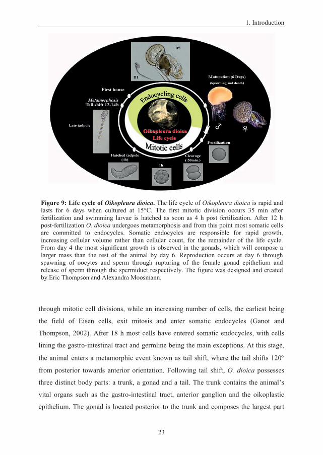
through mitotic cell divisions, while an increasing number of cells, the earliest being
the field of Eisen cells, exit mitosis and enter somatic endocycles (Ganot and
Thompson, 2002). After 18 h most cells have entered somatic endocycles, with cells
lining the gastro-intestinal tract and germline being the main exceptions. At this stage,
the animal enters a metamorphic event known as tail shift, where the tail shifts 120
from posterior towards anterior orientation. Following tail shift, O. dioica possesses
three distinct body parts: a trunk, a gonad and a tail. The trunk contains the animal’s
vital organs such as the gastro-intestinal tract, anterior ganglion and the oikoplastic
epithelium. The gonad is located posterior to the trunk and composes the largest part
Figure 9: Life cycle of Oikopleura dioica. The life cycle of Oikopleura dioica is rapid and lasts for 6 days when cultured at 15°C. The first mitotic division occurs 35 min afterfertilization and swimming larvae is hatched as soon as 4 h post fertilization. After 12 h post-fertilization O. dioica undergoes metamorphosis and from this point most somatic cells are committed to endocycles. Somatic endocycles are responsible for rapid growth, increasing cellular volume rather than cellular count, for the remainder of the life cycle. From day 4 the most significant growth is observed in the gonads, which will compose a larger mass than the rest of the animal by day 6. Reproduction occurs at day 6 through spawning of oocytes and sperm through rupturing of the female gonad epithelium and release of sperm through the spermiduct respectively. The figure was designed and created by Eric Thompson and Alexandra Moosmann.

1. Introduction
24
of the animal at maturity, while the tail contains the notochord and the caudal
ganglion. As O. dioica complete metamorphosis, most somatic cells switch to
endocycles, which facilitate rapid growth best exemplified by the oikoplastic
epithelium, which maintains a perfect bilateral symmetry of differently sized cells with
ploidies ranging from 30-1300C (C = haploid equivalents) at maturity (Ganot and
Thompson, 2002). Metamorphosis is also followed by inflation of the first filter-
feeding house, created by the oikoplastic epithelium (Spada et al., 2001), allowing
feeding. The house is fully replaced every 4th h (Fenaux, 1985). After tail shift, O.
dioica increases trunk size from 200 μm at day 1 to 1000 μm at day 6 (Troedsson et
al., 2007), mostly due to increases in cell volume of endocycling cells. The most
dramatic growth, however, is observed in the gonads, whose growth depends on
nutrient availability, contributing to more than half the volume of the mature animal.
1.6.2 O. dioica oogenesis
The onset of germline development of the ovary occurs through syncytial mitotic
divisions contained within a germline cyst (Pepling et al., 1999). O. dioica oogenesis
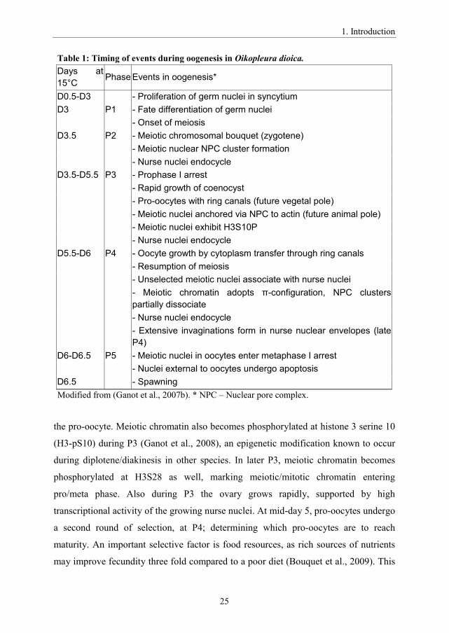
may be broken into five phases, which starts at day 3 as meiosis commences. During
the first phase (P1) of oogenesis, germline nuclei undergo fate selection by either
committing to asynchronous endocycles, establishing polyploid nurse nuclei
supporting oogenesis through high transcriptional activity, or they enter meiosis
committed to seed pro-oocytes. The distribution of meiotic and asynchronously
endocycling nuclei is 1:1, an arrangement defined as a coenocyst describing a
heterogeneous population of nuclei sharing a common cytoplasm (Ganot et al., 2007a;
Ganot et al., 2007b). Nurse nuclei start to endocycle in P2, while the meiotic nuclei
enter zygotene, characterized by the chromosomal bouquet. In P3, the nuclei are
organized by an actin scaffold, which partially encloses meiotic pro-oocytes in
compartments. Similar to Drosophila egg chambers, which contain 15 nurse nuclei
and 1 meiotic oocyte; all nuclei are connected to the same cytoplasm through
structures known as ring canals. P3, which starts at late day 3, lasts until day 5, during
which meiotic nuclei remain arrested in prophase I. The meiotic nuclei are anchored,
via patches rich in nuclear pore complexes, to actin, marking the future animal pole of
1. Introduction
25
the pro-oocyte. Meiotic chromatin also becomes phosphorylated at histone 3 serine 10
(H3-pS10) during P3 (Ganot et al., 2008), an epigenetic modification known to occur
during diplotene/diakinesis in other species. In later P3, meiotic chromatin becomes
phosphorylated at H3S28 as well, marking meiotic/mitotic chromatin entering
pro/meta phase. Also during P3 the ovary grows rapidly, supported by high
transcriptional activity of the growing nurse nuclei. At mid-day 5, pro-oocytes undergo
a second round of selection, at P4; determining which pro-oocytes are to reach
maturity. An important selective factor is food resources, as rich sources of nutrients
may improve fecundity three fold compared to a poor diet (Bouquet et al., 2009). This
Table 1: Timing of events during oogenesis in Oikopleura dioica.Days at 15°C Phase Events in oogenesis*
D0.5-D3 - Proliferation of germ nuclei in syncytiumD3 P1 - Fate differentiation of germ nuclei
- Onset of meiosisD3.5 P2 - Meiotic chromosomal bouquet (zygotene)
- Meiotic nuclear NPC cluster formation- Nurse nuclei endocycle
D3.5-D5.5 P3 - Prophase I arrest- Rapid growth of coenocyst- Pro-oocytes with ring canals (future vegetal pole)- Meiotic nuclei anchored via NPC to actin (future animal pole)- Meiotic nuclei exhibit H3S10P- Nurse nuclei endocycle
D5.5-D6 P4 - Oocyte growth by cytoplasm transfer through ring canals- Resumption of meiosis- Unselected meiotic nuclei associate with nurse nuclei- -configuration, NPC clusters partially dissociate- Nurse nuclei endocycle- Extensive invaginations form in nurse nuclear envelopes (late P4)
D6-D6.5 P5 - Meiotic nuclei in oocytes enter metaphase I arrest- Nuclei external to oocytes undergo apoptosis
D6.5 - SpawningModified from (Ganot et al., 2007b). * NPC – Nuclear pore complex.

1. Introduction
26
also allows opportunistic population growth of O. dioica during periods of algal
blooms (Troedsson et al., 2002). Selection coincides with chromatin condensation into
-configuration and activation of the MAPK pathway, a well-
established transducer of growth and nutrition signals. In addition meiotic nuclei,
selected to seed mature oocytes, become enriched in H3-pS28 and grow rapidly by
cytoplasmic transfer through the ring canals, whereas H3-S28 phosphorylation
diminishes in non-selected nuclei, which then associate with nurse nuclei (Ganot et al.,
2008). When oogenesis reaches its last phase (P5), growing oocytes reach metaphase I
arrest, while nurse nuclei and non-selected nuclei become apoptotic. The fully grown
oocytes will ultimately be released through rupture of the gonad epithelium, at which
point the animal dies. O. dioica oogenesis is summarized in Table 1.
1.7 A perspective on cell cycle evolution
O. dioica responds effectively to available nutrients (Troedsson et al., 2002) and its
rapid growth, short generation time, and efficient modulation of reproductive output,
allow rapid population growth in response to rich food sources occurring during algal
blooms. O. dioica also represents one of the most abundant species of animal plankton
along with copepods, and belong to the closest evolutionary sister group to vertebrates
(Delsuc et al., 2006) (Figure 10). Studying cellular regulatory networks in the light of
evolution provides useful insight into conserved and specialized regulatory modules
amongst species (Doonan and Kitsios, 2009). Common evolutionary mechanisms
include gene expansion, through gene duplication, and gene contraction, through gene
deletions or detrimental mutations. Duplicated genes allow mutations within a
duplicated gene, while retaining original function within the other. This may cause loss
of function mutations of a duplicated gene (non-functionalization), but may also cause
advantageous mutations, giving rise to new functions (neo-functionalization) (Li et al.,
2005). Another possibility is sub-functionalization, where duplicated genes divide
original function amongst paralogs, leading to divided specialization of original
functions. As species evolve, other genes may in turn become redundant, allowing
deleterious mutations of redundant genes, and thus “simplify” regulatory pathways. O.
dioica for instance display both constrictions, as seen with O. dioica notochord genes

1. Introduction
27
which are halved compared to C. intestinalis (Kugler et al., 2011), and expansions,
among O. dioica homeobox genes (Edvardsen et al., 2005), of the genome. Denoeud
et. al. demonstrated that the highly conserved animal genome architecture is shattered
in O. dioica, which illuminates a higher degree of plasticity of genomes than
previously thought (Denoeud et al., 2010).
Regulation of the eukaryotic cell cycle retains similar cell cycle modules in
yeast, plant and metazoans (Doonan and Kitsios, 2009), though there is an obvious
diversification amongst the Cyclin and CDK complements. Budding yeast for instance
possess a single CDK1 ortholog, CDC28, controlling the entire cell cycle, through
interaction with nine Cyclins; Cln 3 (G1), Cln1-2 (G1/S), Clb5-6 (S) and Clb 1-4
(G2/M) (Nurse and Bissett, 1981; Piggott et al., 1982) (Figure 11). Arabidopsis
thaliana (Plant) however requires three CDK1 orthologs namely CDKA1, which
regulates G1/S phase together with five D-type Cyclins and one group of A-type
Cyclins (CycD2-6 and CycA3), and CDKB1 and CDKB2, which regulate G2/M
together with groups of A and B-type Cyclins (Cyc A2 and CycB1-3) (Van Leene et
al., 2010). The plant Cyclin and CDK complement is quite different from the metazoan
Figure 10: Bilaterian evolution. Cephalochordata have traditionally been considered the closest sister group to vertebrates. In the last ten years phylogenetic analysis of genomic data has revealed that the urochordates are closer to vertebrates than cephalochordates in the history of evolution (Delsuc et al., 2006).

1. Introduction
28
cell cycle in that it possesses an amplified complement of D type Cyclins and lacks
Cyclin E all together. Another remarkable difference is that the plant CDK1 homologs
interact with separate cyclin partners, without overlap, which may partly be explained
by their different PSTAIRE motif (conserved only in CDKA1), whereas metazoan
CDK1 and CDK2 share several Cyclin partners. We see that additional CDKs perform
specialized functions in cell cycle regulation from yeast to vertebrates, where three
CDKs are dedicated in control of interphase and one is responsible for M-phase entry.
This classical eukaryotic cell cycle model has however been challenged by CDK
knockout studies in mice, revealing that there is a high level of functional redundancy
amongst cell cycle CDKs (Malumbres and Barbacid, 2009). For instance CDK1, in the
absence of CDK4/6 and CDK2, is sufficient to maintain early embryogenesis in mice,
whereas individual knockout of CDK4/6 and CDK2 display cell type specific defects,
exemplified by CDK2 which is required for meiosis (Adhikari et al., 2012; Berthet et
al., 2003; Santamaria et al., 2007). This suggests that regulation of the basic eukaryotic
cell cycle is in principle largely conserved from yeast. The amplification of cyclins and
CDKs in higher multicellular eukaryotes may thus reflect a more complex composition
of specialized cell types, which possibly require additional specialized regulatory cell
cycle modules.
The invertebrate Cyclin and CDK complements are simple in comparison with
the more complex Cyclin and CDK complements of the vertebrates. Are relatively
simple complements retained in the rapidly evolving O. dioica, which belongs to the
closest sister group to the vertebrates, or have they evolved greater complexity? Has a
rapid evolution and shattered genome architecture also introduced alterations within
the core cell cycle machinery with respect to a growth strategy favoring endocycling?
We know that the homeobox genes of O. dioica have been amplified (Edvardsen et al.,
2005), which suggests higher plasticity of developmental gene expression. A majority
of the duplicated homeobox genes are broadly expressed in the oikoplastic epithelium,
where they have likely neo-functionalized to control patterning and expression of this
functionally important organ (Denoeud et al., 2010; Hosp et al., 2012; Spada et al.,
2001). Thus we set out to determine the core cell cycle regulatory complement of O.

1. Introduction
29
dioica and to begin to explore the functional significance of some of the variant
machinery we uncovered.
Figure 11: Evolution of the Cyclin-CDK complement. The yeast cell cycle depends on a single CDK1 ortholog (CDC28) (Mendenhall and Hodge, 1998), whereas plants (Van Leene et al., 2010) and Metazoans (Campsteijn et al., 2012; Malumbres and Barbacid, 2005; Meyer et al., 2000; Sigrist and Lehner, 1997; Sodergren et al., 2006; van den Heuvel, 2005) subdivide regulation of cell cycle entry (Green lines), G1/S/G2 (Black lines) and G2/M transition (Red lines) between several CDKs. In the chordate lineage there is also an amplification of the Cyclin complement.

1. Introduction
30
2. Aims of study
31
2. Aims of study
O. dioica displays several unusual aspects of the cell cycle for a chordate, involving
extensive use of endocycling required for both somatic and female germline
development. Rapid evolution and unusual genome architecture is reflected in the O.
dioica genomic content, as exemplified by rapidly evolving Lamins and the amplified
homeobox genes, and we were therefore curious to explore to what extent highly
conserved cell cycle machinery had been conserved or modified in this rapidly
evolving chordate. Although the cell cycle Cyclin and CDK complement have
expanded from yeast to metazoan, followed by further amplifications in vertebrates,
the core function of CDK1 has been retained. Our first aim was to explore and
annotate the cyclin and CDK complements of O. dioica, by searching for and aligning
the Cyclin-box motifs, conserved in Cyclins, and kinase domains, conserved in CDKs.
We also wanted to explore retention of other regulatory elements, such as the highly
conserved PSTAIRE motif of CDKs, which could provide clues regarding conserved
function. As a supporting study of the Cyclin and CDK annotation we also wanted to
establish a developmental expression profile of Cyclins and CDKs, which could be
linked to their involvement in mitosis (embryogenesis), endocycling (post-
metamorphosis) and meiosis (late development). Additional support planned for this
work in order to evaluate cell cycle phase specific involvement of Cyclin-CDKs,
included spatio-temporal localization of Cyclins and CDKs. Following identification
of the Cyclin and CDK complements we wanted to explore conserved functions with
respect to cell cycle control, through dsRNA knockdown approaches, especially with
respect to coordinated regulation of endocycling and meiosis within the shared
cytoplasm of the coenocyst. Because the coenocyst is unique compared to germlines of
other metazoans we would also expect to expand this study to include additional
exploration of the architecture and process of oogenesis within the coenocyst, in order
to better comprehend the regulation of this unusual cell cycle environment.

32

3. List of paper
33
3. List of papers
Paper I
Campsteijn, C., J. I. Ovrebo, B. O. Karlsen and E. M. Thompson (2012).
"Expansion of cyclin D and CDK1 paralogs in Oikopleura dioica, a chordate
employing diverse cell cycle variants." Mol Biol Evol 29(2): 487-502.
Paper II
Jan Inge Øvrebø1,2 , Coen Campsteijn3,4, John Kourtesis2, Martina Raasholm2, Harald
Hausen2, Eric Thompson1,2
“Functional specialization of chordate CDK1 paralogs during oogenic meiosis”
Manuscript for submission

4. Summary of results
34
4. Summary of results
35
4. Summary of results
4.1 Expansion of Cyclin D and CDK1 Paralogs in Oikopleura dioica, a Chordate Employing Diverse Cell Cycle Variants (Paper I)
This work characterizes the complete Cyclin and CDK complement of O. dioica using
the assembled genome by Genoscope. We characterized a Cyclin and CDK
complement similar to that of other invertebrates, though some interesting expansions
were identified. The B-type Cyclin complement contained 5 genes, which by
comparison with Ciona intestinalis and Drosophila, each possessing two Cyclin Bs,
represents an expansion even outnumbering the three Cyclin B genes in vertebrates. O.
dioica B-type Cyclins were expressed during early mitotic development as well as in
late development, which is dominated by mitotic and meiotic germline development.
The apparent lack of expression during mid-development, consisting primarily of
somatic endocycles, was consistent with Cyclin B being a G2/M Cyclin. Sex
specificity of B-type Cyclins was also observed in gonads of late animals, which may
be related to specialized functions related to male and female gametogenesis. Also the
O. dioica Cyclin D complement is expanded to 4 genes in contrast to a single Cyclin D
gene in invertebrates and three in vertebrates. The D-type Cyclins displayed an
overlapping expression profile with some being preferentially expressed in early
mitotic development, whereas others were higher expressed during mid-development.
The abundance of D-type Cyclins could be involved in a rheostat like function in
careful regulation of cell size in somatic endocycling cells. Even more surprisingly the
highly conserved G2/M CDK, CDK1, was expanded to five paralogs, making O.
dioica the only known metazoan to possess more than one CDK1 gene. Another
peculiar observation was that none of the five CDK1 paralogs possessed a perfect
PSTAIRE motif, a motif which is highly conserved and invariant in metazoan CDK1s.
Because there are changes within the PSTAIRE motif as well as non-conservative
variations at the Cyclin interaction interface amongst the odCDK1 paralogs, variations
in Cyclin binding preference amongst them may exist. As with Cyclin B, odCDK1
paralogs also displayed sex specificity, suggesting specialization towards
gametogenesis. odCDK1a is most identical to human CDK1 amongst the five

4. Summary of results
36
odCDK1s and is expressed through development, peaking at early mitotic
development and late development. CDK1b and c have a similar expression profile,
being expressed though development with highest expression from early to mid-
development and late male specific expression. CDK1d and e are expressed
exclusively at early and late development and are up regulated in females during
oogenesis.
4.2 Summary of results (Paper II)
This work dissects odCDK1 function in O. dioica meiosis within the coenocyst. We
observed enrichment of odCDK1 paralogs, as well as other meiotic cell cycle
regulators and meiotic kinase activity (MPM-2), within cytoplasmic organelles
juxtaposed to meiotic nuclei. These structures were similar to MTOCs, including
presence of gamma-tubulin, and were defined as organizing centers (OCs). OCs
contained odCDK1 paralogs during P3 (pre-selection) females, but odCDK1 paralogs
translocated from the OCs to non-selected nuclei at P4 (post-selection). Selected nuclei
-configuration but did not contain odCDK1 and did also become
enriched in nuclear Lamin1, an indication of an intact nuclear envelope, indicating
these nuclei are in diakinesis of prophase I and have not resumed meiosis. Nuclear
Lamin1 and lack of nuclear odCDK1 paralogs were observed until spawning and thus
prophase I arrest in diakinesis lasts from day 5 until spawning. To establish whether
odCDK1 paralogs are functionally redundant or possesses specialized functions, we
performed RNAi experiments by injecting dsRNA directly into day 4 gonads. RNAi of
odCDK1e did not give rise to any observable phenotypes, whereas RNAi of odCDK1d
caused release of sterile oocytes failing to resume meiosis, consistent with canonical
CDK1 activity. In addition, odCDK1a RNAi caused release of small and sterile,
oocytes, suggesting problems with cytoplasmic transfer in odCDK1a depleted ovaries.
This work reveals novel sub-functionalization amongst two odCDK1 paralogs.

5. General discussion
37
5. General discussion
The cell cycle control system safeguards mitotic cell division ensuring reliable
inheritance of undamaged genomic material, which is essential in maintenance of cell
fates and healthy cell proliferation. In addition to providing robust passage through the
cell cycle checkpoints, the cell cycle may also be carefully modulated, tailored to the
functional role of the cell, which may involve major restructuring of the cell cycle in
order to switch from mitotic to endocycles or meiotic cell cycles. The mitotic cell
cycle has been scrutinized most extensively, within a diverse range of organisms, and
is thus the best understood. Obtaining a complete picture of the mitotic cell cycle has
many implications in cancer biology and has revealed several cell cycle checkpoints
that are defective in cancer cells. What is known about regulation of the mitotic cell
cycle is often, and rightfully so, transferred to regulation of meiosis, as many mitotic
cell cycle regulators are also involved in meiosis. There are, however, some important
differences to consider, as meiosis consists of two cell division events without an
intervening S-phase in order to decrease cell ploidy. Thus, there are mechanisms
allowing re-entry into M-phase following the first cell division.
Endocyling, which has been studied to a lesser extent, contrasts meiosis by
maintaining repetitive S-phases and lacking M-phase all together in order to increase
ploidy. One of the reasons why there is less knowledge about endocycling might be
that it is less frequent in mammals, which are most relevant for medical research.
Endoreplication is, however, a ubiquitous mechanism deployed, to various extents, in
most eukaryotes, including the giant trophoblast cells of the mammalian placenta.
Endocycling cells share several features with cancer cells including DNA re-
replication, leading to genomic instability (Dutta, 2007), and insensitivity to apoptotic
signals (Mehrotra et al., 2008). In our study we explored cell cycle regulation of the
marine urochordate O. dioica. There are several advantages associated with O. dioica
as a model organism as it deploys mitosis, endocycling and meiosis each
predominating at three different developmental stages, making comparison of
5. General discussion
38
developmental expression with cell cycle variants convenient. As for studying meiosis,
knockdown experiments can easily be performed by microinjection directly into the
exposed gonads, which also allows manipulation of meiosis during prophase I of
meiosis. Gonad injection is also an efficient strategy to distribute synthetic transcripts
in pre-spawned oocytes, allowing manipulation of entire oocyte clutches, providing a
convenient system to study embryonic mitosis. Viewed from an evolutionary
perspective O. dioica stands out as a member of the closest sister group to vertebrates
(Delsuc et al., 2006), as well as being one of the most rapidly evolving metazoans. The
O. dioica Cyclin and CDK complement may therefore illuminate new aspects of cell
cycle evolution within the chordate lineage. An important first step in order to
understand the O. dioica cell cycle was to uncover core cell cycle regulators, namely
the Cyclins and CDKs, within the fully sequenced O. dioica genome (Denoeud et al.,
2010; Seo et al., 2001).
5.1 Specialized function amongst the amplified CDK1 paralogs in O. dioica.
Analysis of the O. dioica genome revealed a cell cycle Cyclin-CDK complement
amplified above that of other invertebrates, and even vertebrates (Figure 12), but the
rationale behind this amplification remains unknown. One obvious feature of O. dioica
is its extensive use of endocycling, both somatic and during germline development.
Similar to growth of O. dioica by endocycling, C. elegans also deploys
endoreplication for somatic growth (Lozano et al., 2006) and in Drosophila
endocycling is observed in several tissues, including nurse nuclei of the ovary
(Hammond and Laird, 1985), but do so with a simpler Cyclin-CDK complement.
Perhaps the most surprising amplification we observed amongst the odCDK
complement was the discovery of five odCDK1 paralogs (Campsteijn et al., 2012),
which was quite unexpected for an organism where endocycling predominates, as
CDK1 is a well-known G2/M regulator in metazoans. Interestingly, CDK1 is also the
most conserved CDK, which single-handedly controls the entire cell cycle in yeast.
Metazoans on the other hand have evolved several cell cycle CDKs. Phylogenetic
analyses indicate that the CDK4/6 subfamily emerged with the appearance of
eumetazoans, while CDK2 diverged from CDK1 by gene duplication in Metazoans
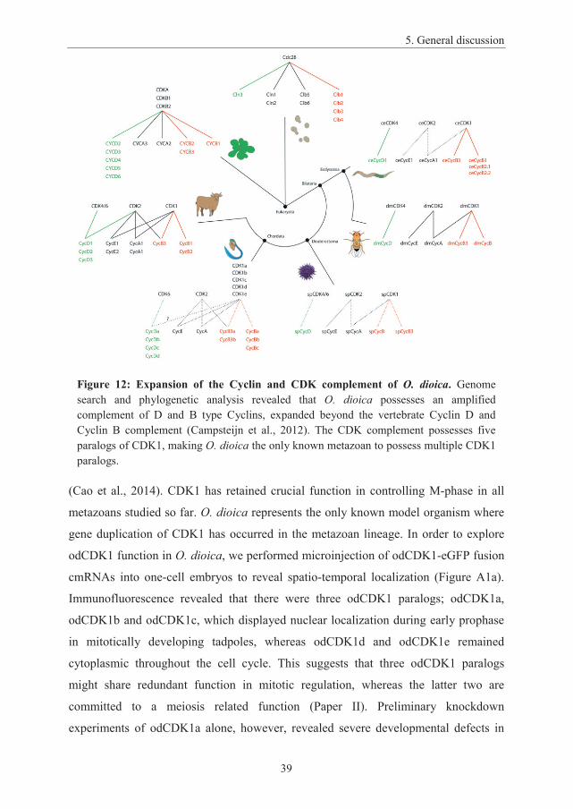
5. General discussion
39
(Cao et al., 2014). CDK1 has retained crucial function in controlling M-phase in all
metazoans studied so far. O. dioica represents the only known model organism where
gene duplication of CDK1 has occurred in the metazoan lineage. In order to explore
odCDK1 function in O. dioica, we performed microinjection of odCDK1-eGFP fusion
cmRNAs into one-cell embryos to reveal spatio-temporal localization (Figure A1a).
Immunofluorescence revealed that there were three odCDK1 paralogs; odCDK1a,
odCDK1b and odCDK1c, which displayed nuclear localization during early prophase
in mitotically developing tadpoles, whereas odCDK1d and odCDK1e remained
cytoplasmic throughout the cell cycle. This suggests that three odCDK1 paralogs
might share redundant function in mitotic regulation, whereas the latter two are
committed to a meiosis related function (Paper II). Preliminary knockdown
experiments of odCDK1a alone, however, revealed severe developmental defects in
Figure 12: Expansion of the Cyclin and CDK complement of O. dioica. Genome search and phylogenetic analysis revealed that O. dioica possesses an amplified complement of D and B type Cyclins, expanded beyond the vertebrate Cyclin D and Cyclin B complement (Campsteijn et al., 2012). The CDK complement possesses five paralogs of CDK1, making O. dioica the only known metazoan to possess multiple CDK1 paralogs.

5. General discussion
40
early tadpoles, suggesting that even amongst the three odCDK1 paralogs involved in
mitosis, there is some degree of non-redundant function concerning embryonic
development (Figure A1b). We also performed co-knockdown of odCDK1b and
odCDK1c, which did not reveal any evident phenotypes. Taken together this suggests
that odCDK1a may act as the primary regulator of mitosis, consistent with canonical
CDK1 regulation, whereas odCDK1b and odCDK1c are functionally redundant to
odCDK1a, or non-essential, in regulation of mitosis. As odCDK1b and odCDK1c are
preferentially expressed in male testes in late development, they may play a more
important function during male germline development, analogous to the specialized
function of odCDK1d and odCDK1e in the female ovary (PaperII). Our findings
suggests that, amongst the odCDK1 paralogs, odCDK1a might be sufficient to
maintain mitotic development, but that specific odCDK1 paralogs are required during
germline development, as observed with odCDK1a and odCDK1d in the ovary (Paper
II). Being functionally different may involve variations in Cyclin interactions,
providing substrate specificity, and thus exploring these interactions would provide
important information for further studies.
5.2 Amplified CDK1 paralogs in O. dioica display variations in the Cyclin
interaction motif.
The CDK1 PSTAIRE motif is highly conserved from yeast (CDC2/CDC28) to
vertebrate CDK1 and CDK2, and is decisive in Cyclin interactions. Plants however do
possess three CDK1 paralogs whose function has sub-functionalized towards
regulation of different cell cycle phases (Van Leene et al., 2010). The highly
conserved PSTAIRE motif is conserved in plant CDKA, whose function is regulation
of G1/S transition, analogous to Metazoan CDK4/6 and CDK2, through interaction
with D and A-type Cyclins. The CDK1 paralogs; CDKB1 and CDKB2, regulate G2/M
through non-overlapping Cyclin A and B interaction, but possess altered PSTAIRE
motifs. Even though CDKA and CDKBs share a sequence similarity of about 83%
(Hirayama et al., 1991), only CDKA is capable of substituting CDC2 function in yeast
(Porceddu et al., 1999). The fact that the PSTAIRE motif, which is decisive in Cyclin
interaction (Child et al., 2010), varies between the three plant CDK1 homologs might

5. General discussion
41
explain the non-overlapping Cyclin interactions, and thus their different functions.
Interestingly, none of the odCDK1 paralogs possess a perfectly conserved motif,
which is invariant amongst other known metazoan CDK1 homologs. Observed
substitutions amongst odCDK1 paralogs includes S2 to A2 in odCDK1c (PA2TSIRE)
and odCDK1e (PA2TSVRE), as well as S2 to P2 in odCDK1b (PP2TSVRE) and
odCDK1d (PP2TSLRE), the latter two creating similar motifs observed in plant
CDKB1 (PPTA/TLRE), which regulates M-phase through interactions with Cyclin B3
(Van Leene et al., 2010). The closest homolog to hsCDK1, odCDK1a, possessed a
single A4 to S4 substitution (PSTS4IRE), shared by the five CDK1 paralogs, mimicking
the S/T sequence of the CDK4 PISTVRE motif (Figure 13), which is known to bind
Cyclin D. If the different PSTAIRE motifs in odCDK1 paralogs provide largely non-
overlapping Cyclin interactions, as seen in plant, the different affinity to Cyclins might
contribute to the specialized functions observed for odCDK1a and odCDK1d. The
amplified complement of D-type Cyclins and the odCDK1 paralog PSTAIRE motifs
that mimic CDK4/6 raises the possibility of interaction between odCDK1 paralogs
with the amplified Cyclin D complement.
5.3 Cyclin Ds and odCDK1 - a possible partnership?
We identified an amplified complement containing four D-type Cyclins, a fourfold
amplification compared with other invertebrates which also exceeds the three D-type
Figure 13. The odCDK1 PSTAIRE motifs. Vertebrate CDK4/6 regulates the G1/S transition through interaction with D-type Cyclins and possesses a PI/LS3T4V/IRE motif. The plant CDK1 homolog, CDKB, also forms active complexes with D-type Cyclins with its PP2T/ALRE motif. odCDK1 paralogs shares an A4 to S4 substitution which mimics the CDK4/6 PI/LS3T4V/IRE motif. odCDK1b and d also possesses a S2 to P2 substitution which mimics the plant CDKB PP2T/ALRE motif.
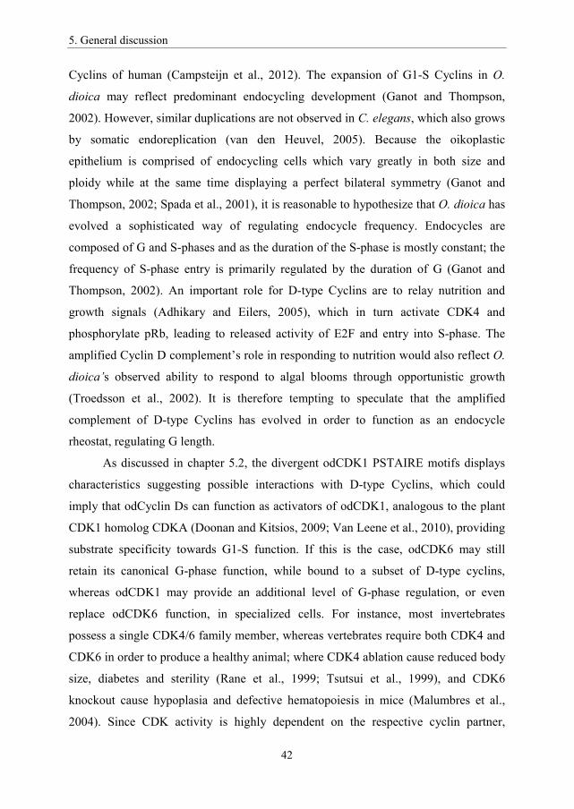
5. General discussion
42
Cyclins of human (Campsteijn et al., 2012). The expansion of G1-S Cyclins in O.
dioica may reflect predominant endocycling development (Ganot and Thompson,
2002). However, similar duplications are not observed in C. elegans, which also grows
by somatic endoreplication (van den Heuvel, 2005). Because the oikoplastic
epithelium is comprised of endocycling cells which vary greatly in both size and
ploidy while at the same time displaying a perfect bilateral symmetry (Ganot and
Thompson, 2002; Spada et al., 2001), it is reasonable to hypothesize that O. dioica has
evolved a sophisticated way of regulating endocycle frequency. Endocycles are
composed of G and S-phases and as the duration of the S-phase is mostly constant; the
frequency of S-phase entry is primarily regulated by the duration of G (Ganot and
Thompson, 2002). An important role for D-type Cyclins are to relay nutrition and
growth signals (Adhikary and Eilers, 2005), which in turn activate CDK4 and
phosphorylate pRb, leading to released activity of E2F and entry into S-phase. The
amplified Cyclin D complement’s role in responding to nutrition would also reflect O.
dioica’s observed ability to respond to algal blooms through opportunistic growth
(Troedsson et al., 2002). It is therefore tempting to speculate that the amplified
complement of D-type Cyclins has evolved in order to function as an endocycle
rheostat, regulating G length.
As discussed in chapter 5.2, the divergent odCDK1 PSTAIRE motifs displays
characteristics suggesting possible interactions with D-type Cyclins, which could
imply that odCyclin Ds can function as activators of odCDK1, analogous to the plant
CDK1 homolog CDKA (Doonan and Kitsios, 2009; Van Leene et al., 2010), providing
substrate specificity towards G1-S function. If this is the case, odCDK6 may still
retain its canonical G-phase function, while bound to a subset of D-type cyclins,
whereas odCDK1 may provide an additional level of G-phase regulation, or even
replace odCDK6 function, in specialized cells. For instance, most invertebrates
possess a single CDK4/6 family member, whereas vertebrates require both CDK4 and
CDK6 in order to produce a healthy animal; where CDK4 ablation cause reduced body
size, diabetes and sterility (Rane et al., 1999; Tsutsui et al., 1999), and CDK6
knockout cause hypoplasia and defective hematopoiesis in mice (Malumbres et al.,
2004). Since CDK activity is highly dependent on the respective cyclin partner,

5. General discussion
43
additional G-phase CDKs may as well arise from CDK1 paralogs with altered cyclin
interaction motifs, as from an existing CDK4/6 family member.
One of the O. dioica D-type Cyclins, odCyclin Dd, displayed sequence
similarities with both D and E-type Cyclins, and immunofluorescence revealed cyclic
nuclear localization in G1-S phase of both mitotic and endocycling cell cycle, thus
based on our observations we concluded that odCyclin Dd acts as a hybrid between
Cyclin D and E (Campsteijn et al., 2012). Surprisingly we also discovered a possible
function related to the meiotic cell cycle in the ovary, where odCyclin Dd was found to
translocate within non-selected nuclei similar to odCDK1 paralogs (Figure A3-1).
Based on the fact that we have concluded a G1-S function for odCyclin Dd
(Campsteijn et al., 2012); we assume odCyclin Dd is not involved in meiosis.
Therefore we still have no clear ideas to why CDKs and Cyclins accumulate in nuclei
destined for an apoptotic fate, but we have an impression that non-selected nuclei
somehow function as “garbage disposals”, clearing the cytoplasm exterior to growing
oocytes of obsolete cell cycle regulators.
5.4 Multiple Cyclin Bs: What are they used for?
The canonical function of CDK1 is regulation of M-phase through interaction with
mitotic Cyclins. The B-type cyclins were also amplified within the O. dioica Cyclin
complement (Campsteijn et al., 2012) to five B-type Cyclins in comparison with
invertebrates possessing two-three Cyclin B genes. Because B-type Cyclins are known
to regulate the G2/M transition whereas most of the O. dioica cells are endocycling,
lacking M-phase all together, it raises the question of why O. dioica requires that
many. The odCyclin B expression profiles also reveal that they are in fact scarcely
expressed during developmental stages composed primarily of endocycling, as would
be expected of B-type Cyclins. odCyclin Bs are, however, expressed pre-
metamorphosis, while cells are still mitotically dividing, and in late development
during which germline differentiation becomes prevalent. In addition, comparison of
expression relative to testes and ovaries, revealed sex specific expression levels, where
odCyclin Ba/b and od were expressed in ovaries, and odCyclin Bc,
odCyclin B3b and od , B1 and B3

5. General discussion
44
Cyclins display functional redundancies during regulation of mitosis in Drosophila.
Although, both are required in production of fertile oocytes, only Cyclin B3 is
dispensable for male fertility (Jacobs et al., 1998). Cyclin B3 regulates the meta/ana-
phase transition, which has been demonstrated in C. elegans where elevated
expression of Cyclin B3 is sufficient to make the meta/ana-phase transition in the
absence of important spindle assembly checkpoint components (Mdf-1/MAD1)
(Tarailo-Graovac et al., 2010). This could partially explain why the male enriched
odCyclin B3b is expressed earlier (at day 5) than the female enriched odCyclin B3a (at
day 6), as meiosis of spermatogenesis is completed at day 6, whereas the meta/ana-
phase transition has not yet occurred in pre-spawned oocytes. Functional specialization
of Cyclins in meiosis control is also reminiscent of the amplified B-type Cyclins of C.
elegans, where RNAi experiments demonstrate overlapping functions among B-type
Cyclins: Cyclin B1 is required for chromosome segregation and Cyclin B3 is required
for sister chromatid separation during meiosis (van der Voet et al., 2009). Because co-
knockdown of Cyclin B1, Cyclin B3 and Cyclin B2.1/B2.2 is required to produce as
severe effect as CDK1 knockdown alone, at least three B-type cyclins are likely to
perform specific functions during meiosis in C. elegans. During embryogenesis, only
Cyclin B1 and Cyclin B3 are required for successful mitosis, whereas during adult
development Cyclin B3 is no longer detected. B-type Cyclins therefore display both
overlapping and specialized function in regulation of meiosis/mitosis, as well as some
functional redundancy during mitosis (van der Voet et al., 2009). Similar to O. dioica,
C. elegans grow primarily through endoreplication (Lozano et al., 2006), so why this
amplification of M-phase cyclins? A possible explanation may be found in the sex-
specific expression patterns of B-type Cyclins, which has been observed in O. dioica
(Campsteijn et al., 2012), allowing sex-specific specialization of duplicated Cyclins,
integrated in tissue/sex-specific expression networks. Sex and tissue-specific
specialization may also be beneficial in hermaphroditic species, such as C. elegans, as
well as O. dioica, which is dioecious but is likely to have evolved from a
hermaphroditic ancestor. This explanation, however, is not necessarily compatible
with another hermaphroditic tunicate, Ciona intestinalis, which possess a single Cyclin
B1 and B3. Nevertheless odCDK1a and odCDK1d do have different functions during

5. General discussion
45
oogenesis, and O. dioica B-type Cyclins may also exhibit functional specialization
during meiosis.
5.5 Cell cycle regulation within the coenocyst
While studying odCDK1 functions in the coenocyst we established that O. dioica
oocytes undergo primary prophase arrest at diakinesis around day 5, persisting until
spawning (Paper II). We then wanted to explore cell cycle regulation within the
coenocyst where there is co-existence of both endocycling and meiotic nuclei within a
shared cytoplasm, which could offer some challenges regarding control of the cell
cycle. In order to prevent reentry into the mitotic cell cycle, endocycles require a
kinase environment poor in CDK1 activity and cycling activity of G1/S CDK activity
to be able to re-enter S-phase (Ullah et al., 2009). Meiosis, however, requires CDK1
activity in order to resume meiosis from prophase I arrest and low Cyclin E-CDK2
activity in order to prevent exit from the meiotic cycle (Hong et al., 2003; Von Stetina
and Orr-Weaver, 2011). Because the timing of meiotic and endocycling entry occurs in
parallel within the O. dioica coenocyst, there is a possibility that canonical CDK1
activity is not required in the ovary after this point, until the oocytes resume meiosis
from prophase I arrest close to the spawning of oocytes. This idea is also supported by
the fact that expression of odCDK1d, which now has been shown to be important for
meiosis resumption (Paper II), peaks at day 6, just prior to spawning and meiosis
resumption. odCDK1a, however, is expressed throughout development, and may
conduct kinase functions which are not involved in meiosis resumption within the
coenocyst. As we have observed functional specialization of odCDK1 paralogs in
control of oogenesis, we may speculate that O. dioica has adapted its Cyclin-CDK
complement in order to manage a heterogeneous cell cycle environment. Similar
genomic Cyclin-CDK amplification is not observed in Drosophila, however, whose
egg chambers contain 15 endocycling nurse cells and a single meiotic oocyte nucleus,
interconnected to a common cytoplasm through ring canals. Drosophila egg chambers
contains a polarized system for mRNA transport, which may facilitate separation of
meiotic and endocycling regulators (Kumano, 2012). An obvious difference between
the two species, however, is that O. dioica maintain thousands of pro-oocytes sharing

5. General discussion
46
cytoplasm with an equal number of asynchronously endocycling nuclei, a situation
where a more localized regulation of the cell cycle would make sense. Further the final
selection of oocytes occurs after entering prophase I arrest in response to available
nutrients, which could be linked to the amplified Cyclin D complement. In this study
we revealed that acentriolar MTOC like organizing centers (OC) could play a vital role
in separation of meiotic and endocycling regulators in a manner which has not been
previously described. Aggregation of CDKs could both contribute to a localized
control of meiotic events and act as a storage node for cell cycle kinases required in
maturation of the oocytes. Canonical centriolar MTOCs play an important function in
mitosis during separation of sister chromatids, and act as an activation center for
Cyclin B-CDK1 before translocation into the nucleus (Jackman et al., 2003), thus
creating localized accumulation of CDK1 activity close to the nuclei. In Drosophila
ovaries, which also possess a meiotic nucleus and endocycling nurse nuclei connected
to a shared cytoplasm via ring canals, centriolar MTOCs from the nurse nuclei migrate
into the developing oocyte (Mahowald and Strassheim, 1970), which is not surprising
in the sense that endocycling nuclei do no longer require MTOCs due to lack of
mitotic division. It may also be possible that MTOC removal plays an additional
function by moving a central mitotic organizing center away from nuclei and thus
restricts CDK1 activity. A similar loss of MTOCs related to endocycling nurse nuclei
are also observed in the O. dioica coenocyst where we observed a 1:1 ratio of OCs
juxtaposed to meiotic nuclei (Paper II). Interestingly, non-selected nuclei also lost
association with their MTOC following oocyte selection, suggesting that they no
longer require a meiotic control point as they are committed to undergo an apoptotic
fate.
Direct interaction between OCs and meiotic nuclei were supported by TEM
(Paper II), where we observed physical contact and NPCs bridging the OC/meiotic
nuclei, as well as in immunofluorescence where NPC clusters were observed in the
OC/meiotic nuclei interface. Interestingly, NPC rich regions of the nuclear envelope
have likewise been observed at sites attached to the actin scaffold (Ganot et al.,
2007b), and could thus be related to OC/meiotic nuclei in a similar manner, although
OCs were not observed to contain actin.

5. General discussion
47
In order to ensure that only one pair of centrioles, provided by sperm, is present
following fertilization, oocytes of most vertebrates eliminate centrioles prior to the
first meiotic division (Mikeladze-Dvali et al., 2012; Szollosi et al., 1972). Acentriolar
MTOCs are still able to focus microtubules in order to separate chromatin during
meiosis I and meiosis II, though do not form a bipolar spindle similar to centrioles but
rather create short microtubule assemblies which are thought to facilitate asymmetric
cell division creating small polar bodies (Luksza et al., 2013). There are still many
questions remaining regarding the O. dioica OCs, including microtubule organization
activity and RNA localization. At some stages there was even observed an envelope of
Lamin surrounding the OCs of P4 and P5 ovaries and additional small Lamin vesicles
surrounding the OCs (Figure A3), suggesting that the OCs are involved in vesicle
transport. Because of difficulties of live imaging we are still unable to fully understand
the dynamics of possible vesicle transport towards the OCs, and thus this feature of the
OC remains an intriguing mystery.
5.6 Meiotic regulation - Functions of odCDK1a and odCDK1d
We wanted to study specific functions of odCDK1 paralogs within the coenocyst and
functional studies revealed that there could be a link between odCDK1 paralogs and
control of the ring canal. Knockdown of odCDK1a by dsRNA injection consistently
produced small infertile oocytes spawned from successfully injected females (Paper
II). This result is consistent with similar effects obtained by knocking down Myosin
phosphatase (DMYPT) and Flw, a PP1 homolog, in Drosophila, causing over-
constriction of the ring canals preventing influx of cytoplasm, which in turn lead to
restricted growth of the oocyte (Ong et al., 2010; Yamamoto et al., 2013). Although,
CDK1 has not yet been directly linked to regulation of the ring canal, but as MYPT is
a confirmed target of CDK1 (Yamashiro et al., 2008), we suggest that CDK1 depletion
could cause a similar effect, resulting in the small oocytes produced in odCDK1a
silenced O. dioica ovaries. Consistent with our assumption is the observed effect of
localized CDK1 inhibition in Drosophila oocytes, which causes premature constriction
of the contractile ring, which is also regulated by MYPT (Menant and Karess, 2012).
The composition of the ring canal and the actomyosin contractile ring is similar in that
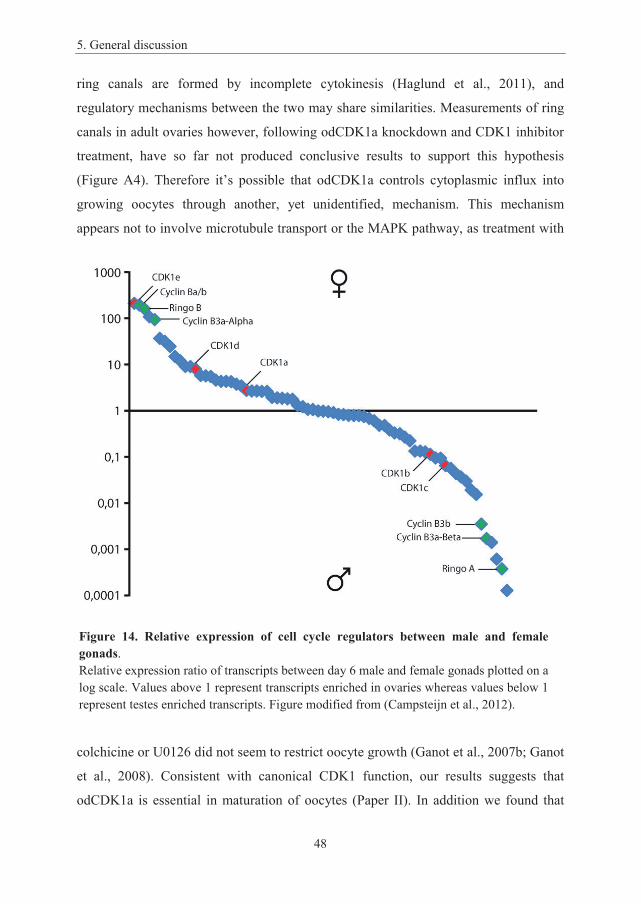
5. General discussion
48
ring canals are formed by incomplete cytokinesis (Haglund et al., 2011), and
regulatory mechanisms between the two may share similarities. Measurements of ring
canals in adult ovaries however, following odCDK1a knockdown and CDK1 inhibitor
treatment, have so far not produced conclusive results to support this hypothesis
(Figure A4). Therefore it’s possible that odCDK1a controls cytoplasmic influx into
growing oocytes through another, yet unidentified, mechanism. This mechanism
appears not to involve microtubule transport or the MAPK pathway, as treatment with
colchicine or U0126 did not seem to restrict oocyte growth (Ganot et al., 2007b; Ganot
et al., 2008). Consistent with canonical CDK1 function, our results suggests that
odCDK1a is essential in maturation of oocytes (Paper II). In addition we found that
Figure 14. Relative expression of cell cycle regulators between male and female gonads.Relative expression ratio of transcripts between day 6 male and female gonads plotted on a log scale. Values above 1 represent transcripts enriched in ovaries whereas values below 1 represent testes enriched transcripts. Figure modified from (Campsteijn et al., 2012).
5. General discussion
49
another odCDK1 paralog, odCDK1d, was required for successful maturation. The
effect, however, appeared more specific as spawned oocytes seemed perfectly normal,
but failed to resume prophase I arrest following spawning. The inability of the two
paralogs to substitute the function of the other implies that they are different regarding
substrate specificity, which is provided by their Cyclin partners. Interestingly, the idea
of different preference of Cyclin partners is supported by the different PSTAIRE
motifs observed amongst odCDK1 paralogs (Campsteijn et al., 2012). Amongst the
five B-type Cyclins discovered in O. dioica, all are expressed during gonad
development, of which odCyclin Ba/b and odCyclin B3a-alpha is expressed
specifically in females (Campsteijn et al., 2012). Further, it has been shown that Ringo
proteins are important CDK1 activators during meiosis resumption (Chauhan et al.,
Figure 15. CDK1 target substrates required for meiosis resumption.CDK1 creates a positive feedback loop through activation of CDC25, inhibition of Wee1/Myt1 and activation of Greatwall. CDK1 also trigger meiosis progression by initiating nuclear envelope breakdown and chromatin separation. CDK1 also inhibits constriction of the contractile ring until CDK1 is deactivated following APC activation. OdCDK1a depletion in O. dioica ovaries causes spawning of small oocytes, suggesting odCDK1a might control growth by activating myosin phosphatase (though see Figure A4). Specific substrates and cyclin partners of odCDK1 paralogs are thus far unknown.

5. General discussion
50
2012), and we have found both a male and a female specific Ringo (Ringo A and
Ringo B respectively) in O. dioica (Figure 14). We therefore argue that odCyclin Ba/b,
odCyclin B3a-alpha and odRingo B are likely candidates to trigger meiosis resumption
in complex with odCDK1a and/or odCDK1d. The different substrates that need to be
phosphorylated in order to resume meiosis from prophase I arrest include; inhibitory
phosphorylation of Wee1/Myt1 and activation of CDC25, NEB through
phosphorylation of Lamins and finally activation of APC/C through CDC20
phosphorylation (Figure 15). A similar distribution of CDK1 function may apply in the
testes, where odCDK1b, odCDK1c and possibly odCDK1a interact with the male
specific CDK activators including odCyclin B3b, odCyclin B3a-beta and odRingo A.
We have, however, not discovered a function of odCDK1e, which did not display any
phenotypes upon knockdown; suggesting that odCDK1e shares a redundant function
with odCDK1a and odCDK1d, or that odCDK1e is in the process of evolving into a
pseudogene. Another possibility is that odCDK1e performs a subtle function which we
are unable to observe, or that RNAi is insufficient to ablate odCDK1e function due to
high expression.
5.7 O. dioica and evolution of the cell cycle.
Regulation of the “essential” cell cycle has been well conserved from yeast to
multicellular organisms, although specialized Cyclins and CDKs appear to have
evolved in order to maintain the cell cycle of specialized cell types in multicellular
eukaryotes (Malumbres and Barbacid, 2009). CDK1 has retained a crucial function in
regulating M-phase entry and is thus essential in mitotic proliferation, whereas
additional CDKs have evolved to maintain progression through interphase, especially
in specialized cells. O. dioica represents the only studied example where additional
CDK1 paralogs have sub-functionalized control of M-phase. We have already
demonstrated that odCDK1d is essential in meiosis resumption of oocytes, although
we have not thus far clearly determined the mechanistic function of odCDK1a during
meiosis, other than it is required for successful production of fertile oocytes of the
correct size. However, the limited expression of odCDK1d suggests that its sole
function is to control meiosis resumption, and possibly early development. Because

5. General discussion
51
mitotic proliferation occurs throughout the life cycle in the intestinal tract and O.
dioica testes, as well as in the ovary until day 3, we may safely assume that mitosis is
controlled by one or several of the three odCDK1 paralogs being expressed throughout
development: odCDK1a, odCDK1b and odCDK1c.
Vertebrates have also amplified their Cyclin complement, which similar to
interphase CDKs display redundant control of the essential cell cycle. As with CDKs,
knockout of individual Cyclins mostly allow proper progression through early
development in mice, but cause various anomalies within tissues composed of
specialized cell types. Vertebrates are composed of a large number of specialized cells
(about 400 different cell types in humans (Vickaryous and Hall, 2006)) and the
amplified Cyclin complement observed in vertebrates is thus likely to facilitate cell
cycle control of a greater variety of cell types. As duplicated genes rapidly diverge in
expression (Gu et al., 2002), duplicated Cyclins and CDKs can be incorporated into
specialized developmental expression networks where they are free to evolve novel
functions within different tissues. It is therefore possible that organisms of higher
cellular complexity, and greater complexity of expression networks, might retain a
greater number of duplicated genes. O. dioica, however, has a less complex
composition of cell types than vertebrates, but still possesses a more complex
complement of D-type Cyclins and CDK1 paralogs. Complex tissue-specific control of
endocycling is however obvious in the oikoplastic epithelium, which displays a perfect
bilateral symmetry of differently shaped cells with a great variety of cell sizes and
ploidies (Spada et al., 2001). Different Cyclin D paralogs may therefore have been
incorporated into different tissue pattern specific expression modules where they
control cell size through regulation of G-phase lengths (Ganot and Thompson, 2002).
odCDK1d may be part of an ovary specific expression network, where odCDK1d has
evolved to conduct a meiosis specific function, whereas odCDK1e, which is
apparently non-essential in oogenesis. In addition we have yet not thoroughly explored
odCDK1 paralog function during mitosis. The male enriched odCDK1b and/or
odCDK1c are possibly involved in spermatogenesis as suggested by male enriched
expression. Another interesting view is that new genes have been proposed to originate
from newly duplicated genes highly expressed in the testes (Kaessmann, 2010). Due to
5. General discussion
52
high selective pressure on competing sperm and the open chromatin state of meiotic
chromatin, which allows promiscuous expression of newly duplicated genes without
established regulatory elements, testis is the most rapidly evolving animal tissue (Assis
and Bachtrog, 2013). Over time testes expressed genes may evolve towards
specialized functions within other tissues, a hypothesis referred to as “Out of testes”
(Assis and Bachtrog, 2013).
5.8 Endocycling and cancer.
Basic research in simple model organisms including yeast, sea urchin and Drosophila
has provided invaluable knowledge about the cell cycle, which has been implemented
in medical research. Currently, most of the knowledge about cell division revolves
around the mitotic cell cycle, but there is yet much to be learned, for instance how
alterations of the cell cycle may lead to polyploidy. Mammalian livers are known to
become increasingly polyploid with age, triggered by telomere damage and the DNA
damage response (Lazzerini Denchi et al., 2006), where endoreplication may offer a
less disruptive solution to DNA damage, opposed to apoptosis, without renouncing
tissue repair and metabolic activity (Fox and Duronio, 2013). Interestingly
developmentally programmed endoreplicating cells are also naturally resistant to
apoptosis (Mehrotra et al., 2008) in addition to dampened effect of genomic instability
due to the gene copy redundancy present in polyploid cells (Comai, 2005). Although
endoreplication offers higher tolerance to genomic stress, with low risk of oncogenic
transformation in liver (Diril et al., 2012; McClendon et al., 2011), polyploidization is
also associated with increased genomic instability and elevated risk of tumourigenesis
in other tissue cells, especially when they revert back to the mitotic cell cycle
(Storchova and Pellman, 2004; Fox and Duronio, 2013). An estimation of the
polyploid fraction of glioblastoma lines revealed that about 5% were polyploid
(Donovan et al., 2014). Error prone cell division of tetra or polyploid cancer precursor
cells is a potent source of aneuploidy, found in 90% percent of solid tumours, and is
responsible for rapid evolution of cell diversity within cancers (Coward and Harding,
2014; Weaver and Cleveland, 2008). The allelic variation that arises with further
polyploidization and aneuploidization is a major contributor to the genetic

5. General discussion
53
heterogenicity in tumours, a major challenge regarding anti-cancer treatment (Coward
and Harding, 2014).
Unscheduled re-replication of DNA occurs occasionally in proliferating cells
when they fail to divide, but is allowed another entry into S-phase (Storchova and
Pellman, 2004). As this form of ploidy increase is not controlled by an
“endoreplication-machinery”, it is rather referred to as an abortive cell cycle. Such
abortive cell cycles will more often than not cause cell cycle arrest and apoptosis,
though “slippage” through the cell cycle checkpoint may occur (Minn et al., 1996).
Abortive cell cycles are a common initiator of cancer cell transformation, as it may
often cause uneven chromosome segregation during cell division, rendering the cell
chromosomally unstable and prone to chromosome loss favoring tumourigenesis
(Storchova and Pellman, 2004). Induced endoreplication is also known to occur in
response to genotoxic stress, including telomere shortening, and regeneration, where
tissue repair is facilitated by increased cell mass in preference to proliferation of
damaged cells (Fox and Duronio, 2013). Although endocycling cells are generally
considered to be terminally differentiated, they are also known to revert back to the
mitotic cell cycle (Fox et al., 2010). This would normally cause problems during
chromatid separation due to a “polyploid” complement of centrosomes. Multiple
centrosomes will either cluster into two spindle poles, but do not create proportionally
bigger spindles, or may nucleate multiple spindle poles, both inevitably causing
asymmetric distribution of chromatids producing aneuploid daughter cells (Holland
and Cleveland, 2009). Such aneuploid cells are thus susceptible to tumourigenesis due
to chromosomal instability (Fox and Duronio, 2013). Another prevailing feature of
cancer cells involves uncontrolled re-entry into the cell cycle, allowing cell cycle entry
independently of growth signals. Mutations in the Cyclin D phospho degron motif may
render Cyclin D insensitive to ubiquitinylation and proteasomal degradation (Barbash
et al., 2009). Such stable Cyclin D mutants can cause over activation of CDK4/6 and
thus also E2F1-3 (Malumbres and Barbacid, 2001), as is found in several human
cancers (Benzeno et al., 2006; Moreno-Bueno et al., 2003). Interestingly, O. dioica
express an odCyclin Db spliced variant which lacks a phospho degron motif, implying
resistance to proteasomal degradation, similar to a Cyclin D variant observed in human
5. General discussion
54
tumors (Lu et al., 2003). On the other hand we have so far been unable to identify
genes coding for p53, the guardian of the genome. It is possible that the short life cycle
of O. dioica and the polyploid cells resistance to genomic instability relives the
pressure to maintain intricate safety mechanisms avoiding tumor development. O.
dioica as a model organism therefore provides an excellent opportunity to increase our
knowledge on polyploidy, as it’s a dominating aspect of the O. dioica life cycle.
5.9 Future perspectives
We have now established fundamental knowledge of the O. dioica Cyclin and CDK
complements, providing a solid foundation for further research on endocycling,
mitosis and meiosis in O. dioica. However, some improvements of O. dioica as a
model organism still remain, such as establishing a protocol for stable transgenics,
which would open up a wide range of experiments to be utilized in O. dioica cell cycle
research. Several transgenic approaches have so far been tried in O. dioica including
transposable elements and transcription activator-like effector nuclease (TALEN)
approach, though none have so far proven effective. Another obstacle of O. dioica,
which could be solved by transgenic lines, is technical limitations of live imaging,
which could shed light on Cyclin-CDK1 dynamics in regulation of mitosis as well as
OC dynamics within the coenocyst.
O. dioica provides an excellent model to study oogenesis, as the adult female
animals are large and easily accessible. This allows manipulation of thousands of pro-
oocytes sharing the same cytoplasm, and may easily become an attractive model to
study oogenesis. Female fertility depends on successful progression through meiosis I
and meiosis II, a process which becomes more error prone with increasing maternal
age (Jones and Lane, 2013), and increased knowledge about oogenesis and meiosis
may thus be applied to medical research concerning infertility in women. Previous
reports have demonstrated that CDK1 is the only essential CDK maintaining meiosis
(Adhikari et al., 2012), but we have now demonstrated a situation where meiosis
depends on two CDK1 paralogs in O. dioica (Paper II). Because CDK1 phosphorylates
a wide range of substrates, this unique subdivision of function serves an excellent
opportunity to narrow down more specific functions of CDK1 during meiosis.
5. General discussion
55
Having established some important functions of the odCDK1 paralogs during
meiosis, our next goal is to further explore their function in regulation of the mitotic
cell cycle through knockdown studies in addition to assigning the functions for each
odCDK1 paralog. Knockdown experiments can be expanded by targeting Cyclins,
focusing on the B-type Cyclins, both during mitotic development and oogenesis in
order to couple CDK1 function with Cyclin interaction. In support of knockdown
studies we need evidence of Cyclin-CDK interactions and therefore our group is
currently working with co-immunoprecipitation experiments. In addition to exploring
interaction between B-type Cyclins and odCDK1 paralogs, it would be interesting to
see whether there is an actual interaction between Cyclin D and odCDK1. This
interaction could reveal novel communication between growth signaling and CDK1
activity, especially if we could link Cyclin D with reproductive output and nutritional
response. Further it would be interesting to examine spatio-temporal localization of
Cyclins, but as we are at present limited by difficulties obtaining Cyclin antibodies, we
would aim to utilize injection of eGFP-Cyclin cmRNA to explore localization within
mitotic cells and the coenocyst. Additionally, in order to assess whether odCDK1 OC
localization within the coenocyst is mediated through Cyclin binding; we have
constructed an eGFP-odCDK1a construct containing mutations within the PSTAIRE
motif. By identifying the mitotic cell cycle components in O. dioica embryogenesis,
we may better comprehend the programmed transition into endocycling.
O. dioica possesses most of the beneficial traits that are desirable in a model
organism and this work pioneers cell cycle research in this urochordate. We have
annotated and revealed an expanded Cyclin-CDK complement where a unique
expansion of CDK1 paralogs raises a number of interesting questions, whose answers
may illuminate processes of cell cycle evolution. This work may have answered some
questions already, as we have demonstrated further sub-functionalization of CDK1
within the metazoan lineage. We have also developed a powerful tool for studying
oogenesis, the coenocyst, which allow manipulation and study of thousands of pro-
oocytes as well as endocycling nurse nuclei. Ultimately, our goal in studying O. dioica
is to provide valuable knowledge about the cell cycle in a chordate model organism.
This knowledge could be applied to obtain a better understanding of the mechanisms
5. General discussion
56
behind human diseases; including reproductive dysfunction as well as cancer
development, and finally, to shed further light on the evolutionary potentials of the
core cell cycle regulators.
6. Appendix
57
6. Appendix
Appendix 1:
Mitotic regulation by multiple odCDK1 paralogs in Oikopleura dioica
Jan Inge Øvrebø, Marine Gueydan, Eric M. Thompson
A1-1. Introduction
From yeast to human, the crucial function of CDK1 controlling mitotic M-phase has
been highly conserved through evolution (Malumbres and Barbacid, 2009). Yeast
possesses a single cell cycle CDK, the CDK1 homolog Cdc2/Cdc28, which
singlehandedly maintains progression through the cell cycle. Metazoans on the other
hand have evolved several cell cycle CDKs. Phylogenetic analysis indicates that the
CDK4/6 subfamily emerged with the appearance of eumetazoans, while CDK2
diverged from CDK1 by gene duplication in metazoans (Cao et al., 2014), although
CDK1 has retained crucial function in controlling M-phase in all metazoans studied so
far. O. dioica represents the only known organism where gene duplication of CDK1
has reoccurred in the metazoan lineage, representing five odCDK1 paralogs
(Campsteijn et al., 2012), raising the possibility of redundant control of M-phase. Due
to variations within the highly conserved cyclin interaction motif, the PSTAIRE motif,
there is also reason to expect functional diversification amongst the odCDK1 paralogs.
In order to explore odCDK1 paralog functions in the mitotic cell cycle, we wanted to
observe spatio-temporal localization of odCDK1 paralogs within mitotically
proliferating cells of O. dioica tadpoles, as well as explore odCDK1 paralogs functions
by RNA knockdown.
A1-2. Materials and Methods
A1-2.1 Animal culture and collection.
O. dioica were maintained in culture at 15 °C (Bouquet et al., 2009). D4-D6 animals
were placed in filtered sea water, chased out of their house and anesthetized in cold
5. General discussion
58
ethyl 3-aminobenzoate methanesulfonate salt (MS-222, 0.125 mg/ml; Sigma), before
collection.
A1-2.2 In vitro fertilization (IVF)
Sperm solution was prepared by transferring 3-4 mature males to a small petri dish
containing artificial seawater. Following spawning of at least one male, sperm solution
was placed on ice and its quality was assessed on Nikon Eclipse E400 microscope.
Mature females were transferred to artificial sea water in 6-well plates coated with 0,1
% gelatin. Oocytes were fertilized by careful addition of 100 μl sperm solution.
Development was documented using a Spot RT-KE camera mounted on a Nikon
Eclipse TE2000-S inverted microscope.
A1-2.3 Micro injection
Injection solution was prepared by mixing 400 ng/μl capped mRNA (cmRNA) and
100 ng/μl Alexa Fluor® 568 fluorescent dye (Molecular probes), adding 400 ng/μl
double stranded RNA (dsRNA) for RNAi silencing, in RNase free water. Anesthetized
D5 animals and 1-cell embryos were transferred to small petri dish coated with 3 %
agarose in filtered seawater. Gonads and 1-cell embryos were injected using a Nikon
Eclipse TE2000-S inverted microscope equipped with Narishige micromanipulators
and transjector 5246 micro injector (Eppendorf). Animals were positioned with
holding pipettes crafted from borosilicate capillaries with an outer diameter of 1 mm
and inner diameter of 0,5 mm (Sutter instruments). Injection needles were fashioned
by pulling Quartz capillaries (Sutter instruments) with outer diameter of 1 mm and
inner diameter of 0,7 mm on a Flaming/Brown P87 Micropipette puller (Sutter
instruments). Injected D5 animals were transferred to watch glasses containing filtered
seawater for recovery until transferred to 3 L beaker and cultured according to their
stage (Bouquet et al., 2009). Animals screened for positive eGFP expression were
raised until spawning for IVF and immunofluorescence analysis.

6. Appendix
59
A1-2.4 CDK1-eGFP constructs
Utilizing cDNA, odCDK1s (paralog a, b, c, d and e) protein coding sequences and
3’UTRs were amplified separately by polymerase chain reaction (PCR), using primers
represented in Table A1-1A. PCR products were cloned into TOPO 2.1 vectors (Life
technologies) which were used to transform One Shot® TOP10 Chemically Competent
E. coli bacteria (Life Technologies) by heat shock, followed by growth in miniprep
culture. Miniprep plasmids were purified using QIAprep spin mini prep kit
(QIAGEN). CDK1 eGFP fusion constructs for cmRNA expression were built using a
T7 promoter driven eGFP construct with ampicillin resistance, inserting
CDK1a/b/c/d/e using restriction sites EcoRV (NotI for CDK1b) and BamHI. 3’UTRs
were inserted using restriction sites BsrGI and XmaI. Finished constructs were
amplified and purified by maxi prep (QIAGEN). Final vector maps are represented in
supplementary figure 1.
A1-2.5 Capped mRNA synthesis
Oikopleura CDK1-eGFP fusion constructs including endogenous 3’UTRs
(supplementary figure 1) were linearized using XmaI (odCDK1a, b, c and d) or AfeI
(odCDK1e), and purified by phenol-chloroform extraction followed by ethanol
precipitation, and were used as template for cmRNA synthesis. cmRNA was
synthesized using mMessage mMachine (Ambion) followed by addition of 3’
polyadenine tails with the poly(A) tailing kit (Ambion) according to supplied protocol,
and purified by Lithium chloride precipitation.
A1-2.6 dsRNA synthesis
DNA templates for dsRNA synthesis were prepared by PCR using gene specific
forward and reverse primers with T7 overhang (Table A1-1B), and purified by phenol-
chloroform extraction. Sense and antisense RNA were synthesized in a single reaction
following recommended protocol for dsRNA synthesis using T7 RiboMAXTM Express
RNAi system (Promega).

5. General discussion
60
A1-2.7 Immunofluorescence sample processing
Samples were processed for immunofluorescence following procedure described in
(Campsteijn et al., 2012). Images were processed in Image J.
A1-2.8 Antibodies
As primary antibodies mitotic phospho protein MPM-2 (Millipore), histone H3-pS28
(Abcam) and GFP (ams Biotechnology) were used at 1:100 dilution. Secondary
antibodies anti-rabbit Alexa488 and anti-rat Alexa568 (Molecular probes) and anti-
mouse Alexa568 (Life Technologies) were used at 1:500 final dilutions.
A1-2.9 Quantitative reverse transcription-PCR
RNA was isolated from 20-30 tadpoles following recommended protocol using
RNAqueous®-Micro Kit (Ambion). Collected tadpoles were first washed with 100 μl
PBS, then PBS was replaced by 100 μl of lysis solution and the samples were snap-
frozen in liquid nitrogen and stored at -80 °C until further processing. Total RNA (200
ng) was reverse transcribed using M-MLV reverse transcriptase (Life Technologies)
following recommended protocol using oligo-dT primers (Promega). Quantitative
reverse transcription–polymerase chain reactions (qRT-PCR) of 20 μl were assembled
using cDNA equivalent to 2 ng total RNA, 500 nM gene specific forward and reverse
primers (Campsteijn et al., 2012) and 10 μl iQ™ SYBR® Green Supermix (Biorad).
qRT-PCR reactions were run on a CFX-96 (Bio-Rad). Relative expressions were
normalized to RPL23 and EF-1 mRNA levels in all qRT-PCRs.
A1-3. Results
A1-3.1 CDK1d and CDK1e do not display canonical CDK1 localization during
mitosis.
We wanted to explore spatio-temporal localizations of the five odCDK1 paralogs
identified in O. dioica. odCDK1a represents the closest homolog to hsCDK1
(Campsteijn et al., 2012) possesses the most conserved PSTAIRE motif (PSTSIRE),
and is expressed throughout development and in about equal proportions between
males and females. odCDK1b and odCDK1c are also expressed throughout
6. Appendix
61
development but are preferentially expressed in males compared to females
(Campsteijn et al., 2012). In contrast, odCDK1d and odCDK1e expression is limited to
oocytes and early embryonic development, dropping after 1h post fertilization before
expression is increased during gametogenesis from day 5 onwards. odCDK1d and
odCDK1e are also preferentially expressed in females, indicating a function related to
oogenesis. In order to observe whether the odCDK1 paralogs are involved in mitosis
we injected odCDK1 paralog-eGFP cmRNA in 1-cell embryos. It has been shown in
various systems that CDK1 targets several substrates within the nuclei during prophase
and thus relies on nuclear translocation. We would therefore expect canonical mitotic
odCDK1s to translocate to the nucleus during the G2-M transition (Gavet and Pines,
2010). Immunofluorescence analysis revealed nuclear accumulation of odCDK1a,
odCDK1b and odCDK1c during prophase, before dispersing before anaphase,
consistent with canonical CDK1 behavior (figures A1-1A). odCDK1d and odCDK1e
however appeared cytoplasmic throughout cell cycle stages, with diffusion
surrounding chromatin following nuclear envelope breakdown (figure A1-1A). Taken
together, these data suggest that odCDK1a, odCDK1b and odCDK1c dynamics reflect
canonical CDK1 and argue for a similar role in Oikopleura, whereas odCDK1d and
odCDK1e localization patterns argue against a canonical role linked to chromatin
translocation for these kinases during mitosis.
A1-3.2 odCDK1a is possibly indispensable for proper mitotic development.
Spatio-temporal localization indicates that odCDK1a, odCDK1b and odCDK1c are
actively involved in mitotic entry. We now wanted to test whether they are all required
for mitotic proliferation or if their functions are redundant, by performing dsRNA
mediated RNA silencing. Because we could expect stockpiling of odCDK1 paralogs in
spawned embryos, suggested by expressed transcripts detected by qRT-PCR in
spawned oocytes (Campsteijn et al., 2012), and in order to limit accumulation of
expressed protein in spawned oocytes, we chose to inject day 5 females. Females
successfully injected with dsRNA targeting odCDK1b and odCDK1c, either
individually or together, spawned oocytes which developed normally following IVF,
suggesting either non-essential function or non-detectable defects in mitotic control
5. General discussion
62
(data not shown). Females successfully injected with dsRNA targeting odCDK1a on
the other hand, displayed failure to develop normally (Figure A1-1B), which indicate
an essential mitotic function for odCDK1a. Because we were unable to verify
successful knockdown by qRT-PCR, we consider these results as very preliminary.
A1-4. DiscussionAmong the five odCDK1 paralogs, odCDK1a, odCDK1b and odCDK1c display
canonical CDK1 spatio-temporal localization during mitotic cell proliferation. In
addition our results give hints towards a central role of odCDK1a maintaining proper
mitotic development. odCDK1a is also the closest homolog to CDK1 of other species
and possesses a PSTSIRE motif with closer resemblance to the PSTAIRE motif than
that of the other four odCDK1 paralogs. It is therefore possible that although O. dioica
has an expanded complement of CDK1, a single CDK1 is apparently still responsible
for M-phase progression, analogous to the initial duplication of CDK1 giving rise to
CDK2 in the metazoan lineage. We know that odCDK1a and odCDK1d possess
functions important for successful oocyte production (Paper II), but we still know little
about the function of odCDK1b, odCDK1c and odCDK1e. We acknowledge the
requirement for experimental adjustments for knockdown experiments during early
development. Because there may be significant changes in expression during the
course of development, a more accurate timing of collection may be crucial in order to
obtain reliable qRT-PCR data. It may also be difficult to compare expression of
necrotic/apoptotic embryos with that of normal tadpoles collected at the same time of
development, and collection of unfertilized oocytes for qRT-PCR prior to fertilization
of remaining oocytes may prove a better approach for further studies. Because
successful knockdown in embryos has still not been confirmed by qRT-PCR, we
cannot exclude that odCDK1b and odCDK1c may still be important mitotic regulators.
Since odCDK1b and odCDK1c are also transcriptionally enriched in males versus
females, another possibility is that they maintain syncytial mitotic divisions in the
male germline, which starts post metamorphosis lasting until meiotic entry at
maturation at day 6.

6. Appendix
63
Figure A1-1. Regulation of the mitotic cell cycle by odCDK1 paralogs. (A) Localization of odCDK1 paralogs in mitotic development. Immunofluorescence using anti-eGFP antibody targeting odCDK1-eGFP fusion constructs reveal cycling nuclear localization of odCDK1a, odCDK1b and CDK1c, in mitotic cells. Background reduced to illustrate nuclear localization only. odCDK1d and odCDK1e appear cytoplasmic, but do not translocate during any of these mitotic cell cycle stages. Cytoplasmic staining illustrates the absence of nuclear translocation. (B) Injection of dsRNA targeting odCDK1a caused abnormal development of Oikopleuradioica embryos. Day 5 animals were injected with 400 ng/μl dsRNA targeting odCDK1a and raised until spawning and were then fertilized. Delayed development was already observed 1 h post fertilization and displayed fatal developmental defects 5 h post fertilization. Data in panel B represents preliminary results produced by Marine Gueydan.

5. General discussion
64
Table A1-1
A: Vector construction primers
Primer name Target Sequence (5’-3’)
JIP34 (forward) CDK1a - ORF GATATCTAATACGACTCACTATAGGGAGAGCCACCATGACGTCCAATATCGCTAATTAC
JIP35 (reverse) CDK1a- ORF CCATGGATCCGCTGCCGTGAATATTCGTGACATTCTTCATTATTTTCG
JIP36 (forward) CDK1a – 3’UTR TGTACAAGTAAGATTTTTGAAATTTAACTATTACAAATTGAAATTCAAACAAGTTC
JIP37 (reverse) CDK1a – 3’UTR CCCGGGAAAAATAAAATTGTCTTTGGCAGGATGACCC
JIP42 (forward) CDK1b - ORF TGGCCATAATACGACTCACTATAGGGAGAGCCACCATGGACGAAGAAGCCAAAATAGC
JIP43 (reverse) CDK1b - ORF TCATGACGGATCCGCTGCCCATGTCCATTTCGTTGAGAGAGG
JIP44 (forward) CDK1ab – 3’UTR TGTACAAGTAACTCGCCGTTCACGCAACAC
JIP45 (reverse) CDK1b – 3’UTR CCCGGGTTTTGAATGTATGGGGCAGGTATGTG
JIP50 (forward) CDK1c - ORF GATATCTAATACGACTCACTATAGGGAGAGCCACCATGAGAAAACGTAATATTGATCGTCCACC
JIP51 (reverse) CDK1c- ORF CCATGGATCCGCTGCCTGAGGCTATGTTTTCTTTCCACTC
JIP52 (forward) CDK1c – 3’UTR TGTACAAGTAACTGCCCTTTTACAGAATAACCCAAATACTG
JIP53 (reverse) CDK1c – 3’UTR CCCGGGCAATGAAGATATATTTTATTGTTTTTCTGCTGTCGGTAG
JIP38 (forward) CDK1d- ORF GATATCTAATACGACTCACTATAGGGAGAGCCACCATGATCAAAGCAAAGTCAGGAACTC
JIP54 (reverse) CDK1d- ORF CCATGGATCCGCTGCCGTTGACAAGCTGAAGACCTCTAACAG
JIP55 (forward) CDK1d – 3’UTR TGTACAAGTAAAAAATTAAGGCCATCACTGTGCATTCTC
JIP41 (reverse) CDK1d – 3’UTR CCCGGGAAATGATGGTTTTAGAAAACCGTCGCG
JIP112 (forward) CDK1e- ORF GATATCTAATACGACTCACTATAGGGCTGCAGCCACCATGCGGCCTTTTAATCCGAG
JIP47 (reverse) CDK1e- ORF CCATGGATCCGCTGCCGTTGACGAGTTGTAGACCTCTG
JIP48 (forward) CDK1e – 3’UTR TGTACAAGTAAAAAATCACGATCACCATTCGACAC
JIP49 (reverse) CDK1e – 3’UTR CCCGGGATATTAATTTTGTGTTTATTTATGCCTAATATGTGATGTCAGTGATAGAAG
B: dsRNA target primers
Primer name Target Sequence (5’-3’)
JIP120 (Forward) CDK1a TAATACGACTCACTATAGGGATGACGAAGGCGTACCAAGCACTA
JIP121 (Reverse) CDK1a TAATACGACTCACTATAGGGAGTCCTTGAAATTCTCGACGCCCT
JIP124 (Forward) CDK1b TAATACGACTCACTATAGGGTTTCGCCTTGGAGACGAGGAAGAA
JIP125 (Reverse) CDK1b TAATACGACTCACTATAGGGTGTCCGAGAAGAATTTCGGGAGCA
JIP128 (Forward) CDK1c TAATACGACTCACTATAGGGTTCTCGGAACACCGACCGAAGAAA
JIP129 (Reverse) CDK1c TAATACGACTCACTATAGGGAGAGGAAGATTCAAGATCGCCGCA
JIP132 (forward) CDK1d TAATACGACTCACTATAGGGAAGATATTCCGCATCCTCGGCACT
JIP133 (reverse) CDK1d TAATACGACTCACTATAGGGACAGCGTTTGCTTTCGAGTTCTCC
JIP136 (forward) CDK1e TAATACGACTCACTATAGGGTACGCATGTCCGGTTGATTGTTGG
JIP137 (reverse) CDK1e TAATACGACTCACTATAGGGAGCAGGGACAGCTGGATATTCCTT

6. Appendix
65
Appendix 2:
No observed effect on ring canal (RC) constriction upon odCDK1a knockdown
Jan Inge Øvrebø, Eric M. Thompson
A2-1. Introduction
During mitosis assembly and constriction of the contractile ring depends on decreased
cellular CDK1 activity through an unknown mechanism (Morgan, 1997). This
requirement has been nicely demonstrated by localized injection of CDK1 inhibitors
into unfertilized Drosophila oocytes, which stimulates assembly and constriction of
the contractile ring at the site of injection (Menant and Karess, 2012). The RC within
Drosophila ovarioles are formed by incomplete cytokinesis by contractile rings (Ong
and Tan, 2010). Limited growth of Drosophila oocytes has been demonstrated by
over-constriction of RCs by inhibiting Myosin phosphatase (MYPT) (Ong et al.,
2010), which requires protein phosph
(Yamamoto et al., 2013). Reduced size of released oocytes was also observed in O.
dioica upon odCDK1a knockdown (Paper II) and we speculated that odCDK1a could
be involved in over-constriction of the O. dioica RCs through a similar mechanism.
We therefore investigated whether reduced size upon odCDK1a knockdown could be
explained by over constriction of RCs. In order to relate odCDK1a with regulation of
RC constriction, we performed knockdown of odCDK1a by injecting dsRNA,
followed by immunofluorescence and measurements of RC diameters.

5. General discussion
66
A2-2. Materials and Methods
A2-2.1 Animal culture and collection
O. dioica were maintained in culture at 15 °C (Bouquet et al., 2009). D4-D6 animals
were placed in filtered sea water, chased out of their house and anesthetized in cold
ethyl 3-aminobenzoate methanesulfonate salt (MS-222, 0.125 mg/ml; Sigma), before
collection.
A2-2.3 odCDK1a knockdown by dsRNA injection
Microinjection and preparation of injection solution, dsRNA and cmRNA were
performed as in (Paper II).
A2-2.7 Immunofluorescence sample processing
Samples were processed for immunofluorescence following procedure described in
(Paper II). For actin detection 0,6 units/ml Alexa488 conjugated phalloidin (Molecular
Probes) were added to the secondary antibody incubation step. Images were processed
in Image J.
A2-3. Results
A2-3.1 RCs did not constrict upon odCDK1a dsRNA injection.
We performed odCDK1a dsRNA injection in day 4 gonads followed by screening for
positive expression of H2B-eGFP as a positive injection control. Immature animals
were collected and processed for immunofluorescence at day 6 where they were
stained for actin in order to measure RCs. We were unable to observe significant
reduction of RC diameters in females expressing H2B-eGFP, compared to RCs of
control animals, measured to about 3μm in diameter.
A2-4. Discussion
Our results indicate that RCs do not constrict upon odCDK1a knockdown and can thus
not explain why reduced odCDK1a causes oocyte size reduction. Technical limitations
to our approach include difficulties in confirming successful knockdown by qRT-PCR

6. Appendix
67
as the same animal cannot be used for immunofluorescence. However, we have
previously experienced positive H2B-eGFP expression as reliable indicator of
successful knockdown. It may yet be possible that oocyte growth is facilitated by
expansion of RC diameters at a later developmental time point and that odCDK1a
knockdown limits this mechanism, although measured diameters of wild type
coenocysts were quite similar in different developmental time points. If the opening
and constriction of the RCs within the coenocyst is a more dynamic process in live
animals, for instance transient opening of RCs, it may be difficult to obtain
representative results by immunofluorescence in fixed animals. It is also possible that
the RCs relax/constricts to 3 μm as a rapid response to stress during fixation of live
animals. We can therefore not entirely exclude the possibility of odCDK1a
involvement in RC constriction. Another possibility of odCDK1a control of oocyte
growth, may involve modulation of the actin cytoskeleton. CDK1 is also known to
activate Rho-family GTPases (Evangelista et al., 1997; Nern and Arkowitz, 2000;
Shimada et al., 2000), which polarize the actin cytoskeleton and is required for
delivery of vesicles to growing cell membranes. Inhibition of CDK1 in budding yeast
demonstrated a possible role during post-golgi traffic to growing membranes
(McCusker et al., 2012); therefore this may be a possible requirement in O. dioica
oocyte growth as well.
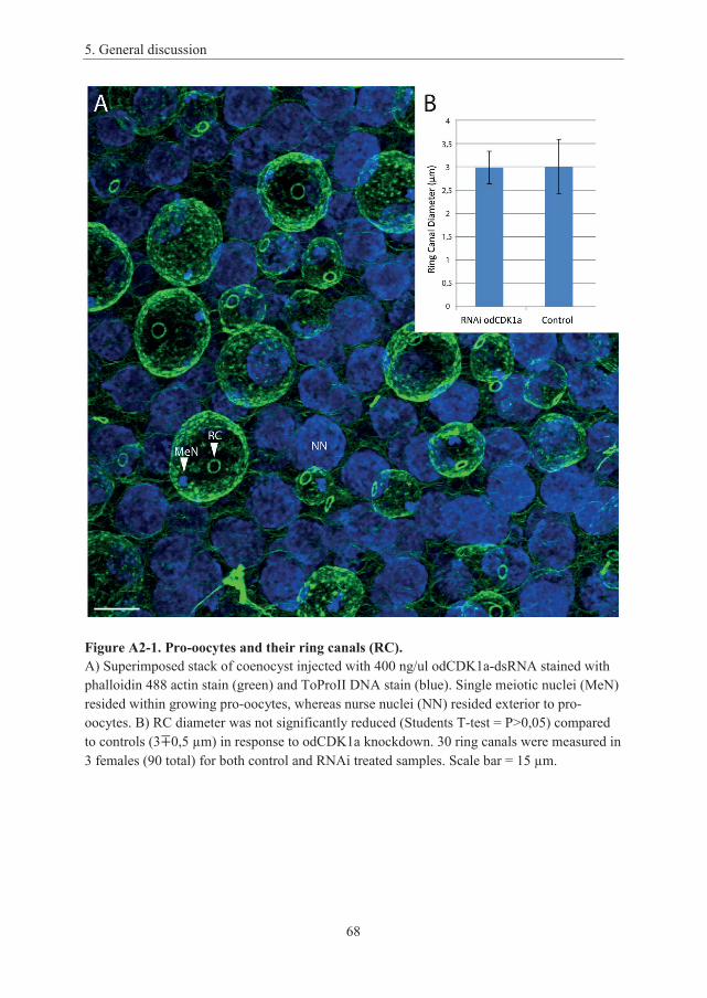
5. General discussion
68
Figure A2-1. Pro-oocytes and their ring canals (RC).A) Superimposed stack of coenocyst injected with 400 ng/ul odCDK1a-dsRNA stained with phalloidin 488 actin stain (green) and ToProII DNA stain (blue). Single meiotic nuclei (MeN) resided within growing pro-oocytes, whereas nurse nuclei (NN) resided exterior to pro-oocytes. B) RC diameter was not significantly reduced (Students T-test = P>0,05) compared to controls (3 0,5 μm) in response to odCDK1a knockdown. 30 ring canals were measured in 3 females (90 total) for both control and RNAi treated samples. Scale bar = 15 μm.

6. Appendix
69
Appendix 3:
Supplementary figures
Figure A3-1. odCyclin Dd in non-selected nuclei. odCyclin Dd translocated selectively to non-selected nuclei, distinguished by negative H3-pS28 staining, following oocyte selection, consistent with behavior of odCDK1 paralogs (Paper II). odCyclin Dd were also observed in endocycling nurse nuclei, consistent with previous observations in endocycling epithelial cells (Campsteijn et al., 2012). As a G1 Cyclin, odCyclin Dd were not expected to perform any particular function during regulation of meiosis and therefore the rationale behind this translocation remains an unsolved mystery. Because the non-selected meiotic nuclei are destined to undergo apoptosis, we speculate that non-selected nuclei may be involved in a mechanism clearing the cytoplasm exterior to growing oocytes for unnecessary cell cycle regulators.
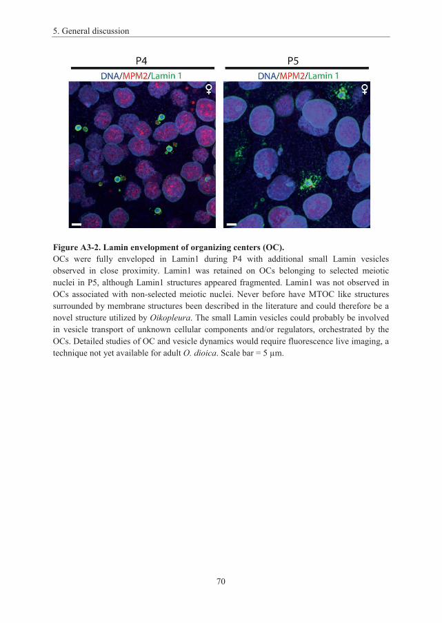
5. General discussion
70
Figure A3-2. Lamin envelopment of organizing centers (OC).OCs were fully enveloped in Lamin1 during P4 with additional small Lamin vesicles observed in close proximity. Lamin1 was retained on OCs belonging to selected meiotic nuclei in P5, although Lamin1 structures appeared fragmented. Lamin1 was not observed in OCs associated with non-selected meiotic nuclei. Never before have MTOC like structures surrounded by membrane structures been described in the literature and could therefore be a novel structure utilized by Oikopleura. The small Lamin vesicles could probably be involved in vesicle transport of unknown cellular components and/or regulators, orchestrated by the OCs. Detailed studies of OC and vesicle dynamics would require fluorescence live imaging, a technique not yet available for adult O. dioica. Scale bar = 5 μm.

7. References
71
7. ReferencesAbe, S., Nagasaka, K., Hirayama, Y., Kozuka-Hata, H., Oyama, M., Aoyagi, Y., Obuse, C., and Hirota, T. (2011). The initial phase of chromosome condensation requires Cdk1-mediated phosphorylation of the CAP-D3 subunit of condensin II. Genes & development 25, 863-874.
Adhikari, D., Zheng, W.J., Shen, Y., Gorre, N., Ning, Y., Halet, G., Kaldis, P., and Liu, K. (2012). Cdk1, but not Cdk2, is the sole Cdk that is essential and sufficient to drive resumption of meiosis in mouse oocytes. Hum Mol Genet 21, 2476-2484.
Adhikary, S., and Eilers, M. (2005). Transcriptional regulation and transformation by MYC proteins. Nat Rev Mol Cell Bio 6, 635-645.
Akamatsu, E., Tanaka, T., and Kato, J. (1998). Transcription factor E2F and cyclin E Cdk2 complex cooperate to induce chromosomal DNA replication in Xenopus oocytes. J Biol Chem 273, 16494-16500.
Assis, R., and Bachtrog, D. (2013). Neofunctionalization of young duplicate genes in Drosophila. P Natl Acad Sci USA 110, 17409-17414.
Barbash, O., Egan, E., Pontano, L.L., Kosak, J., and Diehl, J.A. (2009). Lysine 269 is essential for cyclin D1 ubiquitylation by the SCFFbx4/alpha B-crystallin ligase and subsequent proteasome-dependent degradation. Oncogene 28, 4317-4325.
Bell, S.P., and Dutta, A. (2002). DNA replication in eukaryotic cells. Annual review of biochemistry 71, 333-374.
Bell, S.P., and Stillman, B. (1992). ATP-dependent recognition of eukaryotic origins of DNA replication by a multiprotein complex. Nature 357, 128-134.
Benzeno, S., Lu, F., Guo, M., Barbash, O., Zhang, F., Herman, J.G., Klein, P.S., Rustgi, A., and Diehl, J.A. (2006). Identification of mutations that disrupt phosphorylation-dependent nuclear export of cyclin D1. Oncogene 25, 6291-6303.
Berthet, C., Aleem, E., Coppola, V., Tessarollo, L., and Kaldis, P. (2003). Cdk2 knockout mice are viable. Curr Biol 13, 1775-1785.
Blain, S.W. (2008). Switching cyclin D-Cdk4 kinase activity on and off. Cell Cycle 7, 892-898.
Blais, A., and Dynlacht, B.D. (2004). Hitting their targets: an emerging picture of E2F and cell cycle control. Curr Opin Genet Dev 14, 527-532.
Bouchard, C., Thieke, K., Maier, A., Saffrich, R., Hanley-Hyde, J., Ansorge, W., Reed, S., Sicinski, P., Bartek, J., and Eilers, M. (1999). Direct induction of cyclin D2 by Myc contributes to cell cycle progression and sequestration of p27. Embo Journal 18, 5321-5333.
Bouquet, J.M., Spriet, E., Troedsson, C., Ottera, H., Chourrout, D., and Thompson, E.M. (2009). Culture optimization for the emergent zooplanktonic model organism Oikopleura dioica. Journal of plankton research 31, 359-370.
5. General discussion
72
Boutros, R., Lobjois, V., and Ducommun, B. (2007). CDC25 phosphatases in cancer cells: key players? Good targets? Nat Rev Cancer 7, 495-507.
Bowers, J.L., Randell, J.C., Chen, S., and Bell, S.P. (2004). ATP hydrolysis by ORC catalyzes reiterative Mcm2-7 assembly at a defined origin of replication. Molecular cell 16,967-978.
Calvi, B.R., and Spradling, A.C. (1999). Chorion gene amplification in Drosophila: A model for metazoan origins of DNA replication and S-phase control. Methods 18, 407-417.
Cam, H., and Dynlacht, B.D. (2003). Emerging roles for E2F: Beyond the G1/S transition and DNA replication. Cancer Cell 3, 311-316.
Campsteijn, C., Ovrebo, J.I., Karlsen, B.O., and Thompson, E.M. (2012). Expansion of cyclin D and CDK1 paralogs in Oikopleura dioica, a chordate employing diverse cell cycle variants. Molecular biology and evolution 29, 487-502.
Cao, L., Chen, F., Yang, X., Xu, W., Xie, J., and Yu, L. (2014). Phylogenetic analysis of CDK and cyclin proteins in premetazoan lineages. BMC evolutionary biology 14, 10.
Chauhan, S., Zheng, X., Tan, Y.Y., Tay, B.H., Lim, S., Venkatesh, B., and Kaldis, P. (2012). Evolution of the Cdk-activator Speedy/RINGO in vertebrates. Cellular and molecular life sciences : CMLS 69, 3835-3850.
Cheng, M.G., Olivier, P., Diehl, J.A., Fero, M., Roussel, M.F., Roberts, J.M., and Sherr, C.J. (1999). The p21(Cip1) and p27(Kip1) CDK 'inhibitors' are essential activators of cyclin D-dependent kinases in murine fibroblasts. Embo Journal 18, 1571-1583.
Child, E.S., Hendrychova, T., McCague, K., Futreal, A., Otyepka, M., and Mann, D.J. (2010).A cancer-derived mutation in the PSTAIRE helix of cyclin-dependent kinase 2 alters the stability of cyclin binding. Biochimica et biophysica acta 1803, 858-864.
Churchman, M.L., Brown, M.L., Kato, N., Kirik, V., Hulskamp, M., Inze, D., De Veylder, L., Walker, J.D., Zheng, Z., Oppenheimer, D.G., et al. (2006). SIAMESE, a plant-specific cell cycle regulator, controls endoreplication onset in Arabidopsis thaliana. The Plant cell 18,3145-3157.
Cole, F., Keeney, S., and Jasin, M. (2012). Preaching about the converted: how meiotic gene conversion influences genomic diversity. Ann Ny Acad Sci 1267, 95-102.
Comai, L. (2005). The advantages and disadvantages of being polyploid. Nat Rev Genet 6,836-846.
Corbett, K.D., Yip, C.K., Ee, L.S., Walz, T., Amon, A., and Harrison, S.C. (2010). The Monopolin Complex Crosslinks Kinetochore Components to Regulate Chromosome-Microtubule Attachments. Cell 142, 556-567.
Coward, J., and Harding, A. (2014). Size Does Matter: Why Polyploid Tumor Cells are Critical Drug Targets in the War on Cancer. Frontiers in oncology 4, 123.

7. References
73
Davoli, T., Denchi, E.L., and de Lange, T. (2010). Persistent telomere damage induces bypass of mitosis and tetraploidy. Cell 141, 81-93.
De Bondt, H.L., Rosenblatt, J., Jancarik, J., Jones, H.D., Morgan, D.O., and Kim, S.H. (1993). Crystal structure of cyclin-dependent kinase 2. Nature 363, 595-602.
de Bruin, A., Maiti, B., Jakoi, L., Timmers, C., Buerki, R., and Leone, G. (2003). Identification and characterization of E2F7, a novel mammalian E2F family member capable of blocking cellular proliferation. J Biol Chem 278, 42041-42049.
De Clercq, A., and Inze, D. (2006). Cyclin-dependent kinase inhibitors in yeast, animals, and plants: A functional comparison. Crit Rev Biochem Mol 41, 293-313.
Delsuc, F., Brinkmann, H., Chourrout, D., and Philippe, H. (2006). Tunicates and not cephalochordates are the closest living relatives of vertebrates. Nature 439, 965-968.
Denoeud, F., Henriet, S., Mungpakdee, S., Aury, J.M., Da Silva, C., Brinkmann, H., Mikhaleva, J., Olsen, L.C., Jubin, C., Canestro, C., et al. (2010). Plasticity of animal genome architecture unmasked by rapid evolution of a pelagic tunicate. Science 330, 1381-1385.
Diril, M.K., Ratnacaram, C.K., Padmakumar, V.C., Du, T.H., Wasser, M., Coppola, V., Tessarollo, L., and Kaldis, P. (2012). Cyclin-dependent kinase 1 (Cdk1) is essential for cell division and suppression of DNA re-replication but not for liver regeneration. Proceedings of the National Academy of Sciences of the United States of America 109, 3826-3831.
Donovan, P., Cato, K., Legaie, R., Jayalath, R., Olsson, G., Hall, B., Olson, S., Boros, S., Reynolds, B.A., and Harding, A. (2014). Hyperdiploid tumor cells increase phenotypic heterogeneity within Glioblastoma tumors. Mol Biosyst 10, 741-758.
Doonan, J.H., and Kitsios, G. (2009). Functional evolution of cyclin-dependent kinases. Molecular biotechnology 42, 14-29.
Dutta, A. (2007). Chaotic license for genetic instability and cancer. Nat Genet 39, 10-11.
Dyson, N. (1998). The regulation of E2F by pRB-family proteins. Genes & development 12,2245-2262.
Echalier, A., Endicott, J.A., and Noble, M.E.M. (2010). Recent developments in cyclin-dependent kinase biochemical and structural studies. Bba-Proteins Proteom 1804, 511-519.
Edgar, B.A., and Orr-Weaver, T.L. (2001). Endoreplication cell cycles: more for less. Cell105, 297-306.
Edgar, B.A., Zielke, N., and Gutierrez, C. (2014). Endocycles: a recurrent evolutionary innovation for post-mitotic cell growth. Nature reviews Molecular cell biology 15, 197-210.
Edvardsen, R.B., Seo, H.C., Jensen, M.F., Mialon, A., Mikhaleva, J., Bjordal, M., Cartry, J., Reinhardt, R., Weissenbach, J., Wincker, P., et al. (2005). Remodelling of the homeobox gene complement in the tunicate Oikopleura dioica. Curr Biol 15, R12-13.

5. General discussion
74
Evangelista, M., Blundell, K., Longtine, M.S., Chow, C.J., Adames, N., Pringle, J.R., Peter, M., and Boone, C. (1997). Bni1p, a yeast formin linking cdc42p and the actin cytoskeleton during polarized morphogenesis. Science 276, 118-122.
Fenaux, R. (1985). Rhythm of Secretion of Oikopleurids Houses. B Mar Sci 37, 498-503.
Fox, D.T., and Duronio, R.J. (2013). Endoreplication and polyploidy: insights into development and disease. Development 140, 3-12.
Fox, D.T., Gall, J.G., and Spradling, A.C. (2010). Error-prone polyploid mitosis during normal Drosophila development. Genes & development 24, 2294-2302.
Fujii, S., Nishio, T., and Nishida, H. (2008). Cleavage pattern, gastrulation, and neurulation in the appendicularian, Oikopleura dioica. Development genes and evolution 218, 69-79.
Ganot, P., Bouquet, J.M., Kallesoe, T., and Thompson, E.M. (2007a). The Oikopleura coenocyst, a unique chordate germ cell permitting rapid, extensive modulation of oocyte production. Developmental biology 302, 591-600.
Ganot, P., Kallesoe, T., and Thompson, E.M. (2007b). The cytoskeleton organizes germ nuclei with divergent fates and asynchronous cycles in a common cytoplasm during oogenesis in the chordate Oikopleura. Developmental biology 302, 577-590.
Ganot, P., Moosmann-Schulmeister, A., and Thompson, E.M. (2008). Oocyte selection is concurrent with meiosis resumption in the coenocystic oogenesis of Oikopleura. Developmental biology 324, 266-276.
Ganot, P., and Thompson, E.M. (2002). Patterning through differential endoreduplication in epithelial organogenesis of the chordate, Oikopleura dioica. Developmental biology 252, 59-71.
Gavet, O., and Pines, J. (2010). Progressive Activation of CyclinB1-Cdk1 Coordinates Entry to Mitosis. Developmental cell 18, 533-543.
Gong, D.Q., Pomerening, J.R., Myers, J.W., Gustavsson, C., Jones, J.T., Hahn, A.T., Meyer, T., and Ferrell, J.E. (2007). Cyclin A2 regulates nuclear-envelope breakdown and the nuclear accumulation of cyclin B1. Curr Biol 17, 85-91.
Gu, Z., Nicolae, D., Lu, H.H., and Li, W.H. (2002). Rapid divergence in expression between duplicate genes inferred from microarray data. Trends Genet 18, 609-613.
Haccard, O., and Jessus, C. (2006). Redundant pathways for Cdc2 activation in Xenopus oocyte: either cyclin B or Mos synthesis. EMBO reports 7, 321-325.
Haglund, K., Nezis, I.P., and Stenmark, H. (2011). Structure and functions of stable intercellular bridges formed by incomplete cytokinesis during development. Communicative & integrative biology 4, 1-9.

7. References
75
Hammond, M.P., and Laird, C.D. (1985). Chromosome structure and DNA replication in nurse and follicle cells of Drosophila melanogaster. Chromosoma 91, 267-278.
Han, S.J., Chen, R., Paronetto, M.P., and Conti, M. (2005). Wee1B is an oocyte-specifickinase involved in the control of meiotic arrest in the mouse. Curr Biol 15, 1670-1676.
Hermeking, H., Rago, C., Schuhmacher, M., Li, Q., Barrett, J.F., Obaya, A.J., O'Connell, B.C., Mateyak, M.K., Tam, W., Kohlhuber, F., et al. (2000). Identification of CDK4 as a target of c-MYC. P Natl Acad Sci USA 97, 2229-2234.
Hershko, A. (1999). Mechanisms and regulation of the degradation of cyclin B. Philos T Roy Soc B 354, 1571-1575.
Hirayama, T., Imajuku, Y., Anai, T., Matsui, M., and Oka, A. (1991). Identification of two cell-cycle-controlling cdc2 gene homologs in Arabidopsis thaliana. Gene 105, 159-165.
Hochegger, H., Klotzbucher, A., Kirk, J., Howell, M., le Guellec, K., Fletcher, K., Duncan, T., Sohail, M., and Hunt, T. (2001). New B-type cyclin synthesis is required between melosis I and II during Xenopus oocyte maturation. Development 128, 3795-3807.
Holland, A.J., and Cleveland, D.W. (2009). Boveri revisited: chromosomal instability, aneuploidy and tumorigenesis. Nat Rev Mol Cell Bio 10, 478-487.
Homer, H. (2013). The APC/C in female mammalian meiosis I. Reproduction 146, R61-71.
Hong, A., Lee-Kong, S., Iida, T., Sugimura, I., and Lilly, M.A. (2003). The p27(cip/kip) ortholog dacapo maintains the Drosophila oocyte in prophase of meiosis I. Development 130,1235-1242.
Hopfner, K.P. (2003). Chromosome cohesion: Closing time. Curr Biol 13, R866-R868.
Hosp, J., Sagane, Y., Danks, G., and Thompson, E.M. (2012). The evolving proteome of a complex extracellular matrix, the Oikopleura house. PloS one 7, e40172.
Hu, D., and Cross, J.C. (2010). Development and function of trophoblast giant cells in the rodent placenta. The International journal of developmental biology 54, 341-354.
Iwabuchi, M., Ohsumi, K., Yamamoto, T.M., Sawada, W., and Kishimoto, T. (2000). Residual Cdc2 activity remaining at meiosis I exit is essential for meiotic M-M transition in Xenopus oocyte extracts. Embo Journal 19, 4513-4523.
Jackman, M., Lindon, C., Nigg, E.A., and Pines, J. (2003). Active cyclin B1-Cdk1 first appears on centrosomes in prophase. Nature cell biology 5, 143-148.
Jacobs, H.W., Knoblich, J.A., and Lehner, C.F. (1998). Drosophila Cyclin B3 is required for female fertility and is dispensable for mitosis like Cyclin B. Genes & development 12, 3741-3751.

5. General discussion
76
Jeffrey, P.D., Russo, A.A., Polyak, K., Gibbs, E., Hurwitz, J., Massague, J., and Pavletich, N.P. (1995). Mechanism of CDK activation revealed by the structure of a cyclinA-CDK2 complex. Nature 376, 313-320.
Jones, K.T. (2004). Turning it on and off: M-phase promoting factor during meiotic maturation and fertilization. Mol Hum Reprod 10, 1-5.
Jones, K.T., and Lane, S.I. (2013). Molecular causes of aneuploidy in mammalian eggs. Development 140, 3719-3730.
Kaessmann, H. (2010). Origins, evolution, and phenotypic impact of new genes. Genome research 20, 1313-1326.
Karaiskou, A., Jessus, C., Brassac, T., and Ozon, R. (1999). Phosphatase 2A and Polo kinase, two antagonistic regulators of Cdc25 activation and MPF auto-amplification. J Cell Sci 112,3747-3756.
Kato, J., Matsushime, H., Hiebert, S.W., Ewen, M.E., and Sherr, C.J. (1993). Direct binding of cyclin D to the retinoblastoma gene product (pRb) and pRb phosphorylation by the cyclin D-dependent kinase CDK4. Genes & development 7, 331-342.
Kimura, K., Cuvier, O., and Hirano, T. (2001). Chromosome condensation by a human condensin complex in Xenopus egg extracts. J Biol Chem 276, 5417-5420.
Kishimoto, T. (2003). Cell-cycle control during meiotic maturation. Curr Opin Cell Biol 15,654-663.Klutstein, M., and Cooper, J.P. (2014). The Chromosomal Courtship Dance-homolog pairing in early meiosis. Curr Opin Cell Biol 26, 123-131.
Kugler, J.E., Kerner, P., Bouquet, J.M., Jiang, D., and Di Gregorio, A. (2011). Evolutionary changes in the notochord genetic toolkit: a comparative analysis of notochord genes in the ascidian Ciona and the larvacean Oikopleura. BMC evolutionary biology 11, 21.
Kumano, G. (2012). Polarizing animal cells via mRNA localization in oogenesis and early development. Development, growth & differentiation 54, 1-18.
LaBaer, J., Garrett, M.D., Stevenson, L.F., Slingerland, J.M., Sandhu, C., Chou, H.S., Fattaey, A., and Harlow, E. (1997). New functional activities for the p21 family of CDK inhibitors. Genes & development 11, 847-862.
Labbe, J.C., Capony, J.P., Caput, D., Cavadore, J.C., Derancourt, J., Kaghad, M., Lelias, J.M., Picard, A., and Doree, M. (1989). MPF from starfish oocytes at first meiotic metaphase is a heterodimer containing one molecule of cdc2 and one molecule of cyclin B. The EMBO journal 8, 3053-3058.
Lammens, T., Boudolf, V., Kheibarshekan, L., Zalmas, L.P., Gaamouche, T., Maes, S., Vanstraelen, M., Kondorosi, E., La Thangue, N.B., Govaerts, W., et al. (2008). Atypical E2F activity restrains APC/CCCS52A2 function obligatory for endocycle onset. P Natl Acad Sci USA 105, 14721-14726.
7. References
77
Lazzerini Denchi, E., Celli, G., and de Lange, T. (2006). Hepatocytes with extensive telomere deprotection and fusion remain viable and regenerate liver mass through endoreduplication. Genes & development 20, 2648-2653.
Lee, H.O., Davidson, J.M., and Duronio, R.J. (2009). Endoreplication: polyploidy with purpose. Genes & development 23, 2461-2477.
Lees, J.A., Saito, M., Vidal, M., Valentine, M., Look, T., Harlow, E., Dyson, N., and Helin, K. (1993). The Retinoblastoma Protein Binds to a Family of E2f Transcription Factors. Mol Cell Biol 13, 7813-7825.
Li, A., and Blow, J.J. (2004). Non-proteolytic inactivation of geminin requires CDK-dependent ubiquitination. Nature cell biology 6, 260-267.
Li, W.H., Yang, J., and Gu, X. (2005). Expression divergence between duplicate genes. Trends Genet 21, 602-607.
Lincoln, A.J., Wickramasinghe, D., Stein, P., Schultz, R.M., Palko, M.E., De Miguel, M.P., Tessarollo, L., and Donovan, P.J. (2002). Cdc25b phosphatase is required for resumption of meiosis during oocyte maturation. Nat Genet 30, 446-449.
Lindqvist, A., Kallstrom, H., Lundgren, A., Barsoum, E., and Rosenthal, C.K. (2005). Cdc25B cooperates with Cdc25A to induce mitosis but has a unique role in activating cyclin B1-Cdk1 at the centrosome. J Cell Biol 171, 35-45.
Listovsky, T., Zor, A., Laronne, A., and Brandeis, M. (2000). Cdk1 is essential for mammalian cyclosome/APC regulation. Exp Cell Res 255, 184-191.
Loog, M., and Morgan, D.O. (2005). Cyclin specificity in the phosphorylation of cyclin-dependent kinase substrates. Nature 434, 104-108.
Lozano, E., Saez, A.G., Flemming, A.J., Cunha, A., and Leroi, A.M. (2006). Regulation of growth by ploidy in Caenorhabditis elegans. Curr Biol 16, 493-498.
Lu, F., Gladden, A.B., and Diehl, J.A. (2003). An alternatively spliced cyclin D1 isoform, cyclin D1b, is a nuclear oncogene. Cancer research 63, 7056-7061.
Luksza, M., Queguigner, I., Verlhac, M.H., and Brunet, S. (2013). Rebuilding MTOCs upon centriole loss during mouse oogenesis. Developmental biology 382, 48-56.
Ma, H.T., and Poon, R.Y.C. (2011). How protein kinases co-ordinate mitosis in animal cells. Biochem J 435, 17-31.
Mahowald, A.P., and Strassheim, J.M. (1970). Intercellular migration of centrioles in the germarium of Drosophila melanogaster. An electron microscopic study. J Cell Biol 45, 306-320.
Mailand, N., Podtelejnikov, A.V., Groth, A., Mann, M., Bartek, J., and Lukas, J. (2002). Regulation of G(2)/M events by Cdc25A through phosphorylation-dependent modulation of its stability. Embo Journal 21, 5911-5920.

5. General discussion
78
Malumbres, M., and Barbacid, M. (2001). To cycle or not to cycle: a critical decision in cancer. Nat Rev Cancer 1, 222-231.
Malumbres, M., and Barbacid, M. (2005). Mammalian cyclin-dependent kinases. Trends Biochem Sci 30, 630-641.
Malumbres, M., and Barbacid, M. (2009). Cell cycle, CDKs and cancer: a changing paradigm. Nat Rev Cancer 9, 153-166.
Malumbres, M., Sotillo, R., Santamaria, D., Galan, J., Cerezo, A., Ortega, S., Dubus, P., and Barbacid, M. (2004). Mammalian cells cycle without the D-type cyclin-elependent kinases Cdk4 and Cdk6. Cell 118, 493-504.
Maqbool, S.B., Mehrotra, S., Kolpakas, A., Durden, C., Zhang, B., Zhong, H., and Calvi, B.R. (2010). Dampened activity of E2F1-DP and Myb-MuvB transcription factors in Drosophila endocycling cells. J Cell Sci 123, 4095-4106.
Marston, A.L., and Amon, A. (2004). Meiosis: Cell-cycle controls shuffle and deal. Nat Rev Mol Cell Bio 5, 983-997.
McClendon, A.K., Dean, J.L., Ertel, A., Fu, Z.Y., Rivadeneira, D.B., Reed, C.A., Bourgo, R.J., Witkiewicz, A., Addya, S., Mayhew, C.N., et al. (2011). RB and p53 Cooperate to Prevent Liver Tumorigenesis in Response to Tissue Damage. Gastroenterology 141, 1439-1450.
McCusker, D., Royou, A., Velours, C., and Kellogg, D. (2012). Cdk1-dependent control of membrane-trafficking dynamics. Molecular biology of the cell 23, 3336-3347.
McGarry, T.J., and Kirschner, M.W. (1998). Geminin, an inhibitor of DNA replication, is degraded during mitosis. Cell 93, 1043-1053.
Mehrotra, S., Maqbool, S.B., Kolpakas, A., Murnen, K., and Calvi, B.R. (2008). Endocycling cells do not apoptose in response to DNA rereplication genotoxic stress. Genes & development 22, 3158-3171.
Menant, A., and Karess, R.E. (2012). Inducing "cytokinesis" without mitosis in unfertilized Drosophila eggs. Cell Cycle 11, 2856-2863.
Mendenhall, M.D., and Hodge, A.E. (1998). Regulation of Cdc28 cyclin-dependent protein kinase activity during the cell cycle of the yeast Saccharomyces cerevisiae. Microbiology and molecular biology reviews : MMBR 62, 1191-1243.
Meyer, C.A., Jacobs, H.W., Datar, S.A., Du, W., Edgar, B.A., and Lehner, C.F. (2000). Drosophila Cdk4 is required for normal growth and is dispensable for cell cycle progression. Embo Journal 19, 4533-4542.
Mikeladze-Dvali, T., von Tobel, L., Strnad, P., Knott, G., Leonhardt, H., Schermelleh, L., and Gonczy, P. (2012). Analysis of centriole elimination during C. elegans oogenesis. Development 139, 1670-1679.
7. References
79
Minn, A.J., Boise, L.H., and Thompson, C.B. (1996). Expression of Bcl-xL and loss of p53 can cooperate to overcome a cell cycle checkpoint induced by mitotic spindle damage. Genes & development 10, 2621-2631.
Moreno-Bueno, G., Rodriguez-Perales, S., Sanchez-Estevez, C., Hardisson, D., Sarrio, D., Prat, J., Cigudosa, J.C., Matias-Guiu, X., and Palacios, J. (2003). Cyclin D1 gene (CCND1)mutations in endometrial cancer. Oncogene 22, 6115-6118.
Morgan, D.O. (1997). Cyclin-dependent kinases: engines, clocks, and microprocessors. Annual review of cell and developmental biology 13, 261-291.
Morgan, D.O. (2007). The cell cycle : principles of control (London Sunderland, MA: Published by New Science Press in association with Oxford University Press ; Distributed inside North America by Sinauer Associates, Publishers).
Nakajo, N., Yoshitome, S., Iwashita, J., Iida, M., Uto, K., Ueno, S., Okamoto, K., and Sagata, N. (2000). Absence of Wee1 ensures the meiotic cell cycle in Xenopus oocytes. Genes & development 14, 328-338.
Nasmyth, K. (2002). Segregating sister genomes: The molecular biology of chromosome separation. Science 297, 559-565.
Nern, A., and Arkowitz, R.A. (2000). Nucleocytoplasmic shuttling of the Cdc42p exchange factor Cdc24p. J Cell Biol 148, 1115-1122.
Nishida, H. (2008). Development of the appendicularian Oikopleura dioica: culture, genome, and cell lineages. Development, growth & differentiation 50 Suppl 1, S239-256.
Nowack, M.K., Harashima, H., Dissmeyer, N., Zhao, X., Bouyer, D., Weimer, A.K., De Winter, F., Yang, F., and Schnittger, A. (2012). Genetic framework of cyclin-dependent kinase function in Arabidopsis. Developmental cell 22, 1030-1040.
Nurse, P., and Bissett, Y. (1981). Gene required in G1 for commitment to cell cycle and in G2 for control of mitosis in fission yeast. Nature 292, 558-560.
Ohtani, K., DeGregori, J., and Nevins, J.R. (1995). Regulation of the cyclin E gene by transcription factor E2F1. P Natl Acad Sci USA 92, 12146-12150.
Okano-Uchida, T., Okumura, E., Iwashita, M., Yoshida, H., Tachibana, K., and Kishimoto, T. (2003). Distinct regulators for Plk1 activation in starfish meiotic and early embryonic cycles. Embo Journal 22, 5633-5642.
Ong, S., Foote, C., and Tan, C. (2010). Mutations of DMYPT cause over constriction of contractile rings and ring canals during Drosophila germline cyst formation. Developmental biology 346, 161-169.
Ong, S., and Tan, C. (2010). Germline cyst formation and incomplete cytokinesis during Drosophila melanogaster oogenesis. Developmental biology 337, 84-98.
5. General discussion
80
Pepling, M.E., de Cuevas, M., and Spradling, A.C. (1999). Germline cysts: a conserved phase of germ cell development? Trends in cell biology 9, 257-262.
Piggott, J.R., Rai, R., and Carter, B.L. (1982). A bifunctional gene product involved in two phases of the yeast cell cycle. Nature 298, 391-393.
Pirino, G., Wescott, M.P., and Donovan, P.J. (2009). Protein kinase A regulates resumption of meiosis by phosphorylation of Cdc25B in mammalian oocytes. Cell Cycle 8, 665-670.
Porceddu, A., De Veylder, L., Hayles, J., Van Montagu, M., Inze, D., and Mironov, V. (1999). Mutational analysis of two Arabidopsis thaliana cyclin-dependent kinases in fission yeast. Febs Lett 446, 182-188.
Randell, J.C., Bowers, J.L., Rodriguez, H.K., and Bell, S.P. (2006). Sequential ATP hydrolysis by Cdc6 and ORC directs loading of the Mcm2-7 helicase. Molecular cell 21, 29-39.
Rane, S.G., Dubus, P., Mettus, R.V., Galbreath, E.J., Boden, G., Reddy, E.P., and Barbacid, M. (1999). Loss of Cdk4 expression causes insulin-deficient diabetes and Cdk4 activation results in beta-islet cell hyperplasia. Nat Genet 22, 44-52.
Reed, C.A., Mayhew, C.N., McClendon, A.K., Yang, X., Witkiewicz, A., and Knudsen, E.S. (2009). RB has a critical role in mediating the in vivo checkpoint response, mitigating secondary DNA damage and suppressing liver tumorigenesis initiated by aflatoxin B1. Oncogene 28, 4434-4443.
Reis, A., Chang, H.Y., Levasseur, M., and Jones, K.T. (2006). APC(cdh1) activity in mouse oocytes prevents entry into the first meiotic division. Nature cell biology 8, 539-540.
Roberts, J.M. (1999). Evolving Ideas about Cyclins. Cell 98, 129-132.
Roeder, A.H., Chickarmane, V., Cunha, A., Obara, B., Manjunath, B.S., and Meyerowitz, E.M. (2010). Variability in the control of cell division underlies sepal epidermal patterning in Arabidopsis thaliana. PLoS biology 8, e1000367.
Rubin, S.M., Gall, A.L., Zheng, N., and Pavletich, N.P. (2005). Structure of the RbC-terminal domain bound to E2F1-DP1: A mechanism for phosphorylation-induced E2F release. Cell123, 1093-1106.
Sagata, N. (1996). Meiotic metaphase arrest in animal oocytes: its mechanisms and biological significance. Trends in cell biology 6, 22-28.
Santamaria, D., Barriere, C., Cerqueira, A., Hunt, S., Tardy, C., Newton, K., Caceres, J.F., Dubus, P., Malumbres, M., and Barbacid, M. (2007). Cdk1 is sufficient to drive the mammalian cell cycle. Nature 448, 811-U818.
Scherthan, H. (2001). A bouquet makes ends meet. Nature reviews Molecular cell biology 2,621-627.

7. References
81
Schulze-Gahmen, U., De Bondt, H.L., and Kim, S.H. (1996). High-resolution crystal structures of human cyclin-dependent kinase 2 with and without ATP: bound waters and natural ligand as guides for inhibitor design. Journal of medicinal chemistry 39, 4540-4546.
Seo, H.C., Kube, M., Edvardsen, R.B., Jensen, M.F., Beck, A., Spriet, E., Gorsky, G., Thompson, E.M., Lehrach, H., Reinhardt, R., et al. (2001). Miniature genome in the marine chordate Oikopleura dioica. Science 294, 2506.
Sherr, C.J. (1993). Mammalian G1 cyclins. Cell 73, 1059-1065.
Sherr, C.J. (1995). D-type cyclins. Trends in Biochemical Sciences 20, 187-190.
Sherr, C.J., and Roberts, J.M. (1999). CDK inhibitors: positive and negative regulators of G(1)-phase progression. Genes & development 13, 1501-1512.
Shimada, Y., Gulli, M.P., and Peter, M. (2000). Nuclear sequestration of the exchange factor Cdc24 by Far1 regulates cell polarity during yeast mating. Nature cell biology 2, 117-124.
Sigrist, S.J., and Lehner, C.F. (1997). Drosophila fizzy-related down-regulates mitotic cyclins and is required for cell proliferation arrest and entry into endocycles. Cell 90, 671-681.
Sodergren, E., Weinstock, G.M., Davidson, E.H., Cameron, R.A., Gibbs, R.A., Angerer, R.C., Angerer, L.M., Arnone, M.I., Burgess, D.R., Burke, R.D., et al. (2006). The genome of the sea urchin Strongylocentrotus purpuratus. Science 314, 941-952.
Spada, F., Steen, H., Troedsson, C., Kallesoe, T., Spriet, E., Mann, M., and Thompson, E.M. (2001). Molecular patterning of the oikoplastic epithelium of the larvacean tunicate Oikopleura dioica. J Biol Chem 276, 20624-20632.
Storchova, Z., and Pellman, D. (2004). From polyploidy to aneuploidy, genome instability and cancer. Nat Rev Mol Cell Bio 5, 45-54.
Sugimoto-Shirasu, K., and Roberts, K. (2003). "Big it up": endoreduplication and cell-size control in plants. Curr Opin Plant Biol 6, 544-553.
Symeonidou, I.E., Taraviras, S., and Lygerou, Z. (2012). Control over DNA replication in time and space. Febs Lett 586, 2803-2812.
Szollosi, D., Calarco, P., and Donahue, R.P. (1972). Absence of centrioles in the first and second meiotic spindles of mouse oocytes. J Cell Sci 11, 521-541.
Taieb, F.E., Gross, S.D., Lewellyn, A.L., and Maller, J.L. (2001). Activation of the anaphase-promoting complex and degradation of cyclin B is not required for progression from Meiosis I to II in Xenopus oocytes. Curr Biol 11, 508-513.
Takahashi, N., Kajihara, T., Okamura, C., Kim, Y., Katagiri, Y., Okushima, Y., Matsunaga, S., Hwang, I., and Umeda, M. (2013). Cytokinins control endocycle onset by promoting the expression of an APC/C activator in Arabidopsis roots. Curr Biol 23, 1812-1817.
5. General discussion
82
Tarailo-Graovac, M., Wang, J., Tu, D., Baillie, D.L., Rose, A.M., and Chen, N. (2010). Duplication of cyb-3 (cyclin B3) suppresses sterility in the absence of mdf-1/MAD1 spindle assembly checkpoint component in Caenorhabditis elegans. Cell Cycle 9, 4858-4865.
Troedsson, C., Bouquet, J.M., Aksnes, D.L., and Thompson, E.M. (2002). Resource allocation between somatic growth and reproductive output in the pelagic chordate Oikopleura dioica allows opportunistic response to nutritional variation. Mar Ecol Prog Ser243, 83-91.
Troedsson, C., Ganot, P., Bouquet, J.M., Aksnes, D.L., and Thompson, E.M. (2007). Endostyle cell recruitment as a frame of reference for development and growth in the Urochordate Oikopleura dioica. The Biological bulletin 213, 325-334.
Tsutsui, T., Hesabi, B., Moons, D.S., Pandolfi, P.P., Hansel, K.S., Koff, A., and Kiyokawa, H. (1999). Targeted disruption of CDK4 delays cell cycle entry with enhanced p27(Kip1) activity. Mol Cell Biol 19, 7011-7019.
Ullah, Z., de Renty, C., and DePamphilis, M.L. (2011). Checkpoint kinase 1 prevents cell cycle exit linked to terminal cell differentiation. Mol Cell Biol 31, 4129-4143.
Ullah, Z., Lee, C.Y., Lilly, M.A., and DePamphilis, M.L. (2009). Developmentally programmed endoreduplication in animals. Cell Cycle 8, 1501-1509.
van den Heuvel, S. (2005). Cell-cycle regulation. WormBook : the online review of C elegans biology, 1-16.
van der Voet, M., Lorson, M.A., Srinivasan, D.G., Bennett, K.L., and van den Heuvel, S. (2009). C. elegans mitotic cyclins have distinct as well as overlapping functions in chromosome segregation. Cell Cycle 8, 4091-4102.
Van Leene, J., Hollunder, J., Eeckhout, D., Persiau, G., Van De Slijke, E., Stals, H., Van Isterdael, G., Verkest, A., Neirynck, S., Buffel, Y., et al. (2010). Targeted interactomics reveals a complex core cell cycle machinery in Arabidopsis thaliana. Molecular systems biology 6, 397.
Vickaryous, M.K., and Hall, B.K. (2006). Human cell type diversity, evolution, development, and classification with special reference to cells derived from the neural crest. Biological reviews of the Cambridge Philosophical Society 81, 425-455.
Vodermaier, H.C. (2004). APC/C and SCF: Controlling each other and the cell cycle. Curr Biol 14, R787-R796.
Von Stetina, J.R., and Orr-Weaver, T.L. (2011). Developmental Control of Oocyte Maturation and Egg Activation in Metazoan Models. Cold Spring Harbor perspectives in biology 3.
Von Stetina, J.R., Tranguch, S., Dey, S.K., Lee, L.A., Cha, B., and Drummond-Barbosa, D. (20 -Endosulfine is a conserved protein required for oocyte meiotic maturation in Drosophila. Development 135, 3697-3706.

7. References
83
Walker, J.D., Oppenheimer, D.G., Concienne, J., and Larkin, J.C. (2000). SIAMESE, a gene controlling the endoreduplication cell cycle in Arabidopsis thaliana trichomes. Development127, 3931-3940.
Weaver, B.A., and Cleveland, D.W. (2008). The aneuploidy paradox in cell growth and tumorigenesis. Cancer Cell 14, 431-433.
Weinberg, R.A. (1995). The Retinoblastoma Protein and Cell-Cycle Control. Cell 81, 323-330.
Whitaker, M. (1996). Control of meiotic arrest. Reviews of reproduction 1, 127-135.
Whitman, W.B., Coleman, D.C., and Wiebe, W.J. (1998). Prokaryotes: the unseen majority. P Natl Acad Sci USA 95, 6578-6583.
Yamamoto, S., Bayat, V., Bellen, H.J., and Tan, C. (2013). Protein phosphatase 1ss limits ring canal constriction during Drosophila germline cyst formation. PloS one 8, e70502.
Yamashiro, S., Yamakita, Y., Totsukawa, G., Goto, H., Kaibuchi, K., Ito, M., Hartshorne, D.J., and Matsumura, F. (2008). Myosin phosphatase-targeting subunit 1 regulates mitosis by antagonizing polo-like kinase 1. Developmental cell 14, 787-797.
Yin, L.H., Gater, S.T., and Karrer, K.M. (2010). A Developmentally Regulated Gene, ASI2, Is Required for Endocycling in the Macronuclear Anlagen of Tetrahymena. Eukaryot Cell 9,1343-1353.
Zhou, B.B.S., and Elledge, S.J. (2000). The DNA damage response: putting checkpoints in perspective. Nature 408, 433-439.
Zielke, N., Edgar, B.A., and DePamphilis, M.L. (2013). Endoreplication. Cold Spring Harbor perspectives in biology 5, a012948.
Zielke, N., Kim, K.J., Tran, V., Shibutani, S.T., Bravo, M.J., Nagarajan, S., van Straaten, M., Woods, B., von Dassow, G., Rottig, C., et al. (2011). Control of Drosophila endocycles by E2F and CRL4(CDT2). Nature 480, 123-127.
Zielke, N., Querings, S., Rottig, C., Lehner, C., and Sprenger, F. (2008). The anaphase-promoting complex/cyclosome (APC/C) is required for rereplication control in endoreplication cycles. Genes & development 22, 1690-1703.


![FOH RI D qppMlR]y - Home - Raising Hooks/LIH &\FOH RI D qppMlR]y &D WHU SL OOD U % X W W H U I O \ (JJV & K U \ V D O L V URM yI]M $R qppMlR]y &$ Eg^ci # WZhi dc XVgYhidX`$ '$ 8dadg$](https://static.fdocuments.net/doc/165x107/5f34527579ef5d020e6ab326/foh-ri-d-qppmlry-home-raising-hooks-lih-foh-ri-d-qppmlry-d-whu.jpg)


![2019-04-22 REBA presentation re Smyth case combined ... · wkh uhjxodwlrq dq xqfrqvwlwxwlrqdo wdnlqj ´ 0rvnrzy &rpp¶uri 'hs¶w ri (qywo 0jpw 0dvv ³:kdw pd\ eh fkdudfwhul]hg dv](https://static.fdocuments.net/doc/165x107/5e5a66991f8d4b39b93592a2/2019-04-22-reba-presentation-re-smyth-case-combined-wkh-uhjxodwlrq-dq-xqfrqvwlwxwlrqdo.jpg)


![5HJXODWLRQV RI %XON &DUULHUV...,1752'8&7,21 7KH ,QWHUQDWLRQDO 0DULWLPH 2UJDQL]DWLRQ ,02 KDV DGRSWHG QHZ UHJXODWLRQ RQ HQHUJ\ HIILFLHQF\ IRU VKLSV 0(3& D DFFRUGLQJ WR ZKLFK WKH ,QWHUQDWLRQDO](https://static.fdocuments.net/doc/165x107/60c65a9310f866251012c268/5hjxodwlrqv-ri-xon-duulhuv-17528721-7kh-qwhuqdwlrqdo-0dulwlph.jpg)
