Focus on Chronic Variable Immunodeficiency for Primary Care … · 2019-12-05 · CRITICAL CARE (J...
15
CRITICAL CARE (J GIULIANO AND M BIGHAM, SECTION EDITORS) Focus on Chronic Variable Immunodeficiency for Primary Care Practitioners, the Gatekeepers to Optimal Health Outcomes for Primary Immunodeficiency Syndromes William A. Gerber 1,2 # The Author(s) 2019 Abstract Purpose of Review This review sought to assess the extent and causes of suboptimal healthcare outcomes for chronic variable immunodeficiency (CVID). Recent Findings Significant improvements in diagnostic technology and treatment protocols over time were found, leading to reduced morbidity and mortality for those accessing therapies. Treatments continue to be largely non-curative with financing (mainly insurance coverage) an obstacle. Symptom recognition by primary care practitioners (PCP) remains a gating factor to treatment and a widespread and persistent barrier to optimal health outcomes. Summary CVID is a subtype of primary immunodeficiency (PIDD) associated with under-diagnosis. It has emerged as a health issue more prevalent than historically known. No symptom-recognition framework for early detection of CVID has been generally accepted; those proposed for primary immunodeficiencies have shown low sensitivity, low specificity or both. Positive trends in cases diagnosed have been aided by awareness campaigns and international collaborations. However, treat- ments for CVID will not realize full potential without effective, accepted frameworks for timely identification in the clinic. Keywords CVID . Immunoglobulin . Infection . Diagnosis . Treatment . Diagnosis delay Abbreviations Ig Immunoglobulin IgRT Immunoglobulin replacement therapy PCP Primary care practitioner (here, including emergency room practitioners) PIDD Primary immunodeficiency SCID Severe combined immunodeficiency Introduction The history of scientific understanding of primary immunode- ficiency (PIDD) is relatively short but emerging rapidly. It was not until 1952 that primary immunodeficiency was first rec- ognized as a disease. Before the widespread use of antibiotics, mortality from infections was common, rarely prompting au- topsies [1, 2]. It was only afterwards that chronic or recurrent infections became distinguishable, encouraging further scien- tific investigation [1]. In 1970, the World Health Organization convened the first international committee tasked with forming consensus around PIDD class and subclass defini- tions, best practices for diagnosis and treatment, and establish- ment of patient registries [3]. The committee, now under the International Union of Immunological Societies and named the Inborn Errors of Immunity Committee, meets and pub- lishes an updated, growing classification list every 2 years. From the initial broad “humoral” and “cellular” descrip- tions prior to 1970, 14 distinct subclasses resulted from the first World Health Organization meeting [3]. As an example, it was at this 1970 meeting that the term chronic variable immu- nodeficiency (CVID) was first described as a hypogammaglobulinemia “diagnosis of exclusion” that should not be made before 4 years of age [4•]. By 2003, the number of identified primary immunodeficiencies grew to 80, then to 240 by 2015. A 2017 Inborn Errors of Immunity This article is part of the Topical Collection on Critical Care * William A. Gerber [email protected] 1 Milken Institute School of Public Health, The George Washington University, Washington, DC, USA 2 Fairfield, CT, USA https://doi.org/10.1007/s40124-019-00202-8 Current Pediatrics Reports (2019) 7:130–144 Published online: 16 September 2019
Transcript of Focus on Chronic Variable Immunodeficiency for Primary Care … · 2019-12-05 · CRITICAL CARE (J...

CRITICAL CARE (J GIULIANO AND M BIGHAM, SECTION EDITORS)
Focus on Chronic Variable Immunodeficiency for Primary CarePractitioners, the Gatekeepers to Optimal Health Outcomesfor Primary Immunodeficiency Syndromes
William A. Gerber1,2
# The Author(s) 2019
AbstractPurpose of Review This review sought to assess the extent and causes of suboptimal healthcare outcomes for chronic variableimmunodeficiency (CVID).Recent Findings Significant improvements in diagnostic technology and treatment protocols over time were found, leading toreduced morbidity and mortality for those accessing therapies. Treatments continue to be largely non-curative with financing(mainly insurance coverage) an obstacle. Symptom recognition by primary care practitioners (PCP) remains a gating factor totreatment and a widespread and persistent barrier to optimal health outcomes.Summary CVID is a subtype of primary immunodeficiency (PIDD) associated with under-diagnosis. It has emerged as a healthissue more prevalent than historically known. No symptom-recognition framework for early detection of CVID has beengenerally accepted; those proposed for primary immunodeficiencies have shown low sensitivity, low specificity or both.Positive trends in cases diagnosed have been aided by awareness campaigns and international collaborations. However, treat-ments for CVID will not realize full potential without effective, accepted frameworks for timely identification in the clinic.
Keywords CVID . Immunoglobulin . Infection . Diagnosis . Treatment . Diagnosis delay
AbbreviationsIg ImmunoglobulinIgRT Immunoglobulin replacement therapyPCP Primary care practitioner (here, including emergency
room practitioners)PIDD Primary immunodeficiencySCID Severe combined immunodeficiency
Introduction
The history of scientific understanding of primary immunode-ficiency (PIDD) is relatively short but emerging rapidly. It was
not until 1952 that primary immunodeficiency was first rec-ognized as a disease. Before the widespread use of antibiotics,mortality from infections was common, rarely prompting au-topsies [1, 2]. It was only afterwards that chronic or recurrentinfections became distinguishable, encouraging further scien-tific investigation [1]. In 1970, the World Health Organizationconvened the first international committee tasked withforming consensus around PIDD class and subclass defini-tions, best practices for diagnosis and treatment, and establish-ment of patient registries [3]. The committee, now under theInternational Union of Immunological Societies and namedthe Inborn Errors of Immunity Committee, meets and pub-lishes an updated, growing classification list every 2 years.
From the initial broad “humoral” and “cellular” descrip-tions prior to 1970, 14 distinct subclasses resulted from thefirst World Health Organization meeting [3]. As an example, itwas at this 1970 meeting that the term chronic variable immu-node f i c i ency (CVID) was f i r s t de sc r i bed as ahypogammaglobulinemia “diagnosis of exclusion” thatshould not be made before 4 years of age [4•]. By 2003, thenumber of identified primary immunodeficiencies grew to 80,then to 240 by 2015. A 2017 Inborn Errors of Immunity
This article is part of the Topical Collection on Critical Care
* William A. [email protected]
1 Milken Institute School of Public Health, The George WashingtonUniversity, Washington, DC, USA
2 Fairfield, CT, USA
https://doi.org/10.1007/s40124-019-00202-8Current Pediatrics Reports (2019) 7:130–144
Published online: 16 September 2019

Committee report described 330 primary immunodeficiencieswith 320 specific, associated genetic defects [5]. Just monthslater, its updated February 2018 list included 355 distinct pri-mary immunodeficiencies within 39 subcategories and 341associated genetic defects [6]. Since 2012, the majority ofnew PIDD-associated genetic defects have been discoveredusing next generation sequencing technology, and the paceof discovery is expected to continue as sequencing becomesmore accessible [6].
Understanding of the scope of PIDD as a healthcare issuehas grown in recent years. Prevalence, in total and by subtype,was found to be far from definitively established, but esti-mates increased dramatically over time, aided by diagnosticcapabilities, data collection, and awareness campaigns. Inter-study comparisons were found to be of limited use due tovariability in time, sample size, geographic and demographicscope, disease classification, methodology, and other factors.Prevalence research could not adequately adjust for the exis-tence of large, known undiagnosed populations [4•]. TheImmune Deficiency Foundation sponsored the first survey in1995 of self-identified, diagnosed PIDD patients and physi-cians who treat PIDD. While the primary objective was togather information regarding confirmed cases, an estimate of50,000 U.S. PIDD cases was formulated, equating to preva-lence of 1 in 5300 [7].
Joshi et al. analyzed ICD-9 diagnostic codes collected bythe Mayo Clinic for Olmsted County, Minnesota, over a 31-year period from 1976 through 2006. This study reported anincidence of 1 in 9700 person-years during the 5-year periodending December 2006, representing a 4.3-fold increase com-pared to an earlier 5-year period ending December 1980 (1 in41,700) [8]. Another ICD-9 code-based study by Kobrynskiet al. from a national patient database (MarketScan) estimatedprevalence as 1 in 2000 among privately insured and 1 in 2400amongMedicaid recipients in 2007. These figures represented30 and 42% increases over 2001 (1 in 2600 and 3400, respec-tively) [9].
A national random sample of 10,005 households conduct-ed in 2005 found 23 members of 18 households reportingconfirmed diagnoses of PIDD, equating to prevalence of 1in 1200, more than 8-fold higher than seen by Joshi et al.Extrapolating to the 2005 population, this study’s authors es-timated 250,000 cases in the USA (152,000–361,000, 95%confidence interval), 5-fold greater than the above-describedImmune Deficiency Foundation (1995) estimate, pushingPIDD over the U.S. National Institutes of Health 200,000“rare disease” threshold [2]. Subsequently, 250,000 has beenfrequently cited in published literature but the NationalInstitutes of Health website reports the figure as 500,000, anumber that includes a large, typically asymptomatic selectiveIgA deficiency population [10]. Globally, Bousfiha et al.reviewed multiple studies and applied rates from developedhealthcare economies to those countries assumed to
underreport PIDD. They estimated that only 10% of primaryimmunodeficiencies had been diagnosed, with as many as6,000,000 cases worldwide [11].
Undiagnosed PIDD has not been credibly estimated, but iswidely referred to in published literature as a large population.The Jeffrey Modell Foundation, which funds PIDD research,testing, awareness, and treatment, has developed a global net-work of diagnostic and research centers and expert physiciansat academic hospitals [12]. A survey of their network for the 2-year period from 2013 to 2015 showed an increase of 77.9%in U.S. PIDD referrals (25.5% globally), a 19.4% increase innew diagnoses (8.5% globally), and a 30% increase in patientsevaluated and tracked (13.4% globally) [13••]. PIDD subtypeswere not broken out in this report. Significant intra-study tem-poral increases of primary immunodeficiencies have been re-ported which provide further evidence of historic under-diagnosis [8, 9, 13••]. In fact, Joshi et al. postulated that theincrease between the two time points measured in their studycould be attributable to improved diagnostic technology andphysician awareness [8]. Reviewing ICD-9 codes from thelargest pediatric healthcare database in the USA, the Kids’Inpatient Database, from 2003 through 2012, identified26,794 (1 in 1083) hospitalizations as PIDD related. The in-cidence of PIDD admissions nearly doubled during that periodfrom 1 in 1501 in 2003 to 1 in 789 in 2012 [14].
Studies have attempted to quantify undiagnosed PIDD inspecific populations with varying success. A 2004 studyaimed to identify minorities—generally underrepresented inPIDD patient registries—with undiagnosed PIDD. PIDD-related ICD-9 codes for admissions to one urban hospital be-tween 1995 and 2001 were analyzed. Of this patient pool, 533(or 0.4%) were identified using a scoring system. Fifty-ninepatients of those identified were randomly selected for diag-nostic tests. Seventeen (29%) were ultimately diagnosed withPIDD, 13 (22%) with secondary immunodeficiencies (mostfrequently related to sickle cell anemia), and 29 (49%) hadno detectable immune deficiency. For those diagnosed withPIDD, the most common infections were pneumonia andbronchiectasis [15]. While the specificity of the scoring sys-tem was low, this work succeeded in illustrating significantunder-diagnosis of PIDD in this population. More recent ef-forts to identify undiagnosed primary immunodeficiencies arediscussed below.
Owing in part to technological advances, the pace of dis-covery of biologic drivers and identifiable PIDD phenotypeshas accelerated over the past decade. The history of acuteprimary immunodeficiencies, such as severe combined immu-nodeficiency (SCID), popularly referred to as “bubble boysyndrome,” hints at the underestimation of the prevalence ofless severe primary immunodeficiencies. Untreated SCID of-ten leads to death within the first year of life, but availability oftreatments hastened its inclusion into neonatal blood testpanels in all 50 states using T cell receptor excision circle
Curr Pediatr Rep (2019) 7:130–144131

assays [16]. Consequently, reported SCID incidence nearlydoubled to 1 in 58,000 from 1 in 100,000 [17]. Other primaryimmunodeficiencies are not yet included in routine bloodtests.
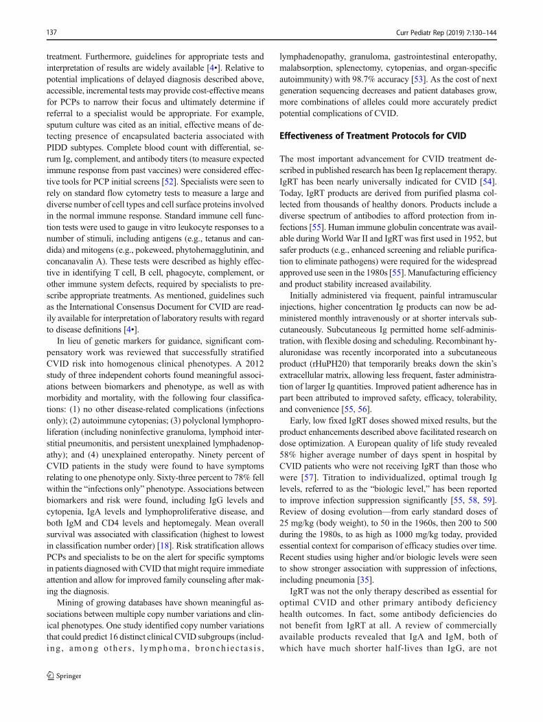
Compared to SCID, CVID is reported to have an incidence ofas high as 1 in 10,000. It results from hypogammaglobulinemia,an inability of the body’s B cells to produce sufficient numbersof functional circulating immunoglobulins (Ig) or antibodies.Without adequate Ig, those with CVID often suffer recurrentand chronic bacterial infections, mostly of the upper and lowerrespiratory tracts, but occasionally of the gastrointestinal tract aswell. CVID is considered a “diagnosis of exclusion,” a basketfor which no molecular or genetic markers can be attributable toa defined antibody deficiency [4•, 18]. It is the most prevalent(27%) type of primary antibody deficiency (also known as a Bcell deficiency or humoral deficiency) and primary antibodydeficiencies are the largest subset (57%) of PIDD. Other majorPIDD subsets involve both B and T cells (20%), phagocytes(4%), and complement (2%) (Fig. 1) [19•]. Secondary (or “ac-quired”) immune deficiency is more common than primary andcan be caused by many indirect factors, such as poor nutrition,
infections (e.g., AIDS), corticosteroids, cancer, and treatmentwith chemotherapy [20]. Finally, a myriad of common environ-mental factors (e.g., frequent exposure to allergens or childrenwith colds in a daycare setting, etc.) that challenge the immunesystem cause symptoms that primary care practitioners (PCPs)routinely see in the clinic and are unrelated to any immunesystem defect [21].
Similar to PIDD discussed above, a wide range of CVIDprevalence estimates were found in published literature. Themost commonly referenced was 1 in 25,000, with ranges from1 in 10,000 to 1 in 100,000. All are based on flawed data andmethods [4•, 6, 22–24]. CVID represented 15.4% of primaryimmunodeficiencies according to a Jeffrey ModellFoundation survey of its network centers (Fig. 1) [19•]. Thisproportion would imply a prevalence of 1 in 7700 if applied toBoyle & Buckley’s findings, or 1 in 12,800–15,400 toKobrynski et al.’s [2, 9]. CVID represented much higher per-centages of primary immunodeficiencies in the EuropeanSociety for Immunodeficiencies (ESID) and U.S.Immunodeficiency Network registries (21 and 30%, respec-tively), which would imply a CVID prevalence of 1 in
57.2%
15.6%
10.0%
4.3%
4.1%
3.5%2.2%
2.1% 0.8% 0.1%
Predominantly antibody deficiencies
Combined immunodeficiencies with associated syndromic featuresUnspecified or other deficiencies
Immunodeficiencies affecting cellular and humoral immunityCongenital defects of phagocyte number, function, or bothDiseases of immune dysregulation
Autoinflammatory disorders
Complement deficiencies
Defects in intrinsic and innate immunity
Phenocopies of primary immunodeficiencies
27.0%
29.5%
14.7%
11.5%
4.8%
4.5%
3.8%2.8%
1.4%Common variable immunodeficiency (CVID), AR
Selective IgA deficiency, unspecified hypogammaglobulinemia, and hyper-IgM syndrome
Specific antibody deficiency (normal Ig and B cells), AR
TACI deficiency
Hypogammaglobulinemia of infancy, transient (normal B cells), AR
IgG subclass deficiency, isolated, AR
BTK deficiency, (X-linked agammaglobulinemia) (XLA)
IgA with IgG subclass deficiency, AR
Other
Fig. 1 Classification and type ofimmunodeficiencies reported byJeffrey Modell Diagnostic/Referral Centers adapted fromModell et al. [19•]
Curr Pediatr Rep (2019) 7:130–144 132

approximately 4000–5700 applied to Boyle & Buckley’s find-ings or 1 in approximately 6700–11,400 if applied toKobrynski et al.’s [2, 4•, 9, 19•].
Specific Aims
The aim of this critical analysis is to assess barriers to optimalhealthcare outcomes in the population with CVID.Specifically, barriers and benefits to reducing diagnosis delayswill be discussed. Current standards and practices for recog-nizing, diagnosing, and treating CVIDwill be reviewed in thiscontext.
Methods
A systematic literature review was conducted to includearticles published in English, limited to the period January1, 2004 through February 15, 2019. Selected literaturepublished prior to 2004 was referenced for historic con-text. Databases used were Pubmed, Scopus, GoogleScholar, and Heal th Informat ion @Himmelfarb.Keywords included CVID, immunoglobulin, infection, di-agnosis, treatment, and delay, yielding over 3000 results.Titles were scanned and abstracts read when titles ap-peared relevant. Approximately 125 articles were identi-fied as relevant. Additional resources included websitesand publications of the Immune Deficiency Foundationand Jeffrey Modell Foundation.
Results
Confounding Factors to Timely Diagnosis
Several factors were found to confound early diagnosis ofCVID. First, unlike SCID, neonatal CVID testing was notrecommended [25]. While CVID has been seen in chil-dren as young as two, immaturity of the immune systemat that age makes current test specificity low [4•]. Earlystudies drawing on data from small registries indicatedCVID diagnoses clustered within two age ranges—6 to10 and 26 to 40, but later studies of larger cohorts showeda continuous curve [24, 26]. Symptom onset, unlike mostprimary immunodeficiencies, has been seen over a verybroad age range, from the first to the ninth decade of life;for this reason the International Consensus Document forCVID recommended that “clinicians must maintain an in-dex of suspicion for antibody deficiency in patients of allages” [4•, 27].
Second, significant heterogeneity within CVID wasfound. Three types of immunoglobulins, or “isotypes”—
IgG, IgA, and IgM—are involved in helping the body fightinfections by identifying and attaching to invading anti-gens, rendering them harmless or marking them for de-struction by other immune system components. The levelsof each in CVID have been found to be highly variable[28]. Considerable effort by international immunology or-ganizations has produced consensus CVID definitions.While there are five classes of Ig—IgG, IgA, IgM, IgD,and IgE—the current definition stipulated that IgG andeither IgA or IgM, or both, be at least 2 standard deviationsunder age and ethnicity-adjusted means [4•, 24]. Underthat threshold, however, considerable variability has beenmeasured [29]. Conversely, patients who were seen pre-senting with CVID-defining symptoms, the requisite IgGdeficiency, but normal IgA or IgM levels, have been cate-gorized as “CVID-like” [30]. Some symptomatic patientshave been assessed with normal total IgG but deficient inone or more subclasses (IgG1, IgG2, IgG3, or IgG4) linkedto multiple phenotypes. Other biomarkers, such as percentof isotype-switched memory B cells, have also been deter-mined to influence phenotype [28].
Third, genetic and other potential biological drivers ofCVID were found to be largely unknown, especially as com-pared to many other primary immunodeficiencies, althoughthis was evolving. There are limited, but growing, numbersof single gene defects discovered to date [31]. While a small,recent study of 36 pediatric CVID patients identified 15 to24% with monogenic drivers, a lower 2 to 10% is the rangeoften cited for all CVID age groups. Monogenic defects werefound to be a larger percentage of pediatric-onset CVID andsomewhat more likely to be hereditary (as opposed to sporad-ic) [4•, 32]. Review of family history was described as animportant physician tool to assess risk for many primary im-munodeficiencies, but the International Consensus Documentfor CVID reported only 5 to 25% of cases are familial [4•].
The majority of diagnosed CVIDs are found to be of spo-radic origin and were attributed to complex interactions be-tween multiple genetic, and possibly epigenetic, factors.Studies are ongoing to understand how sporadic CVID maybe triggered, including environmental factors that influenceDNAmethylation (a mechanism of epigenetics) when geneticpredisposition exists [33]. There are likely many conditionscausing predisposition, one example being selective IgA de-ficiencies progressing to CVID [34].
Fourth, the large majority of CVIDs have been reported aspresenting in the clinic with repeat, common bacterial infec-tions, such as sinus, bronchial, gastrointestinal, and ear infec-tions [24]. In other words, a malfunctioning immune systemwith CVID often manifests in the clinic as well-describedinfections typically not caused by any immune system defect.
Those with PIDD rely predominantly on general healthcarepractitioners to initiate appropriate healthcare workup withsubsequent interventions. For non-SCID PIDD, such as
Curr Pediatr Rep (2019) 7:130–144133

CVID, the prototypical healthcare intervention cycle in theUSAwould flow as follows:
1. PCP recognizes potential symptoms of immune deficien-cy, and may order common tests to guide next steps;
2. PCP provides referral to a specialist, such as an allergistand/or immunologist;
3. Specialist orders advanced blood tests;4. Specialist makes a diagnosis of a specific PIDD subclass
(e.g., CVID);5. Specialist recommends an appropriate treatment protocol;
and6. PCP and specialist monitor patient symptoms, coordinate
treatment, and adjust protocols as necessary for maximumhealth benefit [21].
In the USA, PCPs are typically the gatekeepers to appro-priate treatment for people with CVID; symptom recognitionby PCPs is, therefore, an important gating factor.
Impact of Delay in Diagnosis of CVID
Approximately 80% of CVID diagnoses were reported as pre-ceded by one or more incidences of pneumonia [35].Permanent, structural lung damage, including bronchiectasis,was described as a common consequence of chronic respira-tory tract infections and repeated pneumonia [36]. Severe,opportunistic infections were also seen including bacterialmeningitis and sepsis. A large European multicenter studyexamined 2700 cases in 23 countries and reported non-infectious complications of CVID including autoimmune dis-ease (25%), splenomegaly (24%), gastrointestinal disease/malabsorption (9.9%), granuloma (9.1%), and cancer(14.1%). More than one quarter of the patients included(26.8%) had developed chronic lung disease in the form ofbronchiectasis. This study estimated the individual burden ofCVID using disability life years (DALY) as 36,785 per100,000, whichwas higher than depression disorders, diabetesmellitus, or chronic obstructive pulmonary disease [37•].DALY is a standardized method of calculating the burden ofa specific condition that incorporates data on years of life lost(YLL) and years living with a disability (YLD) linked to thatcondition. Similar comorbidities were seen consistently acrossmost CVID studies [25, 38].
Delays from onset of symptoms to CVID diagnosis wereanalyzed in several large-cohort studies over the past decadeand seen to be widespread and persistent. An average delay of7.46 years (ranging from 0 to 60 years) was calculated among338 patients in the European CVID registry. In this group,delay length was inversely related to age of CVID onset[26]. An Italian study of 224 CVID patients revealed an aver-age delay of 8.9 years [39]. Approximately 20% of adultsultimately diagnosed with CVID were able to identify disease
symptoms from childhood that had gone undiagnosed [40].Non-infectious complications, while less common and oftensevere, did not necessarily raise PCP index of suspicion forCVID. For example, a Norwegian study found that patientswith non-infectious complications had a median time to diag-nosis of 17 years versus 10 for the infection-only cohort. Onaverage, granulomatous disease (a lymphoproliferative dis-ease) had been present 19 years prior to diagnosis, alopecia(an autoimmune disease) 25 years, and gastrointestinal dis-eases 13.5 years [41].
Diagnostic delay of CVID was strongly associated withcomplications and comorbidities in several studies. AGerman pediatric study reported multiple severe and chronicinfections during an average 5.8-year delay to diagnosis andno serious infections after initiation of treatment. This studyalso showed association between delay in diagnosis and bron-chiectasis [24]. The 2700-case CVID study by Odnoletkovaet al. described above revealed diagnostic delay associatedwith bronchiectasis, solid tumors, and enteropathy [37•].Each year of delay was associated with 1.4% increased mor-tality in a pan-European cohort of 2212 patients [42].
The Jeffrey Modell Foundation asked its network physi-cians to compile statistics for the year prior to and year afterdiagnoses of their patients. Their analysis, which incorporatedall primary immunodeficiencies excluding SCID, showedpost-diagnosis reductions of 84% in hospitalization days,83% in physician/emergency room visits, 56% in days onantibiotics, and 74% in work or school days missed(Table 4) [13••]. Improvements can be attributed to effectivetreatments, including immunoglobulin replacement therapy(IgRT) and appropriate use of antibiotics, described below.
Identification of Undiagnosed Patients
Several frameworks proposed to improve PCP identificationof primary immunodeficiencies were reviewed. The JeffreyModell Foundation and Immune Deficiency Foundation werefound to be leaders in ongoing efforts to raise awarenessamong physicians and the public, especially in the USA.The Jeffrey Modell Foundation developed a global awarenesscampaign around its “Ten Warning Signs of PrimaryImmunodeficiency” (“Ten Warning Signs”) starting 1993.The Ten Warning Signs urge follow-up with healthcare pro-fessionals if two or more of the following are present(Table 1).
The Jeffrey Modell Foundation also developed and distrib-uted to provider networks and insurers, free of charge, theSPIRIT Analyzer (Software for Primary ImmunodeficiencyRecognition, Intervention, and Tracking). SPIRIT Analyzermatches patient records to Ten Warning Sign-related ICD-10codes in patient databases. A point scoring system assignseach patient to high, medium, and low, or zero PIDD risk. Atest on a 2,000,000 patient sample from a large dataset (IMS
Curr Pediatr Rep (2019) 7:130–144 134

LifeLink Health Plan Claims Database) identified over 1 in1685 as high and 1 in 680 as moderate risk (combined, 1 in485) [43]. However, no analysis on sensitivity and specificityfor SPIRIT Analyzer could be found that validated the effec-tiveness of this program for real-world application.
The Cleveland Clinic tested the sensitivity of the TenWarning Signs by retrospective analysis of electronic healthrecords for the years 2006 through 2016. They identified 287adults with confirmed PIDD (77% of whom had CVID) and115 pediatric patients. Detailed case review revealed that only45% of these adults and 64% of children had two or more ofthe warning signs [44]. In other words, 55 and 36% respec-tively, would have been missed if the Ten Warning Signs hadbeen the sole identification criterion. A 2011 study inNorthern England determined that only 62% of confirmedPIDD cases exhibited two or more of the warning signs; con-versely, 38% had less than two. The authors of this studydescribed shortcomings of the Ten Warning Signs, includingthat it does not address non-infectious complications. Theyadvocated instead for resource allocation to training and lab-oratory diagnostics [45]. A University of Manchester study of430 pediatric PIDD patients and 133 controls also showed lowsensitivity and specificity. These authors proposed a simpledecision tree framework for PCPs that hinges on past receiptof intravenous antibiotics or antifungals for sepsis [46].
Two German groups, the German Patients’ Organisationfor Primary Immunodeficiencies (DSAI) and the Associationof the Scientific Medical Societies in Germany (AWMF),attempted to improve on the sensitivity and specificity of theTen Warning Signs. A third group from Heinrich HeineUniversity, Dusseldorf (Dusseldorf), tested the DSAI andAWMF frameworks, along with two of its own, on a cohortof 210 PIDD cases. The Jeffrey Modell Foundation’s TenWarning Signs were found to have the highest specificitybut lowest sensitivity. The highest sensitivity (75%) was seenwith Dusseldorf’s maximum negative predictive value (NPV)approach, but specificity was only 51% (Table 2) [47].
ESID developed a significantly more thorough frameworkfor PCPs. Eight distinct clinical presentation categories wereestablished, with corresponding columns for each, including(1) “special features” to look out for; (2) relevant pathogens;(3) potential alternative (non-PIDD) causes of clinical presen-tation; and (4) references for each to distinct step-by-step di-agnostic protocols. Unlike the Ten Warning Signs and similarlists, the ESID framework did not suggest specific numbers ofinfections requiring follow-up. The authors described theirapproach in practical terms from a cost-benefit perspective,calling for immediate laboratory tests if severe, potentiallylife-threatening primary immunodeficiencies were possible(even if not probable), and phased-in examination and testsfor cases unlikely to pose immediate, high risk [48]. The com-plexity of the ESID framework appeared to present a potentialhurdle to widespread adoption in the PCP setting.
The above PCP recognition frameworks were developed byspecialists (such as immunologists) for whom PIDD wouldlikely be a priority. A different perspective on appropriate indexof suspicion was observed in widely used publications for gen-eral PCP practice. For example, Nelson Pediatric Symptom-Based Diagnosis guides that immunodeficiency is fifth of fivecategories of consideration when patients present with recur-rent, common infections. Category 1 is “the patient who isprobably healthy,” guiding that children with no immune de-fects, especially in large families or day care, may have six toten upper respiratory infections or gastroenteritis episodes peryear. Six infection-related criteria that would individually, orcombined, warrant further investigation were listed as follows:(1) more than two systemic bacterial infections (sepsis, menin-gitis, osteomyelitis); (2) two or more serious respiratory infec-tions (pneumonia, sinusitis) or bacterial infections (cellulitis,draining otitis media, lymphadenitis) per year; (3) the presenceof an infection at an unusual site (hepatic or brain abscess); (4)infections with unusual pathogens (Aspergillus pneumonia,disseminated candidiasis, or infection with Serratiamarcescens, Nocardia species, or Burkholderia cepacia); (5)infections of unusual severity; and (6) recurrent mycobacterialinfections or invasive infections with atypical mycobacteria[21]. This framework places more emphasis on serious infec-tions, a focus that couldmiss themajority of recurrent, commoninfections (e.g., frequent or chronic sinus and upper respiratorytract infections) characteristic of most CVIDs.
The diagnostic approach described in Nelson PediatricSymptom-Based Diagnosis and other similar guidebookswas considered in the context of PCP training and awareness.The American Academy of Allergy, Asthma, & Immunology(AAAAI) Primary Immune Deficiency Committee and theImmune Deficiency Foundation collaborated on a mail-based survey of 4500 PCPs in 2009 to assess awareness, train-ing, and practice strategies among family physicians. Twelvepercent (528) were returned, 44% affirming at least one PIDDpatient followed, and 56% not following any. Only 9% of
Table 1 Jeffrey Modell Foundation Ten Warning Signs of PrimaryImmunodeficiency
(1) Four or more new ear infections within 1 year.
(2) Two or more serious sinus infections within 1 year.
(3) Two or more months on antibiotics with little effect.
(4) Two or more pneumonias within 1 year.
(5) Failure of an infant to gain weight or grow normally.
(6) Recurrent, deep skin or organ abscesses.
(7) Persistent thrush in mouth or fungal infection on skin;
(8) Need for IVantibiotics to clear infections;
(9) Two or more deep-seated infections including septicemia; and
(10) A family history of PIDD.
Reproduced from Modell et al. [19•]
Curr Pediatr Rep (2019) 7:130–144135

PCPs followed one or more patient with CVID. PIDD wasdescribed as insufficiently covered in medical school, with11% responding it was not covered at all and 76% just mini-mally. Only 12% reported adequate coverage. Responses re-garding continuing education revealed gaps as well; 99% hadnot participated in PIDD-related education during the preced-ing 6 months but 66% were interested in doing so [49]. Asimilar 2008 survey reported 93% of responding pediatriciansbeing unaware of the existence of published PIDD guidelinesand 35% uncomfortable with PIDD symptom recognition[50]. Of a survey of 1880 PCPs, only 32% knew of one patientwith PIDD within the past 5 years. Only 10% recalled receiv-ing a copy of the Ten Warning Signs [20].
A positive sign that interest and awareness may be highertoday than when the above surveys were taken could beAAAAI ’s 2018 Diagnos t i c Schoo l i n P r ima ryImmunodeficiencies. This was the organization’s first eversuch continuing education seminar for physicians looking to“effectively and appropriately harness diagnostic tools cur-rently available in order to provide the best benefit to theirpatients” [51].
Technical Capabilities to Diagnose CVID
Technology did not appear to be an impediment given theexistence of tests to confer diagnoses sufficient for directing
Table 2 Comparison of three frameworks for PIDD identification in the clinic and two alternatives
Framework JeffreyModellFoundation
DSAI AWMF Duesseldorf (Youden Jstatistic, max. negativepredictive power)
Duesseldorf (Youden Jstatistic, max. positivepredictive power)
Number of symptom types to suspect PIDD
Symptom 2 or more 1 ormore
1 ormore
1 or more 1 or more
Ear infections within 1 year ≥ 4 ≥ 8 ≥ 8Sinus infections within 1 year ≥ 2 ≥ 2Months on antibiotics without effect ≥ 2 ≥ 2Pneumonias within 1 year ≥ 2 ≥ 2 ≥ 2Failure to grow normally √ √ √ √ √Failure to thrive √ √Recurrent deep skin or organ abscesses √ √ √Persistent thrush after age of 1 year √ √Deep-seated infections: meningitis, sepsis ≥ 2 ≥ 2Positive family history √ √ √Chronic graft-versus-host disease (GvHD) in the infant √Need for intravenous antibiotics √ √Recurring infection with atypical mycobacteria √Complications of vaccination √“ELVIS”a—pathological susceptibility to infection:
pathogen, localization, course, intensity, sum√
“GARFIELD”b: granuloma, autoimmunity, recurringfever, eczema, lymphoproliferation, chronic bowelinflammation
√
Abnormal laboratory results √Hypogammaglobulinemia, neutropenia, lymphopenia √ Lymphopenia only √Sensitivity (%) 33 47 69 75c 56
Specificity (%) 79c 64 37 51 76
Positive predictive value (%) 25 21 19 24 32c
Negative Predictive Value (%) 85 85 86 91c 89
Adapted from Lankisch et al. [47]a ELVIS—pathogen [German: erreger], localization, course [German: verlauf], intensity, and sumbGARFIELD—granuloma, autoimmunity, recurring fever [German: fieber], unusual eczema, lymphoproliferation, chronic inflammation of the gut[German: darm]cMaximum percent for given performance metric among frameworks shown
Curr Pediatr Rep (2019) 7:130–144 136
treatment. Furthermore, guidelines for appropriate tests andinterpretation of results are widely available [4•]. Relative topotential implications of delayed diagnosis described above,accessible, incremental tests may provide cost-effective meansfor PCPs to narrow their focus and ultimately determine ifreferral to a specialist would be appropriate. For example,sputum culture was cited as an initial, effective means of de-tecting presence of encapsulated bacteria associated withPIDD subtypes. Complete blood count with differential, se-rum Ig, complement, and antibody titers (to measure expectedimmune response from past vaccines) were considered effec-tive tools for PCP initial screens [52]. Specialists were seen torely on standard flow cytometry tests to measure a large anddiverse number of cell types and cell surface proteins involvedin the normal immune response. Standard immune cell func-tion tests were used to gauge in vitro leukocyte responses to anumber of stimuli, including antigens (e.g., tetanus and can-dida) and mitogens (e.g., pokeweed, phytohemagglutinin, andconcanavalin A). These tests were described as highly effec-tive in identifying T cell, B cell, phagocyte, complement, orother immune system defects, required by specialists to pre-scribe appropriate treatments. As mentioned, guidelines suchas the International Consensus Document for CVID are read-ily available for interpretation of laboratory results with regardto disease definitions [4•].
In lieu of genetic markers for guidance, significant com-pensatory work was reviewed that successfully stratifiedCVID risk into homogenous clinical phenotypes. A 2012study of three independent cohorts found meaningful associ-ations between biomarkers and phenotype, as well as withmorbidity and mortality, with the following four classifica-tions: (1) no other disease-related complications (infectionsonly); (2) autoimmune cytopenias; (3) polyclonal lymphopro-liferation (including noninfective granuloma, lymphoid inter-stitial pneumonitis, and persistent unexplained lymphadenop-athy); and (4) unexplained enteropathy. Ninety percent ofCVID patients in the study were found to have symptomsrelating to one phenotype only. Sixty-three percent to 78% fellwithin the “infections only” phenotype. Associations betweenbiomarkers and risk were found, including IgG levels andcytopenia, IgA levels and lymphoproliferative disease, andboth IgM and CD4 levels and heptomegaly. Mean overallsurvival was associated with classification (highest to lowestin classification number order) [18]. Risk stratification allowsPCPs and specialists to be on the alert for specific symptomsin patients diagnosed with CVID that might require immediateattention and allow for improved family counseling after mak-ing the diagnosis.
Mining of growing databases have shown meaningful as-sociations between multiple copy number variations and clin-ical phenotypes. One study identified copy number variationsthat could predict 16 distinct clinical CVID subgroups (includ-ing , among o the r s , l ymphoma, b ronch iec t a s i s ,
lymphadenopathy, granuloma, gastrointestinal enteropathy,malabsorption, splenectomy, cytopenias, and organ-specificautoimmunity) with 98.7% accuracy [53]. As the cost of nextgeneration sequencing decreases and patient databases grow,more combinations of alleles could more accurately predictpotential complications of CVID.
Effectiveness of Treatment Protocols for CVID
The most important advancement for CVID treatment de-scribed in published research has been Ig replacement therapy.IgRT has been nearly universally indicated for CVID [54].Today, IgRT products are derived from purified plasma col-lected from thousands of healthy donors. Products include adiverse spectrum of antibodies to afford protection from in-fections [55]. Human immune globulin concentrate was avail-able during World War II and IgRTwas first used in 1952, butsafer products (e.g., enhanced screening and reliable purifica-tion to eliminate pathogens) were required for the widespreadapproved use seen in the 1980s [55].Manufacturing efficiencyand product stability increased availability.
Initially administered via frequent, painful intramuscularinjections, higher concentration Ig products can now be ad-ministered monthly intravenously or at shorter intervals sub-cutaneously. Subcutaneous Ig permitted home self-adminis-tration, with flexible dosing and scheduling. Recombinant hy-aluronidase was recently incorporated into a subcutaneousproduct (rHuPH20) that temporarily breaks down the skin’sextracellular matrix, allowing less frequent, faster administra-tion of larger Ig quantities. Improved patient adherence has inpart been attributed to improved safety, efficacy, tolerability,and convenience [55, 56].
Early, low fixed IgRT doses showed mixed results, but theproduct enhancements described above facilitated research ondose optimization. A European quality of life study revealed58% higher average number of days spent in hospital byCVID patients who were not receiving IgRT than those whowere [57]. Titration to individualized, optimal trough Iglevels, referred to as the “biologic level,” has been reportedto improve infection suppression significantly [55, 58, 59].Review of dosing evolution—from early standard doses of25 mg/kg (body weight), to 50 in the 1960s, then 200 to 500during the 1980s, to as high as 1000 mg/kg today, providedessential context for comparison of efficacy studies over time.Recent studies using higher and/or biologic levels were seento show stronger association with suppression of infections,including pneumonia [35].
IgRT was not the only therapy described as essential foroptimal CVID and other primary antibody deficiencyhealth outcomes. In fact, some antibody deficiencies donot benefit from IgRT at all. A review of commerciallyavailable products revealed that IgA and IgM, both ofwhich have much shorter half-lives than IgG, are not
Curr Pediatr Rep (2019) 7:130–144137

material components of IgRT, making them ineffective forselective IgA or IgM deficiencies [60]. At the same time,the paucity of IgA and IgM in commercially availableIgRT was reported to underlie continued susceptibility tocertain types of infections for those with CVID. SecretedIgA2 (one of two types of IgA), for example, lines mucosalsurfaces such as the respiratory and digestive tracts, direct-ly neutralizing antigens there. IgA2 deficiency—often seenwith CVID—has been associated with sinus and upper re-spiratory tract infections even after IgRT initiation, thoughon average at reduced rates [61]. Several studies reportedpneumonia, bacterial meningitis, and sepsis rates dramati-cally reduced once biologic Ig levels were reached, butsinusitis, bronchiectasis, and splenomegaly did not [62].
Antibiotic therapy is an essential, ongoing component oftreatment for breakthrough infections with CVID, regardlessof IgRT use or prophylactic antibiotics. Though overuse ofantibiotics is a growing concern, the benefits of prophylacticuse have outweighed the risks for some [22, 25]. Prophylacticantibiotics have been shown to ameliorate chronic granuloma-tous disease, a potential complication of CVID, for example[63]. However, appropriate patient and family counseling isnecessary when utilizing prophylactic antibiotics. One studyfound that patients on prophylactic antibiotics delayed seekingnecessary high-dose prescriptions for breakthrough infections[64].
Recommendations for vaccinations need to be determinedbased on the specific type of CVID subtype in question. Forexample, IgG1 and IgG3 respond to certain vaccine proteinantigens while IgG2 and IgG4 respond only to polysaccharideantigens. Although those treated with IgRTwill have acquiredmany vaccine-induced antibodies from plasma donors, thereappeared to be flexibility for specialists to recommend specificvaccines depending on individual risks and blood/serummarkers. Live-attenuated vaccines, however, were generallycontraindicated [61].
As mentioned, all 50 states now include SCID in neo-natal blood tests. For babies diagnosed with SCID, stemcell transplantation has been a potentially curative option.Stem cell transplantation was also described as an optionfor non-SCID primary immunodeficiencies, includingCVID, if patients fail to thrive or develop serious comor-bidities such as lymphoma. The low probability of sourc-ing matched stem cells, potential host rejection, and highmortality rate with the procedure were reported as signif-icant constraints to greater stem cell transplant utilization[65]. However, several clinical trials are currently under-way aiming to improve safety and broaden applicabilityof stem cell transplantation for primary immunodefi-ciencies, including CVID [66]. Gene therapy has emergedas a potential option for some forms of severe immuno-deficiencies, particularly when defects are monogenic[67]. That CVID has been found to be driven by
combinations of several genetic (and other) factors makesgene therapy for CVID more complicated and unlikely inthe near term.
Cost, Benefit, and Financing of CVID and Treatments
Strong associations were found between the initiation of opti-mal treatment (including appropriate doses of IgRT and anti-biotics) and reduced morbidity and mortality. Financing oftreatments, however, was reported as a significant impedimentto optimal therapy. Complaints by both patients and physi-cians were found to be pervasive with regard to healthinsurer-erected obstacles to coverage, including pre-authorization requirements for every infusion, frequentswitching to lowest cost products, and dose restrictionscontradicted by evidence [68]. Side effects can be productspecific, so switching may negatively affect patients. TheImmune Deficiency Foundation received 1417 responses(63% diagnosed with CVID) to its 2014 patient survey onhealth insurance. Of those, 1160 had been prescribed IgRTfor at least 6 months, but 372 reported skipping or delayingat least one dose (82% an average of 2.6 doses) that year.Fifty-two percent of those who skipped or delayed did sobecause of insurance-related problems [69].
No comprehensive studies were found that specificallyassessed CVID-related medical costs before or/and after diag-nosis and treatment. A few studies evaluated various catego-ries of infection rates and costs relating to groups of PIDDs.These studies are largely not comparable due to differing co-hort selection criteria, timeframes, and basis for averaging(Table 5). Future comprehensive analyses of pre and post-treatment infection and non-infection comorbidity rates andcosts by subtype could aid the understanding of treatmentintensity and expected benefits at a more granular level; how-ever, the fast-paced discovery of new subtypes of PIDD (de-scribed above) would likely complicate comparability of stud-ies over time.
One study, discussed above, compiled PIDD-related hospi-tal expenditures from The Kids’ Inpatient Database, the larg-est pediatric database in the USA. For the period 2001 to2012, hospital admissions among B cell subtypes accountedfor $342,000,000, averaging $25,357 per admission (Table 3),lower than the average cost of $30,987 across all PIDDs [14].CVID accounted for 17% of B cell subtype hospitalizations,fewer than the proportion of CVID patients in the JeffreyModell Centers Network (27%) (Fig. 1). This disparity doesnot necessarily imply CVID required fewer admissions thanother B cell subtypes. Other potential explanations are that aCVID diagnosis is not recommended before the age of 4,CVID is more likely to be undiagnosed during early child-hood, can initiate as other B cell deficiencies and progress intoCVID at a later age, and that diagnostic granularity has im-proved since 2001. Support for these interpretations come
Curr Pediatr Rep (2019) 7:130–144 138

f r om t h e l a r g e r p e r c e n t a g e o f u n s p e c i f i e dhypogammaglobulinemia in the Kids’ database than in theJeffrey Modell Centers Network, and by very similar totalsof CVID, unspecified hypogammaglobulinemia, hyper-IgMsyndrome, and selective IgA deficiency in both studies. Thisstudy did not distinguish between patients receiving or notreceiving optimal therapy prior to hospitalization, so no treat-ment cost-benefit analysis was possible.
The European quality of life survey mentioned above eval-uated CVID cases within a large PIDD cohort and found 343patients who spent an average of 13.1 days in hospital over12 months. Of these, the 313 receiving IgRT spent an averageof 12.5 days in hospital versus 19.8 (58% more) for the 30patients not on IgRT [57].
One study of PIDD patients receiving IgRT within a verylarge administrative database (including privately insured andMedicare covered lives) provided helpful context for analysesof CVID patients on IgRT. In this study, 1076 patients wereidentified as both diagnosed with PIDD and receiving IgRTfor at least 6 months. Of these, nearly half (48%) had a diag-nosis of CVID, evidencing the large proportion of CVIDwith-in the IgRT-receiving PIDD population as a whole. This entirecohort had a per-patient-year infection rate (pneumonia andbronchitis) of 37% despite stable IgRT. Infection rates prior to
initiation of IgRT were not reported, so no pre-post compari-sons could be made [70]. Another study of a large medicalclaims database identified 490 patients both diagnosed withPIDD and receiving IgRT, and who had suffered from at leastone infection over a 7-month period. Infection-related medicalexpenditures (including hospitalizations) for these 490 pa-tients totaled over $10,000,000, an annualized average of$20,443. The average cost of a hospital stay was $27,952[71]. Costs prior to IgRT initiation were not reported so nopre-post-treatment analysis was possible.
One analysis was found that compared pre and post-diagnosis medical costs for a large group of PIDD patientstreated within the Jeffrey Modell Centers Network. SCIDwas excluded, the intensive treatment requirements for whichwould have skewed average costs. A reasonable estimate ofpercentage of CVID cases included in the study, while notdisclosed, would be 18%, greater than the Jeffrey ModellCenters Network’s 15.4% due to exclusion of SCID (Fig. 1).The authors calculated average pre-diagnosis annual cost as$111,053, which included treatment for infections, physician/emergency room visits, hospital days, and work or schooldays missed. Post-diagnosis annual cost averaged $25,171,before inclusion of IgRT. IgRTwas estimated as an additional$30,000 for a total cost of $55,171. Net average annual per-
Table 3 B cell PIDD-related hospitalizations in the Kids’ Inpatient Database from 2003 through 2012
Hospital admissions by age and subtype (2001 to 2012)
All causes B cell PIDD-related
No. Percent Deaths Mortality%
Cost (millions) Average cost
By age (years)
0–5 Not reported 5174 38% 156 3.02% $ 146.6 $ 28,329
6–10 2857 21% 25 0.88% 60.0 20,966
11–15 2697 20% 14 0.52% 65.0 24,041
> 15 2743 20% 26 0.95% 70.0 25,617
Total 13,471 100% 221 1.64% $ 341.6 $ 25,357
By subtype
CVID 2263 17% 28 1.24%Hypogammaglobulinemia, unspecified 5586 41% 117 2.09%
Other selective immunoglobulin deficiencies 2147 16% 25 1.16% Not reported
Selective IgA immunodeficiency 1760 13% 4 0.23%Other 1715 13% 47 2.74%
Total 13,471 100% 221 1.64%
3-year period ending One in:
2003 7,409,162 2254 3287
2006 7,558,812 2978 2538
2009 7,370,203 3541 2081
2012 6,675,222 4698 1421
Total 29,013,399 13,471 2154
Adapted from Rubin et al. [14]
Curr Pediatr Rep (2019) 7:130–144139

patient savings including and excluding IgRT, achievable bydiagnosing and treating PIDD, was $85,882 and $55,882, re-spectively (Table 4) [13••]. This high level of savings fromtreatment could support aggressive searches by payors withintheir databases for undiagnosed PIDD populations, and couldjustify the cost of incremental screenings for identified, at-riskcases. The report did not include material supporting data ormethodologies used; additional peer-reviewed studies quanti-fying health system savings from treatment could further in-centivize payors to find and treat cases of PIDD within theirmembership pools (Table 5).
Conclusions
Heterogeneity and overlap in clinical manifestations within,between, and unrelated to immune disorders were found toconfound timely symptom recognition of CVID. Therefore,a reliable, widely accepted framework for early CVID identi-fication remains elusive. A logical objective of cliniciansshould be to diagnose early to initiate treatment before chron-ic, recurrent, or severe infections cause irreversible organdamage. Advancements in diagnostic technology and consen-sus guidelines have allowed classification of primary immu-nodeficiencies, including CVID, to become more robust.While only a small percentage of CVID cases have identifi-able genetic drivers currently, laboratory tests that identify
immunoglobulin deficiencies are widely available, informingrequisite initiation of the most important treatment availablefor CVID, namely, IgRT. Prophylactic and aggressive antibi-otic therapies may be indicated for persistent or breakthroughinfections once patients are known to have CVID. Today it isunderstood that patients should be monitored and assessedregularly by their PCPs and specialists for potential protocoladjustments to optimize long-term health. Risk stratificationmay help guide these decisions. Available treatments and pro-tocols have made improved health outcomes and quality oflife possible for those with CVID.
IgRT product enhancements and administration optionshave improved significantly over time, providing greaterconvenience and treatment compliance. IgRT protocolshave moved to higher or “biologic doses” and are individu-alized to suppress rates of infections. The evidence of IgRTeffectiveness in CVID is very strong, but it is rarely func-tionally curative. Those with CVID continue to have certaintypes of infections—especially of the upper respiratory andsinuses—that require medical attention. This, together withthe high cost of IgRT, may be contributing to insurancecoverage barriers encountered by many patients and PCPs.Peer-reviewed studies are needed to support the compellingcost savings achieved through treatment. If savings are sub-stantiated, insurers may be financially incentivized to pro-actively review their insured databases for potentially undi-agnosed CVID and other PIDD cases.
Table 4 Jeffrey Modell Foundation 2013 to 2015 survey of 85 of its network treatment centers, average number of episodes and costs, 1 year beforeand 1 year after diagnosis
Per patient Pre-diagnosis (1 year) Post-diagnosis (1 year) Increase/(decrease) (1 year)
Average # ofepisodes
Averageannual cost
Average # ofepisodes
Averageannual cost
Average # ofepisodes
Averageannual cost
Persistent otitis media 4.2 $2217 1.6 $845 − 62% ($1372)
Serious sinus and upper respiratoryinfections
4.6 5175 2.1 2362 − 54% (2813)
Viral infections 3.7 4717 1.4 1785 − 62% (2932)
Acute bronchitis 3.1 5270 0.8 1360 − 74% (3910)
Bacterial pneumonias 2.8 9945 0.6 2131 − 79% (7814)
COPD and bronchiectasis 4.3 13,609 1.4 4431 − 67% (9178)
Hospitalization days 19.8 49,104 3.1 7688 − 84% (41,416)
Physician/ER visits 70.8 12,744 11.7 2106 − 83% (10,638)
Days on antibiotics 166.2 1662 72.8 728 − 56% (934)
Subtotal 104,443 23,436 (81,007)
School/work days missed 33.9 6610 8.9 1735 − 74% (4875)
Total treatment cost & savings per patientwithout IgRT
$111,053 $25,171 ($85,882)
Average annual cost of IgRT n/a 30,000 30,000
Total treatment costs and savings perpatient with IgRT
$111,053 $55,171 ($55,882)
Adapted from Modell et al. [13••]
Curr Pediatr Rep (2019) 7:130–144 140

Table5
Summaryof
PID
D/CVID
infectionandtreatm
entstatisticsin
studiesreview
ed
Study
Period
Inclusioncriteria
Identified
Measuring
Result
IgRTstatus
%Diagnosisof
CVID
Ref.
The
Kids’Inpatient
(pediatric)Database
study(U
SA)
2001–2012
PIDD
26,794
admissions
Perhospitaladm
ission
cost
$30,987
Not
disclosed
8%a
[14]
PIDDBcellsubtype
13,471
admissions
$25,357
17%
b
EuropeanQualityof
Life
Survey
2004–2006
CVID
313patientsreceivingIgRT
Annuald
aysin
hospital
12.5
days
Yes
100%
[57]
30patientsnotreceiving
IgRT
19.8
days
No
100%
Diff.:7
.3days
Large
administrative
claimsdatabase
(USA
)
2002–2013
PIDD&
≥1inpatient
orERor
≥2outpatient
claims&
min.
12monthsenrollm
entin
health
plan
1076
patients(1571
patient-years)
Infectionrate(pneum
onia&
bronchitis)c
37%
infectionrate
(patient-years)
Yes—min.
6months
48%
d[70]
Large
administrative
claimsdatabase
(USA
)
2008–2010
PIDD,receiving
IgRT,
≥1
infectionin
past7months
490patients(of1742
total
with
PIDDdiagnosis)
Ave.annualized
infection-relatedTxcost
perpatient
$20,443
Yes
Not
disclosed,
butlikelya
largepercent
[70]
[71]
Perhospitaladm
ission
cost
$27,952
Yes
JeffreyModellC
enters
Networksurvey
Not
disclosed
PID
D,excluding
SCID
,in
centersresponding
toJM
CN
survey
#of
patientsnotd
isclosed
Ave.annualized
PIDD-related
costperpatient—pre-Dx
$111,053
No
Not
disclosed,
butlikely
approx.18%
[13••]
Post-D
x(incl.IgRT)
$55,171
Yes
Diff.:
$55,882
aUnspecified
hypogammaglobulin
emiacomprised
21%
bUnspecified
hypogammaglobulin
emiacomprised
41%
cInfectionepisodedefinitio
nassumed
a7-daymaxim
umgapin
claims(longerduratio
nepisodes
may
bedouble-counted)
dUnspecified
hypogammaglobulin
emiacomprised
24%
Curr Pediatr Rep (2019) 7:130–144141

The nature of the CVID symptom recognition is likely toevolve. As the cost of next generation sequencing falls, CVIDregistries grow in size and breadth, and bioinformatics capa-bilities uncover multifactorial drivers of CVIDs, PCPs maysomeday have periodic, routine laboratory tests to guide them.In the meantime, however, symptom recognition by PCPs willremain a gating factor for necessary treatments. Accepted,effective symptom recognition frameworks, along with suffi-cient PCP education and training, are needed to optimizehealth for those with CVID.
Compliance with Ethical Standards
Conflict of Interest William Gerber declares no conflict of interest.
Human and Animal Rights and Informed Consent This article does notcontain any studies with human or animal subjects performed by any ofthe authors.
Open Access This article is distributed under the terms of the CreativeCommons At t r ibut ion 4 .0 In te rna t ional License (h t tp : / /creativecommons.org/licenses/by/4.0/), which permits unrestricted use,distribution, and reproduction in any medium, provided you give appro-priate credit to the original author(s) and the source, provide a link to theCreative Commons license, and indicate if changes were made.
References
Papers of particular interest, published recently, have beenhighlighted as:• Of importance•• Of major importance
1. Ochs HD, Hitzig WH. History of primary immunodeficiency dis-eases. Curr Opin Allergy Clin Immunol. 2012;12(6):577–87.
2. Boyle JM, Buckley RH. Population prevalence of diagnosed pri-mary immunodeficiency diseases in the United States. J ClinImmunol. 2007;27:497–502.
3. Fudenberg H, Good RA, Goodman HC, Hitzig W, Kunkel HG,Roitt IM, et al. Primary immunodeficiencies, report of a WorldHealth Organization committee. Pediatrics. 1971;47(5):927–46.
4.• Bonilla F, Barlan I, Chapel H, Costa-Carvalho B, Cunningham-Rundles C, de La Morena M, et al. International consensus docu-ment (ICON): common variable immunodeficiency disorders. JAllergy Clin Immunol Pract. 2016;4(1):38–59 A comprehensiveoverview of CVID, including epidemiology, clinical manifesta-tions, genetics, diagnosis, treatments and prognosis, resultingfrom extensive international collaboration.
5. Bousfiha AA, Jeddane L, Picard C, Ailal F, Gaspar HB, Al-HerzW,et al. The 2017 IUIS phenotypic classification for primary immu-nodeficiencies. J Clin Immunol. 2018;38:128–43.
6. Tangye SG.. Inborn errors of immunity committee (IEI)—activities;2018. Retrieved February, 2019, from http://www.iuisonline.org/index.php?option=com_content&view=article&id=68&Itemid=9 4 & 5 a 4 8 d 8 7 d c d f 6 f a c 9 6 a 6 b c 0 f f c 1 b 9 e 6 4 c =461e23649300226f90ec7f639f96ce00.
7. Immune Deficiency Foundation, prepared by Schulman, Ronca &Bucuvalas, Inc. Primary immune deficiency diseases in America,the first national survey of patients and specialists, No. IDF0034,Towson, Maryland. 1995.
8. Joshi AY, Iyer VN, Hagan JB, St. Sauver JL, Boyce TG. Incidenceand temporal trends of primary immunodeficiency: a population-based cohort study. Mayo Clin Proc. 2009;84(1):16–22.
9. Kobrynski L, Powell RW, Bowen S. Prevalence and morbidity ofprimary immunodeficiency diseases, United States 2001–2007. JClin Immunol. 2014;34(8):954–61.
10. National Institutes of Health (NIH). National Institutes of Health(NIH), National Institute of Allergy and Infectious Diseases—primary immune deficiency diseases (PIDDs); 2016. RetrievedFebruary 10, 2019, from https://www.niaid.nih.gov/diseases-conditions/primary-immune-deficiency-diseases-pidds.
11. Bousfiha AA, Jeddane L, Ailal F, Benhsaien I, Mahlaoui N,Casanova JL, et al. Primary immunodeficiency diseases worldwide:more common than generally thought. J Clin Immunol. 2013;33:1–7.
12. Jeffrey Modell Foundation (JMF). Our story. Retrieved 01/15,2019, from http://www.info4pi.org/hq/our-story
13.•• Modell V, Quinn J, Orange J, Notarangelo LD, Modell F. Primaryimmunodeficiencies worldwide: an updated overview from theJeffrey Modell Centers global network. Immunol Res. 2016;64:736–53 A unique analysis showing PIDD-related costs for alarge patient population prior to and post diagnosis.
14. Rubin Z, Pappalardo A, Schwartz A, Antoon JW. Prevalence andoutcomes of primary immunodeficiency in hospitalized children inthe United States. J Allergy Clin Immunol in Pract. 2018;6(5):1705–10.
15. Cunningham-Rundles C, Sidi P, Estrella L, Doucette J. Identifyingundiagnosed primary immunodeficiency diseases in minority sub-jects by using computer sorting of diagnosis codes. J Clin Immunol.2004;113:747–55.
16. Immune Deficiency Foundation (IDF). All 50 states now screeningnewborns for severe combined immunodeficiency (SCID); 2018.Retrieved 02/04/2019, 2019, from https://www.primaryimmune.org/news/all-50-states-now-screening-newborns-severe-combined-immunodeficiency-scid.
17. Griffith LM, Cowan MJ, Notarangelo LD, Kohn DB, Puck JM,Shearer WT, et al. Primary immune deficiency treatment consor-tium (PIDTC) update. J Clin Immunol. 2016;138(2):375–85.
18. Chapel H, Lucas M, Patel S, Lee M, Cunningham-Rundles C,Resnick E, et al. Confirmation and improvement of criteria forclinical phenotyping in common variable immunodeficiency disor-ders in replicate cohorts. J Clin Immunol. 2012;130:1197–8.
19.• Modell V, Orange JS, Quinn J, Modell F. Global report on primaryimmunodeficiencies: 2018 update from the Jeffrey Modell CentersNetwork on disease classification, regional trends, treatment mo-dalities, and physician reported outcomes. Immunol Res. 2018;66:367–80 A comprehensive breakdown of PIDD subtypes withina large patient population.
20. Waltenburg R, Kobrynski L, Reyes M, Bowen S, Khoury MJ.Primary immunodeficiency diseases: practice among primary careproviders and awareness among the general public, United States,2008. Genet Med. 2010;12(12):792–800.
21. Verbsky JW, Routes JR. Recurrent fever, infections, immune disor-ders, and autoinflammatory diseases. In: Kliegman RM, Lye PS,Bordini BJ, Toth H, Basel D, editors. Nelson pediatric symptom-based diagnosis. Amsterdam: Elsevier; 2018. p. 746–73.
22. Abolhassani H, Sagvand B, Shokuhfar T, Mirminachi B, Rezaei N,Aghamohammadi A. A review on guidelines for management andtreatment of common variable immunodeficiency. Expert Rev ClinImmunol. 2013;9(6):561–75.
23. Ameratunga R, Woon ST, Gillis D, Koopmans W, Steele R. Newdiagnostic criteria for common variable immune deficiency
Curr Pediatr Rep (2019) 7:130–144 142

(CVID), which may assist with decisions to treat with intravenousor subcutaneous immunoglobulin. Clin Exp Immunol. 2013;174:203–11.
24. Urschel S, Kayikci L, Wintergerst U, Notheis G, Jansson A,Belohradsky B. Common variable immunodeficiency disorders inchildren: delayed diagnosis despite typical clinical presentation. JPediatr. 2009;154(6):888–94.
25. Cunningham-Rundles C. How I treat common variable immunedeficiency. Blood. 2010;116(1):7–15.
26. Chapel H. Common variable immunodeficiency disorders: divisioninto distinct clinical phenotypes. Blood. 2008;112(2):277–86.
27. Grundling K. Common variable immunodeficiency in the very old.J Allergy Clin Immunol. 2014;133(2):AB9.
28. Cunningham-Rundles C. The many faces of common variable im-munodeficiency, vol. 2012: Hematology Am Soc Hematol EducProgram; 2012. p. 301–5.
29. Kardar GA, Oraei M, Shahsavani M, Namdar Z, Kazemisefat GE,Haghi Ashtiani MT, et al. Reference intervals for serum immuno-globulins IgG, IgA, IgM and complements C3 and C4 in Iranianhealthy children. Iran J Public Health. 2012;41(7):59–63.
30. Welch K, Resnick E, Cunningham-Rundles C. Genetic basis ofCVID-like disease. J Allergy Clin Immunol. 2014;133(2)supplement.
31. Schouwenburg PA, Davenport EE, Kienzler AK, Marwah I, WrightB, LucasM, et al. Application of whole genome and RNA sequenc-ing to investigate the genomic landscape of common variable im-munodeficiency disorders. Clin Immunol. 2015;160:301–14.
32. de Valles-Ibáñez G, Esteve-Solé A, Piquer M, González-NavarroEA, Hernandez-Rodriguez J, Laayouni H, et al. Evaluating the ge-netics of common variable immunodeficiency: monogenetic modeland beyond. Front Immunol. 2018;9(636):1–15.
33. Li J, Wei Z, Li YR, Maggadottir SM, Chang X, Desai A, et al.Understanding the genetic and epigenetic basis of common variableimmunodeficiency disorder through omics approaches. BiochimBiophys Acta. 2016;1860:2656–63.
34. Westh L, Mogensen TH, Dalgaard LS, Bernth Jensen JM,Katzenstein T, Hansen ABE, et al. Identification and characteriza-tion of a nationwide Danish adult common variable immunodefi-ciency cohort. Scand J Immunol. 2017;85:450–61.
35. Orange JS, Grossman W, Navickis R, Wilkes M. Impact of troughIgG on pneumonia incidence in primary immunodeficiency: ameta-analysis of clinical studies. Clin Immunol. 2010;137(1):21–30.
36. Cinetto F, Scarpa R, Rattazzi M, Agostini C. The broad spectrum oflung diseases in primary antibody deficiencies. Eur Respir Rev.2018;27(149):180019.
37.• Odnoletkova I, Kindle G, Quinti I, Grimbacher B, Knerr V,Gathmann B, et al. The burden of common variable immunodefi-ciency disorders: a retrospective analysis of the European societyfor immunodeficiency (ESID) registry data. Orphanet J Rare Dis.2018;13(201):1–17A large and unique study analyzing individ-ual and society disease burden of CVID.
38. Resnick ES, Moshier EL, Godbold JH, Cunningham-Rundles C.Morbidity and mortality in common variable immune deficiencyover 4 decades. Blood. 2011;119(7):1650–7.
39. Quinti, I., Soresina A, Spadaro G,Martino S, Donnanno S, AgostiniC, et al. (2007). Long-term follow-up and outcome of a large cohortof patients with common variable immunodeficiency. J ClinImmunol, 27(3), 308–316.
40. Blaese R, Stiehm E, Bonilla F, Younger M, editors. ImmuneDeficiency Foundation patient & family handbook for primary im-munodeficiency diseases. 5th ed. Baltimore: Immune DeficiencyFoundation; 2013.
41. Maarschalk-Ellerbroek LJ, Hoepelman AIM, van Montfrans JM,Ellerbroek PM. The spectrum of disease manifestations in patientswith common variable immunodeficiency disorders and partial
antibody deficiency in a university hospital. J Clin Immunol.2012;32:907–21.
42. Gathmann, B., & Mahlaoui N., , Gérard L., Oksenhendler E.,Warnatz K., Schulze I., Kindle G., Kuijpers T.W., van Beem R.,Guzman D., Workman S., Soler-Palacín P., de Gracia J., Witte T.,Schmidt R.E., Litzman J., Hlavackova E., Thon V., BorteM., BorteS., Kumararatne D., Feighery C., Longhurst H., Helbert M.,Szaflarska A., Sediva A., Belohradsky B.H., Jones A., BaumannU., Meyts I., Kutukculer N., Wågström P., Galal N.M., Roesler J.,Farmaki E., Zinovieva N., Ciznar P., Papadopoulou-Alataki E.,Bienemann K., Velbri S., Panahloo Z., Grimbacher B.,C ERED IH . , D u t c h W ID , E u r o p e a n S o c i e t y f o rImmunodeficiencies Registry Working Party (2014). Clinical pic-ture and treatment of 2212 patients with common variable immu-nodeficiency. J Allergy Clin Immunol, 134(1), 116–126.
43. Modell V, Quinn J, Ginsberg G, Gladue R, Orange J, Modell F.Modeling strategy to identify patients with primary immunodefi-ciency utilizing risk management and outcome measurement.Immunol Res. 2017;65:713–20.
44. Bjelac JA, Yonkof JR, Fernandez J. Differing performance of thewarning signs for immunodeficiency in the diagnosis of pediatricversus adult patients in a two-center tertiary referral population. JClin Immunol. 2018.
45. Arkwright PD, Gennery AR. Ten warning signs of primary immu-nodeficiency: a new paradigm is needed for the 21st century. Ann NYAcad Sci. 2011;1238:7–14.
46. Subbarayan A, Colarusso G, Hughes SM, Gennery AR, Slatter M,Cant AJ, et al. Clinical features that identify children with primaryimmunodeficiency diseases. Pediatrics. 2011;127(5):810–6.
47. Lankisch P, Schiffner J, Ghosh S, Babor F, Borkhardt A, Laws HJ.The Dusseldorf warning signs for primary immunodeficiency: is ittime to change the rules? J Clin Immunol. 2015;35:273–9.
48. de Vries E, et al. Patient-centred screening for primary immunode-ficiency, a multi-stage diagnostic protocol designed for non-immu-nologists: 2011 update. Clin Exp Immunol. 2011(167):108–19.
49. Orange JS, Seeborg FO, Boyle M, Scalchunes C, Hernandez-Trujillo V. Family physician perspectives on primary immunodefi-ciency diseases. Front Med. 2016;3(12).
50. Hernandez-Trujillo VP, Scalchunes C, Hernandez-Trujillo HS,Boyle J, Williams P, Boyle M, et al. Primary immunodeficiencydiseases: an opportunity in pediatrics for improving patient out-comes. Clin Pediatr. 2015;54(13):12652015–1275.
51. American Academy ofAllergy, Asthma, & Immunology (AAAAI);2019. Retrieved 02/15, 2019, from https://education.aaaai.org/immunodeficiency/2018DiagnosticSchool#group-tabs-node-course-default1.
52. Reust CE. Evaluation of primary immunodeficiency disease in chil-dren. Am Fam Physician. 2013;87(11):773–8.
53. Orange JS, Glessner JT, Resnick E, Sullivan KE, LucasM, Ferry B,et al. Genome-wide association identifies diverse causes of com-mon variable immunodeficiency. J Allergy Clin Immunol.2011;127(6):1360–7.
54. Perez EE, Orange JS, Bonilla F, Chinen J, Chinn IK, Dorsey M,et al. Update on the use of immunoglobulin in human disease: areview of evidence. J Allergy Clin Immunol. 2017;139:S1–S46.
55. Berger M. Principles of and advances in immunoglobulin replace-ment therapy for primary immunodeficiency. Immunol AllergyClin N Am. 2008;28:413–37.
56. Pulvirenti F, Cinetto F, Pecoraro A, Carrabba M, Crescenzi L, NeriR, et al. Health-related quality of life in patients with CVID underdifferent schedules of immunoglobulin administration: prospectivemulticenter study. J Clin Immunol. 2019;39:159–70.
57. Eades-Perner AM, Gathmann B, Knerr V, Guzman D, Veit D,Kindle G, et al. The European internet-based patient and researchdatabase for primary immunodeficiencies: results 2004–06. ClinExp Immunol. 2007;147:306–12.
Curr Pediatr Rep (2019) 7:130–144143

58. Bonagura VR, Marchlewski R, Cox A, Rosenthal DW. BiologicIgG level in primary immunodeficiency disease: the IgG level thatprotects against recurrent infection. J Clin Immunol. 2008;122:210–2.
59. Lucas M, Lee M, Lortan J, Lopez-Granados E, Misbah M, ChapelH. Infection outcomes in patients with common variable immuno-deficiency disorders: relationship to immunoglobulin therapy over22 years. J Allergy Clin Immunol. 2010;125(6):1354–60.
60. Langereis JD, van der Flier M, de Jonge MI. Limited innovationsafter more than 65 years of immunoglobulin replacement therapy:potential of IgA- and IgM-enriched formulations to prevent bacte-rial respiratory tract infections. Front Immunol. 2018;9(1925):1–8.
61. Schroeder HW Jr, Cavacini L. Structure and function of immuno-globulins. J Allergy Clin Immunol. 2010;125(202):S41–52.
62. Quinti I, Soresina A, Guerra A, Rondelli R, Spadaro G, Agostini C,et al. Effectiveness of immunoglobulin replacement therapy on clin-ical outcome in patients with primary antibody deficiencies: resultsfrom a multicenter prospective cohort study. J Clin Immunol.2011;31:315–22.
63. Kuruvilla M, de la Morena MT. Antibiotic prophylaxis in primaryimmune deficiency disorders. J Allergy Clin Immunol Pract.2013;1:573–82.
64. Sperlich JM, Grimbacher B, Workman S, Haque T, Seneviratne SL,Burns S, et al. Respiratory infections and antibiotic usage in commonvariable immunodeficiency. J Allergy Clin Immunol. 2018;6:159–68.
65. Wehr, C., Gennery AR, Lindemans C, Schulz A, Hoenig M, MarksR, et al. (2014). Multicenter experience in hematopoietic stem celltransplantation for serious complications of common variable im-munodeficiency. J Allergy Clin Immunol, 135(4), 988–997.
66. Clinicaltrials.gov; 2019. Stem cell transplantation, retrieved 02/11/2019, from https://clinicaltrials.gov/ct2/results?pg=1&load=c a r t& i d=NCT02349906+OR+NCT02990819+OR+NCT01966367+OR+NCT02061800+OR+NCT00919503+OR+NCT02065869+OR+NCT01962415+OR+NCT03301168.
67. Clinicaltrials.gov. 2019, gene therapy trials for primaryimmunodeficiencies, retrieved 02/04/2019, from https://clinicaltrials.gov/ct2/results?pg=1&load=cart&id=NCT03538899+OR+NCT01306019+OR+NCT03601286+OR+NCT03315078+OR+NCT01512888+OR+NCT03645460+OR+NCT03311503+OR+NCT03217617+OR+NCT01347242+OR+NCT01906541+OR+NCT02234934+OR+NCT02757911+OR+NCT01855685+OR+NCT03812263+OR+NCT02333760+OR+NCT03645486.
68. Shapiro RS, Boyle M. Payor issues: barriers to optimal manage-ment of patients with primary immunodeficiency. J Clin Immunol.2012;32(Supplement 2):S410–4.
69. Lyles CA, Scalchunes C, Boyle M, Henderson T, LaMotte L.Health insurance & primary immunodeficiency diseases: a 2014Immune Deficiency Foundation survey (Survey. Towson,Maryland: Immune Deficiency Foundation. (Primary immunodefi-ciency health insurance survey). 2014.
70. Wasserman RL, Ito D, Xiong V, Ye XL, Bonnet P, Li-McLeod J.Impact of site of care on infection rates among patients with primaryimmunodeficiency diseases receiving intravenous immunoglobulintherapy. J Clin Immunol. 2017;37:180–6.
71. Menzin J, SussmanM,MunsellM, ZbrozekA. Economic impact ofinfections among patients with primary immunodeficiency diseasereceiving IVIG therapy. Clinicoecon Outcomes Res. 2014;6:297–302.Further Reading
Baris S, Ercan H, Cagan H, Ozen A, Karakoc-Aydiner E, Ozdemir C,et al. Efficacy of intravenous immunoglobulin treatment in childrenwith common variable immunodeficiency. J Investig Allergol ClinImmunol. 2011;21(7):514–21.
Bonilla FA, Khan DA, Ballas ZK, Chinen JC, Frank MM, Hsu JT, et al.Practice parameter for the diagnosis and management of primaryimmunodeficiency. J Allergy Clin Immunol. 2015;136(5):1186–205.
Buckley RH, editor. Immune Deficiency Foundation diagnostic & clini-cal care guidelines for primary immunodeficiency diseases. 3rd ed.Towson: Immune Deficiency Foundation; 2015.
Busse P, Razvi S, Cunningham-Rundles C. Efficacy of intravenous im-munoglobulin in the prevention of pneumonia in patients with com-mon variable immunodeficiency. J Allergy Clin Immunol.2002;109(6):1001–4.
Chapel H, Cunningham-Rundles C. Update in understanding commonvariable immunodeficiency disorders (CVIDs) and the managementof patients with these conditions. Br J Haematol. 2009;145:709–29.
Llobet M, Soler-Palacin P, Detkova D, Hernández M, Caragol I, EspanolT. Common variable immunodeficiency: 20-yr experience at a sin-gle centre. Pediatr Allergy Immunol. 2009;20(2):113–8.
Modell F, Puente D, Modell V. From genotype to phenotype. Furtherstudies measuring the impact of a physician education and publicawareness campaign on early diagnosis andmanagement of primaryimmunodeficiencies. Immunol Res. 2009;38:132–49.
Orange J, Ballow M, Stiehm R, Ballas Z, Chinen J, De La Morena M,et al. Use and interpretation of diagnostic vaccination in primaryimmunodeficiency: a working group report of the basic and clinicalimmunology interest section of the American Academy of Allergy,Asthma & Immunology. The J Allergy Clin Immunol. 2012;130(3):S1–S24.
Park, C. (2018). Pediatric common variable immunodeficiency. RetrievedOctober 5, 2018, from https://emedicine.medscape.com/article/885935-overview
Samples S, Sharma N, Ownby D. New diagnosis of common variableimmunodeficiency in a 12-year-old with pneumonia: an illustrativecase. Hospital Pediatrics. 2014;4(4):251–5.
Shapiro RS, Wasserman R, Bonagura V, Gupta S. Emerging paradigm ofprimary immunodeficiency disease: individualizing immunoglobu-lin dose and delivery to enhance outcomes. J Clin Immunol.2017;37:190–6.
Shehata N, Palda V, Bowen T, Haddad E, Issekutz T, Mazer B, et al. Theuse of immunoglobulin therapy for patients with primary immunedeficiency: an evidence-based practice guideline. Transfus MedRev. 2010;24(1):S28–50.
Publisher’s Note Springer Nature remains neutral with regard to jurisdic-tional claims in published maps and institutional affiliations.
Curr Pediatr Rep (2019) 7:130–144 144